Glycosylation at Asn91 of H1N1 haemagglutinin affects binding to glycan receptors
Hm
CF
a
AAA
KHALDM
1
atAolcccaHm
saommfl
1h
Journal of Molecular Graphics and Modelling 38 (2012) 50–59
Contents lists available at SciVerse ScienceDirect
Journal of Molecular Graphics and Modelling
j ourna l ho me page: www.elsev ier .com/ locate /JMGM
omology modeling, docking and molecular dynamics of the Leishmaniaexicana arginase: A description of the catalytic site useful for drug design�
arlos A. Méndez-Cuesta, Oscar Méndez-Lucio, Rafael Castillo ∗
acultad de Química, Departamento de Farmacia, Universidad Nacional Autónoma de México, Mexico, DF 04510, Mexico
r t i c l e i n f o
rticle history:ccepted 2 August 2012vailable online 10 August 2012
eywords:omologyrginaseeishmania mexicanaocking
a b s t r a c t
The crystallographic structure of the Leishmania mexicana arginase, an attractive target for the designof leishmanicidal agents, is still unknown. For this reason, we report a computer-assisted homologystudy conducted to build its three-dimensional structure based on the known sequence of amino acidsof this enzyme. In this study, the amino acid sequence in the arginase of the parasite was comparedwith the sequence of the amino acids in the crystallographic structure of rat and human liver arginases.The best similarity was found with the rat liver arginase. The catalytic site of the three-dimensionalarginase structure built for L. mexicana has important structural differences as compared with that of thehuman liver arginase with regard to reasonable design selective compounds against L. mexicana. With this
olecular dynamic information, a docking study was conducted to find the inhibitors of this enzyme. 1439 molecules weredocked and 18 were selective to the L. mexicana arginase. Moreover, molecular dynamics were carried outto study the stability of the homologue protein (including manganeses) and the ligand–enzyme complex.The results indicated that the manganese remains inside the protein throughout the simulation. Besides,hydrogen bonds interactions between the ligand and the arginase were analyzed. These results provide
r the
important information fo. Introduction
Protozoa parasites are responsible for most of the commonnd devastating diseases affecting humans and domestic animals,hreatening the lives of one third of the world population [1].mong these parasites, Leishmania spp, a hemo protozoan, standsut as the etiological cause of leishmaniasis, a disease that currentlyacks safe and efficient drugs for its treatment. This parasitic diseaseauses a high number of deaths in many tropical and subtropi-al regions, thus becoming a major health problem in developingountries [2,3]. In Latin America, the infection exists virtually inll countries [4]. In Mexico, it has affected people since the pre-ispanic era [5]. Today, the causal agent, now known as Leishmaniaexicana, is widely distributed throughout the country [6–9].
The disease-causing organisms are several species and sub-pecies of Leishmania (including L. donovani, L. infantum, L. mexicanand L. amazonensis) which are transmitted to humans by the bitef a female sandfly of the subfamily Phlebotominae [3,10,11]. Leish-
ania spp has a digenetic life cycle that includes a flagellated andobile form called a promastigote, which resides in the sand-y, and a motionless and intracellular form called an amastigote,
� Taken in part from the PhD dissertation of Carlos A. Méndez-Cuesta.∗ Corresponding author. Tel.: +52 5556225287; fax: +52 55562255329.
E-mail address: [email protected] (R. Castillo).
093-3263/$ – see front matter © 2012 Elsevier Inc. All rights reserved.ttp://dx.doi.org/10.1016/j.jmgm.2012.08.003
design of new inhibitors.© 2012 Elsevier Inc. All rights reserved.
which multiplies within macrophages of the infected individual,thus avoiding the immune system [12].
The treatment of leishmaniasis is difficult, a fact that is asso-ciated with a high degree of morbidity. Generally, the first-choicedrugs are Pentavalent antimonials (SbV), such as sodium Stiboglu-conate (Pentostam) and Meglumine antimoniate (Glucantime).However, these types of drugs present difficulties because of theirtoxicity and the pain caused by 20 injections of oily material. More-over, there are several reports of treatment failure, which resultsfrom parasite resistance to antimony. Areas such as Bihar in Indiareported resistance in 70% of the patients treated for visceral leish-maniasis [13–15]. Treatment with Pentamidine isethionate andAmphotericin B (including liposomal formulation) may representa clinical cure despite their toxicity. In fact, Amphotericin B is thefirst-choice drug for visceral leishmaniasis in regions where highresistance to treatment with sodium Stibogluconate has been pre-sented. The use of Paromomycin has also been explored, since ithas demonstrated efficacy that is similar to Amphotericin B and itis cheaper. A new drug, Miltefosine, has recently been introducedin the market with the great advantage of being able to be admin-istered orally. Nevertheless, the teratogenic effects presented byMiltefosine represent a main disadvantage for further development
[13–15].Since the available treatments for leishmaniasis involve manyproblems, the discovery of new leishmanicidal agents is of theutmost importance. For this reason, researchers are now looking
cular
flptsaamoicooolgp
soog[dcattl1
2
2
Afos[Bas[g(MifTts[afo[
SoAwi
C.A. Méndez-Cuesta et al. / Journal of Mole
or novel biochemical targets in order to develop new drugs. In theast two decades experimental approaches such as gene-cloning,rotein crystallography and transfer RNA have been introducedo validate and define new targets for drugs against this para-ite [16–18]. One approach in our research was to use Leishmaniarginase (l-arginine amidinohydrolase, GenBank ID: AAR06176.1)s a possible drug target. This enzyme is a binuclear manganeseetalloenzyme that catalyses the hydrolysis of l-arginine to l-
rnithine, urea and ATP [19]. It is also involved in the process ofnvasion and evasion of the host immune system by reducing theoncentration of l-arginine, a molecule that activates the nitricxide synthase (NOS). As a consequence, the generation of nitricxide in the macrophages is also reduced and the immune responsef the host against the parasite is delayed. This evidence brings toight the biological importance of the Leishmania arginase and sug-ests that it could be used as a relevant drug target against thisarasite [20].
To date, there have been no reports on the three-dimensionaltructure of the L. mexicana arginase. However, previous homol-gy modeling studies conducted in the structural determinationf the L. amazonensis arginase can be used as a guideline forenerating three-dimensional models of the L. mexicana arginase19]. Herein, we report the homology modeling and molecularynamics of the L. mexicana arginase, including a deep study of itsatalytic site. Furthermore, the differences between this enzymend the human arginase were analyzed in order to determinehe potential for the design of new selective inhibitors. Finally,he L. mexicana arginase model and the human arginase crystal-ographic structure were used for a virtual screening, including439 compounds.
. Materials and methods
.1. Homology modeling
The amino acid sequence of the L. mexicana arginase (GenBank:AR06176.1) was obtained from the NCBI database [21]. The search
or sequential homologues to this amino acid sequence was carriedut by using crystal structure reported on this database and variousearch methods online: GenSilico (https://genesilico.pl/meta2)22]; PDB/BLAST (http://blast.ncbi.nlm.nih.gov/Blast.cgi) [23];ioinfobank (http://meta.bioinfo.pl) [24]. The sequencelignments were performed with the Clustal-W programerver (http://www.ebi.ac.uk/Tools/clustalw2/index.html)25]. The models were built with three different pro-rams: Bioinfobank (http://meta.bioinfo.pl) [24], 3D-Jigsawhttp://bmm.cancerresearchuk.org/∼3djigsaw/) [26,27] and
ODELLER 9v1 [28,29]. MODELLER is a general program thatmplements comparative protein structure modeling by satis-ying spatial restraints in terms of probable density functions.he resulting models were evaluated by using PROCHECKo assess the quality of the stereochemistry of the proteintructure [30]. Moreover, WHATCHECK [31] and PROMOTIF31,32], which automatically identify, classify and analyze
number of changes in secondary structure, were usedor the verification of protein structures. All these methodsf assessment were employed on the SWISS-MODEL server33].
All the models were submitted to an energy-minimization usingybyl8.0 in order to remove bad contacts derived from the homol-
gy modeling [34,35]. All the calculations were carried out in thember 4 force field, assigning Gasteiger–Hückel charges at 300 Kith a dielectric constant of 4 and with a maximum of 100,000terations.
Graphics and Modelling 38 (2012) 50–59 51
2.2. Virtual screening
A virtual screening against the homology model of the L. mex-icana arginase and the Homo sapiens arginase (PDB ID: 2aeb)was carried out with AutoDock 4.2 [41]. Compounds from zinc(http://zinc.docking.org/), a database that contains more than 21million molecules that satisfy Lipinski’s rules and with a benz-imidazole nucleus were selected for this study. A total of 1386compounds were obtained from zinc and were complementedwith a set of 53 benzimidazole derivatives with antiprotozoaactivity previously reported by our laboratory [37–40]. The struc-ture of all these compounds was optimized with the Amber 4force field as implemented in Sybyl8.0. Finally, torsional rootsand branches were identified, and Gasteiger–Marsilli atomiccharges were assigned for the 1439 compounds using MGLTools1.5.4 [41].
Before the docking calculations, all water molecules, ligandsand the two Mn2+ were removed from each protein (L. mexicanaand human arginases). Moreover, MGLTools 1.5.4 [41] was used tomerge all non-polar hydrogens and to assign Gasteiger charges foreach atom of the macromolecule. Three-dimensional grids of size126 A × 126 A × 126 A with 0.375 A spacing centered at the center ofthe protein were calculated for each of the following atom types:H, HD, C, A, N, NA, OA, F, P, S, SA, Cl, Br, and I. Electrostatic anddesolvation maps were also calculated.
Molecular docking calculations were performed with AutoDock4.2, using the Lamarckian genetic algorithm (LGA) for ligand con-formational searching. The docking of each molecule consisted ofa total of 25 runs that were carried out with an initial populationof 150 individuals, a maximum of 25 million energy evaluations,a maximum of 270,000 generations, a mutation rate of 0.02, and acrossover rate of 0.8. The resulting conformations for each moleculewere clustered using as a threshold a root-mean-square deviation(RMSD) less than 3.0 A.
The molecules that bind selectively to the active site ofthe L. mexicana arginase were re-docked with grids of size60 A × 60 A × 60 A centered in the catalytic site of this enzyme. Itis important to mention that all possible binding modes were con-sidered throughout all the calculations. For this purpose, flexibilitywas allowed for all rotable bonds of the ligand, while the proteinwas used as a rigid structure.
2.3. Molecular dynamics
The molecular dynamics of the L. mexicana arginase with andwithout a ligand were performed using GROMACS 4.0.5 software.The initial structures of these two simulations were the results ofhomology modeling and the ligand–enzyme complex with the low-est binding energy. The ligand parameters were calculated witha PRODRG [42] server in the framework of the GROMOS forcefield, while C6 = 5.751 C12 = 4.517 were employed for the Mn2+.The enzyme and the complex were solvated with water in a peri-odic cubic box that comprises the enzyme and 0.5 nm of water onall sides. Apart from the two Mn2+, two Na+ atoms were addedrandomly in order to neutralize the charge of the system. All thesimulations were carried out at 1 bar and 300 K.
First, an energy minimization of the system, containing around3179 atoms, was carried out employing the GROMOS96 43a1 forcefield, a single point charge water model and a time step of 0.002 ps.Electrostatic forces were calculated with the PME implementationof the Ewald summation method, and the Lennard–Jones inter-
actions were calculated within a cut-off radius of 0.9 nm. Afterenergy minimization, the system was equilibrated by 20 ps of MDwith position restraints on the enzyme, thus enabling the solventmolecules to relax. Finally, a 3 ns MD was performed in order to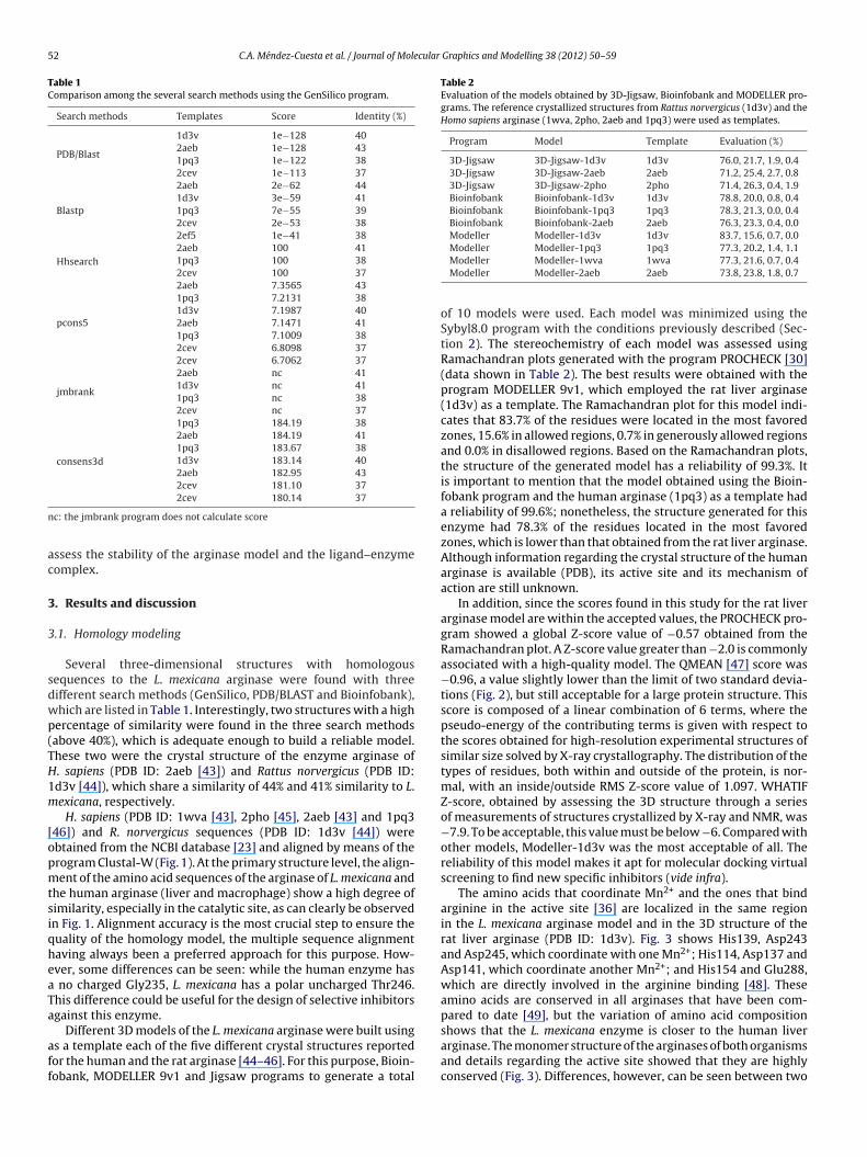
52 C.A. Méndez-Cuesta et al. / Journal of Molecular Graphics and Modelling 38 (2012) 50–59
Table 1Comparison among the several search methods using the GenSilico program.
Search methods Templates Score Identity (%)
PDB/Blast
1d3v 1e−128 402aeb 1e−128 431pq3 1e−122 382cev 1e−113 37
Blastp
2aeb 2e−62 441d3v 3e−59 411pq3 7e−55 392cev 2e−53 382ef5 1e−41 38
Hhsearch2aeb 100 411pq3 100 382cev 100 37
pcons5
2aeb 7.3565 431pq3 7.2131 381d3v 7.1987 402aeb 7.1471 411pq3 7.1009 382cev 6.8098 372cev 6.7062 37
jmbrank
2aeb nc 411d3v nc 411pq3 nc 382cev nc 37
consens3d
1pq3 184.19 382aeb 184.19 411pq3 183.67 381d3v 183.14 402aeb 182.95 432cev 181.10 37
n
ac
3
3
sdwp(TH1m
[opmtsiqheaTa
aff
Table 2Evaluation of the models obtained by 3D-Jigsaw, Bioinfobank and MODELLER pro-grams. The reference crystallized structures from Rattus norvergicus (1d3v) and theHomo sapiens arginase (1wva, 2pho, 2aeb and 1pq3) were used as templates.
Program Model Template Evaluation (%)
3D-Jigsaw 3D-Jigsaw-1d3v 1d3v 76.0, 21.7, 1.9, 0.43D-Jigsaw 3D-Jigsaw-2aeb 2aeb 71.2, 25.4, 2.7, 0.83D-Jigsaw 3D-Jigsaw-2pho 2pho 71.4, 26.3, 0.4, 1.9Bioinfobank Bioinfobank-1d3v 1d3v 78.8, 20.0, 0.8, 0.4Bioinfobank Bioinfobank-1pq3 1pq3 78.3, 21.3, 0.0, 0.4Bioinfobank Bioinfobank-2aeb 2aeb 76.3, 23.3, 0.4, 0.0Modeller Modeller-1d3v 1d3v 83.7, 15.6, 0.7, 0.0Modeller Modeller-1pq3 1pq3 77.3, 20.2, 1.4, 1.1
2cev 180.14 37
c: the jmbrank program does not calculate score
ssess the stability of the arginase model and the ligand–enzymeomplex.
. Results and discussion
.1. Homology modeling
Several three-dimensional structures with homologousequences to the L. mexicana arginase were found with threeifferent search methods (GenSilico, PDB/BLAST and Bioinfobank),hich are listed in Table 1. Interestingly, two structures with a highercentage of similarity were found in the three search methodsabove 40%), which is adequate enough to build a reliable model.hese two were the crystal structure of the enzyme arginase of. sapiens (PDB ID: 2aeb [43]) and Rattus norvergicus (PDB ID:d3v [44]), which share a similarity of 44% and 41% similarity to L.exicana, respectively.
H. sapiens (PDB ID: 1wva [43], 2pho [45], 2aeb [43] and 1pq346]) and R. norvergicus sequences (PDB ID: 1d3v [44]) werebtained from the NCBI database [23] and aligned by means of therogram Clustal-W (Fig. 1). At the primary structure level, the align-ent of the amino acid sequences of the arginase of L. mexicana and
he human arginase (liver and macrophage) show a high degree ofimilarity, especially in the catalytic site, as can clearly be observedn Fig. 1. Alignment accuracy is the most crucial step to ensure theuality of the homology model, the multiple sequence alignmentaving always been a preferred approach for this purpose. How-ver, some differences can be seen: while the human enzyme has
no charged Gly235, L. mexicana has a polar uncharged Thr246.his difference could be useful for the design of selective inhibitorsgainst this enzyme.
Different 3D models of the L. mexicana arginase were built usings a template each of the five different crystal structures reportedor the human and the rat arginase [44–46]. For this purpose, Bioin-obank, MODELLER 9v1 and Jigsaw programs to generate a total
Modeller Modeller-1wva 1wva 77.3, 21.6, 0.7, 0.4Modeller Modeller-2aeb 2aeb 73.8, 23.8, 1.8, 0.7
of 10 models were used. Each model was minimized using theSybyl8.0 program with the conditions previously described (Sec-tion 2). The stereochemistry of each model was assessed usingRamachandran plots generated with the program PROCHECK [30](data shown in Table 2). The best results were obtained with theprogram MODELLER 9v1, which employed the rat liver arginase(1d3v) as a template. The Ramachandran plot for this model indi-cates that 83.7% of the residues were located in the most favoredzones, 15.6% in allowed regions, 0.7% in generously allowed regionsand 0.0% in disallowed regions. Based on the Ramachandran plots,the structure of the generated model has a reliability of 99.3%. Itis important to mention that the model obtained using the Bioin-fobank program and the human arginase (1pq3) as a template hada reliability of 99.6%; nonetheless, the structure generated for thisenzyme had 78.3% of the residues located in the most favoredzones, which is lower than that obtained from the rat liver arginase.Although information regarding the crystal structure of the humanarginase is available (PDB), its active site and its mechanism ofaction are still unknown.
In addition, since the scores found in this study for the rat liverarginase model are within the accepted values, the PROCHECK pro-gram showed a global Z-score value of −0.57 obtained from theRamachandran plot. A Z-score value greater than −2.0 is commonlyassociated with a high-quality model. The QMEAN [47] score was−0.96, a value slightly lower than the limit of two standard devia-tions (Fig. 2), but still acceptable for a large protein structure. Thisscore is composed of a linear combination of 6 terms, where thepseudo-energy of the contributing terms is given with respect tothe scores obtained for high-resolution experimental structures ofsimilar size solved by X-ray crystallography. The distribution of thetypes of residues, both within and outside of the protein, is nor-mal, with an inside/outside RMS Z-score value of 1.097. WHATIFZ-score, obtained by assessing the 3D structure through a seriesof measurements of structures crystallized by X-ray and NMR, was−7.9. To be acceptable, this value must be below −6. Compared withother models, Modeller-1d3v was the most acceptable of all. Thereliability of this model makes it apt for molecular docking virtualscreening to find new specific inhibitors (vide infra).
The amino acids that coordinate Mn2+ and the ones that bindarginine in the active site [36] are localized in the same regionin the L. mexicana arginase model and in the 3D structure of therat liver arginase (PDB ID: 1d3v). Fig. 3 shows His139, Asp243and Asp245, which coordinate with one Mn2+; His114, Asp137 andAsp141, which coordinate another Mn2+; and His154 and Glu288,which are directly involved in the arginine binding [48]. Theseamino acids are conserved in all arginases that have been com-pared to date [49], but the variation of amino acid composition
shows that the L. mexicana enzyme is closer to the human liverarginase. The monomer structure of the arginases of both organismsand details regarding the active site showed that they are highlyconserved (Fig. 3). Differences, however, can be seen between two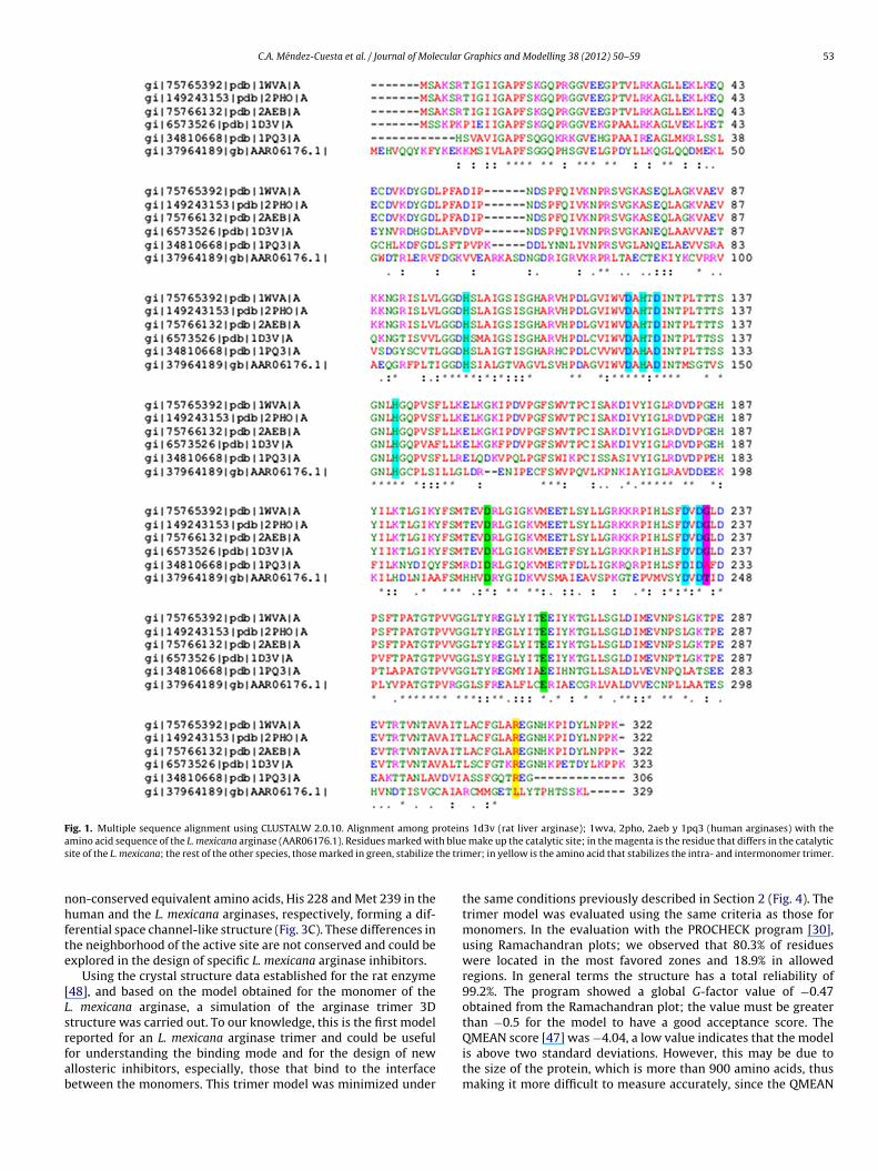
C.A. Méndez-Cuesta et al. / Journal of Molecular Graphics and Modelling 38 (2012) 50–59 53
Fig. 1. Multiple sequence alignment using CLUSTALW 2.0.10. Alignment among proteins 1d3v (rat liver arginase); 1wva, 2pho, 2aeb y 1pq3 (human arginases) with thea h blues the tri
nhfte
[Lsrfab
mino acid sequence of the L. mexicana arginase (AAR06176.1). Residues marked witite of the L. mexicana; the rest of the other species, those marked in green, stabilize
on-conserved equivalent amino acids, His 228 and Met 239 in theuman and the L. mexicana arginases, respectively, forming a dif-
erential space channel-like structure (Fig. 3C). These differences inhe neighborhood of the active site are not conserved and could bexplored in the design of specific L. mexicana arginase inhibitors.
Using the crystal structure data established for the rat enzyme48], and based on the model obtained for the monomer of the. mexicana arginase, a simulation of the arginase trimer 3Dtructure was carried out. To our knowledge, this is the first model
eported for an L. mexicana arginase trimer and could be usefulor understanding the binding mode and for the design of newllosteric inhibitors, especially, those that bind to the interfaceetween the monomers. This trimer model was minimized undermake up the catalytic site; in the magenta is the residue that differs in the catalyticmer; in yellow is the amino acid that stabilizes the intra- and intermonomer trimer.
the same conditions previously described in Section 2 (Fig. 4). Thetrimer model was evaluated using the same criteria as those formonomers. In the evaluation with the PROCHECK program [30],using Ramachandran plots; we observed that 80.3% of residueswere located in the most favored zones and 18.9% in allowedregions. In general terms the structure has a total reliability of99.2%. The program showed a global G-factor value of −0.47obtained from the Ramachandran plot; the value must be greaterthan −0.5 for the model to have a good acceptance score. The
QMEAN score [47] was −4.04, a low value indicates that the modelis above two standard deviations. However, this may be due tothe size of the protein, which is more than 900 amino acids, thusmaking it more difficult to measure accurately, since the QMEAN
54 C.A. Méndez-Cuesta et al. / Journal of Molecular Graphics and Modelling 38 (2012) 50–59
F t norm(
sd
L
FpeO
ig. 2. QMEAN plots for the monomer of the L. mexicana arginase. (a) QMEAN6 ploc) Z-score plot obtained by WHATIF.
tandard is up to 600 residues; over that number the graph is
ispersed in measuring the standard deviation (Fig. 5).The increase in the atomic contact score for the models for the. mexicana arginase and particularly for the trimer in comparison
ig. 3. Leishmania mexicana and the human liver type arginase 3D models. (a) The comparticipate in the active site are indicated by color. (b) Details of the binuclear Mn2+ and onzymes. (c) Difference in the space “channel-like” structure caused by the presence of Mverlay of the structures of L. mexicana human space showing the difference in the “chan
alized shows the standard deviation of the monomer. (b) Density plot for QMEAN.
with the monomer model suggests the existence of a trimeric struc-
ture for the L. mexicana arginase in vivo. In the rat liver arginase, theArg308 residue forms an intramonomer salt bridge with the Glu262and an intermonomer salt bridge with the Asp204 on the adjacentlete monomer model of both human and L. mexicana enzymes. Amino acids thatf the amino acids that directly interact with the substrate in the active site on bothet239 (yellow) in L. mexicana instead of His228 (yellow) observed in humans. (d)
nel-like”. Blue for the human arginase, white for the L. mexicana arginase.
C.A. Méndez-Cuesta et al. / Journal of Molecular
sagitratcl[
3
esi
FR
Fig. 4. Model of trimer of the L. mexicana arginase.
ubunit [50]. Both Glu262 and Asp204 residues are conserved in therginases of rats, humans, mice and several species of frogs of theenus Xenopus. These observations suggest a similar role in stabiliz-ng the oligomeric states of the trimer. In the L. mexicana arginase,he Glu262 and Asp204 are conserved at positions 273 and 215,espectively, but the residue Arg308 is absent. However, in manyrginases from bacteria there are hexameric arginases (dimers ofrimers) rather than trimeric, and the latter three residues are notonserved. For this reason, Arg308 is not essential for the stabi-ization of the trimer in these organisms or for optimal activity49].
.2. Virtual screening
A virtual screening, using molecular docking calculations, wasmployed to identify molecules that could be bound to the catalyticite of the L. mexicana and the H. sapiens arginase. As describedn Section 2, flexibility was allowed in all the rotable bonds of
ig. 5. QMEAN plots for the trimer of the L. mexicana arginase. (a) QMEAN6 plot normamachandran plots for the trimer that shows the most favored zones and disallowed reg
Graphics and Modelling 38 (2012) 50–59 55
the ligand, while the protein was used as a rigid structure. Bothenzymes were used in order to identify selective compounds tothe catalytic site of the parasite arginase. The results of the “blind”docking showed that fewer than 100 molecules bind selectively tothe catalytic site of the L. mexicana arginase, as depicted in Fig. 6.These molecules were submitted to a second docking, focused onthe catalytic site of the arginase in order to rank them by takinginto account the conformation with the lowest estimated bindingenergy of each compound. Based on this ranking only 18 moleculescould be bound selectively to the catalytic site of the parasite withbinding energies <−7.0 kcal/mol. Interestingly, 17 of the 18 com-pounds are analogues of the N-benzyl-1-alkyl-1H-benzimidazol-2-amines and other is a 2-ethyl-3-methyl-1-[(3-morpholin-4-ylpropyl)amino]pyrido[1,2-a]benzimidazole-4-carbonitrile. Thestructures of the 18 selective molecules and their estimated bind-ing energies are listed in Table 3. Analyzing the structure of thesecompounds, it is worth noticed that most of them are 1-alkyl-2-(substituted amine)-1H-benzimidazole derivatives. This resultshows, once again, the versatility of this structure since it presentsbiological activity against several parasites [37].
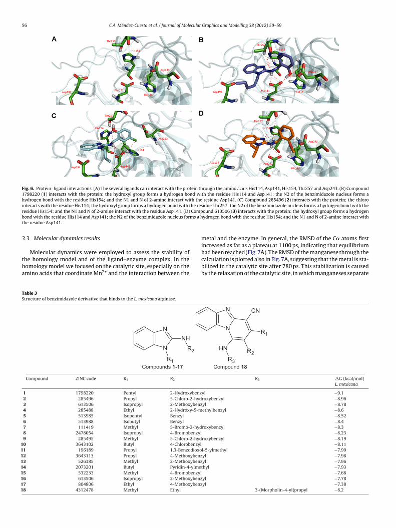
The protein–ligand interactions between the selected moleculesand the L. mexicana arginase are depicted in Fig. 6. As can beobserved, all molecules exhibit the same binding mode. Curiously,important interactions can be found between these molecules andthe residues His114, Asp141 and His154, which directly partici-pate in the catalytic mechanism of this enzyme. The ligand–enzymecomplex is stabilized mainly by hydrogen bonds, especially thosethat R2 forms with His114 and Thr257; the one between N in the2-amine and Asp141; and some presented between N from thebenzimidazole nucleus and the residue His154. Moreover, a �–�interaction can be formed between His139 and the benzyl group ofthe ligand. In this form, the benzyl moiety acts as an anchor towardthe enzyme while benzimidazole helps to maintain the moleculein the binding pocket; the group R1 helps to position the ligand inthe catalytic site.
It is important to mention that none of these molecules hasreports of measured biological activity in vitro against L. mexicanaor against the L. mexicana arginase. The virtual screening reportedin this paper shows N-benzyl-1-alkyl-1H-benzimidazol-2-aminesas possible inhibitors of L. mexicana arginase. Even thought it is
well known that the calculated binding energies do not alwaysreflect the biological response, the specific ligand–enzyme interac-tions could be used as a guideline for the design of new moleculesagainst leishmaniosis.alized shows the standard deviation of trimer. (b) Density plot for QMEAN. (c)ions.
56 C.A. Méndez-Cuesta et al. / Journal of Molecular Graphics and Modelling 38 (2012) 50–59
Fig. 6. Protein–ligand interactions. (A) The several ligands can interact with the protein through the amino acids His114, Asp141, His154, Thr257 and Asp243. (B) Compound1798220 (1) interacts with the protein; the hydroxyl group forms a hydrogen bond with the residue His114 and Asp141; the N2 of the benzimidazole nucleus forms ahydrogen bond with the residue His154; and the N1 and N of 2-amine interact with the residue Asp141. (C) Compound 285496 (2) interacts with the protein; the chlorointeracts with the residue His114; the hydroxyl group forms a hydrogen bond with the residue Thr257; the N2 of the benzimidazole nucleus forms a hydrogen bond with ther Compb ms a ht
3
tha
TS
111111111
esidue His154; and the N1 and N of 2-amine interact with the residue Asp141. (D)ond with the residue His114 and Asp141; the N2 of the benzimidazole nucleus forhe residue Asp141.
.3. Molecular dynamics results
Molecular dynamics were employed to assess the stability of
he homology model and of the ligand–enzyme complex. In theomology model we focused on the catalytic site, especially on themino acids that coordinate Mn2+ and the interaction between theable 3tructure of benzimidazole derivative that binds to the L. mexicana arginase.
N
N
R1
NH
R2
Compounds 1-17
Compound ZINC code R1 R2
1 1798220 Pentyl 2-Hydroxybenz2 285496 Propyl 5-Chloro-2-hyd3 613506 Isopropyl 2-Methoxybenz4 285488 Ethyl 2-Hydroxy-5-m5 513985 Isopentyl Benzyl
6 513988 Isobutyl Benzyl
7 111419 Methyl 5-Bromo-2-hyd8 2478054 Isopropyl 4-Bromobenzyl9 285495 Methyl 5-Chloro-2-hyd0 3643102 Butyl 4-Chlorobenzyl1 196189 Propyl 1,3-Benzodioxo2 3643113 Propyl 4-Methoxybenz3 526385 Methyl 2-Methoxybenz4 2073201 Butyl Pyridin-4-ylme5 532233 Methyl 4-Bromobenzyl6 613506 Isopropyl 2-Methoxybenz7 804806 Ethyl 4-Methoxybenz8 4312478 Methyl Ethyl
ound 613506 (3) interacts with the protein; the hydroxyl group forms a hydrogenydrogen bond with the residue His154; and the N1 and N of 2-amine interact with
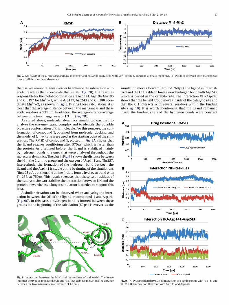
metal and the enzyme. In general, the RMSD of the C� atoms firstincreased as far as a plateau at 1100 ps, indicating that equilibriumhad been reached (Fig. 7A). The RMSD of the manganese through the
calculation is plotted also in Fig. 7A, suggesting that the metal is sta-bilized in the catalytic site after 780 ps. This stabilization is causedby the relaxation of the catalytic site, in which manganeses separateN
N
CN
R1
R2NH
R3
Compound 18
R3 �G (kcal/mol)L. mexicana
yl −9.1roxybenzyl −8.96yl −8.78ethylbenzyl −8.6
−8.52−8.4
roxybenzyl −8.3 −8.23roxybenzyl −8.19
−8.11l-5-ylmethyl −7.99yl −7.98yl −7.96
thyl −7.93 −7.68yl −7.78yl −7.38
3-(Morpholin-4-yl)propyl −8.2
C.A. Méndez-Cuesta et al. / Journal of Molecular Graphics and Modelling 38 (2012) 50–59 57
F ith Mt
taradcab
abftuttbmtIl(Ttpi
a(g
Fib
shows that the benzyl group moves inside of the catalytic site andthat the OH interacts with several residues within the bindingsite (Fig. 10). It is worth mentioning that the ligand remainedinside the binding site and the hydrogen bonds were constant
ig. 7. (A) RMSD of the L. mexicana arginase monomer and RMSD of interaction whrough all the molecular dynamics.
hemselves around 1.3 nm in order to enhance the interaction withcidic residues that coordinate the metals (Fig. 7B). The residuesesponsible for the metal coordination are Asp 141, Asp194, Glu196nd Glu197 for Mn2+−1, while Asp137, Asp243 and Glu288 coor-inate Mn2+−2, as shown in Fig. 8. During these calculations, it islear that the average distance between the manganese and thesecidic residues is 0.21 nm. In addition, the average distance averageetween the two manganeses is 1.3 nm (Fig. 7B).
As stated above, molecular dynamics simulation was used tonalyze the enzyme–ligand complex and to identify the possibleioactive conformation of this molecule. For this purpose, the con-ormation of compound 1, obtained from molecular docking, andhe model of L. mexicana were used as the starting point of the sim-lation. The RMSD of compound 1, plotted in Fig. 9A, shows thathe ligand reaches equilibrium after 570 ps, which is faster thanhe protein. As discussed before, the ligand is stabilized mainlyy hydrogen bonds, the ones that were analyzed throughout theolecular dynamics. The plot in Fig. 9B shows the distance between
he H in the 2-amino group and the oxygen of Asp141 and Thr257.nterestingly, the formation of the hydrogen bond between theigand and the Asp141 is stable at the beginning of the simulationsfirst 95 ps), but then, the amine flips to form a hydrogen bond withhr257, at 750 ps. This result suggests that these two residues ofhe catalytic site can stabilize the interaction between NH and therotein; nevertheless a longer simulation is needed to support this
dea.A similar situation can be observed when analyzing the inter-
ction between the OH of the ligand in compound 1 and Asp141Fig. 9C). In this case, a hydrogen bond is formed between theseroups at the beginning of the calculation (60 ps). However, as the
ig. 8. Interaction between the Mn2+ and the residues of aminoacids. The imagendicates the type of aminoacids (Glu and Asp) that stabilize the Mn and the distanceetween the two manganeses (an average of 1.3 nm).
n2+ of the L. mexicana arginase monomer. (B) Distance between both mangeneses
simulation moves forward (around 760 ps), the ligand is internal-ized and the OH is able to form a new hydrogen bond with Asp243,which is buried in the catalytic site. The interaction OH–Asp243
Fig. 9. (A) Drug positional RMSD. (B) Interaction of 2-Amino group with Asp141 andThr257. (C) Interaction HO group with Asp141 and Asp243.
58 C.A. Méndez-Cuesta et al. / Journal of Molecular Graphics and Modelling 38 (2012) 50–59
Fig. 10. Position of compound 1 inside the protein during the molecular dynamic. (A) At 1 ps time; the interaction is as shown in Fig. 6B. (B) At 1000 ps time; the initiali n His1o ractios
ttIt
poRawo
4
gtfIpTcamduLwNpuarTs
A
d
[
[
[
[
nteraction changes, thus the HO group interacts with the residue Asp243 more thaf the bencimidazole nucleus is stronger than 1 ps. (C) At 2000 ps time; all the inteimilar to B and C.
hroughout the simulation time. Furthermore, it was observed thathe ligand conformation does not involved important changes.ndeed, all these results provide important information regardinghe ligand–enzyme dynamical interaction (Fig. 10).
Furthermore, we have synthesized compounds 1–5 that wereredicted to be the most active of the designed series and carriedut preliminary in vitro evaluation on L. mexicana promastigotes.esults so far obtained indicate that these compounds have goodctivity against this stage of the parasite. However, we are stillorking on the synthesis of the remaining compounds (6–18) to
btain more activity data to complement this study.
. Conclusions
Although several three-dimensional structures with homolo-ous sequences to the L. mexicana arginase were found usinghree different search methods (GenSilico, PDB/BLAST, and Bioin-obank), only the crystal structures of the H. sapiens arginase (PDBD: 2aeb [43]) and R. norvergicus (PDB ID: 1d3v [44]) had a highercentage of similarity (above 40%) with the parasite enzyme.his information, coupled with the reported amino acids in theatalytic site of the three arginases, which differ in one aminocid, suggests that specific inhibitors could be designed for the L.exicana arginase. On the other hand, the best model for the three-
imensional structure of the L. mexicana arginase was obtainedsing the R. norvergicus crystal structure. The best model of the. mexicana arginase was used to perform a virtual screeningith 1439 benzimidazole derivatives. Among these molecules,-benzyl-1-alkyl-1H-benzimidazol-2-amines were identified asossible selective inhibitors of the L. mexicana arginase. The molec-lar dynamics simulations revealed the stability of the monomer ofrginase as well as the stability of the ligand–protein complex. Theesults further suggest that His114, Asp141, Asp194 Asp243 andhr257 could play an important role for the further optimization ofelective inhibitors.
cknowledgements
Méndez-Cuesta CA is very grateful to Consejo Nacionale Ciencia y Tecnología (CONACyT) for granting scholarship
[
[
14; the NH group now changes the interaction with Thr257; the interaction of N2ns in B are conserved and are stronger. (D) At 3000 ps time; the final interaction is
170430/170430. We acknowledge DGSCA, UNAM for the supportwe received to use the HP Cluster Platform 4000 Opteron dual coresupercomputer (KanBalam).
References
[1] M. Ouellette, Biochemical and molecular mechanisms of drug resistance inparasites, Tropical Medicine and International Health 6 (2001) 874–882.
[2] A. Armson, S.W. Kamau, F. Grimm, J.A. Reynoldson, W.M. Best, L.M. MacDon-ald, R.C.A. Thompson, A comparison of the effects of a benzimidazole and thedinitroanilines against Leishmania infantum, Acta Tropica 733 (1999) 303–311.
[3] S. Khabnadideh, D. Pez, A. Musso, R. Brun, L.M. Ruiz Pérez, D. González-Pacanowska, I.H. Gilbert, 2,4-Diaminopyrimidines as inhibitors of leishmanialand trypanosomal dihydrofolate reductase, Bioorganic and Medicinal Chem-istry 11 (2003) 4693–4711.
[4] Organización Mundial de la Salud, Lucha contra las leishmaniasis. Serie deInformes Técnicos No. 793, OMS, Ginebra, 1990.
[5] López de Cogolludo D, Historia de Yucatán, Mérida. Edición original, Madrid,1688, Reeditado por la Universidad de Yucatán, 1978.
[6] F. Biagi, Síntesis de 70 historias clínicas de leishmaniasis tegumentaria deMéxico (úlcera de los chicleros), Rev Facultad de Medicina UNAM 33 (1953)385–396.
[7] N.F. Andrade, M.M. García, R.A. Cruz, L.S. Canto, D.E. Simmonds, Estudio pre-liminar de correlación clínica, histológica e inmunológica de la leishmaniasiscutánea mexicana en el hombre, Archivos de Investigación Médica (México) 15(1984) 267–280.
[8] C.O. Velasco, S.J. Savarino, B.C. Walton, A.A. Gamm, F.A. Neva, Diffuse cutaneousleishmaniasis in Mexico, American Journal of Tropical Medicine and Hygiene41 (1989) 280–288.
[9] Tellaeche M, Informe relativo a prevención y control de las leishmaniasis enMéxico, in: Dirección de Prevención y Control de Enfermedades Transmitidaspor Vectores, Secretaría de Salud, México, 1993.
10] D. Sereno, A. Cordeiro da Silva, F. Mathieu-Daude, A. Ouaissi, Advances andperspectives in leishmania cell based drug-screening procedures, ParasitologyInternational 56 (2007) 3–7.
11] D.O. Santos, C.E. Coutinho, M.F. Madeira, C.G. Bottino, R.T. Vieira, S.B. Nasci-mento, A. Bernardino, S.C. Bourguignon, S. Corte-Real, R.T. Pinho, C.R. Rodrigues,H.C. Castro, Leishmaniasis treatment – a challenge that remains: a review,Parasitology Research 103 (1) (2008) 1–10.
12] UNICEF-UNDP-WHO Reports of TDR (Tropical Diseases Research).http://www.who.int/tdr/diseases-topics/leishmaniasis/en/index.html, 2011(accessed 13.02.12).
13] C.G. Havens, N. Bryant, L. Asher, L. Lamoreaux, S. Perfetto, J.J. Brendle, K.A.Werbovetz, Cellular effects of leishmanial tubulin inhibitors on L. donovani,Molecular and Biochemical Parasitology 110 (2000) 223–236.
14] Reports of the World Health Organization. www.who.int/leishmaniasis/burden/en/, 2011 (accessed 13.02.12).
15] S.L. Croft, G.H. Coombs, Leishmaniasis – current chemotherapy and recentadvances in the search for novel drugs, Trends in Parasitology 19 (11) (2003)503–508.
cular
[
[
[
[
[
[
[
[
[
[
[
[
[
[
[
[
[
[
[
[
[
[
[
[
[
[
[
[
[
[
[
[
[
C.A. Méndez-Cuesta et al. / Journal of Mole
16] M.W. Nowicki, Design, synthesis and trypanocidal activity of lead compoundsbased on inhibitors of parasite glycolysis, Bioorganic & Medicinal Chemistry 16(9) (2008) 5050–5061.
17] C. Doerig, L. Meijer, J.C. Mottram, Protein kinases as drug targets in parasiticprotozoa, Trends in Parasitology 18 (8) (2002) 366–371.
18] D.J. Rigden, S.E. Phillips, P.A. Michels, L.A. Fothergill-Gilmore, The structure ofpyruvate kinase from Leishmania mexicana reveals details of the allosteric tran-sition and unusual effector specificity, Journal of Molecular Biology 291 (1999)615–635.
19] E. Da Silva, T. Castilho, F. Pioker, C. Tomich de Paula, L. Floeter-Winter, Genomicorganization and transcription characterization of the gene encoding Leish-mania amazonensis arginase and its protein structure prediction, InternationalJournal for Parasitology 32 (2002) 727–737.
20] E.R. da Silva, M.F. Laranjeira da Silva, H. Fischer, R.A. Mortara, M.G. Mayer, K.Framesqui, A.M. Silber, L.M. Floeter-Winter, Biochemical and biophysical prop-erties of a highly active recombinant arginase from Leishmania (Leishmania)amazonensis and subcellular localization of native enzyme, Molecular and Bio-chemical Parasitology 159 (2) (2008) 104–111.
21] S.C. Roberts, M.J. Tancer, M.R. Polinsky, K.M. Gibson, O. Heby, B. Ullman,Arginase plays a pivotal role in polyamine precursor metabolism in Leishmania.Characterization of gene deletion mutants, Journal of Biological Chemistry 279(22) (2004) 23668–23678.
22] M.A. Kurowski, J.M. Bujnicki, GeneSilico protein structure prediction meta-server, Nucleic Acids Research 31 (2003) 3305–3307.
23] S.F. Altschul, T.L. Madden, A.A. Schäffer, J. Zhang, Z. Zhang, W. Miller, D.J. Lipman,Gapped BLAST and PSI-BLAST: a new generation of protein database searchprograms, Nucleic Acids Research 25 (1997) 3389–3402.
24] K. Ginalski, A. Elofsson, D. Fischer, L. Rychlewski, 3D-Jury: a simple approachto improve protein structure predictions, Bioinformatics 19 (8) (2003)1015–1018.
25] R. Chenna, H. Sugawara, T. Koike, R. Lopez, T.J. Gibson, D. Higgins, J. Thompson,Multiple sequence alignment with the clustal series of programs, Nucleic AcidsResearch 31 (13) (2003) 3497–3500.
26] P.A. Bates, L.A. Kelley, R.M. MacCallum, M.J.E. Sternberg, Enhancement of Pro-tein modelling by human intervention in applying the automatic programs3D-JIGSAW and 3D-PSSM, Proteins: Structure, Function and Genetics (Suppl.5) (2001) 39–46.
27] B. Contreras-Moreira, P.A. Bates, Domain Fishing: a first step in protein com-parative modelling, Bioinformatics 18 (2002) 1141–1142.
28] A. Sali, T.L. Blundell, Comparative protein modelling by satisfaction of spatialrestraints, Journal of Molecular Biology 234 (1993) 779–815.
29] A. Fiser, R.K. Do, A. Sali, Modeling of loops in protein structures, Protein Science9 (2000) 1753–1773.
30] R.A. Laskowski, M.W. MacArthur, D.S. Moss, J.M. Thornton, PROCHECK: a pro-gram to check the stereochemical quality of protein structures, Journal ofApplied Crystallography 26 (1993) 283–291.
31] R.W. Hooft, G. Vriend, C. Sander, E.E. Abola, Errors in protein structures, Nature381 (1996), p. 272.
32] E.G. Hutchinson, J.M. Thornton, PROMOTIF: a program to identify and analyzestructural motifs in proteins, Protein Science 5 (2) (1996) 212–220.
33] K. Arnold, L. Bordoli, J. Kopp, T. Schwede, The SWISS-MODEL workspace: a web-based environment for protein structure homology modelling, Bioinformatics22 (2006) 195–201.
[
[
Graphics and Modelling 38 (2012) 50–59 59
34] W.L. DeLano, The PyMOL Molecular Graphics System, DeLano Scientific LLC,Palo Alto, CA, 2007, http://www.pymol.org
35] SYBYL Software Package, Version 8.0, Tripos Associates Inc., S. Hanley Rd., St.Louis, MO 631444, USA, 1699.
36] J.J. Irwin, B.K. Shoichet, ZINC – a free database of commercially available com-pounds for virtual screening, Journal of Chemical Information and Modeling 45(1) (2005) 177–182.
37] J. Valdez, R. Cedillo, A. Hernández-Campos, L. Yépez, F. Hernández-Luis, G.Navarrete-Vázquez, A. Tapia, R. Cortés, M. Hernández, R. Castillo, Synthesis andantiparasitic activity of 1H-benzimidazole derivatives, Bioorganic & MedicinalChemistry Letters 12 16 (19) (2002) 2221–2224.
38] S. Díaz, M. de los Remedios, Síntesis de derivados del 1-metilbencimidazol conactividad antihelmíntica potencial, Master of Science Dissertation, UniversidadNacional Autónoma de México, 1999.
39] Méndez-Cuesta, Carlos Alberto, Síntesis y actividad antiparasitaria del 6-Cloro-2-(metiltio)-N-(5-nitro-1,3-tiazol-2-il)-1H-bencimidazol-5-carboxamida ysus derivados 1-metilados. Medicinal Chemistry Dissertation, UniversidadNacional Autónoma de México, 2005.
40] F. Hernández-Luis, A. Hernández-Campos, R. Castillo, G. Navarrete-Vázquez, O.Soria-Arteche, M. Hernández-Hernández, L. Yépez-Mulia, Synthesis and biolog-ical activity of 2-(trifluoromethyl)-1H-benzimidazole derivatives against someprotozoa and Trichinella spiralis, European Journal of Medicinal Chemistry 45(7) (2010) 3135–3141.
41] M.F. Sanner, Python: a programming language for software integration anddevelopment, Journal of Molecular Graphics and Modelling 17 (1999) 57–61.
42] A.W. Schuttelkopf, D.M.F. Van Aalten, PRODRG – a tool for high-throughputcrystallography of protein–ligand complexes, Acta Crystallographica 60 (2004)1355–1363.
43] L. Di Costanzo, G. Sabio, A. Mora, P.C. Rodriguez, A.C. Ochoa, F. Centeno, D.W.Christianson, Crystal structure of human arginase I at 1.29-A resolution andexploration of inhibition in the immune response, Proceedings of the NationalAcademy of Sciences USA 102 (2005) 13058–13063.
44] J.D. Cox, N.N. Kim, A.M. Traish, D.W. Christianson, Arginase–boronic acid com-plex highlights a physiological role in erectile function, Natural StructuralBiology 6 (1999) 1043–1047.
45] L. Di Costanzo, M.E. Pique, D.W. Christianson, Crystal structure of humanarginase I complexed with thiosemicarbazide reveals an unusual thiocarbonyl�-sulfide ligand in the binuclear manganese cluster, Journal of the AmericanChemical Society 129 (2007) 6388–6389.
46] E. Cama, D.M. Colleluori, F.A. Emig, H. Shin, S.W. Kim, N.N. Kim, A.M. Traish,D.E. Ash, D.W. Christianson, Human arginase. II. Crystal structure and phys-iological role in male and female sexual arousal, Biochemistry 42 (2003)8445–8451.
47] P. Benkert, M. Biasini, T. Schwede, Toward the estimation of the absolutequality of individual protein structure models, Bioinformatics 27 (3) (2011)343–350.
48] Z.F. Kanyo, L.R. Scolnick, D.E. Ash, D.W. Christianson, Structure of a uniquebinuclear manganese cluster in arginase, Nature 383 (1996) 554–557.
49] J. Perozich, J. Hempel, MorrisF S.M. Jr., Roles of conserved residues in thearginase family, Biochimica et Biophysica Acta 1382 (1998) 23–37.
50] L.T. Lavulo, T.M. Sossong Jr., M.R.B. Burke, M.L. Doyle, J.D. Cox, D.W. Christianson,D.E. Ash, Subunit–subunit interactions in trimeric arginase, Journal of BiologicalChemistry 276 (2001) 14242–14248.
Copyright © 2022 FDOKUMEN