Structural Basis of 3Phosphoinositide Recognition by Pleckstrin Homology Domains
10
Molecular Cell, Vol. 6, 385–394, August, 2000, Copyright 2000 by Cell Press Structural Basis of 3-Phosphoinositide Recognition by Pleckstrin Homology Domains In unstimulated cells, the levels of PtdIns(3,4)P 2 and PtdIns(3,4,5)P 3 are undetectable. Following receptor stimu- lation, the combined activity of PI 3-kinases and phos- Susan E. Lietzke, Sahana Bose, Thomas Cronin, Jes Klarlund, Anil Chawla, Michael P. Czech, and David G. Lambright* phoinositide phosphatases results in transient produc- Program in Molecular Medicine tion of PtdIns(3,4,5)P 3 and, after a short lag, PtdIns(3,4)P 2 University of Massachusetts Medical Center (Auger and Cantley, 1991). Even at maximal receptor Worcester, Massachusetts 01605 stimulation, the peak levels of these 3-phosphoinositides remain well below the constitutive levels of the relatively abundant precursors, PtdIns(4)P and PtdIns(4,5)P 2 . Conse- Summary quently, intracellular targets of 3-phosphoinositides must selectively recognize PI 3-kinase products against Lipid second messengers generated by phosphoinosi- a high background of chemically and structurally similar tide (PI) 3-kinases regulate diverse cellular functions precursors. through interaction with pleckstrin homology (PH) do- Pleckstrin homology (PH) domains in modular signal- ing proteins represent the primary intracellular targets of mains in modular signaling proteins. The PH domain PtdIns(3,4)P 2 and PtdIns(3,4,5)P 3 . In vitro binding studies of Grp1, a PI 3-kinase-activated exchange factor for employing a variety of inositol head groups, soluble Arf GTPases, selectively binds phosphatidylinositol phosphoinositide analogs, and liposomes containing 3,4,5-trisphosphate with high affinity. We have deter- lipid phosphoinositides have shown that PH domains mined the structure of the Grp1 PH domain in the bind phosphoinositides with a broad range of affinity unliganded form and bound to inositol 1,3,4,5-tetra- and specificity (Harlan et al., 1994; Lemmon et al., 1995; phosphate. A novel mode of phosphoinositide recog- Fukuda et al., 1996; Salim et al., 1996; Franke et al., nition involving a 20-residue insertion within the b6/b7 1997; Frech et al., 1997; Rameh et al., 1997; Isakoff et loop explains the unusually high specificity of the Grp1 al., 1998; Kavran et al., 1998). A subset of PH domains PH domain and the promiscuous 3-phosphoinositide binds one or more PI 3-kinase products with high affinity binding typical of several PH domains including that while discriminating to various degrees against the more of protein kinase B. When compared to other PH do- abundant 3-phosphoinositide precursors. For example, mains, general determinants of 3-phosphoinositide the PH domain of Bruton’s tyrosine kinase (Btk) binds recognition and specificity can be deduced. PtdIns(3,4,5)P 3 with high affinity and specificity (Fukuda et al., 1996; Kojima et al., 1997; Rameh et al., 1997). In contrast, several other PH domains, including that of Introduction PKB, bind both PtdIns(3,4)P 2 and PtdIns(3,4,5)P 3 with similar affinity yet still discriminate against PtdIns(4)P The lipid products of phosphoinositide (PI) 3-kinases and PtdIns(4,5)P 2 (James et al., 1996; Franke et al., 1997; regulate diverse cellular functions including growth, sur- Frech et al., 1997). vival, cytoskeletal dynamics, adhesion, migration and The highly homologous Arf exchange factors Grp1 membrane trafficking (Corvera and Czech, 1998; Fru- (general receptor for 3-phosphoinositides), ARNO (Arf man et al., 1998; Leevers et al., 1999; Rameh and Can- nucleotide binding site opener), and cytohesin are 43 tley, 1999). At least three different classes of mammalian kDa proteins consisting of an N-terminal heptad repeat PI 3-kinases have been identified (Domin and Waterfield, followed by an Arf exchange domain with homology to 1997; Fruman et al., 1998). The p110/p85 and p110g PI the yeast Sec7 protein, a PH domain, and a C-terminal 3-kinases are rapidly activated following ligand binding polybasic region. The PH domain of Grp1 binds to many different cell surface receptors and exhibit PtdIns(3,4,5)P 3 with high affinity (K d <100 nM) and exqui- broad substrate specificity in vitro, phosphorylating the site specificity, such that the affinity for PtdIns(3,4)P 2 3-hydroxyl group of distinct phosphatidylinositol (PtdIns) and PtdIns(4,5)P 2 is between two and three orders of substrates to generate PtdIns 3-phosphate [PtdIns(3)P], magnitude lower (Klarlund et al., 1997; Kavran et al., PtdIns 3,4-bisphosphate [PtdIns(3,4)P 2 ], and PtdIns 1998). Consistent with this observation, the Arf ex- 3,4,5-trisphosphate [PtdIns(3,4,5)P 3 ] (Escobedo et al., change activity of Grp1 is potently stimulated by lipo- 1991; Skolnik et al., 1991; Hiles et al., 1992; Stoyanov somes containing PtdIns(3,4,5)P 3 but not other phos- et al., 1995; Vanhaesebroeck et al., 1997). The functional phoinositides (Klarlund et al., 1998). Experiments in importance of PI 3-kinase signaling is underscored by intact cells indicate that the Grp1 PH domain is both the discovery that a major tumor suppressor known as necessary and sufficient for recruitment of the full-length PTEN is a lipid phosphatase that removes the 3-phos- protein to the plasma membrane following stimulation phate group from PI 3-kinase products (Maehama and with insulin (Klarlund et al., 2000). Dixon, 1998; Wu et al., 1998). Likewise, activation of Despite unusually divergent sequences, PH domains the Akt protooncogene product (also known as protein share a conserved core fold consisting of a seven- kinase B; hereafter PKB) protects proliferating cells stranded b barrel capped on one end by a C-terminal against apoptosis and has been shown to require pro- a helix (Rebecchi and Scarlata, 1998). In the crystal duction of 3-phosphoinositides (Cantley and Neel, structure of the phospholipase Cd (PLCd) PH domain 1999). bound to Ins(1,4,5)P 3 , the head group is located in a pocket flanked by the b1/b2 and b3/b4 loops (Ferguson et al., 1995). A motif of basic residues from the b1 and * To whom correspondence should be addressed (e-mail: david. [email protected]). b2 strands provides critical contacts to the 4- and
-
Upload
independent -
Category
Documents
-
view
2 -
download
0
Transcript of Structural Basis of 3Phosphoinositide Recognition by Pleckstrin Homology Domains

Molecular Cell, Vol. 6, 385–394, August, 2000, Copyright 2000 by Cell Press
Structural Basis of 3-Phosphoinositide Recognitionby Pleckstrin Homology Domains
In unstimulated cells, the levels of PtdIns(3,4)P2 andPtdIns(3,4,5)P3 are undetectable. Following receptor stimu-lation, the combined activity of PI 3-kinases and phos-
Susan E. Lietzke, Sahana Bose, Thomas Cronin,Jes Klarlund, Anil Chawla, Michael P. Czech,and David G. Lambright*
phoinositide phosphatases results in transient produc-Program in Molecular Medicinetion of PtdIns(3,4,5)P3 and, after a short lag, PtdIns(3,4)P2University of Massachusetts Medical Center(Auger and Cantley, 1991). Even at maximal receptorWorcester, Massachusetts 01605stimulation, the peak levels of these 3-phosphoinositidesremain well below the constitutive levels of the relativelyabundant precursors, PtdIns(4)P and PtdIns(4,5)P2. Conse-
Summary quently, intracellular targets of 3-phosphoinositidesmust selectively recognize PI 3-kinase products against
Lipid second messengers generated by phosphoinosi- a high background of chemically and structurally similartide (PI) 3-kinases regulate diverse cellular functions precursors.through interaction with pleckstrin homology (PH) do- Pleckstrin homology (PH) domains in modular signal-
ing proteins represent the primary intracellular targets ofmains in modular signaling proteins. The PH domainPtdIns(3,4)P2 and PtdIns(3,4,5)P3. In vitro binding studiesof Grp1, a PI 3-kinase-activated exchange factor foremploying a variety of inositol head groups, solubleArf GTPases, selectively binds phosphatidylinositolphosphoinositide analogs, and liposomes containing3,4,5-trisphosphate with high affinity. We have deter-lipid phosphoinositides have shown that PH domainsmined the structure of the Grp1 PH domain in thebind phosphoinositides with a broad range of affinityunliganded form and bound to inositol 1,3,4,5-tetra-and specificity (Harlan et al., 1994; Lemmon et al., 1995;phosphate. A novel mode of phosphoinositide recog-Fukuda et al., 1996; Salim et al., 1996; Franke et al.,nition involving a 20-residue insertion within the b6/b71997; Frech et al., 1997; Rameh et al., 1997; Isakoff etloop explains the unusually high specificity of the Grp1al., 1998; Kavran et al., 1998). A subset of PH domains
PH domain and the promiscuous 3-phosphoinositide binds one or more PI 3-kinase products with high affinitybinding typical of several PH domains including that while discriminating to various degrees against the moreof protein kinase B. When compared to other PH do- abundant 3-phosphoinositide precursors. For example,mains, general determinants of 3-phosphoinositide the PH domain of Bruton’s tyrosine kinase (Btk) bindsrecognition and specificity can be deduced. PtdIns(3,4,5)P3 with high affinity and specificity (Fukuda
et al., 1996; Kojima et al., 1997; Rameh et al., 1997). Incontrast, several other PH domains, including that ofIntroductionPKB, bind both PtdIns(3,4)P2 and PtdIns(3,4,5)P3 withsimilar affinity yet still discriminate against PtdIns(4)PThe lipid products of phosphoinositide (PI) 3-kinasesand PtdIns(4,5)P2 (James et al., 1996; Franke et al., 1997;regulate diverse cellular functions including growth, sur-Frech et al., 1997).vival, cytoskeletal dynamics, adhesion, migration and
The highly homologous Arf exchange factors Grp1membrane trafficking (Corvera and Czech, 1998; Fru-(general receptor for 3-phosphoinositides), ARNO (Arfman et al., 1998; Leevers et al., 1999; Rameh and Can-nucleotide binding site opener), and cytohesin are 43tley, 1999). At least three different classes of mammaliankDa proteins consisting of an N-terminal heptad repeatPI 3-kinases have been identified (Domin and Waterfield,followed by an Arf exchange domain with homology to1997; Fruman et al., 1998). The p110/p85 and p110g PIthe yeast Sec7 protein, a PH domain, and a C-terminal3-kinases are rapidly activated following ligand bindingpolybasic region. The PH domain of Grp1 bindsto many different cell surface receptors and exhibitPtdIns(3,4,5)P3 with high affinity (Kd <100 nM) and exqui-broad substrate specificity in vitro, phosphorylating thesite specificity, such that the affinity for PtdIns(3,4)P23-hydroxyl group of distinct phosphatidylinositol (PtdIns)and PtdIns(4,5)P2 is between two and three orders ofsubstrates to generate PtdIns 3-phosphate [PtdIns(3)P],magnitude lower (Klarlund et al., 1997; Kavran et al.,PtdIns 3,4-bisphosphate [PtdIns(3,4)P2], and PtdIns1998). Consistent with this observation, the Arf ex-3,4,5-trisphosphate [PtdIns(3,4,5)P3] (Escobedo et al.,change activity of Grp1 is potently stimulated by lipo-1991; Skolnik et al., 1991; Hiles et al., 1992; Stoyanovsomes containing PtdIns(3,4,5)P3 but not other phos-et al., 1995; Vanhaesebroeck et al., 1997). The functionalphoinositides (Klarlund et al., 1998). Experiments inimportance of PI 3-kinase signaling is underscored byintact cells indicate that the Grp1 PH domain is boththe discovery that a major tumor suppressor known asnecessary and sufficient for recruitment of the full-lengthPTEN is a lipid phosphatase that removes the 3-phos-protein to the plasma membrane following stimulation
phate group from PI 3-kinase products (Maehama and with insulin (Klarlund et al., 2000).Dixon, 1998; Wu et al., 1998). Likewise, activation of Despite unusually divergent sequences, PH domainsthe Akt protooncogene product (also known as protein share a conserved core fold consisting of a seven-kinase B; hereafter PKB) protects proliferating cells stranded b barrel capped on one end by a C-terminalagainst apoptosis and has been shown to require pro- a helix (Rebecchi and Scarlata, 1998). In the crystalduction of 3-phosphoinositides (Cantley and Neel, structure of the phospholipase Cd (PLCd) PH domain1999). bound to Ins(1,4,5)P3, the head group is located in a
pocket flanked by the b1/b2 and b3/b4 loops (Fergusonet al., 1995). A motif of basic residues from the b1 and* To whom correspondence should be addressed (e-mail: david.
[email protected]). b2 strands provides critical contacts to the 4- and

Molecular Cell386
Table 1. Structure Determination and Refinement
Data Collectiona
Crystal SeMet 1 SeMet 1 SeMet 1 SeMet 2 UnligandedWavelength l1 (0.9794 A) l2 (0.9789 A) l3 (0.9680 A) l4 (0.9915 A) 1.54 ASource NSLS X4A NSLS X4A NSLS X4A NSLS X4A LabResolution 20–2.2 A 20–2.2 A 20–2.2 A 20–1.5 A 20–2.0 ARsym (%)b 4.7 (7.3) 5.2 (10.1) 5.5 (11.4) 5.8 (32.7) 7.1 (35.3),I/s. 22.5 (16.0) 21.2 (13.2) 21.5 (12.3) 16.1 (3.9) 15.6 (4.0)Completeness 99.4 (98.8) 99.4 (98.4) 99.5 (98.9) 94.0 (85.3) 98.9 (98.0)Redundancy 4 4 4 4 4
Observed Diffraction Ratiosc
l1 l2 l3
Bijvoet ratio 0.052 0.059 0.045Dispersive ratio (vs. l1) 0.048 0.081Dispersive ratio (vs. l2) 0.051
Phasing Power and FOMa
Bijvoet l1 Bijvoet l2 Bijvoet l3 l1 vs. l3 l2 vs. l3
Acentric PP 2.11 (1.53) 2.84 (1.95) 2.26 (1.54) 2.60 (2.41) 4.33 (4.79)Centric PP 1.73 (1.71) 2.72 (2.86)Acentric FOM 0.73 (0.59) Centric FOM 0.57 (0.41)
Refinement
RMS Deviations
Resolution (A) R Factor Rfreed Bond Length Bond Angle
Ins(1,3,4,5)P4 6.0–1.5 21.3 24.7 0.006 1.3Unliganded 6.0–2.0 22.3 27.7 0.006 1.2
aValues in parentheses represent the highest resolution shell.bRsym 5 ShSj |Ij(h) 2 ,I(h).| / ShSj Ij(h).cValues are ,(D|F|)2.1/2 / ,|F|2.1/2 for data from 20 to 2 A.dR value for a 5% subset of reflections selected at random and omitted from refinement.
5-phosphates and is also present in the sequences of Results and DiscussionPH domains known to bind PI 3-kinase products (Ramehet al., 1997; Isakoff et al., 1998; Kavran et al., 1998). Structure Determination
A selenomethionine-substituted form of the Grp1 PHMutational studies confirm the general importance ofdomain (residues 257–390) was expressed with anthis motif for high-affinity 3-phosphoinositide bindingN-terminal 63 His tag in E. coli, purified without ligand,(Fruman et al., 1999). Although Ins(1,3,4,5)P4 binds to aand crystallized in the presence or absence of inositolsimilar location in the Btk PH domain, the inositol ring1,3,4,5-tetraphosphate [Ins(1,3,4,5)P4], the head groupis flipped such that the conserved basic residues con-of PtdIns(3,4,5)P3 (Experimental Procedures). The struc-tact the 3- and 4-phosphates (Baraldi et al., 1999). Theture of the Grp1 PH domain bound to Ins(1,3,4,5)P4 wasspecific binding of PtdIns(3,4,5)P3 to the Btk PH domainsolved with experimental phases derived from multi-is facilitated by a large b1/b2 loop, which interacts ex-wavelength anomalous diffraction at the Se edge (Tabletensively with the 5-phosphate. However, this mode of1). An initial map calculated at 2.2 A with phases modi-interaction cannot explain the dramatic specificity differ-fied by solvent flipping revealed continuous main andences between the Grp1 and PKB PH domains thatside chain density for the protein core and strong densityhave relatively short b1/b2 loops of identical length andfor the inositol head group. Roughly 70% of the residuessimilar sequence. Evidently, other structural elements,could be modeled into the initial density-modified mapyet to be identified, play a central role in determining whereas the remaining model was placed into unambig-
the specificity of 3-phosphoinositide binding. uous density obtained from partial model combinedHere, we report the crystal structures of the Ins(1,3, maps and sA-weighted 2Fo 2 Fc maps. The current
4,5)P4 bound and unliganded forms of the Grp1 PH do- model, consisting of residues 261–387, was refinedmain. The structures reveal a novel mode of phospho- against data to 1.5 A and has a working R factor ofinositide recognition in which a 20-residue insertion rela- 21.3% and free R factor of 24.7%. The structure of thetive to the canonical PH fold accounts for the ability of unliganded form was solved by molecular replacementthe Grp1 PH domain to selectively bind PtdIns(3,4,5)P3, using a search model based on the protein core of thebut not PtdIns(3,4)P2 or PtdIns(4,5)P2, with high affinity. Ins(1,3,4,5)P4 complex. Following refinement to 2.0 A,The absence of this insertion region provides a simple the current model for the unliganded form has a workingexplanation for the promiscuous binding of PI 3-kinase R factor of 22.3% and free R factor of 27.7%.products typical of other PH domains including that ofPKB. When compared to liganded structures of the Btk Overall Structure and Phosphoinositide Binding Siteand PLCd PH domains, general determinants of 3-phos- As shown in Figure 1, the fold of the Grp1 PH domain
consists of a nine-stranded anti-parallel b barrel cappedphoinositide recognition and specificity can be deduced.

Recognition of PI 3-Kinase Products by PH Domains387
Figure 2. Molecular Surface of the Grp1 PH Domain and Electro-static Properties of the Phosphoinositide Binding Site
Surface representation of the phosphoinositide binding site withthe solvent-accessible surface colored according to electrostaticFigure 1. Overall Structure of the Grp1 PH Domainpotential using GRASP (A. Nichols et al., 1993, Biophys. J., abstract).
Ribbon representation of the overall structure of the Grp1 PH domain Blue denotes positive potential greater than 120 kT and red negativewith the canonical PH fold shown in blue. Regions involved in speci- potential less than 220 kT, where k is Boltzmann’s constant and Tficity determination are highlighted in orange (b6/b7 hairpin inser- is absolute temperature.tion), green (b1/b2 loop), and magenta (b3/b4 loop). TheIns(1,3,4,5)P4 head group is shown in yellow (carbon and phospho-rous atoms) and red (oxygen atoms).
in Grp1 and Btk. However, given the pseudosymmetryof the inositol ring, it is possible that some of these PH
on one end by a C-terminal a helix. In contrast to other domains will bind PtdIns(4,5)P2 in the flipped orientationPH domains (Rebecchi and Scarlata, 1998), a 20-residue observed in the PLCd PH domain, albeit with lower af-insertion within the b6/b7 loop adopts a twisted b hairpin finity.structure that extends the b barrel from seven to ninestrands. Main chain interactions between the b4 andbi1 strands position the b hairpin such that side chains Conserved Determinants of 3-Phosphoinositide
Recognitionon its inner surface are directed toward the phosphoino-sitide binding pocket. A similar b hairpin insertion in the Previous studies identified a motif of three basic resi-
dues (located in the b1 and b2 strands) that are strictlycontext of the immunoglobulin fold occurs in the variabledomains of IgG proteins where the additional residues conserved in PH domains known to bind 3-phosphoino-
sitides with high affinity in vitro (Rameh et al., 1997;(compared to the constant domains) form the secondof three complementarity-determining regions (Chothia Kavran et al., 1998), as well as those predicted to bind
PI 3-kinase products based on an in vivo two-hybridet al., 1989). The b hairpin insertion in the Grp1 subfamilyof PH domains performs an analogous function with assay (Isakoff et al., 1998). However, the invariant basic
residues are also conserved in the PLCd PH domain,respect to the specificity of phosphoinositide binding.Ins(1,3,4,5)P4 binds in a deep pocket located at the which binds PtdIns(4,5)P2 rather than 3-phosphoinosi-
tides. In the course of generating a sequence alignmentopen end of the b barrel and is contacted by invariantresidues from the b1, b2, and b3 strands as well as based on the structure of the Grp1 domain, we observed
that a tyrosine residue located in the b3 strand is strictlynonconserved residues in the b1/b2 loop, the b3/b4loop, and the b hairpin insertion. The concave surface conserved in PH domains that bind PI 3-kinase products
with high affinity but not in other PH domains, includingof the phosphoinositide binding site bears a strikingresemblance to the PtdIns(3,4,5)P3 head group, with those of PLCd, spectrin, dynamin, and pleckstrin (Figure
3A). Accounting for the location of essential hydropho-overlapping yet clearly discernible pockets for each ofthe 3-, 4-, and 5-phosphate groups (Figure 2). Hereafter, bic residues, the original basic motif can be expanded
to a distinctive “signature motif” for 3-phosphoinositidethese will be referred to as the P3, P4, and P5 pockets,respectively. Like the PH domains of Btk and PLCd, binding, φ-X-K-(G/A/S/P)-Xm-φ-X-(K/R)-X-R-X-φ-X-φ-
Xn-φ-X-Y. This expanded motif spans the b1–b3 strandswhich also bind a particular phosphoinositide with highaffinity and specificity, the overall architecture of the and consists of four invariant residues (boldface type)
interspersed with five essential hydrophobic residuesbinding pocket in the Grp1 PH domain accommodatesa unique orientation of the pseudosymmetrical inositol (indicated by F). When used to search the PROSITE
database (z84,000 protein sequences), an explicit ver-polyphosphate head group. This orientation completelyburies the 4-phosphate and places the 3-phosphate in sion of the signature motif indicated in Figure 3B selec-
tively retrieved PH domains known to bind 3-phospho-the most positively charged region of the bindingpocket. A similar head group orientation is observed inositides with only a single false hit.
The conservation of hydrophobic residues within thein the Ins(1,3,4,5)P4 complex with the Btk PH domain(Baraldi et al., 1999), despite large structural differences signature motif is typical of PH domains in general and
reflects the location of these residues in the hydrophobicin the regions that contact the 4- and 5-phosphates.This observation suggests that high-affinity binding of core. However, as shown in Figure 4A, a cluster of the
essential hydrophobic residues (Leu-271, Phe-286, andPI 3-kinase products to other PH domains is likely toinvolve a common head group orientation similar to that Leu-293) packs against a second cluster formed by three

Molecular Cell388
Figure 3. Sequence Comparison of Grp1 and Other PH Domains
(A) Structure-based alignment of representative PH domains, with specificity-determining regions colored coded as in Figure 1. For sequenceslacking corresponding structures, insertions are assumed to occur in the middle of the hypervariable loop regions. Residues strictly conservedin PH domains that bind PI 3-kinase products with high affinity are highlighted in blue. With the exception of PLCd and dynamin, the PHdomains included in the alignment are known to bind PI 3-kinase products with high affinity.(B) Signature motif for PH domains known and/or predicted to bind 3-phosphoinositides with high affinity. A list of residues enclosed in squarebrackets represents the observed range of sequence variability at a given position. X denotes any residue.
of the invariant residues (Lys-273, Arg-284, and Tyr- residues interact simultaneously with the 3- and 4-phos-phate groups. In addition to sensing the presence (or295). Thus, in addition to stabilizing the PH fold, the
essential hydrophobic residues serve a second function absence) of the 3-phosphate, the signature motif playsan important role in distinguishing polyphosphorylatedby supporting the invariant residues as they emerge
from the protein core to contact the phosphoinositide PI 3-kinase products from the more abundant monophos-phoinositides: PtdIns(3)P and PtdIns(4)P. However, inhead group. In the Grp1 PH domain, two of the invariant
basic residues (Lys-273 in b1 and Arg-284 in b2) form view of the pseudosymmetry of the inositol ring, it isunlikely that the conserved interactions mediated bythe positively charged surface of the P3 pocket and
account for all but one interaction with the 3-phosphate the signature motif would be sufficient to distinguish PI3-kinase products from PtdIns(4,5)P2. Thus, additional(Figure 4B). The remaining basic residue (Lys-282 in b2)
contributes ionic and water-mediated interactions with specifying interactions are necessary to selectively en-hance the affinity for PI 3-kinase products.the 1-phosphate. Lys-273 also contacts the 4-phos-
phate as does the side chain hydroxyl of the invarianttyrosine residue (Tyr-295 in b3). Despite dramatic dif-ferences in the mode of 4- and 5-phosphate recogni- Specificity-Determining Regions
Recognition of the 4- and 5-phosphates, and conse-tion described below, the corresponding residues in theBtk PH domain (Lys-12, Lys-26, Arg-28, and Tyr-39) me- quently the specificity or promiscuity of 3-phosphoinosi-
tide binding, depends on nonconserved interactionsdiate equivalent interactions with similar stereochemis-try (Baraldi et al., 1999). These observations provide with residues located in three hypervariable specificity-
determining regions encompassed by the b1/b2 loopstrong evidence favoring a common mechanism of3-phosphoinositide recognition in which the invariant (SDR1), the b3/b4 loop (SDR2), and the b6/b7 insertion

Recognition of PI 3-Kinase Products by PH Domains389
Figure 4. Structural Basis of 3-Phosphoino-sitide Recognition by the Grp1 PH Domain
(A) Location and packing of the invariant(white) and hydrophobic (yellow) residues inthe signature motif. Residues in the b1–b3strands are rendered as solid objectswhereas the remainder of the protein is semi-transparent.(B) Direct interactions between invariant resi-dues in the signature motif and Ins(1,3,4,5)P4.The coloring is similar to Figure 1, except forthe side chains of residues from the signaturemotif, which are shown in white.(C) Direct interactions between the specific-ity-determining regions and Ins(1,3,4,5)P4.Residues from the signature motif are shownas semitransparent.(D) sA weighted 2Fo 2 Fc map contoured at1.2 s showing a network of water-mediatedand supporting interactions with the 4- and5-phosphates.
region (SDR3). In the Grp1 PH domain, four residues an asparagine residue at the C-terminal position medi-ates similar interactions. Interestingly, a small polar resi-from the hairpin insertion (Lys-343 in bi1; Asn-354, His-
355, and Tyr-358 in the bi2/b7 loop) comprise the core due (Thr, Ser or Asn) is conserved in the majority of PHdomains known to bind PI 3-kinase products with highof an extensive network of ionic, hydrogen bonding, and
water-mediated interactions with the 4- and 5-phos- affinity. The structural basis for conservation of residuesat the termini of the b1/b2 loop provides further evidencephates (Figures 4C and 4D). The 4-phosphate is gripped
on one side by Lys-343 and His-355 from the hairpin that most, and perhaps all, of these PH domains bind3-phosphoinositides with a common head group orien-insertion and on the other side by Lys-273 and Tyr-295
from the signature motif. The P5 pocket is located at tation.Despite 90% sequence identity, the PH domains ofthe tightly packed interface between the hairpin inser-
tion and the b1/b2 loop. The positively charged (lower) Grp1, ARNO, and cytohesin exhibit dramatically differ-ent specificity for soluble phosphoinositide analogssurface of the P5 pocket is formed by the side chains
of two residues in the hairpin insertion (Lys-343 and (Klarlund et al., 2000). The Grp1 PH binds di-octoylPtdIns(3,4,5)P3 with high affinity (Kd <100 nM) and exqui-Asn-354) whereas the polar (upper) surface is comprised
of backbone NH groups from the b1/b2 loop (residues site specificity such that the affinity for di-octoylPtdIns(4,5)P2 is nearly three orders of magnitude lower.Gly-276 to Val-278) that donate hydrogen bonds to the
5-phosphate (Figure 4C). Thus, the remarkable specific- In contrast, the PH domains of ARNO and cytohesinbind both analogs with lower affinity (Kd <1 mM) andity of the Grp1 PH domain results from simultaneous and
cooperative recognition of the 3-, 4-, and 5-phosphate little specificity (3-fold difference). These dramatic dif-ferences in specificity are largely due to the insertion ofgroups by the invariant residues of the signature motif
as well as nonconserved residues from all three SDRs. a third Gly residue in the b1/b2 loops of ARNO andcytohesin (Klarlund et al., 2000). A Grp1 mutant withThe short b1/b2 loop in the Grp1 subfamily is encoded
by a conserved sequence, 275GG[G]RVKT280, where the three Gly residues in the b1/b2 loop exhibits moderatelyreduced affinity for di-octoyl PtdIns(3,4,5)P3 but greatlythird glycine is present in certain isoforms of ARNO and
cytohesin but absent in Grp1 (Figure 3A). The glycine enhanced affinity for di-octoyl PtdIns(4,5)P2. The oppo-site effect is observed in a deletion mutant of cytohesinat the N terminus of the loop (Gly-275) is essential, as
a side chain at this position would result in a steric clash having only two Gly residues in the b1/b2 loop. Giventhe relatively small number of contacts contributed bywith the inositol ring. Likewise, the Ca of the second
glycine (Gly-276) packs against the side chain of His- residues in the b1/b2 loop (Figure 4C), it is possible thatthe markedly greater affinity for di-octoyl PtdIns(4,5)P2355 in the bi2/b7 loop; consequently, a glycine at the
second position is strictly conserved only in PH domains results from enhanced interactions between the 5-phos-phate and the backbone NH groups of the larger b1/b2containing the hairpin insertion. The side chain hydroxyl
of the threonine residue at the C-terminal position (Thr- loop. In this case, optimal interaction with the 5-phos-phate would likely require a minor reorientation of the280) simultaneously contacts the axial 2-hydroxyl group
and the 1-phosphate (Figure 4C). In the Btk PH domain, inositol ring at the expense of interactions with 3-phos-
Molecular Cell390
hydrophilic interface of the lipid bilayer (Figure 2). Thering of positive charge surrounding the hydrophobicpatch would in turn be positioned to interact with nega-tively charged head groups of acidic membranes. Inter-estingly, an analogous exposed hydrophobic patchflanking the phosphoinositide binding site is also formedby the b1/b2 loop in the Btk PH domain. These observa-tions suggest that one function of the basic and hy-drophobic residues in the b1/b2 loop may be to enhanceaffinity for acidic membranes through nonspecific inter-actions. A similar function has been proposed for ex-posed hydrophobic residues flanking the PtdIns(3)Pbinding site in FYVE domains (Misra and Hurley, 1999).
A polybasic region immediately C-terminal to the PHdomains of Grp1, ARNO, and cytohesin has been shownto cooperatively enhance affinity for acidic membranes(Chardin et al., 1996; Nagel et al., 1998; Santy et al.,1999). Moreover, phosphorylation of a Ser residue in thepolybasic region of ARNO prevents recruitment to the
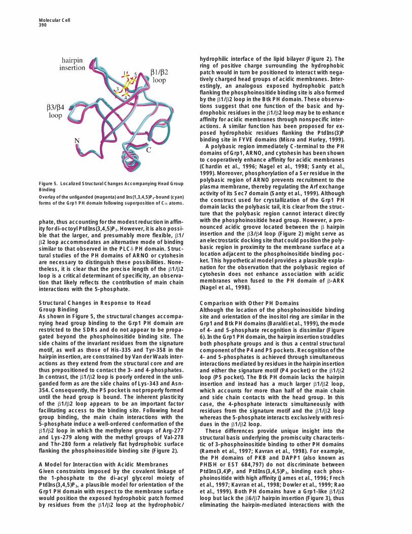
Figure 5. Localized Structural Changes Accompanying Head Groupplasma membrane, thereby regulating the Arf exchangeBindingactivity of its Sec7 domain (Santy et al., 1999). Although
Overlay of the unliganded (magenta) and Ins(1,3,4,5)P4-bound (cyan)the construct used for crystallization of the Grp1 PHforms of the Grp1 PH domain following superposition of Ca atoms.domain lacks the polybasic tail, it is clear from the struc-ture that the polybasic region cannot interact directlywith the phosphoinositide head group. However, a pro-phate, thus accounting for the modest reduction in affin-nounced acidic groove located between the b hairpinity for di-octoyl PtdIns(3,4,5)P3. However, it is also possi-insertion and the b3/b4 loop (Figure 2) might serve asble that the larger, and presumably more flexible, b1/an electrostatic docking site that could position the poly-b2 loop accommodates an alternative mode of bindingbasic region in proximity to the membrane surface at asimilar to that observed in the PLCd PH domain. Struc-location adjacent to the phosphoinositide binding poc-tural studies of the PH domains of ARNO or cytohesinket. This hypothetical model provides a plausible expla-are necessary to distinguish these possibilities. None-nation for the observation that the polybasic region oftheless, it is clear that the precise length of the b1/b2cytohesin does not enhance association with acidicloop is a critical determinant of specificity, an observa-membranes when fused to the PH domain of b-ARKtion that likely reflects the contribution of main chain(Nagel et al., 1998).interactions with the 5-phosphate.
Structural Changes in Response to Head Comparison with Other PH DomainsGroup Binding Although the location of the phosphoinositide bindingAs shown in Figure 5, the structural changes accompa- site and orientation of the inositol ring are similar in thenying head group binding to the Grp1 PH domain are Grp1 and Btk PH domains (Baraldi et al., 1999), the moderestricted to the SDRs and do not appear to be propa- of 4- and 5-phosphate recognition is dissimilar (Figuregated beyond the phosphoinositide binding site. The 6). In the Grp1 PH domain, the hairpin insertion straddlesside chains of the invariant residues from the signature both phosphate groups and is thus a central structuralmotif, as well as those of His-335 and Tyr-358 in the component of the P4 and P5 pockets. Recognition of thehairpin insertion, are constrained by Van der Waals inter- 4- and 5-phosphates is achieved through simultaneousactions as they extend from the structural core and are interactions mediated by residues in the hairpin insertionthus prepositioned to contact the 3- and 4-phosphates. and either the signature motif (P4 pocket) or the b1/b2In contrast, the b1/b2 loop is poorly ordered in the unli- loop (P5 pocket). The Btk PH domain lacks the hairpinganded form as are the side chains of Lys-343 and Asn- insertion and instead has a much larger b1/b2 loop,354. Consequently, the P5 pocket is not properly formed which accounts for more than half of the main chainuntil the head group is bound. The inherent plasticity and side chain contacts with the head group. In thisof the b1/b2 loop appears to be an important factor case, the 4-phosphate interacts simultaneously withfacilitating access to the binding site. Following head residues from the signature motif and the b1/b2 loopgroup binding, the main chain interactions with the whereas the 5-phosphate interacts exclusively with resi-5-phosphate induce a well-ordered conformation of the dues in the b1/b2 loop.b1/b2 loop in which the methylene groups of Arg-277 These differences provide unique insight into theand Lys-279 along with the methyl groups of Val-278 structural basis underlying the promiscuity characteris-and Thr-280 form a relatively flat hydrophobic surface tic of 3-phosphoinositide binding to other PH domainsflanking the phosphoinositide binding site (Figure 2). (Rameh et al., 1997; Kavran et al., 1998). For example,
the PH domains of PKB and DAPP1 (also known asPHISH or EST 684,797) do not discriminate betweenA Model for Interaction with Acidic Membranes
Given constraints imposed by the covalent linkage of PtdIns(3,4)P2 and PtdIns(3,4,5)P3, binding each phos-phoinositide with high affinity (James et al., 1996; Frechthe 1-phosphate to the di-acyl glycerol moiety of
PtdIns(3,4,5)P3, a plausible model for orientation of the et al., 1997; Kavran et al., 1998; Dowler et al., 1999; Raoet al., 1999). Both PH domains have a Grp1-like b1/b2Grp1 PH domain with respect to the membrane surface
would position the exposed hydrophobic patch formed loop but lack the b6/b7 hairpin insertion (Figure 3), thuseliminating the hairpin-mediated interactions with theby residues from the b1/b2 loop at the hydrophobic/

Recognition of PI 3-Kinase Products by PH Domains391
Figure 6. Comparison of Ins(1,3,4,5)P4 Recognition by the Grp1 and Btk PH Domains
(A) Space-filling representation with invariant residues of the signature motif shown in white and nonconserved residues from the three SDRshighlighted in green (b1/b2 loop), magenta (b3/b4 loop), and orange (hairpin insertion).(B) Schematic diagram showing direct interactions with the head group. Residues in the signature motif are indicated in blue.
4- and 5-phosphates as well as the supporting interac- the various loop regions that are often characterizedas hypervariable. However, for PH domains known ortions between the hairpin insertion and b1/b2 loop. Ba-
sic residues at the termini of the b3/b4 and b6/b7 loops predicted to bind PI 3-kinase products, the length ofthe b1/b2 loop is not randomly distributed but insteadof these PH domains could contribute compensatory
interactions with the 3- and 4-phosphates but would be clusters strikingly around two distinct sizes: a relativelyshort Grp1-like b1/b2 loop with 6–7 residues or a largerpoorly positioned to contact the 5-phosphate. In addi-
tion to fewer direct interactions with the 5-phosphate, Btk-like b1/b2 loop with 10–11 residues (Figure 3). Shortb1/b2 loops require a glycine at the N terminus whereasthe loss of supporting interactions between the hairpin
insertion and the b1/b2 loop is likely to weaken the the longer loops accommodate residues with small sidechains (Ala, Ser, or Pro). At the C-terminal position,remaining backbone contacts with the 5-phosphate.
Thus, the absence of the insertion region provides a most PH domains that bind 3-phosphoinositides con-serve a small polar residue (Thr, Asn, or Ser) that cansimple structural explanation for the promiscuous bind-
ing of PI 3-kinase products by PH domains with short be reasonably predicted to contact the 1-phosphate and2-hydroxyl groups. At other positions in the b1/b2 loop,Grp1 like b1/b2 loops.there appear to be no strict sequence requirements;however, there is a clear preference for basic or hy-General Determinants of 3-Phosphoinositidedrophobic residues. Based on the structures of the Grp1Specificityand Btk PH domains, it can be expected that the majorityAmong PH domains as a whole, there is no obvious
pattern in either the length, sequence, or structure of of these residues will not directly contact the head group

Molecular Cell392
defined MOPS media supplemented with 25 mM selenomethionine,or significantly influence the electrostatic environmentinduced at an OD600 of z0.4 by addition of 50 mM IPTG and harvestedwithin the binding pocket but instead are likely to inter-after 15 hr at 228C. The cells were resuspended in 50 mM Tris (pHact nonspecifically with the hydrophobic/charged inter-8.5), 0.1% 2-mercaptoethanol, lysed by sonication, and centrifugedface of acidic phospholipid membranes. A striking simi-at 35,000 3 g for 1 hr, and the supernatant was loaded onto a
larity in the b1/b2 loops of the Grp1 and Btk PH domains NiNTA-agarose column (Qiagen). After washing with ten columnis the use of backbone NH groups as contacts to the volumes of 50 mM Tris (pH 8.5), 500 mM NaCl, 10 mM imidazole,5-phosphate group. This mode of interaction would be 0.1% 2-mercaptoethanol, the fusion protein was eluted with a gradi-expected to depend critically on the size of the loop but ent of 10–150 mM imidazole. Anion exchange chromatography on
Resource Q (Pharmacia) followed by gel filtration on Superdex-75not the sequence except where necessary to facilitate(Pharmacia) yielded 100 mg of a preparation that was .99% purebackbone contacts. Taken together, these observationsas judged by SDS-PAGE.suggest that the structural variation in the b1/b2 loop
is restricted to a relatively small repertoire of main chainCrystallization and Data Collectionconformations, despite considerable sequence varia-Prior to crystallization, a 10 mg/mL solution of the Grp1 PH domain
tion. The Grp1 and Btk PH domains are prototypical (in 10 mM Tris-HCl [pH 8.5], 25 mM NaCl, 10% glycerol) was incu-examples representing two distinct subgroups of the bated at 208C for 1 hr in the presence of a 1.2-fold stoichiometricstructural repertoire. PH domains with 10-residue b1/ excess of Ins(1,3,4,5)P4. Crystals of SeMet-substituted Grp1 PHb2 loops, such as those of Pdk1, Gab1, and Dos, repre- bound to Ins(1,3,4,5)P4 were grown at 208C in microseeded hanging
drops containing 10 mg/mL of the complex, 6%–8% PEG-4000, 50sent a third distinct subgroup. These PH domains allmM sodium acetate (pH 4.8), 100 mM Li2SO4, and 10% glycerol.conserve a proline at the second position of the b1/Crystals appeared in 1–2 days and grew to maximum dimensionsb2 loop and lack the polar residue at the C-terminalof 0.05 3 0.5 3 1 mm over 3 weeks. The crystals are in the primitiveposition.orthorhombic space group P21212 with cell constants a 5 41.3 A,In contrast, the length of the b3/b4 loop is not a gener-b 5 107.0 A, and c 5 36.9 A. The volume of the unit cell is consistent
ally important factor for 3-phosphoinositide recognition. with one molecule in the asymmetric unit and a solvent content ofFor example, the larger b3/b4 loop in the Btk PH domain 44%. Crystals were soaked for 5 min at 208C in a cryoprotectant-does not facilitate additional interactions with the head stabilizer solution (10% PEG-4000, 10% MPD, 10% glycerol, 50 mMgroup compared with the Grp1 PH domain. Indeed, resi- sodium acetate [pH 4.8], and 100 mM Li2SO4) prior to flash freezing
in liquid propane.dues in the middle of the loop are poorly positioned toUsing an ADSC Quantum-4 CCD detector at the NSLS X4a beam-contact the head group. On the other hand, basic resi-
line at Brookhaven, MAD data were collected to 2.2 A on a singledues at the N and C termini of the b3/b4 loop are gener-crystal at three wavelengths near the selenium edge: the f99 maxi-ally well positioned to contact the 3- and/or 4-phos-mum, the f9 minimum, and a high-energy remote wavelength (Tablephates. Similar considerations apply to the relatively1). Friedel mates were collected using inverse beam geometry, and
short b6/b7 loops of PH domains that lack the hairpin the crystal was maintained at 1008K using a nitrogen cryostreaminsertion. In this case, basic or tyrosine residues near (Oxford Cryosystems). A high-resolution data set complete to 1.5 Athe C terminus of the loop would be sufficiently close was collected on a separate crystal at a wavelength below theto contact the 4-phosphate and, possibly, the 5-phos- selenium edge.
Crystals of the unliganded form were obtained at 48C inphate. Thus, the location of residues with respect to themicroseeded hanging drops containing 10 mg/mL protein, 12%termini of the b3/b4 and b6/b7 loops may serve as aPEG-4000, 50 mM sodium acetate (pH 4.8), 100 mM Li2SO4, anduseful predictor of potential specificity-determining in-10% glycerol. The unliganded crystals are also in the space groupteractions with either the 3- and 4-phosphates or the 4-P21212 with similar cell dimensions of a 5 40.7 A, b 5 107.6 A, andand 5-phosphates, respectively.c 5 37.5 A. A data set complete to 2.0 A was collected on a RigakuRUH3R/Mar 30 cm image plate detector. All data were processed
Implications with Denzo and scaled with Scalepack (Otwinowski and Minor,1994).The structure of the Grp1 PH domain provides strong
evidence for an evolutionarily conserved mechanism forStructure Determination and Refinement3- and 4-phosphate recognition coupled with divergentThe structure of the Grp1 PH domain bound to Ins(1,3,4,5)P4 wasmodes of specificity determination, allowing the affinitysolved by multiwavelength anomalous diffraction (MAD) based onfor PI 3-kinase products, and hence the cellular re-a single well-ordered seleno-methionine residue (Table 1). The loca-
sponse, to be adapted to the requirements of particular tion of the Se site was easily deduced from 12 s Harker peakssignaling networks. In addition to providing functional in the Bijvoet difference Patterson map calculated with the datadiversity, this combination of conserved and divergent collected at the f″ maximum. The heavy atom model was refineddeterminants of phosphoinositide recognition and spec- against a maximum likelihood target function using SHARP (de la
Fortelle et al., 1997), and the initial MAD phases were improved byificity enhances the possibility of developing membrane-solvent flipping using Solomon as implemented in CCP4 (1994). Apermeable reagents directed against the phosphoinosi-Fourier summation with sA weights yielded an interpretable electrontide binding sites of PH domains but having specificitydensity map with continuous main chain as well as side chain densityfor particular PI 3-kinase targets. The range of cellularin the hydrophobic core and clearly defined density for the phospho-
responses to such reagents is likely to be considerably inositide head group. An initial polyalanine model corresponding toless pleiotropic than the effects evoked by inhibitors of the central b barrel and C-terminal helix was constructed using OPI 3-kinases. (Jones et al., 1991). Partial model combination yielded unambiguous
density for the omitted loop regions. The resulting model was refinedExperimental Procedures against the 1.5 A data set using X-plor (Brunger, 1992). Except where
noted, structural figures were rendered with Raster3D (Merritt andExpression and Purification Bacon, 1997).A construct (residues 257–390) encompassing the PH domain ofGrp1 was expressed in E. coli using a modified pET15b vector Acknowledgments(Novagen) containing an N-terminal 63 His peptide (MGHHHHHHGS). B834(DE3) cells harboring the modified pET15b plasmid We thank Silvia Corvera for comments on the manuscript, and Craig
Ogatta, John Dumas, and Eric Merithew for assistance with datacontaining the Grp1 PH insert were grown at 228C in 3 liters of
Recognition of PI 3-Kinase Products by PH Domains393
collection. D. G. L. is a Leukemia & Lymphoma Society Scholar. Harlan, J.E., Hajduk, P.J., Yoon, H.S., and Fesik, S.W. (1994). Plecks-trin homology domains bind to phosphatidylinositol-4,5-bisphos-This work was supported in part by National Institutes of Health
Grant DK30648 (M. P. C.). phate. Nature 371, 168–170.
Hiles, I.D., Otsu, M., Volinia, S., Fry, M.J., Gout, I., Dhand, R., Panayo-Received April 5, 2000; revised June 5, 2000. tou, G., Ruiz-Larrea, F., Thompson, A., Totty, N.F., et al. (1992).
Phosphatidylinositol 3-kinase: structure and expression of the 110References kd catalytic subunit. Cell 70, 419–429.
Isakoff, S.J., Cardozo, T., Andreev, J., Li, Z., Ferguson, K.M., Aba-Auger, K.R., and Cantley, L.C. (1991). Novel polyphosphoinositides gyan, R., Lemmon, M.A., Aronheim, A., and Skolnik, E.Y. (1998).in cell growth and activation. Cancer Cells 3, 263–270. Identification and analysis of PH domain-containing targets of phos-Baraldi, E., Carugo, K.D., Hyvonen, M., Surdo, P.L., Riley, A.M., phatidylinositol 3-kinase using a novel in vivo assay in yeast. EMBOPotter, B.V., O’Brien, R., Ladbury, J.E., and Saraste, M. (1999). Struc- J. 17, 5374–5387.ture of the PH domain from Bruton’s tyrosine kinase in complex James, S.R., Downes, C.P., Gigg, R., Grove, S.J., Holmes, A.B., andwith inositol 1,3,4,5-tetrakisphosphate. Structure 7, 449–460. Alessi, D.R. (1996). Specific binding of the Akt-1 protein kinase toBrunger, A.T. (1992). X-plor Version 3.1. A System for X-Ray Crystal- phosphatidylinositol 3,4,5-trisphosphate without subsequent acti-lography and NMR (New Haven, CT: Yale University Press). vation. Biochem. J. 315, 709–713.
Cantley, L.C., and Neel, B.G. (1999). New insights into tumor sup- Jones, T.A., Zou, J.Y., Cowan, S.W., and Kjeldgaard, M. (1991).pression: PTEN suppresses tumor formation by restraining the Improved methods for building protein models in electron densityphosphoinositide 3-kinase/AKT pathway. Proc. Natl. Acad. Sci. USA maps and the location of errors in these models. Acta Crystallogr.96, 4240–4245. A 47, 110–119.Chardin, P., Paris, S., Antonny, B., Robineau, S., Beraud-Dufour, S., Kavran, J.M., Klein, D.E., Lee, A., Falasca, M., Isakoff, S.J., Skolnik,Jackson, C.L., and Chabre, M. (1996). A human exchange factor for E.Y., and Lemmon, M.A. (1998). Specificity and promiscuity in phos-ARF contains Sec7- and pleckstrin-homology domains. Nature 384, phoinositide binding by pleckstrin homology domains. J. Biol. Chem.481–484. 273, 30497–30508.Chothia, C., Lesk, A.M., Tramontano, A., Levitt, M., Smith-Gill, S.J., Klarlund, J.K., Guilherme, A., Holik, J.J., Virbasius, J.V., Chawla,Air, G., Sheriff, S., Padlan, E.A., Davies, D., Tulip, W.R., et al. (1989). A., and Czech, M.P. (1997). Signaling by phosphoinositide-3,4,5-Conformations of immunoglobulin hypervariable regions. Nature trisphosphate through proteins containing pleckstrin and Sec7 ho-342, 877–883. mology domains. Science 275, 1927–1930.Collaborative Computational Project No. 4 (1994). The CCP4 suite: Klarlund, J.K., Rameh, L.E., Cantley, L.C., Buxton, J.M., Holik, J.J.,programs for protein crystallography. Acta Crystallogr. D 50, Sakelis, C., Patki, V., Corvera, S., and Czech, M.P. (1998). Regulation760–763. of GRP1-catalyzed ADP ribosylation factor guanine nucleotide ex-Corvera, S., and Czech, M.P. (1998). Direct targets of phosphoinosi- change by phosphatidylinositol 3,4,5-trisphosphate. J. Biol. Chem.tide 3-kinase products in membrane traffic and signal transduction. 273, 1859–1862.Trends Cell Biol. 8, 442–446. Klarlund, J.K., Tsiaras, W., Holik, J.J., Chawla, A., and Czech, M.P.de la Fortelle, E., Irwin, J.J., and Bricogne, G. (1997). SHARP: a (2000). Distinct polyphosphoinositide binding selectivities for PHmaximum-likelihood heavy-atom parameter refinement and phasing domains of Grp1-like proteins based on diglycine versus triglycineprogram for MIR and MAD methods. In Crystallographic Computing motifs. J. Biol. Chem., in press.7, P. Bourne and K. Watenpaugh, eds. (Oxford: Oxford University Kojima, T., Fukuda, M., Watanabe, Y., Hamazato, F., and Mikoshiba,Press). K. (1997). Characterization of the pleckstrin homology domain of BtkDomin, J., and Waterfield, M.D. (1997). Using structure to define the as an inositol polyphosphate and phosphoinositide binding domain.function of phosphoinositide 3-kinase family members. FEBS Lett. Biochem. Biophys. Res. Commun. 236, 333–339.410, 91–95.
Leevers, S.J., Vanhaesebroeck, B., and Waterfield, M.D. (1999). Sig-Dowler, S., Currie, R.A., Downes, C.P., and Alessi, D.R. (1999). nalling through phosphoinositide 3-kinases: the lipids take centreDAPP1: a dual adaptor for phosphotyrosine and 3-phosphoinosi- stage. Curr. Opin. Cell Biol. 11, 219–225.tides. Biochem. J. 342, 7–12.
Lemmon, M.A., Ferguson, K.M., O’Brien, R., Sigler, P.B., and Schles-Escobedo, J.A., Navankasattusas, S., Kavanaugh, W.M., Milfay, D., singer, J. (1995). Specific and high-affinity binding of inositol phos-Fried, V.A., and Williams, L.T. (1991). cDNA cloning of a novel 85 kd phates to an isolated pleckstrin homology domain. Proc. Natl. Acad.protein that has SH2 domains and regulates binding of PI3-kinase Sci. USA 92, 10472–10476.to the PDGF beta-receptor. Cell 65, 75–82.
Maehama, T., and Dixon, J.E. (1998). The tumor suppressor, PTEN/Ferguson, K.M., Lemmon, M.A., Schlessinger, J., and Sigler, P.B. MMAC1, dephosphorylates the lipid second messenger, phosphati-(1995). Structure of the high-affinity complex of inositol trisphos- dylinositol 3,4,5-trisphosphate. J. Biol. Chem. 273, 13375–13378.phate with a phospholipase C pleckstrin homology domain. Cell 83,
Merritt, E.A., and Bacon, D.J. (1997). Raster3D—photorealistic mo-1037–1046.lecular graphics. Methods Enzymol. 277, 505–524.
Franke, T.F., Kaplan, D.R., Cantley, L.C., and Toker, A. (1997). DirectMisra, S., and Hurley, J.H. (1999). Crystal structure of a phosphatidyl-regulation of the Akt proto-oncogene product by phosphatidylinosi-inositol 3-phosphate specific membrane targeting motif, the FYVEtol-3,4-bisphosphate. Science 275, 665–668.domain of Vps27p. Cell 97, 657–666.
Frech, M., Andjelkovic, M., Ingley, E., Reddy, K.K., Falck, J.R., andNagel, W., Schilcher, P., Zeitlmann, L., and Kolanus, W. (1998). TheHemmings, B.A. (1997). High-affinity binding of inositol phosphatesPH domain and the polybasic c domain of cytohesin-1 cooperateand phosphoinositides to the pleckstrin homology domain of RAC/specifically in plasma membrane association and cellular function.protein kinase B and their influence on kinase activity. J. Biol. Chem.Mol. Biol. Cell 9, 1981–1994.272, 8474–8481.Otwinowski, Z., and Minor, W. (1994). Processing of X-ray diffractionFruman, D.A., Meyers, R.E., and Cantley, L.C. (1998). Phosphoinosi-data in oscillation mode. Methods Enzymol. 276, 307–326.tide kinases. Annu. Rev. Biochem. 67, 481–507.
Rameh, L.E., and Cantley, L.C. (1999). The role of phosphoinositideFruman, D.A., Rameh, L.E., and Cantley, L.C. (1999). Phosphoinosi-3-kinase lipid products in cell function. J. Biol. Chem. 274, 8347–tide binding domains: embracing 3-phosphate. Cell 97, 817–820.8350.Fukuda, M., Kojima, T., Kabayama, H., and Mikoshiba, K. (1996).
Mutation of the pleckstrin homology domain of Bruton’s tyrosine Rameh, L.E., Arvidsson, A., Carraway, K.L., III, Couvillon, A.D., Rath-bun, G., Crompton, A., VanRenterghem, B., Czech, M.P., Ravichan-kinase in immunodeficiency impaired inositol 1,3,4,5-tetrakisphos-
phate binding capacity. J. Biol. Chem. 271, 30303–30306. dran, K.S., Burakoff, S.J., et al. (1997). A comparative analysis of

Molecular Cell394
the phosphoinositide binding specificity of pleckstrin homology do-mains. J. Biol. Chem. 272, 22059–22066.
Rao, V.R., Corradetti, M.N., Chen, J., Peng, J., Yuan, J., Prestwich,G.D., and Brugge, J.S. (1999). Expression cloning of protein targetsfor 3-phosphorylated phosphoinositides. J. Biol. Chem. 274, 37893–37900.
Rebecchi, M.J., and Scarlata, S. (1998). Pleckstrin homology do-mains: a common fold with diverse functions. Annu. Rev. Biophys.Biomol. Struct. 27, 503–528.
Salim, K., Bottomley, M.J., Querfurth, E., Zvelebil, M.J., Gout, I.,Scaife, R., Margolis, R.L., Gigg, R., Smith, C.I., Driscoll, P.C., et al.(1996). Distinct specificity in the recognition of phosphoinositides bythe pleckstrin homology domains of dynamin and Bruton’s tyrosinekinase. EMBO J. 15, 6241–6250.
Santy, L.C., Frank, S.R., Hatfield, J.C., and Casanova, J.E. (1999).Regulation of ARNO nucleotide exchange by a PH domain electro-static switch. Curr. Biol. 9, 1173–1176.
Skolnik, E.Y., Margolis, B., Mohammadi, M., Lowenstein, E., Fischer,R., Drepps, A., Ullrich, A., and Schlessinger, J. (1991). Cloning ofPI3 kinase-associated p85 utilizing a novel method for expression/cloning of target proteins for receptor tyrosine kinases. Cell 65,83–90.
Stoyanov, B., Volinia, S., Hanck, T., Rubio, I., Loubtchenkov, M.,Malek, D., Stoyanova, S., Vanhaesebroeck, B., Dhand, R., Nurnberg,B., et al. (1995). Cloning and characterization of a G protein-activatedhuman phosphoinositide-3 kinase. Science 269, 690–693.
Vanhaesebroeck, B., Leevers, S.J., Panayotou, G., and Waterfield,M.D. (1997). Phosphoinositide 3-kinases: a conserved family of sig-nal transducers. Trends Biochem. Sci. 22, 267–272.
Wu, X., Senechal, K., Neshat, M.S., Whang, Y.E., and Sawyers, C.L.(1998). The PTEN/MMAC1 tumor suppressor phosphatase functionsas a negative regulator of the phosphoinositide 3-kinase/Akt path-way. Proc. Natl. Acad. Sci. USA 95, 15587–15591.
Protein Data Bank Accession Numbers
The accession numbers for the coordinates of the structures re-ported in this article are 1FGY (IP4 bound structure) and 1FGZ (unli-ganded structure).