HIV-1 Is Associated With Lower Group B Streptococcus Capsular and Surface-Protein IgG Antibody...
35
Accepted Manuscript 1 © The Author 2015. Published by Oxford University Press on behalf of the Infectious Diseases Society of America. All rights reserved. For Permissions, please e‐mail: [email protected]. HIV-1 is associated with lower Group B Streptococcus capsular and surface-protein IgG antibody levels and reduced transplacental antibody transfer in pregnant women Ziyaad Dangor 1,2,3 , Gaurav Kwatra 1,2 , Alane Izu 1,2 , Peter Adrian 1,2 , Nadia van Niekerk 1,2 , Clare L. Cutland 1,2 , Yasmin Adam 4 , Sithembiso Velaphi 3 , Sanjay G. Lala 3 , Shabir A. Madhi 1,2,5 1 Medical Research Council: Respiratory and Meningeal Pathogens Research Unit, Faculty of Health Sciences, University of the Witwatersrand, South Africa 2 Department of Science and Technology/National Research Foundation: Vaccine Preventable Diseases, Faculty of Health Sciences, University of the Witwatersrand, South Africa 3 Department of Paediatrics, Faculty of Health Sciences, University of the Witwatersrand, South Africa 4 Department of Obstetrics and Gynaecology, Faculty of Health Sciences, University of the Witwatersrand, South Africa 5 National Institute for Communicable Diseases: a division of National Health Laboratory Service, Centre for Vaccines and Immunology, Sandringham, South Africa * Corresponding Author: Shabir A Madhi, 1 Modderfontein Road, Sandringham, Gauteng; 2131; South Africa, Ph: +27 113866137, Fax: +27 866827159, E-mail: [email protected] Alternate corresponding author: Ziyaad Dangor, 11th Floor, Central West wing, Nurses Residence, Chris Hani Baragwanath Hospital, Soweto, Gauteng, 2013, Ph: +27 119834383, Fax: +27 867656326, E-mail: [email protected] Journal of Infectious Diseases Advance Access published February 4, 2015 by guest on March 17, 2016 http://jid.oxfordjournals.org/ Downloaded from
Transcript of HIV-1 Is Associated With Lower Group B Streptococcus Capsular and Surface-Protein IgG Antibody...
Acce
pted M
anus
cript
1
© The Author 2015. Published by Oxford University Press on behalf of the Infectious Diseases Society of America. All rights reserved. For Permissions, please e‐mail: [email protected].
HIV-1 is associated with lower Group B Streptococcus capsular and surface-protein IgG
antibody levels and reduced transplacental antibody transfer in pregnant women
Ziyaad Dangor1,2,3, Gaurav Kwatra1,2, Alane Izu1,2, Peter Adrian1,2, Nadia van Niekerk1,2,
Clare L. Cutland1,2, Yasmin Adam4, Sithembiso Velaphi3, Sanjay G. Lala3, Shabir A.
Madhi1,2,5
1Medical Research Council: Respiratory and Meningeal Pathogens Research Unit, Faculty of
Health Sciences, University of the Witwatersrand, South Africa
2Department of Science and Technology/National Research Foundation: Vaccine Preventable
Diseases, Faculty of Health Sciences, University of the Witwatersrand, South Africa
3Department of Paediatrics, Faculty of Health Sciences, University of the Witwatersrand, South
Africa
4Department of Obstetrics and Gynaecology, Faculty of Health Sciences, University of the
Witwatersrand, South Africa
5National Institute for Communicable Diseases: a division of National Health Laboratory
Service, Centre for Vaccines and Immunology, Sandringham, South Africa
*Corresponding Author: Shabir A Madhi, 1 Modderfontein Road, Sandringham, Gauteng; 2131;
South Africa, Ph: +27 113866137, Fax: +27 866827159, E-mail: [email protected]
Alternate corresponding author: Ziyaad Dangor, 11th Floor, Central West wing, Nurses
Residence, Chris Hani Baragwanath Hospital, Soweto, Gauteng, 2013, Ph: +27 119834383, Fax:
+27 867656326, E-mail: [email protected]
Journal of Infectious Diseases Advance Access published February 4, 2015 by guest on M
arch 17, 2016http://jid.oxfordjournals.org/
Dow
nloaded from
Acce
pted M
anus
cript
2
Abstract
Background: HIV-exposed infants are at increased risk of invasive Group B Streptococcus
(GBS) disease; however, the reason for this increased susceptibility has not been characterized.
Methods: We compared GBS capsular and surface-protein maternal IgG antibody
concentrations and cord-maternal ratios between HIV-infected and HIV-uninfected mother-
newborn dyads.
Results: Median capsular antibody concentrations (µg/ml) were lower in HIV-infected than
HIV-uninfected women for serotypes Ib (p=0.033) and V (p=0.040); and for pilus island (PI)-1
(p=0.016), PI-2a (p=0.015), PI-2b (p=0.015) and fibrinogen-binding protein A (p<0.001). For
serotypes Ia and III, cord-maternal ratios were 37.4% (p<0.001) and 32.5% (p=0.027) lower in
HIV-infected compared to HIV-uninfected mother-newborn dyads. The adjusted odds of having
capsular antibody concentration ≥2µg/ml when comparing HIV-infected to -uninfected women
were 0.33 (95%CI: 0.15-0.75) and 0.34 (95%CI: 0.12-1.00) for serotypes Ia and III. Antibody
levels and cord-maternal ratios were independent of CD4+ lymphocyte counts or HIV-1 viral
load among HIV-infected women.
Conclusions: The lower GBS antibody concentrations and reduced transplacental antibody
transfer in HIV-infected women, which likely contribute to their infants being at heightened
susceptibility for invasive GBS disease, could possibly be mitigated by vaccination with a
trivalent GBS conjugate vaccine that is currently under clinical development.
by guest on March 17, 2016
http://jid.oxfordjournals.org/D
ownloaded from
Acce
pted M
anus
cript
3
Introduction
Group B Streptococcus (GBS) is a leading cause of sepsis and meningitis in newborns and young
infants [1, 2]. A meta-analysis of studies undertaken from 2000 to 2010, reported the highest
incidence of invasive GBS disease to be in low-middle income countries from Eastern and
Southern Africa [3-7]. Maternal and newborn GBS serotype-specific capsular antibody has been
associated with protection against homotypic serotype invasive GBS disease in infants [8].
Furthermore, GBS surface-proteins which facilitate adherence to host epithelium such as pilus
island (PI) PI-1, PI-2a, PI-2b; Fibrinogen-binding protein A (FbsA) and GBS Immunogenic
Bacterial Adhesin (BibA) have been shown to be immunogenic, and induce antibodies in animal-
model studies that improved survival following systemic GBS inoculation challenges [9-11].
Although maternal HIV-infection is not associated with higher prevalence of recto-vaginal GBS
colonization during pregnancy or at birth [12-16], a greater risk of invasive GBS disease has
been reported in HIV-exposed compared to HIV-unexposed infants [17, 18]. The basis for the
increased susceptibility to invasive GBS in HIV-exposed infants remains to be ascertained and
could include maternal HIV-infection being associated with lower concentrations of protective
GBS antibodies or impaired transplacental antibody transfer [19].
The aim of this study was to determine the effect of maternal HIV-infection on IgG serotype-
specific (Ia, Ib, III and V) capsular antibody and select GBS surface-protein (PI-1, PI-2a, PI-2b,
BibA and FbsA) antibody concentrations in the mother and transplacental transfer to their
newborns.
by guest on March 17, 2016
http://jid.oxfordjournals.org/D
ownloaded from
Acce
pted M
anus
cript
4
Methods
We undertook a cross-sectional study of pregnant women delivering at Chris Hani Baragwanath
Academic Hospital from January to July 2013. This tertiary level care hospital serves the black-
African community of Soweto and surrounding areas. Pregnant women in this region deliver
either at this hospital (approximately 22 000 births annually) or at the midwife-obstetric units
(approximately 9500 births annually) [20].
The HIV-1 sero-prevalence amongst pregnant women in this setting was 28.4% during the study
period [20]. The provision of anti-retroviral therapy (ART) to prevent Mother to Child
Transmission of HIV has been detailed elsewhere [21, 22]. Briefly, following routine
confirmation of HIV-infection in the pregnant women, a CD4+ lymphocyte count is measured,
which at the time if >350 cells/mm3, zidovudine (AZT) was provided until delivery. Pregnant
women with a CD4+ count ≤350 cells/mm3 or WHO stage 3 or 4 were initiated on triple
antiretroviral therapy (ART). From April 2013, all HIV-infected pregnant women irrespective of
CD4+ cell count were initiated on triple ART [22].
The study sample size was calculated based on the assumption that the antibody transfer rate is
normally distributed with a standard deviation of approximately 0.5. We also assumed a trans-
placental antibody transfer ratio of 1.0 in HIV-uninfected mother-newborn dyads [23, 24]. A
sample of 79 HIV-infected and 79 HIV-uninfected pregnant women was required to detect at
least 20% difference in transplacental transfer ratio between HIV-exposed compared to HIV-
unexposed newborns with 80% power and alpha <0.05.
by guest on March 17, 2016
http://jid.oxfordjournals.org/D
ownloaded from
Acce
pted M
anus
cript
5
Study staff enrolled women in the labor and delivery wards during normal working hours from
Monday to Friday. Inclusion criteria were: an infant birth weight ≥2500grams, known maternal
HIV-status during pregnancy and willingness to participate in the study. Gestational age was
estimated using the following hierarchy of methods; antenatal ultrasound examination before 24
completed gestational weeks, the Ballard score done within 24 hours of birth, a reliable history
of the last menstrual period, antenatal sonar done at ≥24 weeks or the fundal symphysis height
(centimeters) examination during labor. Cord blood was taken at the time of birth and maternal
blood within 12 hours of delivery from enrolled participants. Cord blood was withdrawn using a
needled syringe from the umbilical vessels. Blood samples were allowed to clot at room
temperature and transported to the Respiratory and Meningeal Pathogens Research Unit within
4-6 hours for processing and storage. The blood was stored at 2-8ºC if not processed
immediately for a maximum period of 24 hours. Blood was centrifuged for 5 minutes at a 3220
relative centrifugal force and the serum then aliquoted and stored at –70ºC. Serum samples were
thawed and analyzed in batches. Newborns were not tested for HIV-1 infection immediately after
delivery.
The Luminex fluorescence based micro-bead immunosorbent assay was used to measure IgG
antibodies to capsular serotypes Ia, Ib, III, V, and to surface-proteins PI-1, PI-2a, PI-2b, BibA
and FbsA. Capsular and pilus island protein antigens were kindly provided by Novartis Vaccines
and Diagnostics (Italy), while BibA and FbsA protein antigens were provided by Valneva
Austria GmbH. Capsular polysaccharides were coupled to the microsphere beads (Bio-Rad, CA,
USA) with the crosslinking agent 4-(4,6 dimethoxy[1,3,5]triazin-2-yl)-4- methyl-morpholinium
(DMTMM) and protein antigens were coupled to beads with a two-step carbodiimide reaction
by guest on March 17, 2016
http://jid.oxfordjournals.org/D
ownloaded from
Acce
pted M
anus
cript
6
[25, 26]. Polygam (purified pooled commercial gammaglobulin; National Bioproducts, South
Africa) was used as reference serum and calibrated with standard capsular serotype-specific GBS
reference serum kindly provided by Prof. Carol J. Baker. For protein-specific antigen antibody
determination, reference serum was assigned arbitrary units (AU) of 10000 AU/ml. Bead
fluorescence was read with the Bio-plex 200 instrument using Bio-plex manager 5.0, software
(Bio-Rad, Texas, USA). Details are described in the supplementary appendix.
Serum capsular IgG was reported in micrograms per milliliter (µg/ml) with a lower limit of
detection of 0.0008, 0.002, 0.004 and 0.016 µg/ml for serotypes Ia, Ib, III and V, respectively;
while protein-specific IgG was reported in arbitrary units per milliliter (AU/ml) with a lower
limit of detection of 41,110, 46, 6 and 19 per AU/ml for Pil-1, Pil-2a, Pil-2b, BibA and FbsA,
respectively. Samples below these limits were assigned a value of half the lower limit of
detection for statistical analysis.
For analytical specificity of each GBS antigen-microsphere set, reference serum was incubated at
1:100 dilutions with each GBS antigen and incubated at 37˚C for 2 hours. The specificity was
recorded as the difference in reactivity between the absorbed and unabsorbed sera in a multiplex
assay. Homologous inhibition was >90% for all CPS and protein antigens with the exception of
serotype V (88%) and FBS-A protein (32%). Heterologous inhibition across antigens was <15%,
except for serotype Ib which was inhibited by 31% with serotype Ia and serotype V which was
inhibited by 17% with serotype III.
by guest on March 17, 2016
http://jid.oxfordjournals.org/D
ownloaded from
Acce
pted M
anus
cript
7
In HIV-infected women, CD4+ lymphocyte counts measured during pregnancy were recorded
and maternal blood obtained at the time of delivery was tested for HIV-1 RNA viral load using
the real-time PCR COBAS® AmpliPrep/COBAS® TaqMan® HIV-1 Test, version 2.0 (Roche
COBAS®; Roche Molecular Systems, USA), which has a lower limit of detection of 20 copies
per milliliter, with values below this being assigned an arbitrary value of 20.
Maternal GBS colonization was assessed at delivery by performing separate lower vaginal and
rectal swabs. Rayon tipped swabs were used for sampling, which was placed into Amies
transport medium without charcoal (Medical Wire Equipment Co. Ltd. Cat: MW170; U.K) and
transported to the laboratory for processing. The laboratory methods of GBS identification and
serotyping on vaginal and rectal swabs have been described [27].
Data analysis
Maternal and cord blood IgG antibody concentrations were measured, and cord blood to
maternal ratio calculated to compare the efficiency of transplacental antibody transfer between
HIV-exposed and HIV-unexposed newborns.Demographic characteristics were compared
between HIV-uninfected and HIV-infected mother-newborn dyads using Chi-square or Fisher’s
exact test for proportions; whilst the Mann-Whitney test was used to compare the medians.
Antibody concentrations remained non-parametric after log transformation, thus median
concentrations are reported.
Median maternal antibody concentrations were compared between HIV-uninfected and HIV-
infected women at delivery and cord blood antibody concentrations between HIV-unexposed and
by guest on March 17, 2016
http://jid.oxfordjournals.org/D
ownloaded from
Acce
pted M
anus
cript
8
HIV-exposed newborns using the Mann-Whitney test. Using quantile regression, we further
compared median maternal antibody concentrations, cord blood antibody concentrations and
cord-maternal ratios between HIV-uninfected and HIV-infected women, and adjusted for overall
colonization, colonizing serotype for homotypic capsular antibodies, maternal age and parity. We
also compared the proportions of HIV-infected and -uninfected women with capsular antibody
concentrations above various thresholds proposed to be protective against invasive GBS disease
in their infants [8]. In HIV-infected women, CD4+ T-lymphocyte counts and HIV-1 viral load
was correlated with maternal antibody concentrations and cord-maternal ratios using Spearman’s
test. Furthermore, we compared maternal antibody concentrations and cord-maternal ratios at
varying CD4+ lymphocyte counts and HIV-1 viral load thresholds using the Mann-Whitney test.
Data was analyzed using STATA version 13.1 (College Station, Texas, USA) and GraphPad
Prism version 6.05 for Windows (GraphPad Software, California USA). Two-tailed p-values
<0.05 were considered statistically significant. Written informed consent was obtained from the
women at time of study-enrolment. The study was approved by the University of Witwatersrand
Human Research Ethics Committee (HREC number: M120905) and registered as an
observational study on the South African National Clinical Trial Register (DOH-27-0113-4310)
Results
Of the 320 women screened, 70 refused consent and 76 failed to meet the inclusion criteria. We
therefore enrolled 174 mother-newborn dyads, ten of whom were subsequently excluded
(including nine dyads where the newborn gestational age was ≤36 weeks, and one dyad in whom
maternal blood was taken >12 hours following delivery). Thus, 164 mother-newborn dyads were
by guest on March 17, 2016
http://jid.oxfordjournals.org/D
ownloaded from
Acce
pted M
anus
cript
9
analyzed, including 81 HIV-uninfected and 83 HIV-infected women all of whom had singleton
births. Except for HIV-infected women being older (median 30.7 vs 26.0 years; p=0.006), they
were otherwise similar in demographic characteristics compared to HIV-uninfected women
(Table 1). Among the 83 HIV-infected women at the time of delivery, 36 (43.4%) were on triple
ART, 46 (55.4%) on zidovudine (AZT) only and one (1.2%) had not received any ART. The
median duration on triple ART from initiation to delivery was 13.4 weeks (range: 1.4 - >44) and
17.1 weeks (range: 2.4 - 42.7) for women on AZT only. Overall, 49 (29.9%) of 164 women were
colonized with GBS; colonization rates were similar in HIV-uninfected (27.2%) and HIV-
infected (32.5%) women (Table 1). The commonest colonizing serotype was Ia (59.1% of all
serotypes) in HIV-uninfected women and III (40.7% of serotypes) in HIV-infected women
(Table 1). Two infants born to HIV-infected women developed late-onset GBS meningitis from
serotypes Ia and III at 19 and 22 days of age, and among whom their mothers antibody
concentrations were 0.08 and 0.12 for the homotypic serotypes and the transplacental ratio was
0.14 and 0.69, respectively.
All women had detectable antibody levels to all four GBS serotypes, although cord blood
antibody levels were not detected in two samples for serotype Ia and in five samples each for
serotypes Ib, III and V. Regarding surface-protein antibodies, only one woman had undetectable
antibody levels to PI-2a. For cord blood samples, antibody levels were undetectable on two
samples for BibA, four samples for FbsA, five samples for PI-1 and PI-2b, and six samples for
PI-2a. The final analysis included all samples as results were similar when the above samples
were excluded from the analysis (data not shown).
by guest on March 17, 2016
http://jid.oxfordjournals.org/D
ownloaded from
Acce
pted M
anus
cript
10
Maternal HIV-infection status and capsular antibodies
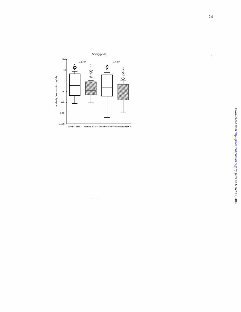
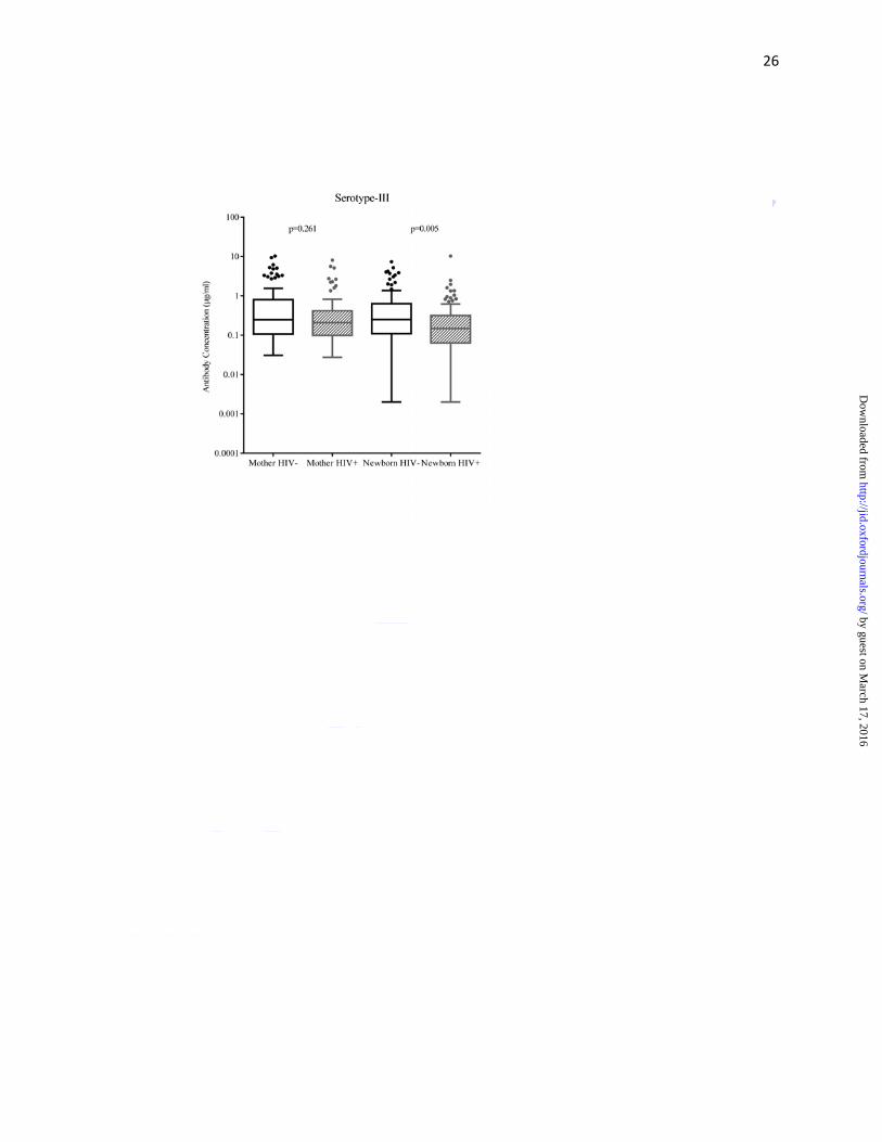
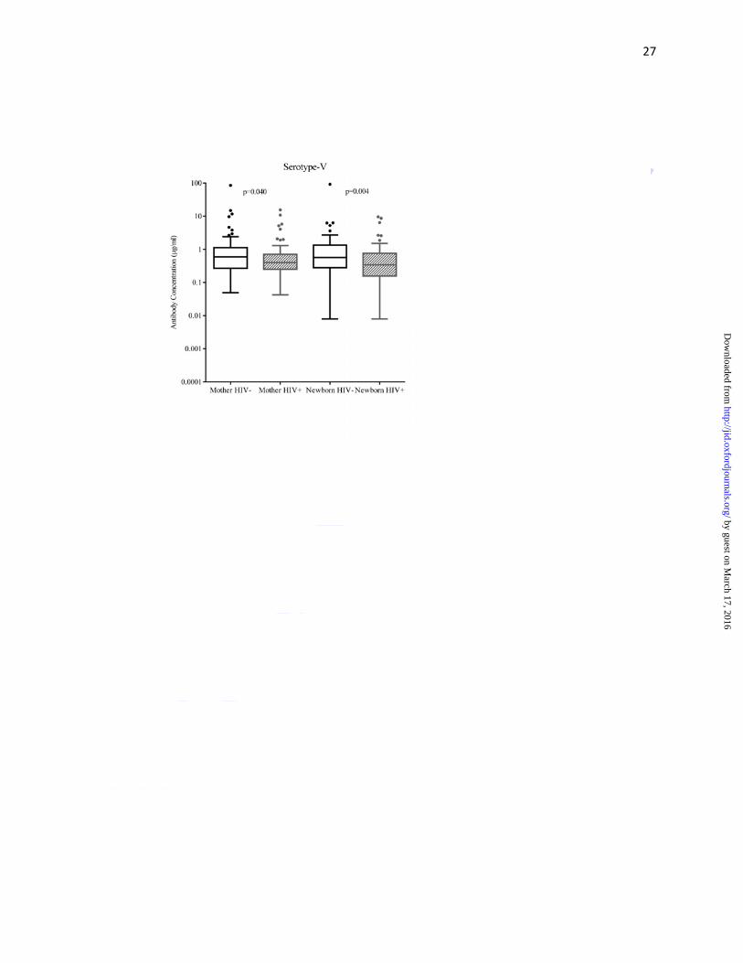
Median capsular antibody concentrations (µg/ml) were lower in HIV-infected than HIV-
uninfected women for serotypes Ib (0.06 vs. 0.09; p=0.033) and V (0.40 vs.0.59; p=0.040);
similar trends were observed for serotype Ia (0.13 vs. 0.36; p=0.077), but this difference was not
significant (Figure 1a-d, Supplementary Table 1). Median cord blood capsular antibody
concentrations (for all serotypes) were significantly lower in HIV-exposed than in HIV-
unexposed newborns; the respective antibody concentrations (μg/ml) for serotypes Ia, Ib, III and
V were 0.07 vs. 0.26 (p=0.005), 0.07 vs. 0.15 (p=0.013), 0.15 vs. 0.25 (p=0.005) and 0.34
vs.0.57 (p=0.004) (Figure 1a-d, Supplementary Table 1).
After adjusting for confounding factors, we compared maternal antibody concentrations between
HIV-infected and -uninfected women at multiple percentiles using quantile regression analysis.
Significant differences in antibody concentrations for serotypes Ia, III and V between HIV-
infected and -uninfected women were found at higher percentiles (above 65th), suggesting that
HIV-infected women also tended to have lower antibody concentrations that HIV-uninfected at
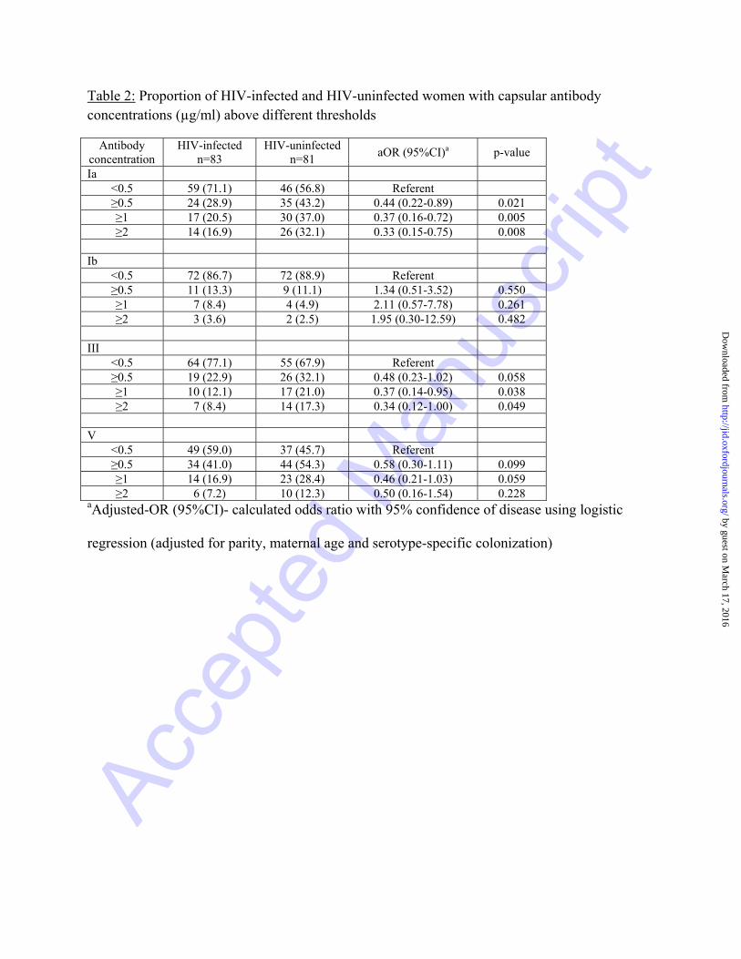
higher percentiles (Supplementary Table 2). Corroborating this, we demonstrated that a lower
proportion of HIV-infected women had capsular antibody concentrations above thresholds of
≥1µg/ml and ≥2µg/ml for serotypes Ia, III and V (Table 2). Using multivariate analysis, with an
antibody concentration of <0.5µg/ml as a referent, the adjusted odds of having capsular antibody
concentration ≥2µg/ml in HIV-infected compared to -uninfected women were 0.33 (95%CI:
0.15-0.75; p=0.008), 0.34 (95%CI: 0.12-1.00; p=0.049) and 0.50 (95%CI: 0.16-1.54; p=0.228)
for serotypes Ia, III and V, respectively (Table 2).
by guest on March 17, 2016
http://jid.oxfordjournals.org/D
ownloaded from
Acce
pted M
anus
cript
11
Overall, median cord-maternal ratios for capsular antibody ranged between 75% to 119% in
HIV-uninfected mother-newborn dyads and 47% to 93% among HIV-infected mother-newborn
dyads (Table 3). In the multivariate model, after adjusting for overall colonization, serotype-
specific colonization, maternal age and parity, the cord-maternal ratio was 37.4% (p<0.001) and
32.5% (p=0.027) lower for serotypes Ia and III in HIV-infected compared to HIV-uninfected
mother-newborn dyads (Table 3).
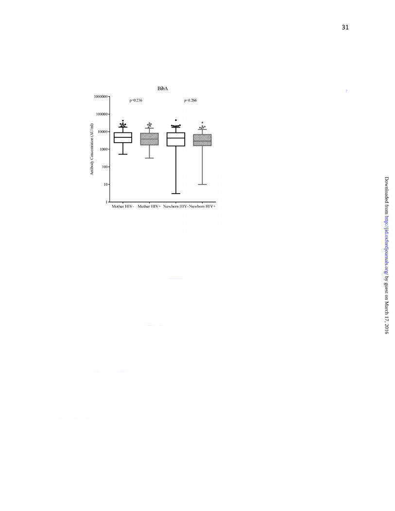
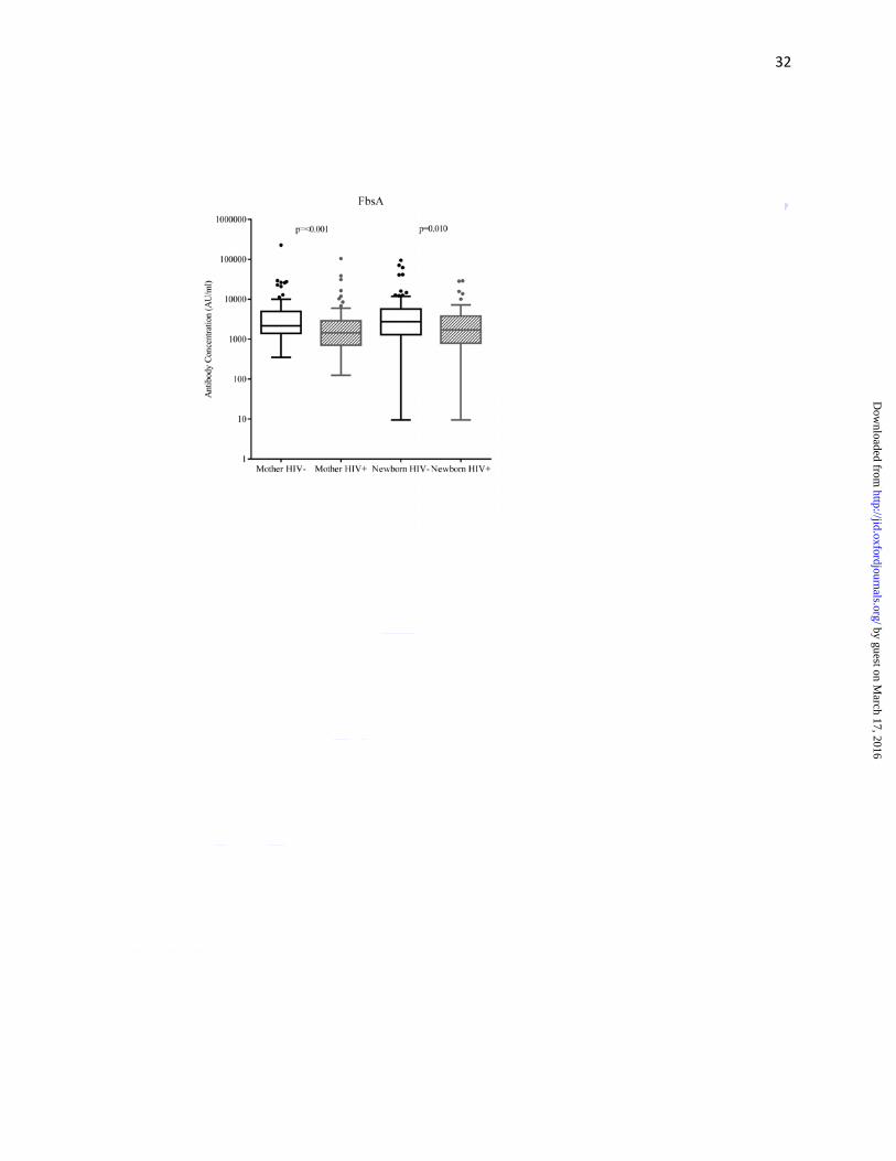
Maternal HIV-infection status and surface-protein antibodies
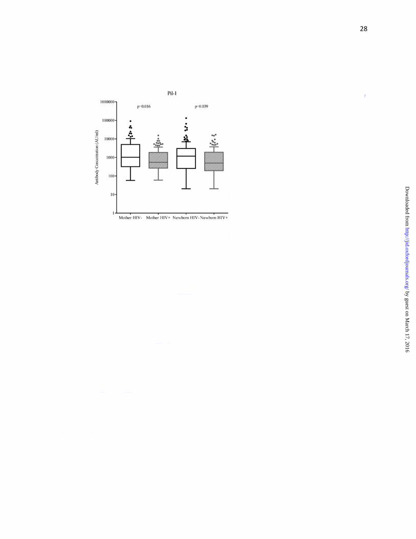
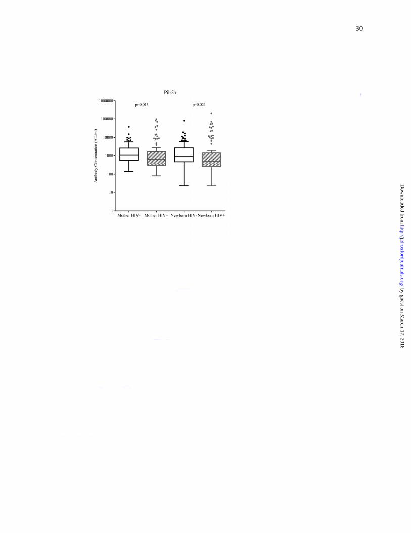
As compared to HIV-uninfected women, HIV-infected women had lower median antibody
concentrations (AU/ml) against surface-protein PI-1 (549 vs. 1020; p=0.016), PI-2a (1130 vs.
1972; p=0.015), PI-2b (611 vs. 1072; p=0.015) and FbsA (1444 vs.2169; p<0.001), but not
significantly so for BibA (3829 vs. 4790; p=0.236) (Figure 2a-e, Supplementary Table 1). Cord
blood median surface-protein antibody concentrations were lower in HIV-exposed compared to
HIV-unexposed newborns for PI-1 (502 vs. 1177; p=0.039), PI-2b (478 vs. 865; p=0.024) and
FbsA (1717 vs.2758; p=0.010) (Figure 2a-e, Supplementary Table 1). The median cord-maternal
ratios (range 76%-126%) were similar for all antibodies directed against surface-proteins
between HIV-uninfected and HIV-infected mother-newborn dyads (Table 3).
Effect of HIV-viral load and CD4+ lymphocyte count on GBS antibody in HIV-infected women
In HIV-infected women, 71 of 83 (85.5%) had a CD4+ lymphocyte count measured within 6
months before delivery with a median CD4+ lymphocyte count of 423 cells/mm3 (range: 46-
1268). The median HIV-1 viral load in 79/83 (95.2%) participants was 96 copies/ml (range: 20-
146 055) and undetectable in 28 of the 79 (35.4%) samples. There was no correlation between
CD4+ lymphocyte counts and maternal antibody concentrations or between CD4+ lymphocyte
by guest on March 17, 2016
http://jid.oxfordjournals.org/D
ownloaded from
Acce
pted M
anus
cript
12
counts and cord-maternal ratios for any of the nine measured antibodies. Furthermore, median
maternal antibody concentrations and cord-maternal ratios were similar when stratified by
different thresholds of CD4+ lymphocyte counts (Supplementary Table 3 and 4). Similarly, there
was no correlation between maternal HIV-1 viral load and maternal antibody concentration or
cord-maternal ratios for any of the nine measured antibodies (Supplementary Table 3 and 4).
Discussion
The findings from our study suggest that the possible mechanisms for the increased susceptibility
to invasive GBS disease in HIV-exposed infants may relate to lower maternal capsular and
surface-protein antibody concentrations, and inefficient transplacental transfer of capsular
antibody to the fetus of HIV-infected women [28-30]. HIV-infected women had lower GBS
capsular antibody concentrations than their HIV-uninfected counterparts, and notably a lower
proportion of HIV-infected women had capsular antibodies above the putative “protective”
thresholds that has been reported to protect against invasive GBS disease in their infants [8]. The
lower GBS antibody concentrations in HIV-infected women could represent waning of natural
acquired antibody or reduced humoral immune responsiveness to recto-vaginal colonization
which likely induces the antibody responses (personal correspondence Gaurav Kwatra-
manuscript under preparation). Additionally, reduced maternal exposure to GBS may also result
in lesser antibody production to various serotype-specific epitopes [8]. This is supported by some
studies which reported a lower prevalence of GBS colonization in HIV-infected women,
including previously in our setting [15, 16]; although this was not observed in the current study
cohort.
by guest on March 17, 2016
http://jid.oxfordjournals.org/D
ownloaded from
Acce
pted M
anus
cript
13
The transplacental transfer of antibodies to serotypes Ia and III, which account for the majority
(72%) of invasive GBS disease globally [3], was 37.4% and 32.5% lower in HIV-exposed
compared to HIV-unexposed newborns. Additionally, maternal capsular antibody concentrations
were lower in HIV-infected women compared to HIV-uninfected women for serotypes Ib and V,
with a trend towards being lower for serotype Ia, but not for serotype III. Serotype III, which has
the highest invasive potential, is the least immunogenic of all serotypes [31, 32] and this may
explain why concentrations were similar in HIV-infected and -uninfected women. Furthermore,
the trend toward higher colonization prevalence of serotype III in HIV-infected compared to
HIV-uninfected women in our study may have contributed to similar serotype III antibody
concentrations between the women.
We also measured antibody concentrations to select GBS surface-proteins which induce antibody
responses and could be possible vaccine targets. There is, however, a paucity of data on these
GBS surface-protein antibody concentrations and no international reference standards exist.
Thus, we can only report on the comparisons using in-house reference serum employed
consistently across all samples. HIV-infected women had lower median concentrations for all
GBS surface-proteins, although antibody differences to BibA were not significant. In addition,
we observed that contrary to the capsular antibody transfer, the transfer of surface-protein
antibodies from mother to fetus was more efficient, and similar between HIV-infected and HIV-
uninfected mother-newborn dyads. This may occur because surface-protein antibodies, which are
mainly subclass IgG1, are more efficiently transferred than capsular antibodies, which are
predominantly of subclass IgG2 [33].
by guest on March 17, 2016
http://jid.oxfordjournals.org/D
ownloaded from
Acce
pted M
anus
cript
14
Our results are consistent with reports showing reduced transplacental transfer of maternal
antibodies directed against epitopes of varicella (31% reduction), measles (35% reduction),
pneumococcus (24-30% reduction), Haemophilus influenzae type b (23% reduction), pertussis
(40% reduction) and tetanus (27-52% reduction) in HIV-infected compared to HIV-uninfected
mother-newborn dyads [28, 29, 34-36]. However, no difference in transplacental antibody
transfer between HIV-infected and -uninfected women for pathogens such as herpes, some
pneumococcal serotypes and influenza have also been reported [29, 34, 37]. Transplacental IgG
antibody transfer is thought to occur via an active transport mechanism utilizing neonatal Fc
receptors found on the placenta [30, 33, 38]. The decrease in transplacental antibody transfer in
HIV-infected women is thought to be as a consequence of maternal hyper-gammaglobulinemia
which saturates the neonatal Fc receptors [39]. Other reasons for the variation in transplacental
antibody transfer may relate to differences in IgG subclass and mechanism of transfer of
antibody (i.e. active or passive transport) [33].
Although our study did not identify a significant association between CD4+ lymphocyte counts
and HIV-1 viral loads on maternal antibody and cord-maternal ratios among HIV-infected
women, the study was not powered (with a sample size of 79) to detect a significant relationship
when the true correlation is between -0.35 and 0.35. Similarly, no association has been observed
between maternal CD4+ lymphocyte counts and transplacental transfer of pneumococcal,
Haemophilus influenzae type b, pertussis and tetanus antibodies in HIV-infected women [28,
29], whereas a positive correlation with CD4+ lymphocyte counts and maternal antibody
concentrations was reported to antibodies to pertussis, pneumococcus and tetanus [28]. More
recently, a large European cohort study reported an increased risk of bacterial infections in HIV-
by guest on March 17, 2016
http://jid.oxfordjournals.org/D
ownloaded from
Acce
pted M
anus
cript
15
exposed infants, particularly in women with low CD4+ lymphocyte counts [40]. Most pregnant
women in our setting had undetectable HIV-1 viral load and had immune reconstituted at the
time of antibody sampling. A study conducted in Nairobi in HIV-infected women reported a
44% decrease of measles antibody transfer with every log10 increase in viral load, indicating that
infants born to women with advanced maternal HIV-infection may be at increased risk of disease
due to reduced acquisition of maternal antibody concentrations [41].
Limitations of our study include that we did not match for age and colonization status in HIV-
infected and -uninfected women, however, we adjusted for these factors in the multivariate
analysis and findings remained consistent. Furthermore, we did not quantify the effect of cross
reactivity of serotype Ib with Ia, as previously documented by Brigsten et al. [42], may have had
on the absolute antibody concentration for serotype Ib. The assay was, however, applied
consistently to both HIV-infected and -uninfected dyads and hence is unlikely to alter the
differences observed between HIV-infected and HIV-uninfected women in our study. Also, our
study only measured IgG antibodies, whilst IgA antibodies may also be transplacentally
transferred; and have been associated with protection against GBS invasive disease in animal
model studies [11, 43]. Additionally, CD4+ lymphocyte counts were measured as part of
standard-of-care at any time within 6 months (mean: 2.8 months) of delivery and the study was
not specifically powered to address whether immunological status or different HIV-1 viral load
were associated with differences in maternal antibody or transplacental antibody transfer.
The lower GBS antibody concentrations and reduced transplacental antibody transfer in HIV-
infected women, which places their infants at risk for invasive GBS disease, may be mitigated by
by guest on March 17, 2016
http://jid.oxfordjournals.org/D
ownloaded from

Acce
pted M
anus
cript
16
maternal GBS vaccination. Furthermore, an investigational trivalent GBS polysaccharide-protein
conjugate vaccine was found to be less immunogenic in HIV-infected than HIV-uninfected
pregnant women [44]. Therefore, in HIV-burden settings, maternal vaccination may require
modified formulations or dosing schedules in HIV-infected women.
Acknowledgements
We are thankful to all mothers and infants that participated in the study. We acknowledge the
Department of Obstetrics at the Chris Hani Baragwanth Academic Hospital. We further
acknowledge the Johannesburg Health District for supplying data on the births in the
Johannesburg metropolitan. We acknowledge Novartis Vaccines and Diagnostics (Italy) and
Valneva Austria GmbH for providing the GBS antigens. We would also like to thank the
nursing, research and laboratory staff at the Respiratory and Meningeal Pathogens Research
Unit.
Financial disclosure/support: ZD is funded in part by the Carnegie Corporation of New York
(Grant number B8749) and the Discovery Foundation (Grant number 20289/1). SGL is funded in
part by a career development award from the Medical Research Council of South Africa. SAM is
funded in part by National Research Foundation/Department of Science and Technology: South
African Research Chair Initiative in Vaccine Preventable Diseases and Medical Research
Council of South Africa. The funders had no role in study design, data collection and analysis,
decision to publish, or preparation of the manuscript.
Conflict of interest: All authors have no conflicts of interest relevant to this article to disclose
by guest on March 17, 2016
http://jid.oxfordjournals.org/D
ownloaded from
Acce
pted M
anus
cript
17
Figure Legends
Figure 1: Tukey box and whisker plots comparing capsular antibody concentrations of (A)
serotype-Ia, (B) serotype-Ib, (C) serotype-III and (D) serotype-V between HIV-uninfected and -
infected mothers, and HIV-unexposed and -exposed newborns
Footnote: Mother HIV- denotes HIV-uninfected and HIV+ denotes HIV-infected, Newborn
HIV- denotes HIV-unexposed and Newborn HIV+ denotes HIV-exposed. The y-axis has been
log10 scaled. For the box and whisker plots; the box represents the distance of the 25th and 75th
percentile with the median represented by the solid line within the box. The upper whisker
represents 1.5 times the interquartile distance from the 75th centile, while the lower whisker
represents 1.5 times the interquartile distance from the 25th centile. The dot symbols represent
outliers above the upper whisker.
Figure 2: Tukey box and whisker plots comparing surface-protein antibody concentrations of (A)
Pil-1, (B) Pil-2a, (C) Pil-2b, (D) BibA and (E) FbsA between HIV-uninfected and -infected
mothers, and HIV-unexposed and -exposed newborns
Footnote: Mother HIV- denotes HIV-uninfected and HIV+ denotes HIV-infected, Newborn
HIV- denotes HIV-unexposed and Newborn HIV+ denotes HIV-exposed. Arbitrary units is
abbreviated AU. The y-axis has been log10 scaled. For the box and whisker plots; the box
represents the distance of the 25th and 75th percentile with the median represented by the solid
line within the box. The upper whisker represents 1.5 times the interquartile distance from the
75th centile, while the lower whisker represents 1.5 times the interquartile distance from the 25th
centile. The dot symbols represent outliers above the upper whisker.
by guest on March 17, 2016
http://jid.oxfordjournals.org/D
ownloaded from
Acce
pted M
anus
cript
18
References
1. Weston EJ, Pondo T, Lewis MM, et al. The burden of invasive early-onset neonatal sepsis in
the United States, 2005-2008. Pediatr Infect Dis J 2011; 30:937-41.
2. Thigpen MC, Whitney CG, Messonnier NE, et al. Bacterial meningitis in the United States,
1998-2007. N Engl J Med 2011; 364:2016-25.
3. Edmond KM, Kortsalioudaki C, Scott S, et al. Group B streptococcal disease in infants aged
younger than 3 months: systematic review and meta-analysis. Lancet 2012; 379:547-56.
4. Dagnew AF, Cunnington MC, Dube Q, et al. Variation in reported neonatal group B
streptococcal disease incidence in developing countries. Clin Infect Dis 2012; 55:91-102.
5. Berkley JA, Lowe BS, Mwangi I, et al. Bacteremia among children admitted to a rural hospital
in Kenya. N Engl J Med 2005; 352:39-47.
6. Gray KJ, Bennett SL, French N, Phiri AJ, Graham SM. Invasive group B streptococcal
infection in infants, Malawi. Emerg Infect Dis 2007; 13:223-9.
7. Cutland CL, Madhi SA, Zell ER, et al. Chlorhexidine maternal-vaginal and neonate body
wipes in sepsis and vertical transmission of pathogenic bacteria in South Africa: a randomised,
controlled trial. Lancet 2009; 374:1909-16.
8. Dangor Z, Kwatra G, Izu A, Lala SG, Madhi SA. Review on the association of Group B
Streptococcus capsular antibody and protection against invasive disease in infants. Expert Rev
Vaccines 2014:1-15.
9. Lindahl G, Stalhammar-Carlemalm M, Areschoug T. Surface proteins of Streptococcus
agalactiae and related proteins in other bacterial pathogens. Clin Microbiol Rev 2005; 18:102-27.
10. Margarit I, Rinaudo CD, Galeotti CL, et al. Preventing bacterial infections with pilus-based
vaccines: the group B streptococcus paradigm. J Infect Dis 2009; 199:108-15.
by guest on March 17, 2016
http://jid.oxfordjournals.org/D
ownloaded from

Acce
pted M
anus
cript
19
11. Meinke AL, Senn BM, Visram Z, et al. Immunological fingerprinting of group B
streptococci: from circulating human antibodies to protective antigens. Vaccine 2010; 28:6997-
7008.
12. El Beitune P, Duarte G, Maffei CM, Quintana SM, De Sa Rosa ESAC, Nogueira AA. Group
B Streptococcus carriers among HIV-1 infected pregnant women: prevalence and risk factors.
Eur J Obstet Gynecol Reprod Biol2006; 128:54-8.
13. Mavenyengwa RT, Moyo SR, Nordbo SA. Streptococcus agalactiae colonization and
correlation with HIV-1 and HBV seroprevalence in pregnant women from Zimbabwe. Eur J
Obstet Gynecol Reprod Biol 2010; 150:34-8.
14. Shah M, Aziz N, Leva N, Cohan D. Group B Streptococcus colonization by HIV status in
pregnant women: prevalence and risk factors. J Womens Health 2011; 20:1737-41.
15. Gray KJ, Kafulafula G, Matemba M, Kamdolozi M, Membe G, French N. Group B
Streptococcus and HIV infection in pregnant women, Malawi, 2008-2010. Emerg Infect Dis
2011; 17:1932-5.
16. Cutland CL, Schrag SJ, Zell ER, et al. Maternal HIV Infection and Vertical Transmission of
Pathogenic Bacteria. Pediatrics 2012; 6:6.
17. Epalza C, Goetghebuer T, Hainaut M, et al. High incidence of invasive group B streptococcal
infections in HIV-exposed uninfected infants. Pediatrics 2010; 126:23.
18. Cutland CL, Schrag SJ, Thigpen MC, et al. HIV-exposure increases Group B Streptococcus
sepsis risk in young South African infants. Emerg Infect Dis 2015; In-press.
19. Afran L, Garcia Knight M, Nduati E, Urban BC, Heyderman RS, Rowland-Jones SL. HIV-
exposed uninfected children: a growing population with a vulnerable immune system? Clin Exp
Immunol 2014; 176:11-22.
by guest on March 17, 2016
http://jid.oxfordjournals.org/D
ownloaded from
Acce
pted M
anus
cript
20
20. District Research Committee. Birth statistics. Johannesburg: Johannesburg Health District.
Gauteng Department of Health, 2014.
21. National Department of Health. Clinical Guidelines: PMTCT (Prevention of Mother-to-Child
Transmission). Pretoria: National Department of Health, South Africa, 2010.
22. National Department of Health. The South African Antiretroviral Treatment Guidelines,
PMTCT Guidelines: Revised March 2013. South Africa: Department of Health, 2013:8.
23. Boyer KM, Papierniak CK, Gadzala CA, Parvin JD, Gotoff SP. Transplacental passage of
IgG antibody to group B streptococcus serotype Ia. J Pediatr 1984; 104:618-20.
24. Lagergard T, Thiringer K, Wassen L, Schneerson R, Trollfors B. Isotype composition of
antibodies to streptococcus group B type III polysaccharide and to tetanus toxoid in maternal,
cord blood sera and in breast milk. Eur J Pediatr 1992; 151:98-102.
25. Schlottmann SA, Jain N, Chirmule N, Esser MT. A novel chemistry for conjugating
pneumococcal polysaccharides to Luminex microspheres. J Immunol Methods 2006; 309:75-85.
26. Ditse Z, Adrian PV, Kuwanda L, Madhi SA. Association of Streptococcus pneumoniae
common protein antigen (CPA) antibodies and pneumococcal nasopharyngeal colonization in
HIV-infected and HIV-uninfected African children. Vaccine 2013; 31:4421-7.
27. Kwatra G, Adrian PV, Shiri T, Buchmann EJ, Cutland CL, Madhi SA. Serotype-specific
acquisition and loss of group B streptococcus recto-vaginal colonization in late pregnancy. PloS
One 2014; 9:e98778.
28. Jones CE, Naidoo S, De Beer C, Esser M, Kampmann B, Hesseling AC. Maternal HIV
infection and antibody responses against vaccine-preventable diseases in uninfected infants.
JAMA 2011; 305:576-84.
by guest on March 17, 2016
http://jid.oxfordjournals.org/D
ownloaded from
Acce
pted M
anus
cript
21
29. Gupta A, Mathad JS, Yang WT, et al. Maternal pneumococcal capsular IgG antibodies and
transplacental transfer are low in South Asian HIV-infected mother-infant pairs. Vaccine 2014;
32:1466-72.
30. Kruczek A, Cutland CL, Madhi SA. Effect of maternal HIV infection on measles
susceptibility during early infancy: implications for optimizing protection of the infant. HIV
Ther 2010; 4:1-12.
31. Davies HD, Adair C, McGeer A, et al. Antibodies to capsular polysaccharides of group B
Streptococcus in pregnant Canadian women: relationship to colonization status and infection in
the neonate. J Infect Dis 2001; 184:285-91.
32. Madzivhandila M, Adrian PV, Cutland CL, Kuwanda L, Schrag SJ, Madhi SA. Serotype
distribution and invasive potential of group B streptococcus isolates causing disease in infants
and colonizing maternal-newborn dyads. PloS One 2011; 6.
33. Palmeira P, Quinello C, Silveira-Lessa AL, Zago CA, Carneiro-Sampaio M. IgG placental
transfer in healthy and pathological pregnancies. Clin Dev Immunol 2012; 2012:985646.
34. de Moraes-Pinto MI, Almeida AC, Kenj G, et al. Placental transfer and maternally acquired
neonatal IgG immunity in human immunodeficiency virus infection. J Infect Dis 1996;
173:1077-84.
35. Scott S, Cumberland P, Shulman CE, et al. Neonatal measles immunity in rural Kenya: the
influence of HIV and placental malaria infections on placental transfer of antibodies and levels
of antibody in maternal and cord serum samples. J Infect Dis 2005; 191:1854-60.
36. Cumberland P, Shulman CE, Maple PA, et al. Maternal HIV infection and placental malaria
reduce transplacental antibody transfer and tetanus antibody levels in newborns in Kenya. J
Infect Dis 2007; 196:550-7.
by guest on March 17, 2016
http://jid.oxfordjournals.org/D
ownloaded from
Acce
pted M
anus
cript
22
37. Madhi SA, Cutland CL, Kuwanda L, et al. Influenza vaccination of pregnant women and
protection of their infants. N Engl J Med 2014; 371:918-31.
38. Leach JL, Sedmak DD, Osborne JM, Rahill B, Lairmore MD, Anderson CL. Isolation from
human placenta of the IgG transporter, FcRn, and localization to the syncytiotrophoblast:
implications for maternal-fetal antibody transport. J Immunol 1996; 157:3317-22.
39. de Moraes-Pinto MI, Farhat CK, Fraser WD, Hart CA, Johnson PM. Human serum beta2-
microglobulin levels: correlation with total serum IgG and placental IgG transfer in HIV-infected
and non-HIV infected individuals. J Reprod Immunol 1999; 42:167-74.
40. Taron-Brocard C, Le Chenadec J, Faye A, et al. Increased Risk of Serious Bacterial
Infections Due to Maternal Immunosuppression in HIV-Exposed Uninfected Infants in a
European Country. Clin Infect Dis : an official publication of the Infectious Diseases Society of
America 2014; 59:1332-45.
41. Farquhar C, Nduati R, Haigwood N, et al. High maternal HIV-1 viral load during pregnancy
is associated with reduced placental transfer of measles IgG antibody. J Acquir Immune Defic
Syndr 2005; 40:494-7.
42. Brigtsen AK, Kasper DL, Baker CJ, Jennings HJ, Guttormsen HK. Induction of cross-
reactive antibodies by immunization of healthy adults with types Ia and Ib group B streptococcal
polysaccharide-tetanus toxoid conjugate vaccines. J Infect Dis 2002; 185:1277-84.
43. Shen X, Lagergard T, Yang Y, Lindblad M, Fredriksson M, Holmgren J. Systemic and
mucosal immune responses in mice after mucosal immunization with group B streptococcus type
III capsular polysaccharide-cholera toxin B subunit conjugate vaccine. Infect Immun 2000;
68:5749-55.
by guest on March 17, 2016
http://jid.oxfordjournals.org/D
ownloaded from
Acce
pted M
anus
cript
23
44. Heyderman R, French N, Madhi S, et al. Safety and Immunogenicity of Investigational
Group B Streptococcus Trivalent Polysaccharide-Conjugate Vaccine in HIV-infected and
Uninfected Pregnant African Women and Newborns. [Abstract 158]. In: European Society for
Paediatric Infectious Diseases. Dublin, Ireland, 2014.
by guest on March 17, 2016
http://jid.oxfordjournals.org/D
ownloaded from
Acce
pted M
anus
cript
24
by guest on March 17, 2016
http://jid.oxfordjournals.org/D
ownloaded from

Acce
pted M
anus
cript
25
by guest on March 17, 2016
http://jid.oxfordjournals.org/D
ownloaded from
Acce
pted M
anus
cript
26
by guest on March 17, 2016
http://jid.oxfordjournals.org/D
ownloaded from
Acce
pted M
anus
cript
27
by guest on March 17, 2016
http://jid.oxfordjournals.org/D
ownloaded from
Acce
pted M
anus
cript
28
by guest on March 17, 2016
http://jid.oxfordjournals.org/D
ownloaded from

Acce
pted M
anus
cript
29
by guest on March 17, 2016
http://jid.oxfordjournals.org/D
ownloaded from
Acce
pted M
anus
cript
30
by guest on March 17, 2016
http://jid.oxfordjournals.org/D
ownloaded from
Acce
pted M
anus
cript
31
by guest on March 17, 2016
http://jid.oxfordjournals.org/D
ownloaded from
Acce
pted M
anus
cript
32
by guest on March 17, 2016
http://jid.oxfordjournals.org/D
ownloaded from
Acce
pted M
anus
cript
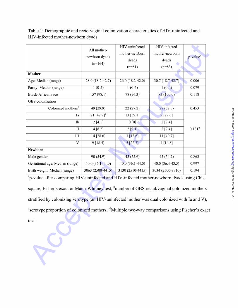
Table 1: Demographic and recto-vaginal colonization characteristics of HIV-uninfected and HIV-infected mother-newborn dyads
ap-value after comparing HIV-uninfected and HIV-infected mother-newborn dyads using Chi-
square, Fisher’s exact or Mann-Whitney test, bnumber of GBS rectal/vaginal colonized mothers
stratified by colonizing serotype (an HIV-uninfected mother was dual colonized with Ia and V),
cserotype proportion of colonized mothers, dMultiple two-way comparisons using Fischer’s exact
test.
All mother-
newborn dyads
(n=164)
HIV-uninfected
mother-newborn
dyads
(n=81)
HIV-infected
mother-newborn
dyads
(n=83)
p-valuea
Mother
Age: Median (range) 28.0 (18.2-42.7) 26.0 (18.2-42.0) 30.7 (18.7-42.7) 0.006
Parity: Median (range) 1 (0-5) 1 (0-5) 1 (0-4) 0.079
Black-African race 157 (98.1) 78 (96.3) 83 (100.0) 0.118
GBS colonization
Colonized mothersb 49 (29.9) 22 (27.2) 27 (32.5) 0.453
Ia 21 [42.9]c 13 [59.1] 8 [29.6]
0.131d
Ib 2 [4.1] 0 [0] 2 [7.4]
II 4 [8.2] 2 [9.1] 2 [7.4]
III 14 [28.6] 3 [13.6] 11 [40.7]
V 9 [18.4] 5 [22.7] 4 [14.8]
Newborn
Male gender 90 (54.9) 45 (55.6) 45 (54.2) 0.863
Gestational age: Median (range) 40.0 (36.1-44.0) 40.0 (36.1-44.0) 40.0 (36.4-43.5) 0.997
Birth weight: Median (range) 3063 (2500-4415) 3130 (2510-4415) 3034 (2500-3910) 0.194
by guest on March 17, 2016
http://jid.oxfordjournals.org/D
ownloaded from
Acce
pted M
anus
cript
Table 2: Proportion of HIV-infected and HIV-uninfected women with capsular antibody concentrations (µg/ml) above different thresholds
Antibody concentration
HIV-infected n=83
HIV-uninfected n=81
aOR (95%CI)a p-value
Ia <0.5 59 (71.1) 46 (56.8) Referent ≥0.5 24 (28.9) 35 (43.2) 0.44 (0.22-0.89) 0.021 ≥1 17 (20.5) 30 (37.0) 0.37 (0.16-0.72) 0.005 ≥2 14 (16.9) 26 (32.1) 0.33 (0.15-0.75) 0.008
Ib
<0.5 72 (86.7) 72 (88.9) Referent ≥0.5 11 (13.3) 9 (11.1) 1.34 (0.51-3.52) 0.550 ≥1 7 (8.4) 4 (4.9) 2.11 (0.57-7.78) 0.261 ≥2 3 (3.6) 2 (2.5) 1.95 (0.30-12.59) 0.482
III
<0.5 64 (77.1) 55 (67.9) Referent ≥0.5 19 (22.9) 26 (32.1) 0.48 (0.23-1.02) 0.058 ≥1 10 (12.1) 17 (21.0) 0.37 (0.14-0.95) 0.038 ≥2 7 (8.4) 14 (17.3) 0.34 (0.12-1.00) 0.049
V
<0.5 49 (59.0) 37 (45.7) Referent ≥0.5 34 (41.0) 44 (54.3) 0.58 (0.30-1.11) 0.099 ≥1 14 (16.9) 23 (28.4) 0.46 (0.21-1.03) 0.059 ≥2 6 (7.2) 10 (12.3) 0.50 (0.16-1.54) 0.228
aAdjusted-OR (95%CI)- calculated odds ratio with 95% confidence of disease using logistic
regression (adjusted for parity, maternal age and serotype-specific colonization)
by guest on March 17, 2016
http://jid.oxfordjournals.org/D
ownloaded from

Acce
pted M
anus
cript
Table 3: Transplacental antibody transfer (cord to maternal blood ratio) between HIV-uninfected and HIV-infected mother-newborn dyads
HIV-uninfected mother-newborn dyads Median CMRa
(IQR)b n=81
HIV-infected mother-newborn dyads Median CMR
(IQR) n=83
Reduction, %c p-valued
Capsular serotypes
Ia 0.749 (0.562-1.021) 0.469 (0.322-0.754) 37.4 <0.001
Ib 1.187 (0.730-1.959) 0.930 (0.593-1.574) 21.7 0.483
III 0.902 (0.605-1.229) 0.609 (0.407-0.976) 32.5 0.027
V 0.954 (0.677-1.310) 0.825 (0.543-1.158) 13.5 0.084
Surface-proteins
PI-1 1.056 (0.835-1.453) 0.948 (0.669-1.431) 10.2 0.379
PI-2a 0.904 (0.545-1.317) 1.262 (0.613-3.000) NRe 0.213
PI-2b 1.006 (0.598-1.588) 0.904 (0.562-1.521) 10.1 0.500
BibA 0.860 (0.687-1.139) 0.759 (0.539-1.126) 11.7 0.207
FbsA 0.964 (0.601-1.695) 1.159 (0.454-2.347) NR 0.385aCMR-cord to maternal ratio, bInterquartile range, cReduction in cord to maternal ratio comparing
HIV-infected and HIV-uninfected mother-newborn dyads; calculated as the ratio of the cord to
maternal ratio from HIV-infected/HIV-uninfected women, subtracted from 1, dUsing quantile
regression (adjusted for overall colonization, colonizing serotype for capsular antibodies,
maternal age and parity), eNo reduction
by guest on March 17, 2016
http://jid.oxfordjournals.org/D
ownloaded from