Histonų deacetilazių raiškos ir viduląstelinės lokalizacijos tyrimai iš pieninio danties...
6
BIOLOGIJA. 2008. Vol. 54. No. 4. P. 306–311 DOI: 10.2478/v10054-008-0061-1 © Lietuvos mokslų akademija, 2008 © Lietuvos mokslų akademijos leidykla, 2008 Expression and subcellular localization of histone deacetylases in mesenchymal stem-like cells from exfoliated deciduous teeth Virginijus Tunaitis 1 *, Ieva Sadzevičienė 1 , Kristina Suriakaitė 1 , Andrejus Surovas 1 , Rūta Aldonytė 1 , Karl-Eric Magnusson 2 , Augustas Pivoriūnas 1 1 Department of Experimental Research, Institute of Experimental and Clinical Medicine at Vilnius University, Žygimantų 9, LT-01102 Vilnius, Lithuania 2 Division of Medical Microbiology, Department of Clinical and Experimental Medicine, Linköping University, SE-581 85 Linköping, Sweden * Corresponding author. E-mail: [email protected] e acetylation of histones and transcription factors plays a crucial role in cell functional ac- tivity. Balance between histone acetyltransferases (HATs) and histone deacetylases (HDACs) is closely related to diverse cellular processes, including differentiation, proliferation, and apop- tosis. Expression of different classes of HDACs is strictly dependent on the cell type, and dif- ferentiated cells possess different members of the HDAC family. In our study, we employed mes- enchymal stem-like cells (MSC) derived from human exfoliated deciduous teeth. Expression of HDACs at mRNA level was analysed using the RT-PCR method. Next, we have assessed the protein levels of different classes of HDACs and their localization within the cell. e cyto- plasm and nuclear fractions of cell lysates were subjected to western blot analysis. Since the functional activity of class II HDACs is determined by their shuttling between the nucleus and the cytoplasm, we have analysed the subcellular localization of HDAC4 in response to the well- known activators of differentiation, i. e. bone morphogenetic protein 2 (BMP-2) and phorbol 12-myristate-13-acetate (PMA), using fluorescent confocal microscopy. Our results suggest that class I and II HDAC family members are expressed and produced in MSC-like cells. e pre- dicted HDAC4 translocation from the cell nucleus was not induced by PMA or BMP-2 in our model. However, our findings open new insights into a possible targeting for HDACs in future transcription activations / derepression studies. Key words: mesenchymal stem cell, MSC, HDAC INTRODUCTION Mesenchymal stem cells (MSC) are characterized by their ability to differentiate into lineages of mesenchymal tissues, including bone, cartilage, muscle and fat under appropriate stimuli [1, 2]. Initially, bone-marrow-derived MSCs were characterized. Since then, populations of the MSC-like cells with similar characteris- tics (morphology, differentiation potential and immunopheno- type) have been isolated from various tissues (adipose, umbili- cal cord, dental pulp, synovium, etc.) [3–6]. Recent studies have demonstrated that MSC-like cells can be isolated from virtually all organs and tissues. Authors assume that MSC-like cells are distributed throughout the organism, possibly occupying the perivascular niche [7, 8]. In the present study, we have used MSC-like cells derived from human exfoliated deciduous teeth. Exfoliated deciduous teeth-derived dental pulp is a relatively new and unexplored source of MSC-like cells. Importantly, these cells demonstrate a very high in vitro proliferation potential and are able to regenerate bone tissue in vivo [9, 10]. us, MSC-like cells derived from exfoliated deciduous teeth can be used as a promising experimental model for studying osteogenic differen- tiation and bone regeneration. e process of cell differentiation is driven by specific genetic programs responsible for mature, specialized phenotype forma- tion. Regulated changes in the chromatin structure play a central role in this process. Histone acetylation, catalyzed by histone acetyl- transferases (HATs), promotes gene transcription by relaxing the chromatin structure. e stimulatory effect of HATs is counter- balanced by histone deacetylases (HDACs) which promote chro- matin condensation and transcriptional repression [11–14]. Class I HDACs (1–3, 8, 11) are ubiquitously expressed. By contrast, the class II HDACs (4, 5, 7, 9) display tissue-restricted (muscle, brain, T and B lymphocytes, chondrocytes) expression patterns [15–17]. In addition, class II HDACs contain an N-terminal regulatory re- gion which mediates interactions with specific transcription fac- tors and also serves as a target for post-transcriptional modifica-
Transcript of Histonų deacetilazių raiškos ir viduląstelinės lokalizacijos tyrimai iš pieninio danties...
biologija. 2008. Vol. 54. No. 4. P. 306–311Doi: 10.2478/v10054-008-0061-1© lietuvos mokslų akademija, 2008© lietuvos mokslų akademijos leidykla, 2008
Expression and subcellular localization of histone deacetylases in mesenchymal stem-like cells from exfoliated deciduous teeth
Virginijus Tunaitis1*,
Ieva Sadzevičienė1,
Kristina Suriakaitė1,
Andrejus Surovas1,
Rūta Aldonytė1,
Karl-Eric Magnusson2,
Augustas Pivoriūnas1
1 Department of Experimental Research, Institute of Experimental and Clinical Medicine at Vilnius University, Žygimantų 9, LT-01102 Vilnius, Lithuania
2 Division of Medical Microbiology, Department of Clinical and Experimental Medicine, Linköping University, SE-581 85 Linköping, Sweden
* Corresponding author. E-mail: [email protected]
The acetylation of histones and transcription factors plays a crucial role in cell functional ac-tivity. Balance between histone acetyltransferases (HATs) and histone deacetylases (HDACs) is closely related to diverse cellular processes, including differentiation, proliferation, and apop-tosis. Expression of different classes of HDACs is strictly dependent on the cell type, and dif-ferentiated cells possess different members of the HDAC family. In our study, we employed mes-enchymal stem-like cells (MSC) derived from human exfoliated deciduous teeth. Expression of HDACs at mRNA level was analysed using the RT-PCR method. Next, we have assessed the protein levels of different classes of HDACs and their localization within the cell. The cyto-plasm and nuclear fractions of cell lysates were subjected to western blot analysis. Since the functional activity of class II HDACs is determined by their shuttling between the nucleus and the cytoplasm, we have analysed the subcellular localization of HDAC4 in response to the well-known activators of differentiation, i. e. bone morphogenetic protein 2 (BMP-2) and phorbol 12-myristate-13-acetate (PMA), using fluorescent confocal microscopy. Our results suggest that class I and II HDAC family members are expressed and produced in MSC-like cells. The pre-dicted HDAC4 translocation from the cell nucleus was not induced by PMA or BMP-2 in our model. However, our findings open new insights into a possible targeting for HDACs in future transcription activations / derepression studies.
Key words: mesenchymal stem cell, MSC, HDAC
INTRODUCTION
Mesenchymal stem cells (MSC) are characterized by their ability to differentiate into lineages of mesenchymal tissues, including bone, cartilage, muscle and fat under appropriate stimuli [1, 2]. Initially, bone-marrow-derived MSCs were characterized. Since then, populations of the MSC-like cells with similar characteris-tics (morphology, differentiation potential and immunopheno-type) have been isolated from various tissues (adipose, umbili-cal cord, dental pulp, synovium, etc.) [3–6]. Recent studies have demonstrated that MSC-like cells can be isolated from virtually all organs and tissues. Authors assume that MSC-like cells are distributed throughout the organism, possibly occupying the perivascular niche [7, 8]. In the present study, we have used MSC-like cells derived from human exfoliated deciduous teeth. Exfoliated deciduous teeth-derived dental pulp is a relatively new and unexplored source of MSC-like cells. Importantly, these
cells demonstrate a very high in vitro proliferation potential and are able to regenerate bone tissue in vivo [9, 10]. Thus, MSC-like cells derived from exfoliated deciduous teeth can be used as a promising experimental model for studying osteogenic differen-tiation and bone regeneration.
The process of cell differentiation is driven by specific genetic programs responsible for mature, specialized phenotype forma-tion. Regulated changes in the chromatin structure play a central role in this process. Histone acetylation, catalyzed by histone acetyl-transferases (HATs), promotes gene transcription by relaxing the chromatin structure. The stimulatory effect of HATs is counter-balanced by histone deacetylases (HDACs) which promote chro-matin condensation and transcriptional repression [11–14]. Class I HDACs (1–3, 8, 11) are ubiquitously expressed. By contrast, the class II HDACs (4, 5, 7, 9) display tissue-restricted (muscle, brain, T and B lymphocytes, chondrocytes) expression patterns [15–17]. In addition, class II HDACs contain an N-terminal regulatory re-gion which mediates interactions with specific transcription fac-tors and also serves as a target for post-transcriptional modifica-
307Expression and subcellular localization of histone deacetylases in mesenchymal stem-like cells from exfoliated deciduous teeth
tions connecting signals from the extracellular environment and chromatin remodelers [18]. Ca2+ / calmodulin-dependent kinases (CaMKs I, II, and IV) and protein kinase D (PKD) phosphorylate serines on N-terminal regulatory region of class II HDACs, cre-ating the binding site for the ubiquitous 14–3–3 adaptor protein which further facilitates subsequent export of II class HDACs from the nucleus. This results in hyperacetylation of specific tran-scription factors, derepression of the target genes and initiation of specific differentiation programs [15, 17, 19].
Besides phosphorylation, class II HDACs may also be sub-jected to ubiquitination and sumoylation. Unlike ubiquitination, modification of N-terminal region by a small ubiquitin-related modifier (SUMO) does not lead to protein degradation, but is rather associated with protein–protein interactions, subcellular protein distribution and stability [20, 21].
Little is known about the role of HDACs in the differentiation of MSC. A recent study has demonstrated that HDAC inhibitors, valproic acid and trichostatin A, accelerate osteogenic differentia-tion of the MSCs. This effect was linked to an increased expres-sion of osteogenic differentiation-related genes Runx2, osterix, osteopontin, and bone morphogenetic protein 2 (BMP-2) [22]. However, the HDAC inhibitors used in this study display a broad activity spectrum. Therefore, the precise role of distinct classes of HDACs during the process of MSC differentiation remains largely unknown. Interestingly, HDAC4 is expressed in pre-hypertrophic chondrocytes and regulates endochondral bone formation by in-teracting with the transcription factor Runx2 which is an impor-tant regulator of osteo- and chondro- genesis [12, 18].
Since MSCs participate in chondro-, osteo-, and adipogen-esis, we speculate that class II HDACs might be involved in these processes. Class II HDACs demonstrate tissue-restricted expres-sion patterns and are regulated by signaling kinases. However, its involvement in MSC differentiation remains to be elucidated. In the present study, we aim to investigate the expression and subcellular distribution of class II HDACs in proliferating MSC-like cells derived from exfoliated deciduous teeth.
MATERIALS AND METHODS
Isolation of MSC-like cells from human exfoliated deciduous teeth. MSC-like cells were isolated according to protocol ap-proved by the Lithuanian Bioethics Committee. Briefly, the pulp from a normal exfoliated deciduous tooth of a 5-year-old child was separated and digested in colagenase solution (3 mg/ml) for 1 h at 37 ºC. Cells were maintained in Dulbecco’s modified Eagle’s medium (DMEM) (Biochrome) supplemented with 100 U/ml penicillin, 100 μg/ml streptomycin and 10% fetal bo-
vine serum (Biochrome) at 37 ºC in a humidified atmosphere containing 5% of CO2.
Induction of osteogenic differentiation. MSC-like cells were seeded at a density 5 × 10³/cm2 in DMEM (low glucose) supplemented with 10% FCS, 100 U/ml penicillin, 100 μg/ml streptomycin, 2 mM L-glutamine and 1 mM sodium pyrophos-phate in 35-mm diameter plastic dishes and cultured until subconfluent. Differentiation was then induced by adding an osteogenic-induction medium prepared according to manu-facturer’s instructions (StemCell Technologies) with addition of dexamethasone (10–8 M) and ascorbic acid (50 μg/ml). When the cell multilayering became evident, β-glycerophosphate was added into the medium (10–6 M). Cell cultures were treated with osteogenic-induction medium for two weeks, the medium was changed twice a week. Osteogenic differentiation was assessed by hydroxy-apatite crystal staining with silver nitrate (von Kossa). The cells were first fixed for 10 min with 1% formalin in PBS and stained with 1% silver nitrate for 30 min, followed by 5% sodium thiosulfate for 5 min. Control cells were cultured without differ-entiation stimuli and were stained in the same manner.
Flow cytometry. MSC-like cells were tested for the presence of the set of MSC markers. Single cell suspension of MSC-like cells (approximately 5 × 10⁵) in 200 μl of PBS buffer was incu-bated with 2 μl of phycoeritrin (PE)-conjugated antibodies against CD14, CD105, CD73, CD34, or fluorescein isothiocyanate (FITC)-conjugated antibodies against CD45, CD90 (Santa Cruz Biotechnology). After three washes, fluorescence was evaluated by flow cytometry in FACSAria (Becton Dickenson, BD).
Reverse-transcriptase (RT)-PCR analysis. Endogenous mRNA of MSC-like cells was isolated using TRIzol reagent (Invitrogen) according to the manufacturer’s instructions. We used the First strand cDNA synthesis kit with (dT)18 primer (Fermentas) to generate the first-strand cDNA. The reaction product (cDNA) was subjected to PCR analysis with the primers shown in Table 1. PCR reactions (35 cycles) were performed in 25 μl of reaction mix (5 μl cDNA, 1 μl Thermo-Start Taq DNA polymeraze (Abgene), 200 nM dNTP and 500 nM forward and reverse primers, 1× reaction buffer with 1.5 mM MgCl2). An automated Mastercycler personal amplifer (Eppendorf) was used for PCR amplification reactions. The final products were separated by agarose gel electrophoresis, incubated in ethidium bromide, and photographed under UV illumination. For sizing the PCR products, GeneRuler™ 50 bp, GeneRuler™ 1 kb Plus and Lambda DNA / Hind III (Fermentas) DNA markers were used.
Western blot analysis. Cells were grown to 70–80% con-fluence in Dulbecco’s modified Eagle’s medium (DMEM) supplemented with 100 U/ml penicillin, 100 μg/ml streptomycin
Ta b l e 1 . Oligonucleotides used in expression analysis of HDACs mRNA
Forward Reverse Ampliconb-actin 5’-TGACGGGGTCACCCACACTGTGCC-3’ 5’-TAGAAGCATTTGCGGTGGACGATG-3’ 600 bpHDAC1 5’-CAAGCTCCACATCAGTCCTTCC-3’ 5’-TGCGGCAGCATTCTAAGGTT-3’ 100 bpHDAC2 5’-AGTCAAGGAGGCGGCAAAA-3’ 5’-TGCGGATTCTATGAGGCTTCA-3’ 100 bpHDAC3 5’-CCGAAATGTTGCCCGCTGCTG-3’ 5’-AGGTGCATGGTTCAGCATCTT-3’ 220 bpHDAC4 5’-AAGGAGTGGACATTGCT-3’ 5’-GATTCAGCAGCTCCACT-3’ 85 bpHDAC5 5’-TCACTGTCACCAACTCAC-3’ 5’-CAGGAATAGAGGATGTGC-3’ 134 bpHDAC7 5’-CTTCTCCACAAGGACAAG-3’ 5’-CTCCAGGGTTCTGTAGG-3’ 150 bp
308 Virginijus Tunaitis, Ieva Sadzevičienė, Kristina Suriakaitė, Andrejus Surovas, Rūta Aldonytė et al.
and 10% fetal bovine serum (Biochrome) at 37 ºC in a humidi-fied atmosphere containing 5% of CO2. Following stimulation with 100 nM phorbol 12-myristate-13-acetate (PMA) (Sigma) for 60 min, the cells were washed three times with ice-cold PBS and lysed in lysis buffer (10 mM Tris-HCl, pH 7.4, 50 mM NaCl, 5 mM EDTA, 50 mM NaF, 1% Triton X-100, 20 mg/ml aprotinin, 1 mM PMSF, 2 mM NaVO4). Lysates were clarified by centrifuga-tion at 20 000 × g for 15 min. Isolation of cytosolic and nuclear protein fractions was performed in parallel using the Nuclei EZ kit (Sigma) according to the manufacturer’s instructions. All samples were fractionated in 10% SDS-PAGE gel and transferred to the PVDF membrane (Bio-Rad). Approximate sizing of pro-teins and transfer efficiency were performed using Precision Plus Protein Dual Color Standards (Bio-Rad). Primary rabbit polyclo-nal anti-HDAC1 and anti-HDAC4 (Cell Signaling Technology) antibodies were used at a 1 : 500 dilution. Secondary antibody (alkaline phosphatase-conjugated goat anti-rabbit IgG (Santa Cruz) was used at 1 : 2000 dilution. Immunoreactivity was de-tected by reaction with BCIP / NBT substrate (Sigma).
Immunocitochemistry. Cells grown on coverslips were in-cubated with 100 nM PMA (Sigma) or 100 ng/ml bone morpho-genetic protein 2 (BMP-2) (R & D Systems) for 60 min at 37 ºC. Control cells were left in the cell culture medium alone. The
coverslips were rinsed three times in ice-cold PBS (pH 7.6) and fixed in 4% (w/v) paraformaldehyde for 15 min at room tem-perature. Then the cells were washed three times in PBS and per-meabilized with 1% Triton X-100 for 15 min. Next, the cells were blocked in 5% goat serum (v/v) (DAKO) for 60 min at room tem-perature, washed with PBS again and incubated with the prima-ry rabbit anti-HDAC4 antibody (Cell Signaling Technology) for 90 min at 37 ºC (dilution 1 : 200). Finally, the cells were washed three times with PBS, incubated with the secondary Alexa 488 coupled goat anti-rabbit (Molecular Probes) antibody at a con-centration of 15 μg/ml and mounted with Fluorescent mount-ing medium (DAKO). Coverslips were subsequently examined by confocal laser scanning microscopy using a Nikon Eclipse TE2000U microscope.
RESULTS AND DISCUSSION
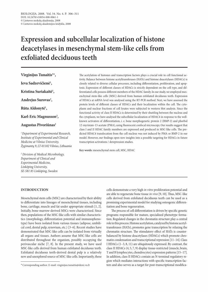
In the present study, we have used MSC-like cells derived from human exfoliated deciduous teeth. Cells were isolated according to a protocol described elsewhere, with minor modifications [9]. The isolated cells displayed a typical fibroblastoid morphology and a high proliferation potential (Fig. 1). Our in vitro studies of MSC-like cells revealed their ability to differentiate into os-
Fig. 1. Morphology of MSC-like cells isolated from human exfoliated deciduous teeth. Cells were cultured in DMEM medium containing 10% FBS for 3 days (A) or for 12 days (B). Phase-contrast images (magnification 100×) represent typical adherent fibroblastoid colony (A) and a subconfluient cell layer (B)
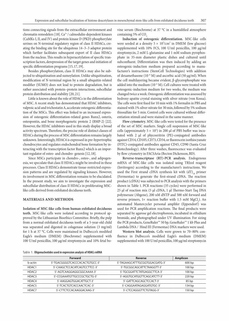
Fig. 2. Osteogenic differentiation properties of MSC-like cells. In vitro osteogenesis. Cells were grown in DMEM media containing 10% FBS (A) or exposed to osteoinducing medium for 14 days (B). Mineral depositions at day 14 were stained by von Kossa method (B)
309Expression and subcellular localization of histone deacetylases in mesenchymal stem-like cells from exfoliated deciduous teeth
teocytic cells. The cells cultured under osteogenic conditions formed multilayers within the first week. Staining with silver nitrate (von Kossa) revealed characteristic patterns of miner-alized matrix two weeks later (Fig. 2). We then analysed the ex-pression of markers recognized as typical of MSCs. According to the International Society for Cellular Therapy [1], MSC should express CD105, CD73 and CD90 and a lack of expres-sion of CD45, CD34, CD14 surface molecules. For that reason, sets of specific positive and negative markers were analysed using FACS (Table 2). We found that our MSC-like cells resem-bled the recognized phenotype of MSC.
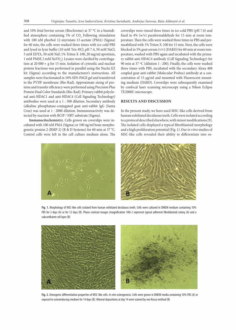
According to previous reports, expression of HDACs de-pends on cell type and regulates cell differentiation [13, 17, 22]. However, there is no direct evidence of expression of all classes of HDACs in MSC. We have analysed HDAC mRNA expression patterns by the RT-PCR method using specific primers to class I HDAC (HDAC1, 2, 3) and class II HDACs (HDAC4, 5, and 7) (Table 1). The housekeeping β-actin gene was used as a refer-ence gene. Surprisingly, we have found mRNA expression of all analysed HDACs at a detectable level (Fig. 3). This is the first re-port to show that both class I and class II HDACs are expressed in MSC-like cells in our model.
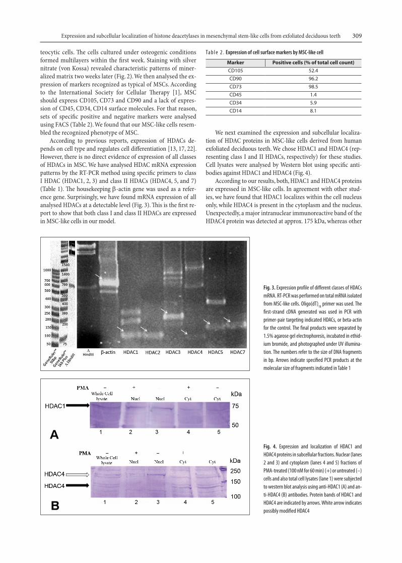
We next examined the expression and subcellular localiza-tion of HDAC proteins in MSC-like cells derived from human exfoliated deciduous teeth. We chose HDAC1 and HDAC4 (rep-resenting class I and II HDACs, respectively) for these studies. Cell lysates were analysed by Western blot using specific anti-bodies against HDAC1 and HDAC4 (Fig. 4).
According to our results, both, HDAC1 and HDAC4 proteins are expressed in MSC-like cells. In agreement with other stud-ies, we have found that HDAC1 localizes within the cell nucleus only, while HDAC4 is present in the cytoplasm and the nucleus. Unexpectedly, a major intranuclear immunoreactive band of the HDAC4 protein was detected at approx. 175 kDa, whereas other
Fig. 3. Expression profile of different classes of HDACs mRNA. RT-PCR was performed on total mRNA isolated from MSC-like cells. Oligo(dT)18 primer was used. The first-strand cDNA generated was used in PCR with primer-pair targeting indicated HDACs, or beta-actin for the control. The final products were separated by 1.5% agarose gel electrophoresis, incubated in ethid-ium bromide, and photographed under UV illumina-tion. The numbers refer to the size of DNA fragments in bp. Arrows indicate specified PCR products at the molecular size of fragments indicated in Table 1
Ta b l e 2 . Expression of cell surface markers by MSC-like cell
Marker Positive cells (% of total cell count)CD105 52.4CD90 96.2CD73 98.5CD45 1.4CD34 5.9CD14 8.1
Fig. 4. Expression and localization of HDAC1 and HDAC4 proteins in subcellular fractions. Nuclear (lanes 2 and 3) and cytoplasm (lanes 4 and 5) fractions of PMA-treated (100 nM for 60 min) (+) or untreated (–) cells and also total cell lysates (lane 1) were subjected to western blot analysis using anti-HDAC1 (A) and an-ti-HDAC4 (B) antibodies. Protein bands of HDAC1 and HDAC4 are indicated by arrows. White arrow indicates possibly modified HDAC4
310 Virginijus Tunaitis, Ieva Sadzevičienė, Kristina Suriakaitė, Andrejus Surovas, Rūta Aldonytė et al.
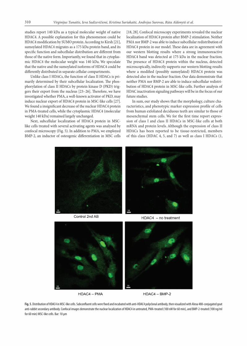
Fig. 5. Distribution of HDAC4 in MSC-like cells. Subconfluent cells were fixed and incubated with anti-HDAC4 polyclonal antibody, then visualized with Alexa 488-conjugated goat anti-rabbit secondary antibody. Confocal images demonstrate the nuclear localization of HDAC4 in untreated, PMA-treated (100 nM for 60 min), and BMP-2-treated (100 ng/ml for 60 min) MSC-like cells. Bar: 10 μm
studies report 140 kDa as a typical molecular weight of native HDAC4. A possible explanation for this phenomenon could be HDAC4 modification by SUMO protein. According to Kirsh [21], sumoylated HDAC4 migrates as a 175 kDa protein band, and its specific function and subcellular distribution are different from those of the native form. Importantly, we found that in cytoplas-mic HDAC4 the molecular weight was 140 kDa. We speculate that the native and the sumoylated isoforms of HDAC4 could be differently distributed in separate cellular compartments.
Unlike class I HDACs, the function of class II HDACs is pri-marily determined by their subcellular localization. The phos-phorylation of class II HDACs by protein kinase D (PKD) trig-gers their export from the nucleus [23–26]. Therefore, we have investigated whether PMA, a well-known activator of PKD, may induce nuclear export of HDAC4 protein in MSC-like cells [27]. We found a insignificant decrease of the nuclear HDAC4 protein in PMA-treated cells, while the cytoplasmic HDAC4 (molecular weight 140 kDa) remained largely unchanged.
Next, subcellular localization of HDAC4 protein in MSC-like cells treated with several activating agents was analysed by confocal microscopy (Fig. 5). In addition to PMA, we employed BMP-2, an inductor of osteogenic differentiation in MSC cells
[18, 28]. Confocal microscopy experiments revealed the nuclear localization of HDAC4 protein after BMP-2 stimulation. Neither PMA nor BMP-2 was able to induce subcellular redistribution of HDAC4 protein in our model. These data are in agreement with our western blotting results where a strong immunoreactive HDAC4 band was detected at 175 kDa in the nuclear fraction. The presence of HDAC4 protein within the nucleus, detected microscopically, indirectly supports our western blotting results where a modified (possibly sumoylated) HDAC4 protein was detected also in the nuclear fraction. Our data demonstrate that neither PMA nor BMP-2 are able to induce subcellular redistri-bution of HDAC4 protein in MSC-like cells. Further analysis of HDAC inactivation signaling pathways will be in the focus of our future studies.
In sum, our study shows that the morphology, culture cha-racteristics, and phenotypic marker expression profile of cells from human exfoliated deciduous teeth are similar to those of mesenchymal stem cells. We for the first time report expres-sion of class I and class II HDACs in MSC-like cells at both mRNA and protein levels. Although the expression of class II HDACs has been reported to be tissue-restricted, members of this class (HDAC 4, 5, and 7) as well as class I HDACs (1,
311Expression and subcellular localization of histone deacetylases in mesenchymal stem-like cells from exfoliated deciduous teeth
18. jeon Ej, lee KY, Choi NS, lee MH et al. j biol Chem 2006; 281(24): 16502–11.
19. Chang S, bezprozvannaya S, li S, olson EN. Proc Natl acad Sci USa 2005; 102(23): 8120–5.
20. Paroni g, Fontanini a, Cernotta N, Foti C et al. Mol Cell biol 2007; 27(19): 6718–32.
21. Kirsh o, Seeler jS, Pichler a, gast a et al. EMbo j. 2002; 21(11): 2682–91.
22. Cho HH, Park HT, Kim Yj et al. j Cell biochem 2005; 96(3): 533–42.
23. Matthews Sa, liu P, Spitaler M, olson EN et al. Mol Cell biol 2006; 26(4): 1569–77.
24. Vega Rb, Harrison bC, Meadows E et al. Mol Cell biol 2004; 24(19): 8374–85.
25. Dequiedt F, Van lint j, lecomte E et al. j Exp Med 2005; 201: 793–804.
26. Wang S, li X, Parra M et al. Proc Natl acad Sci USa 2008; 105(22): 7738–43.
27. Chen j, Deng F, li j, Wang Qj. biochem j 2008; 411(2): 333–42.
28. Celil ab, Campbell Pg. j biol Chem 2005; 280(36): 31353–9.
Virginijus Tunaitis, Ieva Sadzevičienė, Kristina Suriakaitė, Andrejus Surovas, Rūta Aldonytė, Karl-Eric Magnusson, Augustas Pivoriūnas
HISTONų DEACETILAzIų RAIšKOS IR VIDULąSTELINėS LOKALIzACIjOS TyRIMAI Iš PIENINIO DANTIES IšSKIRTOSE MEzENCHIMINėSE KAMIENINėSE LąSTELėSE
SantraukaHistonų ir transkripcijos veiksnių acetilinimas yra vienas iš esminių ląstelių veiklos reguliavimo būdų. Ląstelėje histonų acetiltransferazių (HAT) ir deacetilazių (HDAC) balansas yra susijęs su diferenciacija, pro-liferacija ar apoptoze. Diferencijuotos ląstelės pasižymi skirtinga įvairių tipų HDAC raiška, aktyvavimu, lokalizacija ir funkcija. Šiame darbe ty-rėme iš pieninio danties pulpos išskirtas ląsteles, kurias priskyrėme me-zenchiminių kamieninių ląstelių tipui (MKL tipui). Atvirkštinės trans-kripcijos PGR būdu (RT-PCR) nustatėme I ir II HDAC klasės atstovų (HDAC1, HDAC2, HDAC3 ir HDAC4, HDAC5, HDAC7) raišką mRNR lygyje. Tirdami HDAC1 ir HDAC4 baltymų raišką branduolinėje ir cito-plazminėje ląstelių frakcijose patvirtinome branduolinę HDAC1 loka-lizaciją bei aptikome HDAC4 pasiskirstymo ląstelių frakcijose priklau-somybę nuo jo modifikacijos. Kadangi HDAC4 funkcinis aktyvumas susijęs su baltymo lokalizacijos pokyčiais, fluorescentinės konfokalinės mikroskopijos metodu tyrėme HDAC4 lokalizacijos atsaką paveikus forbolo esteriu (PMA) ir kaulų morfogenetiniu baltymu 2 (BMP-2). Remdamiesi rezultatais galime teigti, kad tirtos I ir II klasės histonų acetilazės yra gaminamos MKL tipo ląstelėse. Darbo metu imunoblo-tais ir konfokaline mikroskopija nustatyta HDAC4 lokalizacija ląstelė-se. Pastebėta, kad mūsų modelyje nei po PMA, nei po BMP-2 poveikio branduolinė HDAC4 lokalizacija pastebimai nepakito. Visi gauti duo-menys rodo, kad tirtose MKL tipo ląstelėse egzistuoja kompleksiškas HDAC4 lokalizacijos ir funkcinio aktyvumo reguliavimo mechanizmas, kurio ištyrimas padės ateityje išaiškinti potencialias MKL diferencija-vimo galimybes.
2, and 3) were found in our cells. Due to HDAC4 importance in osteogenic differentiation, we have assessed the expression and localizations of HDAC4 in cells treated with a strong PKD activator (PMA) and an inductor of osteogenic differentia-tion (BMP-2). According to our data, the possibly sumoylated HDAC4 protein accumulates in the nucleus of the MSC-like cells, and its localization is not changed by PMA or BMP-2 treatment. We speculate that more complex mechanisms are responsible for the HDAC4 activity regulation in cells. Our data elucidate presence of integrated regulatory mechanisms of transcription derepression / activation in MSC-like cells and indicate several candidate targets, i. e. HDACs, for the fu-ture studies.
ACKNOWLEDGEMENTS
This work was supported by the Lithuanian State Science and Studies Foundation (No. B-25/2008 and C-04/2008) and Swedish Institute (Visby program No. 01398/2007).
Received 4 September 2008 accepted 12 December 2008
References
1. Dominici M, le blanc K, Mueller i et al. Cytotherapy 2006; 8(4): 315–7.
2. barry FP, Murphy jM. int j biochem Cell biol 2004; 36: 568–84.
3. Petersen bE, bowen WC, Patrene KD et al. Science 1999; 284: 1168–70.
4. orlic D, Kajstura j, Chimenti S et al. Nature 2001; 410: 701–5.
5. Mezey E, Chandross Kj, Harta g et al. Science 2000; 290: 1779–82.
6. D’ippolito g, Schiller PC, Ricordi C et al. j bone Miner Res 1999; 14: 1115–22.
7. da Silva Meirelles l, Caplan ai, Nardi Nb. Stem Cells 2008; 26(9): 2287–99.
8. da Silva Meirelles l, Chagastelles PC, Nardi Nb. j Cell Sci 2006; 119(Pt 11): 2204–13.
9. Miura M, gronthos S, Zhao M et al. Proc Natl acad Sci USa 2003; 100(10): 5807–12.
10. Shi S, bartold PM, Miura M, Seo bM et al. orthod Craniofac Res 2005; 8(3): 191–9.
11. jenuwein T, alis CD. Science 2001; 293: 1074–80. 12. Vega Rb, Matsuda K, oh j et al. Cell 2004; 119: 555–66. 13. Yang Xj, grégoire S. Mol Cell biol 2005; 25(8): 2873–84. 14. Khochbin S, Verdel a, lemercier C, Seigneurin-berny D.
Curr opin genet Dev 2001; 11(2): 162–6. 15. Verdin E, Dequiedt F, Kasler Hg. Trends genet 2003; 19(5):
286–93. 16. lucio-Eterovic aK, Cortez Ma, Valera ET, Motta Fj et al.
bMC Cancer 2008; 8: 243. 17. de Ruijter aj, van gennip aH, Caron HN, Kemp S,
van Kuilenburg ab. biochem j 2003; 370(Pt 3): 737–49.