HISTOLOGICAL AND IMMUNOHISTOCHEMICAL EVALUATION OF SARCOMA 180 IN MICE AFTER TREATMENT WITH AN...
8
Histological and immunohistochemical evaluation of the chemopreventive role of lycopene in tongue carcinogenesis induced by 4-nitroquinoline-1-oxide Dalia Hussein El-Rouby * Oral Pathology Depart., Faculty of Oral & Dental Medicine, Cairo Univ., Cairo, Egypt 1. Introduction Every year more than 300,000 new cases are being diagnosed with oral squamous cell carcinoma (SCC) world wide. Oral cavity cancer and cancer of oropharynx represents the sixth most common malignancy globally. 1,2 Typically, oral cancer development is a slow and cumulative process, which occurs after exposure of the entire epithelial surface to the repeated insult of carcinogens. This process strongly supports the rationale for a prevention strategy that will inhibit, delay, or reverse carcinogenesis before it becomes invasive disease. Chemoprevention fulfils this strategy and offers a promising treatment for oral cancer. 3 a r c h i v e s o f o r a l b i o l o g y 5 6 ( 2 0 1 1 ) 6 6 4 – 6 7 1 a r t i c l e i n f o Article history: Accepted 1 December 2010 Keywords: 4-Nitroquinoline-1-oxide Chemoprevention Tongue carcinogenesis Lycopene a b s t r a c t Objective: Increasing attention has been given to the potential protective roles of specific antioxidant nutrients found in fruits and vegetables. However, there are relatively few reports on the cancer chemopreventive effects of lycopene or tomato carotenoids in animal models. Therefore, the chemopreventive efficacy of lycopene with regard to oral carcino- genesis was investigated using 4-nitroquinoline-1-oxide (4-NQO) induced tongue squamous cell carcinoma. Materials and methods: Twenty albino rats were equally divided into 2 groups. 4-NQO was dissolved in the drinking water (20 ppm) for rats of both groups. Rats in group 2 were administered lycopene at a dose of 2.5 mg/kg body weight by intragastric intubation once a day. Incidence of oral neoplasms and histopathological changes were microscopically evaluated after 32 weeks of administration of the specific treatments. Moreover, immuno- histochemical expressions of proliferating cell nuclear antigen (PCNA), E-cadherin and b- catenin were analysed in tongue specimens using an image analyser computer system. Results: Lycopene treatment significantly decreased the incidence of 4-NQO induced tongue carcinogenesis. A decreased percentage of PCNA-positive nuclei was associated with lyco- pene treatment. Proliferating cells were mainly confined to the basal and parabasal epithe- lial cell layers. Increased E-cadherin and b-catenin immunoexpression was recorded in the lycopene treated group in comparison to the carcinogen group. Conclusion: Results of the present study indicate that lycopene can exert protective effects against 4-NQO induced tongue carcinogenesis through reduction in cell proliferation and enhanced cellular adhesion, suggesting a new mechanism for the anti-invasive effect of lycopene. Further studies are needed to provide more definitive answers to the question of the anticancer effects of lycopene. # 2010 Elsevier Ltd. All rights reserved. * Tel.: +20 2 24173818; fax: +20 2 23642705. E-mail address: [email protected]. available at w ww.s c ienc ed irec t.c o m journal homepage: http://www.elsevier.com/locate/aob 0003–9969/$ – see front matter # 2010 Elsevier Ltd. All rights reserved. doi:10.1016/j.archoralbio.2010.12.007
Transcript of HISTOLOGICAL AND IMMUNOHISTOCHEMICAL EVALUATION OF SARCOMA 180 IN MICE AFTER TREATMENT WITH AN...

Histological and immunohistochemical evaluation of thechemopreventive role of lycopene in tongue carcinogenesisinduced by 4-nitroquinoline-1-oxide
Dalia Hussein El-Rouby *
Oral Pathology Depart., Faculty of Oral & Dental Medicine, Cairo Univ., Cairo, Egypt
a r c h i v e s o f o r a l b i o l o g y 5 6 ( 2 0 1 1 ) 6 6 4 – 6 7 1
a r t i c l e i n f o
Article history:
Accepted 1 December 2010
Keywords:
4-Nitroquinoline-1-oxide
Chemoprevention
Tongue carcinogenesis
Lycopene
a b s t r a c t
Objective: Increasing attention has been given to the potential protective roles of specific
antioxidant nutrients found in fruits and vegetables. However, there are relatively few
reports on the cancer chemopreventive effects of lycopene or tomato carotenoids in animal
models. Therefore, the chemopreventive efficacy of lycopene with regard to oral carcino-
genesis was investigated using 4-nitroquinoline-1-oxide (4-NQO) induced tongue squamous
cell carcinoma.
Materials and methods: Twenty albino rats were equally divided into 2 groups. 4-NQO was
dissolved in the drinking water (20 ppm) for rats of both groups. Rats in group 2 were
administered lycopene at a dose of 2.5 mg/kg body weight by intragastric intubation once a
day. Incidence of oral neoplasms and histopathological changes were microscopically
evaluated after 32 weeks of administration of the specific treatments. Moreover, immuno-
histochemical expressions of proliferating cell nuclear antigen (PCNA), E-cadherin and b-
catenin were analysed in tongue specimens using an image analyser computer system.
Results: Lycopene treatment significantly decreased the incidence of 4-NQO induced tongue
carcinogenesis. A decreased percentage of PCNA-positive nuclei was associated with lyco-
pene treatment. Proliferating cells were mainly confined to the basal and parabasal epithe-
lial cell layers. Increased E-cadherin and b-catenin immunoexpression was recorded in the
lycopene treated group in comparison to the carcinogen group.
Conclusion: Results of the present study indicate that lycopene can exert protective effects
against 4-NQO induced tongue carcinogenesis through reduction in cell proliferation and
enhanced cellular adhesion, suggesting a new mechanism for the anti-invasive effect of
lycopene. Further studies are needed to provide more definitive answers to the question of
the anticancer effects of lycopene.
# 2010 Elsevier Ltd. All rights reserved.
avai lab le at w ww.s c ienc ed i rec t . c o m
journal homepage: http://www.elsevier.com/locate/aob
1. Introduction
Every year more than 300,000 new cases are being diagnosed
with oral squamous cell carcinoma (SCC) world wide.
Oral cavity cancer and cancer of oropharynx represents
the sixth most common malignancy globally.1,2 Typically,
oral cancer development is a slow and cumulative process,
* Tel.: +20 2 24173818; fax: +20 2 23642705.E-mail address: [email protected].
0003–9969/$ – see front matter # 2010 Elsevier Ltd. All rights reservedoi:10.1016/j.archoralbio.2010.12.007
which occurs after exposure of the entire epithelial
surface to the repeated insult of carcinogens. This process
strongly supports the rationale for a prevention strategy
that will inhibit, delay, or reverse carcinogenesis before
it becomes invasive disease. Chemoprevention fulfils
this strategy and offers a promising treatment for oral
cancer.3
d.

a r c h i v e s o f o r a l b i o l o g y 5 6 ( 2 0 1 1 ) 6 6 4 – 6 7 1 665
Epidemiological studies have indicated that populations
that consume food rich in fruits and vegetables have a lower
incidence of cancers.4,5 A meta-analysis showed that a high
vegetable intake decreases the risk for oral cancers by almost
half.6
High consumption of tomatoes effectively decreases the
risk for cardiovascular disease and cancer by improving the
antioxidant capacity.7 Tomatoes are rich sources of lycopene,
a natural pigment synthesised by plants and microorgan-
isms.8 It is an acyclic isomer of b-carotene with no vitamin A
activity.9 Because of its antioxidant properties, lycopene has
gained attention and became one of the most studied
chemopreventive agents. It is believed that the mechanisms
underlying the inhibitory effects of lycopene on carcinogene-
sis and mutagenesis could also involve improving cell to cell
communication,10 interference with cell proliferation,11–13
induction of gap-junctional communication,14 inhibition of
cell cycle progression and modulation of signal transduction
pathways,7 in addition to induction of apoptosis.15
Lycopene has been postulated to be the protective
compound against prostate cancer.16,17 In cell culture, lyco-
pene strongly inhibited proliferation of human endometrial,
mammary and lung cancer cells.11,18 Lycopene treatment also
resulted in a concentration-dependent reduction in HL-60
leukaemic cells’ growth, which was accompanied by inhibi-
tion of cell cycle progression in the G0-G1 phase.19
Concerning oral cancer, an inverse association was
observed between lycopene and buccal pouch squamous cell
carcinomas in hamsters.20 Moreover, the intake of daily
lycopene capsules effectively improved oral leukoplakia
lesions in a group of patients.21
To develop new methods and improve existing protocols
for diagnosis and treatment, an animal model that mimics
the natural course of SCCs was mandatory. Application of 4-
nitroquinoline-1-oxide (4NQO) to rat tongues is a reliable
procedure for inducing oral SCCs.22 4NQO is a powerful
carcinogen in several organs, and it can specifically
induce tongue SCC when applied in low concentrations
via drinking water. The resulting sequential changes and
morphological features resemble those seen during the
progression of human tongue squamous cell malignancy.
Moreover, level of DNA damage is directly related to the
severity of the histological changes.23 Therefore, 4NQO-
induced rat tongue SCC is an excellent model for studying
early events in oral carcinogenesis24,25 and to investigate
synthetic and natural agents for chemopreventive poten-
tial.26–28
Regarding the need for information focusing on the
chemopreventive potential of naturally occurring compounds,
the current study was conducted to evaluate the role of
lycopene treatment on 4NQO induced tongue carcinogenesis
through quantitative evaluation of the proliferative cell
nuclear antigen (PCNA), b-catenin and E-cadherin immunoex-
pression.
2. Materials and methods
A total of 20 male albino rats were used in this study. Rats were
maintained in plastic cages in an air conditioned room at
22 � 2 8C and 55 � 10% humidity with free access to standard
pellet diet and drinking water. The experimental procedure
was conducted in compliance with ethical principles for
animals’ research as reviewed and approved by institutional
guidelines of Kasr Al-ainy animal and experimental laboratory
(Faculty of Medicine, Cairo University).
After 1 week of acclimatisation, rats were randomly
allocated to two groups (10 rats each). 4-NQO was obtained
as a powder (Sigma, St. Louis, MO, USA, cat. # N8141) and
dissolved in the drinking water for rats of both groups to a
final concentration of 0.02 g/l (20 ppm), and the water was
changed once a week. In addition, rats in group 2 were
administered lycopene (Sigma, cat. # L9879) at a dose
of 2.5 mg/kg body weight by intragastric intubation once
per day.
2.1. Specimens preparation
Animals of both groups were sacrificed after 32 weeks of
administration of the specific treatments. The tongue was cut
in half longitudinally and each tissue specimen was fixed in
10% buffered formalin and embedded in paraffin blocks. Each
tissue was totally submitted to multiple transverse sections
for histological processing.
2.2. Histopathological analysis
Histopathological evaluation was performed by light micros-
copy. Tongue sections were graded as normal, hyperplasia,
carcinoma in situ, dysplasia, and carcinoma per animal, as
modified from Ribeiro et al.29
2.3. Immunohistochemistry
Sections at 5 mm thickness were deparaffinised in three
changes of xylene and rehydrated in a graded series of
ethanol to distilled water. For antigen retrieval, slides were
placed in 0.01 M citrate-buffer pH 6.0 and heated in a
steamer for 30 min. Endogenous peroxidases were
quenched by incubating in 3% H2O2 for 20 min at room
temperature. Sections were further incubated in blocking
serum for 30 min. For application of the primary antibodies,
sections were incubated overnight at 4 8C with anti-PCNA
(proliferating cell nuclear antigen) monoclonal antibody
(PC10, 1:20 diluted, DAKO M0879, Glostrup, Denmark), anti-
E-cadherin antibody, 1:400 diluted (Santa Cruz Biotech.,
Santa Cruz, CA, USA), or b-catenin mouse monoclonal
antibody, 1:100 diluted (Santa Cruz Biotechnology, Santa
Cruz, CA, USA). Subsequently, sections were incubated with
biotinylated secondary antibody (LSAB2, Dako, Glostrup,
Denmark) for 30 min, washed in phosphate buffered saline
(PBS) three times for 10 min each. LSAB2-streptavidin-
peroxidase detection reagent (DAKO, Glostrup, Denmark)
was subsequently added evenly over the sections and
incubated for 30 min. Finally, the reaction was developed
using 3,30-diaminobenzidine tetrahydrocloride (DAB) for
5 min. Slides were briefly counterstained in haematoxylin
and dehydrated, and cover slips added. As negative controls,
tissue sections were processed in parallel by omitting
incubation with the primary antibodies.
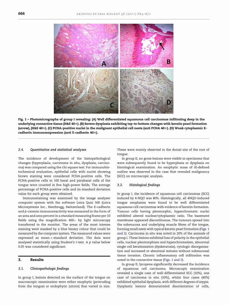
Fig. 1 – Photomicrographs of group I revealing: (A) Well differentiated squamous cell carcinomas infiltrating deep in the
underlying connective tissue (H&E 40T). (B) Severe dysplasia exhibiting top-to-bottom changes with keratin pearl formation
(arrow), (H&E 40T). (C) PCNA-positive nuclei in the malignant epithelial cell nests (anti PCNA 40T). (D) Weak cytoplasmic E-
cadherin immunoexpression (anti E-cadherin 40T).
a r c h i v e s o f o r a l b i o l o g y 5 6 ( 2 0 1 1 ) 6 6 4 – 6 7 1666
2.4. Quantitative and statistical analyses
The incidence of development of the histopathological
changes (hyperplasia, carcinoma in situ, dysplasia, carcino-
ma) was compared using the chi-square test. For immunohis-
tochemical evaluation, epithelial cells with nuclei showing
brown staining were considered PCNA-positive cells. The
PCNA-positive cells in 100 basal and parabasal cells of the
tongue were counted in five high-power fields. The average
percentage of PCNA-positive cells and its standard deviation
value for each group were obtained.
Immunostaining was examined by the image analyser
computer system with the software Leica Quin 500 (Leica
Microsystems Inc., Heerbrugg, Switzerland). The E-cadherin
and b-catenin immunoreactivity was measured in the form of
an area and area percent in a standard measuring frame per 10
fields using the magnification 400� by light microscopy
transferred to the monitor. The areas of the most intense
staining were masked by a blue binary colour that could be
measured by the computer system. The measured values were
expressed as mean � standard deviation. The data were
analysed statistically using Student’s t-test. A p value below
0.05 was considered significant.
3. Results
3.1. Clinicopathologic findings
In group I, lesions detected on the surface of the tongue on
macroscopic examination were either exophytic (protruding
from the tongue) or endophytic (ulcers) that varied in size.
These were mainly observed in the dorsal site of the root of
tongue.
In group II, no gross lesions were visible in specimens that
were subsequently found to be hyperplasia or dysplasia on
histological examination. An exophytic mass of ill-defined
outline was observed in the case that revealed malignancy
(SCC) on microscopic analysis.
3.2. Histological findings
In group I, the incidence of squamous cell carcinomas (SCC)
induced by 4-NQO was 80%. Histologically, all 4NQO-induced
tongue neoplasms were found to be well differentiated
squamous cell carcinomas with evidence of keratin formation.
Tumour cells having pleomorphic, hyperchromatic nuclei
exhibited altered nuclear/cytoplasmic ratio. The basement
membrane appeared discontinuous. The tumours spread into
the submucosa and underlying muscle fibres of the tongue,
forming small nests with typical keratin pearl formation (Figs. 1
and 2). Carcinoma in situ was noted in 20% of the animals of
group I. These lesions exhibited loss of polarity in the epithelial
cells, nuclear pleomorphism and hyperchromatism, abnormal
single cell keratinisation (dyskeratosis), cytologic disorganisa-
tion and increased or abnormal mitoses without submucosal
tissue invasion. Chronic inflammatory cell infiltration was
noted in the connective tissue (Figs. 1 and 2).
In group II, lycopene significantly decreased the incidence
of squamous cell carcinoma. Microscopic examination
revealed a single case of well-differentiated SCC (10%), one
case of carcinoma in situ (10%), whilst four cases (40%)
exhibited epithelial dysplasia, with different degrees of atypia.
Dysplastic lesions demonstrated disorientation of cells,

Fig. 2 – Photomicrographs of group I revealing: (A) Well differentiated squamous cell carcinomas forming small nests with
typical keratin pearl formation (H&E 200T). (B) Severe dysplasia exhibiting top-to-bottom changes without submucosal
tissue invasion (H&E 100T). (C) PCNA-positive nuclei in the basal and suprabasal layers of epithelium (anti PCNA 200T). (D)
Weak cytoplasmic E-cadherin immunoexpression (anti E-cadherin 200T). (E) An almost absent b-catenin immunostaining
in the induced squamous cell carcinomas (anti b-catenin 400T).
a r c h i v e s o f o r a l b i o l o g y 5 6 ( 2 0 1 1 ) 6 6 4 – 6 7 1 667
cellular pleomorphism, nuclear hyperchromatism, prolifera-
tion of basal cells, abnormal keratinisation and elongation of
epithelial rete pegs (Figs. 3 and 4). Hyperplasia with clearly
defined basement membrane was seen in 4 cases (40%). Using
Fig. 3 – Photomicrograph of group II revealing: (A) Mild dysplas
marked hyperkeratosis (arrow), (H&E 40T). (C) PCNA-positive nuc
(arrows), (anti PCNA 40T). (D) Membranous E-cadherin immuno
the Chi square to compare the incidence of development of
carcinomas, carcinoma in situ, dysplasia, or hyperplasia a
highly statistically significance difference (Chi.Sq = 13.778;
p = 0.00322345) was detected between groups I and II.
tic lesion (H&E 40T). (B) Moderate dysplastic lesion with
lei in the basal and parabasal cells of the dysplastic lesions
expression (arrow), (anti E-cadherin 100T).
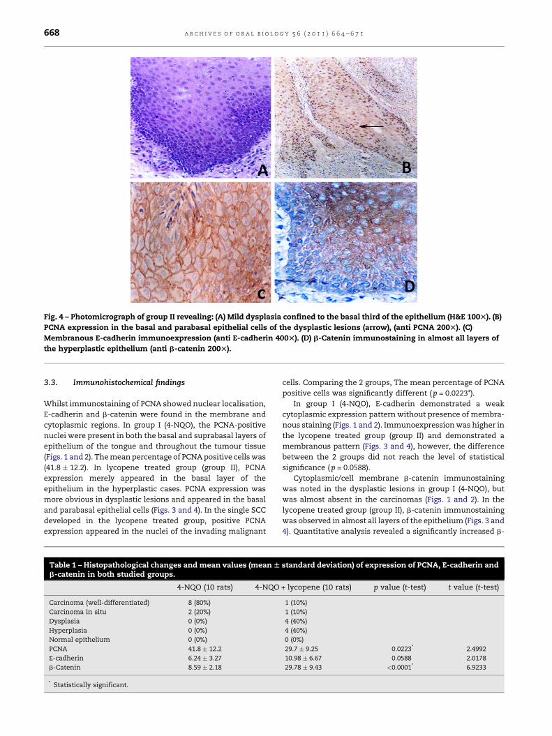
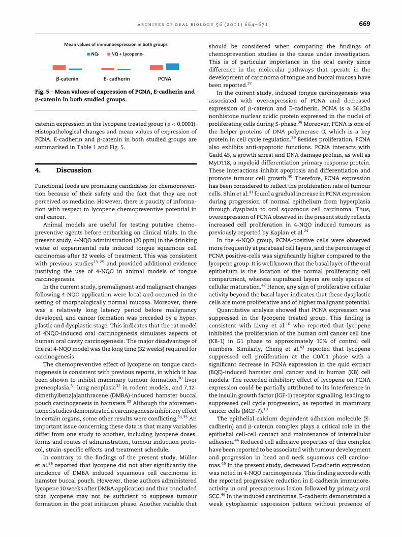
Fig. 4 – Photomicrograph of group II revealing: (A) Mild dysplasia confined to the basal third of the epithelium (H&E 100T). (B)
PCNA expression in the basal and parabasal epithelial cells of the dysplastic lesions (arrow), (anti PCNA 200T). (C)
Membranous E-cadherin immunoexpression (anti E-cadherin 400T). (D) b-Catenin immunostaining in almost all layers of
the hyperplastic epithelium (anti b-catenin 200T).
a r c h i v e s o f o r a l b i o l o g y 5 6 ( 2 0 1 1 ) 6 6 4 – 6 7 1668
3.3. Immunohistochemical findings
Whilst immunostaining of PCNA showed nuclear localisation,
E-cadherin and b-catenin were found in the membrane and
cytoplasmic regions. In group I (4-NQO), the PCNA-positive
nuclei were present in both the basal and suprabasal layers of
epithelium of the tongue and throughout the tumour tissue
(Figs. 1 and 2). The mean percentage of PCNA positive cells was
(41.8 � 12.2). In lycopene treated group (group II), PCNA
expression merely appeared in the basal layer of the
epithelium in the hyperplastic cases. PCNA expression was
more obvious in dysplastic lesions and appeared in the basal
and parabasal epithelial cells (Figs. 3 and 4). In the single SCC
developed in the lycopene treated group, positive PCNA
expression appeared in the nuclei of the invading malignant
Table 1 – Histopathological changes and mean values (mean Wb-catenin in both studied groups.
4-NQO (10 rats) 4-NQO
Carcinoma (well-differentiated) 8 (80%)
Carcinoma in situ 2 (20%)
Dysplasia 0 (0%)
Hyperplasia 0 (0%)
Normal epithelium 0 (0%)
PCNA 41.8 � 12.2
E-cadherin 6.24 � 3.27
b-Catenin 8.59 � 2.18
* Statistically significant.
cells. Comparing the 2 groups, The mean percentage of PCNA
positive cells was significantly different ( p = 0.0223*).
In group I (4-NQO), E-cadherin demonstrated a weak
cytoplasmic expression pattern without presence of membra-
nous staining (Figs. 1 and 2). Immunoexpression was higher in
the lycopene treated group (group II) and demonstrated a
membranous pattern (Figs. 3 and 4), however, the difference
between the 2 groups did not reach the level of statistical
significance ( p = 0.0588).
Cytoplasmic/cell membrane b-catenin immunostaining
was noted in the dysplastic lesions in group I (4-NQO), but
was almost absent in the carcinomas (Figs. 1 and 2). In the
lycopene treated group (group II), b-catenin immunostaining
was observed in almost all layers of the epithelium (Figs. 3 and
4). Quantitative analysis revealed a significantly increased b-
standard deviation) of expression of PCNA, E-cadherin and
+ lycopene (10 rats) p value (t-test) t value (t-test)
1 (10%)
1 (10%)
4 (40%)
4 (40%)
0 (0%)
29.7 � 9.25 0.0223* 2.4992
10.98 � 6.67 0.0588 2.0178
29.78 � 9.43 <0.0001* 6.9233
PCNAE- cadher inβ-ca teni n
Mean value s of immun oexpression in bo th grou ps
-NQ -NQ + Lycop ene
Fig. 5 – Mean values of expression of PCNA, E-cadherin and
b-catenin in both studied groups.
a r c h i v e s o f o r a l b i o l o g y 5 6 ( 2 0 1 1 ) 6 6 4 – 6 7 1 669
catenin expression in the lycopene treated group ( p < 0.0001).
Histopathological changes and mean values of expression of
PCNA, E-cadherin and b-catenin in both studied groups are
summarised in Table 1 and Fig. 5.
4. Discussion
Functional foods are promising candidates for chemopreven-
tion because of their safety and the fact that they are not
perceived as medicine. However, there is paucity of informa-
tion with respect to lycopene chemopreventive potential in
oral cancer.
Animal models are useful for testing putative chemo-
preventive agents before embarking on clinical trials. In the
present study, 4-NQO administration (20 ppm) in the drinking
water of experimental rats induced tongue squamous cell
carcinomas after 32 weeks of treatment. This was consistent
with previous studies23–25 and provided additional evidence
justifying the use of 4-NQO in animal models of tongue
carcinogenesis.
In the current study, premalignant and malignant changes
following 4-NQO application were local and occurred in the
setting of morphologically normal mucosa. Moreover, there
was a relatively long latency period before malignancy
developed, and cancer formation was preceded by a hyper-
plastic and dysplastic stage. This indicates that the rat model
of 4NQO-induced oral carcinogenesis simulates aspects of
human oral cavity carcinogenesis. The major disadvantage of
the rat 4-NQO model was the long time (32 weeks) required for
carcinogenesis.
The chemopreventive effect of lycopene on tongue carci-
nogenesis is consistent with previous reports, in which it has
been shown to inhibit mammary tumour formation,30 liver
preneoplasia,31 lung neoplasia32 in rodent models, and 7,12-
dimethylbenz[a]anthracene (DMBA)-induced hamster buccal
pouch carcinogenesis in hamsters.33 Although the aforemen-
tioned studies demonstrated a carcinogenesis inhibitory effect
in certain organs, some other results were conflicting.34,35 An
important issue concerning these data is that many variables
differ from one study to another, including lycopene doses,
forms and routes of administration, tumour induction proto-
col, strain-specific effects and treatment schedule.
In contrary to the findings of the present study, Muller
et al.36 reported that lycopene did not alter significantly the
incidence of DMBA induced squamous cell carcinoma in
hamster buccal pouch. However, these authors administered
lycopene 10 weeks after DMBA application and thus concluded
that lycopene may not be sufficient to suppress tumour
formation in the post initiation phase. Another variable that
should be considered when comparing the findings of
chemoprevention studies is the tissue under investigation.
This is of particular importance in the oral cavity since
difference in the molecular pathways that operate in the
development of carcinoma of tongue and buccal mucosa have
been reported.37
In the current study, induced tongue carcinogenesis was
associated with overexpression of PCNA and decreased
expression of b-catenin and E-cadherin. PCNA is a 36 kDa
nonhistone nuclear acidic protein expressed in the nuclei of
proliferating cells during S-phase.38 Moreover, PCNA is one of
the helper proteins of DNA polymerase O’, which is a key
protein in cell cycle regulation.39 Besides proliferation, PCNA
also exhibits anti-apoptotic functions. PCNA interacts with
Gadd 45, a growth arrest and DNA damage protein, as well as
MyD118, a myeloid differentiation primary response protein.
These interactions inhibit apoptosis and differentiation and
promote tumour cell growth.40 Therefore, PCNA expression
has been considered to reflect the proliferation rate of tumour
cells. Shin et al.41 found a gradual increase in PCNA expression
during progression of normal epithelium from hyperplasia
through dysplasia to oral squamous cell carcinoma. Thus,
overexpression of PCNA observed in the present study reflects
increased cell proliferation in 4-NQO induced tumours as
previously reported by Kaplan et al.24
In the 4-NQO group, PCNA-positive cells were observed
more frequently at parabasal cell layers, and the percentage of
PCNA positive-cells was significantly higher compared to the
lycopene group. It is well known that the basal layer of the oral
epithelium is the location of the normal proliferating cell
compartment, whereas suprabasal layers are only spaces of
cellular maturation.42 Hence, any sign of proliferative cellular
activity beyond the basal layer indicates that these dysplastic
cells are more proliferative and of higher malignant potential.
Quantitative analysis showed that PCNA expression was
suppressed in the lycopene treated group. This finding is
consistent with Livny et al.10 who reported that lycopene
inhibited the proliferation of the human oral cancer cell line
(KB-1) in G1 phase to approximately 10% of control cell
numbers. Similarly, Cheng et al.43 reported that lycopene
suppressed cell proliferation at the G0/G1 phase with a
significant decrease in PCNA expression in the quid extract
(BQE)-induced hamster oral cancer and in human (KB) cell
models. The recorded inhibitory effect of lycopene on PCNA
expression could be partially attributed to its interference in
the insulin growth factor (IGF-1) receptor signalling, leading to
suppressed cell cycle progression, as reported in mammary
cancer cells (MCF-7).18
The epithelial calcium dependent adhesion molecule (E-
cadherin) and b-catenin complex plays a critical role in the
epithelial cell-cell contact and maintenance of intercellular
adhesion.44 Reduced cell adhesive properties of this complex
have been reported to be associated with tumour development
and progression in head and neck squamous cell carcino-
mas.45 In the present study, decreased E-cadherin expression
was noted in 4-NQO carcinogenesis. This finding accords with
the reported progressive reduction in E-cadherin immunore-
activity in oral precancerous lesion followed by primary oral
SCC.46 In the induced carcinomas, E-cadherin demonstrated a
weak cytoplasmic expression pattern without presence of

a r c h i v e s o f o r a l b i o l o g y 5 6 ( 2 0 1 1 ) 6 6 4 – 6 7 1670
membranous staining. This is in consistence with the reported
alteration in the expression of this molecule in oral SCC.47,48
Differential expression with cytoplasm shifting of E-cadherin
has also been demonstrated in some oral precancerous
lesions, compared with the normal oral mucosa.48 According
to Fujita et al.,49 a redistribution of the E-cadherin complex out
of tight junctions can affect its functions in cell-cell adhesion
and can increase its degradation by cytoplasmic endocytosis.
In the lycopene treated group, E-cadherin immunoreactivi-
ty was higher compared to group I and demonstrated the
typical membranous pattern. These observations clearly
indicate that lycopene is able to prevent the tumour-related
alterations in the expression and localisation of E-cadherin
and can consequently preserve its crucial role in cell adhesion,
thus maintaining normal epithelial polarity and inhibiting
invasion.
In the current 4-NQO tongue carcinogenesis model, b-
catenin immunoreactivity was almost absent in the induced
carcinomas. This observation accords with previous studies
documenting reduced catenin expression in oral SCC.50,51
Moreover, an inverse relationship was reported between the
expression of b-catenin and the degree of cellular differentia-
tion, together with a reduction of membrane expression of
catenins in more undifferentiated cells of human oral SCC.52
This seems to suggest that catenins physiology alterations
may be involved in the transformation process.51
Quantitative analysis demonstrated higher levels of b-
catenin cytoplasmic/cell membrane expression in the hyper-
plasticanddysplasticlesions ofthelycopenetreatedgroupinthe
present study. These findings denote enhanced cellular adhe-
sionandsuggest theanti-invasiveeffect asa newmechanismfor
the chemopreventive role of lycopene, in consistence with
previous reports dealing with other tissues.53,54
5. Conclusion
The present study clearly demonstrates that inhibition of cell
proliferation and induction of cell adhesion may be major
mechanisms through which lycopene exerts its oral anticar-
cinogenic properties. There is a rapid need for more large scale
studies focusing on this naturally occurring compound to
ascertain these observations and hopefully confirm their role
in oral cancer chemoprevention.
Funding: None.
Competing interests: None declared.
Ethical approval: The research was approved by the review
board of the animal and experimental laboratory (Faculty of
Medicine, Cairo University, Egypt).
r e f e r e n c e s
1. Ferslay J, Pisani P, Parkin DM. GLO BOCAN 2002. Cancerincidence, mortality and prevalence worldwide. Lyon: IARCPress; 2004.
2. Pisani B, Bray F, Parkin DM. Estimates of the world-wideprevalence of cancer for 25 sites in the adult population. Int JCancer 2002;97:72–81.
3. Schwartz JL. Biomarkers and molecular epidemiology andchemoprevention of oral carcinogenesis. Crit Rev Oral BiolMed 2000;11:92–122.
4. Block G, Patterson B, Subar A. Fruit, vegetables, and cancerprevention: a review of the epidemiological evidence. NutrCancer 1992;18:1–29.
5. Reddy L, Odhav B, Bhoola KD. Natural products for cancerprevention: a global perspective. Pharmacol Ther 2003;99:1–13.
6. Pavia M, Pileggi C, Nobile CG, Angelillo IF. Associationbetween fruit and vegetable consumption and oral cancer: ameta-analysis of observational studies. Am J Clin Nutr2006;83:1126–34.
7. Bhuvaneswari V, Nagini S. Lycopene: a review of itspotential as an anticancer agent. Curr Med Chem AnticancerAgents 2005;5:627–35.
8. Gruenwald J, Jaenckie C, Freder J. Lycopene, the modernanswer to urban wellness? Nutra Foods 2003;2:21–35.
9. Rao AV, Agarwal S. Role of antioxidant lycopene in cancerand heart disease. J Am Coll Nutr 2000;19:563–9.
10. Livny O, Kaplan I, Reifen R, Polack-Charcon S, Madar Z,Schwartz B. Lycopene inhibits proliferation and enhancesgap-junction communication of KB-1 human oral tumorcells. J Nutr 2002;132:3754–9.
11. Levy J, Bosin E, Feldman B, Giat Y, Miinster A, Danilenko M,et al. Lycopene is a more potent inhibitor of human cancercell proliferation than either alpha-carotene or beta-carotene. Nutr Cancer 1995;24:257–66.
12. Tang L, Jin T, Zeng X, Wang JS. Lycopene inhibits the growthof human androgen independent prostate cancer cellsin vitro and in BALB/c nude mice. J Nutr 2005;135:287–90.
13. Nahum A, Zeller L, Danilenko M, Prall OW, Watts CK,Sutherland RL, et al. Lycopene inhibition of IGF inducedcancer cell growth depends on the level of cyclin D1. Eur JNutr 2006;45:275–82.
14. Zhang LX, Cooney RV, Bertran J. Carotenoids enhance gapjunctional communication and inhibit lipid peroxidation inC3H/10T1/2 cells: relationship to their cancerchemopreventive action. Carcinogenesis 1991;12:2109–14.
15. Hwang ES, Bowen PE. Cell cycle arrest and induction ofapoptosis by lycopene in LNCaP human prostate cancercells. J Med Food 2004;7:284–9.
16. Gann PH, Ma J, Giovannucci E, Willett W, Sacks FM,Hennekens CH, et al. Lower prostate cancer risk in men withelevated plasma lycopene levels: results of a prospectiveanalysis. Cancer Res 1999;59:1225–30.
17. Giovannucci E. Tomatoes, tomato-based products,lycopene, and cancer: review of the epidemiologic literature.J Natl Cancer Inst 1999;91:317–31.
18. Karas M, Amir H, Fishman D, Danilenko M, Segal S, NahumA, et al. Lycopene interferes with cell cycle progression andinsulin-like growth factor I signaling in mammary cancercells. Nutr Cancer 2000;36:101–11.
19. Amir H, Karas M, Giat J, Danilenko M, Levy R, Yermiahu T,et al. Lycopene and 1,25-dihydroxyvitamin D3 cooperate inthe inhibition of cell cycle progression and induction ofdifferentiation in HL-60. Nutr Cancer 1999;33:105–12.
20. Bhuvaneswari V, Velmurugan B, Balasenthil S,Ramachandran CR, Nagini S. Chemopreventive efficacy oflycopene on 7,12-dimethylbenz[a]anthracene-inducedhamster buccal pouch carcinogenesis. Fitoterapia2001;72:865–74.
21. Zakrzewska JM. Oral lycopene—an efficacious treatment fororal leukoplakia? Evid Based Dent 2005;6:17–8.
22. Hawkins BL, Heniford BW, Ackermann DM, Leonberger M,Martinez SA, Hendler FJ. 4NQO carcinogenesis: a mousemodel of oral cavity squamous cell carcinoma. Head Neck1994;16:424–32.

a r c h i v e s o f o r a l b i o l o g y 5 6 ( 2 0 1 1 ) 6 6 4 – 6 7 1 671
23. Ribeiro DA, Favero Salvadori DM, da Silva RN, Ribeiro DarrosB, Alencar Marques ME. Genomic instability in non-neoplastic oral mucosa cells can predict risk during 4-nitroquinoline 1-oxide-induced rat tongue carcinogenesis.Oral Oncol 2004;40:910–5.
24. Kaplan I, Hochstadt T, Dayan D. PCNA in palate and tonguemucosal dysplastic lesions induced by topically applied4NQO in desalivated rat. Med Oral 2002;7:336–43.
25. Tanuma J, Shisa H, Hiai H, Higashi S, Yamada Y, Kamoto T,et al. Quantitative trait loci affecting 4-nitroquinoline 1-oxide-induced tongue carcinogenesis in the rat. Cancer Res1998;58:1660–4.
26. Nishimura A. Changes in Bcl-2 and Bax expression in rattongue during 4-nitroquinoline 1-oxide-inducedcarcinogenesis. J Dent Res 1999;78:1264–9.
27. Shiotani H, Denda A, Yamamoto K, Kitayama W, Endoh T,Sasaki Y, et al. Increased expression of cyclooxygenase-2protein in 4-nitroquinoline-1-oxide induced rat tonguecarcinomas and chemopreventive efficacy of a specificinhibitor, nimesulide. Cancer Res 2001;61:1451–6.
28. Tanaka T, Kohno H, Nomura E, Taniguchi H, Tsuno T, TsudaH. A novel geranylated derivative, ethyl 3-(40-geranyloxy-30-methoxyphenyl)-2-propenoate, synthesized from ferulicacid suppresses carcinogenesis and inducible nitric oxidesynthase in rat tongue. Oncology 2003;64:166–75.
29. Ribeiro D, Kitakawa D, Domingues MA, Cabral LA, MarquesME, Salvadori DM. Survivin and nitric oxide induciblesynthase production during 4NQO-induced rat tonguecarcinogenesis, a possible relationship. Exp Mol Pathol2007;83:131–7.
30. Nagasawa H, Mitamura T, Sakamoto S, Yamamoto K. Effectsof lycopene on spontaneous mammary tumourdevelopment in SHN virgin mice. Anticancer Res1995;15:1173–8.
31. Astorg P, Gradelet S, Berges R, Suschetet M. Dietary lycopenedecreases the initiation of liver preneoplastic foci bydiethylnitrosamine in the rat. Nutr Cancer 1997;29:60–8.
32. Kim DJ, Takasuka N, Kim JM, Sekine K, Ota T, Asamoto M,et al. Chemoprevention by lycopene of mouse lungneoplasia after combined initiation treatment with DEN,MNU and DMH. Cancer Lett 1997;120:15–22.
33. Bhuvaneswari V, Velmurugan B, Nagini S. Lycopenemodulates circulatory antioxidants during hamster buccalpouch carcinogenesis. Nutr Res 2001;21:1447–53.
34. Imaida K, Tamano S, Kato K, Ikeda Y, Asamoto M,Takahashi S, et al. Lack of chemopreventive effects oflycopene and curcumin on experimental rat prostatecarcinogenesis. Carcinogenesis 2001;22:467–72.
35. Guttenplan JB, Chen M, Kosinska W, Thompson S, Zhao Z,Cohen LA. Effects of a lycopene-rich diet on spontaneousand benzo[a] pyrene-induced mutagenesis in prostate,colon and lungs of the lacZ mouse. Cancer Lett 2001;164:1–6.
36. Muller K, Zucoloto S, Albuquerque Jr RF, Vannucchi H. Lackof inhibitory effect of lycopene on dysplastic lesionsinduced by 7,12-dimethyl-benz[a]anthracene in hamsterbuccal pouch. Nutr Res 2007;27:574–9.
37. Sathyan KM, Sailasree R, Jayasurya R, Lakshminarayanan K,Abraham T, Nalinakumari KR, et al. Carcinoma of tongueand the buccal mucosa represent different biologicalsubentities of the oral carcinoma. J Cancer Res Clin Oncol2006;132:601–9.
38. Paunesku T, Mittal S, Protic M, Oryhon J, Korolev SV,Joachimiak A, et al. Proliferating cell nuclear antigen(PCNA): ringmaster of the genome. Int J Radiat Biol2001;77:1007–21.
39. McCormick D, Hall PA. The complexities of proliferating cellnuclear antigen. Histopathology 1992;21:591–4.
40. Vairapandi M, Azam N, Balliet AG, Hoffman B, LibermannDA. Characterization of MyD118, Gadd 45, and proliferatingcell nuclear antigen (PCNA) interacting domains. PCNAimpedes MyD 118 and Gadd45-mediated negative growthcontrol. J Biol Chem 2000;275:16810–9.
41. Shin DM, Voravud N, Ro JY, Lee JS, Hong WK, Hittelman WN.Sequential increases in proliferating cell nuclear antigenexpression in head and neck tumorigenesis: a potentialbiomarker. J Natl Cancer Inst 1993;85:971–8.
42. Krutchkoff DJ, Eisenberg E, Anderson C. Dysplasia of oralmucosa: a unified approach to proper evaluation. Mod Pathol1991;4:113–9.
43. Cheng H-C, Chien H, Liao C, Yang Y, Huang S. Carotenoidssuppress proliferating cell nuclear antigen and cyclin D1expression in oral carcinogenic models. J Nutr Biochem2007;18:667–75.
44. Conacci-Sorrel M, Zhurinsky J, Ben-Zeev A. The cadherincatenin adhesion system in signaling and cancer. J ClinInvest 2002;109:987–91.
45. Andrews NA, Jones AS, Helliwell TR, Kinsella AR. Expressionof the E-cadherin–catenin cell adhesion complex in primarysquamous cell carcinomas of the head and neck and theirnodal metastases. Br J Cancer 1997;75:1474–80.
46. Hung K-F, Chang C-S, Liu C-J, Lui M-T, Cheng C-Y, Kao S-Y.Differential expression of E-cadherin in metastatic lesionscomparing to primary oral squamous cell carcinoma. OralPathol Med 2006;35:589–94.
47. Chow V, Yuen AP, Lam KY, Tsao GS, Ho WK, Wei WI, et al. Acomparative study of the clinicopathological significance ofE-cadherin and Catenins (alpha, beta, gamma) expression insurgical management of the oral tongue carcinoma. J CancerRes Oncol 2001;127:59–63.
48. Wu H, Lotan R, Menter D, Lippman SM, Xu XC. Expression ofE-cadherin is associated with squamous differentiation insquamous cell carcinomas. Anticancer Res 2000;20:1385–90.
49. Fujita Y, Krause G, Scheffner M, Zechner D, Leddy HE,Behrens J, et al. Hakai, a c-Cbl-like protein, ubiquitinatesand induces endocytosis of the E-cadherin complex. Nat CellBiol 2002;4:222–31.
50. Williams HK, Sanders DS, Jankowski JA, Landini G, BrownAM. Expression of cadherins and catenins in oral epithelialdysplasia and squamous cell carcinoma. J Oral Pathol Med1998;27:308–17.
51. Bagutti C, Speight PM, Watt FM. Comparison of integrin,cadherin, and catenin expression in squamous cellcarcinomas of the oral cavity. J Pathol 1998;186:8–16.
52. Lo Muzio L, Staibano S, Pannone G, Grieco M, Mignogna MD,Cerrato A, et al. Beta- and gamma-catenin expression in oralsquamous cell carcinomas. Anticancer Res 1999;19:3817–26.
53. Huang CS, Shih MK, Chuang CH, Hu ML. Lycopene inhibitscell migration and invasion and upregulates nm23-H1 in ahighly invasive hepatocarcinoma, SK-Hep-1 cells. J Nutr2005;135:2119–23.
54. Hwang ES, Lee HJ. Inhibitory effects of lycopene on theadhesion, invasion, and migration of SK-Hep1 humanhepatoma cells. Exp Biol Med 2006;231:322–7.