HighD-glucose reducesSLC29A1 promoter activity and adenosine transport involving specific protein 1...
12
High D-Glucose Reduces SLC29A1 Promoter Activity and Adenosine Transport Involving Specific Protein 1 in Human Umbilical Vein Endothelium CARLOS PUEBLA, 1 MARCELO FARI ´ AS, 1 MARCELO GONZA ´ LEZ, 1 ANDREA VECCHIOLA, 1 CLAUDIO AGUAYO, 1 BERNARDO KRAUSE, 1,2 MARC ¸AL PASTOR-ANGLADA, 3 PAOLA CASANELLO, 1 AND LUIS SOBREVIA 1 * 1 Cellular and Molecular Physiology Laboratory (CMPL) and Perinatal Research Laboratory (PRL), Department of Obstetrics and Gynaecology, Medical Research Centre (CIM), School of Medicine, Faculty of Medicine, Pontificia Universidad Cato´lica de Chile, Santiago, Chile 2 Basic Sciences Department, Faculty of Biological Sciences, Universidad del Bı ´o-Bı ´o, Chile 3 Department of Biochemistry and Molecular Biology, Institute of Biomedicine, (IBUB), University of Barcelona, CIBER EHD, Barcelona, Spain High D-glucose reduces human equilibrative nucleoside transporter 1 (hENT1)-mediated adenosine uptake involving endothelial nitric oxide synthase (eNOS), mitogen-activated protein (MAP) kinase kinases 1 and 2/MAP kinases p42/44 (MEK/ERKs), and protein kinase C (PKC) activation in human umbilical vein endothelium (HUVEC). Since NO represses SLC29A1 gene (hENT1) promoter activity we studied whether D-glucose-reduced hENT1-adenosine transport results from lower SLC29A1 expression in HUVEC primary cultures. HUVEC incubation (24 h) with high D-glucose (25 mM) reduced hENT1-adenosine transport and pGL3-hENT1 1114 construct SLC29A1 reporter activity compared with normal D-glucose (5 mM). High D-glucose also reduced pGL3-hENT1 1114 reporter activity compared with cells transfected with pGL3-hENT1 795 construct. N G -nitro-L-arginine methyl ester (L-NAME, NOS inhibitor), PD-98059 (MEK1/2 inhibitor), and/or calphostin C (PKC inhibitor) blocked D-glucose effects. Insulin (1 nM) and phorbol 12-myristate 13-acetate (PMA, 100 nM, PKC activator), but not 4a-phorbol 12,13-didecanoate (4aPDD, 100 nM, PMA less active analogue) reduced hENT1-adenosine transport. L-NAME and PD-98059 blocked insulin effects. L-NAME, PD-98059, and calphostin C increased hENT1 expression without altering protein or mRNA stability. High D-glucose increased Sp1 transcription factor protein abundance and binding to SLC29A1 promoter, phenomena blocked by L-NAME, PD-98059, and calphostin C. Sp1 overexpression reduced SLC29A1 promoter activity in normal D-glucose, an effect reversed by L-NAME and further reduced by S-nitroso-N-acetyl-L,D-penicillamine (SNAP, NO donor) in high D-glucose. Thus, reduced hENT1-mediated adenosine transport in high D-glucose may result from increased Sp1 binding to SLC29A1 promoter down-regulating hENT1 expression. This phenomenon depends on eNOS, MEK/ERKs, and PKC activity, suggesting potential roles for these molecules in hyperglycemia-associated endothelial dysfunction. J. Cell. Physiol. 215: 645–656, 2008. ß 2007 Wiley-Liss, Inc. An elevated plasma level of D-glucose induces vasodilation (Beckman et al., 2002; Oomen et al., 2002) and oral glucose loading leads to attenuated myogenic vasoconstriction in healthy individuals (Lott et al., 2007), phenomena partially explained by increased endothelium derived nitric oxide (NO; San Martı ´n and Sobrevia, 2006; Casanello et al., 2007). Long-term incubation (24 h) with 25 mM D-glucose (‘‘high D-glucose’’) increases the endothelial NO synthase (eNOS), mitogen-activated protein (MAP) kinase kinases 1 and 2 (MEK1/ 2)/MAP kinases p44 (ERK1) and p42 (ERK2) (i.e., MEK/ERKs), and protein kinase C (PKC) activity in human umbilical vein endothelial cells (HUVEC; Mun ˜oz et al., 2006; Va ´squez et al., Contract grant sponsor: Fondo Nacional de Desarrollo Cientı ´fico y Tecnolo ´gica, Chile (FONDECYT); Contract grant numbers: 1070865, 7070249. Contract grant sponsor: Comisio ´n Nacional de Investigacio ´n Cientı ´fica y Tecnolo ´gica, Chile (CONICYT); Contract grant number: 23070213. Contract grant sponsor: Vice-Rectorı ´a Adjunta de Investigacio ´n y Doctorado, Pontificia Universidad Cato ´lica de Chile (VRAID); Contract grant numbers: BM16/2007, BM14/2007. Contract grant sponsor: Agencia Espan ˜ola de Cooperacio ´n Internacional, Spain (AECI); Contract grant numbers: A/5484/06, SAF2005-01259. Claudio Aguayo’s present address is Faculty of Pharmacy, Universidad de Concepcio ´n, Chile. Paola Casanello and Luis Sobrevia contributed equally to this study. *Correspondence to: Luis Sobrevia, Cellular and Molecular Physiology Laboratory (CMPL), Medical Research Centre (CIM), Department of Obstetrics and Gynaecology, School of Medicine, Faculty of Medicine, Pontificia Universidad Cato ´ lica de Chile, P.O. Box 114-D, Santiago, Chile. E-mail: [email protected] Received 28 July 2007; Accepted 12 October 2007 DOI: 10.1002/21347 ORIGINAL ARTICLE 645 Journal of Journal of Cellular Physiology Cellular Physiology ß 2007 WILEY-LISS, INC.
Transcript of HighD-glucose reducesSLC29A1 promoter activity and adenosine transport involving specific protein 1...

ORIGINAL ARTICLE 645J o u r n a l o fJ o u r n a l o f
CellularPhysiologyCellularPhysiology
High D-Glucose Reduces SLC29A1Promoter Activity and AdenosineTransport Involving SpecificProtein 1 in Human UmbilicalVein Endothelium
CARLOS PUEBLA,1 MARCELO FARIAS,1 MARCELO GONZALEZ,1 ANDREA VECCHIOLA,1CLAUDIO AGUAYO,1 BERNARDO KRAUSE,1,2 MARCAL PASTOR-ANGLADA,3
PAOLA CASANELLO,1 AND LUIS SOBREVIA1*1Cellular and Molecular Physiology Laboratory (CMPL) and Perinatal Research Laboratory (PRL),
Department of Obstetrics and Gynaecology, Medical Research Centre (CIM), School of Medicine, Faculty of Medicine,
Pontificia Universidad Catolica de Chile, Santiago, Chile2Basic Sciences Department, Faculty of Biological Sciences, Universidad del Bıo-Bıo, Chile3Department of Biochemistry and Molecular Biology, Institute of Biomedicine, (IBUB), University of Barcelona, CIBER EHD,
Barcelona, Spain
High D-glucose reduces human equilibrative nucleoside transporter 1 (hENT1)-mediated adenosine uptake involving endothelial nitricoxide synthase (eNOS), mitogen-activated protein (MAP) kinase kinases 1 and 2/MAP kinases p42/44 (MEK/ERKs), and protein kinase C(PKC) activation in human umbilical vein endothelium (HUVEC). Since NO represses SLC29A1 gene (hENT1) promoter activity westudied whether D-glucose-reduced hENT1-adenosine transport results from lower SLC29A1 expression in HUVEC primary cultures.HUVEC incubation (24 h) with high D-glucose (25 mM) reduced hENT1-adenosine transport and pGL3-hENT1�1114 construct SLC29A1reporter activity compared with normal D-glucose (5 mM). High D-glucose also reduced pGL3-hENT1�1114 reporter activity comparedwith cells transfected with pGL3-hENT1�795 construct. NG-nitro-L-arginine methyl ester (L-NAME, NOS inhibitor), PD-98059 (MEK1/2inhibitor), and/or calphostin C (PKC inhibitor) blocked D-glucose effects. Insulin (1 nM) and phorbol 12-myristate 13-acetate (PMA,100 nM, PKC activator), but not 4a-phorbol 12,13-didecanoate (4aPDD, 100 nM, PMA less active analogue) reduced hENT1-adenosinetransport. L-NAME and PD-98059 blocked insulin effects. L-NAME, PD-98059, and calphostin C increased hENT1 expression withoutaltering protein or mRNA stability. High D-glucose increased Sp1 transcription factor protein abundance and binding to SLC29A1promoter, phenomena blocked by L-NAME, PD-98059, and calphostin C. Sp1 overexpression reduced SLC29A1 promoter activity innormal D-glucose, an effect reversed by L-NAME and further reduced by S-nitroso-N-acetyl-L,D-penicillamine (SNAP, NO donor) in highD-glucose. Thus, reduced hENT1-mediated adenosine transport in high D-glucose may result from increased Sp1 binding to SLC29A1promoter down-regulating hENT1 expression. This phenomenon depends on eNOS, MEK/ERKs, and PKC activity, suggesting potentialroles for these molecules in hyperglycemia-associated endothelial dysfunction.
J. Cell. Physiol. 215: 645–656, 2008. � 2007 Wiley-Liss, Inc.
An elevated plasma level of D-glucose induces vasodilation(Beckman et al., 2002; Oomen et al., 2002) and oral glucoseloading leads to attenuated myogenic vasoconstriction inhealthy individuals (Lott et al., 2007), phenomena partiallyexplained by increased endothelium derived nitric oxide(NO; San Martın and Sobrevia, 2006; Casanello et al., 2007).
Contract grant sponsor: Fondo Nacional de Desarrollo Cientıfico yTecnologica, Chile (FONDECYT);Contract grant numbers: 1070865, 7070249.Contract grant sponsor: Comision Nacional de InvestigacionCientıfica y Tecnologica, Chile (CONICYT);Contract grant number: 23070213.Contract grant sponsor: Vice-Rectorıa Adjunta de Investigacion yDoctorado, Pontificia Universidad Catolica de Chile (VRAID);Contract grant numbers: BM16/2007, BM14/2007.Contract grant sponsor: Agencia Espanola de CooperacionInternacional, Spain (AECI);Contract grant numbers: A/5484/06, SAF2005-01259.
Claudio Aguayo’s present address is Faculty of Pharmacy,Universidad de Concepcion, Chile.
� 2 0 0 7 W I L E Y - L I S S , I N C .
Long-term incubation (24 h) with 25 mM D-glucose (‘‘highD-glucose’’) increases the endothelial NO synthase (eNOS),mitogen-activated protein (MAP) kinase kinases 1 and 2 (MEK1/2)/MAP kinases p44 (ERK1) and p42 (ERK2) (i.e., MEK/ERKs),and protein kinase C (PKC) activity in human umbilical veinendothelial cells (HUVEC; Munoz et al., 2006; Vasquez et al.,
Paola Casanello and Luis Sobrevia contributed equally to this study.
*Correspondence to: Luis Sobrevia, Cellular and MolecularPhysiology Laboratory (CMPL), Medical Research Centre (CIM),Department of Obstetrics and Gynaecology, School of Medicine,Faculty of Medicine, Pontificia Universidad Catolica de Chile, P.O.Box 114-D, Santiago, Chile. E-mail: [email protected]
Received 28 July 2007; Accepted 12 October 2007DOI: 10.1002/21347
646 P U E B L A E T A L .
2007). These effects of high D-glucose are associated withreduced uptake of the endogenous vasoactive nucleosideadenosine in this cell type (Aguayo et al., 2005; Munoz et al.,2006).
Adenosine uptake and metabolism by the endotheliumare crucial mechanisms maintaining the physiological plasmaconcentration of this nucleoside (Baldwin et al., 2004; SanMartın and Sobrevia, 2006; Casanello et al., 2007), limiting itsseveral biological effects (Baldwin et al., 2004; Choi et al., 2004;Casanello et al., 2005, 2007; Burnstock, 2006; San Martın andSobrevia, 2006). However, the mechanisms underlyingnucleoside transporters regulation in endothelial cells exposedto high extracellular D-glucose are unknown (San Martın andSobrevia, 2006; Casanello et al., 2007). Under physiologicalconditions adenosine is taken up via the humanequilibrative, Naþ-independent nucleoside transporters 1(hENT1) and hENT2 in HUVEC (Aguayo et al., 2005; Casanelloet al., 2005; Farıas et al., 2006; Munoz et al., 2006). hENT1expression and activity are reduced in this cell type isolatedfrom gestational diabetes (Vasquez et al., 2004; Farıas et al.,2006), an effect associated with reduced SLC29A1 gene(for hENT1) promoter activity likely due to the elevatedendothelium-derived NO characteristic of HUVEC from thispathology (Vasquez et al., 2004; Farıas et al., 2006). Similarly,HUVEC from normal pregnancies exposed to high D-glucoseexhibit reduced hENT1-mediated adenosine transport andincreased NO synthesis, MEK/ERKs, and PKC activity (Aguayoet al., 2005; Munoz et al., 2006). However, the potentialinvolvement of these molecules as inhibitors of SLC29A1transcription in response to elevated D-glucose in humanendothelium is unknown (for reviews see San Martın andSobrevia, 2006; Casanello et al., 2007).
Activity of the zinc finger promoter-selective transcriptionfactor specific protein 1 (Sp1) is modulated by NO (Wang et al.,1999; Bogdan, 2001; Sellak et al., 2002; Starzynski et al., 2006),MEK/ERKs (Fiuza et al., 2003), and PKC (Tanaka et al., 2000;Horovitz-Fried and Sampson, 2007), altering gene expression inseveral cell types (Bogdan, 2001; Jeong et al., 2004; Chu andFerro, 2005; Paradkar and Roth, 2006; Starzynski et al., 2006).SLC29A1 promoter activity of the sequence upstream �795 bpfrom ATG translation start codon is repressed in aNO-dependent manner in primary cultures of HUVEC (Farıaset al., 2006; Puebla et al., 2007). In addition, since the promoterregion between �1,114 and �795 bp contains Sp1 consensusmotifs, it is likely that Sp1 is involved in SCL29A1 repression. Inthis study we examined the Sp1 involvement in high D-glucosetranscriptional modulation of SLC29A1 expression. Ourfindings suggest that reduced hENT1 expression in primarycultures of HUVEC in response to D-glucose may result fromreduced transcriptional SLC29A1 promoter activity. Higher Sp1binding to the SLC29A1 promoter was found to be dependenton NOS, MEK/ERKs, and PKC activity. These results couldbe important to understand the deleterious effect ofhyperglycemia on fetal endothelial function in pregnancydiseases associated with elevated plasma D-glucose, such asgestational diabetes (Garcıa-Carrapato, 2003; Lampl and Jeanty,2004; San Martın and Sobrevia, 2006; Casanello et al., 2007).
Materials and MethodsReagents
Sera, agarose, buffers were from GIBCO Life Technologies(Carlsbad, CA). Collagenase Type II (Clostridium histolyticum) wasfrom Boehringer (Mannheim, F.R.G.) and Bradford reagent andpure nitrocellulose membrane (Trans-blot) were from BioRadLaboratories (Hertfordshire, UK). NG-Nitro-L-arginine methylester (L-NAME), PD-98059, and calphostin C were fromCalbiochem (La Jolla, CA). S-nitroso-N-acetyl-L,D-penicillamine
JOURNAL OF CELLULAR PHYSIOLOGY
(SNAP), cycloheximide, actinomycin D, S-(4-nitrobenzyl)-6-thio-inosine (NBTI), ethidium bromide, salmon sperm DNA and RNAseA, phorbol 12-myristate 13-acetate (PMA), 4a-phorbol 12,13-didecanoate (4aPDD) and insulin were from Sigma (St. Louis,MO). Proteinase K was from Roche Molecular Biochemicals(Indianapolis, IN). [3H]Adenosine (37 Ci/mmol) was from NEN(Dreieich, FRG). b-Actin and Sp1 antibodies were from Santa CruzBiotechnology (Santa Cruz, CA). GST antibody was fromAmersham (Buckinghamshire, UK).
Cell culture
Endothelial cells were isolated by collagenase (0.25 mg/ml)digestion from umbilical cord veins (HUVEC) from normalpregnancies and cultured (378C, 5% CO2) up to passage 2 inmedium 199 (M199) containing 5 mM D-glucose, 10% new borncalf serum, 10% fetal calf serum (FCS), 3.2 mM L-glutamine, and100 U/ml penicillin-streptomycin as described (Montecinos et al.,2000; Munoz et al., 2006; Vasquez et al., 2007).
D-glucose incubation
We previously reported that adenosine transport in primarycultures of HUVEC exposed to different concentrations ofD-glucose (7.5–25 mM) for different periods of time (1–24 h) issignificantly reduced (IC50 �11 mM D-glucose) with a half-maximalinhibition at �6 h, maximally inhibited at 12 h and sustained up to24 h incubation (Montecinos et al., 2000). Thus, we used 24 hincubation with 25 mM D-glucose in this study. Twenty-four hoursprior to experiments incubation medium was changed to sera-freeM199 containing 5 mM (‘‘normal’’) or 25 mM (‘‘high’’) D-glucose.Parallel cultures were also exposed to 5 mM D-glucose plus 20 mML-glucose (osmotic control). None of the experiments performedusing the latter experimental condition was significantly differentfrom cells incubated with 5 mM D-glucose, confirming previousresults in this cell type (Montecinos et al., 2000; Munoz et al., 2006;Vasquez et al., 2007).
Adenosine transport
Overall adenosine transport ([3H]adenosine, 2 mCi/ml, 20 sec,228C) was measured as described (Aguayo et al., 2005; Casanelloet al., 2005; Munoz et al., 2006). Briefly, transport assays wereperformed in cells preincubated (30 min, 228C) with Krebscontaining S-(4-nitrobenzyl)-6-thio-inosine (NBTI, 0.1 or 10 mM,ENTs inhibitor; Aguayo et al., 2005; Casanello et al., 2005; Munozet al., 2006). In experiments where sodium was replaced byN-methylglucamine-HCl or choline chloride, adenosine transportwas unaltered (not shown). Transport of adenosine (10 mM) wasmeasured in absence or presence of NBTI, or 2 mM hypoxanthine,a nucleobase taken up via ENT2, but not via ENT1 in HUVEC andother cell types (Casanello et al., 2007). Difference between totaltransport in absence or presence of 0.1 mM NBTI, inhibited byhypoxanthine, was defined as ENT1-mediated (Aguayo et al., 2005;Casanello et al., 2005). Adenosine transport was also assayedin cells preincubated (30 min) with 100 mM NG-nitro-L-argininemethyl ester (L-NAME, NOS inhibitor; Fleming and Busse, 2003;Casanello et al., 2005; Farıas et al., 2006), 10 mM PD-98059 [MAPkinase kinases 1/2 (MEK1/2) inhibitor] (Crews and Eriksson, 1992),100 mM SNAP (NO donor; Ogonowski et al., 2000), or 100 nMcalphostin C (DAG- and phorbol ester-sensitive PKC isozymesinhibitor; Kobayashi et al., 1989).
In parallel experiments, adenosine transport was also measuredin cells incubated for the last 30 min of the 24 h incubation periodwith 5 or 25 mM D-glucose with 100 nM phorbol 12-myristate13-acetate (PMA, direct PKC activator), 100 nM calphostin Cor the less active PMA analogue 4a-phorbol 12,13-didecanoate(4aPDD, 100 nM; Montecinos et al., 2000). Since phosphorylationof ERK1/ERK2 (p42/p44mapk) and eNOS in the serine 1177 residue,phenomena associated with activation of these molecules (Crewsand Eriksson, 1992; Fleming and Busse, 2003), is increased in

S p 1 - D E P E N D E N T G L U C O S E I N H I B I T I O N O F h E N T 1 647
response to insulin in primary cultures of HUVEC (Gonzalez et al.,2004), and because insulin reduces hENT1-mediated adenosinetransport in this cell type (Munoz et al., 2006), we used acombination of insulin and L-NAME or PD-98059 to test whetherMEK/ERKs and eNOS activation reduced hENT1-mediatedadenosine transport. Cells were incubated for the last 8 h of the 24 hincubation period with 5 or 25 mM D-glucose with insulin (1 nM),insulinþ L-NAME (100 mM), or insulinþ PD-98059 (10 mM) asreported (Gonzalez et al., 2004; Munoz et al., 2006). Cell viabilitywas assayed by Trypan blue exclusion (Montecinos et al., 2000;Aguayo et al., 2005) and was not significantly altered (�98% ofviable cells) by the inhibitors as previously reported (Montecinoset al., 2000). Inhibitors concentrations were selected fromdose–response curves as previously described in primary culturesof HUVEC (Montecinos et al., 2000; Aguayo et al., 2005; Casanelloet al., 2005; Farıas et al., 2006; Munoz et al., 2006).
Anti-hENT1 antibody production
Immunoreactive anti-serum against amino acids 274–283 of thepredicted intracellular loop between transmembrane segments6 and 7 of hENT1 was prepared as described (Farre et al., 2004).
Western blots
Proteins (70 mg) separated by polyacrylamide gel (10%)electrophoresis were transferred to 0.45 mm pore sizenitrocellulose membranes and probed with primary polyclonalrabbit anti-hENT1 (1:500), anti-Sp1 (1:500), mouse anti-b-actin(1:2,000) or goat anti-GST (1:1,000) antibodies, followed byincubation with secondary horseradish peroxidase-conjugatedgoat anti-rabbit or anti-mouse antibodies, or monkey anti-goatantibody (Al-Sarraj et al., 2005; Casanello et al., 2005; Farıas et al.,2006).
Isolation of total RNA and reverse transcription
Total RNA was isolated using the QIAGEN RNeasy kit as described(Casanello et al., 2005; Farıas et al., 2006). RNA quality and integritywere insured by gel visualization and spectrophotometric analysis(OD260/280), quantified at 260 nm and precipitated to obtain 4 mg/ml. Aliquots of 1 mg of total RNA were reverse transcribed intocDNA using oligo (dT18) plus random hexamers (10-mers) andavian Moloney murine leukemia virus-reverse transcriptase(MMLV-RT; Promega, Madison, WI).
Non-quantitative and quantitative RT-PCR
Non-quantitative RT-PCR was performed in 20 ml containing 2 ml10� PCR buffer, 0.6 ml 50 mM Mgþ2, 0.8 ml dNTP’s, 13.2 mlRNAase-free H2O, 0.4 ml Taq DNA polymerase (Invitrogen,Grand Island, NY) and sequence-specific oligonucleotide primersfor human ENT1 (0.5 mM). Samples were incubated 5 min at958C, followed by 25 cycles of 30 sec at 958C, 30 sec at 568C,30 sec at 728C, and 7 min final extension at 728C. 28S mRNAwas internal reference (Casanello et al., 2005). RT-PCR productswere sequenced as described (Casanello et al., 2005; Farıaset al., 2006). Oligonucleotide primers: hENT1 (sense)50-TCTCCAACTCTCAGCCCACCAA-30, hENT1 (anti-sense)50-CCTGCGATGCTGGACTTGACCT-30, 28S (sense)50-TTGAAAATCCGGGGGAGAG-30, 28S (anti-sense)50-ACATTGTTCCAACATGCCAG-30. Expected size productswere hENT1 151 bp and 28S 100 bp.
For quantitative real-time RT-PCR, experiments wereperformed using a LightCyclerTM rapid thermal cycler (RocheDiagnostics, Lewes, UK) as described (Casanello et al., 2005).In brief, reactions in 10 ml volume included 0.5 mM primers, anddNTPs, Taq DNA polymerase and reaction buffer provided in theQuantiTect SYBR Green PCR Master Mix (QIAGEN, Crawley,UK). HotStart Taq DNA polymerase was activated (15 min, 958C),and assays included a 958C denaturation (15 sec), annealing (20 sec)at 588C (hENT1), 568C (28S), and extension at 728C (hENT1,
JOURNAL OF CELLULAR PHYSIOLOGY
15 sec; 28S, 10 sec). Fluorescent product was detected after 3-secstep to 58C below the product melting temperature (Tm). Productspecificity was confirmed by agarose gel electrophoresis (2% v/v)and melting curve analysis. The product Tm values were 79.58Cfor hENT1 and 82.48C for 28S. hENT1 and 28S standards wereprepared as described (Casanello et al., 2005) and oligonucleotideprimers were the same as in non-quantitative RT-PCR.
hENT1 mRNA and protein stability
Total RNA and protein were isolated from HUVEC exposed(0–24 h) to culture medium without or with 1.5mM actinomycin D(transcription inhibitor; Strohm et al., 2002) or 1 mMcycloheximide (protein synthesis inhibitor; Garfinkel et al., 1994) inthe continuous presence of 5 or 25 mM D-glucose (Farıas et al.,2006). hENT1 and 28S mRNA were amplified by RT-PCR, andhENT1 and b-actin proteins detected by Western blot. Changes inthe mRNA number of copies or protein abundance in the absenceor presence of actinomycin D or cycloheximide were numericallyexpressed as the time necessary to induce a reduction equivalent tothe 50% of the maximal reduction reached under the differentexperimental conditions used in this study. Data were fitted toa sigmoid dose–response equation with a Hill slope fixed to 1.
hENT1 promoter cloning
Genomic DNA was isolated using the Wizard1 SV Genomic DNAPurification System (Promega). Preliminary studies show that NOreduces SLC29A1 promoter activity in HUVEC transfected withsequences upstream�795 bp from ATG (Farıas et al., 2006; Pueblaet al., 2007). Thus, two upstream sequences, �1,114 and �795 bpfrom the ATG translation start codon of SLC29A1 gene (GenBank:AF495730), were PCR-amplified using Elongase1 EnzymeSystem (Invitrogen) and cloned into pGL3-basic reporter systemas described (Farıas et al., 2006). The pGL3-hENT1 reporterconstructs generated were pGL3-hENT1�1114 andpGL3-hENT1�795.
Transfection
Aliquots of 0.5 ml of cell suspension (3� 106 cells/ml) were mixedwith 10 mg pGL3-hENT1 reporter constructs, pGL3-Basic (emptypGL3 vector) or pGL3-Control [Simian Virus 40 promoter (SV40)pGL3 vector], and internal transfection control vector pRL-TKexpressing Renilla luciferase (Promega) as described (Farıas et al.,2006). After electroporation (320 V, 975 mF, 9–11 msec;Gene-Pulser1 II System, BioRad, Herts, UK) cells were cultured(48 h) in M199 containing 2% FCS in absence or presence of 100mML-NAME, 10mM SNAP, 100 nM calphostin C, or 10 mM PD-98059.Transfection efficiency was estimated by transfection ofthe pEGFP-N3 vector (Clontech, Palo Alto, CA) and fluorescentcells were counted under a fluorescent-equipped invertedmicroscope (Leica DM IL, Bad Wildbad, Germany) as described(Farıas et al., 2006).
Luciferase assay
Electroporated cells were lysed in 200 ml passive lysis buffer(Promega), and Firefly and Renilla luciferase activity was measuredusing Dual-Luciferase1 Reporter Assay System (Promega) in aSirius luminometer (Berthold Detection System, Bad Wildbad,Germany) as described (Farıas et al., 2006).
Sp1 overexpression
For Sp1 overexpression, cells were transfected by electroporation(Farıas et al., 2006) with the empty expression vector pCGN or theexpression vector containing the human Sp1 full-length cDNAsequence inserted into pCGN (pCGN-Sp1; Parks and Shenk,1996). Parallel experiments were performed transfecting cellswith the pEBG vector containing the glutathione S transferase(GST)-tagged Sp1 dominant negative (pEBG-Sp1DN; Al-Sarrajet al., 2005). Native Sp1 and Sp1DN overexpression was confirmed

648 P U E B L A E T A L .
by detection of Sp1 by Western blot using polyclonal anti-Sp1(1:500) antibody and anti-GST antibody (1:1,000), respectively.
Sub-cellular fractionation
Cells were scraped into ice-cold PBS [(mM): 130 NaCl, 2.7 KCl,0.8 Na2HPO4, 1.4 KH2PO4, pH 7.4] supplemented with 1 mMphenylmethylsulfonyl fluoride (PMSF), centrifuged (5,400 rpm,48C, 6 min) and the pellet resuspended in 50 ml of ice-cold low-saltbuffer A [20 mM HEPES, pH 7.9, 1.5 mM MgCl2, 10 mM KCl, 0.5 mMdithiothreitol (DTT), 0.5 mM PMSF, 20 mM N-ethylmaleimide(NEM)] (Dignam et al., 1983). The mixture was then passedthrough a 25-gauge syringe and centrifuged (14,000 rpm, 448C,2 min). Supernatant (cytoplasmic fraction) was collected and keptat �208C until use. Pellet was resuspended in 30 ml of buffer C(20 mM HEPES, pH 7.9, 0.42 M NaCl, 1.5 mM MgCl2, 0.2 mM EDTA,25% v/v glycerol, 0.5 mM DTT, 0.5 mM PMSF, 20 mM NEM), passedthrough a 1 ml micropipette tip and incubated at 48C for 20 min.After this period, the mixture was centrifuged (14,000 rpm, 48C,20 min) and the supernatant (nuclear fraction) was stored at�208C until use (Dignam et al., 1983).
Chromatin immunoprecipitation (ChIP) assay
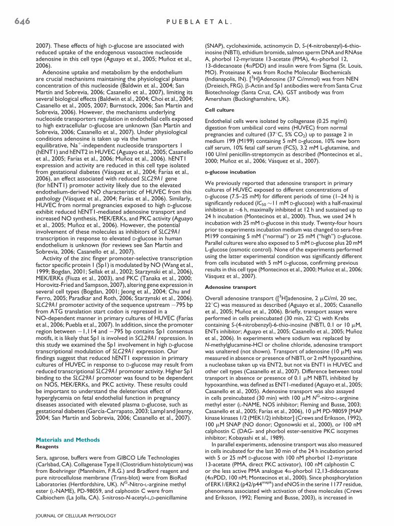
By using the web-based computer softwares MatInspector(�Genomatix Software, GmbH, http://www.genomatix.de/products/matinspector/matinspector2.html; Cartharius et al.,2005) and MOTIF (http://motif.genome.jp), with a cut-offscore¼ 80, the Sp1 response element aggggGGAGggggaa (�815/�801 from ATG) was identified in the region between �1,114 and�795 bp of the SLC29A1 promoter (Fig. 1). After fixing confluentcultures with 1% paraformaldehyde (10 min, 228C) in PBS, cellswere incubated (5 min, 228C) with 125 mM glycine, rinsed withice-cold PBS (48C), scraped and centrifuged (3,000 rpm, 3 min,48C). The pellet was resuspended in 1 ml cell lysis buffer (5 mMHEPES pH 8.0, 85 mM KCl, 0.5% Triton X-100, 1 mM PMSF),incubated (20 min, 48C) and centrifuged (3,000 rpm, 3 min, 48C).The obtained pellet was resuspended in nuclear lysis buffer (50 mMTris–Cl, pH 8.0, 10 mM EDTA, 1% SDS) containing proteaseinhibitors (1 mM leupeptin, 1 mM PMSF, 1 mg/ml aprotinin),incubated (20 min, 48C) and sonicated (20�, 10-sec pulse; MicroUltrasonic Cell Disrupter, KONTES, Haverhill, MA). Suspensionwas then centrifuged (14,000 rpm, 20 min, 48C) and supernatant(nuclear fraction) was collected (100 ml aliquots of this fractionwere saved as input).
Fig. 1. Putative Sp1 element in the 5-untranslated region of the human ENAF495730) isnumbered from the�795 to�1,114bp from theATG translat�801 from ATG of the SLC29A1 promoter was identified using the web-basand MOTIF (http://motif.genome.jp) with a cut-off score of 80, and is indicafragment of the promoter are not shown for clarity.
JOURNAL OF CELLULAR PHYSIOLOGY
Aliquots of the nuclear fractions containing 6 mg of chromatinwere incubated (1 h, 48C, gently shaking) with 20 ml proteinG-agarose beads previously washed (3�, 10 min, 48C) with 1 mlof ChIP dilution buffer (0.01% SDS, 1.1% Triton X-100, 1.2 mMEDTA, 16.7 mM Tris–HCl pH 8.0, 167 mM NaCl, and proteaseinhibitors cocktail as above) and incubated (48C, 12 h, gentlyshaking) with 20 ml of salmon sperm DNA (ssDNA) solution(10 mg/ml). The mix was then centrifuged (3,000 rpm, 3 min) andthe supernatant corresponding to precleared nuclear fractions wascollected. Aliquots of this fraction were diluted to 1 ml in ChIPdilution buffer and incubated (12 h, 48C) with anti-Sp1 (2 mg/ml)antibody under gently shaking. After this incubation period,aliquots were transferred into a tube containing 20 ml proteinG-agarose beads washed (3�, 10 min, 48C) with 1 ml of ChIPdilution buffer and subjected to an initial incubation (3 h, 48C) withBSA (1%) followed by a second incubation (12 h, 48C) with 20 mlof ssDNA solution (10 mg/ml). The mix was further incubatedfor 4 h (DNA samples without antibody were used as G-agarosedrag-non-specific control).
The immunocomplex (protein G-agarose beads, anti-Sp1antibody, Sp1 protein, and chromatin) was centrifuged (3,000 rpm,3 min, 48C), and the pellets were consecutively rinsed (1�, 1 ml,10 min, 228C) with ChIP dilution buffer, dialysis buffer (2 mMEDTA, 50 mM Tris–HCl pH 8.0, 0.2% N-laurylsarcosine sodiumsalt), TSE-500 buffer (500 mM NaCl, 20 mM Tris–HCl pH 8.0, 2 mMEDTA, 1% Triton X-100, 0.1% SDS), and LiCl detergent buffer(500 mM LiCl, 100 mM Tris–HCl pH 8.0, 1% Triton X-100, 1%deoxycholic acid), followed by two further rinses with TE buffer(10 mM Tris–HCl pH 8.0, 1 mM EDTA). Chromatin was elutedfrom the immunocomplex by centrifugation (supernatant fraction,3,000 rpm, 3 min, 228C) after incubation (2�, 15 min, 228C) of thepellets with 150 ml of elution buffer (50 mM NaHCO3, 1% SDS).Eluted (precipitated Sp1-DNA complex) and input (total sonicatedDNA) DNA was obtained by reversing the initialparaformaldehyde reaction by incubation (overnight, 658C) in330 mM NaCl followed by precipitation with isopropanol/glycogen. The resulting pellet was incubated (1 h, 658C) inproteinase K buffer (50 mM Tris–HCl, pH 7.5, 25 mM EDTA, 1.25%SDS, 10 mg proteinase K) and the DNA was purified using phenol/chloroform (1:1 v/v), precipitated with isopropanol/glycogenand then treated with RNAse A (10 mg/ml, 1 h, 378C). Aliquots(1 ml, 1:10 dilution) of DNA (input and precipitate) were usedfor PCR assays. Cycling parameters for amplifications (30 cycles)were 958C for 30 sec, 578C for 30 sec, and 728C for 30 sec.Oligonucleotide primers were: Sp1-hENT-1 promoter-sense
T1 gene SLC29A1. The nucleotide sequence (GenBank Accession No.ion start codon.The putative Sp1responseelement between�815 anded computer softwares MatInspector (�Genomatix Software, GmbH)ted in bold face. Other putative transcription factors elements for this

S p 1 - D E P E N D E N T G L U C O S E I N H I B I T I O N O F h E N T 1 649
50-ATCCACCCCTCCAATCTTCT-30 and Sp1-hENT-1promoter-anti-sense 50-GCAACCCAGATTCCAGTCTC-30
(product size 249 bp).
Statistical analysis
Values are mean� SEM, where n indicates number of different cellcultures (4–8 replicates). Comparisons between two and moregroups were performed by means of Student’s unpaired t-test andanalysis of variance (ANOVA), respectively. If the ANOVAdemonstrated a significant interaction between variables, post hocanalyses were performed by the multiple-comparison Bonferronicorrection test. Data are expressed as mean� SEM, with P< 0.05being considered statistically significant.
ResultsAdenosine transport
Overall adenosine transport was lower (60� 12%, P< 0.04,n¼ 12) in cells exposed to high compared with normalD-glucose, and was reduced to the same extent by 1mM NBTI incells in high D-glucose (67� 16%, P< 0.04, n¼ 12) comparedwith cells in normal D-glucose (not shown; Aguayo et al., 2005;Munoz et al., 2006). Inhibition of ENT1-mediated adenosineuptake by high D-glucose was blocked in cells either incubatedwith L-NAME, PD-98059, or calphostin C alone or incombination (Fig. 2A). Furthermore, L-NAME and PD-98059increased ENT1-mediated adenosine transport over the basalvalues determined in normal D-glucose. However, basalENT1-mediated adenosine transport was unaltered bycalphostin C in cells in normal D-glucose, but was increased in
Fig. 2. Effect of D-glucose and different inhibitors on hENT1-mediated adetransport (10mM,20sec,22-C)inHUVECculturedfor24hin5(&)or25(&)methylester(L-NAME,100mM),PD-98059(10mM),and/orcalphostinC(10fromcellstreatedasinA.28SmRNAwasinternalreference.MP < 0.05versusC. yP < 0.05 versus all other values. Mean W SEM (n U 8–18).
JOURNAL OF CELLULAR PHYSIOLOGY
high D-glucose up to values in normal D-glucose. Cells exposedto PMA, but not to PMA plus calphostin C or to the lessactive PMA analogue 4aPDD, or insulin exhibit reducedENT1-mediated adenosine transport (Table 1). PD-98059 andL-NAME blocked the effect of insulin on transport, and actuallyled to an enhancement of transport over the values determinedin normal D-glucose in absence of insulin (Table 1).
hENT1 expression and stability
High D-glucose reduced the hENT1 mRNA number of copies(Fig. 2B), an effect reversed by L-NAME, PD-98059, andcalphostin C. Basal hENT1 mRNA expression was increasedover the basal mRNA expression detected in normal D-glucoseby L-NAME and PD-98059, but was unaltered by calphostin C.In cells co-incubated with a combination of these inhibitors,hENT1 mRNA expression was higher than controls, but similarto the expression detected in cells incubated with L-NAME orPD-98059 alone.
High D-glucose also reduced hENT1 protein abundance inHUVEC (Fig. 3). High D-glucose effect was reversed andincreased over the basal protein abundance detected in normalD-glucose by L-NAME, PD-98059, or calphostin C in normal orhigh D-glucose. In addition, changes in hENT1 proteinabundance induced by these inhibitors alone were unaltered incells co-incubated with a combination of these inhibitors(Fig. 3). As for adenosine transport, PMA, but not 4aPDD,and insulin reduced hENT1 protein abundance and mRNAexpression in cells in normal D-glucose. Insulin and PMA effectswere blocked by PD-98059 and L-NAME, and calphostin C,
nosine transport and hENT1 mRNA expression. A: hENT1-adenosinemM D-glucose, inabsence(Control)orpresenceofNG-nitro-L-arginine
0nM).B:Real-timeRT-PCRforhENT1mRNA(numberofcopies T 104)valuesin5or25mMD-glucose,exceptincells incubatedwithcalphostin

TABLE 1. Effect of PMA, 4aPDD, calphostin C, and insulin on hENT1 expression and activity
Adenosine transport(pmol/mg protein/sec)
hENT1 mRNA(hENT1/28S mRNA number of copies� 104)
hENT1 protein(hENT1/b-actin arbitrary units)
5 mM D-glucoseControl 0.14� 0.02 6.1� 0.7 1.0� 0.1PMA 0.03� 0.01 1.5� 0.3 0.3� 0.1
PMAþ calphostin C 0.13� 0.02 6.5� 0.6 1.1� 0.24aPDD 0.16� 0.04 5.7� 0.7 1.2� 0.1Insulin 0.06� 0.01 3.5� 0.6 0.5� 0.2
Insulinþ PD-98059 0.26� 0.02, 8.4� 0.6, 1.6� 0.1,
Insulinþ L-NAME 0.27� 0.03, 8.2� 0.5, 1.8� 0.2,
25 mM D-glucoseControl 0.05� 0.01 2.9� 0.2 0.3� 0.1
PMA 0.04� 0.02 2.8� 0.3 0.3� 0.2
PMAþ calphostin C 0.12� 0.03 5.8� 0.5 1.2� 0.3
4aPDD 0.06� 0.01 2.6� 0.3 0.4� 0.2
Insulin 0.04� 0.02 1.9� 0.5 0.2� 0.1
Insulinþ PD-98059 0.23� 0.04,, 7.4� 0.3,, 1.8� 0.2,,
Insulinþ L-NAME 0.22� 0.04,, 7.6� 0.4,, 1.8� 0.2,,
Human umbilical vein endothelial cells (HUVEC) were cultured for 24 h in 5 or 25 mM D-glucose and adenosine transport (10 mM, [3H]adenosine, 2 mCi/ml, 20 sec, 228C) was determined, andtotal protein and mRNA extractions were performed as described in Materials and Methods Section. Cells were incubated for the last 30 min of the 24 h incubation period with 5 or 25 mMD-glucose in absence (Control) or presence of phorbol 12-myristate 13-acetate (PMA, 100 nM), PMAþ calphostin C (100 nM) or 4a-phorbol 12,13-didecanoate (4aPDD, 100 nM). Cells werealso incubated for the last 8 h of the 24 h incubation period with 5 or 25 mM D-glucose in the presence of insulin (1 nM), insulinþNG-nitro-L-arginine methyl ester (L-NAME, 100 mM), orinsulinþ PD-98059 (10 mM) as previously reported (Montecinos et al., 2000). hENT1 protein abundance was determined by Western blot and hENT1 mRNA level by quantitative real-timeRT-PCR as described in Materials and Methods Section. Mean� SEM (n¼ 5).P< 0.05 versus corresponding Control values in 5 mM D-glucose.P< 0.05 versus corresponding values for Insulin in 5 or 25 mM D-glucose.P< 0.05 versus corresponding Control values in 25 mM D-glucose.
650 P U E B L A E T A L .
respectively (Table 1). However, insulin did not significantlyalter hENT1 protein abundance and mRNA expression in highD-glucose.
The 50% of the time at which a maximal reduction of thehENT1 mRNA number of copies (7.4� 1.9 h; Fig. 4A) orprotein abundance (5.3� 1.1 h; Fig. 4B) by high D-glucose wasseen in this study were not significantly different (P< 0.05)from the time detected in cells in normal D-glucose (7.3� 1.5 or5.9� 1.2 h, respectively). hENT1 mRNA level decayed similarly(P> 0.05) in the presence of actinomycin D in normal or highD-glucose. These changes in mRNA and protein were notsignificantly (P> 0.05) altered by L-NAME, calphostin C or
Fig. 3. Effectof D-glucoseanddifferent inhibitors onhENT1protein abund12 similar experiments) in HUVEC cultured for 24 h in 5 or 25 mM D-gluco(L-NAME,100mM),PD-98059(10mM)and/orcalphostinC(100nM).B:hENnormalized to 1 in 5 mM D-glucose. MP < 0.04 and yP < 0.05 versus all other
JOURNAL OF CELLULAR PHYSIOLOGY
PD-98059 alone (Fig. 4) or in combination (not shown). Similarresults were found for hENT1 protein abundance decay in thepresence of cycloheximide (Fig. 4).
SLC29A1 promoter transcriptional activity
The reporter luciferase activity in cells transfected withpGL3-hENT1�1114 construct that were exposed to highD-glucose was lower compared with cells transfected with thisconstruct and exposed to normal D-glucose (Fig. 5A). Equally,reporter activity was also lower in these cells compared withcells transfected with pGL3-hENT1�795 construct. However,
ance. A:Westernblots forhENT1andb-actin (representativeofotherse, in absence (�) or presence (R) of NG-nitro-L-arginine methyl esterT1/b-actinratiodensitometries incells in5(&)or25(&)mM D-glucose,values. Mean W SEM (n U 12).

Fig. 4. hENT1 mRNA and protein level and stability. A: RT-PCRfor hENT1 and 28S mRNA (representative of other eight similarexperiments) in HUVEC cultured for 24 h in sera-free culturemedium containing 5 mM D-glucose. mRNA was obtained fromcultures co-incubated with 5 or 25 mM D-glucose in absence (�) orpresence (R) of 1.5 mM actinomycin D (Act D) for the indicatedperiods of time (see Materials and Methods Section). Lower part:hENT1 mRNA densitometries normalized to 1 corresponding to cellsin 5 or 25 mM D-glucose in absence of actinomycin D (time 0).*, 5 mMD-glucose; *, 5 mM D-glucose R actinomycin D; &, 25 mM D-glucose;&, 25 mM D-glucose R actinomycin D. B: Western blot for hENT1 andb-actin protein (representative of other 11 similar experiments)in HUVEC cultured as in A. Protein was obtained from culturesco-incubated with 5 or 25 mM D-glucose in absence (�) or presence(R) of 1 mM cycloheximide (CLX). Lower part: hENT1 proteindensitometries normalized to 1 corresponding to cells in 5 or 25 mMD-glucose in absence of actinomycin D (time 0). *, 5 mM D-glucose;*, 5 mM D-glucose R cycloheximide; &, 25 mM D-glucose; &, 25 mMD-glucose R cycloheximide. Mean W SEM (n U 8–11).
JOURNAL OF CELLULAR PHYSIOLOGY
S p 1 - D E P E N D E N T G L U C O S E I N H I B I T I O N O F h E N T 1 651
the reporter activity in cells transfected with pGL3-hENT1�795
construct that were exposed to high D-glucose was highercompared with cells transfected with this construct andexposed to normal D-glucose. None of the constructssignificantly altered the reporter activity in cells in normalD-glucose. High D-glucose-reduced reporter activity inpGL3-hENT1�1114 transfected cells was reversed by L-NAME,PD-98059, and calphostin C (Fig. 5B). However, only L-NAMEincreased reporter activity in cells in high D-glucose over thebasal value detected in cells in normal D-glucose.
Sp1 expression and DNA binding
Sp1 protein abundance in the nucleus fraction was increased byhigh D-glucose, an effect reversed by L-NAME, PD-98059, orcalphostin C (Fig. 6A). However, Sp1 protein abundance inwhole cell extracts was unaltered by high D-glucose in absenceor presence of these inhibitors. Parallel experiments show thatSp1 binding to DNA was higher in cells exposed to highcompared with normal D-glucose, an effect blocked by L-NAME,PD-98059, or calphostin C (Fig. 6B). However, only L-NAMEreduced the Sp1 binding below the basal binding detected innormal D-glucose in absence of this inhibitor.
Sp1 native (Sp1) and Sp1 dominant negative(Sp1DN) overexpression
Sp1 protein abundance was higher in cells transfected withpCGN-Sp1 compared with non-transfected cells or cellstransfected with the empty pCGN vector (Fig. 7A).Reporter luciferase activity in HUVEC transfected with thepGL3-hENT1�1114 construct was reduced after transfection ofcells in normal D-glucose with pCGN-Sp1, but unaltered whentransfected with the empty pCGN vector (Fig. 7B). HighD-glucose inhibition of reporter luciferase activity innon-transfected or transfected cells with the empty pCGNvector was unaltered after Sp1 overexpression, but wasreversed to values in normal D-glucose in cells transfectedwith pEBG-Sp1DN containing the Sp1 dominant negative(Fig. 7B). The reduced reporter activity in cells in normalD-glucose transfected with pCGN-Sp1 compared with cellstransfected with the empty pCGN vector, was unaltered byL-NAME or the NO donor, SNAP (Fig. 7C). However, highD-glucose inhibition of reporter activity in cells transfectedwith the empty pCGN vector was unaltered by transfectionwith pCGN-Sp1. The reduced reporter activity detected incells in high D-glucose transfected with pCGN-Sp1, wasblocked by L-NAME, and further reduced by SNAP.
Discussion
This study characterized the effect of elevated extracellularD-glucose on SLC29A1 gene promoter (hENT1) activity andthe involvement of the zinc finger, promoter-selectivetranscription factor Sp1 in this phenomenon in primary culturesof human umbilical vein endothelial cells (HUVEC). We initiallyconfirmed previous observations in HUVEC showing thathENT1-mediated adenosine transport and hENT1 mRNAexpression and protein abundance were reduced in cellsexposed to 25 mM (‘‘high’’) compared with 5 mM (‘‘normal’’)D-glucose. Extending these observations, high D-glucosereduced SLC29A1 promoter activity, a phenomenon dependenton PKC, NOS, and MEK/ERKs activity. This effect of highD-glucose was associated with higher nuclear Sp1 proteinabundance and Sp1 binding to SLC29A1 promoter. Activity ofPKC, NOS and MEK/ERKs are required for Sp1 binding toDNA, SLC29A1 promoter activity and hENT1 expression andactivity in HUVEC cultured in either normal or high D-glucose.

Fig. 5. Role of D-glucose and different inhibitors in SLC29A1 (hENT1) promoter activity. A: Luciferase (Luc) reporter constructs containing serialSLC29A1 promoter truncations (pGL3-hENT1�795 and pGL3-hENT1�1114) were transfected in HUVEC cultured for 24 h in 5 or 25 mM D-glucose,along with Renilla reporter plasmid, and assayed for Firefly and Renilla luciferase activity. Cells were also transfected with the empty pGL3-Basicvector or pGL3-Control vector (SV40 pGL3) as negative or positive controls, respectively. MP < 0.05, yP < 0.04. B: Firefly/Renilla luciferase activityratio in cells transfected with the reporter construct pGL3-hENT1�1114 as in A. After 36 h of transfection, cells were exposed to M199 in absence(�, Control) or presence (R) of NG-nitro-L-arginine methyl ester (L-NAME, 100mM), PD-98059 (10mM), or calphostin C (100 nM). M and yP < 0.05versus Control in 5 and 25 mM D-glucose, respectively. Mean W SEM (n U 8).
652 P U E B L A E T A L .
D-glucose effect on adenosine transport
Uptake of the vasoactive endogenous nucleoside adenosine(Burnstock, 2006) is mainly mediated by the Naþ-independent,human equilibrative nucleoside transporter 1 (hENT1) and in aminor proportion by hENT2 under physiological conditions inprimary cultures of HUVEC (Aguayo et al., 2005; Casanelloet al., 2005; Farıas et al., 2006; Munoz et al., 2006). Our resultssuggest that NOS and the MEK/ERKs, but not PKC activity isrequired for tonic inhibition of basal or insulin-stimulatedhENT1-adenosine transport in HUVEC cultured in normalD-glucose. These findings agree with reports documenting ratENT1 (rENT1) down-regulation by NO in B lymphocytes(Soler et al., 2000), and high D-glucose inhibition ofrENT1-adenosine transport blocked by MEK/ERKs inhibition inrat B lymphocytes (Sakowicz et al., 2005). Since calphostin Cblocked high D-glucose inhibition of hENT1-mediatedadenosine transport, it is likely that PKC activity was requiredfor this phenomenon. This finding agrees with results inimmortalized murine cardiomyocytes HL-1 cells where hypoxiadown-regulation of ENT1 transport activity was PKCdependent (Chaudary et al., 2004). However, involvement ofPKC activity seems not to be similar for different cell types,
JOURNAL OF CELLULAR PHYSIOLOGY
since it has also been reported that rENT1 activity was insteadstimulated by PKA and PKC-d/e in the rat heart myoblast cellline H9c2 (Leung et al., 2007).
L-NAME and PD-98059 reversed the high D-glucose inhibitedhENT1-adenosine transport and, furthermore, increasedtransport values up to those measured in normal D-glucose inthe presence of these inhibitors. Thus, removal of NO- or MEK/ERKs-inhibition of hENT1-adenosine transport could resulteither in a maximal increase of transport activity of a constantnumber of hENT1 proteins at the plasma membrane, a maximalincrease of hENT1 protein availability at the plasma membranewithout altering their maximal transport activity, or both(Deves and Boyd, 1998; Casanello et al., 2007). Since theseinhibitors also increased hENT1 protein abundance in highD-glucose, a lower hENT1-adenosine transport due to areduced number of plasma membrane sites for adenosinetransport is likely. This possibility agrees with the reportedhigh D-glucose induced reduction of the number of sites foradenosine transport from �1.4 to �0.7� 106 per cell inHUVEC consistent with a lower maximal transport velocity(Vmax) for adenosine and maximal specific binding (Bmax) of[3H]NBTI previously reported in this cell type (Aguayo et al.,2005; Munoz et al., 2006). Since calphostin C reversed the high

Fig. 6. Effect of D-glucose and different inhibitors on Sp1 expressionand DNA binding. A: Western blot for Sp1 abundance in total ornucleus protein fractions and b-actin (representative of other 12similar experiments) in HUVEC cultured for 24 h in 5 or 25 mMD-glucose, in absence (�) or presence (R) of NG-nitro-L-argininemethyl ester (L-NAME, 100 mM), PD-98059 (10 mM), or calphostin C(100 nM). Lower part: Sp1 nucleus/Sp1 total protein fractions ratiodensitometries in cells in 5 (&) or 25 (&) mM D-glucose, normalizedto 1 in 5 mM D-glucose. MP < 0.05 versus all other values (n U 12).B: Chromatin immunoprecipitation assay for Sp1 binding to DNA(representative of other eight similar experiments) in HUVEC as in A.The mRNA flanking Sp1 consensus sequence in SLC29A1 promoterregion (�815/�801 bp from ATG) was amplified by RT-PCR (seeMaterials and Methods Section). Input is total DNA. Lower part:Sp1 consensus sequence (Sp1) over Input ratio densitometriesnormalized as in A. MP < 0.05 and yP < 0.04 versus Controls in 5 and25 mM D-glucose, respectively. Mean W SEM (n U 8).
Fig. 7. Role of Sp1 in SLC29A1 gene expression. HUVEC were co-transfected by electroporation with an empty pCGN vector (pCGN),theexpressionvectorcontaininghumannativeSp1(pCGN-Sp1)ortheSp1 dominant negative (pEBG-Sp1DN), and the SLC29A1 promoterpGL3-hENT1�1114 construct along with Renilla reporter plasmid, andthenculturedfor48h(seeMaterialsandMethodsSection).A:Westernblot for native Sp1 protein, the GST-tagged Sp1 dominant negative(Sp1DN), or b-actin protein (representative of other six similarexperiments) in not transfected (�) or transfected (R) HUVEC in5 mM D-glucose. Right part: Sp1/b-actin protein ratio densitometriesnormalized to 1 for values in not transfected cells. MP < 0.05 versus nottransfected or pCGN transfected cells (n U 6). B: Firefly and Renillaluciferase activity in not transfected (�) or transfected HUVEC as in Aexposed for the last 24 h of the 48 h culture period to culture mediumcontaining 5 (&) or 25 (&) mM D-glucose. Mand yP < 0.05 versus control(�) or pCGN in 5 and 25 mM D-glucose, respectively (n U 6). C: Fireflyand Renilla luciferase activity in HUVEC transfected with the emptyvector (pCGN, Control) or pCGN-Sp1 and cultured in absence(Control) or presence of NG-nitro-L-arginine methyl ester (L-NAME,100 mM) or S-nitroso-N-acetyl-L,D-penicillamine (SNAP, 100 mM).MP < 0.05 versus pCGN (Control) in 5 mM D-glucose. yP < 0.05 versuspCGN (Control) in 25 mM D-glucose and Control pCGN-Sp1transfected cells in 25 mM D-glucose. Mean W SEM (n U 6).
JOURNAL OF CELLULAR PHYSIOLOGY
S p 1 - D E P E N D E N T G L U C O S E I N H I B I T I O N O F h E N T 1 653
D-glucose reduced hENT1 protein abundance over the leveldetected in absence of this inhibitor, PKC activity seemsrequired for tonic reduction of hENT1 protein expression.D-glucose and inhibitors effect on hENT1 protein abundancewere paralleled by similar changes in the hENT1 mRNA numberof copies. Thus, reduced hENT1 protein abundance in highD-glucose may result at least from either lower hENT1 mRNAstability, reduced SLC29A1 transcription, or both, and suggestthat this phenomenon also requires PKC, NOS and/or MEK/ERKs activity. Since hENT1 protein and mRNA stability wasunaltered it is unlikely that this phenomenon was responsiblefor hENT1 reduced expression.

654 P U E B L A E T A L .
D-glucose effect on SLC29A1 expression
In a recent study we amplified a sequence spanning from�3,198bp to the ATG translation start codon of SLC29A1 usinggenomic DNA from primary cultures of HUVEC as template(Farıas et al., 2006). Our results show that Firefly luciferaseactivity in cells transfected with pGL3-hENT1�795 was elevatedin high D-glucose compared with normal D-glucose, suggestingthat the region spanning �795 bp from the ATG of SLC29A1promoter confers high D-glucose-associated stimulatoryactivity. On the contrary, cells transfected withpGL3-hENT1�1114 in high D-glucose exhibit lower Fireflyluciferase activity compared with cells in normal D-glucose or incells in high D-glucose transfected with pGL3-hENT1�795. Thisfinding suggests that the region spanning between �1,114 and�795 bp of SLC29A1 promoter confers high D-glucose-associated inhibitory activity of this gene. These results contrastwith our previous study suggesting that this promoter regionconfers stimulatory activity in HUVEC from gestational diabeticcompared with normal pregnancies (Farıas et al., 2006).However, the latter study was performed in HUVEC fromnormal or diabetic pregnancies cultured in normal D-glucosecompared with the present study where cells from normalpregnancies were exposed to normal versus high D-glucose.
Since Firefly luciferase activity was unaltered by the inhibitorsin cells transfected with pGL3-hENT1�1114 in normalD-glucose, PKC, NOS or MEK/ERKs activity may not berequired to sustain basal SLC29A1 promoter activity. On thecontrary, the reduced Firefly luciferase activity detected in cellsin high D-glucose transfected with this construct was blocked bythe inhibitors. Thus, PKC, NOS, and/or MEK/ERKs activity maybe involved in the inhibition of SLC29A1 expression by highD-glucose in HUVEC. Further experiments show that onlyL-NAME increased SLC29A1 reporter activity over the activitydetected in cells in normal D-glucose in absence of this inhibitor,suggesting that NO may act as repressor of SLC29A1 expressionin HUVEC exposed to high D-glucose. This is supported byprevious findings in HUVEC transduced with an adenovirusconstruct carrying siRNA targeting eNOS mRNA, where thereduced eNOS expression and activity was associated withhigher hENT1 protein abundance, mRNA level andhENT1-mediated adenosine uptake (Farıas et al., 2006). Thepossibility that eNOS expression and NO inhibition of hENT1transport activity in high D-glucose contrast with a reportshowing that hENT1 mRNA is unaltered by NG-monoethyl-L-arginine (NOS inhibitor) in human B-lymphocytes derived Rajicells (Soler et al., 2000), a finding that could suggest that NOmay not be required for modulation of hENT1 in this cell lineand that NO effect on ENT1 expression may be cell specific.
Involvement of Sp1
Several studies report that NO modulates transcription factorsincluding Sp1 (Wang et al., 2002; Jeong et al., 2004; Starzynskiet al., 2006) and NF-kB (Park et al., 1997; Park and Wei, 2003).Equally, PKC activators increased Sp1 protein abundance in calfpulmonary artery endothelial cells (Tanaka et al., 2000) andinsulin-increased expression of PKC-d is regulated by PKC-avia a mechanism requiring Sp1 activation in skeletal muscle L6cells (Horovitz-Fried and Sampson, 2007). In addition, Sp1nuclear translocation is associated with transient ERKsphosphorylation in the human microvascular endothelial cellline HMEC-1 (Fiuza et al., 2003). Since HUVEC exposed to highD-glucose exhibit increased NO synthesis (Munoz et al., 2006;Vasquez et al., 2007), higher PKC activity (Montecinos et al.,2000; Vasquez et al., 2004) and activation of ERK1/ERK2(Vasquez et al., 2007), we studied whether Sp1 proteinabundance and its binding to a Sp1 consensus sequence in theSLC29A1 promoter involve activity of these signaling molecules.Our results show that high D-glucose increased nuclear Sp1, but
JOURNAL OF CELLULAR PHYSIOLOGY
did not alter total Sp1 protein abundance in HUVEC. D-glucoseeffect was blocked by L-NAME, PD-98059, and calphostin C,suggesting the involvement of NOS, MEK/ERKs, and PKCactivity in this phenomenon. However, since nuclear and totalSp1 protein abundance was unaltered by these inhibitors in cellsin normal D-glucose, an environment high in D-glucose seemsrequired to trigger these alterations in Sp1 distribution andsignaling pathways in HUVEC. This possibility agrees withfindings showing that reduced apoA-1 mRNA expression byhigh D-glucose (�22 mM, 24–48 h) in HepG2 cells is blocked byactivation of an Sp1 response element in the apoA-1 genepromoter (Murao et al., 1998). Thus, Sp1 seems to be a keymodulator of gene expression in cells exposed to anenvironment with elevated extracellular level of D-glucose orwhere activity of NOS, MEK/ERKs, or PKC is altered (Chu andFerro, 2005; Casanello et al., 2007).
ChIP assays show that Sp1 binding to the consensussequence ‘‘agggGGGAgggggaa’’ (�815/�801 bp) identifiedbetween �1,114 and �795 bp from ATG of the SLC29A1promoter, was increased by high D-glucose and blocked byL-NAME, PD-98059, and calphostin C. Therefore, activitiesof NOS, MEK/ERKs, and PKC seem also required for theformation of Sp1-DNA complexes in the SLC29A1 promoter.In addition, even when L-NAME reduced nuclear Sp1 proteinabundance to values in absence of this inhibitor in cells in normalD-glucose, L-NAME reduced the Sp1 binding to SLC29A1promoter below the basal binding detected in absence of thisinhibitor in normal or high D-glucose. Thus, it is likely that Sp1binding to SLC29A1 promoter requires at least basal NOSactivity in this cell type. This finding has not been documented inendothelium, but NO decreases Sp1 binding to DNA in genepromoters in other cell types (Wang et al., 2002; Jeong et al.,2004; Starzynski et al., 2006). Interestingly, reduced Sp1 bindingto the tumor necrosis factor a (TNF-a) promoter is associatedwith increased gene transcription in response to NO in humanU937 cells (Wang et al., 1999); however, our results suggestthat NO may increase Sp1 binding to SCL29A1 promoterrepressing this gene transcription in primary cultures ofHUVEC. Thus, NO may act as a differential modulator of Sp1binding to DNA depending on the gene promoter (Chu andFerro, 2005).
To directly address the potential role of Sp1 as modulator ofSLC29A1 transcription in response to high D-glucose and NO,we overexpressed native Sp1 and an Sp1 dominant negative(transcriptionally ineffective, Sp1DN) and assayed SLC29A1reporter activity. These experiments show that Firefly luciferaseactivity in cells transfected with pGL3-hENT1�1114 constructwas reduced when native Sp1 was overexpressed, but it wasunaltered when the Sp1DN was overexpressed in normalD-glucose. Since the Sp1DN contains an intact binding domainto DNA, but lacks of the sequence for a transcriptionally activedomain (Al-Sarraj et al., 2005), it is likely that Sp1 binding toSLC29A1 promoter was not enough to repress this geneexpression, but, instead, its transcriptionally active domain isrequired in HUVEC. These results also suggest that repressionof SLC29A1 promoter activity by high D-glucose could bemaximal since Sp1 overexpression was not associated withfurther reduction of SLC29A1 promoter activity in highD-glucose. Interestingly, L-NAME did reverse, but SNAP (a NOdonor) further increased the repressive effect of high D-glucoseon SLC29A1 promoter activity in cells overexpressing nativeSp1. Thus, SLC29A1 promoter is conferred responsiveness toNO via Sp1 in cells exposed to an environment with abnormallyelevated D-glucose. This result agrees with reports showingNO responsiveness achieved after insertion of Sp1 sites into aminimal cytomegalovirus promoter (Wang et al., 1999),reduction in the Sp1-DNA complex formation induced by NOin the RAW 264.7 mouse macrophages iron regulatory protein1 promoter (Starzynski et al., 2006), or NO down-regulated

S p 1 - D E P E N D E N T G L U C O S E I N H I B I T I O N O F h E N T 1 655
transcription of SRG3, the murine homolog of BAF155 inhumans, via inactivation of Sp1 DNA-binding activity to SRG3promoter in primary rat thymocytes and 16610D9 thymomacells (Jeong et al., 2004). Since maximal inhibition of Sp1 bindingby L-NAME was similar in HUVEC in normal or high D-glucose, itis feasible that SLC29A1 promoter activity in high D-glucose ispreferentially modulated by a molecule different to NO, suchas PKC, known to play a key role in the modulation ofhENT1-adenosine transport as well as in activating eNOS andMEK/ERKs signaling pathways in this cell type from normalpregnancies in response to high D-glucose (Montecinos et al.,2000).
In summary, we have found that elevated extracellularD-glucose reduces hENT1 adenosine transport due to lowertranscription of the human SLC29A1 gene (hENT1) in primarycultures of HUVEC. Transcriptional SLC29A1 repression byhigh D-glucose involves increased Sp1 binding to a consensussequence identified between �815 and �801 bp from ATG ofSLC29A1 promoter. hENT1-mediated adenosine transport, Sp1binding and SLC29A1 transcription was dependent on NOS,MEK/ERKs, and PKC activity, and Sp1 activation conferredresponsiveness to high D-glucose via NO, a repressor ofSLC29A1 promoter transcriptional activity, in HUVEC. It islikely that incubation of HUVEC with D-glucose triggers cellsignaling reactions involving activation of PKC, NOS, and MEK/ERK, perhaps in this sequence as previously described foroverall ENTs-mediated adenosine transport in HUVEC(Montecinos et al., 2000). However, further studies arenecessary for a clear characterization of the sequence of eventsaccounting for modulation of hENT1 expression and activityregarding these cell signaling molecules in primary cultures ofHUVEC exposed to high D-glucose. Reduced SLC29A1expression is a phenomenon perceived as reduced hENT1protein abundance and activity, explaining, at least in part, thereported increased extracellular adenosine concentrationdetected in the culture medium of HUVEC exposed to highD-glucose (Munoz et al., 2006), in HUVEC from gestationaldiabetes (Vasquez et al., 2004; Farıas et al., 2006) or in HUVECcultured under a low oxygen environment (Casanello et al.,2005). These results could be important to understand themechanisms by which adenosine-modulated placenta-to-fetusblood flux is altered in pathologies of pregnancy where plasmaD-glucose could be enhanced, such as gestational diabetes(Yoneyama et al., 2002; Lampl and Jeanty, 2004; San Martın andSobrevia, 2006; Casanello et al., 2007).
Acknowledgments
This study was supported by Fondo Nacional de Ciencia yTecnologıa (FONDECYT 1070865, 7070249) Chile, ComisionNacional de Investigacion Cientıfica y Tecnologica (CONICYT23070213) Chile, Vice-Rectorıa de Asuntos Academicos eInvestigacion (VRAID), Pontificia Universidad Catolica de Chile(BM16/2007, BM14/2007), and Agencia Espanola deCooperacion Internacional (AECI A/5484/06, SAF2005-01259)Spain. C. Puebla holds a VRAID-PhD fellowship. M. Farıas holdsSchool of Medicine and CONICYT-PhD fellowships. M.Gonzalez holds CONICYT-PhD fellowship. Authors thank J.Acurio for excellent technical assistance, and the personnel ofHospital Clınico Pontificia Universidad Catolica de Chile laborward for supply of umbilical cords.
Literature Cited
Aguayo C, Casado J, Gonzalez M, Pearson JD, San Martın R, Casanello P, Pastor-Anglada M,Sobrevia L. 2005. Equilibrative nucleoside transporter 2 is expressed in human umbilicalvein endothelium, but is not involved in the inhibition of adenosine transport induced byhyperglycaemia. Placenta 26:641–653.
Al-Sarraj A, Day RM, Thiel G. 2005. Specificity of transcriptional regulation by the zinc fingertranscription factors Sp1, Sp3, and Egr-1. J Cell Biochem 94:153–167.
JOURNAL OF CELLULAR PHYSIOLOGY
Baldwin SA, Beal PR, Yao SY, King AE, Cass CE, Young JD. 2004. The equilibrative nucleosidetransporter family, S LC29. Pflugers Arch 447:735–743.
Beckman JA, Goldfine AB, Gordon MB, Garrett LA, Creager MA. 2002. Inhibition of proteinkinase C b prevents impaired endothelium-dependent vasodilation caused byhyperglycemia in humans. Circ Res 90:107–111.
Bogdan C. 2001. Nitric oxide and the regulation of gene expression. Trends Cell Biol 11:66–75.
Burnstock G. 2006. Vessel tone and remodeling. Nat Med 12:16–17.Cartharius K, Frech K, Grote K, Klocke B, Haltmeier M, Klingenhoff A, Frisch M, Bayerlein M,
Werner T. 2005. MatInspector and beyond: Promoter analysis based on transcriptionfactor binding sites. Bioinformatics 21:2933–2942.
Casanello P, Torres A, Sanhueza F, Gonzalez M, Farıas M, Gallardo V, Pastor-Anglada M, SanMartın R, Sobrevia L. 2005. Equilibrative nucleoside transporter 1 expression isdownregulated by hypoxia in human umbilical vein endothelium. Circ Res 97:16–24.
Casanello P, Escudero C, Sobrevia L. 2007. Equilibrative nucleoside (ENTs) and cationicamino acid (CATs) transporters: Implications in foetal endothelial dysfunction in humanpregnancy diseases. Curr Vasc Pharmacol 5:1–16.
Chaudary N, Naydenova Z, Shuralyova I, Coe IR. 2004. Hypoxia regulates the adenosinetransporter, mENT1, in the murine cardiomyocyte cell line, HL-1. Cardiovasc Res 61:780–788.
Choi DS, Cascini MG, Mailliard W, Young H, Paredes P, McMahon T, Diamond I, Bonci A,Messing RO. 2004. The type 1 equilibrative nucleoside transporter regulates ethanolintoxication and preference. Nat Neurosci 7:855–861.
Chu S, Ferro TJ. 2005. Sp1: Regulation of gene expression by phosphorylation. Gene348:1–11.
Crews CM, Eriksson RL. 1992. Purification of a murine protein-tyrosine/threonine kinasethat phosphorylates and activates the Erk-1 gene product: Relationship to the fission yeastbyr1 gene product. Proc Natl Acad Sci USA 89:8205–8209.
Deves R, Boyd CA. 1998. Transporters for cationic amino acids in animal cells: Discovery,structure, and function. Physiol Rev 78:487–545.
Dignam JD, Lebovitz RM, Roeder RG. 1983. Accurate transcription initiation by RNApolymerase II in a soluble extract from isolated mammalian nuclei. Nucleic Acids Res11:1475–1489.
Farıas M, San Martın R, Puebla C, Pearson J, Casado J, Pastor-Anglada M, Casanello P,Sobrevia L. 2006. Nitric oxide reduces adenosine transport ENT1 gene (SLC29A1)promoter activity in human fetal endothelium from gestational diabetes. J Cell Physiol208:451–460.
Farre X, Guillen-Gomez E, Sanchez L, Hardisson D, Plaza Y, Lloberas J, Casado FJ, Palacios J,Pastor-Anglada M. 2004. Expression of the nucleoside-derived drug transporters hCNT1,hENT1 and hENT2 in gynecologic tumors. Int J Cancer 112:959–966.
Fiuza C, Bustin M, Talwar S, Tropea M, Gerstenberger E, Shelhamer JH, Suffredini AF. 2003.Inflammation-promoting activity of HMGB1 on human microvascular endothelial cells.Blood 101:2652–2660.
Fleming I, Busse R. 2003. Molecular mechanisms involved in the regulation of the endothelialnitric oxide synthase. Am J Physiol 284:R1–R12.
Garcıa-Carrapato MR. 2003. The offspring of gestational diabetes. J Perinat Med 31:5–11.Garfinkel S, Brown S, Wessendorf JH, Maciag T. 1994. Post-transcriptional regulation of
interleukin 1 alpha in various strains of young and senescent human umbilical veinendothelial cells. Proc Natl Acad Sci USA 91:1559–1563.
Gonzalez M, Flores C, Pearson JD, Casanello P, Sobrevia L. 2004. Cell signalling-mediatinginsulin increase of mRNA expression for cationic amino acid transporters-1 and -2and membrane hyperpolarization in human umbilical vein endothelial cells. Eur JPhysiol-Pflugers Arch 448:383–394.
Horovitz-Fried M, Sampson SR. 2007. Involvement of PKCalpha in insulin-induced PKCdeltaexpression: Importance of SP-1 and NFkappaB transcription factors. Biochem Biophys ResCommun 352:78–83.
Jeong SM, Lee KY, Shin D, Chung H, Jeon SH, Seong RH. 2004. Nitric oxide inhibitsglucocorticoid-induced apoptosis of thymocytes by repressing the SRG3 expression. J BiolChem 279:34373–34379.
Kobayashi E, Ando K, Nakano H, Iida T, Ohno H, Morimoto M, Tamaoki T. 1989.Calphostins (UCN-1028), novel and specific inhibitors of protein kinase C.I. Fermentation,isolation, physico-chemical properties and biological activities. J Antibiot 42:1470–1474.
Lampl M, Jeanty P. 2004. Exposure to maternal diabetes is associated with altered fetal growthpatterns: A hypothesis regarding metabolic allocation to growth under hyperglycemic-hypoxemic conditions. Am J Hum Biol 16:237–263.
Leung GP, Tse CM, Man RY. 2007. Characterization of adenosine transport in H9c2cardiomyoblasts. Int J Cardiol 116:186–193.
Lott ME, Hogeman C, Herr M, Gabbay R, Sinoway LI. 2007. Effects of an oral glucosetolerance test on the myogenic response in healthy individuals. Am J Physiol 292:H304–H310.
Montecinos VP, Aguayo C, Flores C, Wyatt AW, Pearson J, Mann G, Sobrevia L. 2000.Regulation of adenosine transport by D-glucose in human fetal endothelial cells:Involvement of nitric oxide, protein kinase C and mitogen-activated protein kinase.J Physiol 529:777–790.
Munoz G, San Martın R, Farıas M, Cea L, Casanello P, Sobrevia L. 2006. Insulin restoresglucose-inhibition of adenosine transport by increasing the expression and activity of theequilibrative nucleoside transporter 2 in human umbilical vein endothelium. J Cell Physiol209:826–835.
Murao K, Wada Y, Nakamura T, Taylor AH, Mooradian AD, Wong NC. 1998. Effects ofglucose and insulin on rat apolipoprotein A–I gene expression. J Biol Chem 273:18959–18965.
Ogonowski AA, Kaesemeyer WH, Jin L, Ganapathy V, Leibach FH, Caldwell RW. 2000.Effects of NO donors and synthase agonists on endothelial cell uptake of L-arg andsuperoxide production. Am J Physiol 278:C136–C143.
Oomen PHN, Kant GD, Dullaart RPF, Reitsma WD, Smit AJ. 2002. Acute hyperglycemiaand hyperinsulinemia enhance vasodilatation in type 1 diabetes mellitus withoutincreasing capillary permeability and inducing endothelial dysfunction. Microvasc Res63:1–9.
Paradkar PN, Roth JA. 2006. Nitric oxide transcriptionally down-regulates specificisoforms of divalent metal transporter (DMT1) via NF-kappaB. J Neurochem 96:1768–1777.
Park SW, Wei LN. 2003. Regulation of c-myc gene by nitric oxide via inactivating NF-kBcomplex in P19 mouse embryonal carcinoma cells. J Biol Chem 278:29776–29782.
Park SK, Lin HL, Murphy S. 1997. Nitric oxide regulates nitric oxide synthase-2 geneexpression by inhibiting NF-kB binding to DNA. Biochem J 332:609–613.
Parks CL, Shenk T. 1996. The serotonin 1a receptor gene contains a TATA-less promoterthat responds to MAZ and Sp1. J Biol Chem 271:4417–4430.

656 P U E B L A E T A L .
Puebla C, Farıas M, Vecchiola A, Casanello P, Sobrevia L. 2007. Transcriptional repression ofequilibrative nucleoside transporter 1 by D-glucose involves nitric oxide and Sp1 in humanfoetal endothelium. Placenta 28:A.24. (Abstract).
Sakowicz M, Szutowicz A, Pawelczyk T. 2005. Differential effect of insulin and elevatedglucose level on adenosine transport in rat B lymphocytes. Int Immunol 17:145–154.
San Martın R, Sobrevia L. 2006. Gestational diabetes and the adenosine/L-arginine/nitricoxide (ALANO) pathway in human umbilical vein endothelium. Placenta 27:1–10.
Sellak H, Yang X, Cao X, Cornwell T, Soff GA, Lincoln T. 2002. Sp1 transcription factor as amolecular target for nitric oxide– and cyclic nucleotide–mediated suppression of cGMP-dependent protein kinase-Ia expression in vascular smooth muscle cells. Circ Res 90:405–412.
Soler C, Felipe A, Casado FJ, Celada A, Pastor-Anglada M. 2000. Nitric oxide regulatesnucleoside transport in activated B lymphocytes. J Leukoc Biol 67:345–349.
Starzynski RR, Goncalves AS, Muzeau F, Tyrolczyk Z, Smuda E, Drapier JC, Beaumont C,Lipinski P. 2006. STAT5 proteins are involved in down-regulation of iron regulatoryprotein 1 gene expression by nitric oxide. Biochem J 400:367–375.
Strohm C, Barancik M, von Bruehl M, Strniskova M, Ullmann C, Zimmermann R, Schaper W.2002. Transcription inhibitor actinomycin-D abolishes the cardioprotective effect ofischemic reconditioning. Cardiovasc Res 55:602–618.
JOURNAL OF CELLULAR PHYSIOLOGY
Tanaka T, Kurabayashi M, Aihara Y, Ohyama Y, Nagai R. 2000. Inducible expression ofmanganese superoxide dismutase by phorbol 12-myristate 13-acetate is mediated by Sp1 inendothelial cells. Arterioscler Thromb Vasc Biol 20:392–401.
Vasquez G, Sanhueza F, Vasquez R, Gonzalez M, San Martın R, Casanello P, Sobrevia L. 2004.Role of adenosine transport in gestational diabetes-induced L-arginine transport and nitricoxide synthesis in human umbilical vein endothelium. J Physiol 560:111–122.
Vasquez R, Farıas M, Vega JL, San Martın R, Vecchiola A, Casanello P, Sobrevia L. 2007. D-glucose stimulation of L-arginine transport and nitric oxide synthesis results fromactivation of mitogen-activated protein kinases p42/44 and Smad2 requiring functionaltype II TGF-beta receptors in human umbilical vein endothelium. J Cell Physiol 212:626–632.
Wang S, Wang W, Wesley RA, Danner RL. 1999. A Sp1 binding site of the tumor necrosisfactor a promoter functions as a nitric oxide response element. J Biol Chem 274:33190–33193.
Wang J, Morita I, Onodera M, Murota SI. 2002. Induction of KDR expression in bovine arterialendothelial cells by thrombin: Involvement of nitric oxide. J Cell Physiol 190:238–250.
Yoneyama Y, Sawa R, Suzuki S, Ishino H, Miura A, Kuwabara Y, Kuwajima T, Ito N, KiyokawaY, Otsubo Y, Araki T. 2002. Regulation of plasma adenosine levels in normal pregnancy.Gynecol Obstet Invest 53:71–74.