High resolution high field rodent cardiac imaging with flow enhancement suppression
8
Pergamon l Original Contribution MagneticResonance Imaging, Vol. 12, No. 8, pp. 1183-1190, 1994 Copyright 0 1994 Elsevier ScienceLtd Printedin the USA. All rightsreserved 0730-725X/94 $6.00 + .OO 0730-725X(94)00061-1 HIGH RESOLUTION HIGH FIELD RODENT CARDIAC IMAGING WITH FLOW ENHANCEMENT SUPPRESSION STEPHEN E. ROSE, STEPHEN J. WILSON, FERNANDO 0. ZELAYA, STUART CROZIER, AND DAVID M. DODDRELL Centre for Magnetic Resonance, University of Queensland, St. Lucia Qld 4072, Australia A method that incorporates cardiorespiratory-gated 2DFT spin-echo imaging with blood flow enhancement sup- pression is described which enables high resolution microimaging of the rodent heart. This methodology was ap- plied to obtain in vivo cardiac mouse and rat images with in-plane resolutions of 100-200 pm using high field vertical bore magnet systems. Suppression of intraventricular blood flow enhancement was achieved using a combined spin-echo/gradient-refocussed sequence to dephase magnetization from flowing spins prior to imaging. Keywords: Rodent cardiac MR imaging; Cardiorespiratory triggered imaging; Blood flow enhancement compensation. INTRODUCTION Magnetic resonance imaging (MRI) is now a widely es- tablished diagnostic imaging modality in clinical radio- logical practice. Similarly, MR microscopy and to a lesser extent image guided localized magnetic resonance spectroscopy (MRS) are currently being extensively used to study human disease in laboratory animal mod- els, in vivo. In this field of research, most imaging studies have primarily targeted the rodent brain and ab- domen, using either small or medium bore (~40 cm) horizontal magnet systems. I*’Rodent thoracic imaging, on the other hand, has not been as extensively investi- gated,3-6 partially due to the additional requirement of suitable respiratory and cardiac gating hardware. Com- pared with human MR cardiology, constraints on ro- dent gating hardware are more severe because of the requirement of monitoring and gating heart rates up to 450 beats/min. In our present study we have utilized a modified cardiorespiratory gated 2DFT spin-echo imaging tech- nique coupled with blood flow enhancement suppres- sion to obtain high resolution cardiac images in both the rat and mouse. All imaging experiments were per- formed on vertical bore magnet systems using custom designed shielded gradient sets, birdcage resonator ra- dio frequency (RF) probes and animal handling systems with cardiorespiratory gating capabilities. Although fast scan methods employing ultra fast gradient echo techniques have been successfully used to assess rodent heart dynamic function,’ gated single and multislice spin-echo techniques were utilized in this study to ensure optimal spatial resolution and morpho- logical information. A flow suppression sequence (see Fig. 1) comprising a combined spin-echo/gradient- refocussed preparation sequence (based on a similar scheme proposed by Nishimura et al8 for flow angi- ography) was modified to suppress signal from flow- ing blood. To our knowledge, this is the first reported application of the use of this sequence to minimize vas- cular and ventricular blood flow enhancement. With this methodology, in vivo mouse heart images with in-plane resolutions of - 100 pm (transverse slices, Figs. 3A and 3B) and - 120 pm (sagittal/coronal slices, Figs. 4A and 4B) and rat heart images of -200 pm (transverse slices, Fig. 5A) were readily achievable. With respect to in-plane resolution, we believe the in vivo images presented in this study represent a signifi- cant improvement over previously reported rodent car- diac imaging results. RECEIVED 11/15/93; ACCEPTED 6/16/94. Address correspondence to David M. Doddrell. 1181
-
Upload
independent -
Category
Documents
-
view
0 -
download
0
Transcript of High resolution high field rodent cardiac imaging with flow enhancement suppression

Pergamon
l Original Contribution
Magnetic Resonance Imaging, Vol. 12, No. 8, pp. 1183-1190, 1994 Copyright 0 1994 Elsevier Science Ltd Printed in the USA. All rights reserved
0730-725X/94 $6.00 + .OO
0730-725X(94)00061-1
HIGH RESOLUTION HIGH FIELD RODENT CARDIAC IMAGING WITH FLOW ENHANCEMENT SUPPRESSION
STEPHEN E. ROSE, STEPHEN J. WILSON, FERNANDO 0. ZELAYA, STUART CROZIER,
AND DAVID M. DODDRELL
Centre for Magnetic Resonance, University of Queensland, St. Lucia Qld 4072, Australia
A method that incorporates cardiorespiratory-gated 2DFT spin-echo imaging with blood flow enhancement sup- pression is described which enables high resolution microimaging of the rodent heart. This methodology was ap- plied to obtain in vivo cardiac mouse and rat images with in-plane resolutions of 100-200 pm using high field vertical bore magnet systems. Suppression of intraventricular blood flow enhancement was achieved using a combined spin-echo/gradient-refocussed sequence to dephase magnetization from flowing spins prior to imaging.
Keywords: Rodent cardiac MR imaging; Cardiorespiratory triggered imaging; Blood flow enhancement compensation.
INTRODUCTION
Magnetic resonance imaging (MRI) is now a widely es- tablished diagnostic imaging modality in clinical radio- logical practice. Similarly, MR microscopy and to a lesser extent image guided localized magnetic resonance spectroscopy (MRS) are currently being extensively used to study human disease in laboratory animal mod- els, in vivo. In this field of research, most imaging studies have primarily targeted the rodent brain and ab- domen, using either small or medium bore (~40 cm) horizontal magnet systems. I*’ Rodent thoracic imaging, on the other hand, has not been as extensively investi- gated,3-6 partially due to the additional requirement of suitable respiratory and cardiac gating hardware. Com- pared with human MR cardiology, constraints on ro- dent gating hardware are more severe because of the requirement of monitoring and gating heart rates up to 450 beats/min.
In our present study we have utilized a modified cardiorespiratory gated 2DFT spin-echo imaging tech- nique coupled with blood flow enhancement suppres- sion to obtain high resolution cardiac images in both the rat and mouse. All imaging experiments were per- formed on vertical bore magnet systems using custom
designed shielded gradient sets, birdcage resonator ra- dio frequency (RF) probes and animal handling systems with cardiorespiratory gating capabilities.
Although fast scan methods employing ultra fast gradient echo techniques have been successfully used to assess rodent heart dynamic function,’ gated single and multislice spin-echo techniques were utilized in this study to ensure optimal spatial resolution and morpho- logical information. A flow suppression sequence (see Fig. 1) comprising a combined spin-echo/gradient- refocussed preparation sequence (based on a similar scheme proposed by Nishimura et al8 for flow angi- ography) was modified to suppress signal from flow- ing blood. To our knowledge, this is the first reported application of the use of this sequence to minimize vas- cular and ventricular blood flow enhancement.
With this methodology, in vivo mouse heart images with in-plane resolutions of - 100 pm (transverse slices, Figs. 3A and 3B) and - 120 pm (sagittal/coronal slices, Figs. 4A and 4B) and rat heart images of -200 pm (transverse slices, Fig. 5A) were readily achievable. With respect to in-plane resolution, we believe the in vivo images presented in this study represent a signifi- cant improvement over previously reported rodent car- diac imaging results.
RECEIVED 11/15/93; ACCEPTED 6/16/94. Address correspondence to David M. Doddrell.
1181

1184 Magnetic Resonance Imaging 0 Volume 12, Number 8, 1994
9OCxl 18OCyl 9OCFxl
[1-_6m
FLOW DEPHASINGJ m GRADIENT
Fig. 1. A combined spin-echo/gradient refocussed sequence for suppression of flow artifact.
METHODS
Imaging and Cardiorespiratory Triggering Hardware
Mouse and rat proton imaging studies were per- formed at 7 T (SWB, super wide bore, 150 mm bore) and 9.4 T (WB, wide bore, 89 mm) in vertical magnet systems. Both magnets were supplied by Bruker Spec- trospin (Fallanden, Switzerland) and interfaced to Bruker AMX consoles equipped for microimaging (Bruker Analytische Messtechnik GmbH, Germany).
RF probes and shielded gradient sets were designed and constructed at the Centre for Magnetic Resonance. The RF probes were 12 rung birdcage resonators’ with dimensions (dia. x length) of 38 x 30 mm (WB) and 64 x 60 mm (SWB). The resonator length-to-diameter ratio was chosen as a compromise between Br sensitiv- ity and homogeneity to allow for maximum spatial res- olution for imaging in the sagittal and coronal planes. This ensured that folding artifact did not perturb im- age quality at the reduced field-of-view (FOV). The shielded gradients are based on a multilayer minimum inductance design. lo The gradients produced a maxi- mum of 25 G/cm in the WB and 19 G/cm in the SWB set. In each case the shielded rms residual field at the bore tube was less than 1% of the unshielded rms field. Approximately 1% of pre-emphasis and Z. compen- sation were applied to cancel residual eddy currents.
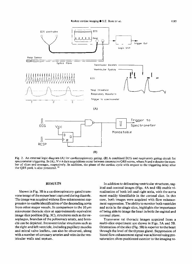
Cardiorespiratory triggering was performed using bipolar ECG leads with Ag/AgCl electrodes (3M HealthCare, St. Paul, MN, USA) and an optical re- spiratory sensor. ” Both signals were displayed on a Physioguard SM 785 NMR Monitor (Bruker Medizin- technik, Germany). Trigger pulses and preset delays from the R wave peak and respiratory threshold were taken from the unit and combined in an external logic circuit (see Fig. 2A).
A trigger pulse (active low) is sent to the spectrom- eter on the detection of the first R wave after the respi- ratory threshold is crossed. The exact timing of this pulse is subject to preset delays within the monitor and within the pulse program. This is achieved in the cir-
cuit as shown (Fig. 2B). A flip-flop is set on crossing of the respiratory threshold and the output state is log- ically ANDed with the next R wave pulse. The result- ing signal is conditioned for the spectrometer and resets the flip-flop for the next cycle. Thus one trigger pulse per respiratory cycle occurs at the first available systole.
Imaging Protocols Rodent thoracic images were acquired with either a
standard cardiorespiratory triggered 2DFT spin-echo technique or a composite flow suppression spin-echo sequence. The blood flow enhancement suppression sequence consisted of three nonselective RF pulses (Fig. l), with symmetrical flow dephasing gradients placed either side of the refocussing 180” pulse.8*‘2 A dephasing gradient strength of either 1.2 or 7.2 G/cm was applied in the slice direction for a total of 2 ms. With this sequence, magnetization of interest is in the transverse plane for 6 ms. The TE time for the spin- echo imaging experiment (without flow compensation) was normally set to 16 ms.
To avoid any saturation effects, a single application of the flow suppression sequence was applied immedi- ately prior to the multi-slice imaging segment. With this methodology, acquisition of up to eight slices of a single cardiac cycle could be achieved between consecutive QRS waves. For some rat thoracic imaging experiments, blood flow suppression was achieved using spatial- presaturation techniques as reported by Felmlee et a1.13 This consisted of two lo-mm presaturation slices po- sitioned exterior to the image volume.
Contiguous images of mouse and rat heart were ac- quired with l-l.5 mm and 2 mm slice thicknesses, re- spectively. For thoracic mouse imaging, FOV of either 25 mm (transverse images) or 30 mm (sagittal/coronal slices) were employed. Transverse images of the rat tho- rax were acquired with a 48 mm FOV. The image ma- trix size was normally 256 x 256 with a single average acquisition. Depending on the animal’s respiratory rate the average imaging time was approximately 15-20 min.
Mouse microtome slices of 10 pm were prepared from a thoracic paraffin tissue block. Sections were stained with H&E to enhance contrast.
Animal Protocol Female Quarkenbush mice weighing 30-40 g and fe-
male Wistar rats weighing 130-150 g were induced with 3% isoflurane in O2 (3 l/mm). Rodents once fitted with paw ECG electrodes and abdominal respiratory sensor, were then placed in a head-up position in the magnet. The animals were maintained with 1.5-2% iso- flurane in O2 (0.5-l l/min). Isoflurane was adminis- tered to the animals via a nose cone.
Rodent cardiac imaging 0 S.E. ROSE ET AL. 1185
ECG electrodes
0 --rkger out
Logic Unit
Resp Sensor
6 /i I Optical Fibre
Ventricular Diastole
Trigger to spectrometer
(4
Trigger to
u- Spectrometer
Monostable
RESP
Fig. 2. An external logic diagram (A) for cardiorespiratory gating. (B) A combined ECG and respiratory gating circuit for spectrometer triggering. In (A), NX n data acquisitions occur between consecutive QRS waves, where Nand n denote the num- ber of slices and averages, respectively. In addition, the phase of the cardiac cycle with respect to the triggering delay after the QRS peak is also presented.18
RESULTS
Shown in Fig. 3B is a cardiorespiratory-gated trans- verse image of the mouse heart captured during diastole. The image was acquired without flow enhancement sup- pression to enable identification of the descending aorta from other major vessels. In comparison to the 10 pm microtome thoracic slice at approximately equivalent image slice position (Fig. 3C), structures such as the oe- sophagus, branches of the pulmonary artery, and bron- chi can be depicted. Intraventricular structures such as the right and left ventricle, including papillary muscles and mitral valve leaflets, can also be observed, along with a number of coronary arteries and veins in the ven- tricular walls and septum.
In addition to delineating ventricular structures, sag- ittal and coronal images (Figs. 4A and 4B) enable vi- sualisation of both left and right atria, with the aorta most readily identifiable in the coronal slice. In this case, both images were acquired with flow enhance- ment suppression. The ability to monitor both ventricles and atria in the single slice, highlights the importance of being able to image the heart in both the sagittal and coronal plane.
Transverse rat thoracic images acquired from a multi-slice experiment are shown in Figs. 5A and 5B. Orientation of the slice (Fig. 5B) is superior to the heart through the level of the thymus gland. Suppression of blood flow enhancement signal was achieved using pre- saturation slices positioned exterior to the imaging re-

1186 Magnetic Resonance Imaging 0 Volume 12, Number 8, 1994
(A)
(Cl
gion. At this level of the aortic arch both ascending and descending aorta are easily observable along with the superior vena cava and pulmonary artery.
DISCUSSION
Multi-slice cardiorespiratory gated spin-echo images of the rodent heart can be severely affected by hyper- intense intraventricular blood signal. This hyperintense signal, which results from the partial refocussing of the
(B)
Fig. 3. (A) and (B) Cardiorespiratory gated 2DFT spin-echo images of the mouse heart captured during diastole. Images were acquired without any bloodflow-enhanced suppression. Spectrometer triggering occurred approximately 100 ms af- ter the R wave. The images were obtained using a 9.4T, 89 mm vertical bore magnet system. Slice thickness, TE time, FOV, matrix size, and number of averages were 1.5 mm, 16 ms, 25 mm, 256 x 256, and 2, respectively. These parameters yielded an in-plane resolution of < 100 pm. (C) Microtome slice (10 pm) of a mouse thorax corresponding to a similar slice position as that in image (B). Abbreviations for (B) and (C): L.V., left ventricle; R.V., right ventricle; P.M., papil- lary muscle; if M.V., leaflets of mitral valve; Cor. A. & V., coronary arteries and veins; b of P.A., branches of pulmo- nary artery; D. Ar . , descending aorta; Oes, Oesophagus; Br . , Bronchi; Lu, Lung.
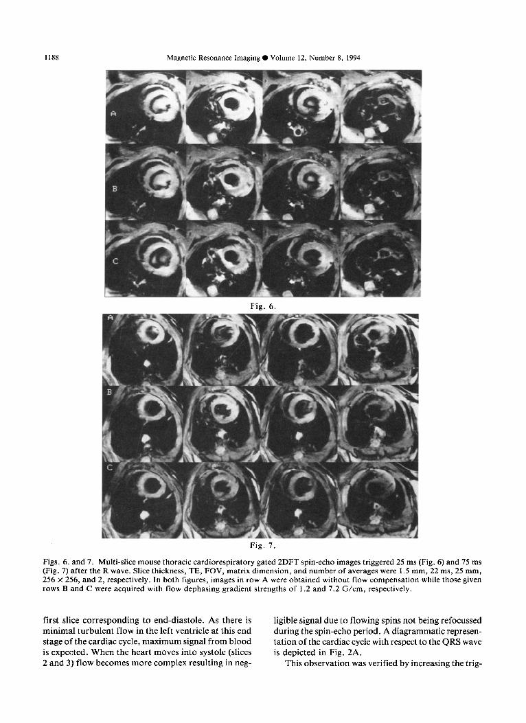
flowing blood magnetization during the spin-echo ex- periment, can lead to loss of morphological informa- tion. The extent to which blood signal appears in the ventricle is dependent on the phase of the cardiac cycle when captured during a multi-slice imaging sequence.14 This effect is clearly demonstrated in the multi-slice im- age array given in Fig. 6. The first image in the four- slice series (A) possesses extensive blood signal in the left ventricle. In this case, the multi-slice experiment was triggered 25 ms after the R wave resulting with the

Rodent cardiac imaging 0 S.E. ROSE ET AL. 1187
W
Fig. 4. Blood flow enhancement suppressed sagittal (A) and coronal (B) images of the mouse thorax acquired approximately 100 ms after the R wave. The images were obtained using a flow dephasing gradient strength of 1.2 G/cm. The slice thickness, TE, FOV, matrix dimension, and number of averages were 1 mm, 22 ms, 30 mm, 256 x 256, and 2, respectively. Additional abbreviations are: L.A., left atrium; R.A., right atrium; Ar, aorta; If Ar., leaflets of the aorta.
(-4 @I
Fig. 5. Rat thoracic cardiorespiratory gated 2DFT spin-echo images obtained using a 7 T, 150 mm vertical magnet system. Image (A) was acquired 100 ms after the QRS wave. Imaging parameters for slice thickness, TE, FOV, matrix size and number of averages were 1.5 mm, 20 ms, 48 mm, 256 x 256 and 1 respectively. The in-plane resolution was 190 pm. Image (B) was acquired superior to the heart at the level of the thymus gland with presaturation pulses to reduce blood flow artifacts. Slice thickness, TE, FOV, matrix dimension, and number of averages were 2 mm, 20 ms, 48 mm, 512 x 512, and 2, respectively. In this case, a 100 pm in-plane resolution was achieved. Assignments: Thy., thymus; A. Ar., ascending aorta; D. Ar., descend- ing aorta; S. Vc., superior vena cava; P.A., pulmonary artery; Oes., Oesophagus.
1188 Magnetic Resonance Imaging 0 Volume 12, Number 8, 1994
Fig. 6.
Fig. 7.
Figs. 6. and 7. Multi-slice mouse thoracic cardiorespiratory gated 2DFT spin-echo images triggered 25 ms (Fig. 6) and 75 ms (Fig. 7) after the R wave. Slice thickness, TE, FOV, matrix dimension, and number of averages were 1.5 mm, 22 ms, 25 mm, 256 x 256, and 2, respectively. In both figures, images in row A were obtained without flow compensation while those given rows B and C were acquired with flow dephasing gradient strengths of 1.2 and 7.2 G/cm, respectively.
first slice corresponding to end-diastole. As there is minimal turbulent flow in the left ventricle at this end stage of the cardiac cycle, maximum signal from blood is expected. When the heart moves into systole (slices 2 and 3) flow becomes more complex resulting in neg-
ligible signal due to flowing spins not being refocussed during the spin-echo period. A diagrammatic represen- tation of the cardiac cycle with respect to the QRS wave is depicted in Fig. 2A.
This observation was verified by increasing the trig-
Rodent cardiac imaging 0 S.E. ROSE ET AL. 1189
gering delay to 75 ms. In this case the heart is imaged predominantly during diastole, but before end-diastole. The results for this experiment are shown in Fig. 7. The images were acquired with the same slice frequencies as those given in Fig. 6. In this experiment, negligible blood flow enhancement signal was detected in any slice. This observation confirms that ventricular blood signal is more pronounced at end-diastole when em- ploying 2DFT spin-echo imaging techniques.
A number of flow suppression sequences have been reported which minimize vascular and ventricular blood flow enhancement signal. The ability to image the heart at end-diastole is important in assessing cardiac status, such as left ventricular mass.’ One approach is the use of spatial presaturation pulses to saturate blood mag- netization flowing into the imaging region.i3 This method was applied to image the ascending and de- scending aorta and pulmonary artery in the rat heart with minimal flow enhancement (Fig. 5A). Alterna- tively, “Black Blood” MR imaging has been proposed where vascular signal is reduced by use of a weighted blood Ti’nulling preparation sequence.15
Our approach to this problem was to utilize a com- bined spin-echo/gradient refocussed sequence which was initially proposed for imaging of flowing spins.*,‘* The modified sequence for suppression of flow en- hancement is given in Fig. 1. With this sequence, both flowing and stationary spins are rotated to the trans- verse plane by the first nonselective 90” pulse. The sym- metrical gradients and the 180” pulse refocus the stationary spins along they axis where they can be re- turned to the longitudinal axis by the last x phased 90” pulse. The advantage in using a spin-echo sequence is
that both chemical shift and B. inhomogeneity are re- focussed in the experiment. The phase of the last pulse can be either +x or -x. The flowing spins, however, are dephased by the gradient leaving only a component of the flowing spin magnetization to be refocussed, the rest is spoiled by the first imaging slice gradient. To re- move the possibility of any gradient modulated “stripe” artifact,15*16 the flow dephasing gradients are applied in the slice direction. It is interesting to note that the sequence can be adapted for angiography by replacing the last pulse with a y-phased 90” pulse.*,‘**” Magne- tization from the flowing spins in this case is restored along the z axis leaving the stationary spins to be de- phased by the imaging slice gradients.
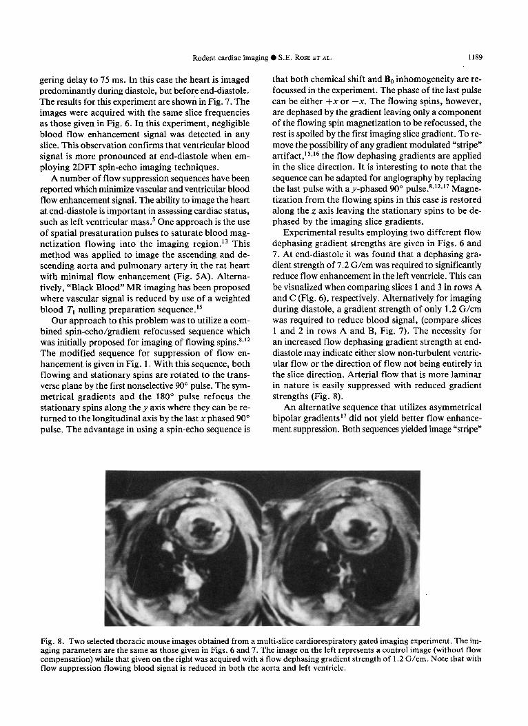
Experimental results employing two different flow dephasing gradient strengths are given in Figs. 6 and 7. At end-diastole it was found that a dephasing gra- dient strength of 7.2 G/cm was required to significantly reduce flow enhancement in the left ventricle. This can be visualized when comparing slices 1 and 3 in rows A and C (Fig. 6), respectively. Alternatively for imaging during diastole, a gradient strength of only 1.2 G/cm was required to reduce blood signal, (compare slices 1 and 2 in rows A and B, Fig. 7). The necessity for an increased flow dephasing gradient strength at end- diastole may indicate either slow non-turbulent ventric- ular flow or the direction of flow not being entirely in the slice direction. Arterial flow that is more laminar in nature is easily suppressed with reduced gradient strengths (Fig. 8).
An alternative sequence that utilizes asymmetrical bipolar gradients ” did not yield better flow enhance- ment suppression. Both sequences yielded image “stripe”
Fig. 8. Two selected thoracic mouse images obtained from a multi-slice cardiorespiratory gated imaging experiment. The im- aging parameters are the same as those given in Figs. 6 and 7. The image on the left represents a control image (without flow compensation) while that given on the right was acquired with a flow dephasing gradient strength of 1.2 G/cm. Note that with flow suppression flowing blood signal is reduced in both the aorta and left ventricle.
1190 Magnetic Resonance Imaging 0 Volume 12, Number 8, 1994
artifacts when the flow dephasing gradients were placed in either read or phase directions both in vivo and flow phantoms. Due to the difficulty of being able to sup- press flow only in the slice direction, cardiac images at end-diastole were never completely void of blood signal.
Nevertheless, the results presented from this study demonstrate that rodent cardiac images with < 100 pm in-plane resolution and minimal signal from vascular and ventricular blood flow enhancement can be ob- tained using vertical bore magnet systems employing the abovementioned microimaging methodology.
Acknowledgment-This research was supported by the National Health and Medical Research Council of Australia (F.O.Z.).
1.
2.
3.
4.
5.
6.
7.
REFERENCES
Callaghan, P.T. Principles of Nuclear Magnetic Reso- nance Microscopy. Oxford: Clarendon Press; 1991: pp. 254-255. Bliimich, B.; Kuhn, W. (Eds). Magnetic Resonance Mi- croscopy, Methods and Applications in Materials Sci- ence, Agriculture and Biomedicine. Weinheim: VCH Verlagsgesellschaft mbH; 1992: pp. 479-586. Mansfield, P.; Hahn, E.L. (Eds). NMR IMAGING, Pro- ceedings of a Royal Society Discussion Meeting, June 1990. Cambridge: University Press; 1990: p. 77. Hedlund, L.W.; Johnson, G.A.; Mills, G.I. Magnetic resonance microscopy of the rat thorax and abdomen. Invest. Radiol. 843; 1986. Manning, W. J.; Burstein, D.; Fossel, E.T.; Wei, J.Y. In- crease in left ventricular mass following aortic valve dis- ruption in the adult rat. In: Book of abstracts: Ninth Annual Meeting of the Society of Magnetic Resonance in Medicine. Berkeley, CA: SMRM; 1990:676. Sarkar, S.K.; Kapadia, R.D. Magnetic resonance micros- copy, methods and applications in materials science, ag- riculture and biomedicine. In: Bltimich, B.; Kuhn, W. (Eds). Weinheim: VCH Verlagsgesellschaft mbH; 1992: pp. 514-515. Burstein, D. MRI of coronary artery flow in isolated and in vivo hearts. In: Book of abstracts: Tenth Annual Meet-
8.
9.
10.
11.
12.
13.
14.
15.
16.
17.
18.
ing of the Society of Magnetic Resonance in Medicine. Berkeley, CA: SMRM; 1991:80. Nishimura, D.G.; Macovski, A.; Pauly, J.M. Magnetic
resonance angiography. IEEE Trans. Med. Imaging MI- 5:140-151; 1986. Hayes, C.E.; Edelstein, W.A.; Schenik, J.F.; Mueller, D.M.; Eash, M. An efficient homogeneous radiofre- quency coil for whole body NMR imaging at 1.5 T. J. Magn. Reson. 63:622; 1985. Eccles, C.D.; Crozier, S.; Roffmann, W.; Doddrell, D.M.; Back, P.; Callaghan, P.T. Practical aspects of shielded gradient coil design for localised in vivo NMR spectroscopy and small scale imaging. Magn. Reson. Im- aging 12:621-630; 1994. Wilson, S.J.; Brereton, I.M.; Hockings, P.; Roffmann, W.; Doddrell, D.M. Respiratory triggered imaging with an optical displacement sensor. Magn. Reson. Imaging 11:1027-1032; 1993. Foster, T.H.; Plewes, D.B.; Szumowski, J. Driven equi- librium MR angiography: A study of static spin suppres- sion. In: Book of abstracts: Sixth Annual Meeting of the Society of Magnetic Resonance in Medicine. Berkeley, CA: SMRM; 1987:30. Felmlee, J.P.; Ehman, R.L. Spatial presaturation: A method for suppressing flow artifacts and improving de- piction of vascular anatomy in MR imaging. Radiology 164:559; 1987. Eldelman, M.D.; Chien, D.; Kim, D. Fast selective black blood MR imaging. Radiology 181:655; 1991.
Axel, L.; Dougherty, L. MR Imaging of motion with spa- tial modulation of magnetization. Radiology 171:841; 1989. Wedeen, V. J.; Rosen, B.R.; Chesler, D.; Brady, T. J. MR velocity imaging by phase display. J. Comput. Assist. Tomogr. 9:530; 1985. Korosec, F.R.; Grist,T.M.; Polzin, J.A.; Weber, D.M.; Mistretta, C.A. MR Angiography using velocity-selective preparation pulses and segmented gradient-echo acqui- sition. Magn. Reson. Med. 30:704; 1993. Budden, R.; Detweiler, D.K.; Zbinden, G. (Eds). The rat electrocardiogram in pharmacology and toxicology. In: Proceedings of an International Workshop held in Hanover, Federal Republic of Germany, July 1980. Per- gamon Press; 1981.