High glucose concentrations induce TNF-α production through the down-regulation of CD33 in primary...
14
RESEARCH ARTICLE Open Access High glucose concentrations induce TNF-a production through the down-regulation of CD33 in primary human monocytes Yolanda Gonzalez 1 , M Teresa Herrera 1 , Gloria Soldevila 3 , Lourdes Garcia-Garcia 4 , Guadalupe Fabián 2 , E Martha Pérez-Armendariz 5 , Karen Bobadilla 1 , Silvia Guzmán-Beltrán 1 , Eduardo Sada 1 and Martha Torres 1* Abstract Background: CD33 is a membrane receptor containing a lectin domain and a cytoplasmic immunoreceptor tyrosine-based inhibitory motif (ITIM) that is able to inhibit cytokine production. CD33 is expressed by monocytes, and reduced expression of CD33 correlates with augmented production of inflammatory cytokines, such as IL-1b, TNF-a, and IL-8. However, the role of CD33 in the inflammation associated with hyperglycemia and diabetes is unknown. Therefore, we studied CD33 expression and inflammatory cytokine secretion in freshly isolated monocytes from patients with type 2 diabetes. To evaluate the effects of hyperglycemia, monocytes from healthy donors were cultured with different glucose concentrations (15-50 mmol/l D-glucose), and CD33 expression and inflammatory cytokine production were assessed. The expression of suppressor of cytokine signaling protein-3 (SOCS-3) and the generation of reactive oxygen species (ROS) were also evaluated to address the cellular mechanisms involved in the down-regulation of CD33. Results: CD33 expression was significantly decreased in monocytes from patients with type 2 diabetes, and higher levels of TNF-a, IL-8 and IL-12p70 were detected in the plasma of patients compared to healthy donors. Under high glucose conditions, CD33 protein and mRNA expression was significantly decreased, whereas spontaneous TNF-a secretion and SOCS-3 mRNA expression were increased in monocytes from healthy donors. Furthermore, the down-regulation of CD33 and increase in TNF-a production were prevented when monocytes were treated with the antioxidant a-tocopherol and cultured under high glucose conditions. Conclusion: Our results suggest that hyperglycemia down-regulates CD33 expression and triggers the spontaneous secretion of TNF-a by peripheral monocytes. This phenomenon involves the generation of ROS and the up-regulation of SOCS-3. These observations support the importance of blood glucose control for maintaining innate immune function and suggest the participation of CD33 in the inflammatory profile associated with type 2 diabetes. Keywords: Antioxidant, Cytokines, Monocytes, ROS, Siaglec-3, Type 2 diabetes Background Both acute and chronic hyperglycemia are associated with inflammation [1]. Patients with newly diagnosed or established diabetes mellitus (DM) have significantly higher levels of acute-phase proteins and pro-inflamma- tory cytokines compared to control subjects without DM [2-5]. Monocytes isolated from patients with type 1 diabetes produce increased levels of IL-6, IL-1b and che- mokines of the CXC family including IL-8 and inter- feron gamma-induced protein 10 (IP-10) [6]. Furthermore, monocytes from patients with DM pro- duce higher levels of TNF-a and IL-8 in comparison to control monocytes [7-9]. TNF- a production is thought to play a role in the generation of microvascular complications associated with diabetes, e.g., by enhancing chronic eye * Correspondence: [email protected] 1 Departamento de Investigación en Microbiología, Instituto Nacional de Enfermedades Respiratorias, Calzada de Tlalpan 4502, Sección XVI, Ciudad de México, 14080, México Full list of author information is available at the end of the article Gonzalez et al. BMC Immunology 2012, 13:19 http://www.biomedcentral.com/1471-2172/13/19 © 2012 Gonzalez et al; licensee BioMed Central Ltd. This is an Open Access article distributed under the terms of the Creative Commons Attribution License (http://creativecommons.org/licenses/by/2.0), which permits unrestricted use, distribution, and reproduction in any medium, provided the original work is properly cited.
-
Upload
independent -
Category
Documents
-
view
4 -
download
0
Transcript of High glucose concentrations induce TNF-α production through the down-regulation of CD33 in primary...
RESEARCH ARTICLE Open Access
High glucose concentrations induce TNF-aproduction through the down-regulation of CD33in primary human monocytesYolanda Gonzalez1, M Teresa Herrera1, Gloria Soldevila3, Lourdes Garcia-Garcia4, Guadalupe Fabián2,E Martha Pérez-Armendariz5, Karen Bobadilla1, Silvia Guzmán-Beltrán1, Eduardo Sada1 and Martha Torres1*
Abstract
Background: CD33 is a membrane receptor containing a lectin domain and a cytoplasmic immunoreceptortyrosine-based inhibitory motif (ITIM) that is able to inhibit cytokine production. CD33 is expressed by monocytes,and reduced expression of CD33 correlates with augmented production of inflammatory cytokines, such as IL-1b,TNF-a, and IL-8. However, the role of CD33 in the inflammation associated with hyperglycemia and diabetes isunknown. Therefore, we studied CD33 expression and inflammatory cytokine secretion in freshly isolatedmonocytes from patients with type 2 diabetes. To evaluate the effects of hyperglycemia, monocytes from healthydonors were cultured with different glucose concentrations (15-50 mmol/l D-glucose), and CD33 expression andinflammatory cytokine production were assessed. The expression of suppressor of cytokine signaling protein-3(SOCS-3) and the generation of reactive oxygen species (ROS) were also evaluated to address the cellularmechanisms involved in the down-regulation of CD33.
Results: CD33 expression was significantly decreased in monocytes from patients with type 2 diabetes, and higherlevels of TNF-a, IL-8 and IL-12p70 were detected in the plasma of patients compared to healthy donors. Underhigh glucose conditions, CD33 protein and mRNA expression was significantly decreased, whereas spontaneousTNF-a secretion and SOCS-3 mRNA expression were increased in monocytes from healthy donors. Furthermore, thedown-regulation of CD33 and increase in TNF-a production were prevented when monocytes were treated withthe antioxidant a-tocopherol and cultured under high glucose conditions.
Conclusion: Our results suggest that hyperglycemia down-regulates CD33 expression and triggers thespontaneous secretion of TNF-a by peripheral monocytes. This phenomenon involves the generation of ROS andthe up-regulation of SOCS-3. These observations support the importance of blood glucose control for maintaininginnate immune function and suggest the participation of CD33 in the inflammatory profile associated with type 2diabetes.
Keywords: Antioxidant, Cytokines, Monocytes, ROS, Siaglec-3, Type 2 diabetes
BackgroundBoth acute and chronic hyperglycemia are associatedwith inflammation [1]. Patients with newly diagnosed orestablished diabetes mellitus (DM) have significantlyhigher levels of acute-phase proteins and pro-inflamma-tory cytokines compared to control subjects without
DM [2-5]. Monocytes isolated from patients with type 1diabetes produce increased levels of IL-6, IL-1b and che-mokines of the CXC family including IL-8 and inter-feron gamma-induced protein 10 (IP-10) [6].Furthermore, monocytes from patients with DM pro-duce higher levels of TNF-a and IL-8 in comparison tocontrol monocytes [7-9].TNF-a production is thought to play a role in the
generation of microvascular complications associatedwith diabetes, e.g., by enhancing chronic eye
* Correspondence: [email protected] de Investigación en Microbiología, Instituto Nacional deEnfermedades Respiratorias, Calzada de Tlalpan 4502, Sección XVI, Ciudad deMéxico, 14080, MéxicoFull list of author information is available at the end of the article
Gonzalez et al. BMC Immunology 2012, 13:19http://www.biomedcentral.com/1471-2172/13/19
© 2012 Gonzalez et al; licensee BioMed Central Ltd. This is an Open Access article distributed under the terms of the CreativeCommons Attribution License (http://creativecommons.org/licenses/by/2.0), which permits unrestricted use, distribution, andreproduction in any medium, provided the original work is properly cited.

inflammation [10,11]. In addition to triggering acute andchronic inflammation, TNF-a regulates glucose andlipid metabolism and inhibits insulin production in pan-creatic beta cells [12]. TNF-a is also produced in adi-pose tissue.In both clinical and experimental conditions, hypergly-
cemia has been shown to alter many cellular parameters.This metabolic state leads to the generation of reactiveoxygen species (ROS), the activity of protein kinase C(PKC), and the expression of p38 mitogen-activated pro-tein kinase, nuclear factor �B (NF-�B), inflammatorycytokines, and chemokines [13-15].Diverse mechanisms have been proposed to explain
how hyperglycemia contributes to inflammation. Forexample, PKC activity may be increased secondarily to apoorly reversible, non-enzymatic protein glycation pro-cess, which could lead to the irreversible production ofadvanced glycation end products (AGEs). AGEs areknown to stimulate the production of inflammatorycytokines in monocytes and macrophages through theactivation of a specific receptor for AGEs (RAGE)[16,17]. Additionally, hyperglycemia may stimulate theproduction of inflammatory cytokines by increasing thelevels of peroxides and free radicals. High serum con-centrations of glucose can lead to enhanced glycolysisand mitochondrial overproduction of superoxide anion(O2
-) and other reactive oxygen species (ROS), whichdirectly induce the activation of protein kinase C (PKC)and nuclear factor �B (NF-�B) [18,19]. Indeed, thesetranscription factors have been shown to induce therelease of IL-1b and IL-6 by human monocytes culturedunder high glucose conditions [20]. The secondaryeffects of PKC and NF- �B activation resulting fromhyperglycemia can further amplify the inflammatoryresponse, resulting in the production of the chemokineIP-10 and the up-regulated expression of TLR2 andTLR4 [6].Although these mechanisms can partially explain the
high levels of inflammatory cytokines observed underacute hyperglycemic conditions, the effects of high glucoseon other regulatory molecules involved in the control ofinflammatory cytokine production have not yet been iden-tified. Low membrane expression levels of CD33 havebeen associated with higher levels of inflammatory cyto-kine production, and CD33 is expressed by myeloid pro-genitor cells of the bone marrow as well as peripheralblood monocytes and lymphocytes [21,22]. CD33, which isalso referred to as human sialic acid-binding Ig superfam-ily lectin (hSiglec-3), is a member of the Siglec family thatincludes 11 human proteins of I-type (Ig-type) lectins witha V-set Ig-like domain and varying numbers of C2-set Ig-like domains, such as sialoadhesins (Siglec-1 and CD169),CD22 (Siglec-2), myelin-associated glycoprotein (MAG;Siglec-4), and additional members from a subgroup that
contains CD33 (Siglec-3) and CD33-related Siglecs(Siglec-5 to -11) [22-25].The regulation of cytokine production via CD33 is
believed to depend on two putative conserved tyrosine-based signaling motifs contained within the cytosolic tailof CD33. These signaling motifs, known as immunore-ceptor tyrosine-based inhibitory motifs (ITIMs), act asregulatory elements that inhibit signaling [22]. CD33activity is regulated by SOCS3, which is a member ofthe suppressor of cytokine signaling (SOCS) proteinfamily. The binding of SOCS3 to the phosphorylatedITIM of CD33 induces the proteosomal degradation ofboth molecules [26], and the reduction of CD33 surfaceexpression on monocytes by silencing with small inter-fering RNA (siRNA) or antibody blockade results in theincreased secretion of IL-1b, IL-8, and TNF-a [27].Interestingly, the role of CD33 in the production of
pro-inflammatory cytokines secondary to hyperglycemiahas not yet been explored. Thus, the aim of the currentstudy was to examine the effects of high glucose concen-trations on the expression of CD33 and the productionof cytokines in human monocytes from healthy indivi-duals. Additionally, from patients with type 2 diabetes,the levels of CD33 expression on freshly obtained mono-cytes and serum cytokine levels were evaluated and com-pared to those from healthy individuals. Our results showthat under hyperglycemic conditions, monocytes CD33mRNA and surface protein expression was decreased,whereas TNF-a production was increased. These changeswere inhibited by antioxidant pre-treatment, suggestingthat the hyperglycemic-dependent decrease in CD33expression involves the generation of oxidative stress.
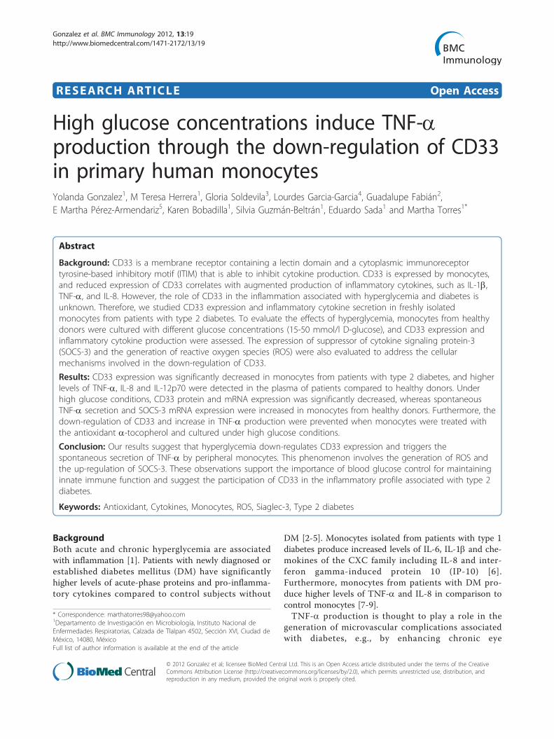
ResultsThe clinical characteristics of the studied subjects aresummarized in Table 1. There were no significant differ-ences in gender, age, BMI, or the levels of creatinine orLDL or HDL cholesterol between the control group andthe type 2 diabetes group. The levels of glucose, HbA1cand triglycerides were significantly higher among type 2diabetes subjects than control subjects. Most patientswith type 2 diabetes had received metformin and Glib-enclamide. Only one of the diabetes patients hadreceived metoprolol, and another had been administeredclonazepam and levopromazine. None of the patientshad been prescribed angiotensin receptor blockers, insu-lin, or statins. Healthy donors did not have any infec-tions or inflammatory diseases and did not take anymedications during the study period.
Diminished CD33 expression in monocytes from patientswith type 2 diabetesTo determine whether CD33 expression is decreased infreshly isolated peripheral monocytes from type 2
Gonzalez et al. BMC Immunology 2012, 13:19http://www.biomedcentral.com/1471-2172/13/19
Page 2 of 14
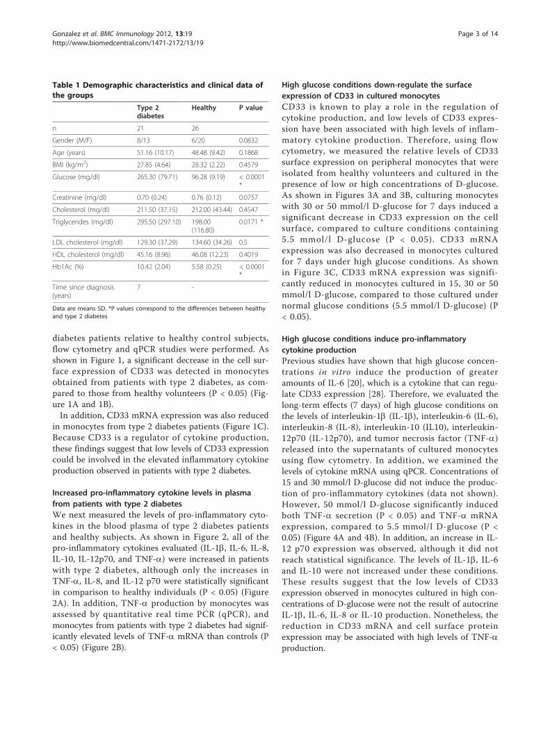
diabetes patients relative to healthy control subjects,flow cytometry and qPCR studies were performed. Asshown in Figure 1, a significant decrease in the cell sur-face expression of CD33 was detected in monocytesobtained from patients with type 2 diabetes, as com-pared to those from healthy volunteers (P < 0.05) (Fig-ure 1A and 1B).In addition, CD33 mRNA expression was also reduced
in monocytes from type 2 diabetes patients (Figure 1C).Because CD33 is a regulator of cytokine production,these findings suggest that low levels of CD33 expressioncould be involved in the elevated inflammatory cytokineproduction observed in patients with type 2 diabetes.
Increased pro-inflammatory cytokine levels in plasmafrom patients with type 2 diabetesWe next measured the levels of pro-inflammatory cyto-kines in the blood plasma of type 2 diabetes patientsand healthy subjects. As shown in Figure 2, all of thepro-inflammatory cytokines evaluated (IL-1b, IL-6, IL-8,IL-10, IL-12p70, and TNF-a) were increased in patientswith type 2 diabetes, although only the increases inTNF-a, IL-8, and IL-12 p70 were statistically significantin comparison to healthy individuals (P < 0.05) (Figure2A). In addition, TNF-a production by monocytes wasassessed by quantitative real time PCR (qPCR), andmonocytes from patients with type 2 diabetes had signif-icantly elevated levels of TNF-a mRNA than controls (P< 0.05) (Figure 2B).
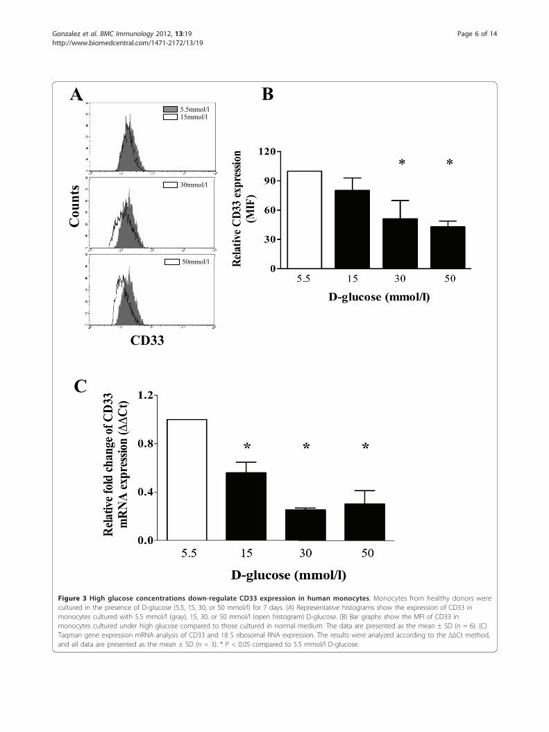
High glucose conditions down-regulate the surfaceexpression of CD33 in cultured monocytesCD33 is known to play a role in the regulation ofcytokine production, and low levels of CD33 expres-sion have been associated with high levels of inflam-matory cytokine production. Therefore, using flowcytometry, we measured the relative levels of CD33surface expression on peripheral monocytes that wereisolated from healthy volunteers and cultured in thepresence of low or high concentrations of D-glucose.As shown in Figures 3A and 3B, culturing monocyteswith 30 or 50 mmol/l D-glucose for 7 days induced asignificant decrease in CD33 expression on the cellsurface, compared to culture conditions containing5.5 mmol/l D-glucose (P < 0.05). CD33 mRNAexpression was also decreased in monocytes culturedfor 7 days under high glucose conditions. As shownin Figure 3C, CD33 mRNA expression was signifi-cantly reduced in monocytes cultured in 15, 30 or 50mmol/l D-glucose, compared to those cultured undernormal glucose conditions (5.5 mmol/l D-glucose) (P< 0.05).
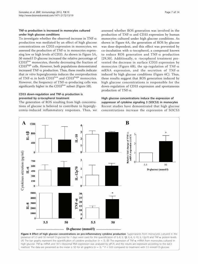
High glucose conditions induce pro-inflammatorycytokine productionPrevious studies have shown that high glucose concen-trations in vitro induce the production of greateramounts of IL-6 [20], which is a cytokine that can regu-late CD33 expression [28]. Therefore, we evaluated thelong-term effects (7 days) of high glucose conditions onthe levels of interleukin-1b (IL-1b), interleukin-6 (IL-6),interleukin-8 (IL-8), interleukin-10 (IL10), interleukin-12p70 (IL-12p70), and tumor necrosis factor (TNF-a)released into the supernatants of cultured monocytesusing flow cytometry. In addition, we examined thelevels of cytokine mRNA using qPCR. Concentrations of15 and 30 mmol/l D-glucose did not induce the produc-tion of pro-inflammatory cytokines (data not shown).However, 50 mmol/l D-glucose significantly inducedboth TNF-a secretion (P < 0.05) and TNF-a mRNAexpression, compared to 5.5 mmol/l D-glucose (P <0.05) (Figure 4A and 4B). In addition, an increase in IL-12 p70 expression was observed, although it did notreach statistical significance. The levels of IL-1b, IL-6and IL-10 were not increased under these conditions.These results suggest that the low levels of CD33expression observed in monocytes cultured in high con-centrations of D-glucose were not the result of autocrineIL-1b, IL-6, IL-8 or IL-10 production. Nonetheless, thereduction in CD33 mRNA and cell surface proteinexpression may be associated with high levels of TNF-aproduction.
Table 1 Demographic characteristics and clinical data ofthe groups
Type 2diabetes
Healthy P value
n 21 26
Gender (M/F) 8/13 6/20 0.0832
Age (years) 51.16 (10.17) 48.48 (9.42) 0.1868
BMI (kg/m2) 27.85 (4.64) 28.32 (2.22) 0.4579
Glucose (mg/dl) 265.30 (79.71) 96.28 (9.19) < 0.0001*
Creatinine (mg/dl) 0.70 (0.24) 0.76 (0.12) 0.0757
Cholesterol (mg/dl) 211.50 (37.15) 212.00 (43.44) 0.4547
Triglycerides (mg/dl) 295.50 (297.10) 198.00(116.80)
0.0171 *
LDL cholesterol (mg/dl) 129.30 (37.29) 134.60 (34.26) 0.5
HDL cholesterol (mg/dl) 45.16 (8.96) 46.08 (12.23) 0.4019
Hb1Ac (%) 10.42 (2.04) 5.58 (0.25) < 0.0001*
Time since diagnosis(years)
7 -
Data are means SD. *P values correspond to the differences between healthyand type 2 diabetes
Gonzalez et al. BMC Immunology 2012, 13:19http://www.biomedcentral.com/1471-2172/13/19
Page 3 of 14

B
C
A
100 101 102 103 104
FL1-H: CD14 FITC
100
101
102
103
104
FL3
-H: C
D3
Per
CP
5.53
100 101 102 103 104
FL2-H: CD33 PE
0
20
40
60
80
100
% o
f Max
Healthy Type 2 diabetes
CD14
CD
3
CD33
% o
f Max
Figure 1 CD33 expression in human monocytes from type 2 diabetes patients. (A) Monocytes were stained with anti-CD3, CD14 and CD33mAbs. At least 50,000 events were acquired for the flow cytometry analysis. CD33 expression is shown after gating for the CD3-CD14+ cells, anda histogram of CD33 expression was plotted for type 2 diabetes patients (tinted histogram) and healthy subjects (open histogram). (B) A bargraph showing the mean intensity fluorescence (MFI) data for CD33 expression in freshly isolated monocytes from patients with type 2 diabetes(n = 10) and healthy donors (n = 10). The data are presented as the mean ± SD. * P < 0.05 compared to healthy donors. (C) Monocytes fromtype 2 diabetes patients (n = 9) and healthy donors (n = 8) were evaluated using Taqman gene expression analysis for CD33 mRNA expression.The results were analyzed according to the ΔΔCt method, and the data are presented as the mean ± SD. * P < 0.05 compared to healthydonors.
Gonzalez et al. BMC Immunology 2012, 13:19http://www.biomedcentral.com/1471-2172/13/19
Page 4 of 14

B
A C
ytok
ines
secr
etio
n (p
g/m
l)
IL-1β
IL-6
IL
-8
IL-1
0 IL
-12p
70
TN
F-α
Healthy Type 2 diabetes Healthy Type 2 diabetes
Figure 2 Pro-inflammatory cytokine production in type 2 diabetes patients. Plasma from type 2 diabetes patients and healthy controls wastested for the presence of pro-inflammatory cytokines using the CBA kit for IL-8, IL-1b, IL-6, IL-10, IL-12p70 and TNF-a. (A) The bar graphs showthe quantification of these cytokines for type 2 diabetes patients (n = 14) and healthy controls (n = 10). All data are presented as the mean ±SD. * P < 0.05 compared to healthy donors. (B) The expression of TNF-a mRNA and 18 S ribosomal RNA was analyzed for monocytes collectedfrom type 2 diabetes patients (n = 7) and healthy subjects (n = 9). The results are expressed according to the ΔΔCt method, and the data arepresented as the mean ± SD. * P < 0.05 compared to healthy donors.
Gonzalez et al. BMC Immunology 2012, 13:19http://www.biomedcentral.com/1471-2172/13/19
Page 5 of 14
A
C
B 5.5mmol/l 15mmol/l
30mmol/l
CD33
50mmol/l
Cou
nts
Figure 3 High glucose concentrations down-regulate CD33 expression in human monocytes. Monocytes from healthy donors werecultured in the presence of D-glucose (5.5, 15, 30, or 50 mmol/l) for 7 days. (A) Representative histograms show the expression of CD33 inmonocytes cultured with 5.5 mmol/l (gray), 15, 30, or 50 mmol/l (open histogram) D-glucose. (B) Bar graphs show the MFI of CD33 inmonocytes cultured under high glucose compared to those cultured in normal medium. The data are presented as the mean ± SD (n = 6). (C)Taqman gene expression mRNA analysis of CD33 and 18 S ribosomal RNA expression. The results were analyzed according to the ΔΔCt method,and all data are presented as the mean ± SD (n = 3). * P < 0.05 compared to 5.5 mmol/l D-glucose.
Gonzalez et al. BMC Immunology 2012, 13:19http://www.biomedcentral.com/1471-2172/13/19
Page 6 of 14
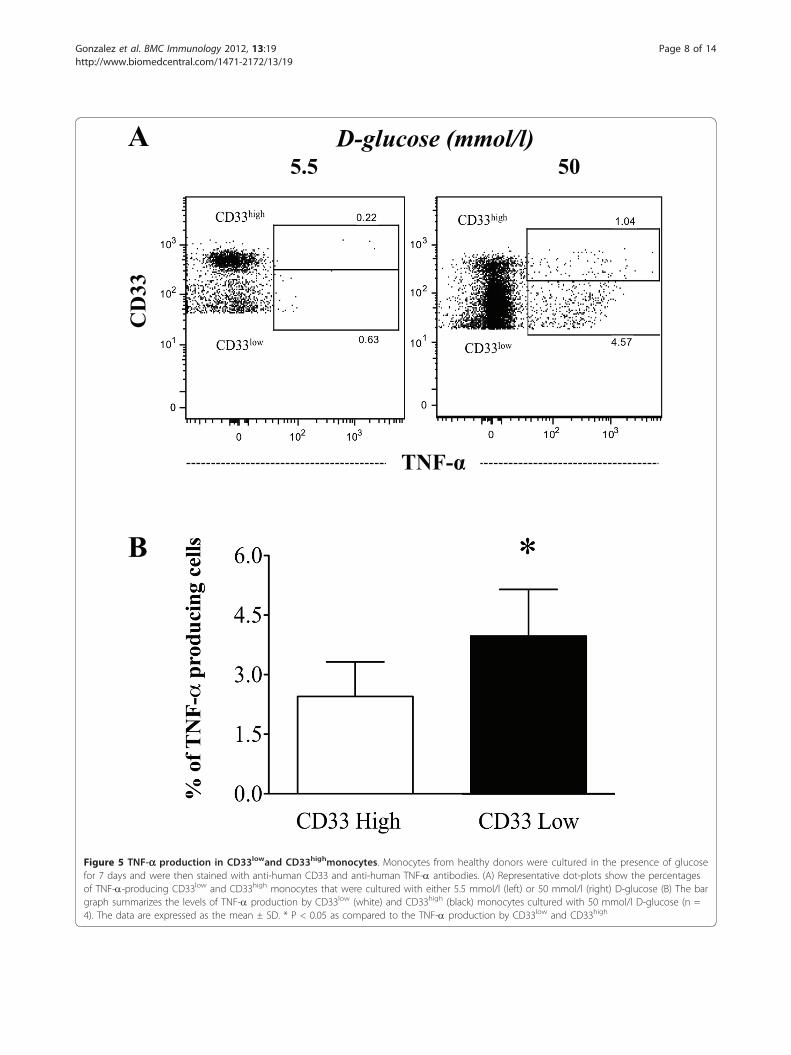
TNF-a production is increased in monocytes culturedunder high glucose conditionsTo investigate whether the observed increase in TNF-aproduction was mediated by an effect of high glucoseconcentrations on CD33 expression in monocytes, weassessed the production of TNF-a in monocytes expres-sing low or high levels of CD33. As shown in Figure 5A,50 mmol/l D-glucose increased the relative percentage ofCD33low monocytes, thereby decreasing the fraction ofCD33high cells. However, both populations demonstratedincreased TNF-a production. Thus, these results indicatethat in vitro hyperglycemia induces the overproductionof TNF-a in both CD33low and CD33high monocytes.However, the frequency of TNF-a-producing cells wassignificantly higher in the CD33low subset (Figure 5B).
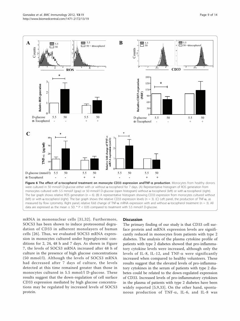
CD33 down-regulation and TNF-a production isprevented by a-tocopherol treatmentThe generation of ROS resulting from high concentra-tions of glucose is believed to contribute to hypergly-cemia-induced inflammatory responses. Thus, we
assessed whether ROS generation was involved in theproduction of TNF-a and CD33 expression by humanmonocytes cultured under high glucose conditions. Asshown in Figure 6A, the generation of ROS by glucosewas dose-dependent, and this effect was prevented byco-incubation with a-tocopherol, a compound knownto reduce ROS generation and TNF-a production[29,30]. Additionally, a -tocopherol treatment pre-vented the decrease in surface CD33 expression bymonocytes (Figure 6B), the up-regulation of TNF-amRNA expression, and the secretion of TNF-ainduced by high glucose conditions (Figure 6C). Thus,these results suggest that ROS generation induced byhigh glucose concentrations is responsible for thedown-regulation of CD33 expression and spontaneousproduction of TNF-a.
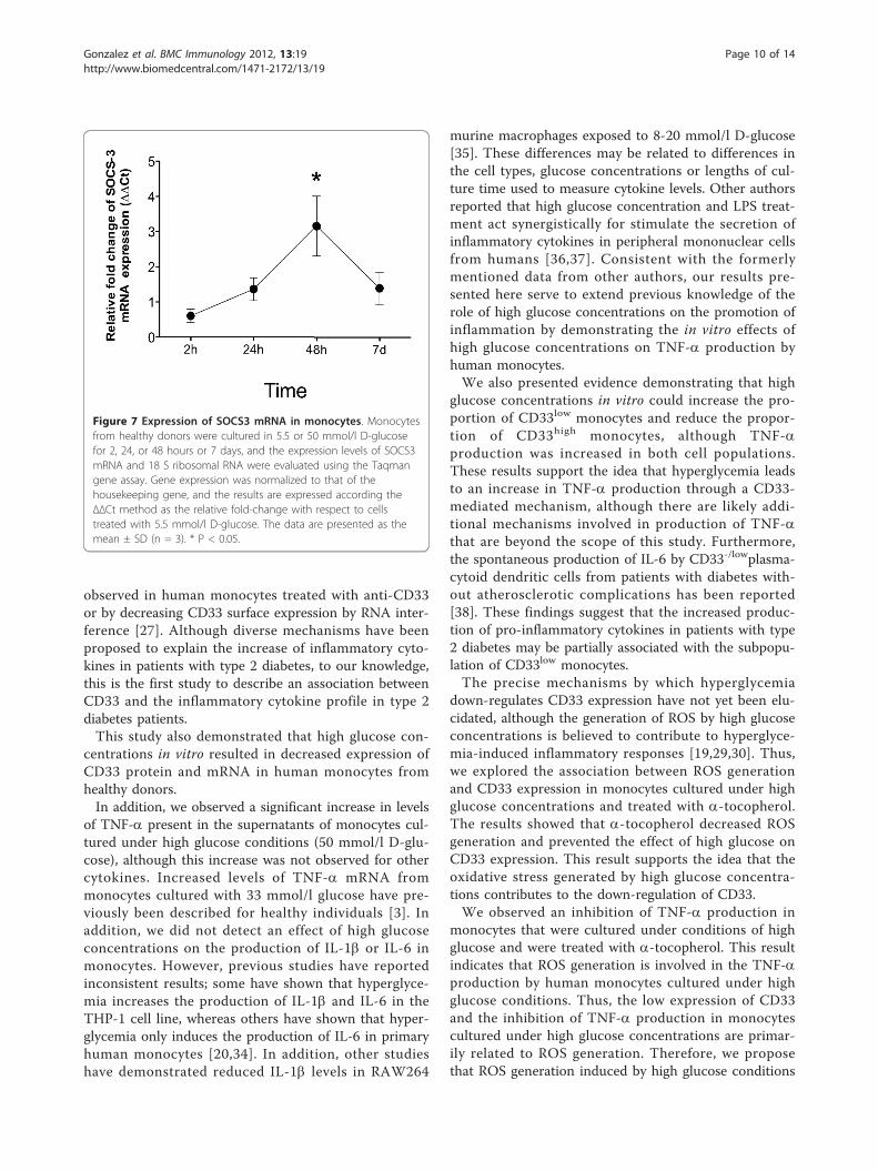
High glucose concentrations induce the expression ofsuppressor of cytokine signaling 3 (SOCS3) in monocytesRecent studies have demonstrated that high glucoseconcentrations increase the expression of SOCS3
A
5.5 50 5.5 50
B
Cyt
okin
es se
cret
ion
(pg/
ml)
0 0
IL-1β
IL-6
IL
-8
IL-1
2p70
IL
-10
TN
F-α
D-glucose (mmol/l)
5.5 50
D-glucose (mmol/l)
Figure 4 Effect of high glucose concentrations on pro-inflammatory cytokine production. Supernatants from monocytes cultured in thepresence of 5.5 and 50 mmol/l D-glucose for 7 days were used for the quantification of IL-8, IL-1b, IL-6, IL-10, IL-12p70 and TNF-a protein levels.(A) The bar graphs represent the quantification of cytokine production (n = 3). (B) The expression of TNF-a mRNA from monocytes cultured inhigh glucose. TNF-a mRNA and 18 S ribosomal RNA expression was analyzed by qPCR, and the results are expressed according to the ΔΔCtmethod. The data are presented as the mean ± SD for all graphics (n = 3). * P < 0.05 compared to treatment with 5.5 mmol/l D-glucose.
Gonzalez et al. BMC Immunology 2012, 13:19http://www.biomedcentral.com/1471-2172/13/19
Page 7 of 14
A
B
D-glucose (mmol/l) 5.5 50
TNF-α
CD
33
Figure 5 TNF-a production in CD33lowand CD33highmonocytes. Monocytes from healthy donors were cultured in the presence of glucosefor 7 days and were then stained with anti-human CD33 and anti-human TNF-a antibodies. (A) Representative dot-plots show the percentagesof TNF-a-producing CD33low and CD33high monocytes that were cultured with either 5.5 mmol/l (left) or 50 mmol/l (right) D-glucose (B) The bargraph summarizes the levels of TNF-a production by CD33low (white) and CD33high (black) monocytes cultured with 50 mmol/l D-glucose (n =4). The data are expressed as the mean ± SD. * P < 0.05 as compared to the TNF-a production by CD33low and CD33high
Gonzalez et al. BMC Immunology 2012, 13:19http://www.biomedcentral.com/1471-2172/13/19
Page 8 of 14
mRNA in mononuclear cells [31,32]. Furthermore,SOCS3 has been shown to induce proteosomal degra-dation of CD33 in adherent monolayers of humancells [26]. Thus, we evaluated SOCS3 mRNA expres-sion in monocytes cultured under hyperglycemic con-ditions for 2, 24, 48 h and 7 days. As shown in Figure7, the levels of SOCS3 mRNA increased after 48 h ofculture in the presence of high glucose concentrations(50 mmol/l). Although the levels of SOCS3 mRNAhad decreased after 7 days of culture, the levelsdetected at this time remained greater than those inmonocytes cultured in 5.5 mmol/l D-glucose. Theseresults suggest that the down-regulation of cell surfaceCD33 expression mediated by high glucose concentra-tions may be regulated by increased levels of SOCS3protein.
DiscussionThe primary finding of our study is that CD33 cell sur-face protein and mRNA expression levels are signifi-cantly reduced in monocytes from patients with type 2diabetes. The analysis of the plasma cytokine profile ofpatients with type 2 diabetes showed that pro-inflamma-tory cytokine levels were increased, although only thelevels of IL-8, IL-12, and TNF-a were significantlyincreased when compared to healthy volunteers. Theseresults suggest that the elevated levels of pro-inflamma-tory cytokines in the serum of patients with type 2 dia-betes could be related to the down-regulated expressionof CD33. Increased levels of pro-inflammatory cytokinesin the plasma of patients with type 2 diabetes have beenwidely reported [5,9,33]. On the other hand, sponta-neous production of TNF-a, IL-6, and IL-8 was
B A
Cou
nts
5.5 50
5.5 50 + αtocopherol
ROS
5.5 50 +αtocopherol
5.5 50
Cou
nts
CD33
α-Tocopherol - - + + - - + + D-glucose (mmol/l) 5.5 50 5.5 50 5.5 50 5.5 50
C
ROS
D-glucose 5.5 50 5.5 50 α-Tocopherol - - + +
D-glucose 5.5 50 5.5 50 α-Tocopherol - - + +
Figure 6 The effect of a-tocopherol treatment on monocyte CD33 expression andTNF-a production. Monocytes from healthy donorswere cultured in 50 mmol/l D-glucose either with or without a-tocopherol for 7 days. (A) Representative histogram of ROS generation frommonocytes cultured with 5.5 mmol/l (gray) or 50 mmol/l D-glucose (open histogram) without a-tocopherol (left) or with a-tocopherol (right).The bar graph shows relative ROS generation (n = 6). (B) A representative histogram showing CD33 expression from monocytes cultured without(left) or with a-tocopherol (right). The bar graph shows the relative CD33 expression levels (n = 3). (C) Left panel, the production of TNF-a, asmeasured by flow cytometry. Right panel, relative fold change of TNF-a mRNA expression with and without a-tocopherol treatment (n = 3). Alldata are expressed as the mean ± SD. * P < 0.05 compared to treatment with 5.5 mmol/l D-glucose.
Gonzalez et al. BMC Immunology 2012, 13:19http://www.biomedcentral.com/1471-2172/13/19
Page 9 of 14
observed in human monocytes treated with anti-CD33or by decreasing CD33 surface expression by RNA inter-ference [27]. Although diverse mechanisms have beenproposed to explain the increase of inflammatory cyto-kines in patients with type 2 diabetes, to our knowledge,this is the first study to describe an association betweenCD33 and the inflammatory cytokine profile in type 2diabetes patients.This study also demonstrated that high glucose con-
centrations in vitro resulted in decreased expression ofCD33 protein and mRNA in human monocytes fromhealthy donors.In addition, we observed a significant increase in levels
of TNF-a present in the supernatants of monocytes cul-tured under high glucose conditions (50 mmol/l D-glu-cose), although this increase was not observed for othercytokines. Increased levels of TNF-a mRNA frommonocytes cultured with 33 mmol/l glucose have pre-viously been described for healthy individuals [3]. Inaddition, we did not detect an effect of high glucoseconcentrations on the production of IL-1b or IL-6 inmonocytes. However, previous studies have reportedinconsistent results; some have shown that hyperglyce-mia increases the production of IL-1b and IL-6 in theTHP-1 cell line, whereas others have shown that hyper-glycemia only induces the production of IL-6 in primaryhuman monocytes [20,34]. In addition, other studieshave demonstrated reduced IL-1b levels in RAW264
murine macrophages exposed to 8-20 mmol/l D-glucose[35]. These differences may be related to differences inthe cell types, glucose concentrations or lengths of cul-ture time used to measure cytokine levels. Other authorsreported that high glucose concentration and LPS treat-ment act synergistically for stimulate the secretion ofinflammatory cytokines in peripheral mononuclear cellsfrom humans [36,37]. Consistent with the formerlymentioned data from other authors, our results pre-sented here serve to extend previous knowledge of therole of high glucose concentrations on the promotion ofinflammation by demonstrating the in vitro effects ofhigh glucose concentrations on TNF-a production byhuman monocytes.We also presented evidence demonstrating that high
glucose concentrations in vitro could increase the pro-portion of CD33low monocytes and reduce the propor-tion of CD33high monocytes, although TNF-aproduction was increased in both cell populations.These results support the idea that hyperglycemia leadsto an increase in TNF-a production through a CD33-mediated mechanism, although there are likely addi-tional mechanisms involved in production of TNF-athat are beyond the scope of this study. Furthermore,the spontaneous production of IL-6 by CD33-/lowplasma-cytoid dendritic cells from patients with diabetes with-out atherosclerotic complications has been reported[38]. These findings suggest that the increased produc-tion of pro-inflammatory cytokines in patients with type2 diabetes may be partially associated with the subpopu-lation of CD33low monocytes.The precise mechanisms by which hyperglycemia
down-regulates CD33 expression have not yet been elu-cidated, although the generation of ROS by high glucoseconcentrations is believed to contribute to hyperglyce-mia-induced inflammatory responses [19,29,30]. Thus,we explored the association between ROS generationand CD33 expression in monocytes cultured under highglucose concentrations and treated with a-tocopherol.The results showed that a-tocopherol decreased ROSgeneration and prevented the effect of high glucose onCD33 expression. This result supports the idea that theoxidative stress generated by high glucose concentra-tions contributes to the down-regulation of CD33.We observed an inhibition of TNF-a production in
monocytes that were cultured under conditions of highglucose and were treated with a-tocopherol. This resultindicates that ROS generation is involved in the TNF-aproduction by human monocytes cultured under highglucose conditions. Thus, the low expression of CD33and the inhibition of TNF-a production in monocytescultured under high glucose concentrations are primar-ily related to ROS generation. Therefore, we proposethat ROS generation induced by high glucose conditions
Figure 7 Expression of SOCS3 mRNA in monocytes. Monocytesfrom healthy donors were cultured in 5.5 or 50 mmol/l D-glucosefor 2, 24, or 48 hours or 7 days, and the expression levels of SOCS3mRNA and 18 S ribosomal RNA were evaluated using the Taqmangene assay. Gene expression was normalized to that of thehousekeeping gene, and the results are expressed according theΔΔCt method as the relative fold-change with respect to cellstreated with 5.5 mmol/l D-glucose. The data are presented as themean ± SD (n = 3). * P < 0.05.
Gonzalez et al. BMC Immunology 2012, 13:19http://www.biomedcentral.com/1471-2172/13/19
Page 10 of 14
directly induces the down-regulation of CD33 expres-sion. Alternatively, ROS generation could induce theproduction of pro-inflammatory cytokines that couldthen regulate the expression of CD33. A study by Sham-sasenjan et al. postulated that IL-6 down-regulates CD33expression in myeloma cells [28]. However, in the cur-rent study, IL-6 production was not increased in thesupernatants of monocytes cultured under high glucoseconditions, and therefore, IL-6 is likely not the mechan-ism responsible for CD33 regulation under hyperglyce-mic conditions.In this study, we also showed that high glucose con-
centrations could up-regulate the expression of SOCS3mRNA in human monocytes, suggesting that this mole-cule may regulate the levels of monocyte CD33 expres-sion. This hypothesis is consistent with results showingthat SOCS3 could contribute to CD33 degradation inperipheral monocytes [26]. Recently, it was reportedthat glucose ingestion induces the over-expression ofSOCS3 in peripheral monocytes [31,32,39]. Interestingly,SOCS3 expression is induced by TNF-a and couldtherefore represent a feedback mechanism for inflamma-tion associated with CD33 regulation [40,41]. However,further studies are needed to assess whether TNF-aproduction regulates SOCS3 expression and its effect onCD33 expression.Our study had limitations, the most critical of which
was the limited ability of our in vitro model to recapitu-late what occurs in patients with type 2 diabetes. None-theless, we demonstrated that a significant increase inTNF-a production and decrease in CD33 protein andmRNA expression were induced by high concentrationsof glucose (30-50 mmol/l). A concentration of 50mmol/l is equivalent to 900 mg/dl of blood glucose,which is a concentration that is rarely attained in type 2diabetes patients is much greater than the mean valuefound in the diabetes patients included in this study(265.3 ± 79.71 mg/dl or 14.73 ± 4.42 mmol/l blood glu-cose). Hence, it is possible that the glucose sensitivity ofTNF-a production associated with CD33 expression isgreater in vivo than in vitro. However, this increasedlevel of sensitivity may occur if other factors in vivocould potentiate glucose-induced ROS generation.Further studies are required to examine this possibility.The increase in TNF-a associated with the down-regula-tion of CD33 expression presented here constitutes aninteresting in vitro model to further investigate themolecular processes involved in the modulation ofinflammation by glucose.
ConclusionWe conclude that hyperglycemic conditions induce thedown-regulation of CD33, which triggers the secretionof the pro-inflammatory cytokine TNF-a by monocytes.
The mechanisms underlying the regulation of the TNF-a release induced by CD33 down-regulation may involvethe generation of oxidative stress and the over-expres-sion of SOCS3.
MethodsParticipantsTwenty-one patients with type 2 diabetes and twenty-sixhealthy subjects were enrolled in this study. Type 2 dia-betes patients were invited to participate in the study atthe metabolic syndrome clinic of the National Instituteof Respiratory Diseases (INER) in Mexico City. Patientswith type 2 diabetes were diagnosed according to thecriteria of the American Diabetes Association (diagnosisand classification of diabetes mellitus) [42]. Sixty millili-ters of heparinized human peripheral venous blood wasobtained from consenting individuals. The study wasapproved by the Institutional Review Board of theNational Institute of Respiratory Diseases.
Monocyte isolationMononuclear cells (PBMCs) were obtained from wholeblood by centrifugation using Lymphoprep® (Nycomed-Pharma, Oslo, Norway) [43]. Monocytes were enrichedby adherence and positive selection using MACS® mag-netic beads coupled to anti-human CD14 antibodies(Miltenyi Biotech, Auburn, CA), according to the manu-facturer’s recommendations. Purity was assessed by con-ventional flow cytometry using anti-human CD14antibodies; the cell preparations were routinely > 95%monocytes. Monocyte viability was evaluated using the(4,5-dimethylthiazol-2-yl)-2,5-diphenyltetrazolium bro-mide (MTT) reduction assay [44], and cell viability wastypically above 95%.
Media and cell cultureMonocytes were cultured in RPMI 1640 (Cambrex,Walkersville, MD) supplemented with 50 μg/ml genta-micin sulfate, 2.0 mmol/l L-glutamine and 10% heat-inactivated pooled human serum (Gemini Bioproducts,Sacramento, CA) at 37°C in a 5% CO2 atmosphere.Control medium contained 5.5 mmol/l D-glucose, andhigh glucose conditions were achieved by supplementa-tion with D-glucose (Sigma, St. Louis, MO) to obtainfinal concentrations of 15, 20, 30, or 50 mmol/l D-glu-cose. CD33 expression at the cell surface was examinedusing flow cytometry. Monocytes from healthy volun-teers were cultured with or without high concentrationsof glucose (15, 20, 30, or 50 mmol/l) for 7 days at 37°Cand 5% CO2 in ultra-low adherence 24-well cell cultureplates. Cells were then mechanically detached by pipet-ting, and cell viability was typically greater than 95%, asassessed by the MTT assay. The cells were then stainedusing phycoerythrin (PE)-labeled anti-human CD33 and
Gonzalez et al. BMC Immunology 2012, 13:19http://www.biomedcentral.com/1471-2172/13/19
Page 11 of 14
PerCP-labeled anti-human CD3 antibodies (BD Bios-ciences, San Jose, CA, USA) or isotype controls. Freshmonocytes from type 2 diabetes patients were alsostained. Cells were fixed with 1% paraformaldehyde, andCD33 surface expression was analyzed by flow cytome-try. The results are expressed as the mean fluorescenceintensity (MFI) of 20,000 acquired events. CellQuestsoftware version 3.1 (BD Biosciences) was used for theanalysis of the samples.
Total RNA extraction, cDNA synthesis and geneexpression analysis by qPCRMonocytes from healthy volunteers were cultured with5.5, 15, 30, or 50 mmol/l D-glucose for 7 days, and thesupernatants were collected and stored at -20°C for thecytokine measurements. Cell viability was routinelyabove 95%, as assessed by the MTT assay. Monocytes (5× 10⁵ cells) were lysed in RTL buffer (Qiagen, German-town, MD), and total RNA was column-isolated accord-ing to the manufacturer’s recommendations (RNasemini kit, Qiagen). RNA was also purified from freshlyisolated monocytes of patients with type 2 diabetes.cDNA was prepared using a SuperScript First-StrandSynthesis System for RT-PCR kit (Invitrogen, Carlsbad,CA). Briefly, RNA was mixed with 10 mMdNTPs andrandom hexamers and incubated at 65°C for 5 minutes.The samples were then mixed with a Reverse Transcrip-tase Mix (10× Reverse Transcriptase buffer, 25 mMMgCl2, RNaseOUT recombinant ribonuclease inhibitorand 0.1 M DTT). cDNA was synthesized using theSuperScript II RT enzyme, and samples were placed inan iCycler (Bio-Rad, Hercules, CA) with the followingcycling conditions: 25°C for 10 min, 42°C for 50 minand 70°C for 10 min. The samples were also treatedwith RNaseH.The levels of CD33, IL-1b, IL-6 and TNF-a mRNA
were measured and normalized to that of the 18 S ribo-somal RNA housekeeping gene. Real-time PCR reactionswere performed in duplicate wells, according to the pro-tocols for Taqman gene assays for CD33(Hs00233544_m1), TNF-a (Hs01000485_m1), IL-1b(IHs0174097_m1), IL-6 (Hs00985639_m1), SOCS-3(Hs01000485_g1), and 18 S ribosomal RNA (AppliedBiosystems, Carlsbad, CA). Briefly, a universal mastermix was added to the target gene primers and probes(CD33, SOCS3, IL-1b, IL-6, TNF-a, and 18 S ribosomalRNA), DEPC water and cDNA samples (diluted 1:5),and 25 μl from each reaction was added to a single wellof a 96-well optical reaction plate, which was thensealed with optical adhesive film. Amplification reactionswere performed using an ABI Prism 7700 SequenceDetection System (Applied Biosystems) with the follow-ing conditions: 2 minutes at 50°C, 10 minutes at 95°C,and 40 cycles consisting of 30 seconds at 95°C and one
minute at 62°C. Gene expression was normalized to thatof 18 S ribosomal RNA, and the results were analyzedaccording to the ΔΔCt method and reported as the fold-change in comparison to samples treated with 5.5mmol/l D-glucose.
Pro-inflammatory cytokine productionMonocytes from healthy volunteers were cultured underlow or high glucose conditions (5.5 and 50 mmol/l D-glucose, respectively) for 7 days at 5 × 10⁵ cells/well in24-well cell culture plates. Supernatants were collectedand frozen at -20°C for cytokine quantification. Secretedcytokines were measured in the supernatants using apro-inflammatory cytometric bead array (CBA) kit (BDBiosciences), according to the manufacturer’s recom-mendations. Cytokine concentrations were also quanti-fied in the blood plasma of patients with type 2diabetes. The cytokines measured included IL-1b, IL-6,IL-8, IL-10, IL-12p70 TNF-a. Cell Quest software wasused for the sample acquisition, and the data were ana-lyzed using the Cytometric Bead Array software (CBA,BD Biosciences). Values were extrapolated from a stan-dard concentration curve and are expressed as pg/ml.
Monocyte culture in the presence of high glucoseconcentrations and a-tocopherola-tocopherol was used as an antioxidant treatment inmonocyte cultures from healthy volunteers. a-toco-pherol was diluted in pooled human serum (PHS) at aconcentration of 100 μmol/l and was protected fromlight. Monocytes (5 × 10⁵ cells) were pre-incubated for20 minutes with 100 μmol/l a-tocopherol. Culture med-ium with the high glucose concentration (50 mmol/l D-glucose) was then added to simulate hyperglycemic con-ditions in vitro. Cells were cultured for 7 days and werethen assessed for the production of reactive oxygen spe-cies (ROS) and the expression of CD33.
Determination of reactive oxygen species (ROS)generationThe fluorescent marker 5-(and 6-) carboxy-2,7-dichloro-dihydrofluorescein diacetate (carboxy-HZDCF-DA;Molecular Probes Inc., Eugene, OR, USA) was used toassess ROS production. Carboxy-HZDCF-DA enters thecell and is deacetylated, oxidized by reactive oxygen andnitrogen species and converted to the fluorescent com-pound 5-(and-6-) carboxy-2,7-dichlorofluorescein (car-boxy-DCF). Following incubation under high glucose orhigh glucose plus a -tocopherol conditions, the cellswere loaded with 10 μMcarboxy-HZDCF-DA for 30 minat 37°C in an atmosphere of 5% CO2. The cells werethen stained using PE-labeled anti-human CD33 andPerCP-labeled anti-human CD3 antibodies or isotypecontrols. Carboxy-DCF fluorescence was evaluated in
Gonzalez et al. BMC Immunology 2012, 13:19http://www.biomedcentral.com/1471-2172/13/19
Page 12 of 14
CD33-positive cells by flow cytometry. The results forROS production are expressed as the carboxy-DCFmean fluorescence intensity (MFI) of 20,000 acquiredevents.
Statistical analysisThe data are expressed as the means ± SD. Nonpara-metric data were analyzed using the Mann Whitney test.For multiple conditions and repeated measures, theFriedman test and Dunn’s test were performed. Thedata were analyzed using Prism software (GraphPadPrism Software version 5.0), and statistical significancewas set at P < 0.05.
AcknowledgementsThis study was supported by the Mexican Secretariat of Health and theMexican Council of Science and Technology (CONACYT) Grant CB 2008-101948, Salud 2004-C01-4749, Salud 2005-C02-14475, and DGAPA IN231011,CONACYT-SEP, 51243Q.YG was the recipient of Doctoral Fellowship CONACYT No. 96404 atDoctorado en Ciencias Biomédicas, Facultad de Medicina, UniversidadNacional Autónoma de México.
Author details1Departamento de Investigación en Microbiología, Instituto Nacional deEnfermedades Respiratorias, Calzada de Tlalpan 4502, Sección XVI, Ciudad deMéxico, 14080, México. 2Clínica del Síndrome Metabólico, Instituto Nacionalde Enfermedades Respiratorias, Calzada de Tlalpan 4502, Sección XVI, Ciudadde México, 14080, México. 3Departamento de Inmunología, Instituto deInvestigaciones Biomédicas, Universidad Nacional Autónoma de México,Ciudad Universitaria 04510, Ciudad de México, 70228, México. 4Centro deInvestigación Sobre Enfermedades Infecciosas, Instituto Nacional de SaludPública, Av. Universidad 655, Cuernavaca, 62508, México.5Departamento de Medicina Experimental, Universidad Nacional Autónomade México, Ciudad Universitaria 04510, Ciudad de México, 70228, México.
Authors’ contributionsYG performed the experiments, data analysis and interpretation and wrotethe manuscript. MTH, KB and SGB collected the data. GS, LGG and EMPAmade valuable contributions to the study design, analysis and interpretationof data and the written manuscript. GF performed the clinical analysis/discussion, and ES participated in the study design and the interpretation ofthe data. MT participated in the study design and wrote the manuscript. Allauthors reviewed and approved the final manuscript.
Received: 8 December 2011 Accepted: 14 April 2012Published: 14 April 2012
References1. Esposito K, Nappo F, Marfella R, Giugliano G, Giugliano F, Ciotola M,
Quagliaro L, Ceriello A, Giugliano D: Inflammatory cytokine concentrationsare acutely increased by hyperglycemia in humans: role of oxidativestress. Circulation 2002, 106:2067-2072.
2. Temelkova-Kurktschiev T, Henkel E, Koehler C, Karrei K, Hanefeld M:Subclinical inflammation in newly detected Type II diabetes andimpaired glucose tolerance. Diabetologia 2002, 45:151.
3. Morohoshi M, Fujisawa K, Uchimura I, Numano F: Glucose-dependentinterleukin 6 and tumor necrosis factor production by human peripheralblood monocytes in vitro. Diabetes 1996, 45:954-959.
4. Stentz FB, Umpierrez GE, Cuervo R, Kitabchi AE: Proinflammatory cytokines,markers of cardiovascular risks, oxidative stress, and lipid peroxidationin patients with hyperglycemic crises. Diabetes 2004, 53:2079-2086.
5. Duncan BB, Schmidt MI, Pankow JS, Ballantyne CM, Couper D, Vigo A,Hoogeveen R, Folsom AR, Heiss G: Low-grade systemic inflammation andthe development of type 2 diabetes: the atherosclerosis risk incommunities study. Diabetes 2003, 52:1799-1805.
6. Devaraj S, Jialal I: Increased secretion of IP-10 from monocytes underhyperglycemia is via the TLR2 and TLR4 pathway. Cytokine 2009, 47:6-10.
7. Satoh N, Shimatsu A, Himeno A, Sasaki Y, Yamakage H, Yamada K,Suganami T, Ogawa Y: Unbalanced M1/M2 phenotype of peripheralblood monocytes in obese diabetic patients: effect of pioglitazone.Diabetes Care 2010, 33:e7.
8. Gacka M, Dobosz T, Szymaniec S, Bednarska-Chabowska D, Adamiec R,Sadakierska-Chudy A: Proinflammatory and atherogenic activity ofmonocytes in type 2 diabetes. J Diabetes Complications 2010, 24:1-8.
9. Giulietti A, van Etten E, Overbergh L, Stoffels K, Bouillon R, Mathieu C:Monocytes from type 2 diabetic patients have a pro-inflammatoryprofile. 1,25-Dihydroxyvitamin D(3) works as anti-inflammatory. DiabetesRes ClinPract 2007, 77:47-57.
10. Demircan N, Safran BG, Soylu M, Ozcan AA, Sizmaz S: Determination ofvitreous interleukin-1 (IL-1) and tumour necrosis factor (TNF) levels inproliferative diabetic retinopathy. Eye (Lond) 2006, 20:1366-1369.
11. Devaraj S, Cheung AT, Jialal I, Griffen SC, Nguyen D, Glaser N, Aoki T:Evidence of increased inflammation and microcirculatory abnormalitiesin patients with type 1 diabetes and their role in microvascularcomplications. Diabetes 2007, 56:2790-2796.
12. Pickup JC: Inflammation and activated innate immunity in thepathogenesis of type 2 diabetes. Diabetes Care 2004, 27:813-823.
13. Iwata H, Soga Y, Meguro M, Yoshizawa S, Okada Y, Iwamoto Y, Yamashita A,Takashiba S, Nishimura F: High glucose up-regulates lipopolysaccharide-stimulated inflammatory cytokine production via c-jun N-terminal kinasein the monocytic cell line THP-1. J Endotoxin Res 2007, 13:227-234.
14. Wuensch T, Thilo F, Krueger K, Scholze A, Ristow M, Tepel M: High glucose-induced oxidative stress increases transient receptor potential channelexpression in human monocytes. Diabetes 2010, 59:844-849.
15. Shanmugam N, Reddy MA, Guha M, Natarajan R: High glucose-inducedexpression of proinflammatory cytokine and chemokine genes inmonocytic cells. Diabetes 2003, 52:1256-1264.
16. Morohoshi M, Fujisawa K, Uchimura I, Numano F: The effect of glucoseand advanced glycosylation end products on IL-6 production by humanmonocytes. Ann N Y AcadSci 1995, 748:562-570.
17. Ichikawa K, Yoshinari M, Iwase M, Wakisaka M, Doi Y, Iino K, Yamamoto M,Fujishima M: Advanced glycosylation end products induced tissue factorexpression in human monocyte-like U937 cells and increased tissuefactor expression in monocytes from diabetic patients. Atherosclerosis1998, 136:281-287.
18. Nishikawa T, Edelstein D, Du XL, Yamagishi S, Matsumura T, Kaneda Y,Yorek MA, Beebe D, Oates PJ, Hammes HP, et al: Normalizingmitochondrial superoxide production blocks three pathways ofhyperglycaemic damage. Nature 2000, 404:787-790.
19. Orie NN, Zidek W, Tepel M: Increased intracellular generation of reactiveoxygen species in mononuclear leukocytes from patients with diabetesmellitus type 2. Exp Clin Endocrinol Diabetes 2000, 108:175-180.
20. Devaraj S, Venugopal SK, Singh U, Jialal I: Hyperglycemia inducesmonocytic release of interleukin-6 via induction of protein kinase c-{alpha} and -{beta}. Diabetes 2005, 54:85-91.
21. Nakamura Y, Noma M, Kidokoro M, Kobayashi N, Takei M, Kurashima S,Mukaiyama T, Kato S: Expression of CD33 antigen on normal humanactivated T lymphocytes. Blood 1994, 83:1442-1443.
22. Crocker PR: Siglecs in innate immunity. Curr Opin Pharmacol 2005,5:431-437.
23. Munday J, Floyd H, Crocker PR: Sialic acid binding receptors (siglecs)expressed by macrophages. J Leukoc Biol 1999, 66:705-711.
24. Cao H, Crocker PR: Evolution of CD33-related siglecs: regulating hostimmune functions and escaping pathogen exploitation? Immunology2011, 132:18-26.
25. Avril T, Attrill H, Zhang J, Raper A, Crocker PR: Negative regulation ofleucocyte functions by CD33-related siglecs. Biochem Soc Trans 2006,34:1024-1027.
26. Orr SJ, Morgan NM, Elliott J, Burrows JF, Scott CJ, McVicar DW, Johnston JA:CD33 responses are blocked by SOCS3 through acceleratedproteasomal-mediated turnover. Blood 2007, 109:1061-1068.
27. Lajaunias F, Dayer JM, Chizzolini C: Constitutive repressor activity of CD33on human monocytes requires sialic acid recognition andphosphoinositide 3-kinase-mediated intracellular signaling. Eur J Immunol2005, 35:243-251.
Gonzalez et al. BMC Immunology 2012, 13:19http://www.biomedcentral.com/1471-2172/13/19
Page 13 of 14

28. Shamsasenjan K, Otsuyama K, Abroun S, Iqbal MS, Mahmoud MS, Asaoku H,Kawano MM: IL-6-induced activation of MYC is responsible for the down-regulation of CD33 expression in CD33+ myeloma cells. Int J Hematol2009, 89:310-318.
29. Wu JH, Ward NC, Indrawan AP, Almeida CA, Hodgson JM, Proudfoot JM,Puddey IB, Croft KD: Effects of alpha-tocopherol and mixed tocopherolsupplementation on markers of oxidative stress and inflammation intype 2 diabetes. Clin Chem 2007, 53:511-519.
30. Muller T, Grandbarbe L, Morga E, Heuschling P, Luu B: Tocopherol longchain fatty alcohols decrease the production of TNF-alpha and NOradicals by activated microglial cells. Bioorg Med Chem Lett 2004,14:6023-6026.
31. Deopurkar R, Ghanim H, Friedman J, Abuaysheh S, Sia CL, Mohanty P,Viswanathan P, Chaudhuri A, Dandona P: Differential effects of cream,glucose, and orange juice on inflammation, endotoxin, and theexpression of Toll-like receptor-4 and suppressor of cytokine signaling-3.Diabetes Care 2010, 33:991-997.
32. Ghanim H, Sia CL, Upadhyay M, Korzeniewski K, Viswanathan P,Abuaysheh S, Mohanty P, Dandona P: Orange juice neutralizes theproinflammatory effect of a high-fat, high-carbohydrate meal andprevents endotoxin increase and Toll-like receptor expression. Am J ClinNutr 2010, 91:940-949.
33. Lee JH, Lee W, Kwon OH, Kim JH, Kwon OW, Kim KH, Lim JB: Cytokineprofile of peripheral blood in type 2 diabetes mellitus patients withdiabetic retinopathy. Ann Clin Lab Sci 2008, 38:361-367.
34. Dasu MR, Devaraj S, Jialal I: High glucose induces IL-1beta expression inhuman monocytes: mechanistic insights. Am J Physiol Endocrinol Metab2007, 293:E337-E346.
35. Hill JR, Kwon G, Marshall CA, McDaniel ML: Hyperglycemic levels ofglucose inhibit interleukin 1 release from RAW 264.7 murinemacrophages by activation of protein kinase C. J Biol Chem 1998,273:3308-3313.
36. Otto NM, Schindler R, Lun A, Boenisch O, Frei U, Oppert M: Hyperosmoticstress enhances cytokine production and decreases phagocytosis invitro. Crit Care 2008, 12:R107.
37. Hancu N, Netea MG, Baciu I: High glucose concentrations increase thetumor necrosis factor-alpha production capacity by human peripheralblood mononuclear cells. Rom J Physiol 1998, 35:325-330.
38. Corrales JJ, Almeida M, Burgo RM, Hernandez P, Miralles JM, Orfao A:Decreased production of inflammatory cytokines by circulatingmonocytes and dendritic cells in type 2 diabetic men withatherosclerotic complications. J Diabetes Complications 2007, 21:41-49.
39. Ghanim H, Abuaysheh S, Sia CL, Korzeniewski K, Chaudhuri A, Fernandez-Real JM, Dandona P: Increase in plasma endotoxin concentrations andthe expression of Toll-like receptors and suppressor of cytokinesignaling-3 in mononuclear cells after a high-fat, high-carbohydratemeal: implications for insulin resistance. Diabetes Care 2009, 32:2281-2287.
40. Emanuelli B, Peraldi P, Filloux C, Chavey C, Freidinger K, Hilton DJ,Hotamisligil GS, Van Obberghen E: SOCS-3 inhibits insulin signaling and isup-regulated in response to tumor necrosis factor-alpha in the adiposetissue of obese mice. J Biol Chem 2001, 276:47944-47949.
41. Ehlting C, Lai WS, Schaper F, Brenndorfer ED, Matthes RJ, Heinrich PC,Ludwig S, Blackshear PJ, Gaestel M, Haussinger D, Bode JG: Regulation ofsuppressor of cytokine signaling 3 (SOCS3) mRNA stability by TNF-alphainvolves activation of the MKK6/p38MAPK/MK2 cascade. J Immunol 2007,178:2813-2826.
42. American Diabetes A: Diagnosis and classification of diabetes mellitus.Diabetes Care 2011, 34:S62-S69, Suppl 1.
43. Boyum A: Isolation of lymphocytes, granulocytes and macrophages.Scand J Immunol 1976, Suppl 5:9-15.
44. Denizot F, Lang R: Rapid colorimetric assay for cell growth and survival.Modifications to the tetrazolium dye procedure giving improvedsensitivity and reliability. J Immunol Methods 1986, 89:271-277.
doi:10.1186/1471-2172-13-19Cite this article as: Gonzalez et al.: High glucose concentrations induceTNF-a production through the down-regulation of CD33 in primaryhuman monocytes. BMC Immunology 2012 13:19.
Submit your next manuscript to BioMed Centraland take full advantage of:
• Convenient online submission
• Thorough peer review
• No space constraints or color figure charges
• Immediate publication on acceptance
• Inclusion in PubMed, CAS, Scopus and Google Scholar
• Research which is freely available for redistribution
Submit your manuscript at www.biomedcentral.com/submit
Gonzalez et al. BMC Immunology 2012, 13:19http://www.biomedcentral.com/1471-2172/13/19
Page 14 of 14