cIAP1 regulates TNF-mediated cdc42 activation and filopodia ...
Upload
khangminh22Category
view
3download
0
New insights into anti-TNF treatment of
ankylosing spondylitis
Mirjam Kirsten de Vries

Publication of this thesis was financially supported by: ABBOTT Immunology,
Dutch Arthritis Foundation, MSD BV, Pfizer BV, Roche Nederland BV, TEVA
Nederland and UCB Pharma BV.
Copyright 2012: M.K. de Vries, Amsterdam, the Netherlands
Cover photo: Pebbles on the beach, Kythira, Greece by C. J. de Vries Cover design: S. van der Wiel
Lay-out: M. Roetman, The Hague, the Netherlands
Printed by: PrintPartners Ispkamp, Enschede ISBN: 978-94-6191-582-5
All rights reserved. No parts of this publication may be reproduced, stored in a retrieval system of any nature, or transmitted in any form or by any means,
electronic, mechanical, photocopying, recording or otherwise, including a
complete or partial transcription, without the prior written permission of the
author.

VRIJE UNIVERSITEIT
New insights into anti-TNF treatment of
ankylosing spondylitis
ACADEMISCH PROEFSCHRIFT
ter verkrijging van de graad Doctor aan
de Vrije Universiteit Amsterdam,
op gezag van de rector magnificus,
prof.dr. L.M. Bouter,
in het openbaar te verdedigen
ten overstaan van de promotiecommissie
van de Faculteit der Geneeskunde
op woensdag 13 februari 2013 om 11.45 uur
in de aula van de universiteit,
De Boelelaan 1105
door
Mirjam Kirsten de Vries
geboren te ’s-Gravenhage

promotor: prof.dr. B.A.C. Dijkmans
copromotor: dr. I.E. van der Horst-Bruinsma

An idea that is developed and put into action
is more important than an idea that exists only as an idea
Boeddha

CONTENTS
Chapter 1 General Introduction 8
Section I Immunogenicity
Chapter 2
2.1 Inefficacy of infliximab in ankylosing spondylitis is 28
correlated with antibody formation. Annals of the Rheumatic Diseases 2007;66(1):133-134.
2.2 Decreased clinical response to infliximab in ankylosing 34
spondylitis is correlated with anti-infliximab formation. Annals of the Rheumatic Diseases 2007;66(9):1252-1254.
Chapter 3
3.1 Decreased clinical response to adalimumab in ankylosing 48 spondylitis is associated with antibody formation.
Annals of the Rheumatic Diseases 2009;68(11):1787-1788.
3.2 Adalimumab in juvenile rheumatoid arthritis. 56 Comment on the article by Lovell and colleagues.
New England Journal of Medicine 2008;359(23):2496.
Chapter 4 Immunogenicity does not influence treatment with 60
etanercept in patients with ankylosing spondylitis.
Annals of the Rheumatic Diseases 2009;68(4):531-535.
Section II Biomarkers of disease activity and cardiovascular risk
Chapter 5
5.1 Erythrocyte sedimentation rate, C-reactive protein level 80 and serum amyloid A protein for patient selection and
monitoring of anti-tumor necrosis factor treatment in
ankylosing spondylitis. Arthritis and Rheumatism 2009;61(11):1484-1490.
5.2 C-reactive protein polymorphisms and haplotypes 100
influence serum C-reactive protein levels independent
of disease activity in ankylosing spondylitis. Submitted

Chapter 6 6.1 Discovertebral (Andersson) lesions of the spine in 122
ankylosing spondylitis revisited.
Clinical Rheumatology 2009;28(8):883-892. 6.2 Discovertebral (Andersson) lesions in severe ankylosing 150
spondylitis: a study using MRI and conventional radiography.
Clinical Rheumatology 2010;29(12):1433-1438.
Chapter 7 Improvement of lipid profile is accompanied by 166
atheroprotective alterations in high-density lipoprotein
composition upon tumor necrosis factor blockade: a prospective cohort study in ankylosing spondylitis.
Arthritis and Rheumatism 2009;60(5):1324-1330.
Section III Extraspinal manifestations Chapter 8 pANCA, ASCA, and OmpC antibodies are present 192
in patients with ankylosing spondylitis without
inflammatory bowel disease. Journal of Rheumatology 2010;37(11):2340-2344.
Chapter 9 The relationship between disease-related characteristics 210 and conduction disturbances in ankylosing spondylitis.
Scandinavian Journal of Rheumatology 2010;39(1):38-41.
Chapter 10 General Discussion and Summary 224
Chapter 11 Nederlandse Samenvatting 242
Appendix Questionnaires and BASMI 254
Abbreviations List 257
Dankwoord 260 Curriculum Vitae 264


General Introduction
General Introduction
10
GENERAL INTRODUCTION
Ankylosing spondylitis (AS) is a chronic rheumatic disease that affects over
40,000 patients in the Netherlands and has a large impact on physical
functioning. Amsterdam has a long tradition in the treatment of AS patients,
especially at Reade, the former Jan van Breemen Institute, and at VU University
Medical Center (VUmc). VUmc was one of the first centers in the world where
AS patients were being treated with infliximab, a new medicine based on the
blockade of the pro-inflammatory protein TNF. The results were amazing.
Patients who did not have any other treatment options before - except for
NSAIDs and physical therapy - experienced a big improvement in pain,
stiffness and physical functions. Other TNF blocking agents followed with
similar impressive responsiveness. The cohort studies of patients, treated with
these agents, provide material for many research projects as described in this
thesis.
Characteristics
AS is a chronic inflammatory disease that mainly involves spine and sacroiliac
joints. The disease often presents itself with pain in the buttock area (due to
inflammation of sacroiliac joints), back pain during the night and morning
stiffness. This morning stiffness will last at least one hour, but often many
hours, and it will improve with exercises, but it is not relieved by rest. Besides
spinal symptoms and joint inflammation, other organs may be affected.
AS belongs to a group of diseases which are defined as spondylarthropathies
(SpA). SpA consist of common inflammatory rheumatic disorders, including
psoriatic arthritis, SpA in patients with inflammatory bowel disease (IBD, such
as Crohn’s disease (CD) or ulcerative colitis (UC)), reactive arthritis, juvenile
SpA and undifferentiated SpA, next to AS. SpA has been classified according to

Chapter 1
11
the European spondylarthropathy study group (ESSG), which includes the
presence of inflammatory spinal pain or synovitis (asymmetrical or
predominantly in the lower limbs) and one or more of the following: positive
family history, psoriasis, inflammatory bowel disease, alternate buttock pain,
enthesopathy or sacroiliitis(1). Nowadays, the nomenclature of SpA has
changed into axial and peripheral SpA(2). Axial SpA comprises AS and non
radiographic axial SpA without radiographic signs of sacroiliitis. Peripheral
SpA is dominated by peripheral arthritis with little axial involvement(3). The
nomenclature of SpA has changed after the data collection of this thesis and
therefore the main focus of research is on AS patients and the nomenclature of
axial SpA is not used.
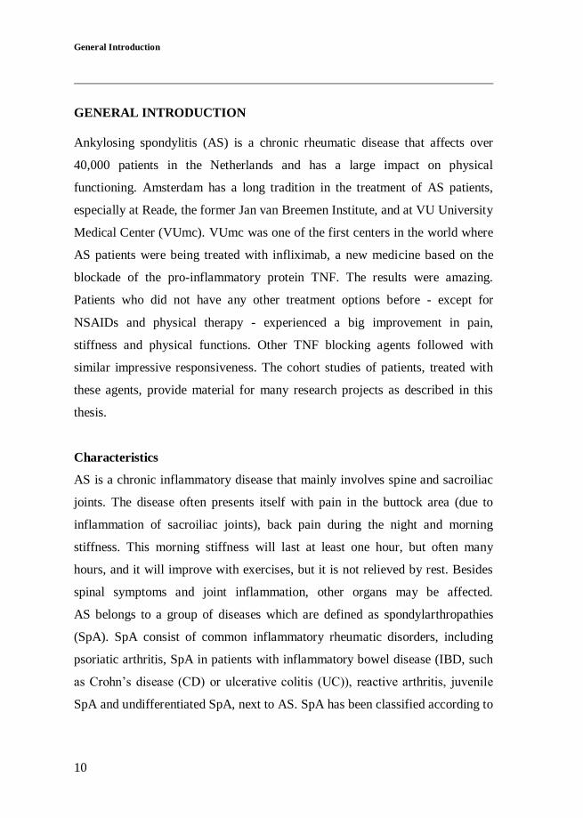
The diagnosis of definite AS has to comply with the 1984 modified New York
criteria: obligatory signs are signs of bilateral sacroiliitis grade 2-4 or unilateral
sacroiliitis grade 3-4 (visible on the X-ray of the pelvis, Figure 1) plus at least
one of the three of the following criteria: inflammatory back pain, limited
lumbar spinal motion in sagittal and frontal planes, and decreased chest
expansion(4). The clinical manifestations can be divided into spinal and
extraspinal features. Extraspinal manifestations are peripheral arthritis,
enthesitis, uveitis, psoriasis, inflammatory bowel disease, cardiac involvement
(such as aortic root and valve regurgitation, heart conduction disturbances and
increased cardiovascular risk) pulmonary involvement (pulmonary fibrosis,
bronchiectasis) or, occasionally, amyloidosis.
Epidemiology
The prevalence of AS ranges between 0.1-1.4% and the male-to-female ratio is
approximately 3:1(5;6). The majority of the patients have their first symptoms
between the ages of 15 and 40(7). In most cases, however, there is a significant
delay in diagnosis with an average delay in Western-Europe of 7.5 years(8).

General Introduction
12
This delay in diagnosis might be explained by the nonspecific, insidious
symptoms of low back pain at the onset of the disease.
Figure 1: Sacroiliitis (grade 2, bilateral).
Pathogenesis
The cause of AS is multifactorial, consisting of both hereditary and
environmental factors, which have not all been elucidated yet. With respect to
hereditary factors it is known that a strong genetic predisposition can be
ascribed to the human leukocyte antigen B27 (HLA-B27), which is prevalent in
more than 95% of the AS patients. This antigen is also present in 8% of the
healthy Dutch Caucasians(9), but is has higher prevalences in the northern parts
of Scandinavia (16%)(10).
Furthermore, it has been postulated that certain bacterial infections, such as
Chlamydia(11) or gastrointestinal infections (with Salmonella, Shigella,
Yersinia or Campylobacter) may contribute to the development of AS, as
similarly described in the onset of reactive arthritis.

Chapter 1
13
There is an increased concentration of T cells, macrophages and pro-
inflammatory cytokines in AS(12).
The course of the disease
The course of AS varies between mild, with little functional disability, and a
very severe, disabling form in a minority of cases. Rudwaleit et al identified
several prognostic factors for severe disease, such as male sex and elevated
CRP(13).
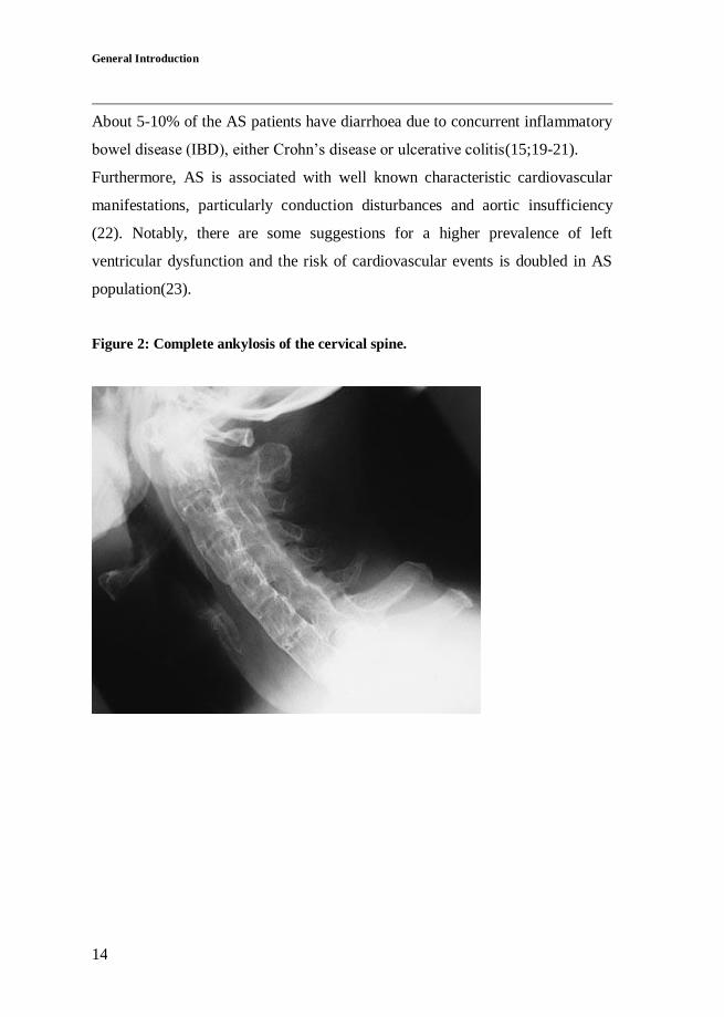
Spinal inflammation can result in formation of syndesmophytes of the spine that
may lead to ankylosis (Figure 2) and thoracic kyphosis. The same process of
inflammation with bone repair resulting in ankylosis occurs in the sacroiliac
joints (grade 4) of most AS patients. Furthermore, extraspinal complications
may occur. Peripheral arthritis occurs in approximately one third of the patients,
predominantly in the knees, hips and shoulders and usually in an asymmetrical
pattern(14). Besides impairment of function, particularly at an early stage of the
disease, arthritis results in cartilage degeneration and it might necessitate joint
replacement procedures. Inflammation at the insertions of ligaments, tendons, or
joint capsules to bone, which is termed enthesitis, is a characteristic feature of
spondyloarthropathy(15).
Vertebral fractures, in particular of the cervical spine, and cervical spine
dislocations can cause neurological deficits after minor trauma as the spine is
fragile due to ankylosis and osteoporosis. This complication has a Standard
Morbidity Ratio of 7.6(16-18) and can be overlooked in daily clinical practice
due to an overlap of symptoms (back pain) and difficulties in radiographic
detection due to additional bone formation of the spine.
Acute anterior uveitis occurs in 25-30% of the patients and can be the
presenting symptom of the disease. It is characterised by a red eye, unilateral
ocular pain and photophobia.
General Introduction
14
About 5-10% of the AS patients have diarrhoea due to concurrent inflammatory
bowel disease (IBD), either Crohn’s disease or ulcerative colitis(15;19-21).
Furthermore, AS is associated with well known characteristic cardiovascular
manifestations, particularly conduction disturbances and aortic insufficiency
(22). Notably, there are some suggestions for a higher prevalence of left
ventricular dysfunction and the risk of cardiovascular events is doubled in AS
population(23).
Figure 2: Complete ankylosis of the cervical spine.

Chapter 1
15
Therapy
Non-pharmacological treatment of AS includes patient information, physical
therapy and stimulating the patient to exercise frequently. Non steroidal anti-
inflammatory drugs (NSAIDs), such as diclofenac or naproxen, are
recommended as first line medical treatment and they generally decrease pain
and morning stiffness. Several studies have shown that selective COX-2
inhibitors, such as etoricoxib and celecoxib, are very effective in AS as
well(24;25). The selective COX-2 inhibitors can be used in case of relative
contraindications for NSAID use, such as dyspepsia or IBD.
Unfortunately, most disease modifying anti-rheumatic drugs (DMARDs) do not
seem to be effective in AS, although properly conducted studies with high
dosages are often lacking(26). Sulphasalazine may be considered for AS
patients with peripheral arthritis(27) and there is some debate about whether it
may be effective for spinal complaints as well(28;29).
Therapy with biologicals, especially tumor necrosis factor (TNF) inhibitors,
have transformed the treatment paradigm in AS. These drugs are produced with
recombinant DNA technologies and they block the protein TNF, which is one of
the main proinflammatory cytokines.
At the time of writing this thesis, three TNF blocking agents were approved and
registered for use in AS after large multicenter randomised clinical trials had
been performed. The first is infliximab, a chimeric monoclonal anti-TNF
antibody of human IgG1(30), the second, etanercept, a fusion protein consisting
of two TNF receptors linked to the Fc region of human IgG1(31), and the third,
adalimumab, a humanized monoclonal anti-TNF antibody of human IgG1(32).
Treatment of AS with anti-TNF has proven to be safe and very effective with
response rates varying from 50% to 76%, which are similar for all three TNF
blocking agents(30-32). These drugs differ in the way of administration.

General Introduction
16
Infliximab is administered intravenously in 6-8 weeks intervals and etanercept
and adalimumab are given as subcutaneous injections once a week or every
other week. Another difference is the efficacy on extra articular manifestations.
In contrast to infliximab and adalimumab, etanercept is not effective IBD(33).
The efficacy of etanercept on the recurrence of attacks of uveitis seems to be
lower than that of the other TNF blocking agents but this needs some further
research(32;34;35). When most studies described in this thesis were finished,
other TNF blocking agents emerged such as golimumab, which has recently
been registered and reimbursed by health insurance for the treatment of AS in
the Netherlands. Phase III trials are currently being conducted with
Certolizumab pegol, a polyethylene glycol (PEG) linked Fab' antibody fragment
of a humanized TNF inhibitor monoclonal antibody(36) in AS.
Increased susceptibility to infections, specifically reactivation of latent
tuberculosis is the most frequent side-effect of TNF blocking agents(37).
Therefore, every patient is screened for latent tuberculosis before start of
treatment according to the Dutch consensus guideline, available at www.nvr.nl.
A complicated side-effect of biologicals is their ability to provoke an immune
response, triggered by foreign components of the protein(38). Additionally,
auto-antibodies can be formed(39).
The high costs of TNF blocking agents (approximately 15.000 €/year)(40)
necessitate the presence of assessment tools in order to determine the disease
activity and response to therapy.
Disease activity scores and functional assessments
Unlike other rheumatic diseases, such as rheumatoid arthritis, acute phase
reactants such as erythrocyte sedimentation rate (ESR) and C-reactive protein
(CRP) do not always reflect disease activity in every AS patient(41). Therefore,
other outcome and disease activity parameters have been developed and

Chapter 1
17
validated over the past years. Up till now, disease activity is mostly determined
with the Bath ankylosing spondylitis disease activity index (BASDAI)(42). This
is a self-administered questionnaire consisting of 6 questions (Appendix). The
BASDAI is used for determination of ASAS response. For example, a 20%
decrease of the BASDAI or decrease with 2 units on a scale from 0 to 10 is
fulfilment of ASAS20 response.
Physical function can be measured with the Bath ankylosing spondylitis
functional index (BASFI), spinal mobility with the Bath ankylosing spondylitis
metrology index (BASMI) and questionnaires (visual analogue scales and
numeric rating scales) have been developed to assess pain and global disease
activity (Appendix).
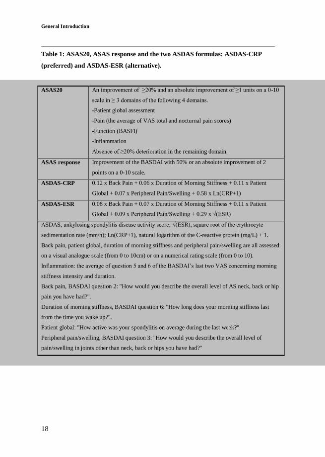
Recently, the Assessment of SpondyloArthritis international Society (ASAS)
has developed a new tool to assess disease activity: the AS disease activity
score, the ASDAS(43). It combines five disease activity variables with only
partial overlap, resulting in one single score with better truth (validity),
enhanced discriminative capacity and improved sensitivity to change as
compared to single-item variables(43;44). The ASDAS was not used as a
disease activity parameter in our studies because at the time the ASAS20
improvement criterion was used in most clinical trials. The definition of this
response parameter is described in Table 1.
General Introduction
18
Table 1: ASAS20, ASAS response and the two ASDAS formulas: ASDAS-CRP
(preferred) and ASDAS-ESR (alternative).
ASAS20 An improvement of ≥20% and an absolute improvement of ≥1 units on a 0-10
scale in ≥ 3 domains of the following 4 domains.
-Patient global assessment
-Pain (the average of VAS total and nocturnal pain scores)
-Function (BASFI)
-Inflammation
Absence of ≥20% deterioration in the remaining domain.
ASAS response Improvement of the BASDAI with 50% or an absolute improvement of 2
points on a 0-10 scale.
ASDAS-CRP 0.12 x Back Pain + 0.06 x Duration of Morning Stiffness + 0.11 x Patient
Global + 0.07 x Peripheral Pain/Swelling + 0.58 x Ln(CRP+1)
ASDAS-ESR 0.08 x Back Pain + 0.07 x Duration of Morning Stiffness + 0.11 x Patient
Global + 0.09 x Peripheral Pain/Swelling + 0.29 x √(ESR)
ASDAS, ankylosing spondylitis disease activity score; √(ESR), square root of the erythrocyte
sedimentation rate (mm/h); Ln(CRP+1), natural logarithm of the C-reactive protein (mg/L) + 1.
Back pain, patient global, duration of morning stiffness and peripheral pain/swelling are all assessed
on a visual analogue scale (from 0 to 10cm) or on a numerical rating scale (from 0 to 10).
Inflammation: the average of question 5 and 6 of the BASDAI’s last two VAS concerning morning
stiffness intensity and duration.
Back pain, BASDAI question 2: "How would you describe the overall level of AS neck, back or hip
pain you have had?".
Duration of morning stiffness, BASDAI question 6: "How long does your morning stiffness last
from the time you wake up?".
Patient global: "How active was your spondylitis on average during the last week?"
Peripheral pain/swelling, BASDAI question 3: "How would you describe the overall level of
pain/swelling in joints other than neck, back or hips you have had?"
Chapter 1
19
Thesis outline
Many AS patients (around 60%) show a good response to treatment with TNF
blocking agents. In a minority however, these drugs do not show efficacy
(primarily) or they lose their effectiveness during treatment (secondarily). In
some cases allergic reactions occur. This topic of unresponsiveness was
explored in Section I (Chapter 2-4). The main issue was whether non-
response and allergic reactions are related to immunogenicity of TNF blocking
agents in AS.
In order to select eligible patients for treatment with anti-TNF, patients were
categorized in according to their disease activity score, the BASDAI. As the
BASDAI is a patient-reported measure, and objectiveness might therefore be
disputable, there is some doubt about the reliability of this tool. In Section II
the changes of other biomarkers of disease activity were studied in relation to
the BASDAI and changes after treatment with anti-TNF were determined. In
Chapter 5.1 three types of inflammatory markers (ESR, CRP and serum
amyloid A protein: SAA) and their change upon TNF-blockade were
investigated. CRP levels are used more frequently to determine disease activity
in AS patients. These levels, though, do not necessarily reflect disease activity
in each patient. In Chapter 5.2, the relation between CRP levels and common
single-nucleotide polymorphisms (SNPs) and haplotypes in the CRP gene were
studied. Additionally, the relation between CRP levels and the BASDAI were
investigated.
Chapter 6 contains a review on Andersson lesions (ALs), destructive vertebral
lesions leading to pseudarthrosis, which can be a severe complication in long
standing AS. The prevalence of these lesions was studied in our cohort of AS
patients, detected with MRI.
General Introduction
20
Inflammation also plays an important role in atherosclerosis(36;45-47).
Therefore, a study on dyslipidemia and its relation with the inflammatory
markers was depicted in Chapter 7. In particular, the effect of anti-TNF on the
composition of HDL particles was studied.
In Section III we focus on extraspinal manifestations. At VUmc, a number of
patients who suffered from both AS and IBD are treated. Therefore, we were
interested in the serological similarities between AS and IBD patients, and
whether serological markers could be detected in AS patients who might be
prone to develop IBD. This study was depicted in Chapter 8. Finally, in
Chapter 9, cardiac manifestations of AS are addressed and the relation between
AS-related characteristics and conduction disturbances was investigated.

Chapter 1
21
REFERENCE LIST
(1) Dougados M, van der Linden S, Juhlin R, Huitfeldt B, Amor B, Calin
A, et al. The European Spondylarthropathy Study Group preliminary criteria for the classification of spondylarthropathy. Arthritis Rheum
1991 Oct;34(10):1218-27.
(2) Rudwaleit M, van der HD, Landewe R, Listing J, Akkoc N, Brandt J, et
al. The development of Assessment of SpondyloArthritis international Society classification criteria for axial spondyloarthritis (part II):
validation and final selection. Ann Rheum Dis 2009 Jun;68(6):777-83.
(3) Rudwaleit M, van der HD, Landewe R, Akkoc N, Brandt J, Chou CT, et al. The Assessment of SpondyloArthritis International Society
classification criteria for peripheral spondyloarthritis and for
spondyloarthritis in general. Ann Rheum Dis 2011 Jan;70(1):25-31.
(4) van der Linden SM, Valkenburg HA, Cats A. Evaluation of diagnostic criteria for ankylosing spondylitis. A proposal for modification of the
New York criteria. Arthritis Rheum 1984 Apr;27(4):361-8.
(5) Calin A, Fries JF. Striking prevalence of ankylosing spondylitis in "healthy" w27 positive males and females. N Engl J Med 1975 Oct
23;293(17):835-9.
(6) Will R, Edmunds L, Elswood J, Calin A. Is there sexual inequality in ankylosing spondylitis? A study of 498 women and 1202 men. J
Rheumatol 1990 Dec;17(12):1649-52.
(7) Feldtkeller E. [Age at disease onset and delayed diagnosis of
spondyloarthropathies]. Z Rheumatol 1999 Feb;58(1):21-30.
(8) Feldtkeller E, Bruckel J, Khan MA. Scientific contributions of
ankylosing spondylitis patient advocacy groups. Curr Opin Rheumatol
2000 Jul;12(4):239-47.
(9) van der Linden SM, Valkenburg HA, de Jongh BM, Cats A. The risk of
developing ankylosing spondylitis in HLA-B27 positive individuals. A
comparison of relatives of spondylitis patients with the general population. Arthritis Rheum 1984 Mar;27(3):241-9.

General Introduction
22
(10) Gran JT, Mellby AS, Husby G. The prevalence of HLA-B27 in
Northern Norway. Scand J Rheumatol 1984;13(2):173-6.
(11) van der Paardt M, van Denderen JC, van den Brule AJ, Morre SA, van
der Horst-Bruinsma IE, Bezemer PD, et al. Prevalence of Chlamydia
trachomatis in urine of male patients with ankylosing spondylitis is not increased. Ann Rheum Dis 2000 Apr;59(4):300-2.
(12) Bollow M, Fischer T, Reisshauer H, Backhaus M, Sieper J, Hamm B, et
al. Quantitative analyses of sacroiliac biopsies in spondyloarthropathies:
T cells and macrophages predominate in early and active sacroiliitis- cellularity correlates with the degree of enhancement detected by
magnetic resonance imaging. Ann Rheum Dis 2000 Feb;59(2):135-40.
(13) Rudwaleit M, Haibel H, Baraliakos X, Listing J, Marker-Hermann E, Zeidler H, et al. The early disease stage in axial spondylarthritis: results
from the German Spondyloarthritis Inception Cohort. Arthritis Rheum
2009 Mar;60(3):717-27.
(14) Resnick D, Niwayama G. Ankylosing Spondylitis. In: Resnick D, ed. Diagnosis of bone and joint disorders. [3
rd edition], 1008-1074. 1995.
Philadelphia, WB Saunders.
(15) McGonagle D, Khan MA, Marzo-Ortega H, O'Connor P, Gibbon W, Emery P. Enthesitis in spondyloarthropathy. Curr Opin Rheumatol 1999
Jul;11(4):244-50.
(16) Mitra D, Elvins DM, Speden DJ, Collins AJ. The prevalence of vertebral fractures in mild ankylosing spondylitis and their relationship
to bone mineral density. Rheumatology (Oxford) 2000 Jan;39(1):85-9.
(17) Donnelly S, Doyle DV, Denton A, Rolfe I, McCloskey EV, Spector
TD. Bone mineral density and vertebral compression fracture rates in ankylosing spondylitis. Ann Rheum Dis 1994 Feb;53(2):117-21.
(18) Ralston SH, Urquhart GD, Brzeski M, Sturrock RD. Prevalence of
vertebral compression fractures due to osteoporosis in ankylosing spondylitis. BMJ 1990 Mar 3;300(6724):563-5.
(19) Brophy S, Pavy S, Lewis P, Taylor G, Bradbury L, Robertson D, et al.
Inflammatory eye, skin, and bowel disease in spondyloarthritis: genetic, phenotypic, and environmental factors. J Rheumatol 2001
Dec;28(12):2667-73.

Chapter 1
23
(20) de Vlam K, Mielants H, Cuvelier C, De Keyser F, Veys EM, De Vos
M. Spondyloarthropathy is underestimated in inflammatory bowel disease: prevalence and HLA association. J Rheumatol 2000
Dec;27(12):2860-5.
(21) Rudwaleit M, Baeten D. Ankylosing spondylitis and bowel disease. Best Pract Res Clin Rheumatol 2006 Jun;20(3):451-71.
(22) Lautermann D, Braun J. Ankylosing spondylitis--cardiac
manifestations. Clin Exp Rheumatol 2002 Nov;20(6 Suppl 28):S11-
S15.
(23) Peters MJ, van Eijk I, Smulders YM, Serne E, Dijkmans BA, Van der
Horst-Bruinsma IE, et al. Signs of accelerated preclinical
atherosclerosis in patients with ankylosing spondylitis. J Rheumatol 2010 Jan;37(1):161-6.
(24) van der Heijde DM, Baraf HS, Ramos-Remus C, Calin A, Weaver AL,
Schiff M, et al. Evaluation of the efficacy of etoricoxib in ankylosing
spondylitis: results of a fifty-two-week, randomized, controlled study. Arthritis Rheum 2005 Apr;52(4):1205-15.
(25) Dougados M, Behier JM, Jolchine I, Calin A, van der Heijde DM,
Olivieri I, et al. Efficacy of celecoxib, a cyclooxygenase 2-specific inhibitor, in the treatment of ankylosing spondylitis: a six-week
controlled study with comparison against placebo and against a
conventional nonsteroidal antiinflammatory drug. Arthritis Rheum 2001 Jan;44(1):180-5.
(26) van der Horst-Bruinsma IE, Clegg DO, Dijkmans BA. Treatment of
ankylosing spondylitis with disease modifying antirheumatic drugs.
Clin Exp Rheumatol 2002 Nov;20(6 Suppl 28):S67-S70.
(27) Chen J, Liu C. Is sulfasalazine effective in ankylosing spondylitis? A
systematic review of randomized controlled trials. J Rheumatol 2006
Apr;33(4):722-31.
(28) Braun J, Van der Horst-Bruinsma IE, Huang F, Burgos-Vargas R,
Vlahos B, Koenig AS, et al. Clinical efficacy and safety of etanercept
versus sulfasalazine in patients with ankylosing spondylitis: a randomized, double-blind trial. Arthritis Rheum 2011 Jun;63(6):1543-
51.

General Introduction
24
(29) Maksymowych WP, Breban M, Braun J. Ankylosing spondylitis and
current disease-controlling agents: do they work? Best Pract Res Clin Rheumatol 2002 Sep;16(4):619-30.
(30) van der Heijde DM, Dijkmans B, Geusens P, Sieper J, DeWoody K,
Williamson P, et al. Efficacy and safety of infliximab in patients with ankylosing spondylitis: results of a randomized, placebo-controlled trial
(ASSERT). Arthritis Rheum 2005 Feb;52(2):582-91.
(31) Davis JC, Jr., van der Heijde DM, Braun J, Dougados M, Cush J, Clegg
DO, et al. Recombinant human tumor necrosis factor receptor (etanercept) for treating ankylosing spondylitis: a randomized,
controlled trial. Arthritis Rheum 2003 Nov;48(11):3230-6.
(32) van der Heijde DM, Kivitz A, Schiff MH, Sieper J, Dijkmans BAC, Braun J, et al. Efficacy and safety of adalimumab in patients with
ankylosing spondylitis: results of a multicenter, randomized, double-
blind, placebo-controlled trial. Arthritis Rheum 2006 Jul;54(7):2136-46.
(33) Braun J, Baraliakos X, Listing J, Davis J, van der Heijde DM, Haibel H, et al. Differences in the incidence of flares or new onset of
inflammatory bowel diseases in patients with ankylosing spondylitis
exposed to therapy with anti-tumor necrosis factor alpha agents. Arthritis Rheum 2007 May 15;57(4):639-47.
(34) Braun J, Baraliakos X, Listing J, Sieper J. Decreased incidence of
anterior uveitis in patients with ankylosing spondylitis treated with the anti-tumor necrosis factor agents infliximab and etanercept. Arthritis
Rheum 2005 Aug;52(8):2447-51.
(35) Guignard S, Gossec L, Salliot C, Ruyssen-Witrand A, Luc M, Duclos
M, et al. Efficacy of tumour necrosis factor blockers in reducing uveitis flares in patients with spondylarthropathy: a retrospective study. Ann
Rheum Dis 2006 Dec;65(12):1631-4.
(36) Schreiber S, Rutgeerts P, Fedorak RN, Khaliq-Kareemi M, Kamm MA, Boivin M, et al. A randomized, placebo-controlled trial of certolizumab
pegol (CDP870) for treatment of Crohn's disease. Gastroenterology
2005 Sep;129(3):807-18.

Chapter 1
25
(37) Keane J, Gershon S, Wise RP, Mirabile-Levens E, Kasznica J,
Schwieterman WD, et al. Tuberculosis associated with infliximab, a tumor necrosis factor alpha-neutralizing agent. N Engl J Med 2001 Oct
11;345(15):1098-104.
(38) Schellekens H. The immunogenicity of therapeutic proteins. Discov Med 2010 Jun;9(49):560-4.
(39) Swale VJ, Perrett CM, Denton CP, Black CM, Rustin MH. Etanercept-
induced systemic lupus erythematosus. Clin Exp Dermatol 2003
Nov;28(6):604-7.
(40) Farmacotherapeutisch Kompas 2012. Kostenoverzicht antirheumatica.
(41) Sheehan NJ, Slavin BM, Donovan MP, Mount JN, Mathews JA. Lack
of correlation between clinical disease activity and erythrocyte sedimentation rate, acute phase proteins or protease inhibitors in
ankylosing spondylitis. Br J Rheumatol 1986 May;25(2):171-4.
(42) Garrett S, Jenkinson T, Kennedy LG, Whitelock H, Gaisford P, Calin
A. A new approach to defining disease status in ankylosing spondylitis: the Bath Ankylosing Spondylitis Disease Activity Index. J Rheumatol
1994 Dec;21(12):2286-91.
(43) van der Heijde DM, Lie E, Kvien TK, Sieper J, van den Bosch F, Listing J, et al. ASDAS, a highly discriminatory ASAS-endorsed
disease activity score in patients with ankylosing spondylitis. Ann
Rheum Dis 2009 Dec;68(12):1811-8.
(44) Lukas C, Landewe R, Sieper J, Dougados M, Davis J, Braun J, et al.
Development of an ASAS-endorsed disease activity score (ASDAS) in
patients with ankylosing spondylitis. Ann Rheum Dis 2009
Jan;68(1):18-24.
(45) Libby P, Ridker PM, Maseri A. Inflammation and atherosclerosis.
Circulation 2002 Mar 5;105(9):1135-43.
(46) Ross R. The pathogenesis of atherosclerosis: a perspective for the 1990s. Nature 1993 Apr 29;362(6423):801-9.
(47) Ross R. Atherosclerosis--an inflammatory disease. N Engl J Med 1999
Jan 14;340(2):115-26.
General Introduction

Chapter 1
Section I
Immunogenicity

General Introduction

Chapter 1
Inefficacy of
ibody formation.
Inefficacy of infliximab in ankylosing spondylitis is
correlated with antibody formation.
Mirjam K. de Vries1, Gerrit Jan Wolbink
2,3, Steven O. Stapel
2, Els R. de Groot
2,
Ben A. C. Dijkmans1,3
, Lucien A. Aarden2, Irene E. van der Horst-Bruinsma
1.
Rheumatology Department VU University Medical Center1, Sanquin Research
2,
Rheumatology Department Jan van Breemen Institute/Reade3, Amsterdam, the
Netherlands.
Annals of the Rheumatic Diseases 2007;66(1):133-134.

Inefficacy of infliximab in AS is correlated with antibody formation
30
Tumor necrosis factor blocking agents such as infliximab have proved to be
effective in patients with ankylosing spondylitis (AS) as up to 60-70% of the
patients meet the 20% response criteria of assessment in ankylosing spondylitis
(ASAS-20)(1;2). However, it cannot be explained why 30% of patients fail to
respond and develop adverse reactions.
In rheumatoid arthritis, inefficacy to infliximab was associated with low serum
trough infliximab levels and the presence of antibodies to infliximab(3).
This study was designed to identify whether infliximab levels and antibodies to
infliximab predict clinical inefficacy and adverse events in AS.
Eight patients with active AS (fulfilling the 1984 modified New York
Criteria(4)) were treated according to the international ASAS consensus
statement(5), with infliximab 5mg/kg given intravenously at baseline, week 2,
6, and 12, and every 6 weeks thereafter. Sera were collected at 12 and 24 weeks
before infusing.
At every visit, questionnaires (e.g., Bath ankylosing spondylitis disease activity
index) to assess ASAS-20 were obtained and routine laboratory tests were
performed. These data were correlated with disease activity (ASAS-20), serum
trough infliximab levels and antibody levels.
All patients were men, with a median (range) age of 47 (24-52) years, and were
human leukocyte antigen B27 (HLA-B27) positive, with a median (range)
disease duration of 11 (1-28 years) years (Table 1). Patient 1 was concomitantly
treated with 15 mg methotrexate weekly and patient 3 was treated with
cyclosporine and sulfasalazine.
Most patients responded well to infliximab with a considerable decline in Bath
ankylosing spondylitis disease activity index (BASDAI), erythrocyte
sedimentation rate (ESR) and C-reactive protein (CRP), high serum trough
levels of infliximab and no development of antibodies to infliximab. However,

Chapter 2.1
31
two non-responders did not show detectable serum trough infliximab levels and
developed antibodies to infliximab after, respectively, 12 and 24 weeks. Patient
3 did not respond to treatment at all, whereas patient 5 met the ASAS-20
response criteria but had an increase in ESR and CRP levels. Both patients
developed an infusion reaction to infliximab.
Table 1: Clinical response to infliximab in ankylosing spondylitis patients in
relation to infliximab levels and antibodies to infliximab after 24 weeks.
Patient BASDAI
Week 0
Mean:5.5
Median:5.2
BASDAI
Week 24
Mean:1.9
Median:1.8
ESR
Week 0
Mean: 43
Median:6.5
ESR
Week 24
Mean: 11
Median:8.5
CRP
Week 0
Mean: 52
Median:25
CRP
Week 24
Mean: 8
Median:5
ASAS-
20
Infliximab
level
(ng/ml)
antibodies
to
infliximab
(ng/ml)
1 6.4 1.2 88 4 115 4 + 17800 0
2 4.5 0.7 90 8 120 6 + 10100 0
**3 * * 22 26 14 21 * 0 7200
4 7.2 0.0 72 18 104 6 + 20600 0
**5 4.7 3.1 12 18 7 20 + 0 15600
6 4.5 1.8 23 9 11 <2.5 + 16000 0
7 5.2 4.1 10 6 7 <2.5 + 10300 0
8 6.3 2.1 30 1 36 <2.5 + 16400 0
BASDAI, Bath ankylosing spondylitis disease activity index (scale 0-10); ESR, erythrocyte sedimentation rate
(mm/hour); CRP, C-reactive protein (mg/l); ASAS-20 response.
* not done due to severe visual impairment.
** are considered as non-responders due to increase of inflammatory parameters.
In this study on eight patients with AS, a correlation between efficacy of
infliximab and high levels of serum trough infliximab was shown. In 25% of
these patients with AS antibodies to infliximab developed within 24 weeks in
association with undetectable serum trough infliximab levels, inefficacy of
infliximab and infusion reactions.
The number of patients, however, is too small to draw definite conclusions, but
interestingly, these data point in the same direction as described previously in
rheumatoid arthritis(3). Lower serum trough infliximab levels could be

Inefficacy of infliximab in AS is correlated with antibody formation
32
explained by enhanced clearance because of immune complex formation
between anti-infliximab antibodies and infliximab. To prevent antibodies to
infliximab formation that might inhibit efficacy of infliximab, it might be
helpful to increase the dosage of infliximab (as occurs in treatment of
rheumatoid arthritis with infliximab), to shorten the interval between infliximab
infusions (as is currently the strategy in Crohn’s disease) or to provide
coadministration of other immunosuppressives (such as methotrexate). These
data should be confirmed in a larger group of patients with AS to develop a
more patient-specific treatment, which might predict the inefficacy of
infliximab at an early stage and might prevent adverse reactions.

Chapter 2.1
33
REFERENCE LIST
(1) Braun J, Baraliakos X, Brandt J, Listing J, Zink A, Alten R, et al.
Persistent clinical response to the anti-TNF-alpha antibody infliximab in patients with ankylosing spondylitis over 3 years. Rheumatology (Oxford)
2005 May;44(5):670-6.
(2) van der Heijde DM, Dijkmans B, Geusens P, Sieper J, DeWoody K,
Williamson P, et al. Efficacy and safety of infliximab in patients with ankylosing spondylitis: results of a randomized, placebo-controlled trial
(ASSERT). Arthritis Rheum 2005 Feb;52(2):582-91.
(3) Wolbink GJ, Vis M, Lems W, Voskuyl AE, de Groot E, Nurmohamed MT, et al. Development of antiinfliximab antibodies and relationship to
clinical response in patients with rheumatoid arthritis. Arthritis Rheum
2006 Mar;54(3):711-5.
(4) van der Linden SM, Valkenburg HA, Cats A. Evaluation of diagnostic criteria for ankylosing spondylitis. A proposal for modification of the
New York criteria. Arthritis Rheum 1984 Apr;27(4):361-8.
(5) Braun J, Pham T, Sieper J, Davis J, van der Linden S, Dougados M, et al. International ASAS consensus statement for the use of anti-tumour
necrosis factor agents in patients with ankylosing spondylitis. Ann Rheum
Dis 2003 Sep;62(9):817-24.

Decreased clinical response to infliximab in AS is correlated with anti-infliximab formation
34

Chapter 2.2
35
Decreased clinical response to infliximab in
ankylosing spondylitis is correlated with
anti-infliximab formation.
Mirjam K. de Vries1, Gert Jan Wolbink
2, Steven O. Stapel
2, Henk de Vrieze
2,
J. Christiaan van Denderen3, Ben A. C. Dijkmans
1, Lucien A. Aarden
2,
Irene E. van der Horst-Bruinsma1.
Rheumatology Department VU University Medical Center1, Sanquin Research
2,
Rheumatology Department Jan van Breemen Institute/Reade3, Amsterdam, the
Netherlands.
Annals of the Rheumatic Diseases 2007;66(9):1252-1254

Decreased clinical response to infliximab in AS is correlated with anti-infliximab formation
36
ABSTRACT
Objectives: Correlation of serum trough infliximab levels and antibodies to
infliximab (anti-infliximab) with clinical response in ankylosing spondylitis
(AS).
Methods: In accordance with the international assessments in ankylosing
spondylitis (ASAS) consensus statement, patients were treated with infliximab
(5 mg/kg) every 6 weeks after a starting regimen. Preinfusion sera were
collected at baseline, 24 and 54 weeks.
At every visit, the 20% improvement response (ASAS-20) was assessed and
laboratory tests performed.
Results: 24 of the 38 (63%) patients fulfilled ASAS-20 response criteria after
24 weeks of treatment and 21 (53%) after 54 weeks. After 54 weeks, 11 (29%)
patients showed undetectable serum trough infliximab levels and detectable
anti-infliximab; six of these patients developed an infusion reaction. Anti-
infliximab was found significantly more often (p=0.04) in ASAS-20 non-
responders compared with responders at week 54. Serum trough infliximab
levels were significantly (p<0.0001) lower in patients with (mean: 0.02 mg/l)
than in those without (mean: 12.7 mg/l) anti-infliximab .
Conclusions: In AS, high levels of serum trough infliximab correlated with a
good clinical response. Detection of anti-infliximab within 54 weeks is
associated with undetectable serum trough infliximab levels, reduced response
to treatment and increased risk of developing an infusion reaction.

Chapter 2.2
37
INTRODUCTION
Large randomised clinical trials have shown that tumor necrosis factor blocking
agents such as infliximab are very effective in ankylosing spondylitis (AS)(1). It
is unknown why more than 30% of patients with AS fail to respond, or why
some initial responders lose responsiveness during treatment and in some cases
even develop an infusion reaction. The non-responsiveness to infliximab might
be due to the development of antibodies against it, which has been described in
patients with rheumatoid arthritis and Crohn’s disease(2-5).
In AS, we recently showed in a small group of patients that detection of anti-
infliximab was associated with undetectable serum trough infliximab levels, a
reduced response to treatment and a higher risk of infusion reactions(6).
The aim of this study was to evaluate these data in a larger group of patients
with AS who were treated for a longer period of time and to specify the
influence on infliximab levels.
METHODS
All consecutive patients with AS (according to the 1984 modified New York
Criteria(7)) who received treatment with infliximab in our center were included
in this study.
Disease activity was measured with the Bath ankylosing spondylitis disease
activity index (BASDAI)(8) and the assessments in ankylosing spondylitis 20%
response criteria (ASAS-20)(9). Active disease was defined as a BASDAI≥4.
Response to treatment with infliximab was defined as fulfilment of the ASAS-
20 response criteria.
Patients with AS were treated with intravenous infliximab, 5 mg/kg bodyweight
at baseline, weeks 2 and 6, and every 6 weeks thereafter. This treatment was
initiated in accordance with the international ASAS consensus statement(9). In

Decreased clinical response to infliximab in AS is correlated with anti-infliximab formation
38
case of decrease of clinical response, the dose of infliximab was increased to 7.5
mg/kg. At each visit the presence of infections, side-effects or infusion
reactions, and the cause for discontinuation of therapy were recorded.
Questionnaires and routine laboratory tests were obtained. Preinfusion sera were
collected at baseline, weeks 24 and 54, before any dose escalation and at two
consecutive visits after dose escalation. After 24 weeks of treatment, serum
samples were collected from 15 patients to measure infliximab levels 2 weeks
after the infliximab infusion.
Validated immunoassays (Sanquin Research, Amsterdam, the Netherlands)
were used for detection of anti-infliximab and serum trough infliximab
levels(5). Trough serum infliximab levels were measured by ELISA, based on
the principle that infliximab is captured through its ability to bind tumor
necrosis factor. The assay, which was described previously, was modified
recently. It currently uses specific polyclonal rabbit antibodies to infliximab for
detection instead of the monoclonal anti human IgG that was previously used.
The sensitivity of detection is 0.0003 mg/l.
A radioimmunoassay (RIA) was used for anti-infliximab detection(5). Arbitrary
units per ml (AU/ml) were expressed as absolute amounts of infliximab-specific
IgG (mg/l)(10). (1 AU = 12 ng of infliximab-specific IgG). The cutoff value for
IgG anti-infliximab was determined by assaying in our anti-infliximab test 100
plasma samples from blood donors sent to Sanquin for IgG anti-tetanus toxoid
testing. The average result (AU/ml) + 6 SD was 12 AU/ml (0.144 mg/l).
The clinical data and presence of human leukocyte antigen B27 (HLA-B27)
were used to correlate disease activity with serum trough infliximab levels and
anti-infliximab levels. Differences between groups were tested with the Mann-
Whitney U test. Associations were calculated with logistic regression. The
threshold for significance was set at p<0.05. The last observation was carried
forward for patients who dropped out before week 54 .

Chapter 2.2
39
RESULTS
Demographic and clinical characteristics of the 38 patients included are shown
in Table 1.
Table 1: Demographic and clinical variables at baseline, week 24 and at week 54 of
patients with ankylosing spondylitis (N=38).
*Except when indicated otherwise, the values are the mean (SD).
HLA-B27, human leukocyte antigen B27; IBD, inflammatory bowel disease; BASDAI, Bath ankylosing
spondylitis disease activity index (0-10 cm); Morning stiffness, mean of item 5+6 of BASDAI (0-10 cm);
Global disease activity Visual Analogue Scale (0-10 cm); CRP, C-reactive protein, normal <8.0 mg/l;
detectable serum trough infliximab (% detectable); anti-infliximab, antibodies to infliximab (% detectable).
Compared with baseline *: p<0.001; #: p=0.005; ~: p=0.003
Variables Baseline Week 24 Week 54
Male sex, N (%) 26 (68)
Age in years 40 (10)
HLA-B27 positivity, N (%) 32 (84)
IBD, N (%) 6 (16)
Use corticosteroids, N (%) 3 (8)
Use other immunosuppressives, N (%) 6 (16)
BASDAI 6.4 (1.2) 3.6 (2.6) * 4.1 (3.0) *
Morning stiffness 6.3 (2.2) 3.0 (2.5) * 3.5 (3.2) *
Global disease activity VAS 6.8 (1.3) 4.3 (2.9) * 4.9 (3.4) #
CRP 37 (34.2) 9.3 (10.7) * 15.8 (21.1) ~
Detectable serum trough infliximab, N (%) 0 31 (82) 27 (71)
Anti-infliximab, N (%) 0 7 (18) 11 (29)

Decreased clinical response to infliximab in AS is correlated with anti-infliximab formation
40
Four patients were lost to follow-up before week 54: one wanted to become
pregnant, one preferred to be treated in a hospital nearby and two because of
comorbidities.
There was a significant decrease in BASDAI , morning stiffness, global disease
activity and C-reactive protein after 24 and 54 weeks of treatment (Table 1) and
all pre-treatment samples showed undetectable infliximab levels and no anti-
infliximab. We did not detect anti-infliximab in the presence of infliximab.
After 24 weeks, 24 patients (63%) met ASAS-20 response criteria. Responders
showed higher mean serum trough infliximab levels, and only two patients (8%)
showed anti-infliximab, compared with 5 (36%) of the non-responders
(p=0.08).
After 54 weeks of treatment, ASAS-20 response criteria were met by 21
patients (53%). The mean serum trough infliximab level for responders was
significantly (p<0.01) higher than that of the non-responders (8.2 mg/l vs 6.3
mg/l; Figure 1) and anti-infliximab was significantly (p<0.04) more often found
in non-responders. Only 5% (1 of 21) of the responders showed anti-infliximab,
compared with 59% (10 of 17) of the non-responders. In total, 9% (1 of 11)
patients with detectable anti-infliximab was classified as a responder at week
54, compared with 74% (20 of 27) of patients without anti-infliximab (Figure
2).

Chapter 2.2
41
Figure 1: Serum trough infliximab level for responders (N=21; 8.2 mg/l) and non-
responders (N=17; 6.3 mg/l) according to the ASAS-20 response criteria, at week
54.
Figure 2: Percentage of patients (N=38) with (9%) and without (74%) anti-
infliximab fulfilling the ASAS-20 response criteria at week 54.
No anti-infliximab Anti-infliximab
0
50
100p<0.001
AS
AS
-20
res
po
nse
Responders Non-responders0
10
20
30
p=0.018
Ser
um
tro
ug
h i
nfl
ixim
ab
lev
el (
mg
/l)

Decreased clinical response to infliximab in AS is correlated with anti-infliximab formation
42
After correction for probable confounding variables such as sex and HLA-B27,
the absence of anti-infliximab remained a significant determinant for ASAS-20
response with an odds ratio (OR) of 100 (95% CI 5.2 to 1000). Remarkably, the
presence of anti-infliximab was significantly associated with the absence of
HLA-B27 (OR=7.1; 95% CI 1.1 to 47.6; Pearson χ², p=0.03).
Two weeks after the infusion of week 24, significantly lower infliximab levels
were measured (20 mg/l compared with 51 mg/l; p<0.01) in patients who
developed anti-infliximab within 54 weeks of treatment.
In 12 patients, dose was increased within the 54 weeks, because of insufficient
clinical response. Nine (75%) of these patients showed anti-infliximab
antibodies. Increase in dose did not result in a significant increase of the serum
trough infliximab level (p=0.33), or a significant decrease in the anti-infliximab
level (p=0.90) and BASDAI (p=0.39). However, 2 of 12 patients reported
longer duration of effect.
Infusion reactions occurred in six patients. Most reactions were mild, and all
patients recovered after supportive therapy. Treatment with infliximab was
stopped in each case. Every infusion reaction was preceded by development of
anti-infliximab and consequently undetectable serum trough infliximab levels.
All antibodies to infliximab consisted of IgG1 and IgG4 subtypes. Although
these infusion reactions resemble a type 1 allergic reaction, no IgE was
detected. One patient’s pre-infusion serum contained an anti-infliximab level of
6.4 g/l, indicating that approximately half of his total serum IgG consisted of
infliximab-specific antibodies.

Chapter 2.2
43
DISCUSSION
A good clinical response of AS to treatment with infliximab was correlated with
the presence of high serum trough infliximab levels and the absence of anti-
infliximab antibodies, and inefficacy with the reverse. Moreover, these data
demonstrate that anti-infliximab antibodies precede an infusion reaction.
The mechanism of the decrease in efficacy can be explained by the lower serum
trough infliximab levels, probably caused by enhanced clearance due to immune
complex formation of anti-infliximab antibodies and infliximab. A recent study
in RA showed an enhanced clearance as a consequence of this process and an
accumulation in the macrophage-phagocyte system (liver and spleen)(11).
Indeed, in those patients with AS who developed detectable anti-infliximab
within 54 weeks of treatment with infliximab, a significantly lower infliximab
level was found 2 weeks after infusion compared with patients who did not
develop anti-infliximab.
Often, the infliximab dose is increased in AS when responsiveness decreases,
but reasons for dose escalation in AS are not yet well defined. In our small
sample, no clear increase in serum trough infliximab level after dose escalation
was shown.
Another option is to try to prevent anti-infliximab formation with the
concomitant administration of other immunosuppressive drugs such as
methotrexate; however, this medication is not efficacious in AS(12).
Remarkably, absence of HLA-27 shows significant correlation with anti-
infliximab formation. Further genetic evaluation will be performed to unravel
this interesting observation.
It also has to be investigated whether coadministration of immunosuppressive
drugs inhibits anti-infliximab formation, and whether infliximab levels can be
used for determination of the optimum dose of infliximab in AS.

Decreased clinical response to infliximab in AS is correlated with anti-infliximab formation
44
In accordance with our previous report, the efficacy of infliximab in AS is
clearly related to infliximab levels and the formation of anti-infliximab
antibodies. Detection of anti-infliximab antibodies within 54 weeks is
associated with undetectable serum trough infliximab levels, reduced response
to treatment and increased risk of development of an infusion reaction.

Chapter 2.2
45
REFERENCE LIST
(1) van der Heijde DM, Dijkmans B, Geusens P, Sieper J, DeWoody K,
Williamson P, et al. Efficacy and safety of infliximab in patients with ankylosing spondylitis: results of a randomized, placebo-controlled trial
(ASSERT). Arthritis Rheum 2005 Feb;52(2):582-91.
(2) Baert F, Noman M, Vermeire S, van Assche G, D'Haens G, Carbonez
A, et al. Influence of immunogenicity on the long-term efficacy of infliximab in Crohn's disease. N Engl J Med 2003 Feb 13;348(7):601-8.
(3) Hanauer SB, Feagan BG, Lichtenstein GR, Mayer LF, Schreiber S,
Colombel JF, et al. Maintenance infliximab for Crohn's disease: the ACCENT I randomised trial. Lancet 2002 May 4;359(9317):1541-9.
(4) St Clair EW, Wagner CL, Fasanmade AA, Wang B, Schaible T,
Kavanaugh A, et al. The relationship of serum infliximab
concentrations to clinical improvement in rheumatoid arthritis: results from ATTRACT, a multicenter, randomized, double-blind, placebo-
controlled trial. Arthritis Rheum 2002 Jun;46(6):1451-9.
(5) Wolbink GJ, Vis M, Lems W, Voskuyl AE, de Groot E, Nurmohamed MT, et al. Development of antiinfliximab antibodies and relationship to
clinical response in patients with rheumatoid arthritis. Arthritis Rheum
2006 Mar;54(3):711-5.
(6) de Vries MK, Wolbink GJ, Stapel SO, de Groot ER, Dijkmans BA,
Aarden LA, et al. Inefficacy of infliximab in ankylosing spondylitis is
correlated with antibody formation. Ann Rheum Dis 2007
Jan;66(1):133-4.
(7) van der Linden SM, Valkenburg HA, Cats A. Evaluation of diagnostic
criteria for ankylosing spondylitis. A proposal for modification of the
New York criteria. Arthritis Rheum 1984 Apr;27(4):361-8.
(8) Garrett S, Jenkinson T, Kennedy LG, Whitelock H, Gaisford P, Calin
A. A new approach to defining disease status in ankylosing spondylitis:
the Bath Ankylosing Spondylitis Disease Activity Index. J Rheumatol 1994 Dec;21(12):2286-91.

Decreased clinical response to infliximab in AS is correlated with anti-infliximab formation
46
(9) Braun J, Pham T, Sieper J, Davis J, van der Linden S, Dougados M, et
al. International ASAS consensus statement for the use of anti-tumour necrosis factor agents in patients with ankylosing spondylitis. Ann
Rheum Dis 2003 Sep;62(9):817-24.
(10) Schuurman J, Perdok GJ, Mueller GA, Benjamin DC, Yong TK, Chapman MD, et al. Mouse/human chimeric IgG1 and IgG4 antibodies
directed to the house dust mite allergen Der p 2: use in quantification of
allergen specific IgG. Clin Exp Allergy 1997 Sep;27(9):1095-102.
(11) van der Laken CJ, Voskuyl AE, Roos JC, Stigter van WM, de Groot ER, Wolbink G, et al. Imaging and serum analysis of immune complex
formation of radiolabelled infliximab and anti-infliximab in responders
and non-responders to therapy for rheumatoid arthritis. Ann Rheum Dis 2007 Feb;66(2):253-6.
(12) Haibel H, Brandt HC, Song IH, Brandt A, Listing J, Rudwaleit M, et al.
No efficacy of subcutaneous methotrexate in active ankylosing
spondylitis: a 16-week open-label trial. Ann Rheum Dis 2007 Mar;66(3):419-21.

Chapter 2.2
47

Decreased clinical response to infliximab in AS is correlated with anti-infliximab formation
48

Chapter 2.2
49
llijijikkgfgdgfddff
Decreased clinical response to adalimumab in
ankylosing spondylitis is associated with antibody
formation.
Mirjam K. de Vries1, Elisabeth Brouwer
5, Irene E. van der Horst-Bruinsma
1,
Anneke Spoorenberg6, J. Christiaan van Denderen
3, Anna Jamnitski
3, Michael
T. Nurmohamed1,3
, Ben A. C. Dijkmans1,3
, Lucien A. Aarden2,4
, Gert Jan
Wolbink2,3
.
Rheumatology Department VU University Medical Center1, Sanquin Research
2,
Rheumatology Department Jan van Breemen Institute/Reade3, Landsteiner
laboratory Academic Medical Center4, Amsterdam, Rheumatology Department
University Medical Center Groningen5, Rheumatology Department Medical
Center Leeuwarden6, the Netherlands.
Annals of the Rheumatic Diseases 2009;68(11):1787-1788.

Decreased clinical response to adalimumab in AS is associated with antibody formation
50
Treatment with anti-tumor necrosis factor (TNF) is very effective in most
patients with ankylosing spondylitis (AS), but inefficacy occurs in about 40% of
cases(1). Antibody formation against TNF blocking agents is an increasingly
recognised problem(2), however, no data have yet been reported on antibody
formation against adalimumab (anti-adalimumab) in AS. Lack of response can
be explained in two ways. Firstly, TNF might not be important for disease
activity in certain patients; and secondly, TNF inhibition might be insufficient.
The latter could be caused by excessive production of TNF, low compliance of
the patient, insufficient dosing or an enhanced clearance of adalimumab due to
antibody formation. Adalimumab is a fully human monoclonal antibody against
TNF but, despite this fact, an immune response still can be provoked by the
antigen binding site also known as the idiotype. In previous studies we have
described the problem of immunogenicity of TNF blocking drugs in patients
with rheumatoid arthritis (RA)(3), in patients with AS treated with infliximab(4)
and in patients with RA treated with adalimumab(5), and concluded that the
presence of antibodies against infliximab or adalimumab was associated with
low or undetectable serum levels of infliximab or adalimumab and clinical non-
response.
The objective of the present study was to investigate the relation between the
formation of anti-adalimumab, serum adalimumab levels and clinical response
in AS.
Patients with AS(6) were treated with adalimumab, 40 mg every other week,
according to the international ASAS consensus statement(7;8). Clinical
response was defined as a 50% improvement or an absolute improvement of 2
points on the BASDAI scale (0-10). Serum samples were collected at baseline
and after 3 and 6 months of treatment. Serum adalimumab levels were
determined with an ELISA and anti-adalimumab was measured with a validated
antigen binding test. The assays used were similar to those described previously

Chapter 3.1
51
for the detection of infliximab levels and antibodies against infliximab(4;5).
Thirty-five patients were included. After 6 months of treatment, 18 were ASAS
responders (Table 1). Within 6 months of treatment, 11 patients developed anti-
adalimumab with low or undetectable adalimumab levels, 9 were ASAS non-
responders (p=0.012) and 1 had an allergic reaction with flushing, dyspnoea and
undetectable serum adalimumab levels (Figure 1).
Table 1: Baseline characteristics and clinical variables.
Baseline
(N=35)
t=6 months
Male sex, N (%) 27 (76)
Mean (SD) age (years) 43 (12)
Disease duration (years) 9 (3.5-16.5)
HLA-B27 positivity, N (%) 24 (69)
Presence of IBD, N (%) 4 (12)
Presence of uveitis, N (%) 15 (46)
Presence of arthritis, N (%) 11 (32)
Anti-TNF used before, N (%) 10 (29)
BASDAI 6.2 (4.9-7.6) 3.0 (1.2-5.3) *
ASAS response (N responders , %) 18 (51%)
Global disease activity 7.2 (5.9-8.0) 2.1 (1.0-5.0) *
ESR 31 (19-44) 7 (3-15)*
CRP 21 (10-35) <5 (2-6) *
Unless otherwise indicated, values are the median (interquartile range).
ASAS, assessments in ankylosing spondylitis (decrease of 50% or > 2; global disease activity (0-10 scale));
BASDAI, Bath ankylosing spondylitis disease activity index (0-10 scale); CRP, C-reactive protein (normal
<10.0 mg/l); ESR, erythrocyte sedimentation rate (normal <15 mm/h); HLA-B27, human leukocyte antigen
B27; IBD, inflammatory bowel disease.
* p<0.0001 compared with baseline.

Decreased clinical response to adalimumab in AS is associated with antibody formation
52
Figure 1: Relation between the presence of anti-adalimumab and response of
ankylosing spondylitis to treatment with adalimumab.
p=0.012
Responders Non-responders0
25
50
75
100No anti-adalimumab
Anti-adalimumab
% o
f p
ati
en
ts
Thus, anti-adalimumab was detected in 31% of the patients after 6 months of
treatment and this corresponded with diminished or undetectable serum
adalimumab levels in these patients. These preliminary observations will need
confirmation in a larger study. In contrast with the treatment of RA,
adalimumab is given without methotrexate in the treatment of AS. This might
be an explanation for the higher incidence of anti-adalimumab formation in AS.
In Crohn’s disease and in RA, the concomitant use of immunosuppressive drugs
or corticosteroids has been proved to decrease antibody formation against
infliximab(9;10).
To date, no other papers have reported on immunogenicity in the treatment of
AS with adalimumab and no systemic allergic reactions have been described.
The detection of antibodies might predict the inefficacy of adalimumab and
should be explored further for use in daily clinical practice.

Chapter 3.1
53
REFERENCE LIST
(1) van der Heijde DM, Schiff MH, Sieper J, Kivitz A, Wong RL, Kupper
H, et al. Adalimumab effectiveness for the treatment of ankylosing spondylitis is maintained for up to 2 years: long-term results from the
ATLAS trial. Ann Rheum Dis 2008 Aug 13.
(2) Aarden LA, Ruuls SR, Wolbink GJ. Immunogenicity of anti-tumor
necrosis factor antibodies-toward improved methods of anti-antibody measurement. Curr Opin Immunol 2008 Jul 19.
(3) Wolbink GJ, Vis M, Lems W, Voskuyl AE, de Groot ER, Nurmohamed
MT, et al. Development of antiinfliximab antibodies and relationship to clinical response in patients with rheumatoid arthritis. Arthritis Rheum
2006 Mar;54(3):711-5.
(4) de Vries MK, Wolbink GJ, Stapel SO, de Vrieze H, Van Denderen JC,
Dijkmans BA, et al. Decreased clinical response to infliximab in ankylosing spondylitis is correlated with anti-infliximab formation. Ann
Rheum Dis 2007 Sep;66(9):1252-4.
(5) Bartelds GM, Wijbrandts CA, Nurmohamed MT, Stapel SO, Lems WF, Aarden LA, et al. Clinical response to adalimumab: The relationship
with anti-adalimumab antibodies and serum adalimumab concentrations
in rheumatoid arthritis. Ann Rheum Dis 2007 Mar 9.
(6) van der Linden S, Valkenburg HA, Cats A. Evaluation of diagnostic
criteria for ankylosing spondylitis. A proposal for modification of the
New York criteria. Arthritis Rheum 1984 Apr;27(4):361-8.
(7) Braun J, Pham T, Sieper J, Davis J, van der Linden S, Dougados M, et al. International ASAS consensus statement for the use of anti-tumour
necrosis factor agents in patients with ankylosing spondylitis. Ann
Rheum Dis 2003 Sep;62(9):817-24.
(8) Braun J, Davis J, Dougados M, Sieper J, van der Linden S, van der
Heijde DM. First update of the international ASAS consensus statement
for the use of anti-TNF agents in patients with ankylosing spondylitis. Ann Rheum Dis 2006 Mar;65(3):316-20.

Decreased clinical response to adalimumab in AS is associated with antibody formation
54
(9) Baert F, Noman M, Vermeire S, van Assche G, D'Haens G, Carbonez
A, et al. Influence of immunogenicity on the long-term efficacy of infliximab in Crohn's disease. N Engl J Med 2003 Feb 13;348(7):601-8.
(10) Maini RN, Breedveld FC, Kalden JR, Smolen JS, Davis D, Macfarlane
JD, et al. Therapeutic efficacy of multiple intravenous infusions of anti-tumor necrosis factor alpha monoclonal antibody combined with low-
dose weekly methotrexate in rheumatoid arthritis. Arthritis Rheum 1998
Sep;41(9):1552-63.

Chapter 3.1
55

Decreased clinical response to adalimumab in AS is associated with antibody formation
56

Chapter 3.1
57
Adalimumab in juvenile rheumatoid arthritis.
Comment on the article by Lovell and colleagues.
Mirjam K. de Vries1, Irene. E. van der Horst-Bruinsma
1 and Gerrit Jan
Wolbink2.
Rheumatology Department VU University Medical Center1, Sanquin Research
2,
Amsterdam, the Netherlands.
New England Journal of Medicine 2008;359(23);2496.

Comment on adalimumab in juvenile rheumatoid arthritis
58
To the editor: Lovell and colleagues conclude that adalimumab appeared to be
efficacious in children with polyarticular-course juvenile rheumatoid arthritis.
Immunogenicity is emerging as an important problem in treatment with
monoclonal antibodies(1). Lovell and colleagues report that approximately 16%
of the patients had anti-adalimumab antibodies, which did not seem to interfere
with the efficacy of adalimumab. This incidence is higher than the 5% incidence
among adults with rheumatoid arthritis, reported by Abbott Laboratories(2). We
wonder which method was used to detect these antibodies, since the use of
different methods hampers the comparison of results(1). The findings of Lovell
and colleagues are in accordance with those of our study, in which anti-
adalimumab antibodies were detected in 17% of the patients with rheumatoid
arthritis after 6 months of treatment(3). In our study, however, the presence of
these antibodies was associated with low or undetectable serum adalimumab
levels and a reduced clinical response, which was also observed in Crohn’s
disease(4). Therefore, it would be interesting to know more details about the
relation among anti-adalimumab antibodies, serum adalimumab levels, and the
American College of Rheumatology Pediatric response.

Chapter 3.2
59
REFERENCE LIST
(1) Aarden LA, Ruuls SR, Wolbink GJ. Immunogenicity of anti-tumor
necrosis factor antibodies-toward improved methods of anti-antibody measurement. Curr Opin Immunol 2008 Jul 19.
(2) Humira (adalimumab) Package Insert. http://www abbott com
(3) Bartelds GM, Wijbrandts CA, Nurmohamed MT, Stapel SO, Lems WF, Aarden LA, et al. Clinical response to adalimumab: The relationship with
anti-adalimumab antibodies and serum adalimumab concentrations in
rheumatoid arthritis. Ann Rheum Dis 2007 Mar 9.
(4) West RL, Zelinkova Z, Wolbink GJ, Kuipers EJ, Stokkers PC, van der
Woude CJ. Immunogenicity negatively influences the outcome of
adalimumab treatment in Crohn;s disease. Aliment Pharmacol Ther 2008
Aug 8.

Comment on adalimumab in juvenile rheumatoid arthritis

Chapter 3.2
Immunogenicity does not influence treatment with
etanercept in patients with ankylosing spondylitis.
Mirjam K. de Vries1, Irene E.van der Horst-Bruinsma
1, Michael T.
Nurmohamed1,3
, Lucien A. Aarden2, Steven O. Stapel
2, Mike J.L. Peters
1,3,
J. Christiaan van Denderen3, Ben A.C. Dijkmans
1, Gerrit J.Wolbink
2,3.
Rheumatology Department VU University Medical Center1, Sanquin Research
2,
Rheumatology Department Jan van Breemen Institute/Reade3, Amsterdam, the
Netherlands.
Annals of the Rheumatic Diseases 2008;68(4):531-535.

Immunogenicity does not influence treatment with etanercept in patients with AS
62
ABSTRACT
Background: Immunogenicity, specifically the onset of antibodies against
Tumor Necrosis Factor (TNF) blocking agents, seems to play an important role
in non-response to treatment with these drugs.
Objectives: To assess the relation of clinical response of ankylosing spondylitis
(AS) to etanercept with etanercept levels, and the presence of antibodies to
etanercept.
Methods: AS patients were treated with etanercept 25 mg twice weekly,
according to the international ASAS consensus statement. Sera were collected
at baseline, after 3 and 6 months of treatment. Clinical response was defined as
a 50% improvement or as an absolute improvement of 2 points on a 0-10 scale
of BASDAI.
Functional etanercept levels were measured by a newly developed ELISA,
measuring the binding of etanercept to TNF. Antibodies against etanercept were
measured with a two-site assay and antigen binding test. Clinical data were used
to correlate disease activity with serum etanercept levels.
Results: 53 consecutive patients were included. After 3 months of treatment 40
patients (76%) fulfilled the response criteria. Mean etanercept levels were 2.7
mg/l and 3.0 mg/l after 3 and 6 months respectively. Characteristics and
etanercept levels of responders and non-responders were similar. No antibodies
to etanercept were detected with any of the assays.
Conclusion: Etanercept levels of responders and non-responders were similar
and no antibodies to etanercept were detected with any of the assays. This study
indicates that etanercept is much less immunogenic compared with the other
TNF blocking agents.

Chapter 4
63
INTRODUCTION
Ankylosing spondylitis (AS) is a chronic inflammatory disease, which can
result in invalidating deformities of the joints and spine at an early age. Until
recently, treatment was mainly based on non-steroidal anti-inflammatory drugs
(NSAIDs) and physical therapy. Most disease modifying anti-rheumatic drugs
(DMARDs) do not seem to be effective in AS, although properly conducted
studies are lacking(1). The introduction of tumor necrosis factor (TNF) blocking
agents, i.e., infliximab(2), etanercept(3) and adalimumab(4) have changed the
treatment options in AS radically. The majority of AS patients, who fulfil the
assessment of ankylosing spondylitis (ASAS) guidelines for anti-TNF
treatment, respond very well. Nevertheless, TNF blocking agents still fail to
reach efficacy in approximately 30% of patients with AS.
A possible explanation for this failure could be the formation of antibodies,
which results in lower or undetectable serum levels of the biological.
For etanercept, however, it is unclear whether a relation between clinical
response and the formation of antibodies is present in AS patients. In addition,
many questions concerning immunogenicity have not been answered yet and
different methods of detection of anti-etanercept are being used, which makes
the results difficult to compare(5;6).
In our previous studies, we demonstrated a correlation between clinical
response and serum trough infliximab levels, adalimumab levels and the onset
of antibodies against these drugs(7;8). In this study, we used the same approach
as in our previous studies, to investigate the relation between clinical response,
functional etanercept levels and the detection of anti-etanercept antibodies in
AS patients. Besides, in a few patients the etanercept levels were measured
daily to investigate their course over time.

Immunogenicity does not influence treatment with etanercept in patients with AS
64
PATIENTS AND METHODS
Patients and study protocol
Consecutive AS patients, attending the outpatient clinics of Jan van Breemen
Institute or VU University Medical Center, who were scheduled for treatment
with etanercept, were included and followed prospectively. All AS patients
fulfilled the modified New York criteria and started using etanercept according
to the ASAS consensus statement on the initiation of TNF blocking agents in
AS(9). According to this ASAS consensus, patients must have an insufficient
response to non-steroid anti-inflammatory drugs (NSAIDs) and a Bath
ankylosing spondylitis disease activity index (BASDAI) above 4 (0-10 scale)
before starting treatment with etanercept. After tuberculosis was excluded by
means of a tuberculin skin test and chest X-rays, subcutaneous injections with
etanercept 25mg were taken twice a week. Concomitant medication remained
unaltered for at least three months after the start of etanercept treatment.
Demographic data collected at baseline were recorded from medical history and
patients’ medical records. The study was approved by the medical ethical
committee and all patients gave their written informed consent.
Outcome measures
Data were collected at baseline, and after 3 and 6 months of treatment. During
every visit questionnaires such as BASDAI, patient global disease activity and
Bath ankylosing spondylitis functional index (BASFI) were obtained and after
three months patients were checked whether they met the ASAS response
criteria.
The primary outcome measure was clinical response after 3 months of treatment
with etanercept, according to the“ International ASAS Consensus Statement for
the use of TNF-agents in patients with AS” which is equivalent to the Dutch

Chapter 4
65
guidelines for continuation of TNF blocking agents(1;9-11). In this consensus
statement, ASAS response was defined as a 50% improvement or as an absolute
improvement of 2 points of the BASDAI (0-10 scale) and an expert opinion in
favour of continuation of treatment after 3 months. Routine laboratory tests
(ESR, CRP) were performed. A CRP below 8.0 mg/l was considered to be
normal.
Etanercept levels and antibodies against etanercept were measured in patients’
sera at baseline, and after 3 and 6 months. Furthermore, in five patients serum
etanercept levels were measured daily in between two consecutive subcutaneous
injections.
Assessment of functional serum etanercept levels
Etanercept levels were measured by means of a newly developed ELISA, based
on the principle that etanercept is captured through its ability to bind TNF
(Sanquin, Diagnostic Services, Amsterdam, the Netherlands). The sensitivity of
detection is 1 ng/ml (=0.001mg/l).
In short, a mouse monoclonal antibody directed against TNF (CLB TNF/5) was
coated overnight at room temperature (0.2 µg/ well) on flat bottomed microtitre
plates. Recombinant TNF (5 ng/well) in HPE buffer (Sanquin, Diagnostic
Services, Amsterdam, The Netherlands) was added and remained in for 1 hour.
After washing the plates with phosphate buffered saline/0.02% Tween, patients’
serum samples were added in different dilutions in high performance ELISA
(HPE) buffer and incubated for 1 hour at 37°C. Plates were washed with
phosphate
buffered saline/0.04% Tween, and incubated with biotinylated
polyclonal rabbit antibodies against etanercept in 100 µl HPE buffer for 1 hour
at 37°C. Subsequently,
after washing the plates, poly-HRP-conjugated
streptavidin was added (30 min at 30°C), followed by incubation with TMB.
The reaction
was stopped with 2M H2SO4. Absorption at 450 nm was

Immunogenicity does not influence treatment with etanercept in patients with AS
66
determined, and results were related to a titration curve of etanercept, which
was present in each plate. Functionally active etanercept was measured because
of its ability to bind TNF.
Assessment of antibodies against etanercept
Two-site assay
Anti-etanercept antibodies were determined by a two-site assay, in the
following manner: etanercept coupled to Sepharose (100 g/100 ml) was used
to bind etanercept-specific antibodies from 1 l of patient serum, diluted in
PBS/BSA (0.3%)/Tween (0.2%), during an overnight incubation on a rotator.
After removal of non-bound serum components by washing, 125
I-radiolabelled
etanercept (~ 1 ng per test) was added, followed by a second overnight rotating
incubation in Freeze buffer (Sanquin, Amsterdam, the Netherlands). The non-
bound radiolabel was washed off, and test results were compared to a
calibration curve made of serum from an etanercept-vaccinated rabbit (Figure
1). Cutoff level for a positive signal was set at 0.95% binding (mean+3SD of
the pre-treatment values).
Antigen binding test
Besides, testing for antibodies was carried out by using an antigen binding test,
which is in principle similar to the tests we routinely use for detection of
antibodies against infliximab(12;13).
Pepsin-treated 125
I-radiolabelled etanercept was used for the detection of
antibodies. The same rabbit-derived calibration curve as applied in the two-site
assay was used for the interpretation of patients’ results. Cutoff level for a
positive signal was set at 1.07% binding (mean+3SD of the pre-treatment
values).

Chapter 4
67
Figure 1: Calibration curve of rabbit anti-etanercept serum as a positive control
for the two-site assay.
Statistical analysis
The last observation was carried forward in patients who had dropped out
before 6 months of treatment, due to ASAS non-response, adverse events or loss
to follow-up.
Data were expressed as mean (SD) or median (interquartile range) where
appropriate. The distribution of variables was tested for normality and
transformed if possible. Independent t-tests were used for variables with a
normal distribution and nonparametric tests (Wilcoxon signed-rank test or
Mann-Whitney U test) for skewed variables. Pearson χ² tests were conducted
for dichotomous variables.
Logistic regression analyses were conducted to examine associations between
ASAS response and serum etanercept level, and were corrected for the possible
influence of demographic, clinical and laboratory variables. Subsequently, a
linear regression model was used to investigate whether serum etanercept levels
0
10
20
30
0 20 40 60 80
nl rabbit serum/test
% b
ind
ing
ra
dio
lab
ell
ed
eta
nercep
t

Immunogenicity does not influence treatment with etanercept in patients with AS
68
were associated with any of the demographic, clinical or laboratory variables.
The following variables were tested: sex, age, ethnicity, BMI and HLA-B27,
baseline bodyweight, baseline BMI, decrease of Patient Global assessment,
absolute decrease of ESR and CRP level. Calculations were made using SPSS
14.0 software. The threshold for significance was set at p<0.05.
RESULTS
Fifty-three patients were included in this study in the period of July 2004 until
March 2006. The demographic data and baseline characteristics are shown in
Table 1.
Table 1: Demographic data and baseline characteristics.
Variables Baseline 3 Months 6 Months
Male sex, N (%) 40 (76)
Age (years) 41 (11)
Caucasian, N (%) 45 (85)
BMI 25 (4.4)
HLA-B27 positivity, N (%) 44 (88)
Peripheral arthritis, N (%) 28 (53)
BASDAI 6.4 (1.3) 3.1 (2.0) * 2.5 (1.7) *
Patient global disease activity VAS 7.2 (1.9) 3.2 (2.4) * 2.5 (2.1) *
BASFI 6.2 (2.1) 4.1 (2.5) * 3.5 (2.5) *
ESR (median, range) 22 (1-114) 5 (1-58) * 4 (1-33) *
CRP (median, range) 17 (1-92) 4 (1-44) * 4 (1-74) *
Serum etanercept levels 0 2.7 (1.2) 3.0 (1.0)
Antibodies to etanercept 0 0 0
Except when indicated otherwise, the values are the mean (SD). * Significance level: p<0.001
BMI, body mass index; HLA-B27, human leukocyte antigen B27; BASDAI, Bath ankylosing spondylitis
disease activity index (0-10); BASFI, Bath ankylosing spondylitis functional index (0-10); VAS, visual
analogue scale; ESR, erythrocyte sedimentation rate, normal<10 mm/hour; CRP, C-reactive protein,
normal<8.0 mg/l; mean serum etanercept levels (mg/l); antibodies to etanercept (mg/l).

Chapter 4
69
Bias due to missing data was excluded, for ASAS response did not differ
between patients with available data and those with missing data at 6 months.
Treatment with etanercept resulted in significant clinical improvement after 3
and 6 months, when compared to baseline (Table 1). The mean BASDAI,
Patient global disease activity, BASFI and ESR were even lower after 6 months
of treatment. Baseline characteristics did not differ significantly for responders
and non-responders (Table 2).
After 3 months of treatment, 40 of the patients (76%) met the ASAS response
criteria. The etanercept levels were measured in 48 patients after 3 months of
treatment and in 41 patients after 6 months of treatment: ten of 41 samples
turned out to be samples of ASAS non-responders. These patients should have
stopped treatment with etanercept after 3 months because of insufficient
improvement of the BASDAI. However, they continued their treatment based
on the expert opinion of the rheumatologist.
At baseline serum etanercept levels were undetectable, mean serum etanercept
levels were 2.7 mg/l (SD 1.2 mg/l) and 3.0 mg/l (SD 1.0 mg/l) after 3 and 6
months of treatment respectively. In each patient etanercept levels could be
measured and were within the same range (Figure 2). The mean etanercept
levels of responders and non-responders were similar, which could be explained
by the fact that antibodies against etanercept were not detected in these patients
with any of the assays (Table 2 and Figure 2). Besides the lack of association
between clinical response and serum etanercept levels, there also turned out to
be no significant association between sex, age, ethnicity, HLA-B27, baseline
bodyweight, baseline BMI, decrease of Patient Global assessment, absolute
decrease of ESR and CRP level, and serum etanercept levels.
In addition, in five patients etanercept levels were measured at daily intervals
between two subsequent injections with a mean of 3.5 mg/l, SD 1.2 mg/l and a
variance of 1.5. Furthermore, sera of six non-responders were taken three

Immunogenicity does not influence treatment with etanercept in patients with AS
70
months after the final etanercept injection and, as expected, etanercept was no
longer present in these sera and antibodies against etanercept could not even be
detected in these patients.
Table 2: Baseline characteristics, baseline ESR and CRP, and etanercept levels
after 3 months of treatment for ASAS responders and non-responders.
Variables Responders
N = 40
Non-responders
N = 13
Significance
level
Male sex, N (%) 29 (73) 11 (85) 0.38
Age in years 41 (11.2) 41 (8.8) 0.82
Caucasian, N (%) 36 (90) 9 (69) 0.07
BMI 25 (4.8) 24 (3.1) 0.53
HLA-B27 positivity, N (%) 34 (90) 10 (83) 0.57
Peripheral arthritis, N (%) 21 (53) 7 (54) 0.99
BASDAI 6.4 (1.3) 6.2 (1.4) 0.57
Patient global disease activity VAS 7.1 (1.5) 7.4 (1.9) 0.31
BASFI 6.2 (2.1) 6.2 (2.2) 0.86
ESR (median, range, mm/h) 22 (1-114) 22 (1-87) 0.82
CRP (median, range) 18 (1-91) 10 (1-92) 0.45
Etanercept level (mg/l) 2.7 (1.3) 2.8 (1.1) 0.50
Except when indicated otherwise, the values are the mean (SD).
BMI, body mass index; HLA-B27, human leucocyte antigen B27; BASDAI, Bath ankylosing spondylitis
disease activity index; VAS, visual analogue scale; BASFI, Bath ankylosing spondylitis functional index;
ESR, erythrocyte sedimentation rate, normal<10 mm/hour; CRP, C-reactive protein, normal<8.0 mg/l.
Adverse events
One patient had an adverse reaction of flushing and dyspnea after 2 months of
treatment with etanercept. Another patient developed urticaria and atopic
dermatitis after 27 months of treatment with the prefilled syringe of etanercept,
however, this patient showed none of these symptoms after retreatment with

Chapter 4
71
etanercept in the form of lyophilized powder. Both patients had previously
developed an infusion reaction to infliximab after which they had switched to
etanercept. No antibodies against etanercept were detected in any of the patients
with an adverse reaction.
Figure 2: Etanercept levels (mg/l) for ASAS responders and non-responders after 3
months of treatment.
R NR0
1
2
3
4
5
6
7
8ASAS responders
ASAS non-respondersp=0.4
Eta
nercep
t le
vel
(mg
/l)
DISCUSSION
Seventy-six percent of the AS patients were classified as responders to
etanercept after 3 months of treatment, which is comparable to the response rate
observed in clinical trials. No correlation was found between etanercept levels,
formation of antibodies against etanercept and clinical response. All patients
had detectable serum levels of etanercept and no antibodies against etanercept
were found. Interestingly, there seemed to be no difference in mean etanercept
levels and the onset of antibodies against etanercept between responders and
non-responders. These findings are in contrast with our previous studies with
infliximab(8;13) and adalimumab(7) in RA and AS, where a strong relation was
found between a decreased response and the onset of antibodies against TNF

Immunogenicity does not influence treatment with etanercept in patients with AS
72
blocking agents. These antibodies against TNF blocking agents often resulted in
low or absent serum levels of these drugs which would explain the decrease in
efficacy, but apparently this is not the case in etanercept. Therefore, this study
seems to confirm the hypothesis that etanercept is less immunogenic than other
TNF blocking agents.
Although etanercept levels have not yet been correlated with clinical response,
some studies do report on the detection of antibodies against etanercept. In
these, etanercept has been labelled as having antibody formation in less than 6%
of the cases and no clear relation with clinical response was detected(5;6;14).
These discrepancies regarding anti-therapeutic drug antibodies could be
explained by the use of different detection methods. ELISA is known to give
more false positive signals than antigen binding assays and it is therefore
difficult to compare other results with our method. The detection method for
anti-etanercept used by our laboratory may be regarded as not very sensitive.
However, this does not agree with the fact that functional etanercept levels
could be measured in all patients, because the presence of antibodies would
have resulted in an enhanced clearance and removal of etanercept. Furthermore,
similar techniques for measuring functional etanercept levels and detecting of
antibodies against etanercept were proven to be sensitive in our previous studies
with other TNF blocking agents.
Several arguments are in favour of the hypothesis that etanercept shows less
immunogenicity compared with other TNF-inhibitors. Firstly, etanercept has a
less immunogenic structure compared with the other TNF blocking agents.
Etanercept is a dimeric fusion protein consisting of two TNF receptors, linked
to the Fc portion of an immunoglobin (Ig)G1. Only the fusion part of the
molecule may contain immunogenic epitopes. Infliximab is a chimerical
monoclonal IgG1 antibody against TNF, partly consisting of murine protein.
Adalimumab is a fully human monoclonal antibody against TNF. These

Chapter 4
73
monoclonal antibodies have more epitopes within the variable region of the
antibody to which an immune response can be directed. Secondly, major
fluctuations in serum levels may precipitate an immune response and the
development of antibodies against the TNF blocking agent(15). This is mainly
the case in treatment with infliximab, which is administered once every six to
eight weeks. Treatment with etanercept, however, produces stable levels
between two injections and is dosed much more frequently, which is in line with
previous findings(16-19). The variance of 1.5 of the etanercept levels measured
at daily intervals between two subsequent injections measured in our five
patients was very low, compared with the variance of all serum trough
infliximab levels measured in our previous study, which was 80. The overall
variance of all etanercept levels was 1.3, which is also very low. Thirdly, there
may be different mechanisms for non-response. Theoretically, non-responders
can be divided into two categories: true non-responders, whose illness is not
mainly caused by excess production of TNF, and non-responders whose illness
is caused by inadequate blocking of TNF. The latter can be caused by enhanced
clearance or as a result of inadequate dosing. A dose-response relation of
etanercept in AS has not been investigated before. There is a possibility that
small differences in etanercept levels could not be found because of the random
timing of sampling between two injections. The most important argument
against this view is probably the observation that etanercept levels, measured at
several time intervals between two subsequent injections in the five patients,
were quite stable. Furthermore, other authors have confirmed that etanercept
levels are likely to be stable between two injections(18). Additionally, the mean
etanercept levels were equal in both responders and non-responders and this
measurement error would influence both groups in the same way.

Immunogenicity does not influence treatment with etanercept in patients with AS
74
To conclude, all AS patients had detectable etanercept levels, regardless of
whether they were responders or non-responders. In contrast with previous
studies with other TNF blocking agents, no antibodies against etanercept were
detected with any of the assays. To summarise, this study indicates that
immunogenicity does not play an important role in explaining the non-response
of AS patients to treatment with etanercept.

Chapter 4
75
REFERENCE LIST
(1) van der Horst-Bruinsma IE, Clegg DO, Dijkmans BA. Treatment of
ankylosing spondylitis with disease modifying antirheumatic drugs.
Clin Exp Rheumatol 2002 Nov;20(6 Suppl 28):S67-S70.
(2) van der Heijde D, Dijkmans B, Geusens P, Sieper J, DeWoody K, Williamson P, et al. Efficacy and safety of infliximab in patients with
ankylosing spondylitis: results of a randomized, placebo-controlled trial
(ASSERT). Arthritis Rheum 2005 Feb;52(2):582-91.
(3) Calin A, Dijkmans BA, Emery P, Hakala M, Kalden J, Leirisalo-Repo
M, et al. Outcomes of a multicentre randomised clinical trial of
etanercept to treat ankylosing spondylitis. Ann Rheum Dis 2004 Dec;63(12):1594-600.
(4) van der Heijde D, Kivitz A, Schiff MH, Sieper J, Dijkmans BA, Braun
J, et al. Efficacy and safety of adalimumab in patients with ankylosing
spondylitis: results of a multicenter, randomized, double-blind, placebo-controlled trial. Arthritis Rheum 2006 Jul;54(7):2136-46.
(5) Dore RK, Mathews S, Schechtman J, Surbeck W, Mandel D, Patel A, et
al. The immunogenicity, safety, and efficacy of etanercept liquid administered once weekly in patients with rheumatoid arthritis. Clin
Exp Rheumatol 2007 Jan;25(1):40-6.
(6) Moreland LW, Baumgartner SW, Schiff MH, Tindall EA, Fleischmann RM, Weaver AL, et al. Treatment of rheumatoid arthritis with a
recombinant human tumor necrosis factor receptor (p75)-Fc fusion
protein. N Engl J Med 1997 Jul 17;337(3):141-7.
(7) Bartelds GM, Wijbrandts CA, Nurmohamed MT, Stapel S, Lems WF, Aarden L, et al. Clinical response to adalimumab: The relationship with
anti-adalimumab antibodies and serum adalimumab concentrations in
rheumatoid arthritis. Ann Rheum Dis 2007 Mar 9.
(8) de Vries MK, Wolbink GJ, Stapel SO, de Groot ER, Dijkmans BA,
Aarden LA, et al. Inefficacy of infliximab in ankylosing spondylitis is
correlated with antibody formation. Ann Rheum Dis 2007
Jan;66(1):133-4.

Immunogenicity does not influence treatment with etanercept in patients with AS
76
(9) Zochling J, van der Heijde D, Burgos-Vargas R, Collantes E, Davis JC,
Jr., Dijkmans B, et al. ASAS/EULAR recommendations for the management of ankylosing spondylitis. Ann Rheum Dis 2006
Apr;65(4):442-52.
(10) Braun J, Pham T, Sieper J, Davis J, van der Linden S, Dougados M, et al. International ASAS consensus statement for the use of anti-tumour
necrosis factor agents in patients with ankylosing spondylitis. Ann
Rheum Dis 2003 Sep;62(9):817-24.
(11) Braun J, Davis J, Dougados M, Sieper J, van der Linden S, van der Heijde D. First update of the international ASAS consensus statement
for the use of anti-TNF agents in patients with ankylosing spondylitis.
Ann Rheum Dis 2006 Mar;65(3):316-20.
(12) Aalberse RC, Dieges PH, Knul-Bretlova V, Vooren P, Aalbers M, van
Leeuwen J. IgG4 as a blocking antibody. Clin Rev Allergy 1983
Jun;1(2):289-302.
(13) Wolbink GJ, Vis M, Lems W, Voskuyl AE, de Groot ER, Nurmohamed MT, et al. Development of antiinfliximab antibodies and relationship to
clinical response in patients with rheumatoid arthritis. Arthritis Rheum
2006 Mar;54(3):711-5.
(14) Enbrel® (etanercept) For Subcutanious Injection (online). 2005 (cited
2005 Sep 26). Available from: URL: http://www.enbrel.com.
(15) Maini RN, Breedveld FC, Kalden JR, Smolen JS, Davis D, Macfarlane JD, et al. Therapeutic efficacy of multiple intravenous infusions of anti-
tumor necrosis factor alpha monoclonal antibody combined with low-
dose weekly methotrexate in rheumatoid arthritis. Arthritis Rheum 1998
Sep;41(9):1552-63.
(16) Korth-Bradley JM, Rubin AS, Hanna RK, Simcoe DK, Lebsack ME.
The pharmacokinetics of etanercept in healthy volunteers. Ann
Pharmacother 2000 Feb;34(2):161-4.
(17) Zhou H, Buckwalter M, Boni J, Mayer P, Raible D, Wajdula J, et al.
Population-based pharmacokinetics of the soluble TNFr etanercept: a
clinical study in 43 patients with ankylosing spondylitis compared with post hoc data from patients with rheumatoid arthritis. Int J Clin
Pharmacol Ther 2004 May;42(5):267-76.

Chapter 4
77
(18) Zhou H. Clinical pharmacokinetics of etanercept: a fully humanized
soluble recombinant tumor necrosis factor receptor fusion protein. J Clin Pharmacol 2005 May;45(5):490-7.
(19) Keystone EC, Schiff MH, Kremer JM, Kafka S, Lovy M, DeVries T, et
al. Once-weekly administration of 50 mg etanercept in patients with active rheumatoid arthritis: results of a multicenter, randomized,
double-blind, placebo-controlled trial. Arthritis Rheum 2004
Feb;50(2):353-63.

Immunogenicity does not influence treatment with etanercept in patients with AS

Chapter 4
Section II
Biomarkers of disease activity
and cardiovascular risk

Immunogenicity does not influence treatment with etanercept in patients with AS
Chapter 4
Erythrocyte sedimentation rate, C-reactive protein
level, and serum amyloid A protein for patient
selection and monitoring of anti-tumor necrosis
factor treatment in ankylosing spondylitis.
Mirjam K. de Vries1, Izhar C. van Eijk
3, Irene E. van der Horst-Bruinsma
1,
Mike J. L. Peters1,3
, Michael T. Nurmohamed1,3
, Ben A.C. Dijkmans1,3
, Bouke
P.C. Hazenberg4, Gerrit Jan Wolbink
2,3.
Rheumatology Department VU University Medical Center1, Immunopathology
Department Landsteiner Laboratory Sanquin Research2, Rheumatology
Department Jan van Breemen Institute3, Amsterdam, Rheumatology
Department University Medical Center Groningen4, the Netherlands.
Arthritis and Rheumatism 2009;61(11):1484-1490.

Erythrocyte sedimentation rate, C-reactive protein level, and serum amyloid A protein for patient
selection and monitoring of anti-tumor necrosis factor treatment in AS
82
ABSTRACT
Objectives: To study the usefulness of erythrocyte sedimentation rate (ESR),
C-reactive protein (CRP), and serum amyloid A (SAA) for response prediction
and monitoring of anti-tumor necrosis factor (anti-TNF) treatment in ankylosing
spondylitis (AS) patients.
Methods: Patients were included consecutively before starting etanercept or
infliximab treatment. Assessment in ankylosing spondylitis (ASAS) response,
defined as a 50% improvement or an absolute improvement of 2 points of the
Bath ankylosing spondylitis disease activity index (BASDAI; 0-10 scale), was
assessed at 3 months. Inflammatory markers and the BASDAI were collected at
baseline and 1 and 3 months. Longitudinal data analysis was performed to
compare associations between inflammatory markers and the BASDAI over
time by calculating standardized betas. Predictive values of baseline levels of
inflammatory markers for ASAS response were calculated.
Results: In total, 155 patients were included, of whom, after 3 months of
treatment, 70% in the etanercept cohort and 71% in the infliximab cohort
responded. All markers, notably SAA, decreased significantly (p<0.0001).
Standardized betas were 0.49 for ESR, 0.43 for CRP and 0.39 for SAA. Normal
baseline levels of CRP and SAA were significantly associated with non-
response. A combination of elevated CRP and SAA levels at baseline revealed
the highest predictive value (81%) for ASAS response.
Conclusion: ESR, CRP and SAA were significantly associated with the
BASDAI over 3 months, and the association with ESR was the strongest.
Elevated baseline CRP and SAA levels revealed the highest predictive value for
response. Together, this study demonstrates that inflammatory markers, and
notably CRP and SAA, may facilitate patient selection and monitoring of
efficacy of anti-TNF treatment in AS, and could be added to response criteria.

Chapter 5.1
83
INTRODUCTION
Disease activity in ankylosing spondylitis (AS) is generally measured with the
Bath ankylosing spondylitis disease activity index (BASDAI)(1). Despite the
fact that the BASDAI is a validated instrument used in many clinical trials as an
outcome parameter for disease activity, it remains a subjective parameter that is
based on a patient questionnaire. A previous study showed that the BASDAI
has a high intra-individual week-to-week variability(2). Theoretically, a high
BASDAI can be caused by 3 factors, including a high level of a) ankylosis or
joint destruction, b) psychological stress, or c) inflammation. There is an unmet
need for more objective biomarkers of disease activity in AS similar to the
erythrocyte sedimentation rate (ESR) or C-reactive protein (CRP) level in
rheumatoid arthritis (RA). ESR and CRP level are sensitive markers of disease
activity in RA and are a reflection of the plasma levels of pro-inflammatory
cytokines, rendering them suitable for monitoring the effectiveness of anti-
tumor necrosis factor (anti-TNF) drugs. In AS, however, the sensitivity of these
inflammatory markers as biomarkers of disease activity is controversial(3).
On one hand, ESR and CRP level are poorly associated with disease activity in
AS, and on the other hand, they may help a clinician to predict the response on
TNF blockers(4-7). Two studies reported a strong association between ESR or
CRP levels at baseline and clinical response to treatment with anti-TNF after 3
months, supporting a potential distinctive exploitation of these biomarkers in
identifying AS patients suitable for treatment with anti-TNF(8;9), which is also
of particular relevance in light of the costs of biologics and the side effects of
these drugs. However, because changes of CRP level may be too small to be
detected in AS with common methods, measurement of high-sensitivity CRP
(hsCRP) might be a more appropriate marker for disease activity. Besides ESR
and CRP, other inflammatory markers are known, such as serum amyloid A

Erythrocyte sedimentation rate, C-reactive protein level, and serum amyloid A protein for patient
selection and monitoring of anti-tumor necrosis factor treatment in AS
84
protein (SAA)(10). SAA is an acute-phase reactant that is mainly transported as
an apolipoprotein in high-density lipoprotein and is predominantly synthesized
in the liver by hepatocytes in response to proinflammatory cytokines(11). SAA
was shown to correlate with disease activity in AS, and one study even
suggested superiority of SAA to ESR and CRP(4;12). However, longitudinal
data and the effects of treatment with anti-TNF agents on these relation are
lacking. Therefore, we explored the usefulness of ESR, CRP, hsCRP, and SAA
for monitoring inflammation in AS patients treated with anti-TNF along with
the association between these inflammatory markers and the BASDAI over
time. In addition, the relation between elevated levels of these markers at
baseline and assessment of ankylosing spondylitis (ASAS) response was studied
in these patients after 3 months of treatment with etanercept or infliximab in
order to predict efficacy of this treatment.
PATIENTS AND METHODS
Patients and study protocol
Consecutive AS patients attending the outpatient clinics of Jan van Breemen
Institute and VU University Medical Center (VUmc) scheduled for treatment
with etanercept were included and followed prospectively, as well as AS
patients scheduled for treatment with infliximab at VUmc.
All patients fulfilled the 1984 modified New York criteria(13) and started anti-
TNF therapy according to the ASAS consensus statement on the initiation of
TNF blocking agents in AS(14). Patients were treated with 25 mg etanercept
twice a week, 50 mg etanercept once a week, or infliximab 5 mg per kilogram
of body weight every 6 weeks after a starting regimen. None of the patients was
treated with adalimumab because adalimumab was not reimbursed for yet at the
start of this study.

Chapter 5.1
85
The study was approved by the medical ethical committees of both participating
centers, and all of the patients gave written informed consent.
Outcome measures
The primary outcome measure was clinical response after 3 months of treatment
with etanercept or infliximab, according to the international ASAS consensus
statement for the use of TNF agents in patients with AS, which is equivalent to
the Dutch guidelines for the continuation of TNF blocking agents. In this
consensus statement, the ASAS response was defined as a 50% improvement or
as an absolute improvement of 2 points of BASDAI (0-10 scale), and an expert
opinion in favour of continuation of treatment after 3 months(15;16).
Data and sera were collected at baseline and after 1 and 3 months of treatment.
During every visit, questionnaires on disease activity (BASDAI) were obtained.
ESR and CRP level were routinely determined. HsCRP and SAA were
measured in a patient’s sera at baseline and 1 and 3 months. Collected sera were
frozen at -20°C until testing. Commercially available kits were used to measure
these inflammatory markers.
Analysis of ESR, CRP, hsCRP, and SAA
ESR was measured with the Westergren method. Values are expressed in
mm/hour. An ESR <15 mm/hour was considered to be normal, according to the
cutoff used at the Jan van Breemen Institute.
Serum CRP levels were determined using the Roche/Hitachi Modular P (VU
University Medical Center) or cobas c (Jan van Breemen Institute) analyzers
(Roche Diagnostics, D-68298, Mannheim, Germany), based on the principle of
particle-enhanced immunologic agglutination. Values are expressed in mg/liter.
A CRP level <10 mg/liter was considered to be normal, according to the cutoff
used at the Jan van Breemen Institute.

Erythrocyte sedimentation rate, C-reactive protein level, and serum amyloid A protein for patient
selection and monitoring of anti-tumor necrosis factor treatment in AS
86
HsCRP levels were determined using the Roche/Hitachi cobas c systems (Roche
Diagnostics), with a detection range of 0.15-20 mg/liter. The test principle
consists of a particle-enhanced immunoturbidimetric assay. Human CRP
agglutinates with latex particles coated with monoclonal anti-CRP antibodies.
SAA levels were assessed with an enzyme-linked immunosorbent assay, as
previously described(17). A value <4 mg/liter was considered to be normal.
Statistical analysis
Continuous variables were reported as the mean ± SD or, if skewed, as the
median (interquartile range). Categorical variables were calculated as
frequencies and percentages. The distribution of variables was tested for
normality and transformed if necessary and possible. Because the distribution of
all the inflammatory markers was skewed, Wilcoxon’s signed rank test was
performed to investigate paired samples. Relative changes (%) of inflammatory
markers were calculated. Generalized estimating equations (GEEs) were
performed to investigate the longitudinal relationship between inflammatory
markers and BASDAI over a period of 3 months by calculating standardized
betas. We investigated the possible influence of demographic or clinical
variables, i.e., sex, age, ethnicity, HLA-B27, presence of peripheral arthritis,
and disease duration.
Logistic regression analysis was performed to investigate the association
between the baseline levels of the inflammatory markers and the dichotomous
outcome variable of ASAS response. Odds ratios (ORs) and 95% confidence
intervals (95% CIs) were calculated for the association between elevated
baseline levels of the inflammatory markers and ASAS response. For ESR, CRP
and SAA as predictors of ASAS response, the sensitivity and specificity were
calculated and receiver operating characteristic (ROC) curves were constructed.
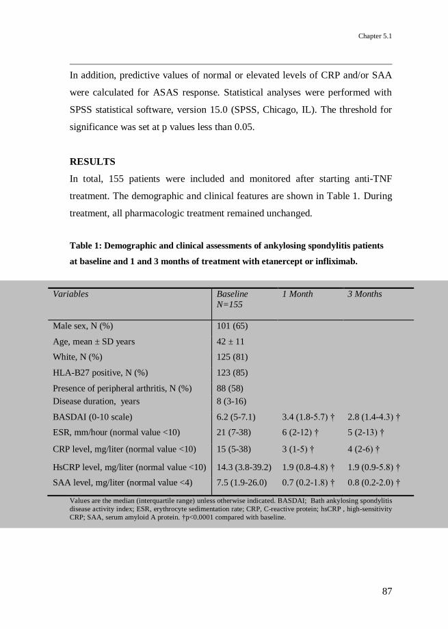
Chapter 5.1
87
In addition, predictive values of normal or elevated levels of CRP and/or SAA
were calculated for ASAS response. Statistical analyses were performed with
SPSS statistical software, version 15.0 (SPSS, Chicago, IL). The threshold for
significance was set at p values less than 0.05.
RESULTS
In total, 155 patients were included and monitored after starting anti-TNF
treatment. The demographic and clinical features are shown in Table 1. During
treatment, all pharmacologic treatment remained unchanged.
Table 1: Demographic and clinical assessments of ankylosing spondylitis patients
at baseline and 1 and 3 months of treatment with etanercept or infliximab.
Variables Baseline
N=155
1 Month
3 Months
Male sex, N (%) 101 (65)
Age, mean ± SD years 42 ± 11
White, N (%) 125 (81)
HLA-B27 positive, N (%) 123 (85)
Presence of peripheral arthritis, N (%) 88 (58)
Disease duration, years 8 (3-16)
BASDAI (0-10 scale) 6.2 (5-7.1) 3.4 (1.8-5.7) † 2.8 (1.4-4.3) †
ESR, mm/hour (normal value <10) 21 (7-38) 6 (2-12) † 5 (2-13) †
CRP level, mg/liter (normal value <10) 15 (5-38) 3 (1-5) † 4 (2-6) †
HsCRP level, mg/liter (normal value <10) 14.3 (3.8-39.2) 1.9 (0.8-4.8) † 1.9 (0.9-5.8) †
SAA level, mg/liter (normal value <4) 7.5 (1.9-26.0) 0.7 (0.2-1.8) † 0.8 (0.2-2.0) †
Values are the median (interquartile range) unless otherwise indicated. BASDAI; Bath ankylosing spondylitis
disease activity index; ESR, erythrocyte sedimentation rate; CRP, C-reactive protein; hsCRP , high-sensitivity
CRP; SAA, serum amyloid A protein. †p<0.0001 compared with baseline.

Erythrocyte sedimentation rate, C-reactive protein level, and serum amyloid A protein for patient
selection and monitoring of anti-tumor necrosis factor treatment in AS
88
The etanercept cohort comprised 117 patients and the infliximab cohort
comprised 38 patients. After 3 months, 70% (80 of 115, because in 2 patients
the ASAS response was missing) and 71% (27 of 38) of patients achieved
ASAS response in the etanercept and infliximab cohort, respectively. Baseline
levels of ESR, CRP, and SAA were elevated in 113 (73%), 96 (62%), and 99
(64%) patients, respectively. In 29 patients (19%), none of these markers was
elevated at baseline. All of the inflammatory markers and the BASDAI
decreased significantly after starting anti-TNF therapy (p<0.0001) (Table 1 and
Figure 1). Notably, the median relative decrease after 1 month was 36% for the
BASDAI (Figure 1A), 67% for the ESR (Figure 1B), 75% for the CRP (Figure
1C), and 84% for the hsCRP (Figure 1D), while SAA decreased by 90% (Figure
1E). Longitudinal linear regression analysis (GEE) showed significant
association between BASDAI and ESR, CRP, hsCRP and SAA over time
(p<0.0001). There were no confounders influencing this association. The
standardized betas were 0.49, 0.43, 0.43, and 0.39 for ESR, CRP, hsCRP and
SAA, respectively. Hereafter, only results of associations with CRP level are
shown as results because the hsCRP level did not differ from the CRP level.
In 2 patients treated with infliximab who showed a secondary increase,
particularly CRP and SAA after initial normalization, antibodies against
infliximab were detected (Figure 2)(18).
Patients with an elevated baseline level of CRP (>10 mg/liter) achieved an
ASAS response after 3 months of treatment significantly more often compared
with patients with normal baseline CRP levels (OR 2.8, 95% CI 1.3-5.7,
adjusted for sex and age). Elevated baseline SAA levels had a similar
association with ASAS response at 3 months of treatment with either infliximab
or etanercept (OR 2.9, 95% CI 1.4-6.1, adjusted for sex and age). Only baseline
ESR levels were not significantly associated with clinical response (OR 1.4,
95% CI: 0.7-3.1).

Chapter 5.1
89
Figure 1: Change of BASDAI (A), ESR (B), CRP (C), hsCRP (D) and SAA (E)
after 1 and after 3 months of treatment with etanercept or infliximab.
IQR, interquartile range; *p<0.0001 compared to baseline.
BASDAI (A)
0 1 30.0
2.5
5.0
7.5
*
*
Time (months)
Med
ian
BA
SD
AI
(IQ
R, 0-1
0 s
cale
)
ESR (B)
0 1 30
10
20
30
40
50
* *
Time (months)
Med
ian
E
SR
(IQ
R, m
m/h
ou
r)
CRP (C)
0 1 30
10
20
30
40
* *
Time (months)
med
ian
CR
P l
evel
(IQ
R, m
g/L
)
hsCRP (D)
0 1 30
10
20
30
40
50
* *
Time (months)
Med
ian
hsC
RP
lev
el (
IQR
, m
g/L
) SAA (E)
0 1 30
10
20
30
* *
Time (months)
Med
ian
SA
A l
evel
(IQ
R, m
g/L
)
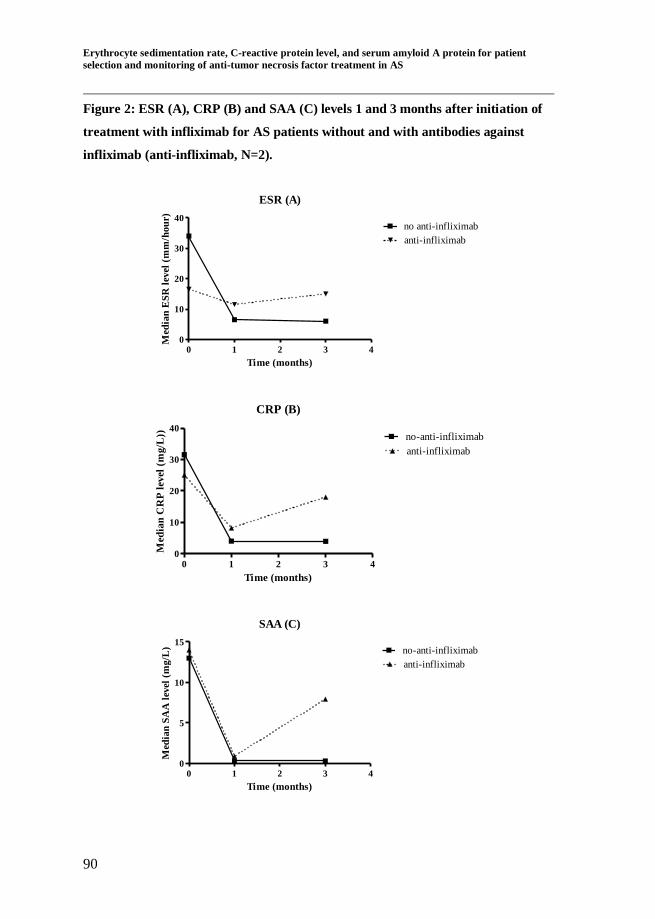
Erythrocyte sedimentation rate, C-reactive protein level, and serum amyloid A protein for patient
selection and monitoring of anti-tumor necrosis factor treatment in AS
90
Figure 2: ESR (A), CRP (B) and SAA (C) levels 1 and 3 months after initiation of
treatment with infliximab for AS patients without and with antibodies against
infliximab (anti-infliximab, N=2).
ESR (A)
0 1 2 3 40
10
20
30
40no anti-infliximab
anti-infliximab
Time (months)
Med
ian
ES
R l
evel
(m
m/h
ou
r)
CRP (B)
0 1 2 3 40
10
20
30
40no-anti-infliximab
anti-infliximab
Time (months)
Med
ian
CR
P l
evel
(m
g/L
))
SAA (C)
0 1 2 3 40
5
10
15no-anti-infliximab
anti-infliximab
Time (months)
Med
ian
SA
A l
evel
(m
g/L
)
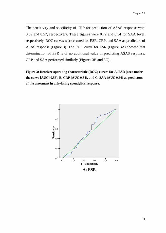
Chapter 5.1
91
The sensitivity and specificity of CRP for prediction of ASAS response were
0.69 and 0.57, respectively. These figures were 0.72 and 0.54 for SAA level,
respectively. ROC curves were created for ESR, CRP, and SAA as predictors of
ASAS response (Figure 3). The ROC curve for ESR (Figure 3A) showed that
determination of ESR is of no additional value in predicting ASAS response.
CRP and SAA performed similarly (Figures 3B and 3C).
Figure 3: Receiver operating characteristic (ROC) curves for A, ESR (area under
the curve [AUC] 0.55), B, CRP (AUC 0.64), and C, SAA (AUC 0.66) as predictors
of the assesment in ankylosing spondylitis response.
1 - Specificity
1,00,80,60,40,20,0
Sen
sit
ivit
y
1,0
0,8
0,6
0,4
0,2
0,0
ROC Curve
Diagonal segments are produced by ties.
A: ESR
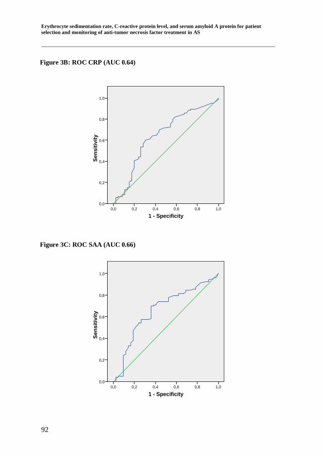
Erythrocyte sedimentation rate, C-reactive protein level, and serum amyloid A protein for patient
selection and monitoring of anti-tumor necrosis factor treatment in AS
92
1 - Specificity
1,00,80,60,40,20,0
Sen
sit
ivit
y1,0
0,8
0,6
0,4
0,2
0,0
ROC Curve
Diagonal segments are produced by ties.
1 - Specificity
1,00,80,60,40,20,0
Sen
sit
ivit
y
1,0
0,8
0,6
0,4
0,2
0,0
ROC Curve
Diagonal segments are produced by ties.
Figure 3B: ROC CRP (AUC 0.64)
Figure 3C: ROC SAA (AUC 0.66)

Chapter 5.1
93
The predictive value of ASAS response of an elevated baseline level of CRP
was 79%. This value was identical to SAA. A combination of the presence of
elevated baseline levels of CRP and SAA of patients in both cohorts displayed
the highest predictive value of ASAS response of 81% (Table 2). Inclusion of
baseline levels of ESR had no additional value. A proportion of 48% of patients
with normal values of both CRP and SAA levels at baseline were non-
responders according to the ASAS criteria.
Table 2: Predictive values of normal or elevated pretreatment levels of CRP and/or
SAA for prediction of ASAS response/non-response after 3 months of treatment
with etanercept or infliximab.
ASAS non-
response, %
ASAS response,
%
Normal CRP (N=59) 44 56
Elevated CRP (N=94) 21 79
Normal SAA (N=55) 45.5 54.5
Elevated SAA (N=98) 21 79
Normal CRP and SAA (N=44) 48 52
Elevated CRP and normal SAA (N=11) 36 64
Normal CRP and elevated SAA (N=15) 33 67
Elevated CRP and SAA (N=83) 19 81
CRP, C-reactive protein; SAA, serum amyloid A protein; ASAS, assessment in ankylosing spondylitis.

Erythrocyte sedimentation rate, C-reactive protein level, and serum amyloid A protein for patient
selection and monitoring of anti-tumor necrosis factor treatment in AS
94
DISCUSSION
The present study demonstrated that a combination of elevated baseline levels
of CRP and SAA can be a valuable instrument for the selection of those AS
patients who are likely to respond to treatment with anti-TNF, unlike the
BASDAI. Moreover, inflammatory markers, CRP and SAA in particular, seem
useful for monitoring the level of inflammation in patients with AS who are
treated with etanercept or infliximab.
Regarding monitoring therapy with anti-TNF, most of the AS patients showed a
significant decrease of several inflammatory markers, most dominantly SAA. In
some cases, a secondary increase of these inflammatory markers can be seen,
which might be caused by a concurrent infection or inadequate therapeutic
levels. The latter may be due to antibody formation, which depletes anti-TNF
levels below therapeutic thresholds(18). This could be an argument to include at
least one inflammatory marker next to the BASDAI in order to assess disease
activity properly. Although ESR showed the strongest association with
BASDAI over time, we consider ESR the least suitable for inclusion because it
has no additional value to the BASDAI, as the half-life of this inflammatory
marker is too long for early detection of changes. HsCRP has been proposed to
be useful as a marker to predict the risk of coronary heart disease due to
inflammation in apparently healthy persons(19). In the current study, however,
measurement of hsCRP level did not provide additional value because the
strength of the association with disease activity of AS patients over time was
not different from and not superior to that of CRP.
Although the majority of the AS patients in this study (62% to 73%) had
elevated inflammatory markers before the start of anti-TNF therapy, it is known
that inflammatory markers do not necessarily reflect disease activity well in
AS(3). This is why inflammatory markers were not implemented for assessment

Chapter 5.1
95
of disease activity or response to treatment, which is in contrast to RA. This
study shows that when inflammatory markers are raised, this is indicative of
active disease. It seems useful to add the decrease of inflammatory markers to
response criteria for continuation of anti-TNF treatment in AS patients who
show elevated inflammatory markers at baseline.
Since anti-TNF therapy is not without risks and is also very costly, it is of great
importance to identify patients likely to (non-)respond to this type of drug.
Although the performance of the tests was poor, the ROC curves showed that
CRP and SAA are superior to ESR. In the present study, we showed that the
combination of elevated CRP and SAA levels at baseline is the strongest
predictor of ASAS response, providing a solid basis for a predictive assessment
of the clinical response of AS patients to treatment with anti-TNF. In contrast,
baseline ESR levels were not associated with clinical response. However, as
demonstrated before, patients with normal baseline levels of CRP and SAA may
respond to anti-TNF therapy as well(20). Therefore, at this moment, we believe
inflammatory markers can be very useful as one of the predictors of a good
response, but a raise of the inflammatory markers should not be mandatory for
allowing AS patients to be treated with anti-TNF.
The fact that good responders do not all necessarily need to have a strong
decline of CRP or SAA levels limits the use of these parameters in making the
decision of whether or not this therapy should continued. Therefore, they can be
useful, but should not be considered obligatory for the decision of whether anti-
TNF therapy is failing or not.
We studied SAA in relation to disease activity in AS. SAA is implicated in
several chronic inflammatory diseases, such as AA amyloidosis, atherosclerosis,
and RA(21). An additional advantage of monitoring SAA levels in AS patients
may therefore be that SAA lowering therapy with anti-TNF could possibly
prevent secondary AA amyloidosis. AA amyloidosis sometimes develops

Erythrocyte sedimentation rate, C-reactive protein level, and serum amyloid A protein for patient
selection and monitoring of anti-tumor necrosis factor treatment in AS
96
secondary to longstanding inflammation and chronically elevated levels of
SAA, the plasma precursor of amyloid A deposits(10). Notably, elevated
baseline levels of SAA were associated with clinical response in AS and
decreased rapidly after initiation of treatment with etanercept or infliximab,
which might prevent AA amyloidosis in the future.
Altogether, in this large prospective cohort of AS patients, measurement of
inflammatory markers, in particular CRP and SAA, served as a powerful tool
not only for monitoring the efficacy of anti-TNF therapy, but also for the
selection of AS patients with a high likelihood of responding to anti-TNF
treatment.

Chapter 5.1
97
REFERENCE LIST
(1) Garrett S, Jenkinson T, Kennedy LG, Whitelock H, Gaisford P, Calin
A. A new approach to defining disease status in ankylosing spondylitis: the Bath Ankylosing Spondylitis Disease Activ ity Index. J Rheumatol
1994 Dec;21(12):2286-91.
(2) Berthelot JM, Tortellier L, Lavy-Bregeon D, Le Goff B, Maugars Y.
High intraindividual week-to-week variability in BASDAI and BASFI values: are several evaluations needed before starting or stopping
TNFalpha antagonist therapy for spondyloarthropathies? Joint Bone
Spine 2008 Mar;75(2):167-71.
(3) Ruof J, Stucki G. Validity aspects of erythrocyte sedimentation rate and
C-reactive protein in ankylosing spondylitis: a literature review. J
Rheumatol 1999 Apr;26(4):966-70.
(4) Jung SY, Park MC, Park YB, Lee SK. Serum amyloid a as a useful indicator of disease activity in patients with ankylosing spondylitis.
Yonsei Med J 2007 Apr 30;48(2):218-24.
(5) Ozgocmen S, Godekmerdan A, Ozkurt-Zengin F. Acute-phase response, clinical measures and disease activity in ankylosing
spondylitis. Joint Bone Spine 2007 May;74(3):249-53.
(6) Spoorenberg A, van der Heijde D, de Klerk E, Dougados M, de Vlam K, Mielants H, et al. Relative value of erythrocyte sedimentation rate
and C-reactive protein in assessment of disease activity in ankylosing
spondylitis. J Rheumatol 1999 Apr;26(4):980-4.
(7) Yildirim K, Erdal A, Karatay S, Melikoglu MA, Ugur M, Senel K. Relationship between some acute phase reactants and the Bath
Ankylosing Spondylitis Disease Activity Index in patients with
ankylosing spondylitis. South Med J 2004 Apr;97(4):350-3.
(8) Luc M, Gossec L, Ruyssen-Witrand A, Salliot C, Duclos M, Guignard
S, et al. C-reactive protein predicts tumor necrosis factor-alpha blocker
retention rate in axial ankylosing spondylitis. J Rheumatol 2007 Oct;34(10):2078-81.

Erythrocyte sedimentation rate, C-reactive protein level, and serum amyloid A protein for patient
selection and monitoring of anti-tumor necrosis factor treatment in AS
98
(9) Rudwaleit M, Listing J, Brandt J, Braun J, Sieper J. Prediction of a
major clinical response (BASDAI 50) to tumour necrosis factor alpha blockers in ankylosing spondylitis. Ann Rheum Dis 2004
Jun;63(6):665-70.
(10) Husby G, Marhaug G, Dowton B, Sletten K, Sipe JD. Serum amyloid A (SAA): biochemistry, genetics and the pathogenesis of AA amyloidosis.
Amyloid 1994;1:119-37.
(11) Uhlar CM, Whitehead AS. The kinetics and magnitude of the
synergistic activation of the serum amyloid A promoter by IL-1 beta and IL-6 is determined by the order of cytokine addition. Scand J
Immunol 1999 Apr;49(4):399-404.
(12) Lange U, Boss B, Teichmann J, Klor HU, Neeck G. Serum amyloid A--an indicator of inflammation in ankylosing spondylitis. Rheumatol Int
2000;19(4):119-22.
(13) van der Linden S, Valkenburg HA, Cats A. Evaluation of diagnostic
criteria for ankylosing spondylitis. A proposal for modification of the New York criteria. Arthritis Rheum 1984 Apr;27(4):361-8.
(14) Zochling J, van der Heijde D, Burgos-Vargas R, Collantes E, Davis JC,
Jr., Dijkmans B, et al. ASAS/EULAR recommendations for the management of ankylosing spondylitis. Ann Rheum Dis 2006
Apr;65(4):442-52.
(15) Braun J, Pham T, Sieper J, Davis J, van der Linden S, Dougados M, et al. International ASAS consensus statement for the use of anti-tumour
necrosis factor agents in patients with ankylosing spondylitis. Ann
Rheum Dis 2003 Sep;62(9):817-24.
(16) Braun J, Davis J, Dougados M, Sieper J, van der Linden S, van der Heijde D. First update of the international ASAS consensus statement
for the use of anti-TNF agents in patients with ankylosing spondylitis.
Ann Rheum Dis 2006 Mar;65(3):316-20.
(17) Hazenberg BP, Limburg PC, Bijzet J, van Rijswijk MH. Monoclonal
antibody based ELISA for human SAA. In: Natvig JB, Førre Ø, Husby
G, Husebekk A, Skogen B, Sletten K, et al., editors. Amyloid and amyloidosis.Dordrecht: Kluwer Academic Publishers; 1991. p. 898-
901.

Chapter 5.1
99
(18) de Vries MK, Wolbink GJ, Stapel SO, de Groot ER, Dijkmans BA,
Aarden LA, et al. Inefficacy of infliximab in ankylosing spondylitis is correlated with antibody formation. Ann Rheum Dis 2007
Jan;66(1):133-4.
(19) Sellmayer A, Limmert T, Hoffmann U. High sensitivity C-reactive protein in cardiovascular risk assessment. CRP mania or useful
screening? Int Angiol 2003 Mar;22(1):15-23.
(20) Gerard S, Le Goff B, Maugars Y, Berthelot JM. Six-month response to
anti-TNF drugs in axial spondylarthropathy according to the fulfillment or not of New-York criteria for ankylosing spondylitis or French
recommendations for anti-TNF use. A "real life" retrospective study on
175 patients. Joint Bone Spine 2008 Dec;75(6):680-7.
(21) De Beer FC, Mallya RK, Fagan EA, Lanham JG, Hughes GR, Pepys
MB. Serum amyloid-A protein concentration in inflammatory diseases
and its relationship to the incidence of reactive systemic amyloidosis.
Lancet 1982 Jul 31;2(8292):231-4.

Erythrocyte sedimentation rate, C-reactive protein level, and serum amyloid A protein for patient
selection and monitoring of anti-tumor necrosis factor treatment in AS

Chapter 5.1
C-reactive protein polymorphisms and haplotypes
influence serum C-reactive protein levels
independent of disease activity in ankylosing
spondylitis.
Theodora A.M. Claushuis1, Mirjam K. de Vries*
1, Mignon A.C. van der
Weijden*1, Ingrid M.Visman
2, Michael T. Nurmohamed
2, Jos W.R Twisk
3,
Irene E. Van der Horst-Bruinsma1, Jakob B.A. Crusius
4.
* both authors contributed equally to this paper
Rheumatology Department VU University Medical Center1, Rheumatology
Department Jan van Breemen Research Institute/Reade2, Methodology and
Applied Biostatistics Department VU University3, Laboratory for
Immunogenetics at the Medical Microbiology and Infection Control Department
VU University Medical Center4, Amsterdam, The Netherlands.
Submitted

C-reactive protein polymorphisms and haplotypes influence serum C-reactive protein levels
independent of disease activity in AS
102
ABSTRACT
Objectives: C-reactive protein (CRP) levels are frequently used for
determination of disease activity in patients with ankylosing spondylitis (AS),
but these levels do not necessarily reflect disease activity. We investigated
whether common single-nucleotide polymorphisms (SNPs) and haplotypes in
the CRP gene influence CRP levels in AS patients. Additionally, the relation
between CRP levels and Bath ankylosing spondylitis disease activity index
(BASDAI) was examined.
Methods: This exploratory cross-sectional study included 189 Dutch Caucasian
AS patients. CRP SNPs rs2794521, rs3091244, rs1800947 and rs876538 were
genotyped and haplotypes constructed. CRP levels were determined by standard
assay. Multivariate linear and logistic regression analyses were used to
investigate the relation between SNPs and CRP levels, adjusting for NSAID
use, BMI, smoking status, age, sex and BASDAI to correct for disease activity.
Results: CRP levels were significantly positively correlated with BASDAI
(p=0.001). AS patients with genotype CA of the tri-allelic (C>T>A) SNP
rs3091244 were associated with significantly higher CRP levels when compared
with genotype CC (CA: 18.6 mg/l vs. CC: 8.3 mg/l; p= 0.02). Carriers of
haplotype 5 (tagged by allele A of rs3091244) had a significantly higher odds
ratio when compared to non-carriers to express a CRP>10 mg/l (OR=2.9,
95%CI 1.0-8.3; p=0.05) in the multivariate regression analyses.
Conclusions: In AS, genotypes and the haplotype tagged by allele A of
rs3091244 associate with high CRP levels, independent of BASDAI and other
confounders. Therefore, carrying distinct genetic variants might explain the lack
of elevated CRP levels despite high disease activity in certain AS patients and
could be important to interpret disease activity scores that incorporate CRP
levels, like the ankylosing spondylitis disease activity score (ASDAS).

Chapter 5.2
103
INTRODUCTION
Ankylosing spondylitis (AS) is a chronic inflammatory rheumatic disease,
characterized by sacroiliitis with low back pain and stiffness, restricted motion
of the spine and chest expansion due to inflammation and bone formation.
Unlike other rheumatic diseases, such as rheumatoid arthritis, it is difficult to
assess clinical disease activity in AS, as patients may not exhibit any peripheral
arthritis (rendering a 44 joint count unsuitable) and absence of increased
inflammatory parameters (such as C-reactive protein (CRP)).
CRP is a plasma protein of the pentraxin family, known for its rapid increased
expression as part of the acute phase reaction and CRP levels are widely used in
the evaluation of infection or systemic inflammation. The CRP level is only
elevated (>10mg/l) in approximately 50% of AS patients with active disease(1-
3). Since 1994, disease activity in AS has been measured with the Bath
ankylosing spondylitis disease activity index (BASDAI), a patient orientated
questionnaire(4). Active disease is specified as a BASDAI ≥ 4 (on a 0-10 scale)
and is used as an indicator to start treatment with TNF blocking agents.
Recently, the assessment of ankylosing spondylitis (ASAS) reported the
ankylosing spondylitis disease activity score (ASDAS), a score that incorporates
plasma levels of CRP(5). This new scoring system necessitates to obtain
information about the value of CRP in AS. CRP levels correlate with the
BASDAI but several studies found that these levels do not necessary reflect
disease activity in all patients(2;6;7). As far back as 1999 Ruof and Stucki
stated that many patients have normal or only minimally increased levels of
acute phase reactants(6). A reason for the lack of increased CRP levels in some
patients with high disease activity might be a genetic contribution to CRP
levels.

C-reactive protein polymorphisms and haplotypes influence serum C-reactive protein levels
independent of disease activity in AS
104
In a healthy population, small variations in the low range of plasma CRP (<10
mg/l) were attributed to genetic factors as well as environmental factors such as
body mass index (BMI), sex, age and smoking(8). Twin and family studies have
illustrated that genetic factors can contribute up to 40-50% of the phenotypic
variation of baseline plasma CRP concentrations(9;10). A meta-analysis of
genome wide association studies (GWAS) in more than 80.000 individuals
identified associations of CRP levels with single-nucleotide polymorphisms
(SNPs) at seven loci within or close to the CRP gene and six other genes as
previously reported in a GWAS scan by Ridker et al.(11) and with 11 novel
loci(12). A weighted genetic risk score explained about 5% of the variation in
CRP levels.
To date, most disease association studies investigated the influence of CRP gene
polymorphisms on CRP levels in relation to cardiovascular disease(13-18).
In these studies SNPs and their haplotypes in the CRP gene have been identified
that modify CRP levels. However, the causal relationship between CRP-
genetics and cardiovascular disease risk remains controversial(8).
When compared with cardiovascular disease, higher ranges of serum CRP
levels are used in the clinical evaluation of AS patients. However, since CRP
levels do not always correlate with disease activity in every AS patient we
investigated the contribution of CRP gene polymorphisms and haplotypes to
these CRP levels in AS patients. This influence is of clinical importance as it
can interfere with diagnostic and treatment choices based on CRP levels.
Additionally, the association between CRP levels and the BASDAI as a
measure of disease activity was studied.

Chapter 5.2
105
PATIENTS AND METHODS
Patients
A cross-sectional study in AS patients was conducted. The study population
included patients treated with tumor necrosis factor (TNF) blocking agents
(infliximab, etanercept or adalimumab) and a group of anti-TNF-naive patients.
All AS patients fulfilled the 1984 modified New York criteria(19), were self-
reported Dutch Caucasians and unrelated to any other patient participating in
this study. Patients were recruited from the VU University Medical Center
(VUmc) and Reade, formerly known as the Jan van Breemen Research Institute,
a large outpatient clinics in Amsterdam.
The study was approved by the medical ethical committees of the VUmc and
Reade and all participants gave written informed consent.
Outcome measures
Filling in questionnaires, sampling of blood, and all measurements, including
serum CRP levels, were performed at the start of each study (i.e. the baseline
visit), making sure that all measurements were of TNF-naive patients. When
using the term ‘CRP levels’ further on, we are referring to these TNF-naive
CRP levels obtained at the start of each study.
Serum CRP levels were determined on Roche/Hitachi Modular P (at VUmc)
and Cobas C (Reade) systems by a particle enhanced immunoturbidimetric
assay. Intra- and interassay coefficients of variation for CRP were 1.9% and
3.4% respectively. These measurements were conducted with a detection limit
of 1 mg/l. A CRP level >10 mg/l was considered a parameter of active disease
in AS. Several other data collected was: age, sex, HLA-B27 status, BMI,
smoking status, disease duration and symptom onset duration, extraspinal
manifestations of AS (presence of uveitis, IBD and psoriasis), medication use,
C-reactive protein polymorphisms and haplotypes influence serum C-reactive protein levels
independent of disease activity in AS
106
and disease activity scores such as BASDAI, BASFI and global disease activity
score (VAS patient).
Genotyping the CRP SNPs
Genomic DNA was isolated from peripheral blood by standard procedures. Four
common CRP gene tagging SNPs with published minor allele frequencies
(MAF) of more than 5% that were associated with CRP levels or their proxies
were selected on the basis of published work(18;20;21).
Functional polymorphisms in the CRP gene region (dbSNP ID) rs2794521
T>C(17;18) and triallelic SNP rs3091244 C>T>A located in the gene
promoter(22), as well as rs1800947 G>C, a synonymous coding SNP in exon
2(15) and SNP rs876538 G>A in the 3' flanking region(18;21) were genotyped.
Genotyping was performed with appropriate quality control measures by the
Taqman SNP allelic discrimination method with an ABI PRISM 7000 Sequence
detection System (Applied Biosystems, Foster City, CA, USA) (details are
available from the authors upon request).
Statistics
Statistical analysis was conducted using SPSS V18.0 (SPSS Inc., Chicago,
Illinois, USA). On basis of published work, sex, BMI, age, smoking status and
NSAID use were selected as possible confounding variables(1;1). CRP levels
were correlated with the disease activity status in each patient using BASDAI.
Two linear regression analyses were performed. First, the crude relation
between polymorphisms and CRP was analysed and secondly an adjustment
was made for sex, BASDAI, BMI, age, smoking status and NSAID use.
Because CRP levels were skewed to the right in all linear regression analyses,
the natural logarithm of CRP was used as outcome variable. Results were
presented as back-transformed geometric means for CRP levels.
Chapter 5.2
107
An independent samples t-test was used to determine the differences in CRP
level between two groups with different disease activity (BASDAI≥4 or <4).
Using the linear regression analyses, the effect of the different genotypes and
the effects of carrier ship of both the minor and the major alleles on CRP level
were tested. For the triallelic SNP rs3091244, the effect of the different
genotypes as well as the effects of carrier ship of both minor alleles were
analysed. Crude and adjusted logistic regression analyses were performed to
determine the effect of SNPs and haplotypes on the dichotomised CRP, with a
cutoff point of 10 mg/l. The same potential confounding variables were used in
the adjusted model.
The publicly available Haploview version 4.2 software (Cambridge, USA) was
applied to estimate pair wise linkage disequilibrium (LD) between the four
SNPs examined(23). Haplotypes were constructed to determine the integrated
effect of multiple SNPs on CRP levels. Genotype data of each of the four CRP
SNPs were used to infer the frequency of the haplotypes present in the
population using the program Phase v2.1. Only patients with >99% certainty in
haplotype estimates were included in the analysis(24).
The same crude and adjusted linear and logistic regression analyses were
applied to determine the effect of each haplotype on CRP levels. Additionally,
the effects of being homozygous, heterozygous or non-carrier for each
haplotype were estimated.
We present otherwise uncorrected two-tailed p values. For all analyses
statistical significance was designated as a two-tailed p value less than 0.05.
C-reactive protein polymorphisms and haplotypes influence serum C-reactive protein levels
independent of disease activity in AS
108
RESULTS
Patient characteristics
In total 233 patients with near complete data were selected for our study.
Excluded were 42 patients because of self-reported non-Caucasian ethnicity and
2 patients who did not fulfil the predefined control criteria for polymorphism
and haplotype determination, leaving 189 patients for statistical analysis. The
main characteristics of the 189 AS patients under study are listed in Table 1.
Our study population of AS patients encompasses a large variety in disease
activity, with a high mean BASDAI of 4.9 (SD 2.2). In total, 47.3% of the
patients had a CRP level ≥10 mg/l. Mean BASFI was 4.5 (SD 2.7) and mean
VASpatient 5.8 (SD 2.6). A high percentage of AS patients (50%) had
peripheral arthritis. CRP levels did not significantly differ in patients with
extraspinal manifestations such as peripheral arthritis, anterior uveitis and
inflammatory bowel disease (data not shown).
Disease activity variables and CRP levels
CRP levels were significantly positively correlated with BASDAI (p=0.001),
BASFI (p<0.001) and VAS (p<0.001). BASDAI, BASFI and VAS were all
significantly positively associated with each other (all p<0.001). CRP values
≥10 mg/l were found in 52% of the AS patients with a high disease activity
(BASDAI ≥4).

Chapter 5.2
109
Table 1: Characteristics of 189 patients with ankylosing spondylitis.
Means ± SD or N (cumulative %)
Characteristics
Age (years) 47.8 ± 11.7
Male 145 (76.7)
Duration of disease (years) 15.0 ± 10.5
HLA-B27 positivity 185 (86.6)
Smoking status 73 (40.6)
Mean BMI (kg/m2) 25.4 ± 4.3
Peripheral arthritis 94 (50.3)
Uveitis 55 (29.3)
Psoriasis 21 (11.2)
IBD 14 (7.4)
Patients on NSAIDs 146 (78.1)
Patients receiving DMARDs 18 (9.6)
Patients on systemic steroids 14 (7.5)
Patients with CRP≥10 mg/l 87 (47.3)
BASDAI ( 0-10 ) 4.9 ± 2.2
Patients with BASDAI≥4 125 (67.2)
BASFI ( 0-10 ) 4.5 ± 2.7
VAS patient ( 0-10 ) 5.8 ± 2.6
HLA-B27, human leucocyte antigen B27; BMI, body mass index; IBD, inflammatory bowel disease;
NSAIDs, non-steroidal anti-inflammatory drugs; DMARDs, disease-modifying antirheumatic drugs; CRP,
C–reactive protein; BASDAI, Bath ankylosing spondylitis disease activity index; BASFI, Bath ankylosing
spondylitis functional index; VAS, visual analogue scale.

C-reactive protein polymorphisms and haplotypes influence serum C-reactive protein levels
independent of disease activity in AS
110
SNPs and CRP levels
The genotype frequencies of all four SNPs were in Hardy-Weinberg
equilibrium. Allele frequencies of SNPs rs2794521 (allele T=0.68, allele
C=0.32), rs3091244 (C=0.65, T=0.30, A=0.05), rs1800947 (G=0.96, C=0.04)
and rs876538 (G=0.77, A=0.23) were in concordance with frequencies found in
the Framingham Offspring cohort, a study of 1640 healthy unrelated
participants of European descent(21). Of the four SNPs genotyped, rs3091244
was significantly associated with CRP levels. (p=0.02) as shown in Table 2.
Patients with the CA genotype showed significantly higher CRP levels when
compared to the reference genotype CC (p=0.02), and this association remained
when adjusted for BASDAI and other confounders in the multivariate
regression analyses (p=0.04). In addition, AS patients carrying the A-allele of
rs3091244 had significantly higher CRP levels compared with patients
homozygous for the C-allele when adjusted for all confounders. Rs876538
genotype GA showed nominal evidence for association with CRP levels
(ptrend=0.06) when compared to genotype GG, but this association did not persist
when adjusted in the multivariate linear regression analyses.
Haplotypes and CRP levels
Six haplotypes defined by the four representative SNPs in the CRP region were
constructed and coded H1-H6 in order of descending frequency (Figure 1A).
Linkage disequilibrium between the CRP SNPs is shown in Figures 1B and 1C.
Haplotype analysis showed that H5 was associated with CRP levels after
adjusting for BASDAI and other confounders. A minority of 10.6% of the AS
patients was an H5 carrier and these were all heterozygotes. H5 is tagged by the
A allele of rs3091244 and AS patients who are carriers of H5 express
significantly higher mean serum CRP levels than non-carriers (p<0.03),

Chapter 5.2
111
bv
showing a mean CRP level of 15.4 mg/l in H5 carriers and 8.6 mg/l in H5 non-
carriers. This result reflects the same significant association as described above
for the A-allele of rs3091244.
Table 2: Mean serum CRP values in mg/l for CRP SNPs genotypes.
Frequency, N
(cumulative %)
Mean CRP 95% CI p-Value
Genotypes
rs2794521 T>C
TT 91 (48.1) 10.6 ref ref
CT 76 (40.2) 7.9 5.9– 10.8 0.17
CC 22 (11.6) 8.0 4.5 – 14.2 0.40
rs3091244 C>T>A
CC 75 (39.7) 8.3 Ref ref
CT 77 (40.7) 8.9 6.6 – 12.0 0.77
CA 17 (9.0) 18.6 9.9 – 34.8 0.02*
TT 17 (9.0) 8.3 4.4 – 15.5 0.98
TA 3 (1.6) 5.4 1.2 – 24.1 0.58
AA N.D. N.D. N.D. N.D.
rs1800947 G>C
GG 175 (92.6) 8.9 ref ref
GC 14 (7.4) 12.9 6.4 – 25.8 0.31
CC N.D. N.D. N.D. N.D.
rs876538 G>A
GG 108 (58.1) 11.0 ref ref
GA 69 (37.1) 7.5 5.5 – 10.2 0.06
AA 9 (4.8) 5.9 2.5 – 13.9 0.17
CRP values were backtransformed after natural logarithmic correction. N.D.= not detected.
*Significant result.

C-reactive protein polymorphisms and haplotypes influence serum C-reactive protein levels
independent of disease activity in AS
112
Figure 1: SNPs in the region of the CRP gene at chromosome 1q21-q23 with their
pairwise linkage disequilibrium (LD), haplotypes, and haplotype frequencies in
ankylosing spondylitis patients.
For clarity, Table 3 shows mean CRP levels in relation to H5 carriage and the
BASDAI. Mean CRP level was elevated (>10 mg/l) despite low disease activity
(BASDAI<4) in H5 carriers. Table 3 also shows that high disease activity
(represented by BASDAI>4) did correlate with CRP levels in the H5 non-
carriers of our study population (P=0.003, independent samples t-test).
Legend to Figure
a Location, haplotypes and
haplotype frequencies of
the four SNPs (with dbSNP
accession numbers) in the
CRP region in 189 AS
patients. In addition, one patient carried a
recombinant haplotype (not
shown).
b LD plot showing
pairwise LD between SNPs
as D' x 100 (100=
maximum possible value)
created by the Haploview
v4.2 program. [23]
c The r2 parameter, as
determined with the Haploview program, that
quantitates intermarker LD
is given (r2 = 0 indicates
no LD, r2 = 100 indicates
complete LD). CRP, C-
reactive protein; H,
haplotype; SNPs, single-
nucleotide polymorphisms.
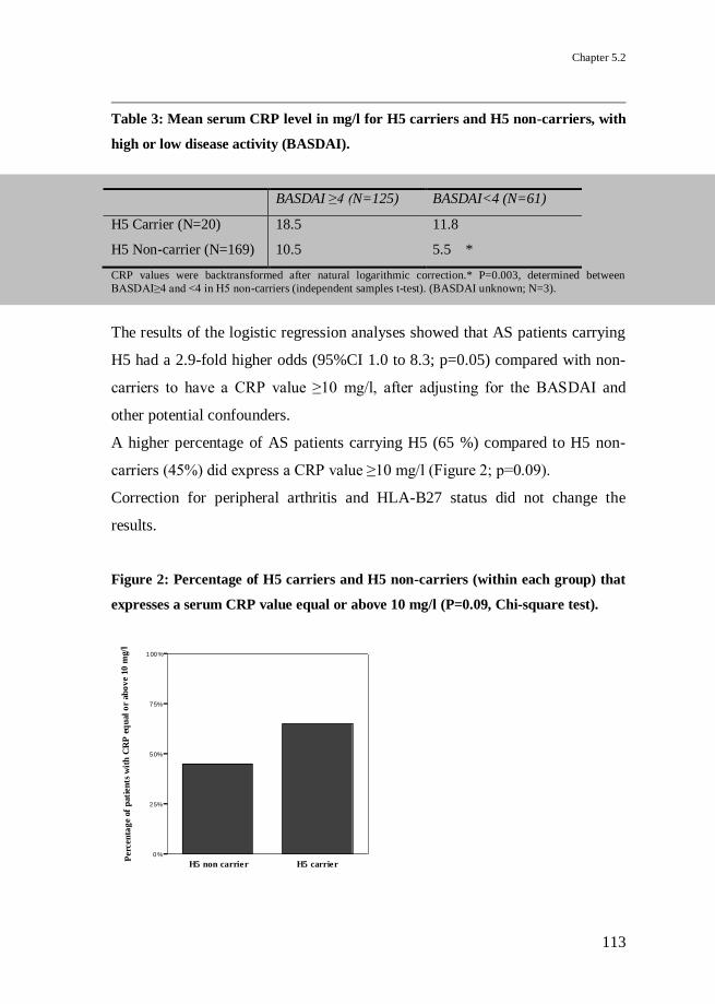
Chapter 5.2
113
Table 3: Mean serum CRP level in mg/l for H5 carriers and H5 non-carriers, with
high or low disease activity (BASDAI).
BASDAI ≥4 (N=125) BASDAI<4 (N=61)
H5 Carrier (N=20) 18.5 11.8
H5 Non-carrier (N=169) 10.5 5.5 *
CRP values were backtransformed after natural logarithmic correction.* P=0.003, determined between
BASDAI≥4 and <4 in H5 non-carriers (independent samples t-test). (BASDAI unknown; N=3).
The results of the logistic regression analyses showed that AS patients carrying
H5 had a 2.9-fold higher odds (95%CI 1.0 to 8.3; p=0.05) compared with non-
carriers to have a CRP value ≥10 mg/l, after adjusting for the BASDAI and
other potential confounders.
A higher percentage of AS patients carrying H5 (65 %) compared to H5 non-
carriers (45%) did express a CRP value ≥10 mg/l (Figure 2; p=0.09).
Correction for peripheral arthritis and HLA-B27 status did not change the
results.
Figure 2: Percentage of H5 carriers and H5 non-carriers (within each group) that
expresses a serum CRP value equal or above 10 mg/l (P=0.09, Chi-square test).
H5 non carrier H5 carrier
0%
25%
50%
75%
100%
Perc
en
tag
e of
pati
en
ts w
ith
CR
P e
qu
al
or
ab
ov
e 10
mg/l

C-reactive protein polymorphisms and haplotypes influence serum C-reactive protein levels
independent of disease activity in AS
114
DISCUSSION
In this exploratory study, we investigated the contribution of four CRP gene
polymorphisms and their haplotypes to CRP levels in AS patients and found a
significant correlation between the minor allele A of triallelic SNP rs3091244
and its corresponding haplotype and serum CRP levels after adjustment for
disease activity (BASDAI) and other confounders. AS patients with the CA
genotype of rs3091244 were associated with significantly higher CRP levels
compared with patients homozygous for the major C-allele. Our observation
that different genotypes of rs3091244 express different CRP levels is in
accordance with other studies(11;18;25).
Moreover, we found that carriers of haplotype 5 (tagged by the A-allele of
rs3091244) have a nearly threefold higher odds (OR=2.9) of expressing a CRP
value ≥10 mg/l, when adjusting for all confounders. Several studies have shown
rs3091244 to be one of the most important SNPs in the CRP gene region
influencing CRP level. Kathiresan et al. showed that rs3091244 explains the
largest part of CRP level variance in the 1640 healthy participants of the
Framingham Offspring cohort study(21). Other studies also established on basis
of haplotype analysis, that rs3091244 was the most important SNP influencing
CRP level variation(15;20). Subsequently, in vitro studies by Szalai et al.
demonstrated rs3091244 to be functional, by affecting the transcriptional
activity of the CRP gene(22).
In our study population a relatively small percentage of 10.6% was a carrier of
haplotype 5. However, this is an interesting finding as many common (and
probably rare) variants genome wide may be influential on elevated CRP level
similar to low-range CRP levels(11;12).
We found no correlation between CRP levels and rs2794521, rs1800947 and
rs876538.

Chapter 5.2
115
In previous literature this is confirmed but other authors did find an association
between CRP levels and these SNPs (rs2794521(13;15;17;18;26;27), rs800947
(13;15;20;21;26;27) and rs876538(18;21;27)).
A possible explanation for the difference between our findings and previous
studies might be due to the fact that in most studies, association between CRP
SNPs and low-range CRP level changes in apparently healthy individuals were
studied, whereas our study investigated the genetic influence of common SNPs
and haplotypes in the CRP gene on CRP ≥10 mg/l in AS patients.
CRP and disease activity
Many AS patients do not express clearly elevated CRP levels. This study
showed that disease activity (BASDAI) and a certain genotype and haplotype in
the CRP gene region are both significantly associated with CRP levels in these
patients. It also supports the hypothesis that genetic polymorphisms may
contribute some patients expressing a lower CRP level, despite high disease
activity, as seen in Table 3.
The results show that the CC-genotype of rs3091244 presents with a lower
mean CRP level and that carrying the A-allele increases CRP level, as also
shown in the haplotype results. AS patients with the CC-genotype in contrast to
those carrying the A-allele could show CRP values <10 mg/l despite high
disease activity. This poses a possible explanation why some AS patients do not
show an increase in CRP despite high disease activity. This is in concordance
with findings by Rhodes et al. in rheumatoid arthritis, who found CRP SNPs to
influence CRP levels in this disease implicating this effect could influence
clinical decision making and lead to suboptimal treatment(25;28).
CRP levels are widely used in AS and recently have been included in the
ASDAS, a new disease activity score reported by van der Heijde et al. to be best
at discriminating between high and low disease activity at baseline and follow

C-reactive protein polymorphisms and haplotypes influence serum C-reactive protein levels
independent of disease activity in AS
116
up and especially being able to correlate with disease activity from a clinical
perspective(5). The present study showed that only 52% of AS patients with
high disease activity show a corresponding rise in CRP level, showing that
genetics influence CRP level. Therefore, one should take into account the
genetic influence on CRP levels when assessing disease activity with the
ASDAS. This is even more important because clinical decisions, such as
starting or stopping treatment with TNF blocking agents, are based on these
disease activity parameters. This might lead to under- or overtreatment of AS
patients depended on their specific genetic architecture.
Also shown in the results (Table 3) is disease activity (BASDAI above or below
4) significantly influences CRP level in AS patients. This association was not
significant in H5-carriers, probably due to small sample size. A recent study by
Kiltz et al. also showed that a high BASDAI influences CRP level(29).
A limitation of this study might be the heterogeneous study population,
including AS patients who are candidates for anti-TNF treatment and those who
do not require this treatment. In order to minimise this difference we have
adjusted for all factors that might influence CRP level, especially BASDAI. In
addition, although strict selection procedures were applied so that only self-
reported Dutch Caucasian patients were included, it can not be ruled out that
there are subtle differences in population ancestry and it is not yet established if
these results can be generalized to other ethnicities.
Furthermore, CRP measurements were not done with a high sensitivity CRP
assay (hsCRP). The more sensitive hsCRP test can measure CRP with the
lowest detected level of 0.1 mg/l. However, this is of less importance in this
study as higher CRP values are more of interest in the population of AS patients
and not the precise differences between low CRP values.
At the time of initiation of this study, the ASDAS was not yet available,
therefore BASDAI was used, the best disease activity score at the time, to adjust
Chapter 5.2
117
for disease activity status in the AS patients. The study is limited by the small
sample size. Even with a modestly sized study population of AS patients, we
were able to demonstrate significant results. However, further research with a
larger study population is required to confirm our results.
To conclude, our data suggest that genetics might play a role in the lack of CRP
rise in some AS patients despite high disease activity. The importance of a
genetic contribution to CRP levels might be taken into consideration when
clinical decisions in AS patients have to be made.

C-reactive protein polymorphisms and haplotypes influence serum C-reactive protein levels
independent of disease activity in AS
118
REFERENCE LIST
(1) Benhamou M, Gossec L, Dougados M. Clinical relevance of C-reactive
protein in ankylosing spondylitis and evaluation of the NSAIDs/coxibs' treatment effect on C-reactive protein. Rheumatology (Oxford) 2010
Mar;49(3):536-41.
(2) Dougados M, Gueguen A, Nakache JP, Velicitat P, Zeidler H, Veys E,
et al. Clinical relevance of C-reactive protein in axial involvement of ankylosing spondylitis. J Rheumatol 1999 Apr;26(4):971-4.
(3) de Vries MK, van Eijk IC, van der Horst-Bruinsma IE, Peters MJ,
Nurmohamed MT, Dijkmans BA, et al. Erythrocyte sedimentation rate, C-reactive protein level, and serum amyloid a protein for patient
selection and monitoring of anti-tumor necrosis factor treatment in
ankylosing spondylitis. Arthritis Rheum 2009 Nov 15;61(11):1484-90.
(4) Garrett S, Jenkinson T, Kennedy LG, Whitelock H, Gaisford P, Calin A. A new approach to defining disease status in ankylosing spondylitis:
the Bath Ankylosing Spondylitis Disease Activity Index. J Rheumatol
1994 Dec;21(12):2286-91.
(5) van der Heijde D, Lie E, Kvien TK, Sieper J, Van den BF, Listing J, et
al. ASDAS, a highly discriminatory ASAS-endorsed disease activity
score in patients with ankylosing spondylitis. Ann Rheum Dis 2009 Dec;68(12):1811-8.
(6) Ruof J, Stucki G. Validity aspects of erythrocyte sedimentation rate and
C-reactive protein in ankylosing spondylitis: a literature review. J
Rheumatol 1999 Apr;26(4):966-70.
(7) Spoorenberg A, van der Heijde D, de Klerk E, Dougados M, de Vlam
K, Mielants H, et al. Relative value of erythrocyte sedimentation rate
and C-reactive protein in assessment of disease activity in ankylosing spondylitis. J Rheumatol 1999 Apr;26(4):980-4.
(8) Shen J, Ordovas JM. Impact of genetic and environmental factors on
hsCRP concentrations and response to therapeutic agents. Clin Chem 2009 Feb;55(2):256-64.

Chapter 5.2
119
(9) Pankow JS, Folsom AR, Cushman M, Borecki IB, Hopkins PN,
Eckfeldt JH, et al. Familial and genetic determinants of systemic markers of inflammation: the NHLBI family heart study.
Atherosclerosis 2001 Feb 15;154(3):681-9.
(10) MacGregor AJ, Gallimore JR, Spector TD, Pepys MB. Genetic effects on baseline values of C-reactive protein and serum amyloid A protein: a
comparison of monozygotic and dizygotic twins. Clin Chem 2004
Jan;50(1):130-4.
(11) Ridker PM, Pare G, Parker A, Zee RY, Danik JS, Buring JE, et al. Loci related to metabolic-syndrome pathways including LEPR,HNF1A,
IL6R, and GCKR associate with plasma C-reactive protein: the
Women's Genome Health Study. Am J Hum Genet 2008 May;82(5):1185-92.
(12) Dehghan A, Dupuis J, Barbalic M, Bis JC, Eiriksdottir G, Lu C, et al.
Meta-analysis of genome-wide association studies in >80 000 subjects
identifies multiple loci for C-reactive protein levels. Circulation 2011 Feb 22;123(7):731-8.
(13) Grammer TB, März W, Renner W, Bohm BO, Hoffmann MM. C-
reactive protein genotypes associated with circulating C-reactive protein but not with angiographic coronary artery disease: the LURIC
study. Eur Heart J 2009 Jan;30(2):170-82.
(14) Lange LA, Carlson CS, Hindorff LA, Lange EM, Walston J, Durda JP, et al. Association of polymorphisms in the CRP gene with circulating
C-reactive protein levels and cardiovascular events. JAMA 2006 Dec
13;296(22):2703-11.
(15) Miller DT, Zee RY, Suk Danik J, Kozlowski P, Chasman DI, Lazarus R, et al. Association of common CRP gene variants with CRP levels
and cardiovascular events. Ann Hum Genet 2005 Nov;69(Pt 6):623-38.
(16) Elliott P, Chambers JC, Zhang W, Clarke R, Hopewell JC, Peden JF, et al. Genetic Loci associated with C-reactive protein levels and risk of
coronary heart disease. JAMA 2009 Jul 1;302(1):37-48.
(17) Wang L, Lu X, Li Y, Li H, Chen S, Gu D. Functional analysis of the C-reactive protein (CRP) gene -717A>G polymorphism associated with
coronary heart disease. BMC Med Genet 2009;10:73.

C-reactive protein polymorphisms and haplotypes influence serum C-reactive protein levels
independent of disease activity in AS
120
(18) Wang Q, Hunt SC, Xu Q, Chen YE, Province MA, Eckfeldt JH, et al.
Association study of CRP gene polymorphisms with serum CRP level and cardiovascular risk in the NHLBI Family Heart Study. Am J
Physiol Heart Circ Physiol 2006 Dec;291(6):H2752-H2757.
(19) van der Linden S, Valkenburg HA, Cats A. Evaluation of diagnostic criteria for ankylosing spondylitis. A proposal for modification of the
New York criteria. Arthritis Rheum 1984 Apr;27(4):361-8.
(20) Carlson CS, Aldred SF, Lee PK, Tracy RP, Schwartz SM, Rieder M, et
al. Polymorphisms within the C-reactive protein (CRP) promoter region are associated with plasma CRP levels. Am J Hum Genet 2005
Jul;77(1):64-77.
(21) Kathiresan S, Larson MG, Vasan RS, Guo CY, Gona P, Keaney JF, Jr., et al. Contribution of clinical correlates and 13 C-reactive protein gene
polymorphisms to interindividual variability in serum C-reactive
protein level. Circulation 2006 Mar 21;113(11):1415-23.
(22) Szalai AJ, Wu J, Lange EM, McCrory MA, Langefeld CD, Williams A, et al. Single-nucleotide polymorphisms in the C-reactive protein (CRP)
gene promoter that affect transcription factor binding, alter
transcriptional activity, and associate with differences in baseline serum CRP level. J Mol Med 2005 Jun;83(6):440-7.
(23) Barrett JC, Fry B, Maller J, Daly MJ. Haploview: analysis and
visualization of LD and haplotype maps. Bioinformatics 2005 Jan 15;21(2):263-5.
(24) Stephens M, Smith NJ, Donnelly P. A new statistical method for
haplotype reconstruction from population data. Am J Hum Genet 2001
Apr;68(4):978-89.
(25) Rhodes B, Merriman ME, Harrison A, Nissen MJ, Smith M, Stamp L,
et al. A genetic association study of serum acute-phase C-reactive
protein levels in rheumatoid arthritis: implications for clinical interpretation. PLoS Med 2010;7(9):e1000341.
(26) Brull DJ, Serrano N, Zito F, Jones L, Montgomery HE, Rumley A, et al.
Human CRP gene polymorphism influences CRP levels: implications for the prediction and pathogenesis of coronary heart disease.
Arterioscler Thromb Vasc Biol 2003 Nov 1;23(11):2063-9.

Chapter 5.2
121
(27) Martinez-Calatrava MJ, Gonzalez-Sanchez JL, Martinez-Larrad MT,
Perez-Barba M, Serrano-Rios M. Common haplotypes of the C-reactive protein gene and circulating leptin levels influence the interindividual
variability in serum C-reactive protein levels. The Segovia study.
Thromb Haemost 2007 Nov;98(5):1088-95.
(28) Rhodes B, Furnrohr BG, Vyse TJ. C-reactive protein in rheumatology:
biology and genetics. Nat Rev Rheumatol 2011 May;7(5):282-9.
(29) Kiltz U, Baraliakos X, Karakostas P, Igelmann M, Kalthoff L, Klink C,
et al. The degree of spinal inflammation is similar in patients with axial spondyloarthritis who report high or low levels of disease activity: a
cohort study. Ann Rheum Dis 2012 Apr 20;71:1207-11.

C-reactive protein polymorphisms and haplotypes influence serum C-reactive protein levels
independent of disease activity in AS

Chapter 5.2
Discovertebral (Andersson) lesions of the spine in
ankylosing spondylitis revisited.
Johannes L. Bron1 , Mirjam K. de Vries
2, Marieke N. Snieders
1, Irene E. van der
Horst-Bruinsma2, Barend J. van Royen
1.
Orthopedic Surgery Department1, Rheumatology Department
2, VU University
Medical Center, Amsterdam, the Netherlands.
Clinical Rheumatology 2009;28(8):883-892.
Discovertebral (Andersson) lesions of the spine in AS revisited
124
ABSTRACT
A well-known complication in patients with ankylosing spondylitis (AS) is the
development of localised vertebral or discovertebral lesions of the spine, which
was first described by Andersson in 1937. Since then, many different terms are
used in literature to refer to these localised lesions of the spine, including the
eponym ‘Andersson lesion’ (AL). The use of different terms reflects an ongoing
debate on the exact aetiology of the AL. In the current study, we performed an
extensive review of the literature in order to align communication on aetiology,
diagnosis and management between treating physicians. AL may result from
inflammation or (stress-) fractures of the complete ankylosed spine. There is no
evidence for an infectious origin. Regardless of the exact aetiology, a final
common pathway exists, in which mechanical stresses prevent the lesion from
fusion and provoke the development of pseudarthrosis. The diagnosis of AL is
established on conventional radiography, but computed tomography and
magnetic resonance imaging both provide additional information. There is no
indication for a diagnostic biopsy. Surgical instrumentation and fusion is
considered the principle management in symptomatic AL that fails to resolve
from a conservative treatment. We advise to use the term Andersson lesion for
these spinal lesions in patients with AS.
INTRODUCTION
Ankylosing spondylitis (AS) is a chronic inflammatory disease that primarily
affects the spine and sacroiliac joints, causing pain, stiffness and a progressive
thoracolumbar kyphotic deformity(1). In the late state of the disease, the spine
demonstrates progressive ossification of the annulus fibrosis, anterior
longitudinal ligament, apophyseal joints, interspinous and flaval ligaments
ligament resulting in a complete ankylosed spine, often referred to as a ‘bamboo
Chapter 6.1
125
spine’(2). A well-known complication in these patients is the development of a
localised vertebral or discovertebral lesions of the spine, which was first
described by Andersson in 1937(3). The exact prevalence of discovertebral
lesions complicating AS in literature is unknown, but reported prevalences
range from 1.5% to over 28%(4-11). This large variation may be explained by
the lack of proper diagnostic criteria and the differences in the extent of spinal
survey undertaken. Since the study of Andersson, many different terms have
been used to refer to these localised lesions of the spine, including the eponym
‘Andersson lesion’ (AL), ‘discovertebral lesion’, ‘vertebral lesion’, ‘destructive
vertebral lesion’, ‘spondylodiscitis’, ‘discitis’, ‘diskitis’, ‘sterile diskitis’,
‘pseudarthrosis’ or ‘(stress-) fracture’(4;6;8;10;12-20). The use of so many
different terms to describe ALs in patients with AS reflects an ongoing debate
on the exact aetiology of these spinal lesions. There also seems to be a
discrepancy in the terminology used by the specialists treating patients with AS,
most often rheumatologists and orthopaedic surgeons. The aim of this study was
to perform an extensive review of the literature of these lesions, in an effort to
align communication on aetiology, diagnosis and management between treating
physicians.
LITERATURE SEARCH METHODS
A comprehensive search was performed for all scientific literature, written in
English and published between 1966 and July 2008 referenced on Medline,
concerning discovertebral lesions in AS. The following search string was used:
((ankylosing spondylitis OR spondylitis ankylopoietica OR bechterew OR
Marie Strumpell disease OR Marie Struempell disease OR Spondyloarthritis
OR bamboo spine) AND (Andersson Lesion OR discovertebral lesion OR
vertebral lesion OR destructive lesion OR spondylodiscitis OR discitis OR
diskitis OR vertebral osteomyelitis OR pseudoarthrosis OR fracture)) AND
Discovertebral (Andersson) lesions of the spine in AS revisited
126
((Humans[Mesh]) AND (English[lang])). The articles were subsequently
screened for opinions on aetiology, diagnosis and management. There are no
randomised controlled trials available studying patients with ALs. In the current
review, we use the term AL to refer to a localised vertebral or discovertebral
lesion complicating AS.
DISCUSSION
aetiology
Since the first report by Andersson(3) several aetiologies for the development of
ALs in patients with AS have been postulated, including infection,
inflammation, trauma and mechanical stress. Because of the radiographic
resemblance with osteomyelitis and spondylodiscitis, an infectious origin has
been suggested by some authors, but this has never gained much popularity in
literature(7;21). Up to now, only two case reports have been reported that might
support an infectious aetiology of the AL(19;22). Lohr et al. described a patient
with AS, with a history of intravenous drugs abuse, with a T11-T12
spondylodiscitis and a positive culture of Staphylococcus aureus in the pleural
fluid, urine and one of eight blood cultures(19). The patient was treated by
antibiotics, and 6 days later, he underwent a right thoracotomy. Unfortunately,
no histology and cultures of the lesion were described after surgery. Nikolaisen
et al. described a case with an active inflammatory histological appearance;
however, they did not achieve positive cultures by biopsy(22). Subsequently,
biopsies have been performed in patients with an AL in many studies, but
positive cultures have never been found and tuberculosis was never
detected(6;10;11;13;21;22). Furthermore, sedimentation rates in AS patients are
often elevated and usually do not further increase when an ALs develops(10).
In Table 1, the clinical studies, including at least six patients with AL from the
past three decades, are summarised.

127
Table 1: Overview of studies with at least six AL patients
Author (year) affiliation
diagnosis N patients (% of AS patients)
N lesions Trauma Posterior elements
(Trans) discal
Trans vertebral
Symptomatic Level
Cawley (1972)
Reu Destructive lesion
15 (-) >18 4 3x fracture all - 14 (93%) >5 T, TL, 8 L
Modena
(1978)
Reu Spondylo-discitis 9 (14%) >22 n.k. n.k.
all - n.k. n.k.
Chan (1987) Rad Pseudarthrosis 18 (-) 22 n.k. 15x fracture 5x unfused fj
22 - 18 (100%) 4 T, 7 TL, 5 L
Wu (1987), Fang (1988)
Ort Pseudarthrosis 35 (15,9%) 40 7 34x fracture, 6x unfused fj
37 3 31 (89%) 13 T 9 TL
18 L
Rasker (1996)
Reu Spondylodiscitis 6 (1.5%) 11 0 n.k. 6 - 6 (100%) 5 T, 1 TL 3 L, 2 LS
Kabasakal (1996)
Reu Spondylodiscitis 12 (8%) 32 0 0 32 - 2 (17%) 6 T, 4 TL 21 L, 1 LS
Shih (2001) Rad Fractures or Pseudarthrosis
16 (-) 16 12 16 x fracture 12 4 16 (100%) 1 C, 8 T 3 TL, 4 L
Langlois (2005)
Reu Aseptic discitis 14 (18%) 16 1 n.k. 16 - 12 (86%) 1 C, 5 T, 3 TL, 6 L, 1 LS
Chan (2006) Ort Pseudarthrosis 30 (-) 30 30 30x fracture 30 - 30 (100%) All between : T9 and L3
Kim (2007) Ort Pseudarthrosis 12 (-) 19 4 n.k. 19 - 7 (58%) 10 T, 1 TL, 7 L, 1 LS
n.k., not known; fj, facet joints; C, cervical; T, thoracic, TL, thoracolumbar; L, lumbar; LS, lumbosacral.
Discovertebral (Andersson) lesions of the spine in AS revisited
128
This table demonstrates that most studies either refer to a traumatic/mechanical
(pseudarthrosis) or inflammatory (spondylodiscitis) aetiology when describing
ALs. Some differences between the studies can be noted; firstly, studies
performed by rheumatologists generally refer to spondylodiscitis, whereas
orthopaedic surgeons or radiologists always refer to pseudarthrosis. Secondly,
when AL is described as spondylodiscitis, there are multiple lesions in most
patients, and these lesions are always (trans)discal. Thirdly, when AL is
described as pseudarthrosis, usually, a single lesion is observed. The lesion can
be transdiscal or transvertebral and is usually accompanied by fractures or non-
fusion of the posterior elements. These differences suggest that the ALs that
have been studied by the authors might represent a heterogeneous group of
lesions. In 1972, Cawley et al. was the first to divide ALs in two different
groups: localised lesions and extensive lesions. Localised lesions occurred early
in the course of AS, supporting an inflammatory mechanism. These lesions
were further subdivided according to their exact location: the discal surface of
the vertebral rim or the cartilaginous part of the vertebral endplate. Extensive
lesions involved both locations and were exclusively seen in patients with an
ankylosed spine(10). Based on radiological features a division in five categories
has also been proposed: pseudodystrophic, pseudotuberculous, extensive
erosions, bone condensation and isolated narrowing of the intervertebral
spaces(8). The first two categories and early erosions should result from an
inflammatory process and the remainder and late erosions from mechanical
factors. However, none of these theories have become generally accepted.
Inflammation
AS is characterised by spinal inflammation, and it is therefore not surprising
that many authors have focussed on an inflammatory aetiology of ALs.
Romanus was the first to describe ‘anterior spondylitis’, which comprised
Chapter 6.1
129
marginal erosions of the anterior vertebral corners related to inflammation of
the anterior annulus fibrosus in patients with AS(23). The erosion becomes
enclosed by a rim of sclerosis and further healing results in the formation of
syndesmophytes finally resulting in a complete ankylosed spinal segment. It has
been postulated that AL might be exceptional extensions of this inflammatory
process(10;17;23;24). Marsh even documented a symptomatic Romanus lesion
rontgenologically, which progressed to a complete destructive lesion within 2
years(25). However, many authors disagree since the Romanus lesion is an
enthesitis limited to the junction of the anterior longitudinal ligament and
annulus fibrosus(14;26). Furthermore, Romanus lesions usually affect multiple
levels and ALs often involves only a single level, indicating that at least
additional factors should be involved(8). The localised lesions in the vertebral
rim described by Cawley et al.(4) seem to include Romanus lesions, and this
might also be the case in other studies. A different theory, for the lesions that
mainly involve the vertebral endplates of the intervertebral disc, is the
herniation of nucleus material through the endplates(27). This process could be
stimulated by vertebral osteoporosis, which is a well-known feature early in the
course of AS. Moreover, apophysal joint disease could attribute to the
herniation process by increasing forces across the discovertebral junction and
stimulating breaks in the endplates and subchondral bone(27). The nucleus
pulposus is an avascularised tissue, and contact with the vascularised
subchondral bone is suspected to provoke a serious inflammatory response(28).
However, there is no evidence thus far to support this hypothesis. A theory that
has become more widely accepted combines the inflammatory process with its
mechanical effects on the spine. During the course of the disease, the extent of
spinal inflammation and spinal fusion is not equally distributed over all
vertebral or discovertebral segments. Local areas with increased spinal
inflammation and decreased spinal fusion permit an excessive degree of

Discovertebral (Andersson) lesions of the spine in AS revisited
130
mobility, resulting in a local non-union of the ankylosed spine(6;15).
ALs show variable results on histological examination (Table 2)(13). Generally,
aspecific reactive changes are found with the intervertebral disc being replaced
by hypovascular fibrous tissue. The endplates show irregular destruction,
extending into the subchondral bone. Fragments of necrotic bone and cartilage
are often found across the vertebral border. Mild inflammatory changes may
also be present, with infiltration of plasma cells, lymphocytes or, less often,
macrophages. Although this infiltration could be a secondary feature,
Nikolaisen et al. revealed infiltration with plasma cells (IgA+) and lymphocytes
(CD3+) in specimens taken in the early course(22). Later biopsies in the same
patients only showed reactive changes with bone formation. According to the
authors, these findings support a primary inflammatory origin for ALs(22).
Further histological evidence favouring an inflammatory origin is sparse, but
several hypotheses have been postulated. Additional factors favouring an
inflammatory origin of ALs include the occurrence of lesions early in the stage
of AS, before the spine has become completely ankylosed, and the occurrence
of multiple lesions within a patient, of asymptomatic lesions and absence of a
history of trauma in many patients.
131
Table 2: Overview of the histological appearances of tissue obtained from ALs reported over the past 20 years
Author (year)
Tissue retrieval Samples Description diagnosis
Fang (1988) and Wu (1987)
Surgical resection
18 Fibrous tissue with a poorly vascularised central zone containing irregular bundles of collagen fibres and aggregates of blood vessels. Irregular destruction of endplates extending into subchondral bone. Fragments of
necrotic bone and cartilage across vertebral border. Inflammatory cells generally absent.
PA
Arnold
(1989)
Surgical
resection
1 Fibrocartilage with low grade inflammatory infiltrate and necrotic bone
fragments.
DL
Peh (1993)
Surgical resection
1 Vascularised fibrous tissue and organizing fibrinous exudate adjacent to fragments of bone and cartilage.
PA
Rasker (1996)
Open biopsy 2 Aspecific reactive changes infiltration of lymfocytes, plasma cells and scarce macrofages and destruction of bone.
SD
Lim (1996) Open biopsy 1 Scanty inflammatory infiltrates. SD
Petterson
(1996)
Open biopsy(1)
Fine needle (1)
2 Non-specific chronic inflammation and degenerative changes. PA
Nikolaisen (2005)
Fine needle aspiration
1 Dense collagenous tissue diffusely infiltrated by regular plasma cells and lymfocytes (CD3 +).
SD
Langlois (2005)
Open biopsy 3 Tissue repair, no evidence of inflammation. D
Kim (2007)
Surgical resection
19 Hypovascular fibrous tissue with fibrinoid necrosis and chondrodysplasia. Irregular destruction of endplates with sclerotic bony spincules. Fragments
of necrotic bone and cartilage within the degenerated fibrotic marrow.
PA
PA, pseudarthrosis; DL, destructive lesion; SD, spondylodiscitis; D, discitis.

Discovertebral (Andersson) lesions of the spine in AS revisited
132
Trauma
In many patients, the AL develops after trauma and is associated with a fracture
of the spine. The ankylosed and osteoporotic spine in AS patients is prone for
fracturing due to loss of spinal mobility(29-34). The bone mineral density in AS
patients is often decreased, and approximately half of the patient population can
be classified as osteoporotic. This is especially the case in the older patients or
patients with long-standing disease. The fractures may occur after a direct
trauma or due to chronic mechanical stress.
Acute fractures in AS are most commonly observed near the thoracolumbar
junction(35). Especially, in a long ankylosed thoracolumbar kyphothic spinal
column, local stresses near the thoracolumbar junction are increased
dramatically. Fracturing of the ankylosed spine in AS results in a fracture of
both the anterior and posterior part of the vertebral column and may pass
through the vertebrae (transvertebral) or more commonly through the calcified
disc region (transdiscal)(36). A transvertebral spinal fracture leads to spinal
instability, comparable to a flexion distraction fracture or Chance fracture of the
vertebral body and the fused posterior facet joints(6;35;37). Traumatic fractures
of the cervical spine, however, are also common in patients with AS, but the
development of an AL is less frequently observed at this location(32;38). An
explanation could be the less severe mechanical loading in the cervical spine
compared to the lower spine.
Repeated stress itself may also lead to a fracture of the ankylosed spine,
comparable with a stress fracture in long bones(5;6;39). Beside the
thoracolumbar junctions, areas that suffer from increased stresses are the levels
proximal to the lumbosacral junction, where conditions like disc degeneration
and sponylolisthesis occur in the non-rigid spine. Stress fractures may also
occur initially in the posterior column prior to the appearance of an anterior
Chapter 6.1
133
discovertebral lesion(6). The thoracolumbar and lumbar spine have disc spaces
most susceptible to shearing or distraction under the effect of gravity in the
kyphotic spine. Bone is more susceptible to distraction than to compressive
forces. Especially during hyperflexion loads, the posterior elements experience
large tension forces and may fail initially(35). A relation between the
occurrence of ALs and heavy manual labour has been reported(4;8). In addition,
cases have been described of patients who developed a symptomatic AL after
improvement of their mobility due to anti-inflammatory treatment or total hip
arthroplasty(40;41).
In the complete ankylosed spine, a (stress-) fracture will be the only moving
segment between the long lever arms. This is similar to the ‘last mobile
segment’ principle as described earlier and is therefore sometimes referred to as
a ‘final common pathway’(6;11). Persistent motion at the fracture site may
hinder fracture healing and union, resulting in a sclerotic unfused spinal
segment. Such a sclerotic spinal lesion can be compared with a hypertrophic
pseudarthrosis of a long bone(4;6;11;14;16;42-45).
Pseudarthrosis
In an attempt to summarise the available data on aetiology, three different
groups may be recognised: firstly, localised lesions that always have an
inflammatory origin. Secondly, extensive lesions without fractured posterior
elements. These lesions result from a combination of inflammatory and
mechanical factors (last mobile segment) and are always transdiscal and
associated with unfused facet joints. Thirdly, extensive lesions with fractured
posterior elements resulting from mechanical factors ((stress-) fracture) and
may be located transdiscal or transvertebral. Lesions from groups 2 and 3 have a
final common pathway and will both result in pseudarthrosis with the typical
appearance of the AL (Figure 1). It might be questioned whether the first group

Discovertebral (Andersson) lesions of the spine in AS revisited
134
of lesions should be considered as ALs since these lesions have a different
aetiology, different mechanical consequences and may require different
management.
Figure 1: Schematic presentation of the development of discovertebral (Andersson)
lesions.
History and clinical presentation
Patients with an AL are most commonly middle-aged males (63-86%), known
with longstanding AS(8;43;46). However, ALs may occur at any level and
during every course of the disease. Cases of patients in whom the AL was the
initial sign of AS have been reported(15). The occurrence of an AL has also
been described in an 11-year-old female patient(47). The main complaint is
progressive localised thoracolumbar pain or sharp localised pain preceded by a
(minor) trauma(4;5;48). Patients may also be asymptomatic, and the AL is
discovered with routine radiological evaluation of the spine. Sometimes,
patients may be unaware or already have forgotten a preceding trauma(8;49).
Lesions may originate
from inflammation
combined with unfused
segments (last mobile
segment; a), fractures
through the ankylosed disc
(b) or fractures through
the vertebral body (c).
Finally, a characteristic
Andersson lesion
develops, with (e) or without (d) a kyphotic
deformity.

Chapter 6.1
135
Occasionally, radiculopathy and neurogenic intermittent claudication due to
nerve root or myelum compression may be the presenting symptom(50). The
symptoms are aggravated by verticalisation and relieved by lying flat. Clinical
investigation may reveal percussion tenderness on the affected spinal level(51).
A thoracolumbar kyphosis is often present and may show progression during
follow-up. Kauppi stated: “If a bamboo spine starts to bend, something is
wrong”. In some patients, multiple lesions may be present or develop
simultaneously, which can make diagnosis more difficult. Fever is not a sign of
AL and should raise the suspicion for other potential conditions, such as
infections. Kabasakal et al. tried to score the severity of the lesion on an
empirical 0-10 scale, in which 1 is defined as suspicious change and 10
represents the end stage of the AL(7). However, this scoring system has not
been followed in literature.
Radiography
Conventional radiographs are the initial imaging study of choice. Plain
radiographs are most reliable to determine the exact level of the lesion when the
lowest rib is included in the image. Especially in AS patient presenting with
localised pain after a trauma, a high index of suspicion is required(49).
Finkelstein et al. showed that a significant part of the spinal fractures is initially
missed, allowing progression to pseudarthrosis(16). The AL is revealed by
osteolytic destruction with a surrounding zone of reactive sclerosis and vertebral
osteophytes(5).
The spinal lesion can be confined to the vertebral body (transvertebral), through
the disc space (transdiscal), or both (discovertebral)(36). In transdiscal lesions,
the disc space is conspicuously more radiolucent than its neighbour disc spaces
which are fused. The lesions may be accompanied by fractures of the posterior
elements, which generally can be exposed by lateral radiographs.
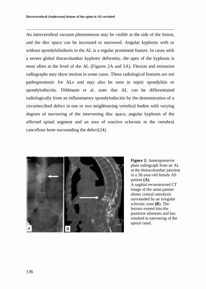
Discovertebral (Andersson) lesions of the spine in AS revisited
136
An intervertebral vacuum phenomenon may be visible at the side of the lesion,
and the disc space can be increased or narrowed. Angular kyphosis with or
without spondylolisthesis in the AL is a regular prominent feature. In cases with
a severe global thoracolumbar kyphotic deformity, the apex of the kyphosis is
most often at the level of the AL (Figures 2A and 3A). Flexion and extension
radiographs may show motion in some cases. These radiological features are not
pathognomonic for ALs and may also be seen in septic spondylitis or
spondylodiscitis. Dihlmann et al. state that AL can be differentiated
radiologically from an inflammatory spondylodiscitis by the demonstration of a
circumscribed defect in one or two neighbouring vertebral bodies with varying
degrees of narrowing of the intervening disc space, angular kyphosis of the
affected spinal segment and an area of reactive sclerosis in the vertebral
cancellous bone surrounding the defect(24).
Figure 2: Anteroposterior
plain radiograph from an AL
at the thoracolumbar junction
in a 56-year-old female AS
patient (A).
A sagittal reconstructed CT
image of the same patient
shows central osteolysis
surrounded by an irregular
sclerotic zone (B). The lesions extend into the
posterior elements and has
resulted in narrowing of the
spinal canal.
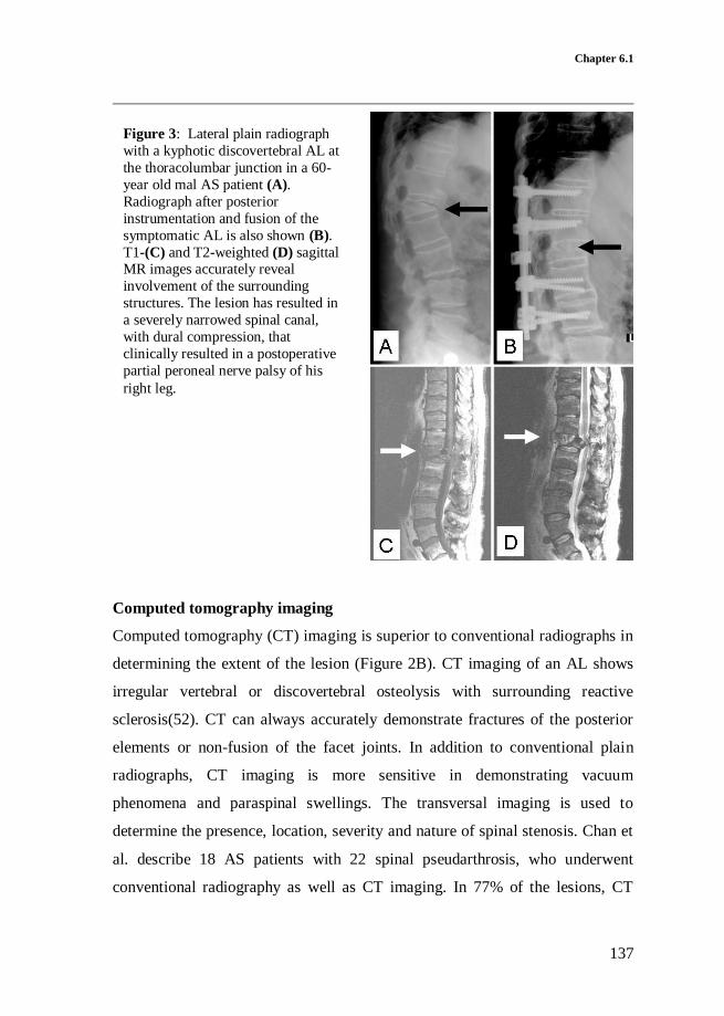
Chapter 6.1
137
Computed tomography imaging
Computed tomography (CT) imaging is superior to conventional radiographs in
determining the extent of the lesion (Figure 2B). CT imaging of an AL shows
irregular vertebral or discovertebral osteolysis with surrounding reactive
sclerosis(52). CT can always accurately demonstrate fractures of the posterior
elements or non-fusion of the facet joints. In addition to conventional plain
radiographs, CT imaging is more sensitive in demonstrating vacuum
phenomena and paraspinal swellings. The transversal imaging is used to
determine the presence, location, severity and nature of spinal stenosis. Chan et
al. describe 18 AS patients with 22 spinal pseudarthrosis, who underwent
conventional radiography as well as CT imaging. In 77% of the lesions, CT
Figure 3: Lateral plain radiograph
with a kyphotic discovertebral AL at
the thoracolumbar junction in a 60-
year old mal AS patient (A).
Radiograph after posterior
instrumentation and fusion of the
symptomatic AL is also shown (B).
T1-(C) and T2-weighted (D) sagittal MR images accurately reveal
involvement of the surrounding
structures. The lesion has resulted in
a severely narrowed spinal canal,
with dural compression, that
clinically resulted in a postoperative
partial peroneal nerve palsy of his
right leg.

Discovertebral (Andersson) lesions of the spine in AS revisited
138
imaging provided data that were missed on conventional radiography.
Therefore, CT offers considerable contributory advantages over conventional
radiography in diagnosing AL complicating AS(52). However, CT scanning had
the disadvantage of irradiation, which should be considered when choosing the
appropriate imaging method.
Magnetic resonance imaging
Magnetic resonance imaging (MRI) is considered the best modality in
visualising AL with the highest sensitivity(17;39;53). Generally, reduced signal
intensity of the disc space and surrounding vertebral bodies and increased signal
intensity after enhancement with contrast medium is noticeable on T1-weighted
images (Figures 3C and 4A). On T2-weighted images, increased signal intensity
in the corresponding area is noticeable (Figures 3D and 4B). In addition,
contrast-enhanced fat suppression imaging allows a better differentiation
between fat and enhanced lesions. Considering a diagnosed AL of the spine in
AS on plain radiographs and CT images, MR images can supplement
information on anterior longitudinal ligament disruption, vertebral translation,
abnormal dural enhancement, epidural lesions and spinal canal stenosis(39).
In addition, MRI is advised to evaluate spinal canal encroachment and the
extend of changes in the dura, spinal cord, nerve roots, soft tissue, and
ligaments(17;39;53). A decreased spinal canal can be observed caused by
anterior and posterior extradural tissue resulting from hypertrophic ligamentum
flavum and facet joints, as well as from hypertrophic callus formation of the
anterior and posterior elements of the AL. Differentiation from infectious
spondylitis and primary or metastatic tumors of the spine is possible if criteria
such as morphology, extension of bone marrow oedema, contrast enhancement
and signal intensity in the disc space on T2-weighted and fat suppression
sequence are taking in consideration(53;54).
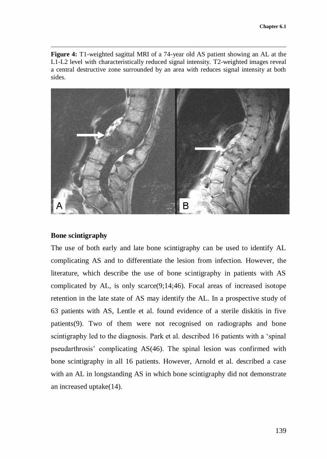
Chapter 6.1
139
Figure 4: T1-weighted sagittal MRI of a 74-year old AS patient showing an AL at the
L1-L2 level with characteristically reduced signal intensity. T2-weighted images reveal
a central destructive zone surrounded by an area with reduces signal intensity at both
sides.
Bone scintigraphy
The use of both early and late bone scintigraphy can be used to identify AL
complicating AS and to differentiate the lesion from infection. However, the
literature, which describe the use of bone scintigraphy in patients with AS
complicated by AL, is only scarce(9;14;46). Focal areas of increased isotope
retention in the late state of AS may identify the AL. In a prospective study of
63 patients with AS, Lentle et al. found evidence of a sterile diskitis in five
patients(9). Two of them were not recognised on radiographs and bone
scintigraphy led to the diagnosis. Park et al. described 16 patients with a ‘spinal
pseudarthrosis’ complicating AS(46). The spinal lesion was confirmed with
bone scintigraphy in all 16 patients. However, Arnold et al. described a case
with an AL in longstanding AS in which bone scintigraphy did not demonstrate
an increased uptake(14).
Discovertebral (Andersson) lesions of the spine in AS revisited
140
Laboratory tests
The role of laboratory tests in patient with an AL is limited. Both erythrocytes
sedimentation rate and C-reactive protein levels may be elevated in patients
with ALs, but this is of little diagnostic value(4;6;8;22;47). Both parameters are
usually more associated with the involvement of peripheral joints(55). White
blood count and blood cultures are of no additional diagnostic value.
Histology
There is no indication for a biopsy in patients with AS suspected from an AL.
However, tissue for histological examination might be obtained when surgical
treatment is performed. The lesions have variable histological appearances
discussed earlier (Table 2).
Management
Conservative treatment is often the first step of treatment, but there are no trials
available showing its optimal duration. Spontaneous relief of symptoms up to 3
years of conservative management has been reported, and the strategy should be
adjusted to each individual patient. In the upper thoracic spine, the presence of
an intact sternal-rib complex provides stability and might prevent kyphosis,
which was recently described as ‘the fourth column’(56). At the more mobile
cervical and thoracolumbar levels, however, conservative management is less
efficient. Stabilisation can be obtained by plaster immobilisation or Halo-jacket
immobilisation. Despite a normal bone-healing capacity in AS patients, fusion
of an extensive AL by plaster immobilisation is not likely(4;6). Even with long-
term plaster immobilisation using a thoracolumbosacral orthosis with one hip
included, minimal persistent motion at the AL may hinder fracture healing and
union. Although plaster immobilisation or Halo-jacket immobilisation has been
reported as a success in the treatment of acute fractures in AS(31), there is no

Chapter 6.1
141
evidence suggesting successful recovery of AL in AS patients after a long time
of immobilisation treatment. However, correction of a progressive cervical
kyphotic deformity resulting from a cervical fracture can be achieved by
immediate halo-jacket immobilisation followed by (gradual) correction by
manipulation.
The use of non-steroidal anti-inflammatory drugs during the active phases of the
disease forms the mainstay of the pharmacological management of AS. Newer
therapies that have been introduced, including anti-tumor necrosis factor
therapy such as infliximab, etanercept and adalimumab. There is no evidence
that treatment with these drugs is also beneficial in the treatment of
symptomatic AL. A rigorous exercise programme, as advocated in the treatment
of AS, is contraindicated in patients with AL complicating AS(41).
Surgery
Surgical treatment is indicated in patients with unbearable pain, progression of
the symptoms, a progressive kyphotic deformity or neurological deficits.
Surgical decompression, stabilisation and fusion form the mainstay of the
surgical management of AL complicating AS (Figure 3B). The goal is both to
decompress the spinal canal and to restore spinal stability facilitating healing
and fusion of the spinal lesion. Numerous surgical techniques have been
advocated, including instrumented and non-instrumented stabilisation through
anterior, posterior or combined approach(57). The surgical procedure depends
on the localisation of the lesion, alignment of the spine, neurological deficit and
the location of thecal sac compression(4;6;39). In patients with a (progressive)
thoracolumbar kyphotic deformity, posterior correction and fixation of the AL
by an anterior opening wedge osteotomy(43) or a posterior transpedicular
wedge resection osteotomy(58) may be considered.
Discovertebral (Andersson) lesions of the spine in AS revisited
142
CONCLUSIONS
In an attempt to structure the broad spectrum of Andersson lesions complicating
AS, a provisory division in localised and extensive lesions can be used.
Localised lesions are limited to certain parts of the intervertebral disc and
always have a inflammatory origin. Extensive lesions affect the whole disc or
vertebral body and may result from both mechanical and inflammatory factors.
Aetiologies range from spinal (stress-) fractures to a local delay in the
ankylosing process compared to adjacent levels, resulting in the last mobile
segment. Regardless of the exact aetiology, mechanical factors in the ankylosed
spine will prevent healing of extensive lesions and promote the formation of
pseudarthrosis. Initially, plain radiographs should be performed. MRI scanning
might be used for further evaluation, and in selected cases, CT scanning can be
performed. At this time, bone scanning does not have a place in the analysis of
Andersson lesions in AS. There is no consensus in literature regarding the
management of these lesions and when surgical intervention should be
performed. The mainstay of surgical treatment, however, consists of
instrumentation and fusion, with the correction of a kyphotic deformity, when
present.
Since radiological appearance, mechanical consequences, prognosis and
management in extensive lesions differ from localised lesions, it may be
questioned whether the latter should be classified as Andersson lesions. The
eponym Andersson lesion should be preserved to extensive lesions, which is
actually a spinal pseudarthrosis and the final common pathway of several
different aetiologies.
Chapter 6.1
143
REFERENCE LIST
(1) Von Bechterew W. Steifigkeit der Wirbelsäule und ihre
Verkrümmung als besondere Erkrankungsform. Neurologisches Zentralblatt 12, 426-434. 1-7-1893.
(2) McGonagle D, Lyn Tan A, Wakefield R, Emery P. Imaging in
ankylosing spondylitis. Van Royen BJ, Dijkmans BAC, editors. Ankylosing spondylitis-diagnosis and management. 71-81. 2006.
New York, Taylor & Francis group.
(3) Andersson O. Röntgenbilden vid spondylarthritis ankylopoetica. Nord Med Tidskr 14, 2000-2002. 1937.
(4) Cawley MID, Chalmers TM, Ball J, Kellgren JH. Destructive Lesions
of Vertebral Bodies in Ankylosing-Spondylitis. Ann Rheum Dis
1972;31(5):345-358.
(5) Chan FL, Ho EK, Fang D, Hsu LC, Leong JC, Ngan H. Spinal
pseudarthrosis in ankylosing spondylitis. Acta Radiol 1987
Jul;28(4):383-8.
(6) Fang D, Leong JC, Ho EK, Chan FL, Chow SP. Spinal pseudarthrosis
in ankylosing spondylitis. Clinicopathological correlation and the
results of anterior spinal fusion. J Bone Joint Surg Br 1988
May;70(3):443-7.
(7) Kabasakal Y, Garrett SL, Calin A. The epidemiology of
spondylodiscitis in ankylosing spondylitis--a controlled study. Br J
Rheumatol 1996 Jul;35(7):660-3.
(8) Langlois S, Cedoz JP, Lohse A, Toussirot E, Wendling D. Aseptic
discitis in patients with ankylosing spondylitis: a retrospective study
of 14 cases. Joint Bone Spine 2005 May;72(3):248-53.
(9) Lentle BC, Russell AS, Percy JS, Jackson FI. Scintigraphic findings
in ankylosing spondylitis. J Nucl Med 1977 Jun;18(6):524-8.
(10) Rasker JJ, Prevo RL, Lanting PJ. Spondylodiscitis in ankylosing
spondylitis, inflammation or trauma? A description of six cases. Scand J Rheumatol 1996;25(1):52-7.

Discovertebral (Andersson) lesions of the spine in AS revisited
144
(11) Wu PC, Fang D, Ho EKW, Leong JCY. The Pathogenesis of
Extensive Discovertebral Destruction in Ankylosing-Spondylitis. CLIN ORTHOP RELAT RES 1988;(230):154-61.
(12) Adler CP. [Spondylitis--spondylodiscitis. Pathologico-anatomical
morphology and diagnostic problems]. Radiologe 1985 Jul;25(7):291-8.
(13) Agarwal AK, Reidbord HE, Kraus DR, Eisenbeis CH, Jr. Variable
histopathology of discovertebral lesion (spondylodiscitis) of
ankylosing spondylitis. Clin Exp Rheumatol 1990 Jan;8(1):67-9.
(14) Arnold MH, Brooks PM, Ryan M, Francis H. A destructive
discovertebral lesion: septic discitis, ankylosing spondylitis, or
rheumatoid arthritis? Clin Rheumatol 1989 Jun;8(2):277-81.
(15) Bourqui M, Gerster JC. Ankylosing spondylitis presenting as
spondylodiscitis. Clin Rheumatol 1985 Dec;4(4):458-64.
(16) Finkelstein JA, Chapman JR, Mirza S. Occult vertebral fractures in
ankylosing spondylitis. Spinal Cord 1999 Jun;37(6):444-7.
(17) Kenny JB, Hughes PL, Whitehouse GH. Discovertebral Destruction
in Ankylosing-Spondylitis - the Role of Computed-Tomography and
Magnetic-Resonance-Imaging. BR J RADIOL 1990;63(750):448-55.
(18) Kurugoglu S, Mihmanli I, Kanberoglu K, Kanberoglu A. Destructive
diskovertebral lesions in ankylosing spondylitis: Appearance on
magnetic resonance imaging. Southern Medical Journal 2001;94(8):837-41.
(19) Lohr KM, Barthelemy CR, Schwab JP, Haasler GB. Septic
spondylodiscitis in ankylosing spondylitis. J Rheumatol 1987
Jun;14(3):616-20.
(20) Wise CM, Irby WR. Spondylodiscitis in ankylosing spondylitis:
variable presentations. J Rheumatol 1983 Dec;10(6):1004-6.
(21) Eschelman DJ, Beers GJ, Naimark A, Yablon I. Pseudoarthrosis in ankylosing spondylitis mimicking infectious diskitis: MR appearance.
AJNR Am J Neuroradiol 1991 Nov;12(6):1113-4.
Chapter 6.1
145
(22) Nikolaisen C, Nossent H. Early histology in ankylosing spondylitis
related spondylodiscitis supports its inflammatory origin. Scand J Rheumatol 2005 Sep;34(5):396-8.
(23) Romanus R, Yden S. Diskography in ankylosing spondylitis. Acta
Radiol 1952 Dec;38(6):431-9.
(24) Dihlmann W, Delling G. Disco-Vertebral Destructive Lesions (So-
Called Andersson Lesions) Associated with Ankylosing-Spondylitis.
Skelet Radiol 1978;3(1):10-6.
(25) Marsh CH. Internal fixation for stress fractures of the ankylosed spine. J R Soc Med 1985 May;78(5):377-9.
(26) Frank P, Gleeson JA. Destructive vertebral lesions in ankylosing
spondylitis. BR J RADIOL 1975 Sep;48(573):755-8.
(27) Calin A, Robertson D. Spondylodiscitis and pseudarthrosis in a
patient with enteropathic spondyloarthropathy. Ann Rheum Dis 1991
Feb;50(2):117-9.
(28) Albert HB, Kjaer P, Jensen TS, Sorensen JS, Bendix T, Manniche C. Modic changes, possible causes and relation to low back pain. Med
Hypotheses 2008;70(2):361-8.
(29) Cooper C, Carbone L, Michet CJ, Atkinson EJ, O'Fallon WM, Melton LJ, III. Fracture risk in patients with ankylosing spondylitis: a
population based study. J Rheumatol 1994 Oct;21(10):1877-82.
(30) Geusens P, Vosse D, van der Linden S. Osteoporosis and vertebral fractures in ankylosing spondylitis. Curr Opin Rheumatol 2007
Jul;19(4):335-9.
(31) Hitchon PW, From AM, Brenton MD, Glaser JA, Torner JC.
Fractures of the thoracolumbar spine complicating ankylosing spondylitis. J NEUROSURG 2002 Sep;97(2 Suppl):218-22.
(32) Hunter T, Dubo HI. Spinal fractures complicating ankylosing
spondylitis. A long-term followup study. Arthritis Rheum 1983 Jun;26(6):751-9.

Discovertebral (Andersson) lesions of the spine in AS revisited
146
(33) Mullaji AB, Upadhyay SS, Ho EK. Bone mineral density in
ankylosing spondylitis. DEXA comparison of control subjects with mild and advanced cases. J Bone Joint Surg Br 1994 Jul;76(4):660-5.
(34) Olerud C, Frost A, Bring J. Spinal fractures in patients with
ankylosing spondylitis. Eur Spine J 1996;5(1):51-5.
(35) Gelman MI, Umber JS. Fractures of the thoracolumbar spine in
ankylosing spondylitis. AJR Am J Roentgenol 1978 Mar;130(3):485-
91.
(36) Thorngren KG, Liedberg E, Aspelin P. Fractures of the thoracic and lumbar spine in ankylosing spondylitis. Arch Orthop Trauma Surg
1981;98(2):101-7.
(37) Chance GQ. Note on a type of flexion fracture of the spine. BR J RADIOL 1948 Sep;21(249):452.
(38) Mountney J, Murphy AJ, Fowler JL. Lessons learned from cervical
pseudoarthrosis in ankylosing spondylitis. Eur Spine J
2005;14(7):689-93.
(39) Shih TT, Chen PQ, Li YW, Hsu CY. Spinal fractures and
pseudoarthrosis complicating ankylosing spondylitis: MRI
manifestation and clinical significance. J Comput Assist Tomogr 2001 Mar;25(2):164-70.
(40) Mahesh BH, Jayaswal A, Bhan S. Fracture dislocation of the spine
after total hip arthroplasty in a patient with ankylosing spondylitis with early pseudoarthrosis. Spine J 2008 May;8(3):529-33.
(41) Sakaura H, Hosono N, Mukai Y, Fujii R, Yoshikawa H. Paraparesis
due to exacerbation of preexisting spinal pseudoarthrosis following
infliximab therapy for advanced ankylosing spondylitis. Spine J 2006;6(3):325-9.
(42) Bergmann EW. Fractures of the ankylosed spine. J Bone Joint Surg
Am 1949 Jul;31A(3):669-71.
(43) Chang KW, Tu MY, Huang HH, Chen HC, Chen YY, Lin CC.
Posterior correction and fixation without anterior fusion for
pseudoarthrosis with kyphotic deformity in ankylosing spondylitis. Spine 2006;31(13):E408-E413.
Chapter 6.1
147
(44) Deburge A, Guigui P, Ouahes M, Barre E, Simmons EH. Cervical
pseudarthrosis in ankylosing spondylitis: A case report. Spine 1996;21(23):2801-5.
(45) Kewalramani LS, Taylor RG, Albrand OW. Cervical spine injury in
patients with ankylosing spondylitis. J Trauma 1975 Oct;15(10):931-4.
(46) Park WM, Spencer DG, McCall IW. The detection of spinal
pseudarthrosis in ankylosing spondylitis. BR J RADIOL
1981;54(642):467-72.
(47) Unsal E, Arici AM, Kavukcu S, Pirnar T. Andersson lesion:
spondylitis erosiva in adolescents. Two cases and review of the
literature. Pediatr Radiol 2002 Mar;32(3):183-7.
(48) Dunn N, Preston B, Jones KL. Unexplained Acute Backache in
Longstanding Ankylosing-Spondylitis. BR MED J
1985;291(6509):1632-4.
(49) Hunter T, Forster B, Dvorak M. Ankylosed spines are prone to fracture. Can Fam Physician 1995 Jul;41:1213-6.
(50) Jobanputra P, Kirkham B, Duke O, Crockard A, Panayi GS.
Discovertebral destruction in ankylosing spondylitis complicated by spinal cord compression. Ann Rheum Dis 1988 Apr;47(4):344-7.
(51) Chang KW, Chen YY, Lin CC, Hsu HL, Pai KC. Closing wedge
osteotomy versus opening wedge osteotomy in ankylosing spondylitis with thoracolumbar kyphotic deformity. Spine 2005 Jul
15;30(14):1584-93.
(52) Chan FL, Ho EK, Chau EM. Spinal pseudarthrosis complicating
ankylosing spondylitis: comparison of CT and conventional tomography. AJR Am J Roentgenol 1988 Mar;150(3):611-4.
(53) Obradov M, Schonfeld DH, Franssen MJ, de Rooy DJ. Andersson
lesion in ankylosing spondylitis. JBR-BTR 2001 Apr;84(2):71.
(54) Jevtic V, Kos-Golja M, Rozman B, McCall I. Marginal erosive
discovertebral "Romanus" lesions in ankylosing spondylitis
demonstrated by contrast enhanced Gd-DTPA magnetic resonance imaging. Skeletal Radiol 2000 Jan;29(1):27-33.

Discovertebral (Andersson) lesions of the spine in AS revisited
148
(55) Ozgocmen S, Godekmerdan A, Ozkurt-Zengin F. Acute-phase
response, clinical measures and disease activity in ankylosing spondylitis. Joint Bone Spine 2007 May;74(3):249-53.
(56) Shen FH, Samartzis D. Successful nonoperative treatment of a three-
column thoracic fracture in a patient with ankylosing spondylitis: existence and clinical significance of the fourth column of the spine.
Spine 2007 Jul 1;32(15):E423-E427.
(57) Fox MW, Onofrio BM, Kilgore JE. Neurological complications of
ankylosing spondylitis. J NEUROSURG 1993;78(6):871-8.
(58) Van Royen BJ, Kastelijns RC, Noske DP, Oner FC, Smit TH.
Transpedicular wedge resection osteotomy for the treatment of a
kyphotic Andersson lesion-complicating ankylosing spondylitis. Eur Spine J 2006 Feb;15(2):246-52.

Chapter 6.1
149

Discovertebral (Andersson) lesions of the spine in AS revisited
150

Chapter 6.1
151
Discovertebral (Andersson) lesions in severe
ankylosing spondylitis: a study using MRI and
conventional radiography.
M. K. de Vries1, A.S van Drumpt
2, B. J. van Royen
2, J.C. van Denderen
4, R.A.
Manoliu3, I. E. van der Horst-Bruinsma
1.
Rheumatology Department
1, Orthopaedic Surgery Department
2 and Radiology
Department3 VU University Medical Center, Rheumatology Department
4, Jan
van Breemen Institute/Reade, Amsterdam, the Netherlands.
Clinical Rheumatology 2010;29(12):1433-1438.

Discovertebral (Andersson) lesions in severe AS: a study using MRI and conventional radiography
152
ABSTRACT
Objectives: To investigate the prevalence of Andersson lesions (ALs) in
ankylosing spondylitis (AS) patients who will start anti-tumor necrosis factor
(TNF) treatment.
Methods: Radiographs and magnetic resonance imaging (MRI) of the spine
were performed before therapy with anti-TNF. ALs were defined as
discovertebral endplate destructions on MRI, associated with bone marrow
edema and fat replacement or sclerosis, a decreased signal on T1, enhancement
after contrast administration gadolinium diethylenetriamine pentaacetic acid
(Gd-DTPA), and increased signal on T2 and short tau inversion recovery
(STIR). Additionally, conventional radiography showed a fracture line, irregular
endplates, and increased sclerosis of adjacent vertebral bodies.
Results: Fifty-six AS patients were included, 68% males, mean age of 43 years,
and mean disease duration of 11 years. The mean Bath ankylosing spondylitis
disease activity index was 6.4, and 24% of all patients had ankylosis. Only one
patient showed a discovertebral abnormality with bone marrow edema of more
than 50% of the vertebral bodies adjacent to the intervertebral disc of T7/T8 and
T9/T10, a hypodense signal area on T1, and a high signal on STIR. Irregular
endplates were depicted, and T1 after Gd-DTPA demonstrated high signal
intensity around the disc margins. However, no fracture line was visible on
conventional radiology, and therefore, this case was not considered to be an AL.
Conclusion: No AL was detected in our AS patients, who were candidates for
anti-TNF treatment. One patient showed a discovertebral abnormality on MRI,
without a fracture line on conventional radiology. The relative small proportion
of patients with a long-established disease might explain this finding for,
particularly, an ankylosed spine is prone to develop an AL.
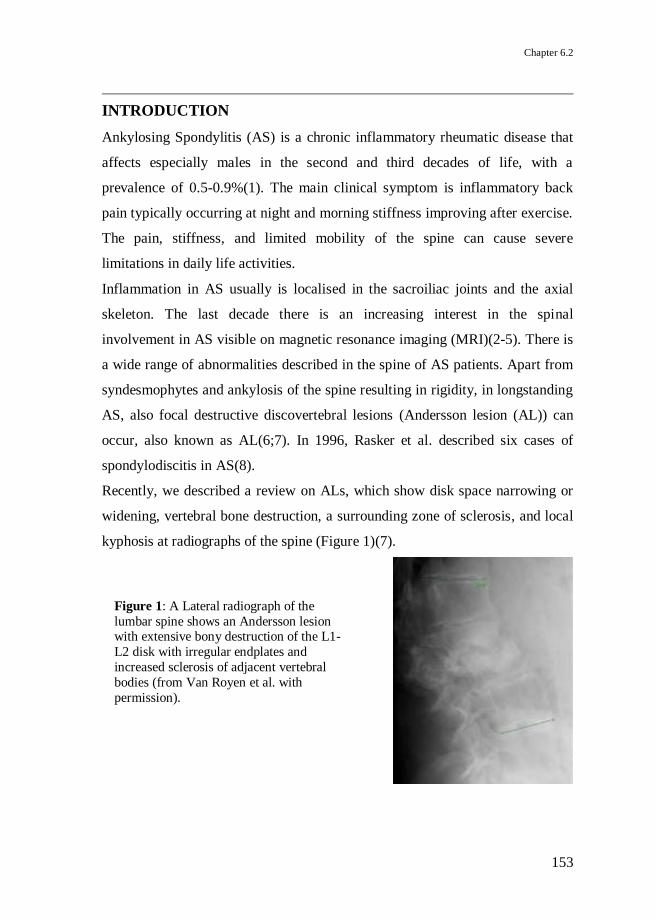
Chapter 6.2
153
INTRODUCTION
Ankylosing Spondylitis (AS) is a chronic inflammatory rheumatic disease that
affects especially males in the second and third decades of life, with a
prevalence of 0.5-0.9%(1). The main clinical symptom is inflammatory back
pain typically occurring at night and morning stiffness improving after exercise.
The pain, stiffness, and limited mobility of the spine can cause severe
limitations in daily life activities.
Inflammation in AS usually is localised in the sacroiliac joints and the axial
skeleton. The last decade there is an increasing interest in the spinal
involvement in AS visible on magnetic resonance imaging (MRI)(2-5). There is
a wide range of abnormalities described in the spine of AS patients. Apart from
syndesmophytes and ankylosis of the spine resulting in rigidity, in longstanding
AS, also focal destructive discovertebral lesions (Andersson lesion (AL)) can
occur, also known as AL(6;7). In 1996, Rasker et al. described six cases of
spondylodiscitis in AS(8).
Recently, we described a review on ALs, which show disk space narrowing or
widening, vertebral bone destruction, a surrounding zone of sclerosis, and local
kyphosis at radiographs of the spine (Figure 1)(7).
Figure 1: A Lateral radiograph of the
lumbar spine shows an Andersson lesion with extensive bony destruction of the L1-
L2 disk with irregular endplates and
increased sclerosis of adjacent vertebral
bodies (from Van Royen et al. with
permission).
Discovertebral (Andersson) lesions in severe AS: a study using MRI and conventional radiography
154
One of the causes of an AL is the local inflammation in the spine in
combination with a minor trauma. This lesion can be differentiated from the
signs of inflammation at the MRI of the spine of active disease in AS, as is
often observed in patients who are candidates for tumor necrosis factor (TNF)
blocking treatment. An AL might be difficult to detect on clinical symptoms
alone because most AS patients suffer from back pain. More important is the
fact that an AL requires a different treatment, for instance immobilisation, in
contrast with the physical therapy normally prescribed in AS patients.
The aim of this study was to investigate whether in AS patients, who are
candidates for treatment with anti-TNF, ALs could be detected.
METHODS
Patients
AS patients were derived from the outpatient clinics of VU University Medical
Center (VUmc) and Jan van Breemen Institute (JBI) in Amsterdam. All AS
patients who fulfilled the modified New York criteria and met the criteria for
starting treatment with TNF blocking agents according to the international
ASAS consensus statement(9;10) were included.
Following this ASAS statement, all patients had an active disease as defined by
a Bath ankylosing spondylitis disease activity index (BASDAI) >4 (scale 0-
10)(11), had failed to previous therapy with at least two nonsteroidal anti-
inflammatory drugs (NSAIDs) and sulphasalazine in case of peripheral arthritis,
and should be treated with TNF blocking agents according to an expert. Patients
were enrolled consecutively, but mainly because of limited availability of the
MR scanner, several patients could not be included. Furthermore, one patient
did not fit into the scanner due to severe kyphosis, and several patients suffered
from claustrophobia. At baseline, data, conform to daily clinical practice during

Chapter 6.2
155
treatment with TNF blocking agents, were collected in all patients who gave
their written informed consent.
Radiology
Standing anteroposterior and lateral full-length plain films and a MRI of the
whole spine were performed before the start of anti-TNF therapy. MRI were
obtained at baseline and after 6-24 months of anti-TNF therapy, using T1-
weighted spin-echo sequences (T1) before and after administration of the
contrast medium gadolinium diethylenetriamine pentaacetic acid (Gd-DTPA),
T2-weighted sequences (T2), and fat-suppression sequences (short tau inversion
recovery (STIR)). In patients included in the JBI, MRI of the spine was
performed in the Amstelland Hospital Amstelveen with a field strength of 0.5
tesla, starting from the third cervical vertebra (C3) and downwards. In patients
included in Vumc, MRI of the whole spine was performed with a field strength
of 1.5 tesla. Pre- and post- (Gd) T1 TSE, and STIR MRIs were acquired. In
Amstelland Hospital Amstelveen, T2 was acquired as well.
The conventional radiographs and MRI were read by two observers, one
orthopedic surgeon (BvR) and a radiologist (RM), who were blinded to the
status of the subjects. The consensus score was accepted. First, the conventional
radiographs were analyzed followed by an assessment of the MRI.
The extent of spinal ankylosis in the total study cohort was estimated by
counting extensive syndesmophyte formation or bamboo spine features on
conventional radiographs counted by segment. The extent of abnormalities in
the discovertebral unit was compared with the same discovertebral unit in
different saggital MR sequences and the other way around.
An AL was defined as an abnormality in the discovertebral junction that shows
on MRI diffuse endplate destruction, with associated bone marrow edema and
fat replacement or sclerosis(12). The fracture cleft thought the disk space or

Discovertebral (Andersson) lesions in severe AS: a study using MRI and conventional radiography
156
vertebra itself is depicted as decreased signal on T1 and increased signal on T2,
STIR, and T1 imaging after Gd-DTPA administration, reflecting reactive edema
of the fracture. Conventional radiography of the AL must show a fracture line,
irregular endplates, and increased sclerosis of adjacent vertebral bodies.
Statistical analysis
This concerns a descriptive study. Continuous variables were reported as the
mean ± SD or, if skewed, as the median (range). Categorical variables were
calculated as frequencies and percentages.
RESULTS
In total, 56 AS patients were included, 36 in VUmc and 20 in JBI, and the
baseline characteristics of all patients are summarised in Table 1. Patients from
the two centers did not differ in sex, age, or disease activity at baseline.
Radiographs at baseline were available in 55 of the total 56 patients. MRI was
performed at baseline in all 56 patients. In two patients, an alternative MRI
procedure was performed due to a severe kyphotic posture. These MR scans
were still suitable for the purpose of our study.
Radiology
Systematic examination of conventional radiographs of the cervical, thoracic,
and lumbar spine did not reveal any lesions that met the definition of AL.
Distinct ankylosis with extensive syndesmophyte formation was seen in 13 of
55 (24%) of the patients determined with conventional radiography of the spine.
Spinal ankylosis or bridging syndesmophytes over multiple vertebrae were seen
at the cervical, thoracic, and lumbar segment in 16%, 6% and 7% of patients,
respectively.

Chapter 6.2
157
Table 1: Baseline characteristics
Mean +/- SD (range)
Number 56
Age (in years) 43 +/- 10.8 (22 - 73)
Male sex (%) 38 (68)
Disease duration (in years)ª 11 +/- 8.7 (1 - 41)
Symptom duration (in years )b 21 +/- 11.3 (1 - 49)
BASDAI (0-10)c 6.4 +/- 1.4 (4.0 – 9.7)
Tragus-to-wall distance (cm; normal, <15 cm) 16 +/- 6.0 (11 – 44)
Lumbar flexion index (cm; normal, >5 cm) 2.5 +/- 1.2 (0.3 – 5)
Lumbar side flexion (cm; normal, >10 cm) 10 +/- 4.9 (3.8 – 19)
Chest expansion (cm; normal, >5 cm) 3.4 +/- 1.5 (0.5 - 7)
a Disease duration: mean time between the diagnosis and baseline .
b Symptom duration: mean time between the first symptoms and baseline.
c BASDAI : Bath ankylosing spondylitis disease activity index; mean value (11).
Conventional radiographs and MRIs of the whole spine showed no AL
according to our definition. However, one patient showed a discovertebral
abnormality on MRI. This abnormality showed bone marrow edema of more
than 50% of the vertebral bodies adjacent to the intervertebral disk of T7/T8 and
T9/T10 (Figure 2), a hypodense signal area on T1 and high signal on STIR.
Irregular endplates of the vertebra at these levels were depicted on all
sequences. T1 after administration of Gd-DTPA demonstrated high signal
intensity around the disk margins of T7/T8 and T9/T10. However, no fracture
line was visible on conventional radiology. The clinical characteristics of this
patient did not differ from the other patients. He was a male, aged 65 years, with
a BASDAI and CRP of 5 and 35 mg/L, respectively, indicative of an active
disease. He did not report any specific localising complaints or a preceding
(minor) trauma. He had suffered from complaints of low back pain for 40 years
and was diagnosed with AS at the age of 51.

Discovertebral (Andersson) lesions in severe AS: a study using MRI and conventional radiography
158
Figure 2:
A. T1-weighted Gd-DTPA post contrast image demonstrated high-signal intensity
around the disk margins of T7-T8 and T9-T10.
B. T1-weighted image shows low-signal intensity bone marrow edema in the adjacent vertebral endplates of the T7/T8 and T9/T10 disk spaces.
C. T1-weighted image, obtained 24 months after treatment with anti-TNF agents.
After 24 months treatment with infliximab, T1 weighted images showed a
decrease of signs of acute inflammation such as bone marrow edema and maybe
even an increase of post-inflammatory fatty bone marrow degeneration around
the disk margins. The T1-weighted post-contrast images demonstrated no high
signal intensity around the disk margins of T7/T8 and T9/T10 anymore. There
were signs of progression of the ankylosis and kyphosis in the thoracic segment
after 2 years of follow-up despite anti-TNF treatment. Apart from the focus on
ALs, other abnormalities involving the discovertebral unit as described in
literature on MRI in AS were encountered. Romanus lesions, which are acute
inflammatory lesions at the corners of the endplates of the vertebra, were often
detected. These lesions show bone marrow edema in the corners of vertebral
endplates and are depicted as hypodense signal areas in T1- and high signal in
T2-weighted images(29).
A B C

Chapter 6.2
159
DISCUSSION
In 56 AS patients, who fulfilled the criteria for treatment with TNF blocking
agents, only one lesion resembling an AL of the thoracic spine was detected
with MRI, but this lesion lacked a fracture line on the conventional radiograph.
The low prevalence of ALs was unexpected because this group of AS patients
had a relatively high rate of ankylosis of the spine (24%) and signs of active
inflammation. This was in concordance with the moderate impairment of the
mobility of the spine in these patients. The mean tragus-to-wall distance was 16
cm (normal:<15cm), mean lumbar flexion index was 2.5 cm (normal:>5cm), the
mean lumbar side flexion was 10 cm (normal:>10cm) and the mean chest
expansion was 3.4 (normal:>5cm)(13). The percentage of ALs as described in
literature in this subset of severe AS patients varies between 1% and 28%(14-
20).
Many studies report that the presence of an AL is associated with long standing
disease in AS and with a rigid, ankylosed spine(21-23), concluding that an AL
is a late complication of AS(17). The low prevalence of ALs in our study was
probably caused by the relatively short disease duration, with a mean of 11
years and a mean age of 43 years. This could be due to a selection bias, for
mostly young patients with an active disease were referred for treatment with
TNF blocking agents, whereas the efficacy of TNF blockers in older and more
severe cases of AS with complete ankylosis of the spine was still doubtful at the
time of the study.
A severe kyphotic deformity and an osteoporotic spine puts a patient at risk of
development of a stress fracture in the ankylosed spine(24;25). Our study
contains patients with a severe, kyphotic, thoracic spine. However, we did not
find any features of stress fractures in this susceptible subgroup of patients,
probably due to the small group size.
Discovertebral (Andersson) lesions in severe AS: a study using MRI and conventional radiography
160
Another cause of the low prevalence of ALs in this study might be the due to
the radiographic techniques used to detect them, although the combination of
conventional radiographs of the spine and MRI with gadolinium should be
sensitive enough. Hermann et al. compared MRI and conventional radiography
of spinal changes in patients with spondyloarthritis(21). They concluded that the
best way to detect syndesmophytes was through radiography; ankylosis was
detected equally well by both radiography and MRI, but for all other lesions,
MRI was the preferred method. A disadvantage of conventional radiographs of
the spine is the difficult analysis of the vertebrae of the thoracic segment
because of the overimposed lung tissue(26), while most disorders of the
discovertebral junction occur in the lower part of the thoracic and upper parts of
the lumbar spine(27;28). MRI is important for early detection of discovertebral
lesions and described as a sensitive method to detect ALs, in particular, after
administration of gadolinium(29). Some MRIs in our study showed features of
severe degenerative disk disease (DDD), specifically lumbar-sacral. The MRI
features of these degenerative abnormalities were sometimes difficult to
distinguish from destructive discovertebral lesions as was described by Jevtic et
al., who showed that the appearance of an AL can resemble Modic type III
degenerative lesion(29). One point to differentiate between AL and DDD in AS
is the fact that an AL occurs as a result of inflammation in combination with
mechanical stress of the ankylosed spine. Another point is that AL differs from
DDD in the localisation, because most AL lesions occur at the level of the
cervical and thoracolumbar spine, whereas the DDD predominate at the
lumbosacral level(7).
The debate about the prevalence of ALs is hampered by the lack of uniformity
in the nomenclature of these lesions. For example, the term AL has been
employed for severe destructive discovertebral lesions, but also for minor

Chapter 6.2
161
abnormalities such as degenerative changes and local inflammation around in
the discovertebral junction, as described in our case(7;12).
To conclude, no ALs were detected by using conventional radiographs and MRI
in this study in 56 severe AS patients, whereas a higher prevalence was
described in literature. The discrepancy might be due to the relatively small
group of patient with a short disease duration and young age despite the severe
ankylosed spine in our study cohort.
Despite the absence of ALs in our study, we would like to increase the
awareness of this complication in AS. In case of a minor spine trauma, a MRI
should be combined with the conventional spine radiographs in order to detect
this lesion in the stiff and vulnerable spine, which is often osteoporotic as well.
The treatment of an AL lesion consists of immobilisation instead of the
routinely advised exercise programme or even surgical decompression in case
of neurological deficits(7).
In previous studies, there has been no sequential MRI study of the spine in
patients with AS that visualizes the evolution from an early discovertebral
lesion, as described in our patient, into a severe destructive discovertebral
lesion, by some authors called an AL. Thus, it remains unclear whether and how
often an abnormality in the discovertebral junction develops into an AL. More
research with a long-term follow-up of discovertebral lesions is necessary to
clarify the evolution of these lesions.

Discovertebral (Andersson) lesions in severe AS: a study using MRI and conventional radiography
162
REFERENCE LIST
(1) Braun J, Bollow M, Remlinger G, Eggens U, Rudwaleit M, Distler A, et
al. Prevalence of spondylarthropathies in HLA-B27 positive and negative blood donors. Arthritis Rheum 1998 Jan;41(1):58-67.
(2) Braun J, Baraliakos X, Golder W, Brandt J, Rudwaleit M, Listing J, et
al. Magnetic resonance imaging examinations of the spine in patients
with ankylosing spondylitis, before and after successful therapy with infliximab: evaluation of a new scoring system. Arthritis Rheum 2003
Apr;48(4):1126-36.
(3) Maksymowych WP, Landewe R. Imaging in ankylosing spondylitis. Best Pract Res Clin Rheumatol 2006 Jun;20(3):507-19.
(4) Marc V, Dromer C, Le Guennec P, Manelfe C, Fournie B. Magnetic
resonance imaging and axial involvement in spondylarthropathies.
Delineation of the spinal entheses. Rev Rhum Engl Ed 1997 Jul;64(7-9):465-73.
(5) van der Heijde D, Landewe R. Imaging in spondylitis. Curr Opin
Rheumatol 2005 Jul;17(4):413-7.
(6) Andersson O. Röntgenbilden vid spondylarthritis ankylopoetica. Nord
Med Tidskr 14, 2000-2002. 1937.
(7) Bron JL, de Vries MK, Snieders MN, van der Horst-Bruinsma IE, Van Royen BJ. Discovertebral (Andersson) lesions of the spine in
ankylosing spondylitis revisited. Clin Rheumatol 2009 Aug;28(8):883-
92.
(8) Rasker JJ, Prevo RL, Lanting PJ. Spondylodiscitis in ankylosing spondylitis, inflammation or trauma? A description of six cases. Scand J
Rheumatol 1996;25(1):52-7.
(9) Braun J, Pham T, Sieper J, Davis J, van der Linden S, Dougados M, et al. International ASAS consensus statement for the use of anti-tumour
necrosis factor agents in patients with ankylosing spondylitis. Ann
Rheum Dis 2003 Sep;62(9):817-24.

Chapter 6.2
163
(10) Braun J, Davis J, Dougados M, Sieper J, van der Linden S, van der
Heijde D. First update of the international ASAS consensus statement for the use of anti-TNF agents in patients with ankylosing spondylitis.
Ann Rheum Dis 2006 Mar;65(3):316-20.
(11) Garrett S, Jenkinson T, Kennedy LG, Whitelock H, Gaisford P, Calin A. A new approach to defining disease status in ankylosing spondylitis:
the Bath Ankylosing Spondylitis Disease Activity Index. J Rheumatol
1994 Dec;21(12):2286-91.
(12) Van Royen BJ, Kastelijns RC, Noske DP, Oner FC, Smit TH. Transpedicular wedge resection osteotomy for the treatment of a
kyphotic Andersson lesion-complicating ankylosing spondylitis. Eur
Spine J 2006 Feb;15(2):246-52.
(13) Wanders A, Landewe R, Dougados M, Mielants H, van der Linden S,
van der Heijde D. Association between radiographic damage of the
spine and spinal mobility for individual patients with ankylosing
spondylitis: can assessment of spinal mobility be a proxy for radiographic evaluation? Ann Rheum Dis 2005 Jul;64(7):988-94.
(14) Cawley MID, Chalmers TM, Ball J, Kellgren JH. Destructive Lesions
of Vertebral Bodies in Ankylosing-Spondylitis. Ann Rheum Dis 1972;31(5):345-358.
(15) Guma M, Olive A, Perez R, Holgado S, Ortiz-Santamaria V, Tena X.
Aseptic spondylodiskitis in rheumatic diseases. Clin Exp Rheumatol 2001;19(6):740-7.
(16) Hehne HJ, Becker HJ, Zielke K. The spondylodiscitis in kyphotic
deformities of ankylosing spondylitis and the influence of dorsal
correction osteotomies. Report on 33 patients. Z ORTHOP IHRE GRENZGEB 1990;128(5):494-502.
(17) Kabasakal Y, Garrett SL, Calin A. The epidemiology of
spondylodiscitis in ankylosing spondylitis--a controlled study. Br J Rheumatol 1996 Jul;35(7):660-3.
(18) Langlois S, Cedoz JP, Lohse A, Toussirot E, Wendling D. Aseptic
discitis in patients with ankylosing spondylitis: a retrospective study of 14 cases. Joint Bone Spine 2005 May;72(3):248-53.

Discovertebral (Andersson) lesions in severe AS: a study using MRI and conventional radiography
164
(19) Lanting PJ, Rasker JJ, Kruijsen MW, Prevo RL. [Spondylodiscitis in
Bechterew's disease; inflammation or trauma? Description of 6 patients]. Ned Tijdschr Geneeskd 1994 Oct 1;138(40):1997-2001.
(20) Tsuchiya K, Nagamine R, Iwamoto Y. Discovertebral lesion in
ankylosing spondylitis: Differential diagnosis with discitis by magnetic resonance imaging. Mod Rheumatol 2002;12(2):113-7.
(21) Hermann KGA, Althoff CE, Schneider U, Zuhlsdorf SZ, Lembcke A,
Hamm B, et al. Spinal changes in patients with spondyloarthritis:
Comparison of MR imaging and radiographic appearances. Radiographics 2005;25(3):559-U1.
(22) Obradov M, Schonfeld DH, Franssen MJ, de Rooy DJ. Andersson
lesion in ankylosing spondylitis. JBR-BTR 2001 Apr;84(2):71.
(23) Yau ACMC, Chan RNW. Stress Fracture of Fused Lumbo-Dorsal Spine
in Ankylosing-Spondylitis. Journal of Bone and Joint Surgery-British
Volume 1974;B 56(4):681-7.
(24) Cooper C, Carbone L, Michet CJ, Atkinson EJ, O'Fallon WM, Melton LJ, III. Fracture risk in patients with ankylosing spondylitis: a
population based study. J Rheumatol 1994 Oct;21(10):1877-82.
(25) Maillefert JF, Aho LS, El Maghraoui A, Dougados M, Roux C. Changes in bone density in patients with ankylosing spondylitis: a two-
year follow-up study. Osteoporos Int 2001;12(7):605-9.
(26) Braun J, Baraliakos X, Golder W, Hermann KG, Listing J, Brandt J, et al. Analysing chronic spinal changes in ankylosing spondylitis: a
systematic comparison of conventional x rays with magnetic resonance
imaging using established and new scoring systems. Ann Rheum Dis
2004 Sep;63(9):1046-55.
(27) Baraliakos X, Landewe R, Hermann KG, Listing J, Golder W, Brandt J,
et al. Inflammation in ankylosing spondylitis: a systematic description
of the extent and frequency of acute spinal changes using magnetic resonance imaging. Ann Rheum Dis 2005 May;64(5):730-4.
(28) Braun J, Golder W, Bollow M, Sieper J, van der Heijde D. Imaging and
scoring in ankylosing spondylitis. Clin Exp Rheumatol 2002 Nov;20(6 Suppl 28):S178-S184.

Chapter 6.2
165
(29) Jevtic V, Kos-Golja M, Rozman B, McCall I. Marginal erosive
discovertebral "Romanus" lesions in ankylosing spondylitis demonstrated by contrast enhanced Gd-DTPA magnetic resonance
imaging. Skeletal Radiol 2000 Jan;29(1):27-33.

Discovertebral (Andersson) lesions in severe AS: a study using MRI and conventional radiography

Chapter 6.2
Improvement of lipid profile is accompanied by
atheroprotective alterations in high-density
lipoprotein composition upon tumor necrosis factor
blockade.
A prospective cohort study in ankylosing
spondylitis.
Izhar C. van Eijk1, Mirjam K. de Vries
2, J.H.M. Levels
3, Mike J.L. Peters
2,
Esther E. Huizer1, Ben A.C. Dijkmans
1-2, Irene E. van der Horst-Bruinsma
2,
B.P.C. Hazenberg5, Rob J. van de Stadt
1, Gert Jan Wolbink
1-4, Michael T.
Nurmohamed1.
Rheumatology Department, Jan van Breemen Institute/Reade1; Rheumatology
Department, VU University Medical Center2; Experimental Vascular Medicine
Department, Academic Medical Center, University of Amsterdam3; Sanquin
Research, Amsterdam4; Rheumatology and Clinical Immunology Department,
University Medical Center Groningen5, the Netherlands.
Arthritis and Rheumatism 2009;60(5):1324-1330.

Improvement of lipid profile is accompanied by atheroprotective alterations in high-density lipoprotein
composition upon tumor necrosis factor blockade in AS
168
ABSTRACT
Objective: Cardiovascular mortality is increased in ankylosing spondylitis
(AS), and inflammation plays an important role. Inflammation deteriorates lipid
profile and alters high-density lipoprotein cholesterol (HDL-c) composition,
reflected by increased concentrations of serum amyloid A (SAA) within the
particle. Anti-tumor necrosis factor (anti-TNF) treatment may improve these
parameters. We therefore undertook the present study to investigate the effects
of etanercept on lipid profile and HDL composition in AS.
Methods: In 92 AS patients, lipid levels and their association with the
inflammatory markers C-reactive protein (CRP), erythrocyte sedimentation rate
(ESR), and serum amyloid A protein (SAA) were evaluated serially during 3
months of etanercept treatment. HDL composition and its relation to
inflammatory markers was determined in a subgroup of patients, using surface-
enhanced laser desorption/ionization time-of-flight analysis.
Results: With anti-TNF treatment, levels of all parameters of inflammation
decreased significantly, whereas total cholesterol, HDL-c, and apolipoprotein
A-I (Apo A-I) levels increased significantly. This resulted in a better total
cholesterol:HDL-c ratio (from 3.9 to 3.7) (although the difference was not
statistically significant), and an improved Apo B:Apo A-I ratio, which
decreased by 7.5% over time (p=0.008). In general, increases in levels of all
lipid parameters were associated with reductions in inflammatory activity. In
addition, SAA was present at high levels within HDL particles from AS patients
with increased CRP levels and disappeared during treatment, in parallel with
declining plasma levels of SAA.
Conclusion: Our results show for the first time that during anti-TNF therapy of
AS, along with favorable changes in the lipid profile, HDL composition is
actually altered whereby SAA disappears from the HDL particle, increasing its
atheroprotective ability.
Chapter 7
169
These findings demonstrate the importance of understanding the role of
functional characteristics of HDL-c in cardiovascular diseases related to chronic
inflammatory conditions.
INTRODUCTION
Ankylosing spondylitis (AS) is a chronic inflammatory disease of the sacroiliac
joints and spine affecting up to 1% of the population(1). Patients with AS have
approximately a 2-fold increased mortality rate compared with general
population, which is predominantly due to an increased cardiovascular (CV)
risk(2-4). Although specific CV disorders (valvular disease and conduction
disturbances) occur more frequently in AS(4;5), accelerated atherosclerotic
disease probably contributes to the increased CV risk as well(6;7).
Atherosclerosis is a multifactorial process, but its most well-established risk
factor is dyslipidemia. Important prognostic indicators of CV disease are the
ratio of total cholesterol to high-density lipoprotein cholesterol (HDL-c) and the
ratio of apolipoprotein B (Apo B) to Apo A-I.
Inflammation deteriorates the lipid profile, as characterized by low HDL, total
cholesterol, and Apo A-I levels and increased levels of low-density lipoprotein
cholesterol (LDL-c), triglycerides, and Apo B. Indeed, several investigators
have reported that patients with inflammatory rheumatic diseases have a
deteriorated lipid profile(4;6;8-10).
In addition to changes in lipid levels, inflammation can affect HDL
qualitatively(11). During inflammation, specific enzyme and protein
components of HDL, contributing to its (anti)atherogenic potential, such as
serum amyloid A (SAA) and Apo A-I, are modified and may even render it
proatherogenic(12). Tumor necrosis factor (TNF) is a pivotal pro-inflammatory
cytokine in inflammatory diseases and causes a deterioration of the lipid profile
in inflammatory conditions(12).

Improvement of lipid profile is accompanied by atheroprotective alterations in high-density lipoprotein
composition upon tumor necrosis factor blockade in AS
170
Treatment with TNF blocking agents, in addition to the known powerful anti-
inflammatory effects, may therefore have a beneficial effect on the lipid profile
as well as and on HDL composition(13;14).
The current study was designed to investigate whether modulation of
inflammatory activity by TNF blockade therapy in patients with active AS is
associated with alterations in lipid profile and qualitative changes in HDL
composition.
PATIENTS AND METHODS
Patients
Consecutive AS patients attending the outpatient clinics of the Jan van Breemen
Institute and VU University Medical Center, in whom etanercept treatment was
initiated according to the assessment in ankylosing spondylitis (International
Working Group) consensus statement for initiation of anti-TNF treatment(15),
were enrolled and followed up prospectively. All patients fulfilled the 1984
modified New York criteria for AS(16) and were treated with subcutaneous
etanercept 25 mg twice weekly or 50 mg once weekly. High disease activity
was defined as a score of 4 on the Bath ankylosing spondylitis disease activity
index (BASDAI)(17). Patients were included if a baseline serum sample and at
least one follow-up serum sample were available. The study was approved by
the local medical ethics committee, and all patients provided written informed
consent.
Study Design
Data were collected at baseline and after 1 month and 3 months of treatment.
During every visit, questionnaires on disease activity (BASDAI) were
administered. Total cholesterol, HDL-c, LDL-c, triglycerides, Apo A-I, Apo B,
SAA, erythrocyte sedimentation rate (ESR), and C-reactive protein (CRP) were

Chapter 7
171
measured in sera obtained at each time point. Collected sera were stored at
-20°C until testing. Commercially available kits were used to measure acute-
phase reactants.
Assessment of lipids
Serum total cholesterol and triglycerides were analyzed by an enzymatic
method using the appropriate assays (Roche Diagnostics, Almere, The
Netherlands), on a Cobas 6000 analyzer (Roche Diagnostics), according to the
instructions of the manufacturer. Polyethylene glycol-modified enzymes were
used for assessing the HDL-c levels. Apo A-I and Apo B were analyzed by an
immunoturbidimetric method, using appropriate assays (Roche Diagnostics).
Since we were not able to directly measure LDL-c levels at our laboratory, the
Friedewald formula was used, when triglyceride levels were lower than 400
mg/dl, to calculate LDL-c levels, although, in the strictest sense, this formula
may not be the most appropriate method for measuring LDL-c in nonfasting
samples. The total cholesterol:HDL-c ratio was calculated.
Assessment of inflammation markers
CRP levels were determined using the Roche/Hitachi Cobas 6000 analyzer,
based on the principle of particle-enhanced immunologic agglutination (Roche
Diagnostics); values of <10 mg/liter were considered normal. High-sensitivity
CRP (hsCRP) levels were determined using the Roche/Hitachi Cobas 6000
system, with a detection range of 0.15-20 mg/liter. The test is based on the
principle of a particle-enhanced immunoturbidimetric assay; human CRP
agglutinates with latex particles coated with anti-CRP monoclonal antibodies.
ESR was determined with local measurement techniques (Westergren method);
values of <20 mm/hour and <30 mm/hour were considered normal in men and
women, respectively. SAA was assessed with an enzyme-linked immunosorbent
Improvement of lipid profile is accompanied by atheroprotective alterations in high-density lipoprotein
composition upon tumor necrosis factor blockade in AS
172
assay as described previously(18); values of <4 mg/liter were considered
normal.
Preparation of samples
HDL protein profiling was performed as described previously(19). For coating
of antibody, a 5 ml mixture containing 2.8 nM anti-Apo A-I monoclonal
antibodies, 3 mM ethylenediamine, and 0.1M Na2SO 4 was added per spot of a
PS-20 protein chip, and covalent binding of antibodies through primary amine-
epoxide chemistry was achieved by incubating the chip in a humid chamber
overnight at 4°C. Excess antibody was removed by one wash with distilled
water, and subsequently, free amine binding places were blocked by incubating
the chip with 1M Tris buffer (pH 8.4) for 30 minutes at room temperature.
For HDL capture, after mounting of the PS-20 protein chip(s) in a 96-well
bioprocessor, 100 ml of plasma aliquots (diluted 1:2 with Tris buffered saline
[TBS]) (50 mM Tris [pH 7.4], and 150 mM NaCl) was applied onto each spot
and allowed to bind for 2 hours at room temperature on a horizontal shaker. The
protein chips were washed 4 times with TBS (5 minutes for each wash),
followed by a 2-minute rinse with TBS-Tween (0.005%). A final wash step with
HEPES solution (5 mM) was carried out to remove the excess salt. All spots
were allowed to dry, and subsequently, 1.2 l of sinapinic acid (10 mg/ml) in a
50:49.9:0.1% acetonitrile-water-trifluoric acid mix was applied onto each spot.
All chips were air-dried and stored at room temperature in the dark until
analysis. These measurements were conducted on the same day as the chip
processing.

Chapter 7
173
Surface-enhanced laser desorption/ionization time-of-flight (SELDI-TOF)
analysis
SELDI-TOF analysis was carried out with a PBS IIc protein chip reader
(Ciphergen Biosystems, Fremont, CA), using an automated data collection
protocol within the Protein-Chip software (version 3.1). Data were collected up
to 200 kd. Laser intensity was set in a range of 190-200 arbitrary units at a
sensitivity of 7, and the focus mass was set at 28 kd, specific for anti-Apo A-I
capture. Measurement of the spectra was performed with an average of 100
shots at 13 positions per SELDI spot. Calibration was done using a protein
calibration chip (Ciphergen). Spectra were normalized on total ion current.
Detected peaks having a signal-to-noise ratio of 5 were recognized as
significant peaks. Data on the reproducibility of the Seldi technique have been
reported previously(19).
Statistical analysis
Data are expressed as the mean ± SD or the median and interquartile range, as
appropriate. The distribution of variables was tested for normality and
transformed if necessary. Independent t-tests were used for variables with a
normal distribution, and nonparametric tests (Wilcoxon’s signed rank test or
Mann-Whitney U test) for skewed variables. Pearson’s χ² test was performed
for dichotomous variables. Correlation coefficients (Pearson’s) were calculated
to evaluate correlations between SAA and lipid levels at baseline.
The generalized estimating equations (GEE) approach was used (a) to analyse
longitudinal data on lipids, lipoproteins, and acute-phase reactants measured at
3 different time points (i.e., a longitudinal logistic regression analysis was
performed) and (b) to investigate associations between changes in disease
activity markers, HDL-c and Apo A-I levels over time.
Improvement of lipid profile is accompanied by atheroprotective alterations in high-density lipoprotein
composition upon tumor necrosis factor blockade in AS
174
Absolute and relative changes in lipid levels were calculated in relation to
changes in disease activity parameters. Since the total cholesterol:HDL-c ratio
and triglyceride levels were not normally distributed, data were analyzed with
the logarithms of these values. For clarity, the regression coefficients of these
lipids were retransformed to geometric means. Calculations were performed
using SPSS 16.0 software. P values less than 0.05 were considered significant.
RESULTS
Characteristics of the patients
A total of 92 consecutive AS patients were enrolled (60 men (65%), 32 women
(35%)). The median age was 40.6 years. The mean BASDAI was 6.0, and the
median disease duration was 9 years. Ninety-four percent of the patients were
taking nonsteroidal anti-inflammatory drugs, 22% were taking concomitant
disease-modifying anti-rheumatic drugs, and 8% were known to take statins.
During the anti-TNF treatment period of this study, all pharmacologic treatment
was unchanged. Baseline characteristics of the patients are shown in Table 1.
Inflammation markers
Concentrations of ESR, CRP and hsCRP were elevated at baseline and declined
during treatment (p<0.001) (Table 2). The same was true for SAA, another
acute-phase protein, with elevated levels at baseline that decreased significantly
after 1 month and remained low and stable thereafter (p<0.001) (Table 2).
At baseline, SAA levels correlated negatively with Apo A-I levels (r = -0.28,
p=0.08), indicating that higher plasma SAA levels were accompanied by lower
Apo A-I levels. Baseline SAA levels did not correlate with HDL-c levels
(r = -0.07, p=0.5).

Chapter 7
175
Table 1: Baseline characteristics of the 92 AS patients.
Demographic features
Age, years 43 ± 11.2
Disease duration, median (IQR ) years 8.5 (3-18)
N males/females 60/32
HLA-B27 positivity, N (%)† 74 (88)
Disease activity parameters
ESR, median (IQR) mm/hour 21 (6-38)
CRP, median (IQR) mg/liter 13 (3-35)
hsCRP, median (IQR) mg/liter 11.3 (3.1-33.2)
SAA, median (IQR) mg/liter 5 (2-18)
BASDAI, 0-10 scale 6.0 ± 1.5
Lipids
Total cholesterol, mmoles/liter 4.87 ± 0.9
HDL-c, mmoles/liter 1.29 ± 0.4
Total cholesterol: HDL-c ratio, median (IQR)‡ 3.89 (3.01-4.90)
LDL-c, mmoles/liter 2.92 ± 0.8
Triglycerides, median (IQR) mmoles/liter 1.17 (0.89-1.74)
Apo A-I, gm/liter 1.39 ± 0.3
Apo B, gm/liter 0.88 ± 0.2
Apo B:Apo A-I ratio 0.67 ± 0.23
Except when indicated otherwise, values are the mean ± SD. AS, ankylosing spondylitis; IQR, interquartile
range; ESR, erythrocyte sedimentation rate; hsCRP, high-sensitivity C-reactive protein; SAA, serum amyloid
A; BASDAI, Bath ankylosing spondylitis disease activity index; HDL-c, high-density lipoprotein cholesterol;
LDL-c, low-density lipoprotein cholesterol; Apo A-I, apolipoprotein A-I.
†Data not available of 8 patients.
‡Atherogenic index is based on this ratio.
Improvement of lipid profile is accompanied by atheroprotective alterations in high-density lipoprotein
composition upon tumor necrosis factor blockade in AS
176
Lipid levels over time
Lipid levels and disease activity parameters in AS patients were measured
before and during anti-TNF treatment (Table 2). Total cholesterol, HDL-c, and
Apo A-I levels increased significantly during treatment (p<0.001, p<0.001, and
p=0.004, respectively). Levels of LDL-c and triglycerides increased slightly
during treatment (p=0.04 and p=0.03 respectively), and Apo B remained stable.
The total cholesterol:HDL-c ratio decreased from 3.9 at baseline to 3.7 after 3
months (5% reduction), but this did not reach statistical significance.
The Apo B:Apo A-I ratio declined by 7.5%, from 0.67 to 0.62 (p=0.008).
Associations between lipids levels and disease activity markers
Since CRP and hsCRP levels were comparable, only CRP was used in the
association models. GEE analyses demonstrated several significant associations
between lipid levels and disease activity parameters, including CRP, ESR, SAA,
and BASDAI, over time, i.e., the degree of disease activity as assessed by the
selected disease activity parameters significantly influenced lipid levels. The
influence of disease activity parameters on lipid levels is demonstrated by the
data shown in Table 3. During the 3-months follow-up period, decreasing levels
of CRP, ESR, SAA, and BASDAI levels were significantly associated with
increasing total cholesterol levels (p≤0.003) (with regression coefficients of
0.01, 0.015, 0.006, and 0.063, respectively), increasing HDL-c levels (p≤0.014)
(with regression coefficients of 0.004, 0.005, 0.002, and 0.025, respectively),
increasing Apo A-I levels (p≤0.001) (with regression coefficients of 0.004,
0.005, 0.003, and 0.018, respectively) and decreasing Apo B:Apo A-I ratios
(p<0.01) (with regression coefficients of -0.002, -0.002, -0.001, and -0.001,
respectively). Changes in disease activity parameters were not associated with
changes in atherogenic index (total cholesterol:HDL-c ratio).
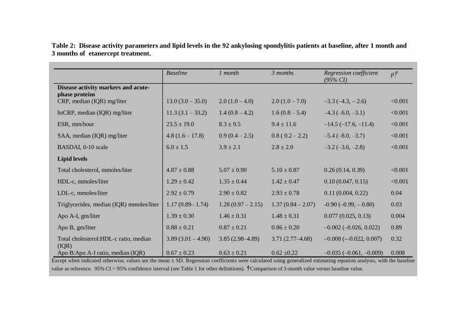
177
Table 2: Disease activity parameters and lipid levels in the 92 ankylosing spondylitis patients at baseline, after 1 month and
3 months of etanercept treatment.
Except when indicated otherwise, values are the mean ± SD. Regression coefficients were calculated using generalized estimating equation analysis, with the baseline
value as reference. 95% CI = 95% confidence interval (see Table 1 for other definitions). †Comparison of 3-month value versus baseline value.
Baseline 1 month 3 months Regression coefficient (95% CI)
p†
Disease activity markers and acute-
phase proteins
CRP, median (IQR) mg/liter 13.0 (3.0 – 35.0) 2.0 (1.0 – 4.0) 2.0 (1.0 – 7.0) –3.3 (–4.3, – 2.6) <0.001
hsCRP, median (IQR) mg/liter 11.3 (3.1 – 33.2) 1.4 (0.8 – 4.2) 1.6 (0.8 – 5.4) –4.3 (–6.0, –3.1) <0.001
ESR, mm/hour 23.5 ± 19.0 8.3 ± 9.5 9.4 ± 11.6 –14.5 (–17.6, –11.4) <0.001
SAA, median (IQR) mg/liter 4.8 (1.6 – 17.8) 0.9 (0.4 – 2.5) 0.8 ( 0.2 – 2.2) –5.4 (–8.0, –3.7) <0.001
BASDAI, 0-10 scale 6.0 ± 1.5 3.9 ± 2.1 2.8 ± 2.0 –3.2 (–3.6, –2.8) <0.001
Lipid levels
Total cholesterol, mmoles/liter 4.87 ± 0.88 5.07 ± 0.90 5.10 ± 0.87 0.26 (0.14, 0.39) <0.001
HDL-c, mmoles/liter 1.29 ± 0.42 1.35 ± 0.44 1.42 ± 0.47 0.10 (0.047, 0.15) <0.001
LDL-c, mmoles/liter 2.92 ± 0.79 2.90 ± 0.82 2.93 ± 0.78 0.11 (0.004, 0.22) 0.04
Triglycerides, median (IQR) mmoles/liter 1.17 (0.89– 1.74) 1.28 (0.97 – 2.15) 1.37 (0.84 – 2.07) -0.90 (–0.99, – 0.80) 0.03
Apo A-I, gm/liter 1.39 ± 0.30 1.46 ± 0.31 1.48 ± 0.31 0.077 (0.025, 0.13) 0.004
Apo B, gm/liter 0.88 ± 0.21 0.87 ± 0.21 0.86 ± 0.20 –0.002 (–0.026, 0.022) 0.89
Total cholesterol:HDL-c ratio, median
(IQR)
3.89 (3.01 – 4.90) 3.85 (2.98–4.89) 3.71 (2.77–4.68) –0.008 (-–0.022, 0.007) 0.32
Apo B:Apo A-I ratio, median (IQR) 0.67 ± 0.23 0.63 ± 0.21 0.62 ±0.22 –0.035 (–0.061, –0.009) 0.008
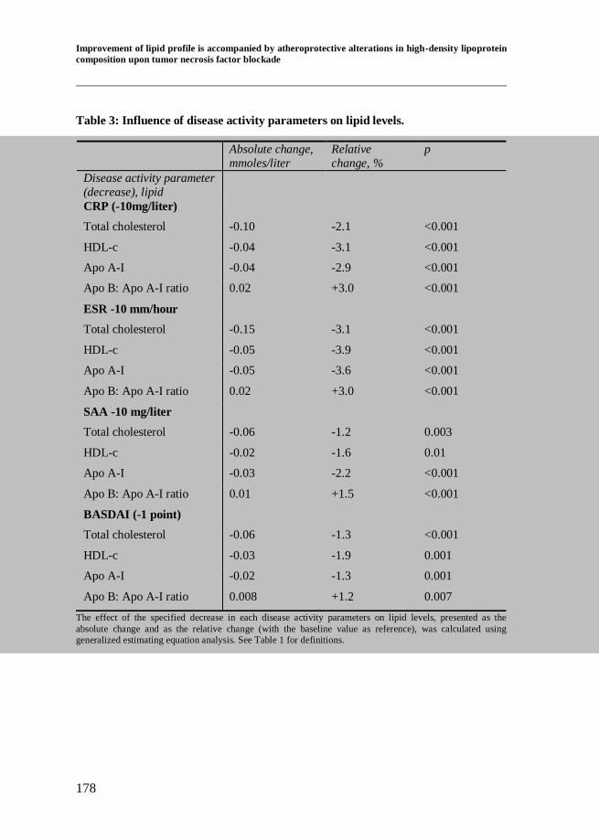
Improvement of lipid profile is accompanied by atheroprotective alterations in high-density lipoprotein
composition upon tumor necrosis factor blockade
178
Table 3: Influence of disease activity parameters on lipid levels.
The effect of the specified decrease in each disease activity parameters on lipid levels, presented as the
absolute change and as the relative change (with the baseline value as reference), was calculated using
generalized estimating equation analysis. See Table 1 for definitions.
Absolute change,
mmoles/liter
Relative
change, %
p
Disease activity parameter
(decrease), lipid
CRP (-10mg/liter)
Total cholesterol -0.10 -2.1 <0.001
HDL-c -0.04 -3.1 <0.001
Apo A-I -0.04 -2.9 <0.001
Apo B: Apo A-I ratio 0.02 +3.0 <0.001
ESR -10 mm/hour
Total cholesterol -0.15 -3.1 <0.001
HDL-c -0.05 -3.9 <0.001
Apo A-I -0.05 -3.6 <0.001
Apo B: Apo A-I ratio 0.02 +3.0 <0.001
SAA -10 mg/liter
Total cholesterol -0.06 -1.2 0.003
HDL-c -0.02 -1.6 0.01
Apo A-I -0.03 -2.2 <0.001
Apo B: Apo A-I ratio 0.01 +1.5 <0.001
BASDAI (-1 point)
Total cholesterol -0.06 -1.3 <0.001
HDL-c -0.03 -1.9 0.001
Apo A-I -0.02 -1.3 0.001
Apo B: Apo A-I ratio 0.008 +1.2 0.007

Chapter 7
179
SELDI-TOF findings
Additional analyses were performed in a subgroup of 10 patients, 5 of whom
had high levels of CRP (>30 mg/liter) at baseline and 5 of whom had low levels
of CRP (<15 mg/liter) at baseline. After SELDI-TOF analysis, protein spectra
from HDL were obtained. Figure 1 shows the HDL profile and Figure 2 shows
the plasma SAA levels in 3 representative patients over time. At baseline, a
higher density of mass charge (m/z) marker 11,695, which represents SAA, was
found in the subgroup of AS patients with high CRP levels. During treatment,
all spectra exhibited virtually similar profiles, and m/z marker 11,695
disappeared from HDL as inflammation regressed in the patients in whom CRP
levels had been increased at baseline (patients A and B in Figure 1). Moreover,
in patient B, a proteolytically generated isoform of SAA appeared to be present;
it is known that 1-3 amino acids can be cleaved from either the N- or C-
terminus of SAA(20).

Improvement of lipid profile is accompanied by atheroprotective alterations in high-density lipoprotein
composition upon tumor necrosis factor blockade
180
SAA m/z region
A
B
C
0
1
3
0
1
3
0
1
3
11400 11600 11800 12000
11400 11600 11800 12000
11400 11600 11800 12000
11400 11600 11800 12000
11400 11600 11800 12000
11400 11600 11800 12000
11400 11600 11800 12000
11400 11600 11800 12000
11400 11600 11800 12000
11400 11600 11800 12000
11400 11600 11800 12000
11400 11600 11800 12000
11400 11600 11800 12000
11400 11600 11800 12000
11400 11600 11800 12000
11400 11600 11800 12000
11400 11600 11800 12000
11400 11600 11800 12000
11400 11600 11800 12000
m/z
SAA m/z region
A
B
C
0
1
3
0
1
3
0
1
3
11400 11600 11800 12000
11400 11600 11800 12000
11400 11600 11800 12000
11400 11600 11800 12000
11400 11600 11800 12000
11400 11600 11800 12000
11400 11600 11800 12000
11400 11600 11800 12000
11400 11600 11800 12000
11400 11600 11800 12000
11400 11600 11800 12000
11400 11600 11800 12000
11400 11600 11800 12000
11400 11600 11800 12000
11400 11600 11800 12000
11400 11600 11800 12000
11400 11600 11800 12000
11400 11600 11800 12000
11400 11600 11800 1200011400 11600 11800 12000
m/z
Figure 2: Plasma SAA levels in
patients A, B and C at each time point.
0 1 30
5
10
15A
B
C
SA
A (
mg
/L)
0
5
10
15
SA
A (
mg
/L)
0 1 30
5
10
15A
B
C
SA
A (
mg
/L)
0
5
10
15
SA
A (
mg
/L)
Figure 1: Representative
examples of high-density
lipoprotein spectra in gel
views obtained from
surface-enhanced laser
desorption/ionization time-
of-flight analysis in 2
representative ankylosing
spondylitis patients with
high baseline C-reactive
protein (CRP) levels (patients A and B) and 1
representative AS patient
with low baseline CRP
level (patient C). Spectra
bin the specific ass/charge
(m/z) range of serum
amyloid A (SAA) are
shown by arrows. Each
spectrum was measured in
duplicate at baseline, 1
month after initiation of anti-tumor necrosis factor
(anti-TNF) treatment, and 3
months after initiation of
anti-TNF treatment (time
points 0, 1, and 2,
respectively. All spectra
were normalized on total
ion current.
Chapter 7
181
DISCUSSION
In the present study, ankylosing spondylitis with high inflammatory activity was
characterized by decreased levels of total cholesterol, HDL-c and Apo A-I
accompanied by biochemical changes in the HDL particle. Along with
improvement of the lipid profile, reflected by increased HDL-c and Apo A-I
levels and an improved Apo B:Apo A-I ratio, anti-TNF treatment led to
favourable alterations in HDL composition, i.e., diminishing of the SAA
concentration within the HDL particles.
This is the first report of a study investigating alterations in apolipoprotein
levels in AS patients during anti-TNF treatment. Apo A-I is the major
atheroprotective apolipoprotein in the HDL particle, whereas Apo B reflects the
total number of potentially atherogenic particles, being present in very low-
density lipoprotein, intermediate-density lipoprotein, and LDL.
Comparable with the total cholesterol:HDL-c ratio, the Apo B:Apo A-I ratio has
emerged as a very good predictor of future CV events, with the practical
advantage that fasting blood samples are not required(21-24). This ratio reflects
the balance of cholesterol transport in a simple way. The higher the Apo B:Apo
A-I ratio, the more cholesterol is circulating in the plasma compartment, and
this cholesterol is likely to be deposited in the arterial wall, causing
atherogenesis and risk of CV events. In the AS patients in the present study, the
Apo B:Apo A-I ratio was positively associated with disease activity parameters
and a 7.5% decrease in this ratio was accomplished during anti-TNF treatment,
suggesting a beneficial effect on the risk for CV morbidity and mortality,
although, due to the relatively small change the in Apo B:Apo A-I ratio, this
should be interpreted with caution.
Anti-TNF treatment resulted in a less atherogenic lipid profile, which is
consistent with previous findings(10). Although the observed changes in lipid

Improvement of lipid profile is accompanied by atheroprotective alterations in high-density lipoprotein
composition upon tumor necrosis factor blockade
182
levels were small, even these small changes may well have a clinically relevant
effect on CV risk, since AS is a chronic inflammatory disease that persists over
many years(25). However, beyond focusing solely on HDL-c levels, it seems
important to investigate actual HDL composition and thereby its functional
characteristics, to learn more about its effects on the vascular system and CV
risk.
HDL protein profiling is increasingly being used to determine the biochemical
composition of HDL(19;26;27). During acute systemic inflammation HDL
becomes pro-inflammatory, loses its protective properties, and can even
enhance atherogenesis(12;28). Interestingly, in addition to showing reduced
plasma levels of HDL-c during active AS, SELDI-TOF analysis enabled us to
demonstrate actual alterations in HDL composition; i.e., in contrast to findings
in AS patients with low CRP levels at baseline, in whom virtually no SAA was
found on HDL, SAA was markedly present on the surface of HDL in AS
patients with increased CRP levels at baseline, but after treatment of these
patients to suppress inflammation, the SAA on HDL almost disappeared.
SAA is an acute-phase reactant which is synthesized mainly in the liver in
response to pro-inflammatory cytokines such as interleukin-1, interleukin-6, and
TNF(29), and elevated levels of SAA are associated with increased CV risk(30).
SAA is transported mainly in HDL as an apolipoprotein(31;32). Increased
serum SAA levels during the acute-phase response in patients with active AS
thus seem to be accompanied by an increased presence of SAA within the HDL
particle. Recently, increased SAA within the HDL particle was also found in
patients with active Crohn’s disease, another chronic inflammatory disease,
which is associated with spondylarthropathies including AS(33). This is of
interest since it is known that SAA is able to replace anti-atherogenic Apo A-I
in the HDL particles, which renders them less protective(34;35). Moreover,

Chapter 7
183
SAA-rich HDL particles are rapidly cleared from plasma, and thus the increase
in SAA during inflammation could also contribute to the decrease in total
HDL-c concentrations(36). However, other mechanisms may also play a role in
decreased HDL-c levels during inflammation as well. It has been suggested that
remodelling of HDL through activation of secretory phospholipase A2 may be
an alternate explanation for reduced HDL-c levels during the acute-phase
response. Overexpression of this enzyme in mice leads to decreased HDL-c
levels and enhanced HDL-c catabolism(28;37-39). In addition, inflammation
may convert HDL de novo into a more proatherogenic form by coordinate but
inverse transcriptional regulation of SAA and Apo A-I in the liver(29).
This may explain the observed inverse correlation between plasma levels of
SAA and Apo A-I, but not between plasma levels of SAA and levels of HDL-c,
at baseline. Changes in total cholesterol, HDL-c and Apo A-I levels were
significantly inversely associated with changes in levels of disease activity
parameters over time, confirming the role of inflammatory activity in lipid
profile changes.
In conclusion, findings of the present study demonstrate for the first time that
during anti-TNF treatment for AS, along with favourable changes in lipid
profile, HDL composition is actually altered, with SAA disappearing from the
HDL particle, which rendered it more atheroprotective. Our results highlight the
importance of understanding the role of functional characteristics of HDL
cholesterol in CV diseases related to chronic inflammatory conditions such as
AS.

Improvement of lipid profile is accompanied by atheroprotective alterations in high-density lipoprotein
composition upon tumor necrosis factor blockade
184
REFERENCE LIST
(1) Braun J, Bollow M, Remlinger G, Eggens U, Rudwaleit M, Distler A, et
al. Prevalence of spondylarthropathies in HLA-B27 positive and
negative blood donors. Arthritis Rheum 1998 Jan;41(1):58-67.
(2) Lautermann D, Braun J. Ankylosing spondylitis--cardiac manifestations. Clin Exp Rheumatol 2002 Nov;20(6 Suppl 28):S11-
S15.
(3) Lehtinen K. Mortality and causes of death in 398 patients admitted to hospital with ankylosing spondylitis. Ann Rheum Dis 1993
Mar;52(3):174-6.
(4) Peters MJ, van der Horst-Bruinsma IE, Dijkmans BA, Nurmohamed MT. Cardiovascular risk profile of patients with spondylarthropathies,
particularly ankylosing spondylitis and psoriatic arthritis. Semin
Arthritis Rheum 2004 Dec;34(3):585-92.
(5) Roldan CA, Chavez J, Wiest PW, Qualls CR, Crawford MH. Aortic root disease and valve disease associated with ankylosing spondylitis. J
Am Coll Cardiol 1998 Nov;32(5):1397-404.
(6) Divecha H, Sattar N, Rumley A, Cherry L, Lowe GD, Sturrock R. Cardiovascular risk parameters in men with ankylosing spondylitis in
comparison with non-inflammatory control subjects: relevance of
systemic inflammation. Clin Sci (Lond) 2005 Aug;109(2):171-6.
(7) Heeneman S, Daemen MJ. Cardiovascular risks in spondyloarthritides.
Curr Opin Rheumatol 2007 Jul;19(4):358-62.
(8) Joven J, Rubies-Prat J, Ras MR, de la Figuera M, Lience E, Masdeu S.
High density lipoprotein cholesterol subfractions and apoprotein A-I in patients with rheumatoid arthritis and ankylosing spondylitis. Arthritis
Rheum 1984 Oct;27(10):1199-200.
(9) Han C, Robinson DW, Jr., Hackett MV, Paramore LC, Fraeman KH, Bala MV. Cardiovascular disease and risk factors in patients with
rheumatoid arthritis, psoriatic arthritis, and ankylosing spondylitis. J
Rheumatol 2006 Nov;33(11):2167-72.

Chapter 7
185
(10) van Halm V, van Denderen JC, Peters MJ, Twisk JW, van der Paardt
M, van der Horst-Bruinsma IE, et al. Increased disease activity is associated with a deteriorated lipid profile in patients with ankylosing
spondylitis. Ann Rheum Dis 2006 Nov;65(11):1473-7.
(11) Khovidhunkit W, Kim MS, Memon RA, Shigenaga JK, Moser AH, Feingold KR, et al. Effects of infection and inflammation on lipid and
lipoprotein metabolism: mechanisms and consequences to the host. J
Lipid Res 2004 Jul;45(7):1169-96.
(12) Navab M, Anantharamaiah GM, Fogelman AM. The role of high-density lipoprotein in inflammation. Trends Cardiovasc Med 2005
May;15(4):158-61.
(13) Kiortsis DN, Mavridis AK, Filippatos TD, Vasakos S, Nikas SN, Drosos AA. Effects of infliximab treatment on lipoprotein profile in
patients with rheumatoid arthritis and ankylosing spondylitis. J
Rheumatol 2006 May;33(5):921-3.
(14) Popa C, Barrea P, Netea MG, Stalenhoef AF, van der Meer JW. Anti-TNF therapy and plasma HDL cholesterol concentration.
Atherosclerosis 2005 Oct;182(2):375.
(15) Zochling J, van der Heijde D, Burgos-Vargas R, Collantes E, Davis JC, Jr., Dijkmans B, et al. ASAS/EULAR recommendations for the
management of ankylosing spondylitis. Ann Rheum Dis 2006
Apr;65(4):442-52.
(16) van der Linden S, Valkenburg HA, Cats A. Evaluation of diagnostic
criteria for ankylosing spondylitis. A proposal for modification of the
New York criteria. Arthritis Rheum 1984 Apr;27(4):361-8.
(17) Garrett S, Jenkinson T, Kennedy LG, Whitelock H, Gaisford P, Calin A. A new approach to defining disease status in ankylosing spondylitis:
the Bath Ankylosing Spondylitis Disease Activity Index. J Rheumatol
1994 Dec;21(12):2286-91.
(18) Hazenberg BP, Limburg PC, Bijzet J, van Rijswijk MH. Monoclonal
antibody based ELISA for human SAA. Natvig JB, Forre O, Husby G,
Husebekk A, Skogen B, Sletten K et al., editors. [Amyloid and amyloidosis.], 898-901. 1991. Dordrecht (the Netherlands), Kluwer
Academic Publishers.

Improvement of lipid profile is accompanied by atheroprotective alterations in high-density lipoprotein
composition upon tumor necrosis factor blockade
186
(19) Levels JH, Bleijlevens B, Rezaee F, Aerts JM, Meijers JC. SELDI-TOF
mass spectrometry of High-Density Lipoprotein. Proteome Sci 2007;5:15.
(20) Ducret A, Bruun CF, Bures EJ, Marhaug G, Husby G, Aebersold R.
Characterization of human serum amyloid A protein isoforms separated by two-dimensional electrophoresis by liquid
chromatography/electrospray ionization tandem mass spectrometry.
Electrophoresis 1996 May;17(5):866-76.
(21) van der Steeg WA, Boekholdt SM, Stein EA, El-Harchaoui K, Stroes ES, Sandhu MS, et al. Role of the apolipoprotein B-apolipoprotein A-I
ratio in cardiovascular risk assessment: a case-control analysis in EPIC-
Norfolk. Ann Intern Med 2007 May 1;146(9):640-8.
(22) Ingelsson E, Schaefer EJ, Contois JH, McNamara JR, Sullivan L, Keyes
MJ, et al. Clinical utility of different lipid measures for prediction of
coronary heart disease in men and women. JAMA 2007 Aug
15;298(7):776-85.
(23) Walldius G, Jungner I, Holme I, Aastveit AH, Kolar W, Steiner E. High
apolipoprotein B, low apolipoprotein A-I, and improvement in the
prediction of fatal myocardial infarction (AMORIS study): a prospective study. Lancet 2001 Dec 15;358(9298):2026-33.
(24) Rasouli M, Kiasari AM, Mokhberi V. The ratio of apoB/apoAI, apoB
and lipoprotein(a) are the best predictors of stable coronary artery disease. Clin Chem Lab Med 2006;44(8):1015-21.
(25) Sharrett AR, Ballantyne CM, Coady SA, Heiss G, Sorlie PD, Catellier
D, et al. Coronary heart disease prediction from lipoprotein cholesterol
levels, triglycerides, lipoprotein(a), apolipoproteins A-I and B, and HDL density subfractions: The Atherosclerosis Risk in Communities
(ARIC) Study. Circulation 2001 Sep 4;104(10):1108-13.
(26) Rezaee F, Casetta B, Levels JH, Speijer D, Meijers JC. Proteomic analysis of high-density lipoprotein. Proteomics 2006 Jan;6(2):721-30.
(27) Vaisar T, Pennathur S, Green PS, Gharib SA, Hoofnagle AN, Cheung
MC, et al. Shotgun proteomics implicates protease inhibition and complement activation in the antiinflammatory properties of HDL. J
Clin Invest 2007 Mar;117(3):746-56.
Chapter 7
187
(28) van der Westhuyzen DR, de Beer FC, Webb NR. HDL cholesterol
transport during inflammation. Curr Opin Lipidol 2007 Apr;18(2):147-51.
(29) Han CY, Chiba T, Campbell JS, Fausto N, Chaisson M, Orasanu G, et
al. Reciprocal and coordinate regulation of serum amyloid A versus apolipoprotein A-I and paraoxonase-1 by inflammation in murine
hepatocytes. Arterioscler Thromb Vasc Biol 2006 Aug;26(8):1806-13.
(30) Ridker PM, Hennekens CH, Buring JE, Rifai N. C-reactive protein and
other markers of inflammation in the prediction of cardiovascular disease in women. N Engl J Med 2000 Mar 23;342(12):836-43.
(31) Eriksen N, Benditt EP. Isolation and characterization of the amyloid-
related apoprotein (SAA) from human high density lipoprotein. Proc Natl Acad Sci U S A 1980 Nov;77(11):6860-4.
(32) Coetzee GA, Strachan AF, van der Westhuyzen DR, Hoppe HC, Jeenah
MS, de Beer FC. Serum amyloid A-containing human high density
lipoprotein 3. Density, size, and apolipoprotein composition. J Biol Chem 1986 Jul 25;261(21):9644-51.
(33) van Leuven SI, Hezemans R, Levels JH, Snoek S, Stokkers PC,
Hovingh GK, et al. Enhanced atherogenesis and altered high density lipoprotein in patients with Crohn's disease. J Lipid Res 2007
Dec;48(12):2640-6.
(34) Van Lenten BJ, Hama SY, de Beer FC, Stafforini DM, McIntyre TM, Prescott SM, et al. Anti-inflammatory HDL becomes pro-inflammatory
during the acute phase response. Loss of protective effect of HDL
against LDL oxidation in aortic wall cell cocultures. J Clin Invest 1995
Dec;96(6):2758-67.
(35) Steinmetz A, Hocke G, Saile R, Puchois P, Fruchart JC. Influence of
serum amyloid A on cholesterol esterification in human plasma.
Biochim Biophys Acta 1989 Nov 28;1006(2):173-8.
(36) Hoffman JS, Benditt EP. Plasma clearance kinetics of the amyloid-
related high density lipoprotein apoprotein, serum amyloid protein
(apoSAA), in the mouse. Evidence for rapid apoSAA clearance. J Clin Invest 1983 Apr;71(4):926-34.
Improvement of lipid profile is accompanied by atheroprotective alterations in high-density lipoprotein
composition upon tumor necrosis factor blockade
188
(37) de Beer FC, Connell PM, Yu J, de Beer MC, Webb NR, van der
Westhuyzen DR. HDL modification by secretory phospholipase A(2) promotes scavenger receptor class B type I interaction and accelerates
HDL catabolism. J Lipid Res 2000 Nov;41(11):1849-57.
(38) Tietge UJ, Maugeais C, Cain W, Grass D, Glick JM, de Beer FC, et al. Overexpression of secretory phospholipase A(2) causes rapid
catabolism and altered tissue uptake of high density lipoprotein
cholesteryl ester and apolipoprotein A-I. J Biol Chem 2000 Apr
7;275(14):10077-84.
(39) de Beer FC, de Beer MC, van der Westhuyzen DR, Castellani LW,
Lusis AJ, Swanson ME, et al. Secretory non-pancreatic phospholipase
A2: influence on lipoprotein metabolism. J Lipid Res 1997 Nov;38(11):2232-9.

Chapter 7
189

Improvement of lipid profile is accompanied by atheroprotective alterations in high-density lipoprotein
composition upon tumor necrosis factor blockade
190

Chapter 7
191
Section III
Extraspinal manifestations

Improvement of lipid profile is accompanied by atheroprotective alterations in high-density lipoprotein
composition upon tumor necrosis factor blockade
192
Chapter 7
193
pANCA, ASCA, and OmpC antibodies in patients
with ankylosing spondylitis without inflammatory
bowel disease.
Mirjam K. de Vries1, Ingrid M.W. van Hoogstraten
2, Adriaan. A. van
Bodegraven3, B. Mary E. von Blomberg
2, Hana Ratnawati
4, Ben A.C.
Dijkmans1, Irene E. van der Horst-Bruinsma
1.
Departments of Rheumatology1, Clinical Immunology
2 and Gastroenterology
3,
VU University Medical Center, Amsterdam, the Netherlands, Bandung4,
Indonesia.
Journal of Rheumatology 2010;37(11):2340-2344.

pANCA, ASCA, and OmpC Antibodies are present in many patients with AS without inflammatory
bowel disease
194
ABSTRACT
Objectives: Patients with ankylosing spondylitis (AS) can suffer concurrently
from inflammatory bowel disease (IBD), as ulcerative colitis (UC) or Crohn’s
disease (CD). Serological markers have been described to diagnose IBD. We
investigated IBD serological markers in AS patients without IBD and whether
these antibodies enable differentiating patients with AS and IBD from those
without IBD.
Methods: Frequencies of perinuclear antineutrophil cytoplasmic antibodies
(pANCA), antibodies to the cell-wall mannan of Saccharomyces cerevisiae
(ASCA), and antibodies to porin protein C of Escherichia coli (OmpC) were
evaluated in 179 patients: 52 with AS, 50 with UC, 51 with CD, and 26 with
IBD and AS. Patient groups were matched for age and sex. All AS patients
fulfilled the 1984 modified New York criteria. IBD was ascertained by clinical,
endoscopic, and microscopic findings.
Results: In 55% of the AS patients without manifest IBD at least one antibody
associated with IBD was observed. pANCA, ASCA (IgA and/or IgG), and
OmpC antibodies were found in 21%, 30%, and 19% of the AS patients,
respectively. pANCA was more frequently present in AS with concurrent UC
than in AS alone (OR 8.2, 95%CI 1.2-55.6), thus being an indicator for UC in
AS patients.
Conclusion: Antibodies associated with IBD are detectable in more than half of
AS patients without symptoms or signs of IBD. A relatively recent marker in
this setting, OmpC antibodies, does not contribute to the differentiation between
AS and type of IBD. Presence of pANCA, however, is significantly increased in
AS patients who also have UC, and is an indicator to perform endoscopy. These
results corroborate a pathophysiological link between AS and IBD.

Chapter 8
195
INTRODUCTION
Several clinical observations imply a common pathogenetic trunk of ankylosing
spondylitis (AS) and inflammatory bowel disease (IBD). About 5%-10% of the
AS patients have concurrent IBD, either Crohn’s disease (CD) or ulcerative
colitis (UC)(1-3). In patients with AS without abdominal complaints, 25-49%
show microscopic lesions in colon biopsies(4-8). It has been postulated that
intestinal inflammation is of (etio)pathogenic importance for development of
AS(9). Chronic IBD is usually diagnosed by means of an ileocolonoscopy
including histological sampling, which is an invasive, burdensome, patient
unfriendly, and costly procedure. Therefore, use of serological markers
associated with bowel disease to select AS patients at risk of IBD could be
helpful to increase the likelihood of a positive diagnosis following an invasive
procedure. In IBD, several of such markers have been proposed to help identify
patients at risk for either CD or CU(10-13).
Previous research in our center showed that in UC perinuclear antineutrophil
cytoplasmic antibodies (pANCA) had a sensitivity of 63% and a specificity of
86%(12). In CD, antiglycan antibodies to the cell-wall mannan of
Saccharomyces cerevisiae (ASCA) had a sensitivity of 72% and a specificity of
82%(10;12). Other investigators have reported similar results(10). An even
higher specificity is obtained by combining ASCA and pANCA(12). Antibodies
to porin protein C of Escherichia coli (OmpC antibodies) are also observed in
CD, present in about 40% of patients(10;14). These serological markers, in
particular ASCA and OmpC antibodies, are rarely positive (<5%) in healthy
controls(10;14-16).
Generally, IBD and AS respond well to treatment with tumor necrosis factor
(TNF) blocking agents, although several TNF blockers do not show therapeutic
efficacy in IBD. In AS the registered TNF blocking agents infliximab,
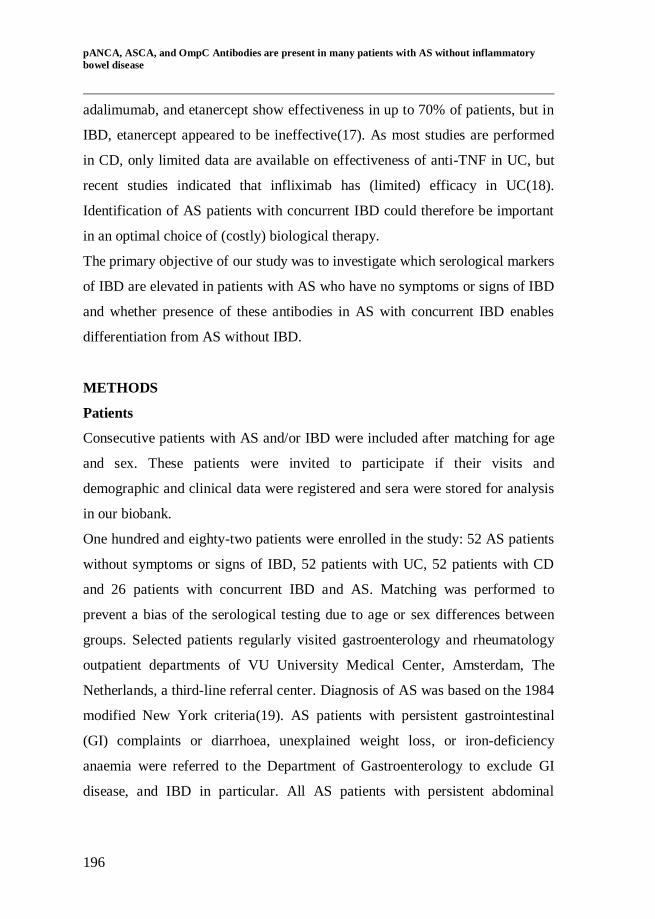
pANCA, ASCA, and OmpC Antibodies are present in many patients with AS without inflammatory
bowel disease
196
adalimumab, and etanercept show effectiveness in up to 70% of patients, but in
IBD, etanercept appeared to be ineffective(17). As most studies are performed
in CD, only limited data are available on effectiveness of anti-TNF in UC, but
recent studies indicated that infliximab has (limited) efficacy in UC(18).
Identification of AS patients with concurrent IBD could therefore be important
in an optimal choice of (costly) biological therapy.
The primary objective of our study was to investigate which serological markers
of IBD are elevated in patients with AS who have no symptoms or signs of IBD
and whether presence of these antibodies in AS with concurrent IBD enables
differentiation from AS without IBD.
METHODS
Patients
Consecutive patients with AS and/or IBD were included after matching for age
and sex. These patients were invited to participate if their visits and
demographic and clinical data were registered and sera were stored for analysis
in our biobank.
One hundred and eighty-two patients were enrolled in the study: 52 AS patients
without symptoms or signs of IBD, 52 patients with UC, 52 patients with CD
and 26 patients with concurrent IBD and AS. Matching was performed to
prevent a bias of the serological testing due to age or sex differences between
groups. Selected patients regularly visited gastroenterology and rheumatology
outpatient departments of VU University Medical Center, Amsterdam, The
Netherlands, a third-line referral center. Diagnosis of AS was based on the 1984
modified New York criteria(19). AS patients with persistent gastrointestinal
(GI) complaints or diarrhoea, unexplained weight loss, or iron-deficiency
anaemia were referred to the Department of Gastroenterology to exclude GI
disease, and IBD in particular. All AS patients with persistent abdominal
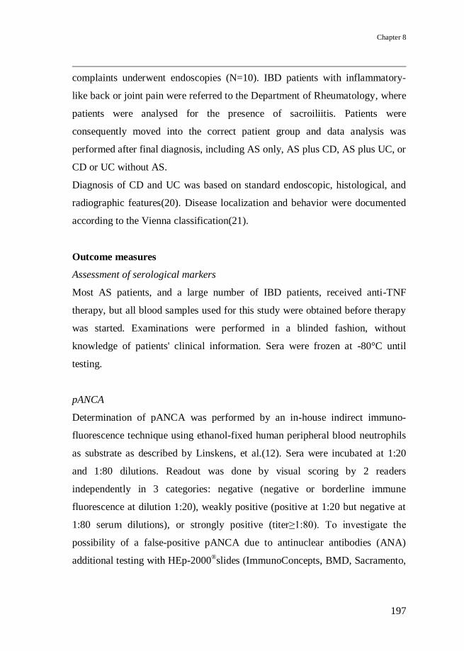
Chapter 8
197
complaints underwent endoscopies (N=10). IBD patients with inflammatory-
like back or joint pain were referred to the Department of Rheumatology, where
patients were analysed for the presence of sacroiliitis. Patients were
consequently moved into the correct patient group and data analysis was
performed after final diagnosis, including AS only, AS plus CD, AS plus UC, or
CD or UC without AS.
Diagnosis of CD and UC was based on standard endoscopic, histological, and
radiographic features(20). Disease localization and behavior were documented
according to the Vienna classification(21).
Outcome measures
Assessment of serological markers
Most AS patients, and a large number of IBD patients, received anti-TNF
therapy, but all blood samples used for this study were obtained before therapy
was started. Examinations were performed in a blinded fashion, without
knowledge of patients' clinical information. Sera were frozen at -80°C until
testing.
pANCA
Determination of pANCA was performed by an in-house indirect immuno-
fluorescence technique using ethanol-fixed human peripheral blood neutrophils
as substrate as described by Linskens, et al.(12). Sera were incubated at 1:20
and 1:80 dilutions. Readout was done by visual scoring by 2 readers
independently in 3 categories: negative (negative or borderline immune
fluorescence at dilution 1:20), weakly positive (positive at 1:20 but negative at
1:80 serum dilutions), or strongly positive (titer≥1:80). To investigate the
possibility of a false-positive pANCA due to antinuclear antibodies (ANA)
additional testing with HEp-2000®slides (ImmunoConcepts, BMD, Sacramento,
pANCA, ASCA, and OmpC Antibodies are present in many patients with AS without inflammatory
bowel disease
198
CA, USA) was performed in case of a (weakly) positive pANCA test result.
Cutoff for positivity was set at 1:80.
ASCA
ASCA-IgA and ASCA-IgG were determined using commercial enzyme-linked
immunosorbent assay (ELISA) kits (Inova, Uniprom Diagnostics BV, Krimpen
aan de IJssel, The Netherlands).
The antigen consisted of phosphopeptidomannan (PPM) extracted from S.
cerevisiae. ASCA ELISA were performed according to the manufacturer’s
instructions; results were expressed as arbitrary units with a cutoff for positivity
at 25 U/ml as advised by the manufacturer, resulting in a sensitivity for CD of
49% for ASCA-IgA and 74% for ASCA-IgG, and in 1.4% versus 4% positive in
healthy controls. Serum was considered positive if either IgA or IgG or both
were positive. Serum was considered negative if both ASCA-IgA and ASCA-
IgG were negative.
OmpC antibodies
IgA antibodies to bacterial OmpC were detected according to the manufacturer's
protocol (Inova, Uniprom Diagnostics BV). The results are presented as
arbitrary units with a cutoff for positivity at 25 U/ml.
Serological profiles
The usual UC serological profile was arbitrarily defined as pANCA+ in 1:80,
ASCA (IgA or IgG) <25U/ml, and OmpC antibodies <25U/ml.
The classical CD serological profile was arbitrarily defined as: pANCA
negative or positive in 1:20, ASCA (IgA or IgG) >25U/ml or OmpC antibodies
>25U/ml.

Chapter 8
199
Statistical analysis
Frequencies and percentages of pANCA, ANA, ASCA-IgA and ASCA-IgG,
and OmpC antibodies were calculated in each group of patients. χ² analysis or
Fisher’s test was performed to examine dichotomous variables.
Logistic regression analyses were conducted to examine associations between
serological markers and serological profiles. The influence of confounders on
this relation was investigated. The following variables were investigated: age,
sex, and presence of HLA-B27. Statistical analyses were performed with SPSS
15.0 software. The threshold for statistical significance was set at p<0.05.
RESULTS
We recruited 182 patients for the study, 52 patients with AS without IBD, 26
with concurrent AS and IBD (11 AS plus UC, 15 AS plus CD); and 50 patients
with UC and 51 with CD, both without AS. Two UC patients and 1 CD patient
were excluded as serum samples were unavailable. Demographic data of these
groups were similar (Table 1). Nonsteroidal anti-inflammatory drugs (NSAIDs)
were used daily by 67 (86%) of all AS patients, immunosuppressive drugs were
used in a minority, and TNF blocking agents were started in a majority of AS
and CD patients (Table 1).
As expected, within the IBD group, pANCA was more frequently seen in UC
(p<0.0001), whereas ASCA and OmpC antibodies were more frequent in CD
(p<0.0001, p=0.006, respectively; Table 2). These results were comparable with
our previous evaluation of pANCA and ASCA in patients with UC and CD
(12). In the overlap group with concurrent AS and IBD, pANCA was more
frequent in patients with UC as well (p=0.024); as well, ASCA was more
frequently observed in CD although this did not reach statistical significance.
OmpC antibodies did not differ between the 2 AS groups.

pANCA, ASCA, and OmpC Antibodies are present in many patients with AS without inflammatory
bowel disease
200
Table 1: Patient characteristics (N= 179) and clinical variables.
Group
AS
N=52
UC
N=50
CD
N=51
AS+UC
N=11
AS+CD
N=15
Characteristic
Male, N (%) 29 (56) 23 (46) 21 (41) 5 (46) 6 (40)
Age, yrs, mean (SD) 41 (12) 37 (11) 36 (13) 43 (11) 38 (12)
Caucasian, N (%) 46 (89) 35 (78) 36 (78) 9 (82) 12 (80)
HLA-B27 positivity, N (%) 42 (84) 5 (83) 10 (67)
Peripheral arthritis, N (%) 21 (40) 1 (9) 1 (7)
Uveitis, N (%) 16 (31) 1 (9) 0 (0)
CRP at baseline 11
(2.5-163)
9
(2.5-37)
29
(2.5-294)
Duration of IBD, yrs, mean (SD) NA 8 (7) 11 (9) 9(8) 15 (12)
BASDAI at baseline, mean (SD) 6.3 (1.6) NA NA 6.2 (1.0) 6.0 (1.9)
Vienna classification
Age: onset <40 yrs, N (%) 43 (88) 8 (89)
Behaviour
Inflammatory, N (%) 17 (35) 6 (67)
Stricturing, N (%) 13 (27) 3 (33)
Perforating, N (%) 19 (39) 0 (0)
Localization
Terminal ileum, N (%) 10 (20) 0 (0)
Colon, N (%) 14 (29) 4 (44)
Ileocolonic, N (%) 24 (49) 5 (56)
Upper GI, N (%) 1 (2) 0 (0)
Immunosuppressive,N (%) 3 (6) 5 (10) 4 (8) 3 (27) 4 (27)
Anti-TNF, N (%) 51 (98) 1 (2) 23 (46) 3 (27) 11 (73)
AS, ankylosing spondylitis; UC, ulcerative colitis; CD, Crohn’s disease; BASDAI, Bath ankylosing
spondylitis disease activity index (0-10). NA: not applicable

Chapter 8
201
Table 2: Antibodies present in serum of patients (N=179) with ankylosing
spondylitis (AS) , ulcerative colitis (UC), Crohn’s disease (CD), AS with concurrent
UC, and AS with concurrent CD.
AS UC CD AS+UC AS+CD
pANCA IgG (%) 11 (21) 33 (66) 8 (16) 7 (64) 3 (20)
ASCA IgA (%) 10 (19) 4 (8) 19 (37) 1 (9) 4 (27)
ASCA IgG (%) 4 (8) 5 (10) 27 (53) 1 (9) 2 (13)
OmpC antibodies IgA (%) 10 (19) 6 (12) 18 (35) 3 (27) 5 (33)
UC profile 8 (17) 25 (50) 4 (8) 6 (67) 0 (0)
CD profile 13 (28) 3 (6) 27 (54) 2 (20) 4 (27)
pANCA, perinuclear antineutrophil cytoplasmic antibodies, positive: titer ≥1:80; ANA, antinuclear antibodies,
tested if pANCA-positive; ASCA, antiglycan antibodies to the cell wall mannan of Saccharomyces cerevisiae,
positive: >25 U/ml; OmpC antibodies, antibodies to porin protein C of Escherichia coli: >25 U/ml.
Prevalences of serological markers in AS without IBD
Presence of any out of the 4 serological markers was observed in 55% of AS
patients without symptoms or signs of IBD. ASCA (IgA and/or IgG) and OmpC
antibodies, markers of CD, were found to be positive in 30% and 19%,
respectively, of AS patients without IBD, versus the reported prevalence of 5%
or lower in healthy controls(10). pANCA was found in 21% of the AS patients
without IBD.
Differences between patient groups
Differences in frequencies of pANCA between patient groups were not due to
ANA: a positive ANA was found in about 25% of each group (results not
shown). pANCA was statistically significant more frequent in AS with
concurrent UC than in AS alone (OR 8.2, 95% CI 1.2-55.6, adjusted for the
presence of HLA-B27), thus being an indicator for UC in AS. Moreover, the
serological profile for UC differentiated between AS patients with concurrent

pANCA, ASCA, and OmpC Antibodies are present in many patients with AS without inflammatory
bowel disease
202
UC versus AS alone. In patients with CD no serological marker or profile
differentiated between AS with concurrent CD and AS alone. No association
was found between presence of HLA-B27 and presence of IBD in AS patients
(data not shown).
Colonoscopy
All AS patients without IBD who had persistent abdominal complaints
underwent endoscopies and IBD was excluded. One out of 10 patients had signs
of microscopic chronic inflammation in the cecum, but there were not enough
arguments for IBD, according to the usual definition(20). The 10 patients did
not have more frequent peripheral arthritis, higher C-reactive protein, or
positive serology (pANCA, ASCA-IgA or ASCA-IgG, anti-OmpC), nor did
they have higher levels of anti-OmpC compared to the AS patients without GI
complaints. Hence, this particular cohort/group could not be distinguished by
serological profile (data not shown).
DISCUSSION
Our study reports on the prevalence of serological markers associated with IBD
(pANCA, ASCA, and OmpC antibodies) in patients with AS. For proper
evaluation, 3 groups of patients with chronic inflammatory diseases were
included: patients with AS, with IBD, and with concurrent AS and IBD.
All the serological markers measured were frequently observed in AS patients:
pANCA, ASCA-IgA, and ASCA-IgG antibodies in 21%, 19%, and 8% of the
52 AS patients, respectively. Further, we demonstrated for the first time that
OmpC antibodies are highly prevalent in AS patients (19%). These markers,
notably ASCA and OmpC antibodies, rarely occur in healthy controls(10). The
frequencies of serological markers in AS patients with concurrent UC or CD,
except for OmpC antibodies, were comparable with our reference findings in
Chapter 8
203
UC or CD alone. Our findings are in concordance with reports showing
increased prevalence of pANCA (22), in particular ASCA, in patients with
AS(15;22;23). Positive ASCA-IgA was found in 19% of AS patients without
IBD and in 27% in the 15 patients with AS and CD, which is not in
concordance with the study of Hoffman, et al.(23), who found no difference
among positive ASCA tests in a small series of AS patients with IBD (N=6) and
without IBD (N=12). The observed prevalence for the various markers in these
series, however, was higher than that reported in a recent study by Mundwiler,
et al.(24).
pANCA was statistically significantly more frequently present in AS patients
with concurrent UC than in AS alone, with an OR of 8.2 (95% CI 1.2-55.6).
pANCA thus might be a valuable tool to screen AS patients with abdominal
complaints: if pANCA is present, performance of an endoscopy is indicated.
Despite this high prevalence of IBD markers in AS patients, our study might be
flawed due to the fact that invasive colonoscopy was performed in 10 of 52 AS
patients (20%). Indeed, colonoscopy was performed only in patients with
persistent GI complaints, because the a priori chance of detecting IBD in
asymptomatic patients is considered to be very low. However, even in the AS
patients with intestinal symptoms, and therefore a relatively ‘high-risk’ group,
the frequency and level of antibodies did not differ from the asymptomatic
patients. The explanation for the increased levels of IBD markers in AS is in
accord with the studies performed by Mielants, et al.(25), who found a high
number of mucosal lesions in asymptomatic AS patients, in particular in the
terminal ileum, without convincing macroscopic signs of inflammation by
microscopic examination. Intestinal damage (and increased intestinal leakage)
itself may be associated with increased IBD markers, but our study was not
designed to confirm this association. Interpretation is thwarted due to group
sizes. It is interesting, however, that 6.5 % of the cases reported by Mielants

pANCA, ASCA, and OmpC Antibodies are present in many patients with AS without inflammatory
bowel disease
204
developed IBD after 2-9 years of follow-up(8).
Involvement of the GI tract in AS can be interpreted in 3 ways: as an aberrant
immune response following a GI infection, as part of an inflammatory disease
sharing a common genetic background, or as a result of intestinal leakage due to
treatment with NSAID. As a first hypothesis, AS is induced by a pathogenic
microbial, such as Klebsiella(26;27), similarly to the onset of another form of
spondylarthropathy, i.e., reactive arthritis, which is related to the culprit
intestinal pathogen (Salmonella species), which induces an aberrant immune
response. However, previously no correlation was found between the presence
of ASCA in serum and Saccharomyces in the intestine(28).
A second hypothesis assumes that a common genetic background in
combination with external factors triggers an (auto) immune response against
different organs, such as intestines, spine, joints, eyes, or skin(1;29).
Corroborating this hypothesis, extraspinal manifestations are frequently
observed in AS patients, such as those with uveitis, which also occurs in IBD.
The third hypothesis is that serological IBD markers, notably ASCA and OmpC
antibodies, reflect intestinal damage. Fitting in with this hypothesis is that in
celiac disease, which is associated with damage to the intestinal barrier, the
prevalence of ASCA is increased(30). Moreover, chronic use of (maximum
dosages of) NSAID may result in increased intestinal leakage, leading to an
(auto) immune response against intestinal antigens(31). From this perspective
one might expect higher prevalence of ASCA in other patient groups taking
NSAID on a regular basis, such as patients with rheumatoid arthritis, but these
data have not been reported.
Together, IBD-associated serological markers are insufficient sensitive for
identifying IBD in AS, but presence of pANCA might differentiate AS patients
with concurrent UC from patients without UC, and these patients are candidates
for colonoscopy.

Chapter 8
205
We demonstrate that the presence of IBD-associated markers in AS patients is
indicative that AS and IBD share a similar pathophysiological origin. These
findings apply to AS patients with and without proven IBD since serum markers
were also found in AS patients without (symptoms of) IBD.
Prospective follow-up of patients with AS and positive IBD serology markers in
comparison with seronegative patients might shed new light on this discussion
and contribute to the decision on whether to perform ileocolonoscopy, and on
which TNF blocking agents might be most effective, as some (e.g. etanercept)
are not effective in colitis(16).

pANCA, ASCA, and OmpC Antibodies are present in many patients with AS without inflammatory
bowel disease
206
REFERENCE LIST
(1) Brophy S, Pavy S, Lewis P, Taylor G, Bradbury L, Robertson D, et al.
Inflammatory eye, skin, and bowel disease in spondyloarthritis: genetic, phenotypic, and environmental factors. J Rheumatol 2001
Dec;28(12):2667-73.
(2) de Vlam K, Mielants H, Cuvelier C, De Keyser F, Veys EM, De Vos
M. Spondyloarthropathy is underestimated in inflammatory bowel disease: prevalence and HLA association. J Rheumatol 2000
Dec;27(12):2860-5.
(3) Rudwaleit M, Baeten D. Ankylosing spondylitis and bowel disease. Best Pract Res Clin Rheumatol 2006 Jun;20(3):451-71.
(4) De Vos M, Cuvelier C, Mielants H, Veys E, Barbier F, Elewaut A.
Ileocolonoscopy in seronegative spondylarthropathy. Gastroenterology
1989 Feb;96(2 Pt 1):339-44.
(5) Grillet B, de Clerck L, Dequeker J, Rutgeerts P, Geboes K. Systematic
ileocolonoscopy and bowel biopsy study in spondylarthropathy. Br J
Rheumatol 1987 Oct;26(5):338-40.
(6) Leirisalo-Repo M, Turunen U, Stenman S, Helenius P, Seppala K. High
frequency of silent inflammatory bowel disease in spondylarthropathy.
Arthritis Rheum 1994 Jan;37(1):23-31.
(7) Lionetti P, Pupi A, Veltroni M, Fonda C, Cavicchi MC, Azzari C, et al.
Evidence of subclinical intestinal inflammation by 99m technetium
leukocyte scintigraphy in patients with HLA-B27 positive juvenile
onset active spondyloarthropathy. J Rheumatol 2000 Jun;27(6):1538-41.
(8) Mielants H, Veys EM, Goemaere S, Goethals K, Cuvelier C, De Vos
M. Gut inflammation in the spondyloarthropathies: clinical, radiologic, biologic and genetic features in relation to the type of histology. A
prospective study. J Rheumatol 1991 Oct;18(10):1542-51.
Chapter 8
207
(9) Mielants H, Veys EM, Cuvelier C, De Vos M, Goemaere S, de Clerck
L, et al. The evolution of spondyloarthropathies in relation to gut histology. III. Relation between gut and joint. J Rheumatol 1995
Dec;22(12):2279-84.
(10) Dubinsky MC, Taylor K, Targan SR, Rotter JI. Immunogenetic phenotypes in inflammatory bowel disease. World J Gastroenterol 2006
Jun 21;12(23):3645-50.
(11) Joossens S, Reinisch W, Vermeire S, Sendid B, Poulain D, Peeters M,
et al. The value of serologic markers in indeterminate colitis: a prospective follow-up study. Gastroenterology 2002 May;122(5):1242-
7.
(12) Linskens RK, Mallant-Hent RC, Groothuismink ZM, Bakker-Jonges LE, van de Merwe JP, Hooijkaas H, et al. Evaluation of serological
markers to differentiate between ulcerative colitis and Crohn's disease:
pANCA, ASCA and agglutinating antibodies to anaerobic coccoid rods.
Eur J Gastroenterol Hepatol 2002 Sep;14(9):1013-8.
(13) Rutgeerts P, Vermeire S. Serological diagnosis of inflammatory bowel
disease. Lancet 2000 Dec 23;356(9248):2117-8.
(14) Mei L, Targan SR, Landers CJ, Dutridge D, Ippoliti A, Vasiliauskas EA, et al. Familial expression of anti-Escherichia coli outer membrane
porin C in relatives of patients with Crohn's disease. Gastroenterology
2006 Apr;130(4):1078-85.
(15) Aydin SZ, Atagunduz P, Temel M, Bicakcigil M, Tasan D, Direskeneli
H. Anti-Saccharomyces cerevisiae antibodies (ASCA) in
spondyloarthropathies: a reassessment. Rheumatology 2007 Dec 26.
(16) Riente L, Chimenti D, Pratesi F, Delle Sedie A, Tommasi S, Tommasi C, et al. Antibodies to tissue transglutaminase and Saccharomyces
cerevisiae in ankylosing spondylitis and psoriatic arthritis. J Rheumatol
2004 May;31(5):920-4.
(17) Sandborn WJ, Hanauer SB, Katz S, Safdi M, Wolf DG, Baerg RD, et al.
Etanercept for active Crohn's disease: a randomized, double-blind,
placebo-controlled trial. Gastroenterology 2001 Nov;121(5):1088-94.
(18) Wilhelm SM, McKenney KA, Rivait KN, Kale-Pradhan PB. A review
of infliximab use in ulcerative colitis. Clin Ther 2008 Feb;30(2):223-30.

pANCA, ASCA, and OmpC Antibodies are present in many patients with AS without inflammatory
bowel disease
208
(19) van der Linden S, Valkenburg HA, Cats A. Evaluation of diagnostic
criteria for ankylosing spondylitis. A proposal for modification of the New York criteria. Arthritis Rheum 1984 Apr;27(4):361-8.
(20) Lennard-Jones JE. Classification of inflammatory bowel disease. Scand
J Gastroenterol Suppl 1989;170:2-6.
(21) Gasche C, Scholmerich J, Brynskov J, D'Haens G, Hanauer SB, Irvine
EJ, et al. A simple classification of Crohn's disease: report of the
Working Party for the World Congresses of Gastroenterology, Vienna
1998. Inflamm Bowel Dis 2000 Feb;6(1):8-15.
(22) Torok HP, Glas J, Gruber R, Brumberger V, Strasser C, Kellner H, et
al. Inflammatory bowel disease-specific autoantibodies in HLA-B27-
associated spondyloarthropathies: increased prevalence of ASCA and pANCA. Digestion 2004;70(1):49-54.
(23) Hoffman IE, Demetter P, Peeters M, De Vos M, Mielants H, Veys EM,
et al. Anti-saccharomyces cerevisiae IgA antibodies are raised in
ankylosing spondylitis and undifferentiated spondyloarthropathy. Ann Rheum Dis 2003 May;62(5):455-9.
(24) Mundwiler ML, Mei L, Landers CJ, Reveille JD, Targan S, Weisman
MH. Inflammatory bowel disease serologies in ankylosing spondylitis patients: a pilot study. Arthritis Res Ther 2009 Nov 23;11(6):R177.
(25) Mielants H, Veys EM, Cuvelier C, De Vos M. Macroscopic and
histological changes of the ileum HLA-B27 related diseases: therapeutic consequences. Int J Clin Pharmacol Res 1984;4(6):409-14.
(26) Martinez A, Pacheco-Tena C, Vazquez-Mellado J, Burgos-Vargas R.
Relationship between disease activity and infection in patients with
spondyloarthropathies. Ann Rheum Dis 2004 Oct;63(10):1338-40.
(27) Rashid T, Ebringer A. Ankylosing spondylitis is linked to Klebsiella--
the evidence. Clin Rheumatol 2007 Jun;26(6):858-64.
(28) Mallant-Hent RC, Mooij M, von Blomberg BM, Linskens RK, van Bodegraven AA, Savelkoul PH. Correlation between Saccharomyces
cerevisiae DNA in intestinal mucosal samples and anti-Saccharomyces
cerevisiae antibodies in serum of patients with IBD. World J Gastroenterol 2006 Jan 14;12(2):292-7.

Chapter 8
209
(29) Thjodleifsson B, Geirsson AJ, Bjornsson S, Bjarnason I. A common
genetic background for inflammatory bowel disease and ankylosing spondylitis: a genealogic study in Iceland. Arthritis Rheum 2007
Aug;56(8):2633-9.
(30) Mallant-Hent RC, Mary B, von Blomberg BM, Yuksel Z, Wahab PJ, Gundy C, et al. Disappearance of anti-Saccharomyces cerevisiae
antibodies in coeliac disease during a gluten-free diet. Eur J
Gastroenterol Hepatol 2006 Jan;18(1):75-8.
(31) Graham DY, Opekun AR, Willingham FF, Qureshi WA. Visible small-intestinal mucosal injury in chronic NSAID users. Clin Gastroenterol
Hepatol 2005 Jan;3(1):55-9.

pANCA, ASCA, and OmpC Antibodies are present in many patients with AS without inflammatory
bowel disease
210
Chapter 8
211
Inefficacy of
ibody formation.
The relationship between disease-related
characteristics and conduction disturbances in
ankylosing spondylitis.
Vincent K. Dik1, Mike J.L. Peters
1,2, Pieter A. Dijkmans
1, Mignon A.C. van der
Weijden1, Mirjam K. de Vries
1, Ben A.C. Dijkmans
1,2, Irene E. van der Horst-
Bruinsma1,2
, Michael T. Nurmohamed1,2
.
Rheumatology Department VU University Medical Center1, Rheumatology
Department Jan van Breemen Institute/Reade2, Amsterdam, The Netherlands.
Scandinavian Journal of Rheumatology 2010;39(1):38-41.
The relation between disease-related characteristics and conduction disturbances in AS
212
ABSTRACT
Objectives: Ankylosing spondylitis (AS) is associated with an increased
cardiovascular (CV) risk. Conduction disturbances (CD) may explain the CV
burden, as they are independently associated with cardiac disease. The aim of
this study was (a) to determine the prevalence of CD in AS, and (b) to evaluate
the relation between CD and demographic and AS-related characteristics.
Methods: A rheumatological evaluation assessing demographic and AS-related
characteristics and a resting standard 12-lead electrocardiogram (ECG) were
performed in 131 consecutive AS patients.
Results: A first-degree atrioventricular (AV) block was found in six (4.6%)
patients. One (0.8%) patient suffered from a complete right bundle branch block
(RBBB) and one (0.8%) patient had a left anterior hemiblock. A prolonged QRS
(pQRS) interval was observed in 38 (29.2%) patients, including those with a
complete or incomplete BBB. Age, disease duration, and body mass index
(BMI) were significantly associated with the PR interval, and male sex, disease
duration, and the Bath ankylosing spondylitis metrology index (BASMI) with
QRS interval. In multivariate analyses, disease duration remained independently
associated with both PR and QRS intervals.
Conclusion: Intraventricular CD is highly prevalent in AS, particularly in
patients with long-standing disease. Further research is needed to determine
whether intraventricular CD contribute to the increased CV risk and long-term
CV mortality in AS.
Chapter 9
213
INTRODUCTION
Spondyloarthropathies are a group of common inflammatory rheumatic
disorders, with ankylosing spondylitis (AS) as the major subtype.
The prevalence of AS in the population is in the range 0.1-1.4%, and AS occurs
predominantly in early adulthood in men. AS is characterized primarily by
sacroilliitis and inflammation of the (lumbar) spine, causing inflammatory back
pain. In addition, extra-spinal manifestations such as uveitis, psoriasis and
inflammatory bowel disease are common.
Human leukocyte antigen (HLA)-B27 is present in approximately 90-95% of
patients with AS, and is thought to play an important role in the
pathophysiology of AS(1).
AS is, in accordance with other rheumatic diseases, associated with an increased
risk of developing cardiovascular (CV) complications, including atherosclerotic
disease, aortic root and valve regurgitation, and a decreased myocardial
performance(2;3). Inflammation and/or fibrosis may play an important role in
affecting the aortic root and mitral valve, which may extend to the membranous
portion of the interventricular septum, the atrioventricular (AV) node, and both
bundle branches, causing conduction disturbances (CD)(4-6). It has also been
recognized that HLA-B27 is associated with CD(7).
Several studies have reported a higher CV mortality in AS, which may be
explained partially by a higher prevalence of CD. However, literature on this
topic is sparse and not well defined(2;8). Therefore, the aim of this study was
(a) to determine the prevalence of CD in AS, and (b) to evaluate the relation
between CD and demographic and AS-related characteristics.

The relation between disease-related characteristics and conduction disturbances in AS
214
METHODS
Patients
Patients attended the rheumatology outpatient clinics. A total of 131
consecutive AS patients derived from two different cohorts were included.
Forty-four consecutive patients were included from an ‘early AS-cohort’, which
comprised AS patients with a diagnosis duration of less than 2 years, and 87
patients were included from an ‘AS cohort’, which comprised AS patients with
an active disease (Bath ankylosing spondylitis disease activity index4
(BASDAI)), for which treatment with tumor necrosis factor (TNF) blocking
agents was initiated. Baseline data were used for the current analyses.
Rheumatological evaluation
Disease duration was defined as the moment of diagnosis until the moment of
entry in this study, in years. Disease activity was determined by using BASDAI
(scale 0-10) and Bath ankylosing spondylitis functional index (BASFI, scale 0-
10). Physical function and spinal mobility were tested by using Bath ankylosing
spondylitis metrology index (BASMI, scale 0-10).
Blood samples were taken to assess HLA-B27 status, and the inflammatory
markers erythrocyte sedimentation rate (ESR) and C-reactive protein (CRP). In
addition, body mass index (BMI) and medication use were determined.
Electrocardiography
Standard 12-lead electrocardiograms (ECGs) were recorded at 25 mm/s paper
speed. One cardiologist, unaware of the patients’ health status, analysed all
ECGs according to a standardized protocol (heart rate, rhythm, electrical axis,
and conduction intervals). The PR interval was defined as prolonged when ≥200
ms (pPR) and the threshold for a prolonged QRS (pQRS) interval was set at
≥100 ms (9).

Chapter 9
215
Statistics
The distribution of variables was tested for normality and expressed as mean
(± standard deviation) or median (interquartile range) where appropriate.
Univariate and multivariate linear regression analyses were performed to
determine the association between demographic and AS-related characteristics
on the outcome variables PR and QRS intervals. Additional analyses were
performed to investigate the influence of anti-rheumatic, pulmonary, and/ or CV
treatment on the PR and QRS intervals. Statistical analyses were performed
using SPSS version 14.0. The threshold for statistical significance was set at
p<0.05.

The relation between disease-related characteristics and conduction disturbances in AS
216
RESULTS
Baseline characteristics
One patient was diagnosed with the Brugada syndrome. This patient was
excluded, leaving 130 AS patients for further analyses. The demographic and
AS-related characteristics of these patients are summarized in Table 1. None of
the patients were using TNF blocking agents.
Table 1: Demographic and AS-related characteristics in all patients (N=130).
Age (years) 39.4 ± 10.4
Disease duration (years) 3.9 (1.1 - 10.9)
Male 63.8
HLA-B27 positive 86.2
BMI 25.1 ± 4.0
ESR (mm/h) 12.0 (6.0 - 30.0)
CRP (mg/L) 10.0 (2.0 - 28.3)
BASDAI 5.9 (4.1 - 7.3)
BASMI 3.0 (1.0 - 6.0)
BASFI 5.9 (3.1 - 7.4)
Beta blocker 7.0
Beta2 agonist 4.7
NSAIDs 90.7
DMARDs 10.9
BMI, body mass index; ESR,erythrocyte sedimentation rate; CRP, C-reactive protein; BASDAI,
Bath ankylosing spondylitis disease activity index; BASMI, Bath ankylosing spondylitis
metrology index; BASFI, Bath ankylosing spondylitis functional index; NSAID, non-steroidal
anti-inflammatory drug; DMARD, disease-modifying anti-rheumatic drug. Values are given as mean
± standard deviation, median (interquartile range), or percentage.

Chapter 9
217
Arrhythmias and CD
Electrocardiographic data are shown in Table 2. Mean PR and QRS intervals
were 151 ± 21 and 94 ± 10 ms, respectively. The mean heart rate was 65 ± 12
beats/min, and 39 (30.0%) patients were bradycardic (heart rate <60 beats/min).
A first-degree AV block (i.e. a PR interval ≥200 ms) was found in six (4.6%)
patients and no second- or third-degree AV blocks were observed.
An incomplete right bundle branch block (RBBB) was observed in 11 (8.5%)
patients and one (0.8%) patient suffered from a complete RBBB. None of our
patients had a complete or incomplete left bundle branch block (LBBB) and
only one (0.8%) patient had a left anterior hemiblock. A pQRS interval (i.e. a
QRS interval ≥100 ms) was present in 38 (29.2%) patients, including those with
a complete or an incomplete BBB.
Table 2: Arrhythmias and conduction disturbances in all patients (N=130).
Heart rate (beats/min) 65 ± 12
Bradycardia (<60 beats/min) 39 (30.0)
PR interval (ms) 151 ± 21
QRS interval (ms) 94 ± 10
First-degree AV block 6 (4.6)
Second-/third-degree AV block 0 (0.0)
Prolonged QRS interval 38 (29.2)
Incomplete RBBB 11 (8.5)
Complete RBBB 1 (0.8)
Incomplete LBBB 0 (0.0)
Complete LBBB 0 (0.0)
Left anterior hemiblock 1 (0.8)
AV, atrioventricular; RBBB, right bundle branch block; LBBB, left bundle branch block.
Values are given as mean ± standard deviation or number (percentage).

The relation between disease-related characteristics and conduction disturbances in AS
218
Conduction intervals, demographic and AS-related characteristics
Univariate linear regression analyses revealed a significant association between
age, disease duration, BMI, and PR interval (Table 3). In addition, QRS interval
was significantly associated with disease duration, male sex, and BASMI. A
trend was found suggesting an association between pQRS interval and presence
of HLA-B27. Moreover, when focusing on male patients only, the prevalence of
a pQRS appeared to be significantly higher in HLA-B27-positive patients than
in HLA-B27-negative patients (44.3% vs. 10.0%, p=0.045). In addition, HLA-
B27-positive male patients more often had a pQRS than HLA-B27-positive
female patients (44.3% vs. 11.1%, p=0.001). Multivariate regression analyses
revealed an independent association between age, disease duration, and PR
interval, and also an independent association between disease duration, male
sex, and QRS-interval. These results were not influenced by anti-rheumatic,
pulmonary, and/or cardioprotective treatment.

Chapter 9
219
Table 3: Univariate associations between conduction intervals and demographic and ankylosing spondylitis-related
characteristics.
PR interval QRS interval
Demographic and AS-related
characteristics
B (95% CI) p-value B (95% CI) p-value
Age (years) 0.60 (0.26 - 0.93) 0.001 0.06 (-0.10 to 0.23) 0.456
Disease duration (years) 0.75 (0.38 - 1.11) <0.001 0.32 (0.14 - 0.49) <0.001
Male 3.28 (-4.29 to 10.85) 0.393 8.84 (5.58 - 12.10) <0.001
HLA-B27 positive -2.94 (-13.94 to 8.05) 0.597 4.85 (-0.32 to 10.01) 0.066
BMI (kg/m2) 1.23 (0.23 - 2.22) 0.016 0.35 (-0.10 to 0.79) 0.124
ESR (mm/h) 0.06 (-0.12 to 0.24) 0.487 0.02 (-0.06 to 0.10) 0.621
CRP (mg/L) 0.02 (-0.13 to 0.17) 0.801 0.05 (-0.02 to 0.12) 0.149
BASDAI -0.17 (-1.92 to 1.57) 0.845 -0.41 (-1.24 to 0.43) 0.334
BASMI 0.62 (-0.85 to 2.09) 0.404 1.02 (0.33 - 1.70) 0.004
BASFI -0.21 (-1.64 to 1.21) 0.769 0.11 (-0.57 to 0.80) 0.745
B,regression coefficient; CI, confidence interval; BMI, body mass index; ESR, erythrocyte sedimentation rate; CRP, C-reactive protein; BASDAI, Bath ankylosing spondylitis disease activity index; BASMI, Bath ankylosing spondylitis metrology index; BASFI, Bath ankylosing spondylitis functional index.

The relation between disease related characteristics and conduction disturbances in AS
220
DISCUSSION
The most important finding of this study is the high prevalence of first-degree
AV blocks and pQRS intervals (4.6% and 29.2%, respectively). In addition, we
observed that both PR and QRS intervals were independently associated with
disease duration, suggesting that the inflammatory burden affects the
conduction system of the heart.
It is being increasingly recognised that AS is associated with an increased CV
risk. The high prevalence of prolonged conduction intervals may be a potential
explanation for the CV burden among AS patients, as CD are independently
associated with an increased long-term CV mortality(10). The lack of a control
group in the present study is a limitation and comparison of the prevalence of
CD with that observed in other cohorts needs to be made with caution.
Nevertheless, the prevalence of CD among healthy controls reported in large-
scale observational studies is considerably lower. For example, in a large study
with 122,043 aircrew men, aged between 16 and 50 years, the prevalence of
first-degree AV blocks was 0.65%(11). In addition, the prevalence of pQRS
(QRS interval ≥100 ms) in the Framingham study was 6.1% in male and 2.4%
in female subjects(12;13). It should be noted that, although a high prevalence of
pQRS was observed, only two (1.5%) AS patients were diagnosed with a
complete BBB. However, many patients already had either a pPR or a pQRS at
a relatively young age, and in light of the independent association between the
QRS interval and disease duration, it could be argued that the number of
complete BBBs will increase over time. Previous observations from studies
investigating CD and their association with AS-related characteristics are
contradictory. Some state that CD in AS patients occur as an intermittent and
inflammatory process, while others believe that CD occur particularly in
patients with long-standing and more severe disease(14;15).

Chapter 9
221
Our data are inconsistent with the first hypothesis, as we observed no
association between the inflammatory markers (i.e., ESR and CRP) and the
duration of the QRS interval, but are consistent with the latter, as the duration of
the QRS interval appeared to be significantly associated with disease duration
and BASMI.
Inflammation and fibrosis located in the membranous part of the interventricular
septum may cause intraventricular CD in AS(5;6). However, most previous
studies indicate that CD in AS are mainly located in the AV node, which might
be the consequence of an abnormality of the AV node artery, rather than septal
fibrosis(5). Our observations suggest that the inflammatory burden might induce
septal inflammation and fibrosis as well as abnormalities of the AV node artery,
causing supra- and intraventricular CD in AS. It should be noted that
information about the presence or absence of ischaemic or valvular heart
disease was lacking, which prevented us from evaluating the potential
contribution of these diseases towards the development of CD.
HLA-B27 is another potential risk factor for the development of CD. In fact, it
has been demonstrated that, even in the absence of rheumatic manifestations
such as AS, HLA-B27 is strongly associated with CD in males(5;7).
In agreement with these observations, we observed that a pQRS was more
prevalent in HLA-B27-positive male patients than in HLA-B27-negative male
patients, supporting a role for HLA-B27 in the development of CD in male AS
patients.
In conclusion, our observations strengthen the need for further (prospective)
research to unravel the pathophysiological mechanisms responsible for the
increased prevalence of CD and to investigate the extent to which CD may lead
to clinically relevant CV disease in AS.

The relation between disease related characteristics and conduction disturbances in AS
222
REFERENCE LIST
(1) Braun J, Sieper J. Ankylosing spondylitis. Lancet 2007 Apr
21;369(9570):1379-90.
(2) Peters MJ, van der Horst-Bruinsma IE, Dijkmans BAC, Nurmohamed
MT. Cardiovascular risk profile of patients with spondylarthropathies, particularly ankylosing spondylitis and psoriatic arthritis. Semin
Arthritis Rheum 2004 Dec;34(3):585-92.
(3) Moyssakis I, Gialafos E, Vassiliou VA, Boki K, Votteas V, Sfikakis PP, et al. Myocardial performance and aortic elasticity are impaired in
patients with ankylosing spondylitis. Scand J Rheumatol 2009
May;38(3):216-21.
(4) Roldan CA, Chavez J, Wiest PW, Qualls CR, Crawford MH. Aortic
root disease and valve disease associated with ankylosing spondylitis. J
Am Coll Cardiol 1998 Nov;32(5):1397-404.
(5) Bergfeldt L. HLA-B27-associated cardiac disease. Ann Intern Med 1997 Oct 15;127(8 Pt 1):621-9.
(6) Lautermann D, Braun J. Ankylosing spondylitis--cardiac
manifestations. Clin Exp Rheumatol 2002 Nov;20(6 Suppl 28):S11-S15.
(7) Bergfeldt L, Insulander P, Lindblom D, Moller E, Edhag O. HLA-B27:
an important genetic risk factor for lone aortic regurgitation and severe conduction system abnormalities. Am J Med 1988 Jul;85(1):12-8.
(8) Boonen A, van der Linden SM. The burden of ankylosing spondylitis. J
Rheumatol Suppl 2006 Sep;78:4-11.
(9) Prineas R, Crow R, Blackburn H. The Minnesota code manual of electrocardiographic findings. 1982. Littleton, MA: John Wright-PSG.
(10) Miller WL, Hodge DO, Hammill SC. Association of uncomplicated
electrocardiographic conduction blocks with subsequent cardiac morbidity in a community-based population (Olmsted County,
Minnesota). Am J Cardiol 2008 Jan 1;101(1):102-6.

Chapter 9
223
(11) Averill KH, Lamb LE. Electrocardiographic findings in 67,375
asymptomatic subjects. I. Incidence of abnormalities. Am J Cardiol 1960 Jul;6:76-83.
(12) Hiss RG, Lamb LE. Electrocardiographic findings in 122,043
individuals. Circulation 1962 Jun;25:947-61.
(13) Kreger BE, Anderson KM, Kannel WB. Prevalence of intraventricular
block in the general population: the Framingham Study. Am Heart J
1989 Apr;117(4):903-10.
(14) O'Neill TW, King G, Graham IM, Molony J, Bresnihan B. Echocardiographic abnormalities in ankylosing spondylitis. Ann Rheum
Dis 1992 May;51(5):652-4.
(15) Nagyhegyi G, Nadas I, Banyai F, Luzsa G, Geher P, Molnar J, et al. Cardiac and cardiopulmonary disorders in patients with ankylosing
spondylitis and rheumatoid arthritis. Clin Exp Rheumatol 1988
Jan;6(1):17-26.

The relation between disease related characteristics and conduction disturbances in AS

Chapter 9
General Discussion and Summary
General Discussion and Summary
226
GENERAL DISCUSSION and SUMMARY
TNF blocking agents were introduced in 1998, after decades of limited
possibilities for treatment of AS patients, such as exercises and NSAID’s. The
use of TNF blocking agents has diminished the disease burden of many AS
patients dramatically. Patients who did not have any other treatment options
before, received a treatment which made their lives almost back to normal again
without pain and stiffness. The majority of AS patients (60%) respond very
well, but many others, unfortunately, do not. There is no difference in response
rate between the various TNF blocking agents: infliximab, adalimumab and
etanercept(1-3). In primarily non responsive cases, this lack of response may be
due to the fact that disease activity is monitored with a questionnaire, the
BASDAI, which is a validated outcome parameter for AS, but which has the
disadvantage of subjectivity without a direct relation with inflammatory
parameters, such as acute phase reactants (ESR e.g.). A relatively high pain
score, due to enthesitis or pain caused by structural deformities, for instance,
may raise the BASDAI, even when there is little inflammation in the spine or
sacroiliac joints.
On the other hand, patients who initially respond very well, might lose this
responsiveness during continuous treatment with TNF blockers. Part of the
mechanism behind this secondary non-response was unravelled by measurement
of serum levels of anti-TNF and the detection of antibodies against these drugs,
as described in Section I.
Immunogenicity
In AS patients, inefficacy of infliximab or adalimumab correlated with the
presence of low serum trough infliximab or adalimumab levels and the presence
of antibodies against infliximab or adalimumab (Chapter 2 and 3), whereas

Chapter 10
227
cases with good clinical responses showed the opposite (high serum trough
levels and no antibodies against anti-TNF). Moreover, our data have
demonstrated that development of anti-infliximab antibodies can precede an
infusion reaction. The mechanism of the decrease in efficacy can be explained
by lower serum trough infliximab or adalimumab levels, caused by neutralizing
antibodies against the idiotype of infliximab and adalimumab, and enhanced
clearance due to immune complex formation of antibodies against biologicals
and the biologicals(4). Our hypothesis is that when drug levels decrease below a
critical limit, the foreign protein might no longer be tolerated and might become
immunogenic. The starting dose of infliximab (5 mg/kg) is higher than that of
RA (3 mg/kg), which might suppress immunogenic reactions(5). Often, the
infliximab dose is increased in AS when responsiveness subsides, but reasons
for dose escalation in AS have not yet been well defined. More research is
needed to assess whether anti-TNF levels can be used for to determine the
optimum dose of anti-TNF in AS in daily clinical practice.
Another option for trying to prevent ineffectiveness caused by antibody
formation is the concomitant administration of other immunosuppressive drugs.
In contrast with treatment of RA, anti-TNF in AS is given without methotrexate.
This may be an explanation for the higher incidence of anti-infliximab and anti-
adalimumab formation in AS. In Crohn’s disease and in RA, the concomitant
use of immunosuppressive drugs or corticosteroids has proven to decrease
antibody formation against infliximab(5;6). It has to be investigated whether
coadministration of immunosuppressive drugs (such as methotrexate) inhibits
antibody formation, because despite the use of immunosuppressive drugs in RA,
anti-infliximab and anti-adalimumab formation occurs in around 17% of RA
patients treated with adalimumab(7) compared with 31% of AS patients.
Moreover, methotrexate has so far not proven to be effective on clinical
symptoms in AS in contrast to RA, so the benefits of an additional drug that

General Discussion and Summary
228
might slow down or inhibit formation of anti-TNF antibodies should be
weighed against the side effects of this drug.
Another TNF blocking agent, etanercept, does not seem to have high
immunogenic properties. In Chapter 4, no correlation was found between
etanercept levels, formation of antibodies against etanercept and clinical
response. All patients had detectable serum levels of etanercept and no
antibodies against etanercept were found during 6 months of treatment.
Interestingly, there seemed to be no difference in mean etanercept levels
between responders and primary non-responders. These findings are in contrast
with our previous studies with infliximab(8;9) and adalimumab in AS(8).
Therefore, this study seems to confirm the hypothesis that etanercept is less
immunogenic than other TNF blocking agents, although the duration of this
study was too short to exclude anti-etanercept formation after a longer period of
therapy with etanercept.
Several arguments are in favour of the hypothesis that etanercept shows less
immunogenicity than other TNF-inhibitors. Firstly, etanercept has a less
immunogenic structure compared with other TNF blocking agents. Etanercept is
a dimeric fusion protein consisting of two TNF receptors, linked to the Fc
portion of an immunoglobin (Ig)G1. Only the fusion part of the molecule may
contain immunogenic epitopes. Infliximab is a chimerical monoclonal IgG1
antibody against TNF, partly consisting of murine protein. Adalimumab is a
fully human monoclonal antibody against TNF. These monoclonal antibodies
have more epitopes within the variable region of the antibody to which an
immune response can be directed. Secondly, major fluctuations in serum levels
may precipitate an immune response and the development of antibodies against
the TNF blocking agent. This is mainly the case in treatment with infliximab,
which is administered once every six to eight weeks. Treatment with etanercept,
however, produces stable levels between two injections and it is dosed more

Chapter 10
229
frequently (once a week). Thirdly, there may be different mechanisms for non-
response, for example caused by inadequate blocking of TNF. This can be
caused by enhanced clearance or as a result of inadequate dosing. A dose-
response relation of etanercept in AS has not been investigated systematically.
We are very interested to learn to what extent new TNF blocking agents such as
golimumab and certolizumab pegol will prove to be immunogenic. Golimumab
is a fully human monoclonal IgG1 against TNF, which is administered once a
month, whereas certolizumab has a totally different structure, containing a Fab’
fragment of monoclonal IgG1 anti-TNF antibody linked to polyethylene glycol
which is dosed every other week.
As discussed in Section I there is some doubt about whether validated patient
questionnaires, such as the BASDAI, are the most optimal outcome parameters
to measure disease activity in AS. Therefore some studies were performed to
explore other biomarkers (Section II ) of disease activity.
Inflammation in AS does not only cause symptoms such as pain and stiffness,
but also increases comorbidity due to cardiovascular disease by accelerating the
process of atherosclerosis. Next to biomarkers of disease activity, studies were
performed to examine biomarkers of cardiovascular disease in AS. The second
chapter in Section II contains a review on Andersson lesions and its prevalence
in our AS cohort. It is important to consider the presence of such a lesion, as
specifically AS patients with an ankylosed spine, who have had a (minor)
trauma, have an increased risk at an AL.
Biomarkers of disease activity: inflammatory markers
The study in Chapter 5.1, in our large prospective cohort of AS patients, has
demonstrated that a combination of elevated baseline levels of CRP and SAA
can be a valuable tool in the selection of AS patients who are likely to respond
to treatment with anti-TNF. Moreover, inflammatory markers, CRP and SAA in

General Discussion and Summary
230
particular, seem to be useful in monitoring the level of inflammation in patients
with AS who are treated with etanercept or infliximab.
Most AS patients showed a significant decrease of several inflammatory
markers upon treatment with anti-TNF. In some cases, a secondary increase of
these inflammatory markers was seen, which may have been caused by a
concurrent infection or inadequate therapeutic levels. Although about 68% of
the AS patients in this study had elevated inflammatory markers before the start
of anti-TNF therapy, it is known that normal inflammatory markers do not
necessarily indicate a low disease activity in AS(10). That is why inflammatory
markers were, until recently, not implemented for assessment of disease activity
or response to treatment, which is in contrast to RA. This study supports the
previous data that raised inflammatory markers are indicative of active disease
in AS. It seems useful to add decrease of inflammatory markers to response
criteria for continuation of anti-TNF treatment in AS patients showing elevated
inflammatory markers at baseline. In 2009, a new ASAS endorsed disease
activity score for AS was developed, the ASDAS(11). This tool for assessment
of disease activity in AS was derived in analogy with the DAS used in RA. The
ASDAS includes the domains of back pain, duration of morning stiffness,
patient’s global assessment, peripheral pain or swelling and CRP or ESR. This
score promises to improve comparison of individual patients’ disease activity,
and the individual score gives a more reliable reflection of the disease activity at
that particular moment.
Despite the fact that ESR showed the strongest association with the BASDAI
over time, we consider ESR the least suitable parameter for inclusion in the
ASDAS, because in our study, it had no additional value and the half-life of this
inflammatory marker was too long for early detection of changes.
Since anti-TNF therapy is very expensive and not without risks, it is of great
importance to identify patients who are likely to respond to this type of drug. At

Chapter 10
231
this moment, we believe that inflammatory markers can be very useful
predictors for a good response, but a raise of inflammatory markers should not
be mandatory for allowing AS patients to be treated with anti-TNF, because
patients with normal baseline levels of CRP and SAA may respond to anti-TNF
therapy as well.
Additionally, in Chapter 5.2, we investigated whether CRP levels, the
important acute phase reactant, are influenced by common single-nucleotide
polymorphisms (SNPs) and haplotypes in the CRP gene. We saw that genotypes
and the haplotype tagged by allele A of rs3091244 associated with high CRP
levels, independent of BASDAI and other confounders. Therefore, the carrying
of distinct genetic variants might explain the lack of elevated CRP levels despite
high disease activity in some AS patients. This observation can be important for
interpreting disease activity scores that incorporate CRP levels, such as
ASDAS.
Discovertebral lesions in AS
Apart from reversible inflammatory signs of the pelvis and spine, visible on
MRI, chronic structural lesions can occur in AS, such as the development of
localised vertebral or discovertebral lesions of the spine, first described by
Andersson in 1937(16) (Chapter 6). We conducted an extensive review of
literature in order to align communication on aetiology, diagnosis and
management between treating physicians. In an attempt to structure the broad
spectrum of Andersson lesions (ALs) complicating AS, a provisory division in
localised and extensive lesions can be used. Localised lesions are limited to
certain parts of the intervertebral disk and they always have an inflammatory
origin. Extensive lesions affect the whole disk or vertebral body and may be
caused by both mechanical and inflammatory factors. Aetiologies range from
spinal (stress-) fractures to a local delay in the ankylosing process compared to

General Discussion and Summary
232
adjacent levels, resulting in the last mobile segment. There is no evidence for an
infectious origin. Regardless of the exact aetiology, mechanical factors in the
ankylosed spine will prevent the healing of extensive lesions and promote the
formation of pseudarthrosis. The diagnosis of AL is established with
conventional radiography, but computed tomography and magnetic resonance
imaging will both provide additional information. Surgical instrumentation and
fusion with the correction of a kyphotic deformity, when present, is considered
to be the principle treatment of a symptomatic AL that does not respond to
conservative treatment(17;18). The eponym Andersson lesion should be
preserved to extensive lesions, which is actually a spinal pseudarthrosis and the
final common pathway of several different aetiologies.
In our AS patients, with a high disease activity, only one lesion resembling an
AL of the thoracic spine was detected with MRI before start of the therapy, but
this lesion lacked a fracture line on conventional radiograph. The low
prevalence of ALs was unexpected because this group of AS patients had a
relatively high rate of ankylosis of the spine and signs of active inflammation.
The percentage of ALs, as described in literature in this subset of severe AS
patients, varies between 1-28%(19-25). The low prevalence of ALs in our study
was probably caused by the relatively short disease duration and small sample
size. This could be due to a selection bias mainly for young patients with an
active disease were referred for treatment with TNF blocking agents, whereas
the efficacy of TNF blockers in older and more severe cases of AS with
complete ankylosis of the spine was still doubtful at the time of this study.
Despite the absence of ALs in our study we would like to increase the
awareness of this complication in AS. In case of a minor spine trauma an MRI
should be combined with conventional spine radiographs in order to detect this
lesion in the stiff and vulnerable spine, which is often osteoporotic as
well(26;27). In previous studies, there has been no sequential MRI study of the
Chapter 10
233
spine in patients with AS that visualizes the evolution from an early
discovertebral lesion - as described in our patient - into a severe destructive
discovertebral lesion, by some authors also known as AL. Thus it remains
unclear whether and how often an abnormality in the discovertebral junction
develops into an AL. More research with a long term follow up of
discovertebral lesions is necessary to clarify the evolution of these lesions.
Biomarkers of cardiovascular risk: lipid profile
Patients with AS have approximately a twofold increased mortality rate
compared to general population. This is predominantly caused by an increased
cardiovascular (CV) risk(12). Inflammation has shown to deteriorate the lipid
profile, which is the main risk factor for atherosclerosis. In Chapter 7, we noted
favourable changes in lipid profile and HDL composition upon TNF blockade.
This was reflected by increased HDL-c and Apo A-I levels and an improved
Apo B:Apo A-I ratio. Anti-TNF treatment also led to favourable alterations in
HDL composition, by diminishing the SAA concentration within the HDL
particles, which rendered the lipid profile more atheroprotective.
SAA is an acute-phase reactant, which is synthesized mainly in the liver in
response to pro-inflammatory cytokines such as interleukin-1, interleukin-6, and
TNF(13), and elevated levels of SAA are associated with increased CV risk(14).
Moreover, SAA-rich HDL particles are rapidly cleared from plasma, and thus
the increase in SAA during inflammation could also contribute to the decrease
of total HDL-c concentrations(15). However, other mechanisms may also play a
role in decreased HDL-c levels during inflammation as well. It has been
suggested that remodelling HDL through activation of secretory phospholipase
A2 may be an alternate explanation for reduced HDL-c levels during the acute-
phase response. In addition, inflammation may convert HDL de novo into a
more proatherogenic form by coordinate but inverse transcriptional regulation

General Discussion and Summary
234
of SAA and Apo A-I in the liver(13). This may explain the observed inverse
correlation between plasma levels of SAA and Apo A-I, but not between plasma
levels of SAA and levels of HDL-c, at baseline. Changes in total cholesterol,
HDL-c and Apo A-I levels were significantly inversely associated with changes
in levels of disease activity parameters over time, confirming the role of
inflammatory activity in lipid profile changes.
Our results highlight the importance of understanding the role of functional
characteristics of HDL cholesterol in CV diseases related to chronic
inflammatory conditions, such as AS.
We were also interested in studying extraspinal manifestations, such as IBD and
conduction disturbances in the heart, as described in Section III.
Extraspinal manifestations: Pathophysiological link between AS and IBD
The study in Chapter 8 reports on the prevalence of serological markers
associated with IBD (pANCA, ASCA and OmpC antibodies) in AS patients.
For a proper evaluation, three groups of patients with chronic inflammatory
diseases were included in this study: one with AS, one with IBD, and one with
patients with concurrently AS and IBD.
All determined serological markers were frequently observed in AS patients:
pANCA, ASCA IgA, and ASCA IgG antibodies in 21%, 19% and 8% of 52 AS
patients, respectively. Furthermore, we demonstrated for the first time that
OmpC antibodies are highly prevalent in AS patients (19%). These markers,
notably ASCA and OmpC antibodies, rarely occur in healthy controls(28).
pANCA was statistically significantly more often present in AS patients with
concurrent UC than in AS alone with an OR of 8.2 (95%CI 1.2-55.6). Thus,
pANCA might be a valuable tool to screen AS patients with abdominal
complaints: if pANCA is present an endoscopy is indicated.

Chapter 10
235
The involvement of the gastrointestinal tract in AS can be interpreted in three
different ways: as an aberrant immune response following gastrointestinal
infection, as part of an inflammatory disease sharing a common genetic
background(29;30) or as a result of intestinal leakage due to treatment with
NSAIDs(31).
We have demonstrated that the presence of IBD-associated markers in AS
patients is indicative that AS and IBD share a similar pathophysiological origin.
These findings apply to AS patients with and without proven IBD since serum
markers were also found in AS patients without (symptoms of) IBD.
Prospective follow-up of AS patients with positive IBD serology markers in
comparison with seronegative patients might shed new light on this discussion
and might contribute to the decision whether or not to perform ileocolonoscopy
in symptomatic patients and which TNF blocking agents might be most
effective, as some (e.g. etanercept) seem to be ineffective in colitis.
Extraspinal manifestations: Conduction disturbances in the heart
Previous literature has revealed that AS patients have an increased risk of
conduction disturbances (CD) which is mainly associated with HLA-B27
antigen(32). These studies were mainly based on hospitalized AS patients with a
long disease duration and therefore a prospective study was started in our out-
patients population of 131 cases (Chapter 9). A first-degree AV-block was
found in 6 of our AS patients. One patient suffered from a complete right
bundle branch block and 1 patient had a left anterior hemiblock. A prolonged
QRS-interval (pQRS >100ms) was observed in 38 patients, including those with
a complete or incomplete bundle branch block. Age, disease duration and body
mass index were significantly associated with PR-interval, and male gender,
disease duration, and BASMI with QRS-interval. In the multivariate analyses,
General Discussion and Summary
236
disease duration remained independently associated with both the PR- and
QRS-interval.
To conclude, intraventricular CD are highly prevalent in AS, particularly in
patients with longstanding disease. Further research is needed to determine
whether intraventricular CD may contribute to increased CV risks and long-
term cardiovascular mortality in AS.
Future goals
Our main goal is to detect and treat AS, and its complications, at an early stage
in order to prevent damage. One of the trials that should be performed is to test
the efficacy of very early treatment with anti-TNF, even before abnormalities of
AS are visible on radiographs. The goals of such a trial would be to prevent
damage and to stop progression of the disease. At this moment we have started
such a placebo-controlled trial with etanercept (PREVAS study) at VUmc.
Concerning extraspinal manifestations, it is interesting to see whether AS
patients with serological markers of IBD will develop manifest IBD in time or
not. Particularly AS patients with pANCA and gastrointestinal complaints
probably have a higher risk of developing ulcerative colitis. Cardiovascular
risks, such as conduction disorders and increased risk of atherosclerosis, can be
determined by performing an electrocardiogram and assessment of lipid profile
in daily practice, and cardiovascular risk management should be considered.
Lowering inflammatory activity by optimum use of TNF blocking agents can be
supported by development of reliable biomarkers of disease activity and
damage. Further research is needed whether serum trough levels of anti-TNF
can be used for clinical decision making and adjustment of the anti-TNF dose. It
is possible that non-responsive AS patients require a higher dose, but it is also
possible that a lower dose suffices in responsive AS patients. This would lead to
a considerable cost reduction in the future. New research has to be done to see

Chapter 10
237
whether concomitant immunosuppressive medication can prevent antibody
formation against anti-TNF, which is a significant problem in AS.
To conclude, with the introduction of anti-TNF, future perspectives of AS
patients have improved dramatically and future studies should aim on
refinement of this treatment for individual patients.

General Discussion and Summary
238
REFERENCE LIST
(1) Davis JC, Jr., van der Heijde DM, Braun J, Dougados M, Cush J, Clegg
DO, et al. Recombinant human tumor necrosis factor receptor (etanercept) for treating ankylosing spondylitis: a randomized,
controlled trial. Arthritis Rheum 2003 Nov;48(11):3230-6.
(2) van der Heijde DM, Dijkmans B, Geusens P, Sieper J, DeWoody K,
Williamson P, et al. Efficacy and safety of infliximab in patients with ankylosing spondylitis: results of a randomized, placebo-controlled trial
(ASSERT). Arthritis Rheum 2005 Feb;52(2):582-91.
(3) van der Heijde DM, Kivitz A, Schiff MH, Sieper J, Dijkmans BAC, Braun J, et al. Efficacy and safety of adalimumab in patients with
ankylosing spondylitis: results of a multicenter, randomized, double-
blind, placebo-controlled trial. Arthritis Rheum 2006 Jul;54(7):2136-46.
(4) van der Laken CJ, Voskuyl AE, Roos JC, Stigter van WM, de Groot ER, Wolbink G, et al. Imaging and serum analysis of immune complex
formation of radiolabelled infliximab and anti-infliximab in responders
and non-responders to therapy for rheumatoid arthritis. Ann Rheum Dis 2007 Feb;66(2):253-6.
(5) Maini RN, Breedveld FC, Kalden JR, Smolen JS, Davis D, Macfarlane
JD, et al. Therapeutic efficacy of multiple intravenous infusions of anti-tumor necrosis factor alpha monoclonal antibody combined with low-
dose weekly methotrexate in rheumatoid arthritis. Arthritis Rheum 1998
Sep;41(9):1552-63.
(6) Baert F, Noman M, Vermeire S, van Assche G, D'Haens G, Carbonez A, et al. Influence of immunogenicity on the long-term efficacy of
infliximab in Crohn's disease. N Engl J Med 2003 Feb 13;348(7):601-8.
(7) Bartelds GM, Wijbrandts CA, Nurmohamed MT, Stapel S, Lems WF, Aarden L, et al. Clinical response to adalimumab: relationship to anti-
adalimumab antibodies and serum adalimumab concentrations in
rheumatoid arthritis. Ann Rheum Dis 2007 Jul;66(7):921-6.

Chapter 10
239
(8) de Vries MK, Brouwer E, van der Horst-Bruinsma IE, Spoorenberg A,
van Denderen JC, Jamnitski A, et al. Decreased clinical response to adalimumab in ankylosing spondylitis is associated with antibody
formation. Ann Rheum Dis 2009 Nov;68(11):1787-8.
(9) Wolbink GJ, Vis M, Lems W, Voskuyl AE, de Groot E, Nurmohamed MT, et al. Development of antiinfliximab antibodies and relationship to
clinical response in patients with rheumatoid arthritis. Arthritis Rheum
2006 Mar;54(3):711-5.
(10) Ruof J, Stucki G. Validity aspects of erythrocyte sedimentation rate and C-reactive protein in ankylosing spondylitis: a literature review. J
Rheumatol 1999 Apr;26(4):966-70.
(11) van der Heijde DM, Lie E, Kvien TK, Sieper J, van den Bosch F, Listing J, et al. ASDAS, a highly discriminatory ASAS-endorsed
disease activity score in patients with ankylosing spondylitis. Ann
Rheum Dis 2009 Dec;68(12):1811-8.
(12) Peters MJ, Van der Horst-Bruinsma IE, Dijkmans BA, Nurmohamed MT. Cardiovascular risk profile of patients with spondylarthropathies,
particularly ankylosing spondylitis and psoriatic arthritis. Semin
Arthritis Rheum 2004 Dec;34(3):585-92.
(13) Han CY, Chiba T, Campbell JS, Fausto N, Chaisson M, Orasanu G, et
al. Reciprocal and coordinate regulation of serum amyloid A versus
apolipoprotein A-I and paraoxonase-1 by inflammation in murine hepatocytes. Arterioscler Thromb Vasc Biol 2006 Aug;26(8):1806-13.
(14) Ridker PM, Hennekens CH, Buring JE, Rifai N. C-reactive protein and
other markers of inflammation in the prediction of cardiovascular
disease in women. N Engl J Med 2000 Mar 23;342(12):836-43.
(15) Hoffman JS, Benditt EP. Plasma clearance kinetics of the amyloid-
related high density lipoprotein apoprotein, serum amyloid protein
(apoSAA), in the mouse. Evidence for rapid apoSAA clearance. J Clin Invest 1983 Apr;71(4):926-34.
(16) Andersson. [Andersson lesion (spondylodiscitis in Bechterew disease)].
Rontgenpraxis 1996 Apr;49(4):90-1.

General Discussion and Summary
240
(17) Chang KW, Tu MY, Huang HH, Chen HC, Chen YY, Lin CC.
Posterior correction and fixation without anterior fusion for pseudoarthrosis with kyphotic deformity in ankylosing spondylitis.
Spine 2006;31(13):E408-E413.
(18) van Royen BJ, Kastelijns RC, Noske DP, Oner FC, Smit TH. Transpedicular wedge resection osteotomy for the treatment of a
kyphotic Andersson lesion-complicating ankylosing spondylitis. Eur
Spine J 2006 Feb;15(2):246-52.
(19) Cawley MID, Chalmers TM, Ball J, Kellgren JH. Destructive Lesions of Vertebral Bodies in Ankylosing-Spondylitis. Ann Rheum Dis
1972;31(5):345-&.
(20) Guma M, Olive A, Perez R, Holgado S, Ortiz-Santamaria V, Tena X. Aseptic spondylodiskitis in rheumatic diseases. Clin Exp Rheumatol
2001;19(6):740-7.
(21) Hehne HJ, Becker HJ, Zielke K. The spondylodiscitis in kyphotic
deformities of ankylosing spondylitis and the influence of dorsal correction osteotomies. Report on 33 patients. Z ORTHOP IHRE
GRENZGEB 1990;128(5):494-502.
(22) Kabasakal Y, Garrett SL, Calin A. The epidemiology of spondylodiscitis in ankylosing spondylitis--a controlled study. Br J
Rheumatol 1996 Jul;35(7):660-3.
(23) Langlois S, Cedoz JP, Lohse A, Toussirot E, Wendling D. Aseptic discitis in patients with ankylosing spondylitis: a retrospective study of
14 cases. Joint Bone Spine 2005 May;72(3):248-53.
(24) Lanting PJ, Rasker JJ, Kruijsen MW, Prevo RL. [Spondylodiscitis in
Bechterew's disease; inflammation or trauma? Description of 6 patients]. Ned Tijdschr Geneeskd 1994 Oct 1;138(40):1997-2001.
(25) Tsuchiya K, Nagamine R, Iwamoto Y. Discovertebral lesion in
ankylosing spondylitis: Differential diagnosis with discitis by magnetic resonance imaging. Mod Rheumatol 2002;12(2):113-7.
(26) Magrey M, Khan MA. Osteoporosis in ankylosing spondylitis. Curr
Rheumatol Rep 2010 Oct;12(5):332-6.

Chapter 10
241
(27) Van der Weijden MA, van Denderen JC, Lems WF, Heymans MW,
Dijkmans BA, van der Horst-Bruinsma IE. Low bone mineral density is related to male gender and decreased functional capacity in early
spondylarthropathies. Clin Rheumatol 2010 Aug 10.
(28) Dubinsky MC, Taylor K, Targan SR, Rotter JI. Immunogenetic phenotypes in inflammatory bowel disease. World J Gastroenterol 2006
Jun 21;12(23):3645-50.
(29) Brophy S, Pavy S, Lewis P, Taylor G, Bradbury L, Robertson D, et al.
Inflammatory eye, skin, and bowel disease in spondyloarthritis: genetic, phenotypic, and environmental factors. J Rheumatol 2001
Dec;28(12):2667-73.
(30) Thjodleifsson B, Geirsson AJ, Bjornsson S, Bjarnason I. A common genetic background for inflammatory bowel disease and ankylosing
spondylitis: a genealogic study in Iceland. Arthritis Rheum 2007
Aug;56(8):2633-9.
(31) Graham DY, Opekun AR, Willingham FF, Qureshi WA. Visible small-intestinal mucosal injury in chronic NSAID users. Clin Gastroenterol
Hepatol 2005 Jan;3(1):55-9.
(32) Lautermann D, Braun J. Ankylosing spondylitis--cardiac manifestations. Clin Exp Rheumatol 2002 Nov;20(6 Suppl 28):S11-
S15.

General Discussion and Summary

Chapter 10
Nederlandse Samenvatting
Nieuwe inzichten in de behandeling van de ziekte
van Bechterew met anti-TNF medicatie.

Nederlandse Samenvatting
244
NEDERLANDSE SAMENVATTING
Nieuwe inzichten in de behandeling van de ziekte van Bechterew met anti-
TNF medicatie.
Achtergrond
De ziekte van Bechterew is een chronische ontstekingsziekte van de
wervelkolom en heiligbeen (sacroiliacaal) gewrichten waarbij tevens gewrichts-
en peesontstekingen elders kunnen optreden. Naast het bewegingsapparaat
kunnen ook organen aangedaan zijn zoals de ogen (regenboogvliesonsteking
(uveitis anterior)), de huid (psoriasis), de darmen (de ziekte van Crohn en colitis
ulcerosa), het hart, de longen en soms de nieren. De ziekte leidt tot klachten van
rugpijn en stijfheid, die met name ‘s nachts optreden en verminderen door
beweging. In de loop van de tijd kan de ontsteking leiden tot het vastgroeien
van de rug (ankylose), verandering in een kromme stand (kyfose), en schade aan
de gewrichten toebrengen. In Nederland lijden meer dan 40.000 patiënten aan
de ziekte van Bechterew.
De ziekteactiviteit kan onder andere worden gemeten met behulp van een
vragenlijst, de BASDAI, die bestaat uit 6 vragen (Appendix).
Tot voor kort bestond de behandeling uit oefentherapie en ontstekings-
remmende pijnstillers. Sinds enige jaren is de behandeling met anti-TNF
medicatie (gericht tegen het ontstekingseiwit TNF) mogelijk die bij 60% van de
patiënten een sterke afname van de ziekteactiviteit geeft. Ten tijde van het
schrijven van dit proefschrift waren er 3 middelen beschikbaar, te weten
infliximab, adalimumab en etanercept. Infliximab en adalimumab zijn
monoklonale antilichamen, waarbij infliximab deels muizeneiwit en
adalimumab gehumaniseerd eiwit bevat. Etanercept is een fusie eiwit tussen 2
TNF receptoren. Infliximab wordt 1x/6 weken intraveneus toegediend en

Chapter 11
245
adalimumab en etanercept subcutaan, respectievelijk 2-wekelijks en wekelijks.
De meest voorkomende bijwerkingen zijn injectieplaatsreacties en verhoogde
gevoeligheid voor infecties.
Sectie I: Immunogeniciteit
In Hoofdstuk 2 en 3 is onderzocht waarom 40% van de Bechterew patiënten
geen baat heeft bij anti-TNF medicatie en waarom bij sommige patiënten de
werkzaamheid in de loop van de tijd afneemt.
Patiënten met een goede klinische respons bleken hoge dalspiegels te hebben.
Daarnaast bleek er een sterk verband te zijn tussen het niet werken van
infliximab of adalimumab en antistofvorming tegen deze middelen. Deze
antistoffen werken neutraliserend en zorgen door complexvorming met het
medicijn voor een versnelde klaring uit de circulatie. Antistoffen tegen
infliximab kunnen uiteindelijk ook een allergische reactie veroorzaken. Voor
etanercept hebben wij het verband tussen antistofvorming tegen etanercept en
non-respons niet aan kunnen tonen (Hoofdstuk 4). Dit zou kunnen komen door
de andere structuur van etanercept of doordat het middel vaker wordt
toegediend, en daardoor stabielere serumspiegels geeft. Het is ook mogelijk dat
er een dosisrespons effect is, maar dit is nog niet onderzocht.
Ineffectiviteit van anti-TNF zou mogelijk kunnen worden tegengegaan door
ophoging van de dosis of door voorkoming van antistofvorming door
gelijktijdige toediening van immunosuppressiva, zoals methotrexaat of
corticosteroiden. Deze gelijktijdige behandeling wordt al toegepast bij patiënten
met reumatoïde artritis en met de ziekte van Crohn. Bij deze patiënten lijken
inderdaad minder vaak antistoffen gevormd te worden. Naar de rol van
gelijktijdige toediening van immunosuppressiva bij anti-TNF voor de ziekte van
Bechterew moet nog nader onderzoek worden verricht.
Nederlandse Samenvatting
246
Een andere reden voor ineffectiviteit kan zijn dat de ziekteactiviteit, tot voor
kort gemeten met de BASDAI, door de patiënt als ‘hoog’ gescoord wordt door
schade die in de loop der jaren door de ontstekingen ontstaan is, terwijl er op dat
moment geen hoge ontstekingsactiviteit in het lichaam aanwezig is.
Daarom zijn wij op zoek gegaan naar andere biomarkers voor ziekteactiviteit.
Sectie II: Biomarkers
Biomarkers van ziekteactiviteit
In Hoofdstuk 5.1, bleken de ontstekingsfactoren in het bloed zoals de
bloedbezinking, maar met name andere acute fase eiwitten zoals C-reactief
proteïne (CRP) en serum amyloid A (SAA) geschikt voor het meten van de
ziekteactiviteit bij de ziekte van Bechterew. Deze waarden daalden significant
na behandeling met infliximab of etanercept. Ook bleek dat patiënten die voor
behandeling verhoogde waarden van CRP en SAA hadden vaker goed te
reageren op behandeling met antiTNF.
In 2009 werd een nieuwe ziekteactiviteitsscore ontwikkeld: de ASDAS. Deze
score is een combinatie tussen vragen over rugpijn, ochtendstijfheid, algemene
beoordeling, pijn of zwelling in de gewrichten en de bezinking of het CRP. Wij
vinden dat de bezinking minder geschikt is voor het gebruik in de ASDAS door
de lange halfwaardetijd in vergelijking tot CRP. In de praktijk kunnen we deze
acute fase markers niet altijd gebruiken omdat niet alle Bechterew patiënten
reageren met verhoogde ontstekingswaarden in het bloed. In ons onderzoek had
68% van de patiënten met een hoge ziekteactiviteit ook verhoogde
ontstekingswaarden. Dit lijkt tevens samen te hangen met genetische aanleg. Uit
het onderzoek in Hoofdstuk 5.2 blijkt dat de CRP spiegel beïnvloed wordt door
genetische varianten van het CRP-gen, de SNP’s (common single nucleotide
polymorfismen) en de samenstelling van de genenparen: de haplotypen. SNP

Chapter 11
247
rs3091244 genotypes en het haplotype gelabeld A bleken geassocieerd te zijn
met hoge CRP spiegels, onafhankelijk van de ziekteactiviteit (gemeten met de
BASDAI) of andere invloeden.
Hoofdstuk 6 beschrijft het voorkomen van typische gelokaliseerde laesies van
de wervel en tussenwervelschijf bij Bechterew patiënten: de Andersson laesies.
Deze laesies ontstaan waarschijnlijk door een samenspel van schade door
ontsteking en mechanische factoren. Uiteindelijk kan er pseudoartrose ontstaan,
met name als de wervelkolom vast gegroeid (geankyloseerd) is. Meestal kan de
diagnose gesteld worden aan de hand van röntgenonderzoek, maar CT of MRI
kunnen extra informatie bieden. De behandeling is chirurgisch en bestaat uit het
vastzetten van de wervels en correctie van de kromming (kyfose) van de
wervelkolom.
In ons Bechterew cohort werd slechts 1 Andersson laesie gezien, maar deze was
alleen zichtbaar op de MRI en niet op de röntgenfoto van de wervelkolom. Het
is belangrijk deze laesies tijdig op te sporen, omdat met name de
geankyloseerde wervelkolom kwetsbaar is en vaak ook een slechte botkwaliteit
heeft (osteoporotisch). Een klein ongeluk kan hierdoor al tot schade en mogelijk
neurologische uitval leiden.
Biomarkers van cardiovasculair risico: lipidenprofiel
Bechterew patiënten hebben een verdubbelde mortaliteit ten opzichte van de
gehele populatie. Dat komt voornamelijk door een verhoogd risico op hart- en
vaatziekten. Het is aangetoond dat ontsteking een slechter lipidenprofiel geeft,
hetgeen een hoger risico op aderverkalking (atherosclerose) geeft.
In Hoofdstuk 7 is te zien dat het lipidenprofiel en de HDL samenstelling van
Bechterew patiënten verbeterde na behandeling met anti-TNF. Dit was te zien
aan verhoogde HDL-c en Apo A-I spiegels en een betere Apo B:Apo A-I ratio.
Nederlandse Samenvatting
248
Ook leidde de behandeling tot een lagere SAA concentratie in de HDL deeltjes
wat een beschermend effect heeft op het ontstaan van atherosclerose. De
stijging van totaal cholesterol, HDL-c en Apo A-I hing significant samen met
een daling van de ziekteactiviteit.
Sectie III: extraspinale manifestaties
Pathofysiologisch verband tussen Bechterew en inflammatoire darmziekten
In Hoofdstuk 8 wordt het voorkomen van serologische markers vergeleken bij
patiënten met Bechterew, patiënten met inflammatoire darmziekten, en
patiënten met beide aandoeningen. pANCA, ASCA IgA en IgG, en OmpC
antilichamen zijn geassocieerd met inflammatoire darmziekten. Deze markers
kwamen ook voor bij Bechterew patiënten in respectievelijk 21%, 19%, 8% en
19% van de gevallen. Deze markers komen bij gezonde mensen zelden voor.
pANCA kwam significant vaker voor bij Bechterew patiënten met colitis
ulcerosa dan bij patiënten zonder deze ziekte met een odds ratio van 8,2 (95%
betrouwbaarheidsinterval 1,2-55,6). Daarom kan pANCA een hulpmiddel zijn
om colitis ulcerosa te detecteren bij een Bechterew patiënt met darmklachten.
Ook bij Bechterew patiënten zonder darmklachten werden deze markers
aangetroffen.
Een verklaring voor het voorkomen van deze markers kan liggen in een
afwijkende immuunrespons na een darminfectie, een gemeenschappelijke
genetische achtergrond, of als gevolg van darmlekkage door behandeling met
ontstekingsremmende pijnstillers. Het lijkt er op dat de ziekte van Bechterew en
inflammatoire darmziekten een gemeenschappelijke pathofysiologische origine
hebben.
Chapter 11
249
Prospectief onderzoek moet in de eerste plaats uitwijzen of de Bechterew
patiënten met markers (die met IBD geassocieerd zijn) een inflammatoire
darmziekte ontwikkelen en, in de tweede plaats, bij wie van hen er een
ileocolonoscopie verricht moet worden.
Geleidingsstoornissen van het hart
Hoofdstuk 9 beschrijft het verhoogd voorkomen van geleidingsstoornissen in
het hart bij 131 Bechterew patiënten met lange ziekteduur.
Bij 6 van de patiënten werd op het electrocardiogram (ECG) een 1e graads AV
blok gevonden, 1 patiënt had een compleet rechter bundeltakblok en 1 patiënt
had een linker anterior hemiblok. Bij 38 van de patiënten werd een verlengd
QRS interval (>100ms) gevonden. In de multivariate analyse was de ziekteduur
geassocieerd met het PR en QRS interval. Het is nog onduidelijk of deze
geleidingsstoornissen bijdragen aan de verhoogde cardiovasculaire mortaliteit
bij Bechterew patiënten.
Toekomstige doelen
Het doel is om de ziekte van Bechterew en de complicaties ervan in een zo
vroeg mogelijk stadium op te sporen en te behandelen nog voordat de schade is
aangericht. Een van de studies die moet worden gedaan is onderzoek van de
effeciviteit van zeer vroege anti-TNF behandeling van de ziekte van Bechterew
in het stadium voordat de afwijkingen op het röntgenonderzoek te zien zijn. Het
doel van deze behandeling is om de ziekte in een zo vroeg mogelijk stadium tot
stilstand te brengen (preventie) en mogelijk zelfs te voorkomen. Op dit moment
is zo’n placebogecontroleerde preventiestudie met etanercept opgestart in het
VUmc (PREVAS).
Follow-up van de patiënten met markers ten opzichte van de patiënten zonder
markers die kunnen duiden op IBD kan interessant zijn, omdat deze groep op

Nederlandse Samenvatting
250
den duur mogelijk een hogere kans heeft op het ontwikkelen van manifeste
IBD.
De verhoogde kans op cardiale complicaties, zoals het verhoogd voorkomen
van geleidingsstoornissen, kan opgemerkt worden door routinematig een ECG
te verrichten, het lipidenprofiel te bepalen en afwijkingen zo nodig te
behandelen (cardiovasculair risicomanagement).
Bestrijding van ontsteking door het optimaal inzetten van anti-TNF medicatie
kan worden ondersteund door de ontwikkeling van goede biomarkers van
ziekteactiviteit en schade. Ook moet onderzocht worden wat een therapeutische
spiegel van de antiTNF medicatie is. Daarna kan deze spiegel gebruikt worden
voor aanpassing van de dosis om een zo goed mogelijke respons en daling van
de ontsteking te bereiken. Het is ook mogelijk dat bepaalde patiënten voldoende
hebben aan een lagere anti-TNF dosis wat in de toekomst een aanzienlijke
kostenbesparing kan opleveren. Omdat antistofvorming tegen infliximab en
adalimumab bij patiënten met de ziekte van Bechterew vaker voorkomt, moeten
wij onderzoeken of comedicatie met immunosuppressiva kan helpen dit te
voorkomen, zodat meer patiënten langer baat hebben bij deze medicatie.
Tot slot kan gezegd worden dat het toekomstperspectief voor veel patiënten met
de ziekte van Bechterew er door de komst van anti-TNF medicatie aanzienlijk
op vooruit is gegaan. De komende studies zullen zich vooral moeten richten op
verfijning van deze behandeling voor de individuele patiënt.

Chapter 11
251

Nederlandse Samenvatting
252

Chapter 11
253
Questionnaires and BASMII
Abbreviations List
Dankwoord
Curriculum Vitae

Questionnaires and BASMI
254

Appendix
255

Questionnaires and BASMI
256

Appendix
257
ABBREVIATIONS LIST
AL Andersson lesion
ANA antinuclear antibodies
Anti-TNF TNF blocking medication
apoA-1 apolipoprotein A1
apoB apolipoprotein B
AS ankylosing spondylitis
ASAS assessments in ankylosing spondylitis
ASCA antibodies to the cell-wall mannan of Saccharomyces cerevisiae
ASDAS ankylosing spondylitis disease activity score
AU arbitrary units
BASDAI Bath ankylosing spondylitis disease activity index
BASFI Bath ankylosing spondylitis disease functional index
BASG Bath ankylosing spondylitis disease global
BASMI Bath ankylosing spondylitis disease metrology index
BMI body mass index
C cervical
CD Crohn’s disease (chapter 9)
CD conduction disturbance (chapter 10)
CI confidence interval
CRP C-reactive protein
CV cardiovascular
DDD degenerative disk disease
DDL destructive discovertebral lesion
DMARDs disease modifying anti-rheumatic drugs
e.g. exempli gratia/for example
ELISA enzyme-linked immunosorbent assay
ESR erythrocyte sedimentation rate
Gd-DTPA gadolinium diethylenetriamine pentaacetic acid
GEE generalized estimating equations
GI gastrointestinal
HDL-c high density lipoprotein cholesterol
HLA-B27 human leucocyte antigen B27
hsCRP high sensitive CRP

Abbreviations List
258
IBD inflammatory bowel disease
i.e. id est/that means
Ig immunoglobin
IQR interquartile range
JBI Jan van Breemen Institute
kg kilogram
L lumbar
LDL-c low density lipoprotein cholesterol
LS lumbosacral
MRI magnetic resonance imaging
MTX methotrexate
N number
n.k. not known
NA not applicable
NR non-responder
NRS numeric rating scale
NSAID nonsteroidal anti-inflammatory drug
OmpC antibodies to porin protein C of Escherichia coli
OR odds ratio
pANCA perinuclear antineutrophil cytoplasmic antibodies
pPR prolonged PR interval
pQRS prolonged QRS interval
PPM phosphopeptidomannan
R responder
RA rheumatoid arthritis
RIA radioimmunoassay
ROC receiver operating characteristic
SAA serum amyloid A protein
SD standard deviation
SELDI-TOF surface-enhanced laser desorption/ionization time-of-flight
SNPs single-nucleotide polymorphisms
SpA spondylarthropathy
SPSS statistical package for the social sciences
STIR short tau inversion recovery
Appendix
259
T thoracic
TC total cholesterol
TL thoracolumbar
TNF tumor necrosis factor
t-test students t-test
UC ulcerative colitis
VAS visual analogue scale
VU vertebral unit
VUmc VU University medical center
Yrs years

Dankwoord
260
DANKWOORD
Graag wil ik iedereen bedanken die zijn steentje heeft bijgedragen bij de
totstandkoming van dit proefschrift! Een aantal mensen wil ik speciaal
bedanken.
Als eerste wil ik alle Bechterew patiënten bijzonder bedanken die mee hebben
gewerkt aan al deze onderzoeken. Zij hebben mij veel geleerd over de ziekte en
verleenden zeer toegewijd hun medewerking aan de onderzoeken. Dankzij hen
hebben we kunnen zorgen voor enige opheldering rondom de behandeling van
de ziekte van Bechterew met anti-TNF medicatie.
Professor Dijkmans, bij u was het niet alleen mogelijk te promoveren, maar ook
mijzelf te ontplooien als epidemioloog. Het was een onmisbaar onderdeel van
mijn promotie. Dank voor uw vertrouwen in mijn kunnen. Ik voel mij
bevoorrecht dat u als emeritus hoogleraar terugkomt om mijn promotor te zijn.
Zeer belangrijk is mijn copromotor die mij introduceerde in de wereld van de
reumatologie: Irene! Je spoorde mij aan de samenhang tussen de diverse
steentjes te ontdekken. Je gaf me de mogelijkheid mijzelf te ontwikkelen als
onderzoeker. Je stelde steeds veel vertrouwen in mij. Ik heb zo veel geleerd, ook
op het gebied van patiëntenzorg. Dank voor je raad en daad tijdens de moeilijke
momenten.
Gertjan, dank voor alle verhelderende en inspirerende gesprekken. Al pratend
werden mij de ingewikkeldste mechanismen duidelijk. De biologicals
besprekingen op Sanquin wierpen een belangrijk licht op de mechanismen van

Appendix
261
immunogeniciteit. Ook al heb ik niet veel proeven zelf gedaan, ik heb zeker ook
van het laboratoriumwerk een goede indruk gekregen. In het speciaal wil ik
Lucien, Steven, Henk en Els bedanken. Wat fijn dat jullie er nooit gras over
lieten groeien!
De leescommissie en promotiecommissie bestaande uit Dr. A. Boonen, Prof. dr.
D.L. Baeten, Mw. Prof. D.M.F.M. van der Heijde, Prof. dr. M. den Heijer, Prof.
Dr. C.J.J. Mulder, Prof. dr. B.J. van Royen en Prof. dr. C.L. Verweij. Veel dank
voor het beoordelen van mijn proefschrift.
Voorts wil ik alle reumatologen van het VU medisch centrum en Reade
(voormalig Jan van Breemen Instituut) hartelijk danken voor hun aanvullingen
en kritiek, met name op de woensdagochtend bespreking. Jullie droegen veel bij
aan mijn beslissing om reumatoloog te worden.
Natuurlijk alle co-auteurs veel dank voor de hulp bij mijn onderzoeken. Met
name professor Manoliu, Marlies Meursinge Reynders, Lucien Aarden, Ad van
Bodegraven, Mary von Blomberg, Ingrid van Hoogstraten en Bouke Hazenberg
bedankt voor het delen van kennis en expertise; ik heb er veel van geleerd.
Statistiek wordt vaak als taaie kost gezien, maar het enthousiasme en de heldere
uitleg van Jos Twisk waren een enorme stimulans om het zelf aan te pakken.
Iez, wij zijn een dynamisch duo als het gaat om data verzamelen, database
bouwen en longitudinale data analyse. Het was goed samenwerken! Wat leuk
dat we collega’s blijven.

Dankwoord
262
Ook wil ik de researchnurses mevrouw Abrahams, Houkje Hofman, in het
bijzonder Elleke Verkerke, en natuurlijk Bianca van Wesep en Simone Dalm
bedanken voor ondersteuning van de cohortonderzoeken. We vormden een
super team. Verder behoren de verpleegkundigen van de dagbehandeling tot de
besten van het ziekenhuis. Veel dank voor de samenwerking en jullie
betrokkenheid.
Voorts dank aan de laboranten van het VU medisch centrum, Reade en met
name ook Sanquin voor het doen van alle bepalingen en assays.
Marjo, Ida en Noortje bedankt voor alle praktische ondersteuning. Jullie leven
enorm met iedereen mee. Noortje, jij had gouden tips op het gebied van het
Engels. Thanks a million!
Mede-onderzoekers Ruud, Joost, Gerrit, Mike, Ernst, Mignon, Daniëlle, Debbie,
Hennie, Izhar, Wouter, Jennie en Margret; met jullie deelde ik lief en leed.
Dank voor het brainstormen, de kopjes koffie of thee, de kledingsadviezen, het
filosoferen, het schelden, het relativeren, het reizen, het lunchen, het fietsen en
vooral het lachen. We houden contact.
Mayke, je bent een kei; ook op het gebied van lay-out en ICT. Je was een
fantastische hulp bij het realiseren van dit boekje!
Ver weg of dichtbij, Inger en Paloma, jullie waren er altijd voor mij!
Mam en Esther bedankt voor de onvoorwaardelijke steun en het vertrouwen.

Appendix
263
Een dikke kus voor mijn paranimfen: Marijn en Hugo. Marijn, jij hebt enorm
bijgedragen aan het geluk in mijn leven doordat je Hugo aan mij voorgesteld
hebt! Wie had ooit gedacht dat wij familie zouden worden.
Hugo, bedankt voor je liefde. Bij jou ben ik thuis. Met Fenna is ons geluk
gegroeid!

Curriculum Vitae
264
CURRICULUM VITAE
The author of this thesis was born in The Hague on the 16th
of February 1978.
After finishing her secondary school in 1996 at the Dalton school in The Hague
she studied medicine at the Leiden University. In 1998, she performed physical
anthropological research on ten skeletons from an excavation in Breda, under
the supervision of professor Maat. In 2001, she received her master’s degree
and in 2002, after the internships, the last one being an internship internal
medicine in Paramaribo, Suriname, she received her medical doctor’s degree.
After working as an internal medicine resident in The Hague, she started
working on her thesis at the rheumatology department of VU University
medical center in 2004. Then, she became interested in becoming a
rheumatologist. During this period she also studied clinical epidemiology and
she received her master’s degree in 2008. Starting October 2010 until March
2012, she worked as a resident internal medicine at the Spaarne hospital in
Hoofddorp, and subsequently VUmc. At the moment she works at Reade as part
of her formal training to become a rheumatologist, which she will complete in
2015 at VUmc.

Appendix
265
De auteur van dit proefschrift werd op 16 februari 1978 geboren te ’s-
Gravenhage. Na het behalen van haar VWO diploma aan de Daltonschool te ’s-
Gravenhage startte zij haar studie Geneeskunde aan de Universiteit Leiden. In
1998 deed zij fysisch antropologisch onderzoek op tien skeletten uit een
opgraving in Breda onder supervisie van professor Maat. In 2001 behaalde zij
haar doctoraalexamen en na het doorlopen van de coschappen, met als laatste
een coschap interne geneeskunde in Paramaribo te Suriname, behaalde zij in
2002 haar artsexamen. Hierna werkte zij enige tijd als arts-assistent interne
geneeskunde in ’s-Gravenhage, waarna zij in 2004 op de afdeling reumatologie
aan het VU medisch centrum te Amsterdam aan haar promotieonderzoek begon
te werken. Hier werd de interesse voor het werken als reumatoloog gewekt. Ook
volgde zij tijdens deze periode de masteropleiding epidemiologie waarvan in
2008 haar bul ontving. Vanaf oktober 2010 tot maart 2012 werkte zij
achtereenvolgens als arts-assistent interne geneeskunde in het Spaarne
ziekenhuis te Hoofddorp en het VUmc. Op dit moment werkt zij in Reade in het
kader van de opleiding tot reumatoloog, die zij in 2015 aan het VUmc zal
afronden.
Copyright © 2022 FDOKUMEN