Anti-hypercholesterolemic and anti-hyperglycaemic effects of ...
Upload
independentCategory
view
0download
0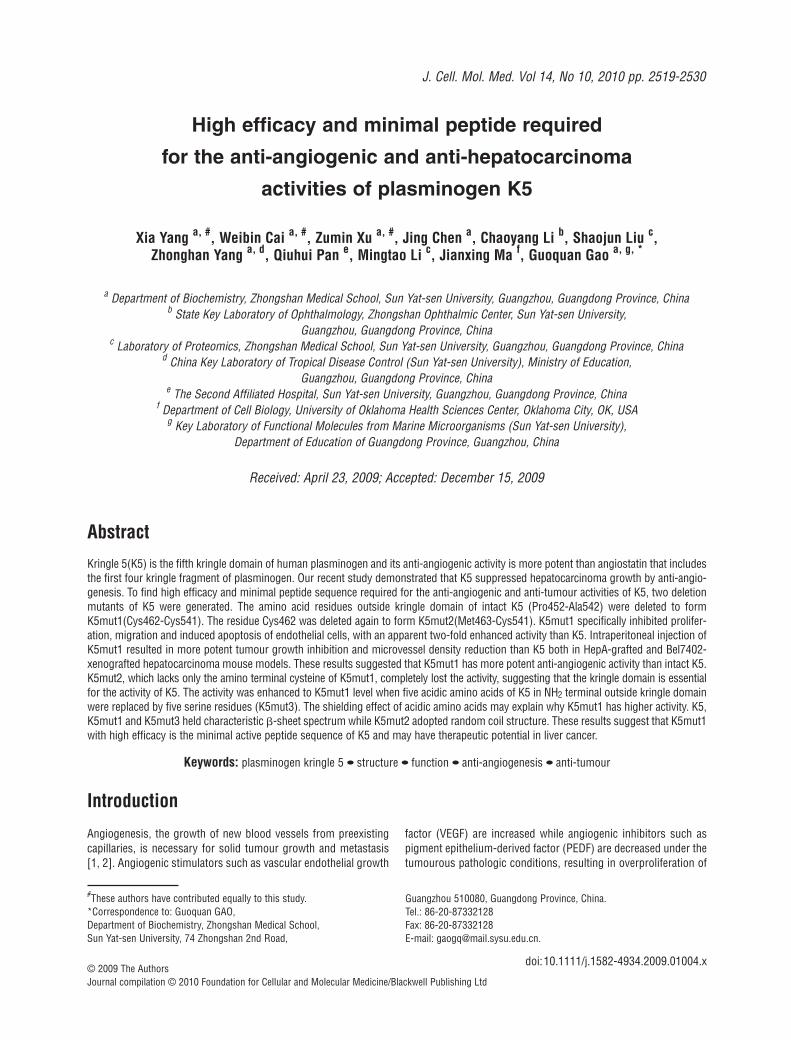
Introduction
Angiogenesis, the growth of new blood vessels from preexistingcapillaries, is necessary for solid tumour growth and metastasis[1, 2]. Angiogenic stimulators such as vascular endothelial growth
factor (VEGF) are increased while angiogenic inhibitors such aspigment epithelium-derived factor (PEDF) are decreased under thetumourous pathologic conditions, resulting in overproliferation of
High efficacy and minimal peptide required
for the anti-angiogenic and anti-hepatocarcinoma
activities of plasminogen K5
Xia Yang a, #, Weibin Cai a, #, Zumin Xu a, #, Jing Chen a, Chaoyang Li b, Shaojun Liu c, Zhonghan Yang a, d, Qiuhui Pan e, Mingtao Li c, Jianxing Ma f, Guoquan Gao a, g, *
a Department of Biochemistry, Zhongshan Medical School, Sun Yat-sen University, Guangzhou, Guangdong Province, Chinab State Key Laboratory of Ophthalmology, Zhongshan Ophthalmic Center, Sun Yat-sen University,
Guangzhou, Guangdong Province, Chinac Laboratory of Proteomics, Zhongshan Medical School, Sun Yat-sen University, Guangzhou, Guangdong Province, China
d China Key Laboratory of Tropical Disease Control (Sun Yat-sen University), Ministry of Education, Guangzhou, Guangdong Province, China
e The Second Affiliated Hospital, Sun Yat-sen University, Guangzhou, Guangdong Province, Chinaf Department of Cell Biology, University of Oklahoma Health Sciences Center, Oklahoma City, OK, USA
g Key Laboratory of Functional Molecules from Marine Microorganisms (Sun Yat-sen University), Department of Education of Guangdong Province, Guangzhou, China
Received: April 23, 2009; Accepted: December 15, 2009
Abstract
Kringle 5(K5) is the fifth kringle domain of human plasminogen and its anti-angiogenic activity is more potent than angiostatin that includesthe first four kringle fragment of plasminogen. Our recent study demonstrated that K5 suppressed hepatocarcinoma growth by anti-angio-genesis. To find high efficacy and minimal peptide sequence required for the anti-angiogenic and anti-tumour activities of K5, two deletionmutants of K5 were generated. The amino acid residues outside kringle domain of intact K5 (Pro452-Ala542) were deleted to formK5mut1(Cys462-Cys541). The residue Cys462 was deleted again to form K5mut2(Met463-Cys541). K5mut1 specifically inhibited prolifer-ation, migration and induced apoptosis of endothelial cells, with an apparent two-fold enhanced activity than K5. Intraperitoneal injection ofK5mut1 resulted in more potent tumour growth inhibition and microvessel density reduction than K5 both in HepA-grafted and Bel7402-xenografted hepatocarcinoma mouse models. These results suggested that K5mut1 has more potent anti-angiogenic activity than intact K5.K5mut2, which lacks only the amino terminal cysteine of K5mut1, completely lost the activity, suggesting that the kringle domain is essentialfor the activity of K5. The activity was enhanced to K5mut1 level when five acidic amino acids of K5 in NH2 terminal outside kringle domainwere replaced by five serine residues (K5mut3). The shielding effect of acidic amino acids may explain why K5mut1 has higher activity. K5,K5mut1 and K5mut3 held characteristic �-sheet spectrum while K5mut2 adopted random coil structure. These results suggest that K5mut1with high efficacy is the minimal active peptide sequence of K5 and may have therapeutic potential in liver cancer.
Keywords: plasminogen kringle 5 • structure • function • anti-angiogenesis • anti-tumour
J. Cell. Mol. Med. Vol 14, No 10, 2010 pp. 2519-2530
#These authors have contributed equally to this study.*Correspondence to: Guoquan GAO,Department of Biochemistry, Zhongshan Medical School,Sun Yat-sen University, 74 Zhongshan 2nd Road,
Guangzhou 510080, Guangdong Province, China.Tel.: 86-20-87332128Fax: 86-20-87332128E-mail: [email protected].
© 2009 The AuthorsJournal compilation © 2010 Foundation for Cellular and Molecular Medicine/Blackwell Publishing Ltd
doi:10.1111/j.1582-4934.2009.01004.x
2520
capillary endothelial cells and abnormal formation of new bloodvessels in neoplasm [3]. Therefore, angiogenic inhibitor may holdgreat therapeutic potential in the treatment of solid tumour [4, 5].
Some proteolytic fragments from human plasminogen arereported as angiogenic inhibitors. Kringle 5 (K5) is the fifth kringledomain of human plasminogen and has been widely demonstratedto inhibit proliferation of endothelial cells [6]. Moreover, itsinhibitory activity is more potent than angiostatin that includes thefirst four kringle modules of plasminogen (kringles1–4) [6, 7]. Inaddition, K5 also inhibits endothelial cell migration, an importantprocess in angiogenesis [8], and exerts its effect on endothelialcells by inducing cell cycle arrest and apoptosis. We previouslydemonstrated the anti-angiogenic activity of K5 in rat model ofoxygen-induced retinopathy (OIR) [9, 10] and rabbit model ofalkali-burn-induced corneal neovascularization [11]. Recently, weproved the anti-tumour activity of K5 in hepatocarcinoma [12].Down-regulation of VEGF and up-regulation of PEDF, thus leadingtoward restoration of the balance in angiogenic control, may rep-resent a mechanism for the anti-angiogenic activity of K5 [13].
To find more potent and smaller peptide sequence required forthe anti-angiogenic activity and define the structure/function relationship of K5, two deletion mutants of K5 were generated inthe present study according to the distribution of three disulfidebonds in intact K5. The amino acid residues outside kringledomain of K5 were deleted to form K5mut1 (Cys462-Cys541). The residue Cys462 was deleted again to form K5mut2 (Met463-Cys541). The disulfide bridging conformation and dimensionalstructure of recombinant K5 and mutants were predicted by 3D-JIGSAW Comparative Modelling Server (UK) and confirmed bymass spectrometry and circular dichroism (CD) analysis. Theiranti-angiogenic and anti-hepatoma activities were evaluated bothin vitro and in vivo. Our findings demonstrated for the first timethat K5mut1 has more potent anti-angiogenic and anti-hepatomaactivities than intact K5 and the complete kringle structure with appropriate folding of three disulfide bonds is required for the activity of K5.
Materials and methods
Construction and production of human plasminogen kringle 5 and its deletion mutants
The cDNAs encoding K5 and mutants were generated by polymerase chainreaction using a human plasminogen cDNA as the template with the followingprimers: K5 P(�): 5�-TGTGAATTCGCCAGATGTAGAGACTCCTTC-3�,P(-):5�-GGAAAGCTTGGCACACTGAGGGACATCACAG-3�; K5mut1 P(�): 5�-ATGAATTCGTGTATGTTTGGGAATGGG-3�, P(-):5�-GCCAAGCTTACACT-GAGGGACATCACAGTAG-3�; K5mut2 P(�): 5�-CGGAATTCCAT-GTTTGGGAATGGGAAAGG-3�, P(-): 5�-CGGAAGCTTACACTGAGGGACAT-CACAGT-3�; K5mut3 P(�):5�-ACTGAATTCGCCATCTGTATCGACTCCTTC-CTCATCATCCTGTATGTTTGG-3�, P(-): 5�-TGCTGCAAGCTTCGCACACTGAGGGACATCACAGTAGTC-3�. The PCR products were cloned intothe pET-22b(�) at EcoRI and HindIII sites. These constructs were intro-
duced into Escherichia coli BL21 (DE3) (Novagen Co., Madison, WI, USA.)strain for protein expression. The expression and purification followed theprotocol recommended by Novagen. The purity and identity of recombi-nant peptides were examined by SDS-PAGE and Western blot analysisusing an antibody specific to His-tag (Novagen Co.).
Molecule mass and disulfide bridging conformation analysis of purified soluble K5 and K5 mutants
Molecule mass analysis of K5 and K5 mutants was performed asdescribed previously [14]. Orthogonal digestion combining with MALDI-QTOF mass spectrometry was used for disulfide bridging conformationanalysis of recombinant K5 and K5 mutants. For single enzyme digest,lyophilized K5 or K5 mutants samples were suspended in digest buffer(50 mM Tris-Cl, 1 mM CaCl2, pH7.6) containing 100 ng of Trypsin Gold(Promega, Madison, WI, USA), and digested overnight. For dual enzymedigest, lyophilized K5 or K5 mutants were first digested overnight withimmobilized trypsin (Pierce, Rockford, IL, USA) followed by an overnightdigestion with 100 ng of Endoproteinase-Asp-N (Roche Diagnostics,Laval, Quebec, Canada). The singly or dually digested solutions wereequated into two aliquots and one aliquot was treated with TCEP in 60�Cwater for 10 min. to reducing the peptides. Peptide fragment analysis wasdone with an ETTAN MALDI-TOF Pro mass spectrometer (ETTAN MALDI-TOF Pro, Amersham Biosciences, Sweden). Spectra analysis and peptideidentity assignment were done using Ettan MALDI-ToF Pro Control mod-ule version 2.01 [15].
Cell culture
Human umbilical vein endothelial cells (HUVEC) were freshly isolatedfrom human umbilical cord veins, as previously described [16, 17] andgrown in human endothelial-SFM basal growth medium (GIBCO,Grand Island, NY, USA) supplemented with 20% foetal calf serum, 100 �g/ml streptomycin, 100 U/ml penicillin, 5 �g/ml amphotericin B(GIBCO, Grand Island, NY, USA), 2 mmol/l L-glutamine, 15 mg/l ECGS(Upstate, NY, USA). The identity and purity of HUVEC were determinedby the incorporation of acetylated low-density lipoprotein labelled witha fluorescent probe DiI (Biomedical Technologies, Inc., Stoughton,MA, USA) [18].
The liver cell line and hepatocarcinoma cell line Bel7402, HepA wereprovided by Cell Bank of China Science Academy (Shanghai, China). Thecells were maintained in DMEM medium (GibcoBRL, Gaithersburg, MD,USA) supplemented with 10% heat-inactivated foetal bovine serum (FBS)and antibiotic-antimycotic.
Cell proliferation assay
The viable cells were quantified by the 3- [4, 5-dimethylthiazol -2-yl]-2,5-dephenyl tetrazolium bromid MTT (Sigma Chemical Co., St. Louis,MO, USA) colorimetric assay following a protocol recommended by themanufacturer [12, 19]. The inhibitory effects of K5 on cell proliferationwere expressed as IC50 values, which were determined from three independent tests.
© 2009 The AuthorsJournal compilation © 2010 Foundation for Cellular and Molecular Medicine/Blackwell Publishing Ltd
J. Cell. Mol. Med. Vol 14, No 10, 2010
2521
Endothelial cell migration assay
Migration assays were performed as described previously [8, 20]. HUVECsin SFM (1�105) were seeded into the top chamber with polycarbonatemembrane (8 �m pore sizes, Corning Costar Corp, Cambridge, MA, USA)precoated with 10 �g/ml fibronectin. Top chambers containing HUVEC wereplaced into a 24-well plate containing 600 �l DMEM with 1% serum andincubated for 30 min. at 37�C. HUVEC migration was stimulated by additionof the VEGF (10 ng/ml) to the lower well of the Boyden chamber. A total of640 nM of K5, K5mut1 and K5mut2 were added to the lower chamber,respectively. After 6 hrs, the chemotaxis chamber was dismantled and thepolycarbonate membranes of top chambers were fixed in formaldehyde for10 min. The non-migrated cells were scraped off the upper surface of themembrane and the migrated cells on the lower surface of the membranewere stained with haematoxylin and eosin. Each sample was tested in trip-licate and the average number of migrating cells per field was assessed bycounting three random high-power fields per filter.
Quantitative analysis of apoptosis by flow cytometry
Apoptosis analysis was carried out as described previously [12, 21, 22].Briefly, HUVEC cells were treated with 320 nM K5 and K5 mutants for 24 hrs.Prior to analysis, cells were washed again with PBS and incubated for 30 min. in propidium iodide staining solution. The suspension was analysedby flow cytometry, using a Coulter (Hialeah, FL, USA) Epic Elite apparatus.
Animals
Care, use and treatment of all animals in this study were in strict agreementwith the institutionally approved protocol according to the USPHS Guidefor the care and use laboratory animals, as well as the guidelines set forthin the Care and Use of Laboratory Animals by the Sun Yat-sen University.Male 5–6-week-old Kunming mice and male 4–5-week-old athymic nudemice were obtained from the Laboratory Animal Center of Guangdong,China and the animal license number is SCXK(YUE)2004-0011.
Inhibition of liver cancer growth
Hepatocarcinoma mouse model was established as described previouslyby subcutaneously injection of mouse hepatoma cells (1�106) into theoxter of mice [12]. When the tumour grew to about 200 mm3, mice werethen randomized and divided into different groups and received three timesperitoneal injection with 72 hrs alternation of PBS, K5 and K5 mutants,respectively. The total amount is 7.5 mg/kg. Two weeks later from the firstinjection, the mice were executed and tumours were dissected, weighed.
Similarly, male athymic nude mice (4–5-week-old) were used forxenografted hepatocarcinoma model. A single human hepatoma Bel7402cell suspension of 1.5�105 was implanted subcutaneously in the middledorsum of each animal. When the tumours became visible (about 9 daysafter inoculation), mice were randomized into different groups. Animalsreceived five times peritoneal injection with 48 hrs alternation with K5 andK5 mutants at a total amount of 12.5 mg/kg, respectively. Tumour volumewas calculated by the following formula: tumour volume (mm3) �
(a�b2)/2, where a � length in mm and b � width in mm [23, 24]. One
month later from the first injection, the mice were executed and tumourswere dissected, weighed.
Microvessel density assay
Immunostaining was performed as described previously with minor mod-ification [16]. Briefly, frozen tissues were cut into 10-�m sections, fixed inacetone at 4�C for 5 min., and blocked for endogenous peroxidase. Tumourvasculature was stained using a monoclonal antibody against mouse CD31at 1:100 dilutions. Tumour microvessel density was quantified by theWeidenr’s method. In negative-control staining, the primary antibodieswere omitted.
Circular dichroism spectroscopy
The purified proteins were dialysed against 25 mmol/l boric acid (pH7.4)for 8 hrs. The CD spectra were measured with a JASCO-J-810 spectropo-larimeter under nitrogen flush in 0.2 cm path length cells at 25�C. Thespectra were recorded between 260 nm and 200 nm and the average ofthree recordings was taken. The calibration was carried out with 25 mmol/lboric acid (pH7.4). The proteins were scanned at concentrations of 0.50mg/ml. The optical activity was normalized to molar ellipticity.
Statistical analysis
Means and SDs were calculated for continuous variables. For two-groupcomparison, the t-test method was used. For more than two groups’ com-parison, one-way ANOVA was used firstly to detect the difference amongstthese groups. If the P-value was less than 0.05, then multiple comparisonwas performed using LSD-t test. All statistical tests were 2-sided, with P-value less than 0.05 considered significant.
Results
Expression and purification of K5 and its deletion mutants
Two deletion mutants were designed according to the structureand disulfide bond distribution of K5 (Pro452-Ala542). K5mut1(Cys462-Cys541) retained the intact kringle domain and the aminoacid residues outside kringle domain of K5 were deleted. Theresidue Cys462 was deleted again to form K5mut2 (Met463-Cys541). K5mut2 opened the first disulfide bond (Fig. 1B).Recombinant K5 and its mutants were expressed in E. coli. andpurified to homogeneity with metal affinity chromatography. Thepurified recombinant protein K5, K5mut1, K5mut2 showed anapparent molecular weight of 16, 14 and 14 kD, respectively (Fig. 1C). Accurate molecular weight of K5mut1, K5mut2 obtainedby MALDI-TOF MS analysis matched the calculated molecularweight from the sequence. The identity of the band was confirmed
© 2009 The AuthorsJournal compilation © 2010 Foundation for Cellular and Molecular Medicine/Blackwell Publishing Ltd
2522
by Western blot analysis using an anti-His tag antibody (Fig. 1C).An average of 15 mg of purified protein per litre culture and over90% purity were achieved for recombinant K5 and mutants andbeing used in this study.
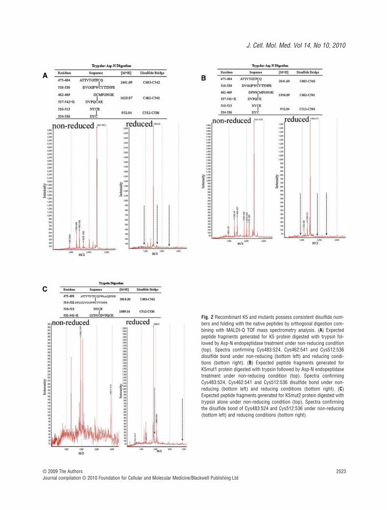
Recombinant K5 and mutants possess consistentdisulfide numbers and folding with the corresponding native peptides as displayed within plasminogen
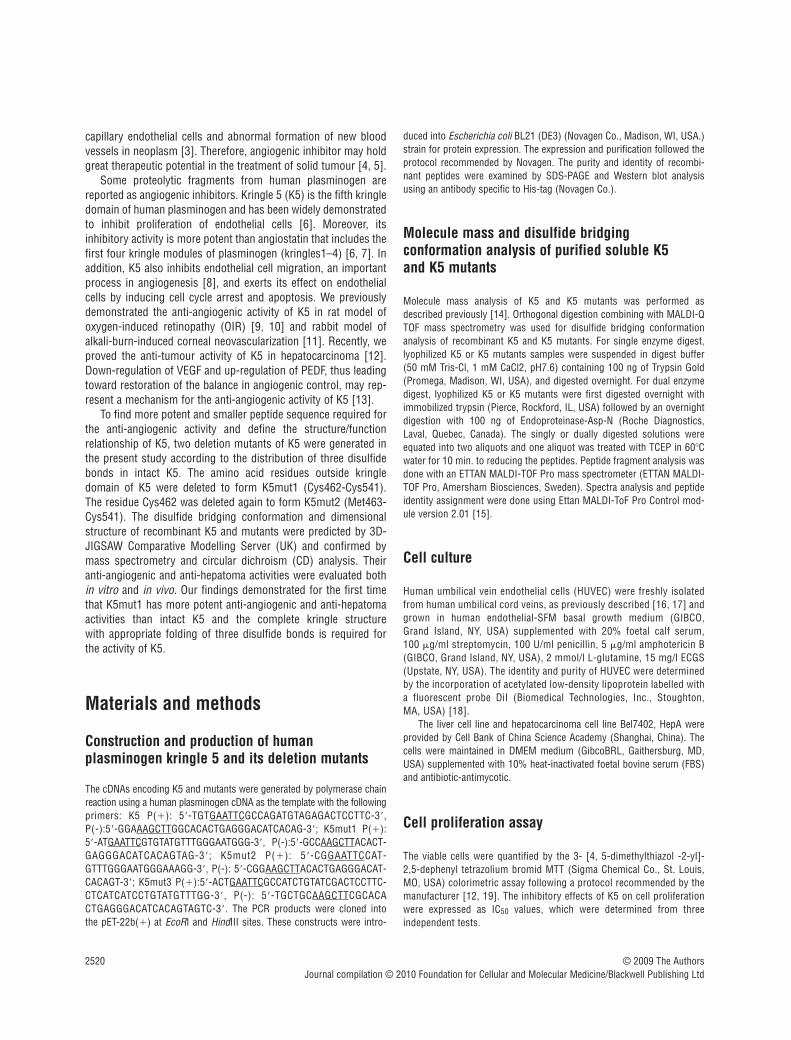
To identify the disulfide bridging in recombinant K5 and mutants,an orthogonal digestion was carried out either with trypsin aloneor with agarose-immobilized trypsin followed by Asp-N endopep-tidase under reducing and nonreducing conditions. MALDI-Q TOFmass spectrometry of K5 and mutants protease digests confirmedthe presence of three disulfide bridges between the cysteine pairsof Cys462:541, Cys483:524 and Cys512:536 in K5 and K5mut1
(Fig. 2A and B), but the presence of only two disulfide bridgesbetween the cysteine pairs of Cys483:524 and Cys512:536 inK5mut2 (Fig. 2C). These findings confirmed that the disulfidebridges present in recombinant K5 and K5mut1 are in the sameconformation as displayed within plasminogen whereas theK5mut2 preserved only two disulfide bridges (Fig. 2).
More potent inhibiting effect of K5mut1 on endothelial cell proliferation
Both K5 and K5mut1 but not K5mut2 exhibited inhibition ofendothelial cell proliferation. The effect appeared to be concentra-tion-dependent. At a concentration as low as 10 nmol/l, K5mut1treatment resulted in significantly fewer viable cells than the PBScontrol group and K5-treated group (P 0.05). K5mut1 showedan IC50 of approximately 35 nmol/l while intact K5 has an IC50
of approximately 70 nmol/l in inhibiting HUVEC proliferation
© 2009 The AuthorsJournal compilation © 2010 Foundation for Cellular and Molecular Medicine/Blackwell Publishing Ltd
Fig. 1 Schematic structure and generation of K5 and its deletionmutants. (A) The primary sequence of K5 and its mutants. (B) Theschematic structure of K5 and its mutants. Two deletion mutants weredesigned according to the structure and disulfide bond distribution ofK5 (Pro452-Ala542). K5mut1 (Cys462-Cys541) retained the intactkringle domain and deleted the amino acid residues of both terminalsoutside kringle domain of K5. K5mut2 (Met463-Cys541) opened thefirst disulfide bond by deleting the amino acids of Cys462 on the basisof K5mut1 structure. (C) The production and identification of recom-binant K5 and its mutant proteins. The top panel is SDS-PAGE withCoomassie blue staining while the bottom panel is Western blot analy-sis with antibody specific to His-tag; Marker: Protein marker; Lane1:Purified recombinant protein of K5; Lane2: Purified recombinant pro-tein of K5mut1; Lane3: Purified recombinant protein of K5mut2.
J. Cell. Mol. Med. Vol 14, No 10, 2010
2523© 2009 The AuthorsJournal compilation © 2010 Foundation for Cellular and Molecular Medicine/Blackwell Publishing Ltd
Fig. 2 Recombinant K5 and mutants possess consistent disulfide num-bers and folding with the native peptides by orthogonal digestion com-bining with MALDI-Q TOF mass spectrometry analysis. (A) Expectedpeptide fragments generated for K5 protein digested with trypsin fol-lowed by Asp-N endopeptidase treatment under non-reducing condition(top). Spectra confirming Cys483:524, Cys462:541 and Cys512:536disulfide bond under non-reducing (bottom left) and reducing condi-tions (bottom right). (B) Expected peptide fragments generated forK5mut1 protein digested with trypsin followed by Asp-N endopeptidasetreatment under non-reducing condition (top). Spectra confirmingCys483:524, Cys462:541 and Cys512:536 disulfide bond under non-reducing (bottom left) and reducing conditions (bottom right). (C)Expected peptide fragments generated for K5mut2 protein digested withtrypsin alone under non-reducing condition (top). Spectra confirmingthe disulfide bond of Cys483:524 and Cys512:536 under non-reducing(bottom left) and reducing conditions (bottom right).
2524 © 2009 The AuthorsJournal compilation © 2010 Foundation for Cellular and Molecular Medicine/Blackwell Publishing Ltd
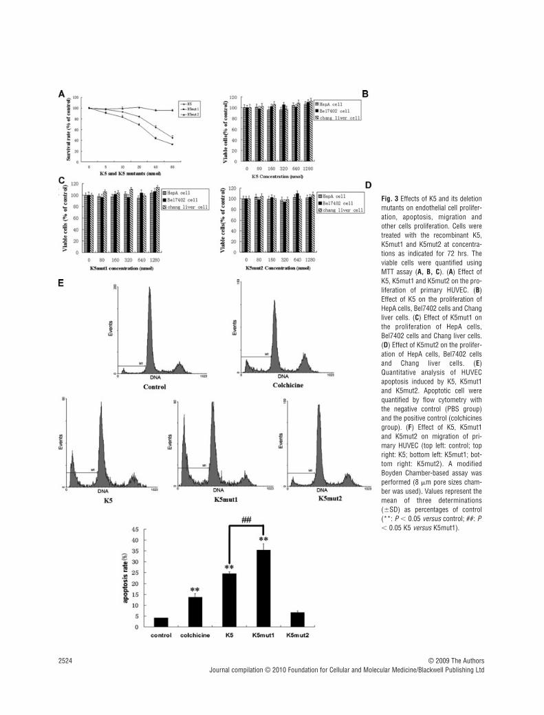
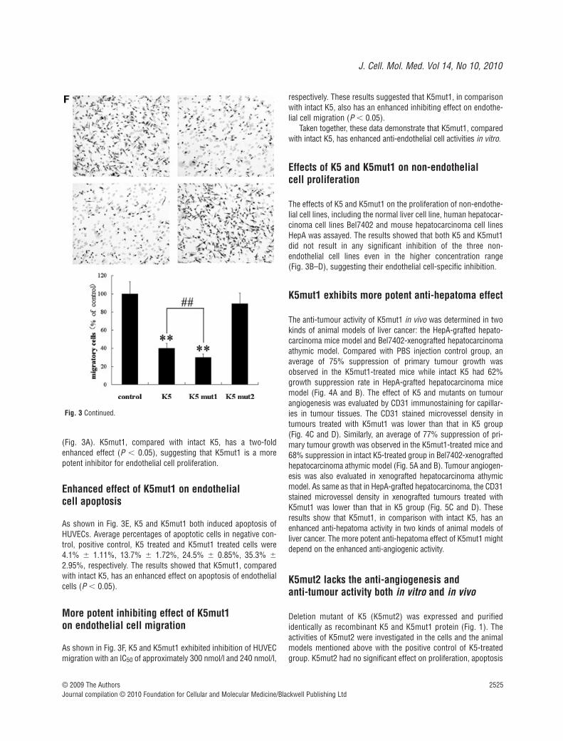
Fig. 3 Effects of K5 and its deletionmutants on endothelial cell prolifer-ation, apoptosis, migration andother cells proliferation. Cells weretreated with the recombinant K5,K5mut1 and K5mut2 at concentra-tions as indicated for 72 hrs. Theviable cells were quantified usingMTT assay (A, B, C). (A) Effect ofK5, K5mut1 and K5mut2 on the pro-liferation of primary HUVEC. (B)Effect of K5 on the proliferation ofHepA cells, Bel7402 cells and Changliver cells. (C) Effect of K5mut1 onthe proliferation of HepA cells,Bel7402 cells and Chang liver cells.(D) Effect of K5mut2 on the prolifer-ation of HepA cells, Bel7402 cellsand Chang liver cells. (E)Quantitative analysis of HUVECapoptosis induced by K5, K5mut1and K5mut2. Apoptotic cell werequantified by flow cytometry withthe negative control (PBS group)and the positive control (colchicinesgroup). (F) Effect of K5, K5mut1and K5mut2 on migration of pri-mary HUVEC (top left: control; topright: K5; bottom left: K5mut1; bot-tom right: K5mut2). A modifiedBoyden Chamber-based assay wasperformed (8 �m pore sizes cham-ber was used). Values represent themean of three determinations(SD) as percentages of control(**: P 0.05 versus control; ##: P 0.05 K5 versus K5mut1).
J. Cell. Mol. Med. Vol 14, No 10, 2010
2525
(Fig. 3A). K5mut1, compared with intact K5, has a two-foldenhanced effect (P 0.05), suggesting that K5mut1 is a morepotent inhibitor for endothelial cell proliferation.
Enhanced effect of K5mut1 on endothelial cell apoptosis
As shown in Fig. 3E, K5 and K5mut1 both induced apoptosis ofHUVECs. Average percentages of apoptotic cells in negative con-trol, positive control, K5 treated and K5mut1 treated cells were4.1% 1.11%, 13.7% 1.72%, 24.5% 0.85%, 35.3%
2.95%, respectively. The results showed that K5mut1, comparedwith intact K5, has an enhanced effect on apoptosis of endothelialcells (P 0.05).
More potent inhibiting effect of K5mut1 on endothelial cell migration
As shown in Fig. 3F, K5 and K5mut1 exhibited inhibition of HUVECmigration with an IC50 of approximately 300 nmol/l and 240 nmol/l,
respectively. These results suggested that K5mut1, in comparisonwith intact K5, also has an enhanced inhibiting effect on endothe-lial cell migration (P 0.05).
Taken together, these data demonstrate that K5mut1, comparedwith intact K5, has enhanced anti-endothelial cell activities in vitro.
Effects of K5 and K5mut1 on non-endothelial cell proliferation
The effects of K5 and K5mut1 on the proliferation of non-endothe-lial cell lines, including the normal liver cell line, human hepatocar-cinoma cell lines Bel7402 and mouse hepatocarcinoma cell linesHepA was assayed. The results showed that both K5 and K5mut1did not result in any significant inhibition of the three non-endothelial cell lines even in the higher concentration range (Fig. 3B–D), suggesting their endothelial cell-specific inhibition.
K5mut1 exhibits more potent anti-hepatoma effect
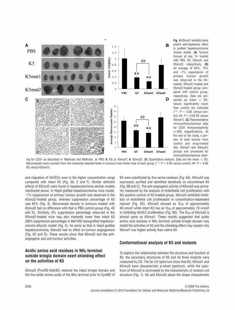
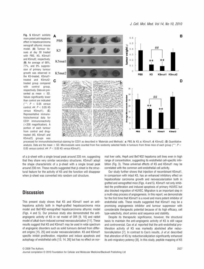
The anti-tumour activity of K5mut1 in vivo was determined in twokinds of animal models of liver cancer: the HepA-grafted hepato-carcinoma mice model and Bel7402-xenografted hepatocarcinomaathymic model. Compared with PBS injection control group, anaverage of 75% suppression of primary tumour growth wasobserved in the K5mut1-treated mice while intact K5 had 62%growth suppression rate in HepA-grafted hepatocarcinoma micemodel (Fig. 4A and B). The effect of K5 and mutants on tumourangiogenesis was evaluated by CD31 immunostaining for capillar-ies in tumour tissues. The CD31 stained microvessel density intumours treated with K5mut1 was lower than that in K5 group (Fig. 4C and D). Similarly, an average of 77% suppression of pri-mary tumour growth was observed in the K5mut1-treated mice and68% suppression in intact K5-treated group in Bel7402-xenograftedhepatocarcinoma athymic model (Fig. 5A and B). Tumour angiogen-esis was also evaluated in xenografted hepatocarcinoma athymicmodel. As same as that in HepA-grafted hepatocarcinoma, the CD31stained microvessel density in xenografted tumours treated withK5mut1 was lower than that in K5 group (Fig. 5C and D). Theseresults show that K5mut1, in comparison with intact K5, has anenhanced anti-hepatoma activity in two kinds of animal models ofliver cancer. The more potent anti-hepatoma effect of K5mut1 mightdepend on the enhanced anti-angiogenic activity.
K5mut2 lacks the anti-angiogenesis and anti-tumour activity both in vitro and in vivo
Deletion mutant of K5 (K5mut2) was expressed and purifiedidentically as recombinant K5 and K5mut1 protein (Fig. 1). Theactivities of K5mut2 were investigated in the cells and the animalmodels mentioned above with the positive control of K5-treatedgroup. K5mut2 had no significant effect on proliferation, apoptosis
© 2009 The AuthorsJournal compilation © 2010 Foundation for Cellular and Molecular Medicine/Blackwell Publishing Ltd
Fig. 3 Continued.
2526 © 2009 The AuthorsJournal compilation © 2010 Foundation for Cellular and Molecular Medicine/Blackwell Publishing Ltd
and migration of HUVECs even in the higher concentration rangecompared with intact K5 (Fig. 3A, E and F). Similar deficienteffects of K5mut2 were found in hepatocarcinoma animal modelsmentioned above. In HepA-grafted hepatocarcinoma mice model,–7% suppression of primary tumour growth was observed in theK5mut2-treated group, whereas suppression percentage of K5was 62% (Fig. 4). Microvessel density in tumours treated withK5mut2 had no difference with that in PBS control group (Fig. 4Cand D). Similarly, 9% suppression percentage observed in theK5mut2-treated mice was also markedly lower than intact K5(68% suppression percentage) in Bel7402-xenografted hepatocar-cinoma athymic model (Fig. 5). As same as that in HepA-graftedhepatocarcinoma, K5mut2 had no effect on tumour angiogenesis(Fig. 5C and D). These results show that K5mut2 lost the anti-angiogenic and anti-tumour activities.
Acidic amino acid residues in NH2 terminal outside kringle domain exert shielding effect on the activities of K5
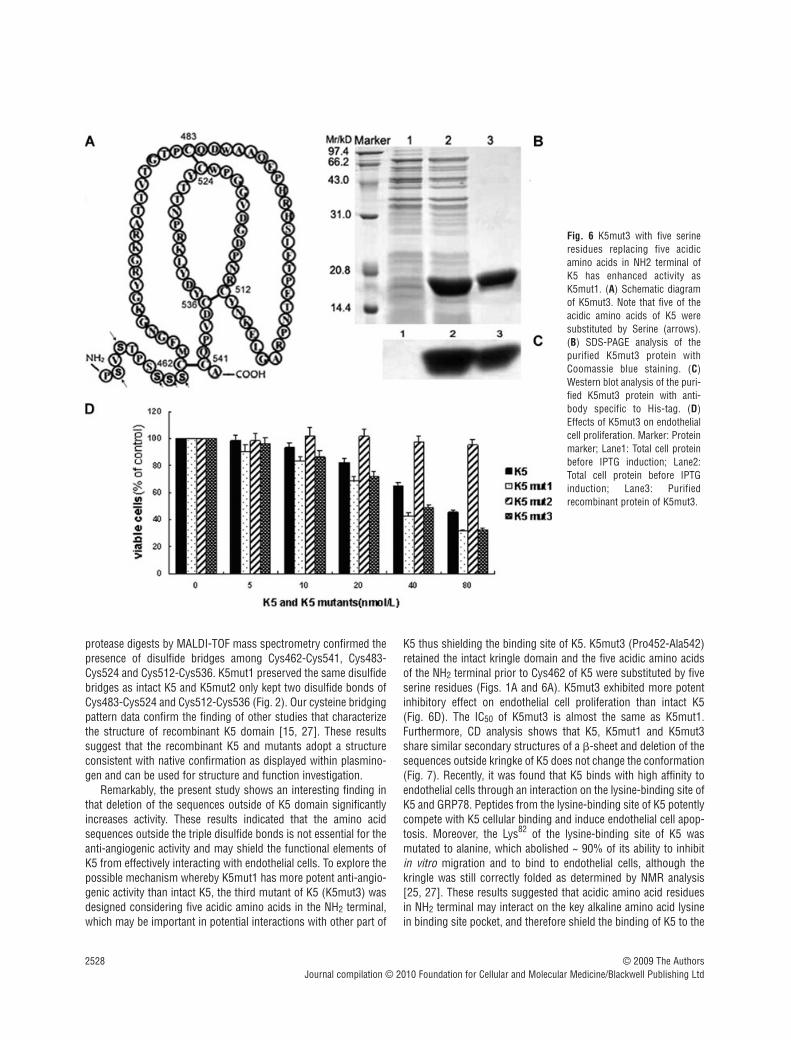
K5mut3 (Pro452-Ala542) retained the intact kringle domain andthe five acidic amino acids of the NH2 terminal prior to Cys462 of
K5 were substituted by five serine residues (Fig. 6A). K5mut3 wasexpressed, purified and identified identically as recombinant K5(Fig. 6B and C). The anti-angiogenic activity of K5mut3 was prima-rily measured by the analysis of endothelial cell proliferation withthe positive control of K5-treated group. K5mut3 exhibited inhibi-tion of endothelial cell proliferation in concentration-dependentmanner (Fig. 6D). K5mut3 showed an IC50 of approximately 40 nmol/l while intact K5 has an IC50 of approximately 70 nmol/lin inhibiting HUVEC proliferation (Fig. 6D). The IC50 of K5mut3 isalmost same as K5mut1. These results suggested that acidicamino acid residues in NH2 terminal outside kringle domain mayshield the activities of K5 and the shielding effect may explain whyK5mut1 has higher activity than native K5.
Conformational analysis of K5 and mutants
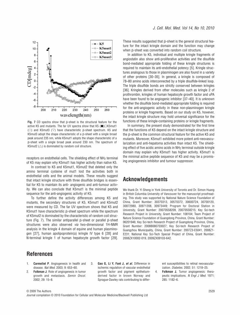
To explore the relationship between the structure and function ofK5, the secondary structures of K5 and its three mutants weremeasured by CD. The far UV spectrum show that K5, K5mut1 andK5mut3 have characteristic �-sheet spectrum, while the spec-trum of K5mut2 is dominated by the characteristic of random coilstructure (Fig. 7). K5 and K5mut3 adopt the shape characteristic
Fig. 4 K5mut1 exhibits morepotent anti-hepatoma effectin grafted hepatocarcinomamouse model. (A) Tumourtissues at day 14 treated with PBS, K5, K5mut1 andK5mut2, respectively. (B)An average of 62%, 75% and –7% suppression of primary tumour growth was observed in the K5-treated, K5mut1-treated andK5mut2-treated group com-pared with control group,respectively. Data are pre-sented as mean SD.Values significantly lowerthan control are indicated(**: P 0.05 versus con-trol; ##: P 0.05 K5 versusK5mut1). (C) Representativeimmunohistochemical datafor CD31 immunoreactivity(�200 magnification). At the end of the study, a por-tion of each tumour fromcontrol and drug-treated (K5, K5mut1 and K5mut2)groups was processed forimmunohistochemical stain-
ing for CD31 as described in ‘Materials and Methods’. a: PBS; b: K5; c: K5mut1; d: K5mut2. (D) Quantitative analysis. Data are the mean SD.Microvessels were counted from five randomly selected fields in tumours from three mice of each group (**: P 0.05 versus control; ##: P 0.05K5 versus K5mut1).
J. Cell. Mol. Med. Vol 14, No 10, 2010
2527© 2009 The AuthorsJournal compilation © 2010 Foundation for Cellular and Molecular Medicine/Blackwell Publishing Ltd
of a �-sheet with a single broad peak around 235 nm, suggestingthat they share very similar secondary structures. K5mut1 adoptthe shape characteristic of a �-sheet with a single broad peakaround 230 nm. These results suggested that �-sheet is the struc-tural feature for the activity of K5 and the function will disappearwhen �-sheet was converted into random coil structure.
Discussion
This present study shows that K5 and K5mut1 exert an anti-hepatoma activity both in HepA-grafted hepatocarcinoma micemodel and Bel7402-xenografted hepatocarcinoma athymic model(Figs. 4 and 5). Our previous study also demonstrated the anti-angiogenic activity of K5 in rat model of OIR [9, 10] and rabbitmodel of alkali-burn-induced corneal neovascularization [11]. Theseresults suggest that K5 and K5mut1 may be used in wide spectrumof angiogenic disorders such as solid tumours derived from differ-ent origins [15, 25] and ocular neovascularization. K5 and K5mut1specific inhibit proliferation, migration and induce apoptosis andautophagy of endothelial cells [13, 14, 26] but has no effect on nor-
mal liver cells, HepA and Bel7402 hepatoma cell lines even in highrange of concentration, suggesting its endothelial cell-specific inhi-bition (Fig. 3). These universal effects of K5 and K5mut1 may becorrelated with the common anti-endothelial cell activity.
Our study further shows that injection of recombinant K5mut1,in comparison with intact K5, has an enhanced inhibitory effect onhepatocellular carcinoma growth and neovascularization both ingrafted and xenografted mice (Figs. 4 and 5). K5mut1 not only inhib-ited the proliferation and induced apoptosis of primary HUVEC butalso blocked migration of HUVEC. Migration is an important step ina multi-step process of angiogenesis. In this report, we demonstratefor the first time that K5mut1 is a novel and more potent inhibitor ofendothelial cells. These results suggested that K5mut1 may be apromising angiogenesis inhibitor and tumour suppressor with considerable therapeutic potential because of its high efficacy, celltype-selectivity, short amino acid sequence and stability.
Despite its therapeutic significance, however, the structuralbasis to maintain the anti-angiogenic activity of K5 is still vagueand controversial. Cao et al. reported that the anti-endothelial pro-liferation activity of K5 was markedly abolished after reduc-tion/alkylation [7]. In contrast to Cao’s results, Ji et al. describedthat alteration of K5 by reduction/alkylation significantly enhancedits anti-migratory potency [8]. In this study, peptide mapping of K5
Fig. 5 K5mut1 exhibitsmore potent anti-hepatomaeffect in hepatocarcinomaxenograft athymic mousemodel. (A) Tumour tis-sues at day 30 treatedwith PBS, K5, K5mut1and K5mut2, respectively.(B) An average of 68%,77%, and 9% suppres-sion of primary tumourgrowth was observed inthe K5-treated, K5mut1-treated and K5mut2-treated group comparedwith control group,respectively. Data are pre-sented as mean SD.Values significantly lowerthan control are indicated(**: P 0.05 versuscontrol; ##: P 0.05 K5versus K5mut1). (C)Representative immuno-histochemical data forCD31 immunoreactivity(�200 magnification). Aportion of each tumourfrom control and drug-treated (K5, K5mut1 andK5mut2) groups wasprocessed for immunohistochemical staining for CD31 as described in ‘Materials and Methods’. a: PBS; b: K5; c: K5mut1; d: K5mut2. (D) Quantitativeanalysis. Data are the mean SD. Microvessels were counted from five randomly selected fields in tumours from three mice of each group (**: P
0.05 versus control; ##: P 0.05 K5 versus K5mut1).
2528
protease digests by MALDI-TOF mass spectrometry confirmed thepresence of disulfide bridges among Cys462-Cys541, Cys483-Cys524 and Cys512-Cys536. K5mut1 preserved the same disulfidebridges as intact K5 and K5mut2 only kept two disulfide bonds ofCys483-Cys524 and Cys512-Cys536 (Fig. 2). Our cysteine bridgingpattern data confirm the finding of other studies that characterizethe structure of recombinant K5 domain [15, 27]. These resultssuggest that the recombinant K5 and mutants adopt a structureconsistent with native confirmation as displayed within plasmino-gen and can be used for structure and function investigation.
Remarkably, the present study shows an interesting finding inthat deletion of the sequences outside of K5 domain significantlyincreases activity. These results indicated that the amino acidsequences outside the triple disulfide bonds is not essential for theanti-angiogenic activity and may shield the functional elements ofK5 from effectively interacting with endothelial cells. To explore thepossible mechanism whereby K5mut1 has more potent anti-angio-genic activity than intact K5, the third mutant of K5 (K5mut3) wasdesigned considering five acidic amino acids in the NH2 terminal,which may be important in potential interactions with other part of
K5 thus shielding the binding site of K5. K5mut3 (Pro452-Ala542)retained the intact kringle domain and the five acidic amino acidsof the NH2 terminal prior to Cys462 of K5 were substituted by fiveserine residues (Figs. 1A and 6A). K5mut3 exhibited more potentinhibitory effect on endothelial cell proliferation than intact K5 (Fig. 6D). The IC50 of K5mut3 is almost the same as K5mut1.Furthermore, CD analysis shows that K5, K5mut1 and K5mut3share similar secondary structures of a �-sheet and deletion of thesequences outside kringke of K5 does not change the conformation(Fig. 7). Recently, it was found that K5 binds with high affinity toendothelial cells through an interaction on the lysine-binding site ofK5 and GRP78. Peptides from the lysine-binding site of K5 potentlycompete with K5 cellular binding and induce endothelial cell apop-tosis. Moreover, the Lys82 of the lysine-binding site of K5 wasmutated to alanine, which abolished ~ 90% of its ability to inhibitin vitro migration and to bind to endothelial cells, although thekringle was still correctly folded as determined by NMR analysis[25, 27]. These results suggested that acidic amino acid residuesin NH2 terminal may interact on the key alkaline amino acid lysinein binding site pocket, and therefore shield the binding of K5 to the
© 2009 The AuthorsJournal compilation © 2010 Foundation for Cellular and Molecular Medicine/Blackwell Publishing Ltd
Fig. 6 K5mut3 with five serineresidues replacing five acidicamino acids in NH2 terminal ofK5 has enhanced activity asK5mut1. (A) Schematic diagramof K5mut3. Note that five of theacidic amino acids of K5 weresubstituted by Serine (arrows).(B) SDS-PAGE analysis of thepurified K5mut3 protein withCoomassie blue staining. (C)Western blot analysis of the puri-fied K5mut3 protein with anti-body specific to His-tag. (D)Effects of K5mut3 on endothelialcell proliferation. Marker: Proteinmarker; Lane1: Total cell proteinbefore IPTG induction; Lane2:Total cell protein before IPTGinduction; Lane3: Purifiedrecombinant protein of K5mut3.
J. Cell. Mol. Med. Vol 14, No 10, 2010
2529
receptors on endothelial cells. The shielding effect of NH2 terminalof K5 may explain why K5mut1 has higher activity than native K5.
In contrast to K5 and K5mut1, K5mut2 that deleted only theamino terminal cysteine of mut1 lost the activities both inendothelial cells and the animal models. These results suggestthat intact kringle structure with three disulfide bridges is essen-tial for K5 to maintain its anti- angiogenic and anti-tumour activ-ity. We can also conclude that K5mut1 is the minimal peptidesequence for the anti-angiogenic activity of K5.
To further define the activity differences among K5 andmutants, the secondary structures of K5, K5mut1 and K5mut2were measured by CD. The far UV spectrum shows that K5 andK5mut1 have characteristic �-sheet spectrum while the spectrumof K5mut2 is dominated by the characteristic of random coil struc-ture (Fig. 7). The similar antiparallel �-sheet or parallel �-sheetstructures were also observed via two-dimensional 1H-NMRanalysis in the kringle 4 domain of equine and human plasmino-gen [27], human apolipoprotein(a) kringle IV type 6 [28] and N-terminal kringle 1 of human hepatocyte growth factor [29].
These results suggested that �-sheet is the general structural fea-ture for the intact kringle domain and the function may changewhen �-sheet was converted into random coil structure.
In addition to K5, individual and multiple kringle fragments ofangiostatin also show anti-proliferative activities and the disulfidebond-mediated appropriate folding of these kringle structures isrequired to maintain its anti-endothelial potency [5]. Kringle struc-tures analogous to those in plasminogen are also found in a varietyof other proteins [30–35]. In general, a kringle is composed of78–80 amino acids interconnected by a triple disulfide-linked loop.The triple disulfide bonds are strictly conserved between kringles[36]. Kringles derived from other molecules such as kringle 2 ofprothrombin, kringles of human hepatocyte growth factor and uPAhave been found to be angiogenic inhibitor [37–40]. It is unknownwhether the disulfide bond-mediated appropriate folding is requiredfor the anti-angiogenic activity in these non-plasminogen kringleproteins or kringle fragments. Based on our study on K5, however,the intact kringle structure may hold universal significance for thefunctions of these kringle-containing proteins or kringle fragments.
In summary, the present study demonstrated for the first timethat the functions of K5 depend on the intact kringle structure andthe �-sheet is the common structural feature for the active K5 andmutants. Moreover, K5mut1 exhibited more potent anti-neovascu-larization and anti-hepatoma activities than intact K5. The shield-ing effect of five acidic amino acids in NH2 terminal outside kringledomain may explain why K5mut1 has higher activity. K5mut1 isthe minimal active peptide sequence of K5 and may be a promis-ing angiogenesis inhibitor and tumour suppressor.
Acknowledgements
We thank Dr. Yi Sheng in York University of Toronto and Dr. Simon Huangin British Columbia University of Vancouver for the manuscript proofread-ing. This study was supported by National Nature Science Foundation ofChina, Grant Number: 30370313, 30570372, 30600724, 30700120,30872980, 30971208, 30973449; Program for Doctoral Station inUniversity, Grant Number: 20070558209, 20070558215; Key Sci-techResearch Project in University, Grant Number: 108104; Team Project ofNature Science Foundation of Guangdong Province, China, Grant Number:06201946; Key Sci-tech Research Project of Guangdong Province, China,Grant Number: 2008B080703027; Key Sci-tech Research Project ofGuangzhou Municipality, China, Grant Number: 2007Z3-E5041, 2008Z1-E231; National Key Sci-Tech Special Project of China, Grant Number:2008ZX10002-019, 2009ZX09103-642.
© 2009 The AuthorsJournal compilation © 2010 Foundation for Cellular and Molecular Medicine/Blackwell Publishing Ltd
References
1. Carmeliet P. Angiogenesis in health anddisease. Nat Med. 2003; 9: 653–60.
2. Folkman J. Role of angiogenesis in tumorgrowth and metastasis. Semin Oncol.2002; 29: 15–8.
3. Gao G, Li Y, Fant J, et al. Difference inischemic regulation of vascular endothelialgrowth factor and pigment epithelium-derived factor in brown Norway andSprague-Dawley rats contributing to differ-
ent susceptibilities to retinal neovascular-ization. Diabetes. 2002; 51: 1218–25.
4. Folkman J. Tumor angiogenesis: thera-peutic implications. N Engl J Med. 1971;285: 1182–6.
Fig. 7 CD spectra show that �-sheet is the structural feature for theactive K5 and mutants. The far UV spectra show that K5 (�)�K5mut1(�) and K5mut3 (�) have characteristic �-sheet spectrum. K5 andK5mut3 adopt the shape characteristic of a �-sheet with a single broadpeak around 235 nm, while K5mut1 adopts the shape characteristic of a�-sheet with a single broad peak around 230 nm. The spectrum ofK5mut2 (�) is dominated by random coil structure.
2530
5. Cao Y. Antiangiogenic cancer therapy.Semin Cancer Biol. 2004; 14: 139–45.
6. Cao Y, Chen A, An SS, et al. Kringle 5 of plasminogen is a novel inhibitor ofendothelial cell growth. J Biol Chem. 1997;272: 22924–8.
7. Cao Y, Ji RW, Davidson D, et al.Kringle domains of human angiostatin.Characterization of the anti-proliferativeactivity on endothelial cells. J Biol Chem.1996; 271: 29461–7.
8. Ji WR, Barrientos LG, Llinás M, et al.Selective inhibition by kringle 5 of humanplasminogen on endothelial cell migration,an important process in angiogenesis.Biochem Biophys Res Commun. 1998;247: 414–9.
9. Zhang D, Kaufman PL, Gao G, et al.Intravitreal injection of plasminogenkringle 5, an endogenous angiogenicinhibitor, arrests retinal neovascularizationin rats. Diabetologia. 2001; 44: 757–65.
10. Zhang SX, Sima J, Shao C, et al.Plasminogen kringle 5 reduces vascularleakage in the retina in rat models of oxy-gen-induced retinopathy and diabetes.Diabetologia. 2004; 47: 124–131.
11. Zhang Z, Ma JX, Gao G, et al.Plasminogen kringle 5 inhibits alkali-burn-induced corneal neovascularization. InvestOphthalmol Vis Sci. 2005; 46: 4062–71.
12. Yang X, Cheng R, Li C, et al. Kringle 5 ofhuman plasminogen suppresses hepatocel-lular carcinoma growth both in grafted andxenografted mice by anti-angiogenic activ-ity. Cancer Biol Ther. 2006; 5: 399–405.
13. Gao G, Li Y, Gee S, et al. Down-regulationof vascular endothelial growth factor andup-regulation of pigment epithelium-derived factor: a possible mechanism forthe anti-angiogenic activity of plasminogenkringle 5. J Biol Chem. 2002; 277: 9492–7.
14. Cai W, Ma J, Li C, et al. Enhanced anti-angiogenic effect of a deletion mutant ofplasminogen kringle 5 on neovasculariza-tion. J Cell Biochem. 2005; 96: 1254–61.
15. Perri SR, Nalbantoglu J, Annabi B, et al.Plasminogen kringle 5-engineered gliomacells block migration of tumor-associatedmacrophages and suppress tumor vascu-larization and progression. Cancer Res.2005; 65: 8359–65.
16. Cooke BM, Usami S, Perry I, et al. A sim-plified method for culture of endothelialcells and analysis of adhesion of bloodcells under conditions of flow. MicrovascRes. 1993; 45: 33–45.
17. Houliston RA, Pearson JD, Wheeler-Jones CP. Agonist-specific cross talkbetween ERKs and p38(mapk) regulates
PGI(2) synthesis in endothelium. Am JPhysiol Cell Physiol. 2001; 281: C1266–76.
18. Grant MB, Guay C. Plasminogen activatorproduction by human retinal endothelialcells of nondiabetic and diabetic origin.Invest Ophthalmol Vis Sci. 1991; 32: 53–64.
19. Carmichael J, DeGraff WG, Gazdar AF, et al. Evaluation of a tetrazolium-basedsemiautomated colorimetric assay: assess-ment of chemosensitivity testing. CancerRes. 1987; 47: 936–42.
20. Annabi B, Lachambre MP, Bousquet-Gagnon N, et al. Green tea polyphenol(-)-epigallocatechin 3-gallate inhibits MMP-2secretion and MT1-MMP-driven migrationin glioblastoma cells. Biochim BiophysActa. 2002; 1542: 209–20.
21. Ormerod MG, Collins MK, Rodriguez-Tarduchy G, et al. Apoptosis in interleukin-3-dependent haemopoietic cells: quantifi-cation by two flow cytometric methods. J Immunol Methods. 1992; 153: 57–65.
22. Dive C, Gregory CD, Phipps DJ, et al.Analysis and discrimination of necrosisand apoptosis (programmed cell death) bymultiparameter flow cytometry. BiochimBiophys Acta. 1992; 1133: 275–85.
23. Gr Griscelli F, Li H, Bennaceur-GriscelliA, et al. Angiostatin gene transfer: inhibi-tion of tumor growth in vivo by blockage ofendothelial cell proliferation associatedwith a mitosis arrest. Proc Natl Acad SciUSA. 1998; 95: 6367–72.
24. Li H, Lu H, Griscelli F, et al. Adenovirus-mediated delivery of a uPA/uPAR antago-nist suppresses angiogenesis-dependenttumor growth and dissemination in mice.Gene Ther. 1998; 5: 1105–13.
25. Davidson DJ, Haskell C, Majest S, et al.Kringle 5 of human plasminogen inducesapoptosis of endothelial and tumor cellsthrough surface-expressed glucose-regu-lated protein 78. Cancer Res. 2005; 65:4663–72.
26. Nguyen TMB, Subramanian IV, KelekarA, et al. Kringle 5 of human plasminogen,an angiogenesis inhibitor, induces bothautophagy and apoptotic death in endothe-lial cells. Blood. 2007; 109: 4793–802.
27. Cox M, Schaller J, Boelens R, et al.Kringle solution structures via NMR: two-dimensional 1H-NMR analysis of horseplasminogen kringle 4. Chem Phys Lipids.1994; 67–68: 43–58.
28. Maderegger B, Bermel W, Hrzenjak A, et al. Solution structure of humanapolipoprotein(a) kringle IV type 6.Biochemistry. 2002; 41: 660–8.
29. Ultsch M, Lokker NA, Godowski PJ, et al.Crystal structure of the NK1 fragment of
human hepatocyte growth factor at 2.0 Aresolution. Structure. 1998; 6: 1383–93.
30. McLean JW, Tomlinson JE, Kuang WJ, etal. cDNA sequence of human apolipopro-tein(a) is homologous to plasminogen.Nature. 1987; 330: 132–7.
31. Walz DA, Hewett-Emmett D, SeegersWH. Amino acid sequence of human pro-thrombin fragments 1 and 2. Proc NatlAcad Sci USA. 1977; 74: 1969–72.
32. Gunzler WA, Steffens GJ, Otting F, et al.The primary structure of high molecularmass urokinase from human urine. Thecomplete amino acid sequence of the Achain. Hoppe Seylers Z Physiol Chem.1982; 363: 1155–65.
33. Pennica D, Holmes WE, Kohr WJ, et al.Cloning and expression of human tissue-type plasminogen activator cDNA in E. coli.Nature. 1983; 301: 214–21.
34. Wilson C, Goberdhan DC, Steller H. Dror,a potential neurotrophic receptor gene,encodes a Drosophila homolog of the ver-tebrate Ror family of Trk-related receptortyrosine kinases. Proc Natl Acad Sci USA.1993; 90: 7109–13.
35. Lokker NA, Presta LG, Godowski PJ.Mutational analysis and molecular model-ing of the N-terminal kringle-containingdomain of hepatocyte growth factor identi-fies amino acid side chains important forinteraction with the c-Met receptor.Protein Eng. 1994; 7: 895–903.
36. Castellino FJ, Beals JM. The genetic rela-tionships between the kringle domains ofhuman plasminogen, prothrombin, tissueplasminogen activator, urokinase, andcoagulation factor XII. J Mol Evol. 1987;26: 358–69.
37. Lee TH, Rhim T, Kim SS. Prothrombinkringle-2 domain has a growth inhibitoryactivity against basic fibroblast growth fac-tor-stimulated capillary endothelial cells. JBiol Chem. 1998; 273: 28805–12.
38. Xin L, Xu R, Zhang Q, et al. Kringle 1 ofhuman hepatocyte growth factor inhibitsbovine aortic endothelial cell proliferationstimulated by basic fibroblast growth factorand causes cell apoptosis. BiochemBiophys Res Commun. 2000; 277: 186–90.
39. Kuba K, Matsumoto K, Date K, et al.HGF/NK4, a four-kringle antagonist of hepato-cyte growth factor, is an angiogenesis inhibitorthat suppresses tumor growth and metasta-sis in mice. Cancer Res. 2000; 60: 6737–43.
40. Kim KS, Hong YK, Joe YA, et al. Anti-angiogenic activity of the recombinantkringle domain of urokinase and its spe-cific entry into endothelial cells. J BiolChem. 2003; 278: 11449–56.
© 2009 The AuthorsJournal compilation © 2010 Foundation for Cellular and Molecular Medicine/Blackwell Publishing Ltd
Copyright © 2022 FDOKUMEN