Hierarchical Mechanochemical Switches in Angiostatin
10
DOI: 10.1002/cbic.200600227 Hierarchical Mechanochemical Switches in Angiostatin Fabio Grandi, [a] Massimo Sandal, [a] Giovanni Guarguaglini, [a, b] Emidio Capriotti, [c] Rita Casadio, [c] and Bruno Samorȶ* [a, b] Introduction The extent and fate of many biological processes are drastically affected by the stresses and strains that are exerted during their development. [1–3] To understand the behavior of these processes, force as a physical variable must also be taken into account. [4] A mechanical tension can alter the functional states of proteins and activate processes. In the case of PECAM-1 for instance, mechanical tension switches on the binding of this protein with a kinase. [5] Likewise, it promotes the exposure of binding sites that serve as nucleation centers for the self-as- sembly fibronectin into a fibrillar form. [6] Disulfide bonds are believed to have been added during evolution to enhance the stability of proteins that function in fluctuating cellular environments. Recent evidence indicates that disulfide bonds can be more than inert structural motifs and that they can act as switches for different protein func- tions. [7] The switching capabilities of the coupling of disulfide redox equilibria with a mechanical force have been addressed by Discher and colleagues in a vascular cell-adhesion protein. [8] The chemical kinetics of disulfide reduction under an external force have been more recently investigated by Fernandez and colleagues. [9] Angiostatin (ANG) is a multimodular protein that contains ACHTUNGTRENNUNGdisulfide bonds and is located on the basement membrane where different reductases are present, [10] and mechanical stretching forces are constantly being developed. [11] This com- bination of mechanochemical stress and redox conditions in the in vivo ANG environment makes this protein an ideal model system for addressing the issue of the possible trigger- ing of biochemical signals by the combination of mechano- chemical forces and redox equilibria. ANG is an extracellular fragment of plasminogen, and is composed of five highly homologous, tandem kringle domains (K1–K5). All of them contain three internal disulfide bonds, which define a triple-loop topology like that displayed in Figure 1. [12] It is in clinical trials as an antitumor agent because of its ability to inhibit angiogenesis. [13, 14] However, the deter- ACHTUNGTRENNUNGmination of the inhibition mechanism at the molecular level ACHTUNGTRENNUNGremains a very open challenge. We wish to propose a novel mechanism by which the triggering of a biochemical signal can be controlled by the hierarchical cou- pling between a protein redox equilibrium and an external me- chanical force. We have characterized this mechanochemical mechanism in angiostatin, and we have evidence that it can switch the access to partially unfolded structures of this protein. We have identified a metastable intermediate that is specifically accessible under thioredoxin-rich reducing conditions, like those met by angiostatin on the surface of a tumor cell. The structure of the same intermediate accounts for the unexplained antian- giogenic activity of angiostatin. These findings demonstrate a new link between redox biology and mechanically regulated pro- cesses. Figure 1. The triple-loop topology of an ANG kringle domain (K4) defined by the three internal disulfide bonds (black). The two critical noncovalent inter- actions that control the mechanochemical unfolding of the ANG domains are the hydrogen bonds connecting the N and C termini (light gray) and a short b-sheet. [a] F. Grandi, + M. Sandal, + G. Guarguaglini, Prof. B. Samorȶ Department of Biochemistry, University of Bologna Via Irnerio 48, 40126 Bologna (Italy) Fax: (+ 39) 51-2094387 E-mail : [email protected] [b] G. Guarguaglini, Prof. B. Samorȶ National Center of Nanostructures and bioSystems at Surfaces (S3) of INFM Via G.Campi 2, 41100 Modena (Italy) [c] E. Capriotti, Prof. R. Casadio CIRB Laboratory of Biocomputing, Department of Biology University of Bologna Via Irnerio 42, 40126, Bologna (Italy) [ + ] These authors contributed equally to this work. Supporting information for this article is available on the WWW under http://www.chembiochem.org or from the author. ChemBioChem 0000, 00, 1 – 10 # 2006 Wiley-VCH Verlag GmbH & Co. KGaA, Weinheim &1& These are not the final page numbers! ÞÞ
Transcript of Hierarchical Mechanochemical Switches in Angiostatin

DOI: 10.1002/cbic.200600227
Hierarchical Mechanochemical Switches inAngiostatinFabio Grandi,[a] Massimo Sandal,[a] Giovanni Guarguaglini,[a, b] Emidio Capriotti,[c]
Rita Casadio,[c] and Bruno Samor�*[a, b]
Introduction
The extent and fate of many biological processes are drasticallyaffected by the stresses and strains that are exerted duringtheir development.[1–3] To understand the behavior of theseprocesses, force as a physical variable must also be taken intoaccount.[4] A mechanical tension can alter the functional statesof proteins and activate processes. In the case of PECAM-1 forinstance, mechanical tension switches on the binding of thisprotein with a kinase.[5] Likewise, it promotes the exposure ofbinding sites that serve as nucleation centers for the self-as-sembly fibronectin into a fibrillar form.[6]
Disulfide bonds are believed to have been added duringevolution to enhance the stability of proteins that function influctuating cellular environments. Recent evidence indicatesthat disulfide bonds can be more than inert structural motifsand that they can act as switches for different protein func-tions.[7] The switching capabilities of the coupling of disulfideredox equilibria with a mechanical force have been addressedby Discher and colleagues in a vascular cell-adhesion protein.[8]
The chemical kinetics of disulfide reduction under an externalforce have been more recently investigated by Fernandez andcolleagues.[9]
Angiostatin (ANG) is a multimodular protein that containsACHTUNGTRENNUNGdisulfide bonds and is located on the basement membranewhere different reductases are present,[10] and mechanicalstretching forces are constantly being developed.[11] This com-bination of mechanochemical stress and redox conditions inthe in vivo ANG environment makes this protein an idealmodel system for addressing the issue of the possible trigger-ing of biochemical signals by the combination of mechano-chemical forces and redox equilibria.
ANG is an extracellular fragment of plasminogen, and iscomposed of five highly homologous, tandem kringle domains(K1–K5). All of them contain three internal disulfide bonds,which define a triple-loop topology like that displayed in
Figure 1.[12] It is in clinical trials as an antitumor agent becauseof its ability to inhibit angiogenesis.[13,14] However, the deter-ACHTUNGTRENNUNGmination of the inhibition mechanism at the molecular levelACHTUNGTRENNUNGremains a very open challenge.
We wish to propose a novel mechanism by which the triggeringof a biochemical signal can be controlled by the hierarchical cou-pling between a protein redox equilibrium and an external me-chanical force. We have characterized this mechanochemicalmechanism in angiostatin, and we have evidence that it canswitch the access to partially unfolded structures of this protein.We have identified a metastable intermediate that is specifically
accessible under thioredoxin-rich reducing conditions, like thosemet by angiostatin on the surface of a tumor cell. The structureof the same intermediate accounts for the unexplained antian-giogenic activity of angiostatin. These findings demonstrate anew link between redox biology and mechanically regulated pro-cesses.
Figure 1. The triple-loop topology of an ANG kringle domain (K4) defined bythe three internal disulfide bonds (black). The two critical noncovalent inter-actions that control the mechanochemical unfolding of the ANG domainsare the hydrogen bonds connecting the N and C termini (light gray) and ashort b-sheet.
[a] F. Grandi,+ M. Sandal,+ G. Guarguaglini, Prof. B. Samor&Department of Biochemistry, University of BolognaVia Irnerio 48, 40126 Bologna (Italy)Fax: (+39)51-2094387E-mail : [email protected]
[b] G. Guarguaglini, Prof. B. Samor&National Center of Nanostructures and bioSystems at Surfaces (S3) of INFMVia G.Campi 2, 41100 Modena (Italy)
[c] E. Capriotti, Prof. R. CasadioCIRB Laboratory of Biocomputing, Department of BiologyUniversity of BolognaVia Irnerio 42, 40126, Bologna (Italy)
[+] These authors contributed equally to this work.
Supporting information for this article is available on the WWW underhttp://www.chembiochem.org or from the author.
ChemBioChem 0000, 00, 1 – 10 @ 2006 Wiley-VCH Verlag GmbH&Co. KGaA, Weinheim &1&
These are not the final page numbers! ��

Here we show how redox andmechanical switches can be cou-pled in ANG to generate ahigher level of switching com-plexity.
Results and Discussion
ANG acts within the extracellularmatrix (ECM).[15] It also binds cell-surface receptors, like focal ad-hesion integrin avb3, angiomotinand cell-surface F1-ATPase(ref. [16] and references therein)in actively migrating endothelialcells. In this environment, stress-es of the order of 5.5 nNmm�2
are generated.[17] It has beenproven that these stresses are able to unfold ECM multimodu-lar proteins, like fibronectin.[3] Because of the covalent disulfidebonds in its kringle domains (Figure 1), ANG can be mechani-cally unfolded under those stresses only under reducing condi-tions. Incubation of ANG with increasing concentrations of areducing agent, like dithiothreitol (DTT) leads to the reductionand sequential opening of its internal disulfide bonds in thedifferent domains: first, Cys1–Cys78 only, then Cys22–Cys61and Cys50–Cys73, in sequence, as reported in a previouspaper.[18]
We have studied the mechanical unfolding of the ANG do-mains in their different redox states at the simulation level bythe steered molecular-dynamics (SMD) approach,[19] and at theexperimental level by new experiments following the single-molecule force spectroscopy (SMFS) methodology, which isbased on atomic force microscopy (AFM).[18,20–25] Because ofthe setup of the SMFS experiments (see below), the simula-tions addressed only the K2, K3 and K4 domains, and did notallow us to obtain unfolding data for the two terminal K1 andK5 domains.
Steered molecular-dynamics simulations of the angiostatinmechanical unfolding under reducing conditions
The simulations showed that, when the three disulfide bondsof the ANG domains are fully reduced, the mechanical unfold-ing of K4 and K2, is controlled by the sequential rupture oftwo critical sets of interactions: first the hydrogen bonds seal-ing the C- to the N-terminal break, then a short antiparallel b-sheet ruptures (Figure 1). The failing of the three hydrogenbonds that sustain this b-sheet corresponds to the dominantpeak in the simulated force–elongation profiles (Figure 2A)and to an energy barrier located at an elongation of 23�4 Efrom the fully folded, native structure. This peak is precededby 13�2 E along the elongation coordinate by another peakor hump (Figure 2A), which is due to the rupture of the set ofhydrogen bonds connecting the N and C termini. The structureof this intermediate is shown in Figure 3.
The SMD simulations were also performed on the partiallyreduced states of K2 and K4, in which their two internalCys22–Cys61, and Cys50–Cys73 bonds are not reduced. Asshown in Figure 4, their simulated force–elongation profiles donot substantially change with respect to the totally reducedstate; the double humped profiles of Figure 2 are maintained,despite the topological blockades due to the unreduced disul-fide bonds (see Figure 5). In conclusion the simulationsshowed that, when at least the most external Cys1–Cys78bond is reduced, the mechanical unfolding of K2 and K4 iscontrolled by a double-well, free energy landscape like thatshown in Figure 2B, which has a metastable intermediate,whose structure is depicted in Figure 3.
In the case of K3, the rupture of the five hydrogen bondsconnecting the N and C termini and the failing of the b-sheetoccurred without any relaxation into an intermediate in be-tween. (see Figure 2A). The intertwining of these two rupturesin K3 is due to the greater number of hydrogen bonds con-
Figure 2. Force–elongation profiles of the A) K2, K3 and K4 ANG domains, and the corresponding B) double- orC) single-well free-energy landscapes that can be inferred from the SMD simulations on the K2–K4 or K3 domains.
Figure 3. Structure of the partially unfolded and partially reduced interme-ACHTUNGTRENNUNGdiate that is mechanochemically populated in the K2 and K4 domains. Theside chains evidenced are those involved in the hydrogen bonds betweenthe N and C termini that break apart in the first unfolding step.
&2& www.chembiochem.org @ 2006 Wiley-VCH Verlag GmbH&Co. KGaA, Weinheim ChemBioChem 0000, 00, 1 – 10
�� These are not the final page numbers!
B. Samor& et al.

necting its N and C termini: there are five instead oftwo or three, as in the case of K2 and K4, respective-ly. In the case of K3, the rupture of the fifth hydrogenbond takes place when the b-sheet is already undertension.
Stretching single angiostatin molecules under re-ducing conditions by the experimental SMFS meth-odology.
When single molecules of ANG are experimentallystretched by the SMFS methodology, force curveswith sawtooth shaped peaks like that in Figure 6 arerecorded. Therein, each peak corresponds to the un-folding of one single domain.
As demonstrated by a previous study,[18] the stateof reduction of the relevant domain can be assignedto one of the three reduction stages based on thecontour length increments of each peak with respectto the previous one: I (Cys1–Cys80 opened), II (Cys1–Cys80+Cys22–Cys63 opened) and III (Cys1–Cys80+
Cys22–Cys63+Cys51–Cys75 opened). This is madepossible by the fact that as the reduction of the di-sulfide bonds proceeds, a larger portion of eachmodule can unfold under the external force, and thedistance between the peaks increases, as also shownby different authors in other proteins with internal di-sulfide bonds.[8,9,18,26]
About 50% of the recorded peaks exhibit either ashoulder or a double tip at their crests, like that inFigure 6C–F. This statistic is maintained for all threereduction states. The length increment between thetwo tips, or between the tip and the shoulder, ranges from 10to 20 E, and their force is approximately equal (ratio 1st/2ndtip: 0.95�0.15). Therefore, the profile of these double-tippedpeaks matches the energy profile that results from the simula-tions, and is shown in Figure 2B for the K2 and K4 domains.
The assignment of these double-tipped peaks to K2 and K4is confirmed by the frequency of their occurrence, which is in
accord with the expected mechanochemical population of theintermediate that was identified by the SMD simulations ofthose two domains. The other peaks with a single-tippedshape fit the case of K3, which according to the same simula-tions, should not have any on-path intermediate.
Our previously reported study on this molecule[18] was per-formed by pulling the ANG molecules deposited on a polystyr-
Figure 4. The double-humped force-elongation profiles obtainedby SMD simulations of the K4 domains in the (I–III) reductionstages (see text). The double-well energy landscape inferred inFigure 2B for the completely reduced case therefore applies alsoto the other reduction stages.
Figure 5. Snapshots of the SMD simulation of the mechanical unfolding of the K4domain with A) only the most external disulfide bond unlocked (state I) B) all three disul-fide bonds reduced (state III). In both cases the first barrier to mechanical unfolding isconnected to the rupture of the hydrogen bonds between the N- and C-termini. Thesecond barrier is due to the general disruption of the protein core. This takes place in Bwith the full unzipping of the b strands that is made possible by the reduction of all thethree disulfide bonds. The S�S bridge in A allows only a distortion and twisting of the b-sheet. On the other hand, this distortion is sufficient to disrupt the core structure of theprotein through its invasion by solvent.
ChemBioChem 0000, 00, 1 – 10 @ 2006 Wiley-VCH Verlag GmbH&Co. KGaA, Weinheim www.chembiochem.org &3&
These are not the final page numbers! ��
Mechanochemical Switches in Angiostatin

ene surface at one velocity only. We have now modified themethodology of sample preparation and have adopted the so-called dynamic approach of the SMFS methodology.[24,27–29] Infact, when a pulling force is applied to a folded protein, thedominant energy barrier that confines the folded structure islowered. The most probable unfolding force therefore decreas-es with increasing applied force.[27,28,30] The decrease of theforce depends on the pulling velocity, that is, on the loadingrate of the external force. The dynamic approach of the SMFSmethodology is based on the study of this dependence.[24,27–29]
This methodology makes it possible to locate the dominantbarrier and estimate the folding lifetime extrapolated at zeroforce.
Normally the SMD simulations are used to provide an atom-istic interpretation for the experimental SMFS force extensiondata. In this case, due to intrinsic limitations of these data (seebelow), the SMFS dynamic approach data have been used to
support the energy landscape depicted by the SMD simula-tions. We recorded and analyzed about a thousand forcecurves under different reducing conditions and at differentpulling speeds (from 100 nms�1 to 5600 nms�1). All the peakswere then assigned to the I, II, or III state of reduction (see Ex-perimental Section, and Figure 7), and each data set, corre-
sponding to each state was analyzed separately. For each pull-ing velocity, a histogram like that in Figure 7 was obtained,and the most probable unfolding forces, corresponding toeach of the three peaks, that is, to each different reducedstage, were reported vs. the natural logarithm of the loadingrate (Figure 8).
For the reduction stage III, an energy barrier at an extensionof 16�8 E (as shown in Figure 8B) was mapped from theslope of the linear dependences of the force upon the loadingrate (Figure 8A). The fit of the experimental unfolding data byMonte Carlo simulations[31] confirmed the value of the energybarrier position, and estimated a lifetime, extrapolated at zeroforce, of the order of 1010 s, which is consistent with those re-ported for domains with b-sheet structures.[32] This 16�8 E po-sition most likely results from the limited capability of the dy-namic SMFS to resolve the two barriers that were found by theSMD simulations, and to confine the intermediate identified inK2 and K4 at about 10 and 23 E. This lack of resolution of theSMFS data is likely due to the intrinsic limitations we have metin the curve analysis, which come from the dissimilar structuresof the domains.
Figure 6. A) and B) Typical ANG force–elongation curves (black) recorded byAFM-based single-molecule force spectroscopy. The worm-like chain linesare colored according to the state of reduction (I, II and III) that can be as-signed to each unfolding peak. The last peak of all curves is the detachmentpeak (fit line in gray). The corresponding unfolded portion of the kringledomain is colored in red in the 2D sketches over the fitting lines. C) to F) Un-folding curves showing typical double-tipped unfolding peaks (arrows) re-vealing the population of the on-path intermediate predicted by SMD simu-lations with the structure reported in Figure 3. Scale bars (in the center) are100 pN (vertical) and 10 nm (horizontal). The last peak of each curve is thedetachment peak. The large vertical portion visible at the left of C, D and Eis the final part of the unspecific adhesion signal, and is not related to pro-tein unfolding. All the scale bars are 100 pN (vertical) and 10 nm (horizon-tal).
Figure 7. A triple Gaussian function with three peaks centered at 12.5, 21and 30 nm fits the distribution of the unfolding length increments of the un-folded domains, obtained from the contour length increment of each singlepeak with respect to the previous one. According to Bustanji et al.[18] thesevalues are in good agreement with the unfolding lengths expected for thethree (I–III, see text) thiol–disulfide reduction stages (about 13.5, 20 and28 nm). These stages are sketched on the top of each relative band with theportion of the domain accessible to mechanical unfolding (and, therefore,corresponding to the observed average length increment) shaded in darkgray.
&4& www.chembiochem.org @ 2006 Wiley-VCH Verlag GmbH&Co. KGaA, Weinheim ChemBioChem 0000, 00, 1 – 10
�� These are not the final page numbers!
B. Samor& et al.

The dynamic-force spectroscopy analysis for the reductionstates I and II (see Figure 8C and D) locates the barrier at ex-tensions of 10�8 and 8�4 E, respectively. The barrier positionis therefore almost cut in half when the reduction of the disul-fide bonds is not complete. The simulations suggest that, inthe reduction states I and II, the b structure is partially blockedby the two internal S�S bonds (Cys22–Cys61, and Cys50–Cys73; see Figure 1), and the nature of the second and finalunfolding barrier is markedly different from that of state III : thestrands of the b-sheet are not fully broken apart, as in the caseof that latter state, but rather distorted, with the residues 14–22 fully exposed to the solvent (see Figure 5).
In conclusion, we should expect that when a molecule ofthis protein experiences a force, once at least the Cys1–Cys78disulfide bridge has been reduced, an intermediate becomespopulated in the K4 and K2 domains (Figure 3). This structuredoes not correspond to any intermediate in K3.
Stretching ANG in a condition that mimics the redox envi-ronment met by this protein on the surface of a tumor cell.
The physiological significance of the states and the structuresidentified by the above study of the unfolding energy land-scapes of the different reduction states of ANG domains, withregard to their antiangiogenic activity, is linked to the possibili-ty that the same protein experiences in vivo the conditionsthat are required to reach them. We therefore translated theSMFS experiments to conditions mimicking the in vivo environ-
ments that this protein experien-ces on the surface of a tumorcell.
Due to the absence of in vivostudies, and because of the ex-treme difficulty of assigning aACHTUNGTRENNUNGreduction state in that environ-ment, there is considerable ex-perimental uncertainty on thephysiological reduction state ofangiostatin kringle domains. Wehave seen how the SMFS canmake this kind of assignmentpossible[18] in vitro, but notunder in vivo conditions. Wetherefore carried out SMFS invitro experiments aimed at re-producing the redox environ-ment that is possibly met byANG on the surface of a tumorcell.
Human thioredoxin (hTRX) is awidespread disulfide reductasepresent in the extracellularspace[10] and it is active in alter-ing the thiol–disulfide equilibri-um of cell surface proteins.[33,34]
Its reducing activity is ensuredby the concurrent extracellular secretion of thioredoxin reduc-tase (TRXR).[35] Furthermore TRX is greatly overexpressed onthe surface of highly metastatic tumors.[36,37]
We performed the pulling experiments after having treatedANG with hTRX at a concentration of 20 mm, which is of thesame order of magnitude as that found on the surface ofmammalian tissues.[37] After this treatment, we blocked the re-duced state by treating the sample with iodoacetamide. Forcecurves with the characteristic sequence of saw-tooth peaks,like those previously obtained with DTT (see Figure 6) were ob-tained. From the length increments in the force curves record-ed after the treatment with hTRX, the statistical distribution ofthe reduction stage of each unfolded kringle domain was ob-tained (see Figure 9). This histogram demonstrates that underthose conditions, this enzyme almost selectively reduces theCys1–Cys78 disulfide bond. The coupling of this reducingenzyme to mechanical stress can therefore lead to the inter-mediate structure of Figure 3.
This result strongly indicates that the ANG kringle domainscan be present in vivo, not only with their native, fully oxidizedand folded structure, but also with a series of partially reducedand partially unfolded structures, including that in Figure 3. Ifwe empirically assume that these structures can act as the an-tiangiogenic active forms of this protein rather than the nativeone, as commonly believed so far, a set of puzzling activitydata can be accounted for, which has been so far unexplained.As explained in the following two paragraphs, one can bothexplain by using this assumption the triggering mechanism ofthe cell antimigratory activity of several fragments of ANG, and
Figure 8. According to the dynamic approach of the SMFS methodology, the most probable unfolding forces thatcorrespond to the kringle domains in their three reduction stages (I, II and III) were reported vs. the natural loga-rithm of the loading rate in A (stage III), C (stage I) and D (stage II). The energy landscape extrapolated from theplot in A is sketched in B.
ChemBioChem 0000, 00, 1 – 10 @ 2006 Wiley-VCH Verlag GmbH&Co. KGaA, Weinheim www.chembiochem.org &5&
These are not the final page numbers! ��
Mechanochemical Switches in Angiostatin

propose a cooperative binding mechanism of ANG to the en-dothelial cell surface ATPase.
Mechanochemical tuning of the binding of ANG withATPase.
The binding of ANG K1–K5 with the endothelial cell surfaceATPase has been recently proposed to mediate its antiprolifer-ative activity by triggering caspase-mediated endothelial cellapoptosis activation.[38] A simple structural rationale for thebinding of ANG to a single ATPase a-helix has been proposedby Geiger and Cnudde.[16] The docking was based on the krin-gle’s ability to bind a-helices.[12,16,40] According to these authorsthe binding could be sustained by the interactions betweenexposed helices in the a and b subunits of ATPase, and thesurface containing the LBS binding sites of the kringle do-mains.
By performing docking simulations[39] between ATPase, anda stretched and partially reduced (see below) ANG K2–K3 frag-ment, we found that a cooperative binding between ATPaseand adjacent kringle domains can be settled. An increasedbinding affinity should result from this mechanically-induced,two-site interaction (see legend of Figure 12). The simulationsshowed that when the K2–K3 fragment is stretched, once itsexternal disulfide bond has been reduced, its elongation takesplace first at the expense of K3 only (Figure 10). K2 starts un-folding only after the opening of K3, and then reaches theACHTUNGTRENNUNGintermediate conformation described above (see Figure 3). Inthis elongated structure of K2–K3, the Glu221 and Arg220 resi-dues of K2 are at an average distance of 51 E from the Asp309and Lys311 of K3 (Figure 10).
The 51 E distance makes it possible to those two ANG sitesto bind in concert to the residues Lys496–Glu499 in chain Aand Lys472–Asp471 in chain E of F1-ATPase (Figure 11A andFigure 12). Those ANG binding sites are instead only 12 Eapart in the native structure of the K2–K3 fragment,[12] thuspreventing its concerted binding to ATPase to take place.
Mechanochemical tuning of the exposure of the K4 frag-ments with the highest antimigratory activity.
The binding of ANG to an ectopic, plasma membrane form ofATPase, is one of the mechanisms postulated for its antiangio-genic activity, even if certainly not the only one.[14,16,38] By thesame mechanically induced elongation process that can makeit possible for the K2–K3 fragment to settle a two-site bindingto F1-ATPase, the metastable intermediate of the K4 domain(see Figure 3) becomes thermally populated. This intermediatecan account for a set of puzzling data on the cell antimigratoryactivity of several fragments of ANG.
Among its five kringle domains, K4 is the one with the high-est activity,[41] and its linear K4-A and cyclic K4-C fragments
Figure 9. The distribution of the unfolding contour lengths obtained by me-chanically unfolding ANG previously incubated with human thioredoxin. Thepeak at 12 nm fits the estimated 13.4 nm elongation that corresponds tothe reduction of the Cys1–Cys78 disulfide bond only.
Figure 10. Mechanically stretching the K2–K3 fragment. SMD simulationsshow that when the K2–K3 fragment with the Cys1–Cys78 disulfide bondsreduced in both domains is mechanically stretched, the unfolding starts inK3 first (A). The five hydrogen bonds connecting the C and N termini arebroken first. Afterwards, because of the rotation of one of the two strandsof the b-sheet with respect to the other, the three hydrogen bonds that sus-tain this sheet fail (B). The unfolding of K2 starts at this point and reachesthe conformation of its intermediate (C). In the meantime the conformationof K3 remains almost unchanged with respect to that in (B). The folding ofthe two more internal topological loops of the two domains is preventedby the Cys22–Cys61 and the Cys50–Cys73 disulfide bonds, which are notACHTUNGTRENNUNGreduced. The preserved integrity of the folding of most sections of the twodomains is confirmed by the RMSD values of the structures of the K2 andK3 domains as in (C), with respect to their native structures: 0.8 E and 2.2 E,respectively. The resolution of the structure (1KI0) used in the simulations isin fact of the same order of magnitude as these two values.
&6& www.chembiochem.org @ 2006 Wiley-VCH Verlag GmbH&Co. KGaA, Weinheim ChemBioChem 0000, 00, 1 – 10
�� These are not the final page numbers!
B. Samor& et al.

showed even higher activity than the entire K4 domain[42] (Fig-ure 11B). These two fragments are partially buried in thenative structure of the kringle. In the intermediate form, how-ever, the opening of the C–N termini ensures accessibility tothe most active linear fragment K4-A, and to the K4-C loop. Itshould be mentioned, that the looped conformation of the K4-C fragment, which is apparently required for its antimigratoryactivity, is in this intermediate that is preserved by the integrityof both the b-sheet and the other two Cys22–Cys61 and theCys50–Cys73 disulfide bonds (see Figures 3 and 10). We shouldtherefore expect that whenever the reduction has gonebeyond the rupture of these latter disulfide bonds, a decreasein activity might result. This prediction is confirmed by the ac-tivity data reported by Ji et al. .[41]
Conclusion
In conclusion, the SMFS experiment carried out with hTRX indi-cates that the ANG kringle domains can be present in vivo notonly in their native, fully oxidized and folded structures, butwith a series of partially reduced and partially unfolded struc-tures, including that of the intermediate in Figure 3. We pro-pose that these structures act as the active antiangiogenicforms of this protein, rather than the native one, as commonlybelieved so far. In Figure 13 a model is proposed for the me-chanochemical control of the triggering of antiproliferativeand antimigrative activities of ANG through the production ofthose partially reduced and partially unfolded structures. Thismodel follows closely a general model for the combinedaction of redox and mechanical switches that we have recentlyhypothesized.[43]
The mechanochemical paradigm proposed here has broadimplications for redox biology. It applies in particular to otherkringle fragments of multimodular proteins, like prothrombin,apolipoprotein(a) and hepatocyte growth factor, whose role inangiogenesis has been proved.[14,44] Moreover it also applies tothe main components of the basement membrane-like throm-bospondin-1, laminin or perlecan, which, like ANG, are com-posed of independent modules containing internal disulfidebonds[45–47] and experience mechanical stresses in vivo.
Experimental Section
Force spectroscopy experiments with DTT. A drop of purified an-giostatin K1–5 (ANG; Calbiochem) solution (20 mL, 0.5 mm) in HEPESbuffer (pH 7.4), previously incubated with DTT (100 mm ; 2 h, 37 8C;which is more than enough to reduce all kringle disulfidebonds[18,48]) was deposited on a clean glass surface that had beenrinsed with water, acetone and ethanol, drawn through a Bunsenflame and rinsed again. In the previous paper[18] we used a poly-styrene surface as a substrate for the deposition of ANG. Weswitched to glass to avoid possible artifacts due to the possiblepulling of polystyrene chains from the surface.
After a deposition time of 30 min, the sample was then insertedinto a fluid cell. The force spectroscopy experiments were per-formed in micro filtered PBS buffer with DTT (100 mm) with aNanoscope IIIa multimode atomic force microscope (AFM; DigitalInstruments) using sharpened silicon nitride (Si3N4) tips (from Park
Figure 11. A) Mechanochemically induced binding of the b subunit (in blue)and the a subunit (in brown) of the F1-ATPase with the K2–K3 fragment (inmagenta), which was elongated according to the energy landscapes inFigure 2 B and C, and the structure in Figure 10. Amino acid numberingrefers to the crystal structures used in the simulation. B) Topology of the K4domain that indicates the fragments (K4-A: red and K4-C: blue) whose anti-migratory activity is higher than that of the full domain, according to ref.[42]
Figure 12. Binding energies and specific interactions that sustain the K2 andK3 binding to ATPase. The two most critical amino acid binding determi-nants of the docking showed in Figure 11A are for K2: Glu221–Lys496, andArg220–Glu499, whereas for K3 they are: Asp309–Lys472, Asp471–Lys311,and Asp471–Arg312. The K2 and K3 amino acids are numbered followingthe 1KI0 structure in the Protein Data Bank; the ATPase amino acids arenumbered following the 1BMF structure in the PDB. The docking energieswere calculated according to S. Liu et al.[55] Binding energy values of �4.99and �5.20 Kcalmol�1 were obtained for the interaction of ATPase with K2 orK3 in their native structure, respectively. When a K2–K3 fragment is in itsnative form, only one of those two interactions can be settled: either withK2 or with K3. When K2–K3 is instead in the elongated form shown in Fig-ACHTUNGTRENNUNGures 10C and 11A, its binding to ATPase can be sustained by the interac-tions with both domains. The single binding-energy values were found to in-crease to �5.14 and �5.61 Kcalmol�1 for K2 and K3 respectively, leading toan overall estimated binding energy of �10.75 Kcalmol�1. In conclusion, theelongation of the K2–K3 fragment not only increases the ANG–ATPase bind-ing affinity, by making possible a concerted binding of both domains, but italso increases the binding affinity of each of them.
ChemBioChem 0000, 00, 1 – 10 @ 2006 Wiley-VCH Verlag GmbH&Co. KGaA, Weinheim www.chembiochem.org &7&
These are not the final page numbers! ��
Mechanochemical Switches in Angiostatin

Scientific), at room temperature. Cantilever spring constants weremeasured by their thermal spectrum (range between 40 and60 pNnm�1). The loading rate values reported in Figure 8 werecomputed by taking into account that they are related to the pull-ing speed through the elasticity of the bridge, which is composedof by both the molecule under stress, and the AFM cantilever.After analyzing the experimental data,[49] we estimated that anequivalent spring constant of 27�3 pNnm�1 can be assigned tothe bridge for the speed range that we used.
Force spectroscopy experiments with thioredoxin. ANG (0.13 mm)was incubated for 30 min at 37 8C with an activated human thiore-doxin (hTRX; from Calbiochem) solution (20 mm, ANG/hTRX molarratio about 1:150). After the reduction, ANG was alkylated for30 min in the dark by adding iodoacetamide (from Fluka) at a 5–10 mm final concentration in order to avoid the oxidation of hTRX-reduced Cys residues, and to block hTRX activity. ANG was finallypurified from hTRX and iodoacetamide by using YM-30 ultra freeMicrocon (from Millipore; 4 8C). We found that this simple purifica-tion step was enough to avoid interference in the force spectros-copy experiments, due to engagements of the residual hTRXrather than ANG molecules. One drop of the final solution (0.5 mm)was used for the force spectroscopy experiments, following theprotocol described above, but without DTT in the fluid–cell buffer.
The activation of the enzymatic site of hTRX was obtained by incu-bation with DTT (from Sigma–Aldrich; 1 h, 37 8C, 1 mm) in HEPESbuffer (pH 7.4). The activated hTRX was purified by using YM-3ultra free Microcon (from Millipore) at 4 8C (final DTT concentrationbelow 50 mm). Control force spectroscopy experiments performedwith DTT at 50 mm ruled out any detectable reduction of ANG,which would have been contributed by the DTT.
Force spectroscopy raw data analysis. The unfolding force curveswere selected by keeping only those clearly presenting the saw-tooth profile that is characteristic of angiostatin single-moleculemechanical unfolding for analysis.[18] More specifically, the mea-sured curves showed a) from 2 to 4 peaks (1–3 unfolding peaksplus final detachment); b) peaks whose height was similar besidesthe case of the last peak that must be the highest (or at least ashigh as the others) ; c) no indication of plateaus or other featuressuperimposed on the sawtooth profile.
We observed no significant evidence of the two-stage unfoldingdescribed for disulfide-bonded domains,[8] probably because in ourcase the disulfide bonds are already naturally accessible to the sol-vent; however we cannot completely rule out that some sporadicoccurrence of these effects might have entered our statistics.
The selected curves have been analyzed by a custom MatLab pro-gram which performs a double-parameter fit on each unfoldingpeak. The fit function is a worm-like chain,[50] and the two fitted pa-rameters are the persistence length (range of variation from 3.5 to7 E) and the contour length. The fit is not normally performed onthe entire peak profile: it is advisable in fact to exclude the finalpart of the peak, because it can be affected by small enthalpic de-viations from the entropic elasticity profile. The implemented anal-ysis software allows one to directly select the fit interval on thecurve plot.
Dynamic single-molecule force spectroscopy analysis. The singleunfolding peaks have then been separated into three classesACHTUNGTRENNUNGaccording to their contour length increase: I (12.5�4 nm), II (21�2.5 nm) and III (30.5�3 nm), according to their triple Gaussian dis-tribution (see Figure 7). Only the peaks that belonging to one ofthese three classes have been used for dynamic SMFS analysis. Forthe amount of data used for each class, see the Supporting Infor-mation.
Monte Carlo simulations : Monte Carlo simulations of the stretch-ing experiments were performed according to a previously pub-lished procedure.[23] See the Supporting Information for details.
Steered molecular dynamics simulations. SMD simulations wereperformed according to ref.[19] The kringle domain was solvatedwith the TIP3 water model, and the energy of the system was mini-mized. After this, the kringle–water box was heated over 10 ps andthen equilibrated with a thermal bath at 300 K for another 10 ps.The SMD simulation was performed with a time step of 1 fs, a uni-form dielectric constant of 1, and a cut-off of Coulumb forces witha switching function from 10–13 E. The molecular dynamics werecarried out using the CHARMM19 force field, and by fixing the C-aatom of one terminus and applying an external force to the otherC-a terminus. For each kringle, 5 different simulations were per-formed at 1 Eps�1 pulling speed. The pulling direction was chosenalong the vector from the fixed atoms to pulled one. The value ofthe spring constant k was set to 10 KBTE
�2, corresponding to thethermal fluctuation of constrained C-a at 300 K. Simulations withthese values of k and v correspond to pulling with a stiff spring inthe drift regime. The K2 and K3 structures have been extractedfrom the human K1–3 protein structure[12] (PDB ID: 1KI0, http://
Figure 13. An example model of how force and oxidation state can work intandem for an enhanced antiproliferative and antimigratory activity of ANG.The reducing environment met by ANG on the surface of highly metastatictumors[36,37] is able to reduce the most external disulfide bonds of its kringledomains. This reduction creates the possibility that the mechanical stressthat is constantly being developed in the ECM environment, where ANG islocated,[11] can mechanically unfold a topological loop of 40 amino acids.The resulting partially elongated, and partially unfolded structure of ANG(see Figure 3) can activate new binding capabilities and trigger new bio-chemical signals, as shown for the cases of a K2–K3 fragment and the K4kringle domain. The hierarchical activation of those two switches can leadi) to an enhanced binding of a K2–K3 fragment to ATPase and therefore toan increased antiproliferative activity of ANG (see Figure 11A); ii) to a moreefficient exposure of the two segment chains that ensure, through the inter-action with still unknown receptors, antimigratory activities higher than thatof the native K4 full domain (see Figure 11B).
&8& www.chembiochem.org @ 2006 Wiley-VCH Verlag GmbH&Co. KGaA, Weinheim ChemBioChem 0000, 00, 1 – 10
�� These are not the final page numbers!
B. Samor& et al.
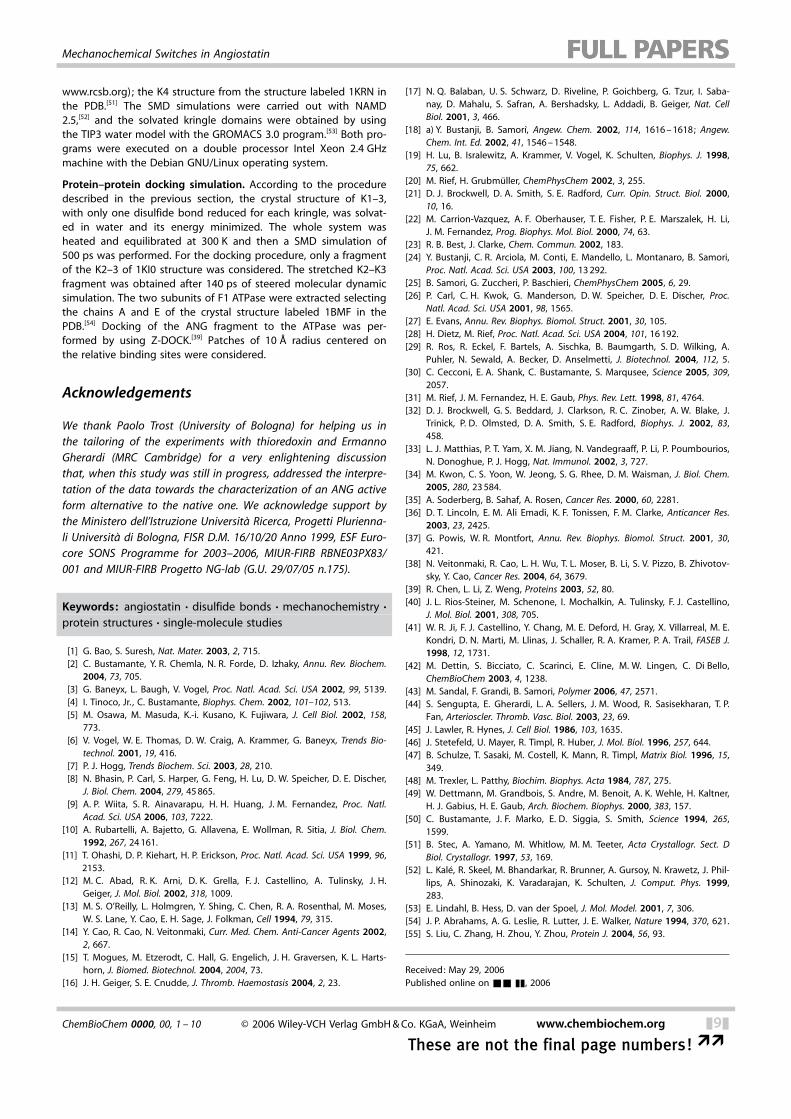
www.rcsb.org); the K4 structure from the structure labeled 1KRN inthe PDB.[51] The SMD simulations were carried out with NAMD2.5,[52] and the solvated kringle domains were obtained by usingthe TIP3 water model with the GROMACS 3.0 program.[53] Both pro-grams were executed on a double processor Intel Xeon 2.4 GHzmachine with the Debian GNU/Linux operating system.
Protein–protein docking simulation. According to the proceduredescribed in the previous section, the crystal structure of K1–3,with only one disulfide bond reduced for each kringle, was solvat-ed in water and its energy minimized. The whole system washeated and equilibrated at 300 K and then a SMD simulation of500 ps was performed. For the docking procedure, only a fragmentof the K2–3 of 1KI0 structure was considered. The stretched K2–K3fragment was obtained after 140 ps of steered molecular dynamicsimulation. The two subunits of F1 ATPase were extracted selectingthe chains A and E of the crystal structure labeled 1BMF in thePDB.[54] Docking of the ANG fragment to the ATPase was per-formed by using Z-DOCK.[39] Patches of 10 E radius centered onthe relative binding sites were considered.
Acknowledgements
We thank Paolo Trost (University of Bologna) for helping us inthe tailoring of the experiments with thioredoxin and ErmannoGherardi (MRC Cambridge) for a very enlightening discussionthat, when this study was still in progress, addressed the interpre-tation of the data towards the characterization of an ANG activeform alternative to the native one. We acknowledge support bythe Ministero dell’Istruzione UniversitB Ricerca, Progetti Plurienna-li UniversitB di Bologna, FISR D.M. 16/10/20 Anno 1999, ESF Euro-core SONS Programme for 2003–2006, MIUR-FIRB RBNE03PX83/001 and MIUR-FIRB Progetto NG-lab (G.U. 29/07/05 n.175).
Keywords: angiostatin · disulfide bonds · mechanochemistry ·protein structures · single-molecule studies
[1] G. Bao, S. Suresh, Nat. Mater. 2003, 2, 715.[2] C. Bustamante, Y. R. Chemla, N. R. Forde, D. Izhaky, Annu. Rev. Biochem.
2004, 73, 705.[3] G. Baneyx, L. Baugh, V. Vogel, Proc. Natl. Acad. Sci. USA 2002, 99, 5139.[4] I. Tinoco, Jr. , C. Bustamante, Biophys. Chem. 2002, 101–102, 513.[5] M. Osawa, M. Masuda, K.-i. Kusano, K. Fujiwara, J. Cell Biol. 2002, 158,
773.[6] V. Vogel, W. E. Thomas, D. W. Craig, A. Krammer, G. Baneyx, Trends Bio-
technol. 2001, 19, 416.[7] P. J. Hogg, Trends Biochem. Sci. 2003, 28, 210.[8] N. Bhasin, P. Carl, S. Harper, G. Feng, H. Lu, D. W. Speicher, D. E. Discher,
J. Biol. Chem. 2004, 279, 45865.[9] A. P. Wiita, S. R. Ainavarapu, H. H. Huang, J. M. Fernandez, Proc. Natl.
Acad. Sci. USA 2006, 103, 7222.[10] A. Rubartelli, A. Bajetto, G. Allavena, E. Wollman, R. Sitia, J. Biol. Chem.
1992, 267, 24161.[11] T. Ohashi, D. P. Kiehart, H. P. Erickson, Proc. Natl. Acad. Sci. USA 1999, 96,
2153.[12] M. C. Abad, R. K. Arni, D. K. Grella, F. J. Castellino, A. Tulinsky, J. H.
Geiger, J. Mol. Biol. 2002, 318, 1009.[13] M. S. O’Reilly, L. Holmgren, Y. Shing, C. Chen, R. A. Rosenthal, M. Moses,
W. S. Lane, Y. Cao, E. H. Sage, J. Folkman, Cell 1994, 79, 315.[14] Y. Cao, R. Cao, N. Veitonmaki, Curr. Med. Chem. Anti-Cancer Agents 2002,
2, 667.[15] T. Mogues, M. Etzerodt, C. Hall, G. Engelich, J. H. Graversen, K. L. Harts-
horn, J. Biomed. Biotechnol. 2004, 2004, 73.[16] J. H. Geiger, S. E. Cnudde, J. Thromb. Haemostasis 2004, 2, 23.
[17] N. Q. Balaban, U. S. Schwarz, D. Riveline, P. Goichberg, G. Tzur, I. Saba-nay, D. Mahalu, S. Safran, A. Bershadsky, L. Addadi, B. Geiger, Nat. CellBiol. 2001, 3, 466.
[18] a) Y. Bustanji, B. Samor�, Angew. Chem. 2002, 114, 1616–1618; Angew.Chem. Int. Ed. 2002, 41, 1546–1548.
[19] H. Lu, B. Isralewitz, A. Krammer, V. Vogel, K. Schulten, Biophys. J. 1998,75, 662.
[20] M. Rief, H. GrubmRller, ChemPhysChem 2002, 3, 255.[21] D. J. Brockwell, D. A. Smith, S. E. Radford, Curr. Opin. Struct. Biol. 2000,
10, 16.[22] M. Carrion-Vazquez, A. F. Oberhauser, T. E. Fisher, P. E. Marszalek, H. Li,
J. M. Fernandez, Prog. Biophys. Mol. Biol. 2000, 74, 63.[23] R. B. Best, J. Clarke, Chem. Commun. 2002, 183.[24] Y. Bustanji, C. R. Arciola, M. Conti, E. Mandello, L. Montanaro, B. Samori,
Proc. Natl. Acad. Sci. USA 2003, 100, 13292.[25] B. Samori, G. Zuccheri, P. Baschieri, ChemPhysChem 2005, 6, 29.[26] P. Carl, C. H. Kwok, G. Manderson, D. W. Speicher, D. E. Discher, Proc.
Natl. Acad. Sci. USA 2001, 98, 1565.[27] E. Evans, Annu. Rev. Biophys. Biomol. Struct. 2001, 30, 105.[28] H. Dietz, M. Rief, Proc. Natl. Acad. Sci. USA 2004, 101, 16192.[29] R. Ros, R. Eckel, F. Bartels, A. Sischka, B. Baumgarth, S. D. Wilking, A.
Puhler, N. Sewald, A. Becker, D. Anselmetti, J. Biotechnol. 2004, 112, 5.[30] C. Cecconi, E. A. Shank, C. Bustamante, S. Marqusee, Science 2005, 309,
2057.[31] M. Rief, J. M. Fernandez, H. E. Gaub, Phys. Rev. Lett. 1998, 81, 4764.[32] D. J. Brockwell, G. S. Beddard, J. Clarkson, R. C. Zinober, A. W. Blake, J.
Trinick, P. D. Olmsted, D. A. Smith, S. E. Radford, Biophys. J. 2002, 83,458.
[33] L. J. Matthias, P. T. Yam, X. M. Jiang, N. Vandegraaff, P. Li, P. Poumbourios,N. Donoghue, P. J. Hogg, Nat. Immunol. 2002, 3, 727.
[34] M. Kwon, C. S. Yoon, W. Jeong, S. G. Rhee, D. M. Waisman, J. Biol. Chem.2005, 280, 23584.
[35] A. Soderberg, B. Sahaf, A. Rosen, Cancer Res. 2000, 60, 2281.[36] D. T. Lincoln, E. M. Ali Emadi, K. F. Tonissen, F. M. Clarke, Anticancer Res.
2003, 23, 2425.[37] G. Powis, W. R. Montfort, Annu. Rev. Biophys. Biomol. Struct. 2001, 30,
421.[38] N. Veitonmaki, R. Cao, L. H. Wu, T. L. Moser, B. Li, S. V. Pizzo, B. Zhivotov-
sky, Y. Cao, Cancer Res. 2004, 64, 3679.[39] R. Chen, L. Li, Z. Weng, Proteins 2003, 52, 80.[40] J. L. Rios-Steiner, M. Schenone, I. Mochalkin, A. Tulinsky, F. J. Castellino,
J. Mol. Biol. 2001, 308, 705.[41] W. R. Ji, F. J. Castellino, Y. Chang, M. E. Deford, H. Gray, X. Villarreal, M. E.
Kondri, D. N. Marti, M. Llinas, J. Schaller, R. A. Kramer, P. A. Trail, FASEB J.1998, 12, 1731.
[42] M. Dettin, S. Bicciato, C. Scarinci, E. Cline, M. W. Lingen, C. Di Bello,ChemBioChem 2003, 4, 1238.
[43] M. Sandal, F. Grandi, B. Samori, Polymer 2006, 47, 2571.[44] S. Sengupta, E. Gherardi, L. A. Sellers, J. M. Wood, R. Sasisekharan, T. P.
Fan, Arterioscler. Thromb. Vasc. Biol. 2003, 23, 69.[45] J. Lawler, R. Hynes, J. Cell Biol. 1986, 103, 1635.[46] J. Stetefeld, U. Mayer, R. Timpl, R. Huber, J. Mol. Biol. 1996, 257, 644.[47] B. Schulze, T. Sasaki, M. Costell, K. Mann, R. Timpl, Matrix Biol. 1996, 15,
349.[48] M. Trexler, L. Patthy, Biochim. Biophys. Acta 1984, 787, 275.[49] W. Dettmann, M. Grandbois, S. Andre, M. Benoit, A. K. Wehle, H. Kaltner,
H. J. Gabius, H. E. Gaub, Arch. Biochem. Biophys. 2000, 383, 157.[50] C. Bustamante, J. F. Marko, E. D. Siggia, S. Smith, Science 1994, 265,
1599.[51] B. Stec, A. Yamano, M. Whitlow, M. M. Teeter, Acta Crystallogr. Sect. D
Biol. Crystallogr. 1997, 53, 169.[52] L. KalS, R. Skeel, M. Bhandarkar, R. Brunner, A. Gursoy, N. Krawetz, J. Phil-
lips, A. Shinozaki, K. Varadarajan, K. Schulten, J. Comput. Phys. 1999,283.
[53] E. Lindahl, B. Hess, D. van der Spoel, J. Mol. Model. 2001, 7, 306.[54] J. P. Abrahams, A. G. Leslie, R. Lutter, J. E. Walker, Nature 1994, 370, 621.[55] S. Liu, C. Zhang, H. Zhou, Y. Zhou, Protein J. 2004, 56, 93.
Received: May 29, 2006Published online on && &&, 2006
ChemBioChem 0000, 00, 1 – 10 @ 2006 Wiley-VCH Verlag GmbH&Co. KGaA, Weinheim www.chembiochem.org &9&
These are not the final page numbers! ��
Mechanochemical Switches in Angiostatin

ARTICLES
F. Grandi, M. Sandal, G. Guarguaglini,E. Capriotti, R. Casadio, B. Samor&*
&& –&&
Hierarchical MechanochemicalSwitches in Angiostatin
A novel protein signaling switch basedon the coupling of redox conditionsand mechanical stresses upon a disul-fide bond is reported. Single-moleculeexperiments and simulations on angios-tatin (a protein with antitumor activity)indicate that this switching mechanismcan control the access to partially re-duced and partially unfolded structures(see figure) ; these are proposed hereinto be the active forms of angiostatin,rather than the native one, as common-ly believed so far.
&10& www.chembiochem.org @ 2006 Wiley-VCH Verlag GmbH&Co. KGaA, Weinheim ChemBioChem 0000, 00, 1 – 10
�� These are not the final page numbers!