Hemodynamic responses on prefrontal cortex related to meditation and attentional task
13
SYSTEMS NEUROSCIENCE ORIGINAL RESEARCH ARTICLE published: 17 February 2015 doi: 10.3389/fnsys.2014.00252 Hemodynamic responses on prefrontal cortex related to meditation and attentional task Singh Deepeshwar *, Suhas Ashok Vinchurkar , Naveen Kalkuni Visweswaraiah and Hongasandra RamaRao Nagendra ANVESANA Research Laboratory, Department of Yoga and Life Sciences, Swami Vivekananda Yoga Research Foundation, Bangalore, Karnataka, India Edited by: Mikhail Lebedev, Duke University, USA Reviewed by: José M. Delgado-García, University Pablo de Olavide, Seville, Spain Laura Marzetti, University “G. d’Annunzio” of Chieti-Pescara, Italy *Correspondence: Singh Deepeshwar, ANVESANA Research Laboratory, Department of Yoga and Life Sciences, Swami Vivekananda Yoga Research Foundation, #19 Eknath Bhavan, Gavipuram Circle, K.G. Nagar, Bangalore – 560019, Karnataka, India e-mail: deepeshwar.singh@ gmail.com Recent neuroimaging studies state that meditation increases regional cerebral blood flow (rCBF) in the prefrontal cortex (PFC). The present study employed functional near infrared spectroscopy (fNIRS) to evaluate the relative hemodynamic changes in PFC during a cognitive task. Twenty-two healthy male volunteers with ages between 18 and 30 years (group mean age ± SD; 22.9 ± 4.6 years) performed a color-word stroop task before and after 20 min of meditation and random thinking. Repeated measures ANOVA was performed followed by a post hoc analysis with Bonferroni adjustment for multiple comparisons between the mean values of “During” and “Post” with “Pre” state. During meditation there was an increased in oxy-hemoglobin (1HbO) and total hemoglobin (1THC) concentration with reduced deoxy-hemoglobin (1HbR) concentration over the right prefrontal cortex (rPFC), whereas in random thinking there was increased 1HbR with reduced total hemoglobin concentration on the rPFC. The mean reaction time (RT) was shorter during stroop color word task with concomitant reduction in 1THC after meditation, suggestive of improved performance and efficiency in task related to attention. Our findings demonstrated that meditation increased cerebral oxygenation and enhanced performance, which was associated with activation of the PFC. Keywords: meditation, attention task, Stroop task, fNIRS, cerebral blood flow INTRODUCTION Meditation is a complex mental process that aims to calm the fluctuations of the mind and improve cognitive functions. Several meditation techniques from diverse traditions (e.g., Transcendental meditation, Buddhists, Zen, Yoga, Vipassana, Brahmakumari, Mindfulness-based stress reduction (MBSR) etc.,) demonstrated that regular practice of meditation develops awareness to the contents of subjective experience, including thoughts, sensations, intentions, and emotions (Saggar et al., 2012). It is considered as a voluntary means of mental training to achieve greater control of higher mental functions. Traditional yoga texts like Patanjali’s Yoga Sutras (the Sage Patanjali’, Circa 900 B.C.) and Bhagavad Gita (Circa 400–600 B.C.) very well describe the connection between meditation and mental modifications. Traditionally, two states of meditation have been described, viz., (i) focused meditation (dharana in Sanskrit, Patanjali’s Yoga Sutras, Chapter III, Verse 1), and this state is supposed to lead to the next stage of effortless mental expansion i.e., (iii) meditation (dhyana in Sanskrit; Patanjali’s Yoga Sutras, Chapter III, Verse 2). When not in meditation, it is said that the mind may be in two other states (Telles et al., 2012). These are (i) random thinking (cancalata in Sanskrit; Bhagavad Gita, chapter VI, verse 34); and (ii) non-meditative focused thinking (ekagrata in Sanskrit; Bhagavad Gita, chapter VI, verse 12) (Telles et al., 2014). In recent years, there have been a number of neuroimaging studies showing that meditation improves cognitive performance as signified by behavioral and neurophysiological measures (Tang et al., 2007; Lutz et al., 2009). Previous studies have shown that the practice of meditation enhances behavioral performance viz., perceptual discrimination and sustained attention during visual discrimination task (MacLean et al., 2010). Meditation practice develops the ability to engage the attention onto an object for extended periods of time (Carter et al., 2005; Jha et al., 2007; Lutz et al., 2008). It improves the control over the distribution of limited brain resources in the temporal domain, as measured by the attentional blink task (van Leeuwen et al., 2009; Slagter et al., 2011). Long term meditation practice has been found to enhance cognitive performance (Cahn and Polich, 2006), attentional focus, alerting (Jha et al., 2007), processing speed (Lutz et al., 2009; Slagter et al., 2009), and overall information processing (van Vugt and Jha, 2011). In a study, Buddhist meditation practitioners showed mindfulness meditation was positively correlated with sustained attention, when compared to non-meditation practitioners (Moore and Malinowski, 2009). Improvements in sustained attention and attentional error monitoring demonstrated a positive correlation with increased activation in executive attention networks in meditators (Short et al., 2010). Other studies have shown that meditation is associated with improved conflict scores on the attention network test (Tang et al., 2007), reduced interference (Chan and Woollacott, 2007) and enhanced attentional performance during the stroop task compared to meditation-naïve control Frontiers in Systems Neuroscience www.frontiersin.org February 2015 | Volume 8 | Article 252 | 1
Transcript of Hemodynamic responses on prefrontal cortex related to meditation and attentional task

SYSTEMS NEUROSCIENCEORIGINAL RESEARCH ARTICLE
published: 17 February 2015doi: 10.3389/fnsys.2014.00252
Hemodynamic responses on prefrontal cortex related tomeditation and attentional taskSingh Deepeshwar *, Suhas Ashok Vinchurkar , Naveen Kalkuni Visweswaraiah andHongasandra RamaRao Nagendra
ANVESANA Research Laboratory, Department of Yoga and Life Sciences, Swami Vivekananda Yoga Research Foundation, Bangalore, Karnataka, India
Edited by:Mikhail Lebedev, Duke University,USA
Reviewed by:José M. Delgado-García, UniversityPablo de Olavide, Seville, SpainLaura Marzetti, University “G.d’Annunzio” of Chieti-Pescara, Italy
*Correspondence:Singh Deepeshwar, ANVESANAResearch Laboratory, Departmentof Yoga and Life Sciences, SwamiVivekananda Yoga ResearchFoundation, #19 Eknath Bhavan,Gavipuram Circle, K.G. Nagar,Bangalore – 560019,Karnataka, Indiae-mail: [email protected]
Recent neuroimaging studies state that meditation increases regional cerebral bloodflow (rCBF) in the prefrontal cortex (PFC). The present study employed functional nearinfrared spectroscopy (fNIRS) to evaluate the relative hemodynamic changes in PFC duringa cognitive task. Twenty-two healthy male volunteers with ages between 18 and 30years (group mean age ± SD; 22.9 ± 4.6 years) performed a color-word stroop taskbefore and after 20 min of meditation and random thinking. Repeated measures ANOVAwas performed followed by a post hoc analysis with Bonferroni adjustment for multiplecomparisons between the mean values of “During” and “Post” with “Pre” state. Duringmeditation there was an increased in oxy-hemoglobin (1HbO) and total hemoglobin(1THC) concentration with reduced deoxy-hemoglobin (1HbR) concentration over theright prefrontal cortex (rPFC), whereas in random thinking there was increased 1HbRwith reduced total hemoglobin concentration on the rPFC. The mean reaction time (RT)was shorter during stroop color word task with concomitant reduction in 1THC aftermeditation, suggestive of improved performance and efficiency in task related to attention.Our findings demonstrated that meditation increased cerebral oxygenation and enhancedperformance, which was associated with activation of the PFC.
Keywords: meditation, attention task, Stroop task, fNIRS, cerebral blood flow
INTRODUCTIONMeditation is a complex mental process that aims to calmthe fluctuations of the mind and improve cognitive functions.Several meditation techniques from diverse traditions (e.g.,Transcendental meditation, Buddhists, Zen, Yoga, Vipassana,Brahmakumari, Mindfulness-based stress reduction (MBSR)etc.,) demonstrated that regular practice of meditation developsawareness to the contents of subjective experience, includingthoughts, sensations, intentions, and emotions (Saggar et al.,2012). It is considered as a voluntary means of mental trainingto achieve greater control of higher mental functions. Traditionalyoga texts like Patanjali’s Yoga Sutras (the Sage Patanjali’, Circa 900B.C.) and Bhagavad Gita (Circa 400–600 B.C.) very well describethe connection between meditation and mental modifications.Traditionally, two states of meditation have been described, viz.,(i) focused meditation (dharana in Sanskrit, Patanjali’s YogaSutras, Chapter III, Verse 1), and this state is supposed to lead tothe next stage of effortless mental expansion i.e., (iii) meditation(dhyana in Sanskrit; Patanjali’s Yoga Sutras, Chapter III, Verse 2).When not in meditation, it is said that the mind may be in twoother states (Telles et al., 2012). These are (i) random thinking(cancalata in Sanskrit; Bhagavad Gita, chapter VI, verse 34);and (ii) non-meditative focused thinking (ekagrata in Sanskrit;Bhagavad Gita, chapter VI, verse 12) (Telles et al., 2014).
In recent years, there have been a number of neuroimagingstudies showing that meditation improves cognitive performance
as signified by behavioral and neurophysiological measures (Tanget al., 2007; Lutz et al., 2009). Previous studies have shownthat the practice of meditation enhances behavioral performanceviz., perceptual discrimination and sustained attention duringvisual discrimination task (MacLean et al., 2010). Meditationpractice develops the ability to engage the attention onto anobject for extended periods of time (Carter et al., 2005; Jhaet al., 2007; Lutz et al., 2008). It improves the control over thedistribution of limited brain resources in the temporal domain,as measured by the attentional blink task (van Leeuwen et al.,2009; Slagter et al., 2011). Long term meditation practice has beenfound to enhance cognitive performance (Cahn and Polich, 2006),attentional focus, alerting (Jha et al., 2007), processing speed(Lutz et al., 2009; Slagter et al., 2009), and overall informationprocessing (van Vugt and Jha, 2011). In a study, Buddhistmeditation practitioners showed mindfulness meditation waspositively correlated with sustained attention, when comparedto non-meditation practitioners (Moore and Malinowski, 2009).Improvements in sustained attention and attentional errormonitoring demonstrated a positive correlation with increasedactivation in executive attention networks in meditators (Shortet al., 2010). Other studies have shown that meditation isassociated with improved conflict scores on the attentionnetwork test (Tang et al., 2007), reduced interference (Chanand Woollacott, 2007) and enhanced attentional performanceduring the stroop task compared to meditation-naïve control
Frontiers in Systems Neuroscience www.frontiersin.org February 2015 | Volume 8 | Article 252 | 1

Deepeshwar et al. Hemodynamic changes in meditation and attention
group (Moore and Malinowski, 2009). These studies providesignificant evidence of meditation promoting the higher-order cognitive processing (Zeidan et al., 2010), particularly,the features of conflict monitoring and cognitive controlprocesses.
The stroop task is one of the most frequently used modelsof the conflict processing (Sz"ucs et al., 2012) in cognitiveneuroscience. Stroop color word task performance evaluatesflexibility in the purview of cognitive processes and behaviorwhich requires both attention and impulse control. Thesimultaneous presentation of the prime color and a writtenword stimulus will either facilitate (when the color and wordstimuli are congruent, e.g., “b-l-u-e” written in the colorblue) or interfere (the incongruent stroop trial, e.g., “blue”written in red) with color naming (MacLeod, 1991; Petersonet al., 1999). Previous studies on stroop test have consistentlyshown that responses in naming the ink color of incongruentcolor word are much slower than in naming the ink colorof neutral (Zysset et al., 2007), and responses are often,but not always, faster when color and word are congruentthan in the neutral condition. It supports the hypothesisthat, both the task relevant and task irrelevant dimensionsof stroop task activate the same response in the congruentcondition, in contrast, these dimensions stimulate opposingresponse tendencies in the incongruent condition (Mortonand Chambers, 1973; Posner and Snyder, 1975; Sz"ucs et al.,2012).
Recent studies reported that regular practice of meditationmay alter brain structure and function related to attention (Lazaret al., 2005; Holzel et al., 2011; Kozasa et al., 2012). A studyon 20 experienced participants of extensive Insight meditation,that involves focused attention to internal experiences, reportedincreased cortical thickness in prefrontal cortex (PFC) andright anterior insula associated with attention, interoception andsensory processing in meditation participants compared withmatched controls (Lazar et al., 2005).
In order to examine neuronal activity and hemodynamicchanges in the brain regions during meditation, the applicationof different neuroimaging techniques (viz., fMRI and MEG)would be beneficial. The neuronal activity during meditationhas been reported in several electroencephalography (EEG)and magnetoencephalography (MEG) studies. Experiencedmeditators showed an increased EEG power in lower frequencybands (theta, delta and alpha) (Kubota et al., 2001; Takahashiet al., 2005) compared to controls. An EEG study onTranscendental Meditation, showed intermittent prominentbursts of frontally dominant theta activity at an average maximalamplitude of 135 µV in 21 practitioners (Hebert and Lehmann,1977). Zen meditators showed fast theta and slow alpha powerduring meditation (Takahashi et al., 2005) demonstratingenhanced automatic memory and reduction in conceptualthinking following meditation (Faber et al., 2014). In a singleMEG study on twelve long term Buddhist meditators wereassessed in two distinct types of self-awareness, i.e., “narrative”and “minimal” in mindfulness-induced selflessness awareness(Dor-Ziderman et al., 2013). It was found that there was areduction in gamma band (60–80 Hz) power in frontal, and
medial prefrontal areas, and reduced beta band (13–25 Hz)power in ventral medial prefrontal, medial posterior and lateralparietal regions (Dor-Ziderman et al., 2013) and right inferiorparietal lobules. These studies are consistent with fMRI and NIRSfindings. Functional magnetic resonance imaging (fMRI) posesseveral challenges such as high sensitivity to participant’s motion,a loud, restrictive environment, low temporal resolution, andrelatively high cost (Cui et al., 2011). Some of these challenges areovercome with new optical imaging technique: NIRS measure’schanges in oxy-hemoglobin and deoxy-hemoglobin (∆HbOand ∆HbR) concentration changes from the cortical surfaceand less invasive and expensive than fMRI (Bunce et al., 2006).Functional near infrared spectroscopy (fNIRS) is a compactand portable optical technique to monitor hemodynamicsof the brain in real time (Son and Yazici, 2006; Lin et al.,2009).
Brain hemodynamic responses during meditation, i.e.,∆HbO, ∆HbR and total hemoglobin changes (∆THC)are in its infancy. In fact, there is only one study thatassessed deoxyhemoglobin changes with a single wavelengthprobe placed over the left PFC during Qigong meditation(Cheng et al., 2010). Practitioners showed decrease in deoxy-hemoglobin and increase in oxy-hemoglobin concentrationthat suggest, meditation lead to left prefrontal activation duringmeditation.
With this background, the present study was designedto assess the bilateral prefrontal hemodynamic responses inmeditation and random thinking. Additionally, we investigatedthe hemodynamic changes and performance during a stroopcolor word task before and after meditation and randomthinking. Since, stroop color word task is known to measureattention, interference, processing speed, and executive attention,we expected that this task to be the most sensitive to the effects ofmeditation.
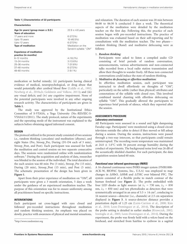
MATERIALS AND METHODSPARTICIPANTSA total of 25 right handed healthy male participants with agesranging from 19 and 30 years (Mean, SD; 23.4 ± 3.7 years)were recruited from S-VYASA (a Yoga University), South India.All participants had a minimum of 12-month experience inmeditation (group average experience ± S.D., 15.6 ± 14.2months) on the Sanskrit syllable “OM”. Three participantswere excluded from the study because of large motion artifactsin the signals due to head movements or because of failurein probe placement due to obstruction by hair (Taga et al.,2003; Minagawa-Kawai et al., 2011). Thus, only data from 22participants (mean age 22.9 ± 4.6 years) were included inthe final analysis. Participants fulfilling the following criteriawere included in the study: (i) the participants with at least12 months of meditation experience; (ii) male participantsalone were studied as cognitive abilities and cerebral bloodflow (Brackley et al., 1999) have been shown to fluctuatewhich the phases of menstrual cycle (Yadav et al., 2002);and (iii) no history of smoking; and (iv) normal healthon a routine clinical examination. Participants with followingcriteria were excluded from the study: (i) persons on any
Frontiers in Systems Neuroscience www.frontiersin.org February 2015 | Volume 8 | Article 252 | 2
Deepeshwar et al. Hemodynamic changes in meditation and attention
Table 1 | Characteristics of 22 participants.
Characteristics
Age (in years) (group mean ± S.D.) 22.9 ± 4.6 yearsYears of education17 years and more 6 (27.3%)Upto 15 years 10 (45.5%)Upto 12 years 6 (27.3%)Type of meditation Meditation on the
Sanskrit syllable “OM”Experience of meditationpractice (in months)6–12 months 4 (18.2%)13–24 months 3 (13.6%)25–36 months 7 (31.8%)37–48 months 6 (27.3%)48–60 months 2 (9.1%)
medication or herbal remedy; (ii) participants having clinicalevidence of medical, neuropsychological, or drug abuse thatwould potentially alter cerebral blood flow (Liddle et al., 1992;Newberg et al., 2010a,b; Goldstein and Volkow, 2011); and (iii)any visual deficit; and (iv) any cognitive impairment. None ofthe potential participants were involved in any other ongoingresearch activity. The characteristics of participants are given inTable 1.
The study was approved by the Institutional EthicsCommittee of S-VYASA, a Yoga University (No.-RES/IEC-S-VYASA/11/2011). The study protocol, nature of the experimentsand the operating mode of the instrument was explained to thesubjects before obtaining signed informed consent.
DESIGNThe protocol utilized in the present study consisted of two sessionsi.e., random thinking (cancalata) and meditation (dhyana), andeight States (Pre, Stroop_Pre, During (D1-D4 each of 5 min),Stroop_Post, and Post). Each participant was assessed for boththe meditation and control session on two separate consecutivedays. The sessions were randomized online with randomizationsoftware.1 During the acquisition and analysis of data, researcherwas blinded to the session of the individual. The total duration ofthe each session was 60 min: Pre (5 min), Stroop_Pre (15 min),During (20 min), Stroop_Post (15 min), and Post (5 min).The schematic presentation of the design has been given inFigure 1.
Apart from their prior experience of meditation on “OM”, allparticipants were given a 3 month orientation, 5 days a weekunder the guidance of an experienced meditation teacher. Thepurpose of this orientation was for to ensure uniformity amongall practitioners based on specific instructions.
INTERVENTIONSEach participant sat cross-legged with eyes closed andfollowed pre-recorded instructions throughout meditationand random thinking sessions. An emphasis was placed onslowly, practice with awareness of physical and mental sensations,
1http://www.randomizer.org
and relaxation. The duration of each session was 20 min between06:00 to 06:30 h conducted 5 days a week. The theoreticalaspects of the meditation were detailed by the meditationteacher on the first day. Following this, the practice of eachsession began with pre-recorded instructions. The practice ofmeditation was evaluated based on their self-reporting and byconsultations with the meditation teacher. The two phases—random thinking (Rand) and meditative defocusing were asfollows:
1. Random thinking:Participants were asked to listen a compiled audio CDconsisting of brief periods of random conversation,announcements, various advertisements and non-connectedtalks recorded from a local radio station transmission andallow their thoughts to wander freely. All these non-connectedconversations could induce the state of random thinking.
2. Meditative de-focusing or effortless meditation:In effortless meditation session, each participant wasinstructed to dwell effortlessly on thoughts of “OM”,particularly on the subtle (rather than physical) attributes andconnotations of the syllable with closed eyes. This involvedcombined mental chanting with effortless defocusing onsyllable “OM”. This gradually allowed the participants toexperience brief periods of silence, which they reported afterthe session.
ASSESSMENTS PROCEDURELaboratory environmentAll Participants were assessed in a sound and light dampeningFaraday cage. Participants’ were monitored using a closed circuittelevision outside the cabin to detect if they moved or fell asleepduring a session. During the session, instructions were passedthrough a two-way intercom, so that participants could remainuninterrupted. The recording room temperature was maintainedat 24.0 ± 1.0◦C with 56 percent average humidity during theconduct of experiments. The background noise level was 26 dB ofthe acoustically shielded chamber. For each participant, the dataacquisition session lasted 60 min.
Functional near infrared spectroscopy (fNIRS)A 16-channel continuous wave fNIRS imager system (FNIR1000-ACK-W, BIOPAC Systems, Inc., U.S.A) was employed to mapchanges in 1HbO, 1HbR and 1THC over bilateral PFC. Thesystem consisted of a flexible probe to match contour of thehuman forehead (see Figure 2). The probe embedded withfour LED diodes as light sources (at λ1 = 730 nm, λ2 = 830nm, λ3 = 850 nm) and ten photodiodes as detectors that weresymmetrically arranged in an area of 3.5 × 14 cm2, conducing to16 nearest source—detector (i.e., channels) at 2.5 cm separationdisplayed in Figure 3. A source-detector distance provides apenetration depth of 1.25 cm (León-Carrion et al., 2008; Kimet al., 2010; Leon-Dominguez et al., 2014). The description ofthe probe setting is detailed in earlier studies (Krawczyk, 2002;Izzetoglu et al., 2005; Leon-Dominguez et al., 2014). During theexperiment, the probe was firmly held with a velcro band on theforehead, and stretched from hairline to eyebrow in a sagittal
Frontiers in Systems Neuroscience www.frontiersin.org February 2015 | Volume 8 | Article 252 | 3

Deepeshwar et al. Hemodynamic changes in meditation and attention
FIGURE 1 | Schematic representation of the study design. Note: Sessions were modified for each participant D1: During 1; D2: During 2; D3: During 3; D4:During 4.
direction and from ear to ear in axial direction (Tian et al., 2009).The probes were positioned bilaterally on forehead, over the leftand right frontal poles, a part of dorsolateral PFC, and a portionof the ventrolateral PFC. Regional cerebral blood flow (rCBF),∆HbO, ∆HbR, and ∆THC for each hemisphere were updatedevery 0.5 s. The four LEDs flashed in sequence; the reflected lightfrom the brain as detected with the nearest photodiodes of eachLED and converted into digital signals using an analog-digitalconverter (ADC) card in the control box. The digital data weresent to the laptop though a serial port. The sampling rate was 3 Hzacross all 16 channels. The principles of measurement were basedon the modified Beer-Lambert law for highly scattering media(Plichta et al., 2006) that agrees assessing changes in ∆HbO and∆HbR at a certain measured point (Hoshi and Tamura, 1993).Increases in ∆HbO and corresponding decrease in ∆HbR can beinterpreted as a sign of functional brain activation.
Stroop color word taskSubjects were seated comfortably on a reclining chair in a Faradaycage, facing a 21 inch LCD monitor placed at a distance of70 cm from their eyes. Participants were required to focus onthe center of the screen which was guided by a fixation object“+” followed by stimuli. Participants did a modified multiple-trial stroop task and were confronted with neutral, congruent,and incongruent stimuli on a black background using E-Prime2.0.8.90 (Psychological Software Tools, Inc., Pittsburgh, PA, USA).The stroop color word task consisted of red, green and bluecolored boxes and the corresponding written words “RED”,“BLUE” and “GREEN”. The color was presented as color square(4.5 × 4.5 cm) boxes on a black background. The durationof the presented square boxes and words was 500 ms each.Congruent trials comprised of square color boxes followed bywords describing the color of the box written in the same color(e.g., the BLUE square box and the printed word “BLUE” in blue
FIGURE 2 | The complete setup employed is herein presented. ThefNIRS sensor is displayed with 4 light sources and 10 detectors (top) and 16optode (channel) measurement locations registered on the sensor.
ink); incongruent trials comprised of words describing the colorof the box written in a color other than that of the box (e.g., theRED square box and word RED written in blue ink); neutral trialscomprised words written in white (e.g., the BLUE square box andword BLUE printed in white ink). Participants were instructedto reply as speedily and accurately as possible to the name ofthe color word (while ignoring the color itself) consistent to thecolor of the Box with a button press of the response key usingthe thumb of their right hand. To increase the potency of theconflict stimulus, 20% of trials were congruent (approximately 45trials), 20% were incongruent (approximately 45 trials) and 50%were neutral (90 trials). The duration of the stimulus was 500 ms,with a variable interstimulus interval (ISI) of 1000–2500 ms theexperimental steps are illustrated in Figure 4.
Frontiers in Systems Neuroscience www.frontiersin.org February 2015 | Volume 8 | Article 252 | 4

Deepeshwar et al. Hemodynamic changes in meditation and attention
FIGURE 3 | The 16 fNIRS optode (channel) measurement locationsregistered on the brain surface image are presented.
FIGURE 4 | Experimental steps of Color word Stroop Task.
Data acquisitionThe participants were assessed in two separate sessions i.e.,random thinking and meditation while recording hemodynamicactivity on the PFC using 16-channel continuous wave fNIRSsystem. On the preceding day and on the day of the recording,participants were asked to avoid tea and coffee which are knownto influence cognitive performance (Nehlig, 2010) and cerebralblood flow (Addicott et al., 2009). Where this was unavoidable
the session was engaged on another day. The participants worea flexible sensor pad over prefrontal region and covered with ablack cloth. The probable artifacts such as heart rate pulsation,respiration and high frequency noise in raw data, which maypossibly be induced by autonomic arousal caused during strooptask, was eliminated with pre designed finite impulse response(FIR) filters based on type, order, window function and cut-offfrequency. For the present study, raw data were acquired fromthe probe, which is pre-filtered by two filters and processed in thedata processing unit using COBI filter module. The first filter isa 10th order low-pass filter with cutoff frequency of 0.1 Hz withBlackman window. The second filter is a 20th order low-pass,with the normalized cut-off frequency of 0.1 Hz which uses aHamming window. The filtered data were averaged according tothe tasks and conditions for further statistical analysis.
Data analysisThe hemodynamic responses of bilateral PFC were recordedand data were averaged according to the task condition (pre,stroop_pre, during, stroop_post and post). Statistical analysis hasbeen carried out on these differential values. Filtered data weretested with Kolmogorov-Smirnov test for normality. Repeatedmeasures analysis of variance (RM-ANOVA) was used becausethe same individuals were assessed in repeated sessions on twoseparate days (i.e., random thinking and meditation). RM-ANOVA was performed with three “within subjects” factors, i.e.,Factor 1: Sessions (random thinking and meditation); Factor2: PFC (right and left). Factor 3: States (“Pre”, “Stroop_Pre”,“During” (D1 to D4), “Stroop_Post” and “Post”). The repeatedmeasures ANOVAs were carried out for concentration changes ofoxygenated and deoxygenated hemoglobin and total hemoglobinchange (∆HbO, ∆HbR and ∆THbC) across the right and leftPFC. This was followed by a post hoc analysis with Bonferroniadjustment for multiple comparisons between the mean values ofdifferent states (“During” and “Post”) and all comparisons weremade with the respective “Pre” state.
Moreover, for analysis of stroop task we compared themean reaction time (ms) of neutral, congruent and incongruentconditions and hemodynamic responses of stroop color word taskbefore and after the sessions (random thinking and meditation).The results were averaged for each side of PFC (right and left),parameter and subject separately to compare between differentconditions and sessions. A repeated measures ANOVA was carriedfor multiple comparisons following Bonferroni adjustment.Statistical analyses were carried out using the Statistical softwareSPSS version 20.0 (SPSS Inc., Chicago, USA). The alpha level wasset at p< 0.05. The effect size (d) defined by Cohen (1988), as themean change score divided by the standard deviation of change,calculated for further statistical analysis.
RESULTSBEHAVIORAL RESULTSReaction times (RTs) were computed solely from thecorrectly answered trials. With respect to RT, a repeated—measures 3 way ANOVA with Sessions (random thinkingand meditation) × States (“Stroop_Pre”, “Stroop_Post”) ×
Conditions (neutral vs. congruent vs. incongruent). Repeated
Frontiers in Systems Neuroscience www.frontiersin.org February 2015 | Volume 8 | Article 252 | 5

Deepeshwar et al. Hemodynamic changes in meditation and attention
measures ANOVA demonstrated a significant main effect forSessions (F(1,21) = 4.862, p = 0.039, η2p = 0.188); Conditions(F(2,42) = 24.12, p < 0.001, η2p = 0.535); States (F(1,21) = 6.696,p < 0.023, η2p = 0.242), and the significant interaction betweenSessions × States (F(1,21) = 45.36, p< 0.001, η2p = 0.684).
Post hoc analysis revealed that there was a significantimprovement in cognitive performance after meditation in allthree conditions (neutral, congruent and incongruent) comparedto random thinking session given in Table 1. The RTs differedin all the conditions (neutral vs. congruent vs. incongruent)in both the sessions. These findings verify that our attentionalmanipulation was indeed effective.
The RTs were compared using two-tailed paired sample t-test, revealed significant differences among all three conditions(neutral, congruent and incongruent) in two different sessions(meditation and random thinking). In random thinking session,there were significant differences in neutral vs. congruent: t(21)
= −3.86, p = 0.001; congruent vs. incongruent: t(21) = −2.31,p = 0.031; neutral vs. incongruent: t(21)= −5.92, p < 0.001whereas in meditation session, there was a significant differencein neutral—congruent: t(21) = −4.47, p < 0.001; congruent—incongruent: t(21) = −1.85, p > 0.05 (NS); neutral—incongruent:t(21) = −6.148, p< 0.001. The mean RTs were significantly shorterin the neutral (p = 0.002), congruent (p < 001) and incongruent(p < 0.003) conditions after meditation session whereas afterthe random thinking session, mean RTs were delayed in theneutral (p = 0.034) and incongruent (p = 0.008) conditions.The average RTs for neutral, congruent, and incongruent trialsof the stroop color word task are given in Table 2. Subjectsmade negligible errors during the color word matching strooptask. For error rates, we did not make any statistical test, sincetheir distributions are clearly not Gaussian. However, it can besupposed that interference effect also reveals itself in error rates.In summary, behavioral results of the stroop color word taskare in accordance with the literature, as demonstrated by a clearinterference effect in the participants for meditation and randomthinking sessions.
HEMODYNAMIC RESPONSES IN STROOP COLOR WORD TASKIn the present study, the 16 channel fNIRS device provided aset of time series recorded over the PFC. The locations of theprobed regions are shown in Figure 2. The order of the channelsis from left to right, i.e., “1” is on the left and “16” is on the
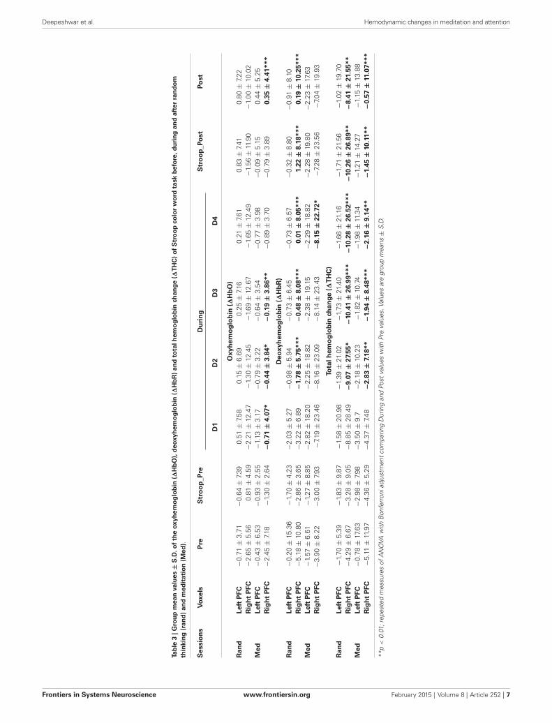
right as depicted in Figure 3. Analysis of hemoglobin signalsi.e., ∆HbO or ∆HbR is still a controversial issue, specificallywhich hemoglobin signal is more reliably associated with brainactivity still remain unclear (Schroeter et al., 2002). In thisstudy, we have utilized three wavelengths (i.e.,750, 803 and 850nm). This combination is suitable only for detecting ∆HbOsignal. Therefore we used ∆HbO, ∆HbR and ∆THC signals forstatistical analysis. The groups mean values ± S.D. for the ∆HbO,∆HbR and ∆THC in stroop task and the two sessions (randomthinking and meditation) in “Pre”, “During” and “Post” states aregiven in Table 3.
For ∆HbO, the repeated—measures ANOVA for Sessions(Random thinking and Meditation) × PFC (Left and Right)× States (“Stroop_Pre”, “Stroop_Post”) revealed no significantmain effect for Sessions, States and PFC. There was a significantinteraction between PFC × States (F(1,175) = 9.87, p < 0.01,η2p = 0.053); Sessions × PFC × States (F(1,175) = 3.17, p < 0.01,η2p = 0.040).
For ∆HbR, the repeated—measures ANOVA demonstratedsignificant main effect for Sessions (F(1,175) = 9.99, p < 0.01,η2p = 0.054); PFC (F(1,175) = 4.57, p < 0.05, η2p = 0.025).Also, there was a significant interaction between Sessions ×
PFC (F = 5.11, p < 0.05, η2p = 0.028); Sessions × States(F(1,175) = 22.13, p< 0.001, η2p = 0.112); Sessions × PFC × States(F(1,175) = 9.81, p< 0.01, η2p = 0.053).
For total hemoglobin (∆THC), the repeated—measuresANOVA revealed that there was a significant main effect forPFC (F(1,175) = 9.71, p < 0.01, η2p = 0.053), and the significantinteraction between Sessions × PFC (F(1,175) = 5.33, p < 0.01,η2p = 0.03); Sessions × States (F(1,175) = 19.87, p < 0.001,η2p = 0.102); PFC × States (F(1,175) = 5.96, p< 0.05, η2p = 0.033);Sessions × PFC × States (F(1,175) = 14.20, p< 0.001, 0.075).
The post hoc analysis with Bonferroni correctionsdemonstrated forehead hemodynamic responses during strooptask related to random thinking and meditation sessions are givenin Table 3. The results demonstrated a significant decrease in theconcentration of ∆HbO in left PFC (p = 0.016) and in the rightPFC (p = 0.032) after random thinking session during stroopcolor word task, whereas, there was a significant improvementin ∆HbO in left PFC (p = 0.006) and right PFC (p = 0.046)following the meditation session.
From the above observations, it can be concluded thatmeditation enhances bilaterally activation of the anterior PFC
Table 2 | Group mean values ± S.D. of the reaction time scores (ms) of Stroop color word Task.
Sessions States Pre Post t-value P value % Change
Rand Neutral 643.18 ± 130.654 660.00 ± 113.641 −2.274 0.034* 2.62Congruent 783.64 ± 117.333 790.91 ± 119.440 −0.876 0.391 0.93Incongruent 871.41 ± 136.070 892.73 ± 136.004 −2.920 0.008** 2.45
Med Neutral 638.64 ± 118.615 617.73 ± 121.653 3.533 0.002** −3.27Congruent 794.55 ± 118.029 764.55 ± 112.238 6.205 <0.001*** −3.78Incongruent 865.00 ± 137.797 819.09 ± 133.627 3.302 0.003** −5.31
*p < 0.05; p < **0.01; ***p < 0.001; repeated measures of ANOVA with Bonferroni adjustment comparing Post values with Pre values. Values are group means ±
S.D. Rand—Random Thinking; Med—Meditation.
Frontiers in Systems Neuroscience www.frontiersin.org February 2015 | Volume 8 | Article 252 | 6
Deepeshwar et al. Hemodynamic changes in meditation and attention
Tab
le3
|Gro
up
mea
nva
lues
±S
.D.o
fth
eox
yhem
og
lob
in(∆
Hb
O),
deo
xyh
emo
glo
bin
(∆H
bR
)an
dto
talh
emo
glo
bin
chan
ge(∆
TH
C)
of
Str
oo
pco
lor
wo
rdta
skb
efo
re,d
uri
ng
and
afte
rra
nd
om
thin
kin
g(r
and
)an
dm
edit
atio
n(M
ed).
Ses
sio
ns
Vox
els
Pre
Str
oo
p_P
reD
uri
ng
Str
oo
p_P
ost
Post
D1
D2
D3
D4
Oxy
hem
og
lob
in(∆
Hb
O)
Ran
dLe
ftP
FC−
0.71
±3.
71−
0.64
±7.
390.
51±
7.58
0.15
±6.
690.
25±
7.16
0.21
±7.
610.
83±
7.41
0.80
±7.
22R
igh
tP
FC−
2.65
±5.
560.
81±
4.59
−2.
21±
12.4
7−
1.30
±12
.45
−1.
69±
12.6
7−
1.65
±12
.49
−1.
56±
11.9
0−
1.00
±10
.02
Med
Left
PFC
−0.
43±
6.53
−0.
93±
2.55
−1.
13±
3.17
−0.
79±
3.22
−0.
64±
3.54
−0.
77±
3.98
−0.
09±
5.15
0.44
±5.
25R
igh
tP
FC−
2.45
±7.
18−
1.30
±2.
64−
0.71
±4.
07*
−0.
44±
3.84
*−
0.19
±3.
86**
−0.
89±
3.70
−0.
79±
3.89
0.35
±4.
41**
*
Deo
xyh
emo
glo
bin
(∆H
bR
)R
and
Left
PFC
−0.
20±
15.3
6−
1.70
±4.
23−
2.03
±5.
27−
0.98
±5.
94−
0.73
±6.
45−
0.73
±6.
57−
0.32
±8.
80−
0.91
±8.
10R
igh
tP
FC−
5.18
±10
.80
−2.
86±
3.65
−3.
22±
6.89
−1.
78±
5.75
***
−0.
48±
8.08
***
0.01
±8.
05**
*1.
22±
8.18
***
0.19
±10
.25*
**M
edLe
ftP
FC−
1.57
±6.
61−
1.27
±8.
85−
2.82
±18
.20
−2.
25±
18.8
2−
2.38
±19
.15
−2.
29±
18.8
2−
2.28
±19
.80
−2.
23±
17.6
3R
igh
tP
FC−
3.90
±8.
22−
3.00
±7.
93−
7.19
±23
.46
−8.
16±
23.0
9−
8.14
±23
.43
−8.
15±
22.7
2*−
7.28
±23
.56
−7.
04±
19.9
3
Tota
lhem
og
lob
inch
ange
(∆T
HC
)R
and
Left
PFC
−1.
70±
5.39
−1.
83±
9.87
−1.
58±
20.9
8−
1.39
±21
.02
−1.
73±
21.4
0−
1.66
±21
.16
−1.
71±
21.5
6−
1.02
±19
.70
Rig
ht
PFC
−4.
29±
6.67
−3.
28±
9.05
−8.
85±
28.4
9−
9.07
±27
.55*
−10
.41
±26
.99*
**−
10.2
8±
26.5
2***
−10
.26
±26
.89*
*−
8.41
±21
.55*
*M
edLe
ftP
FC−
0.78
±17
.63
−2.
98±
7.98
−3.
50±
9.7
−2.
18±
10.2
3−
1.82
±10
.74
−1.
98±
11.3
4−
1.21
±14
.27
−1.
15±
13.8
8R
igh
tP
FC−
5.11
±11
.97
−4.
36±
5.29
−4.
37±
7.48
−2.
83±
7.18
**−
1.94
±8.
48**
*−
2.16
±9.
14**
−1.
45±
10.1
1**
−0.
57±
11.0
7***
**p<
0.01
;rep
eate
dm
easu
res
ofA
NO
VAw
ithB
onfe
rron
iadj
ustm
ent
com
parin
gD
urin
gan
dPo
stva
lues
with
Pre
valu
es.V
alue
sar
egr
oup
mea
ns±
S.D
.
Frontiers in Systems Neuroscience www.frontiersin.org February 2015 | Volume 8 | Article 252 | 7

Deepeshwar et al. Hemodynamic changes in meditation and attention
and consequently, a stronger increase of oxygenation and cerebralblood flow during stroop task at the right PFC due to interferencereduction.
HEMODYNAMICS RESPONSES IN MEDITATION AND RANDOMTHINKINGFor ∆HbO, the repeated—measures ANOVA for Sessions(Random thinking and Meditation) × PFC (Left and Right) ×
States (Pre Stroop_Pre, D1-D4, Stroop_Post, Post) demonstrateda significant main effects for States (F(7,1225) = 5.23, p < 0.001,η2p = 0.029). There was a significant interaction between the PFC× States (F(7,1225) = 2.42, p < 0.001, η2p = 0.014); Sessions ×
Hemispheres × States (F(7,1225) = 7.32, p< 0.05, η2p = 0.040).For ∆HbO, the repeated—measures ANOVA showed there
was a significant main effect for Sessions (F(1,175) = 12.20,p < 0.001, η2p = 0.065); PFC (F(1,175) = 7.89, p < 0.01,η2p = 0.043) and States (F(7,1225) = 3.55, p < 0.001, η2p = 0.019).There was a significant interaction between the Sessions × PFC(F(1,175) = 4.13, p < 0.001, η2p = 0.023); Sessions × States(F(7,1225) = 9.99, p< 0.001, η2p = 0.054); Sessions × PFC × States(F(7,1225) = 10.37, p< 0.001, η2p = 0.056).
For total hemoglobin change (∆THC), there was a significantmain effect for Sessions (F(1,175) = 5.07, p < 0.05, η2p = 0.028);PFC (F(1,175) = 12.20, p < 0.001, η2p = 0.065); and States(F(1,175) = 2.79, p < 0.01, η2p = 0.016) and a significantinteraction between the Sessions × PFC (F(1,175) = 6.45, p< 0.05,η2p = 0.036); Sessions × States (F(7,1225) = 9.06, p < 0.001,η2p = 0.049); PFC × States (F(7,1225) = 2.34, p < 0.05,η2p = 0.036]; Session × PFC × State (F(7,1225) = 14.51, p< 0.001).
Post hoc analyses with Bonferroni corrections were performedon ∆HbO, ∆HbR and ∆THC and all comparisons were madewith respective “Pre” state. These have been summarized inTable 3. There was a significant increase in ∆HbR at the rightPFC (p = 0.005) after random thinking session whereas there wasa significant increase in the left PFC (p = 0.02) and in right PFC(p < 0.001) after meditation session. Similarly, in ∆THC, therewas a significant decrease in blood flow change in the right PFC(p < 0.001) after the random thinking session whereas there wasa significant increase in blood flow change in the left (p = 0.03)and in right PFC (p< 0.001) after meditation session.
In summary, as described in Table 3 and in Line diagrams(Figures 5–7), there was a positive trend to show a significantincrease in the concentration of oxyhemoglobin change (∆HbO)during meditation session at right PFC (as shown in Figure 5).There was a significant decrease in deoxyhemoglobin change(∆HbR) (as shown in Figure 6) during meditation sessionwhereas there was a significant increase in the concentration ofdeoxyhemoglobin change during random thinking session at theright PFC. Additionally, there was also a significant increase in thetotal hemoglobin change (∆THC) during and after meditationsessions (Figure 7) and decrease in the total hemoglobin change(∆THC) during and after random thinking session.
DISCUSSIONThe primary goal of the present study was to ascertain whethermeditation increases rCBF at bilateral PFC, measured withfNIRS, compared to random thinking. Our secondary goal was
to observe the RT scores and relative changes in cerebral bloodflow, and to determine if there are persistent effects followingmeditation session compared to random thinking session.Results as confirmed with recent studies on meditation withspectroscopy (Cheng et al., 2010), SPECT imaging (Newberget al., 2001, 2010a,b; Cohen et al., 2009) and fMRI (Short et al.,2010; Guleria et al., 2013; Zeidan et al., 2014) have revealed thatmeditation program resulted in significant increases in baselineCBF ratios in the prefrontal, superior, inferior and orbital frontalcortex, dorsolateral prefrontal cortex (DLPFC), right dorsalmedial frontal lobe, cingulate gyrus and right sensorimotorcortex. In present study, we found that brain activation, measuredby changes in ∆HbO and ∆THC concentration in the rightprefrontal area was followed by a strong decrease in ∆HbRconcentration during meditation. Additionally, the rCBFsignificantly increased in the right frontal lobe during strooptask after meditation, which suggest the improvement in theparticipant’s performance (reaction time) during the task. Thetotal blood oxygenation (∆THC) level in the PFC could risewith increasing task load from neutral to congruent, and thenincongruent; this would demonstrate a positive correlation withperformance measures. The changes in regional blood flow ismediated by changes in neural activity in a single region or inseveral selective regions of the brain (Lauritzen, 2001).
Earlier studies have demonstrated that the PFC is activatedparticularly on the right PFC and anterior cingulate cortex (ACC)in willful act and tasks that require intense focused and sustainedattention (Frith et al., 1991; Pardo et al., 1991; Vogt et al., 1992;Petersen and Posner, 2012). A study on eight Tibetan Buddhistmeditators demonstrated improved activity in the PFC bilaterally(though greater on the right hemisphere) and the cingulate gyrusduring meditation (Newberg and Iversen, 2003). This suggeststhat meditation begins with activation of the PFC and anteriorcingulate gyrus associated with the will or intent to clear the mindof thoughts or to focus on an object (Edwards et al., 2012).
Meditation increases CBF and decreases cerebrovascularresistance (CVR) suggesting a contributing vascular mechanism(Jevning et al., 1996) which reflect cerebral activation. TheCVR reduction being associated with cognitive improvementwhich suggests a vascular contribution to cognitive enhancement(Nation et al., 2013). During meditation, the activation of rightPFC is theoretically associated with the activity in the reticularnucleus of the thalamus. This activation may be accomplishedby the PFC’s production and distribution of glutamate, a knownexcitatory neurotransmission (Cheramy et al., 1987; Finkbeiner,1987), which communicate with other brain structures such aslateral geniculate and lateral posterior nuclei of the thalamus(Portas et al., 1998). An early study on meditation with singlephoton emission computed tomography (SPECT) demonstrateda general increase in thalamic activity that was proportional tothe activity levels in the PFC (Newberg et al., 2001; Edwards et al.,2012). The activation on the right PFC causes increased activityin the reticular nucleus during meditation, the results may bedecreased sensory input entering into the posterior superiorparietal lobule which is involved in the analysis and integrationof higher order visual, auditory, and somesthetic information(Adair et al., 1995).
Frontiers in Systems Neuroscience www.frontiersin.org February 2015 | Volume 8 | Article 252 | 8

Deepeshwar et al. Hemodynamic changes in meditation and attention
FIGURE 5 | Line graph represents averaged Oxy-hemoglobinchange at right prefrontal cortex (rPFC) in two sessions i.e.,random thinking and meditation and Stroop task. Note: Linegraph represents comparisons between baseline, stroop_pre, during
sessions (random thinking and meditation), stroop_post, and post.Stroop Pre showed higher Oxy-hemoglobin change compared tobaseline. During and after meditation, the cerebral oxygenation washigher in rPFC compared to random thinking.
FIGURE 6 | Line graph represents averaged Deoxy-hemoglobin change atright PFC in two sessions i.e., random thinking and meditation andStroop task. Note: Line graph represents de-oxyhemoglobin changes was
higher in right PFC during random thinking (D2, D3, and D4), stroop task andafter random thinking. In other hand, during meditation, there was a decreasein de-oxyhemoglobin in D3 level in rPFC.
A major strength of the present study was to examine thestates of meditation and random thinking related hemodynamicresponses in cerebral oxygenation during performance of thestroop color word task. It is a well established phenomenon thatexecutive processes are facilitated by the frontal lobe and due tostroop interference brain activity may depend on increased abilityto recruit frontal neural resources (Schroeter et al., 2004b). Thisallowed us to examine whether there is an increase in oxygenationwith meditation corresponding to an ability to recruit appropriateresources for task performance or a decrease in activationcorresponding to better optimization and possible reduction intask difficulty with meditation. In a study, fNIRS showed stroopinterference is consistently associated with the ACC and thelateral prefrontal cortex (LPFC), especially the DLPFC, wherethe ACC is considered to be susceptible to conflict, and theDLPFC is purported to implement cognitive control (Carter
et al., 2000; Leung et al., 2000). DLPFC may involve attentionalmaintenance while ACC monitors performance (MacDonaldet al., 2000). Another similar study suggested meditation mayenhance specific subcomponents of attention such as conflictmonitoring or performance (Jha et al., 2007). Although fNIRScannot monitor the cortical activation in the ACC becauseits measurement is limited to lateral cortical surfaces, it hassuccessfully monitored the activation of the LPFC associated withstroop interference (Schroeter et al., 2002, 2003, 2004a,b; Ehliset al., 2005).
There have been several neuroimaging studies evaluating thecerebral blood flow and performance of different meditationpractices using behavioral, EEG and (Carter et al., 2005) fMRIimaging. Previous studies on meditation and EEG reported,greater midline theta power and slow alpha power in thefrontal area during meditation (Takahashi et al., 2005; Chan
Frontiers in Systems Neuroscience www.frontiersin.org February 2015 | Volume 8 | Article 252 | 9

Deepeshwar et al. Hemodynamic changes in meditation and attention
FIGURE 7 | Line graph represents averaged total hemoglobin change atrPFC in two sessions i.e., random thinking and meditation and Strooptask. Note: Line graph represents total hemoglobin change was higher in
rPFC during meditation (D2, D3, and D4), in stroop task, and in post session.In other hand, there was a decrease in rPFC during random thinking (D2, D3,and D4), in stroop task and in post session.
et al., 2008). Zazen meditation showed increased alpha-1 andalpha-2 frequency activity of EEG in right prefrontal areasincluding insula, parts of the somatosensory, motor corticesand temporal areas (Faber et al., 2014). A subsequent study,on Satyananda Yoga meditation practice, showed greater sourceactivity in low frequencies (particularly theta and alpha 1) duringmental calculation, body-steadiness and mantra meditation(Thomas et al., 2014). Additionally, body-steadiness and mantrameditation showed greatest activity in right side of superiorfrontal and precentral gyri, parietal and occipital lobes. Similarly,neuroimaging studies on meditation practice, when comparedto the control session showed significantly increased oxy-hemoglobin and CBF in the medial PFC which was associatedwith the intense focus-based component of the practice (Wanget al., 2011). Meditation involves attentional regulation and leadsto increased activity in brain regions associated with attentionsuch as DLPFC and ACC. The long-term practitioners hadsignificantly more consistent and sustained activation in theDLPFC and the ACC during meditation vs. control in comparisonto short-term practitioners (Baron Short et al., 2010). Thesestudies suggest that willful acts and tasks that require sustainedattention are initiated via activity in the PFC, particularly in theright hemisphere (Posner and Petersen, 1990; Frith et al., 1991;Pardo et al., 1991; Ingvar, 1994). Meditation requires focus ofattention on objects which thereby activates PFC, particularly inthe right hemisphere (Cohen et al., 2009), as well as the cingulategyrus (Herzog et al., 1990; Lazar et al., 2000; Newberg et al.,2001). This demonstrated that during meditation there was anincreased activity in the PFC bilaterally (greater on the right)and the cingulate gyrus (Newberg and Iversen, 2003). Therefore,the process of meditation seems to happen by activation of theprefrontal and cingulate cortex which are associated with thewill or intent to clear one’s mind of thoughts or to focus on anobject.
In other imaging studies on meditation, there have beeninconsistent results regarding the frontal cortex. A recent studyshowed decreased frontal activity during externally guided word
generation compared to internal or volitional word generation(Cross et al., 2012). Thus, prefrontal and cingulate activationmay be associated with the volitional aspects of meditation.Meditation with fluorodeoxyglucose (FDG) PET in eight subjectsundergoing Yoga meditative relaxation (Herzog et al., 1990)reported increased rCBF in the frontal: occipital ratio of cerebralmetabolism. Specifically, there was a mild increase in the frontallobe, but marked decreases in metabolism in the occipital andsuperior parietal lobes. In addition to these studies, the PFCis reported to have a crucial role in social cognitive skillsand along with the cingulate gyrus governs social behaviortasks related to Theory of Mind, empathy, moral reasoning,and evaluation of emotional states (Declerck et al., 2006). ThePFC is essential for flexible behavior because it inhibits thehabitual responses that have become inappropriate (Mesulam,1998). But, an increase in the activity of PFC (determined byfNIRS) is not necessarily beneficial always. For example, animalexperimentation has shown that the electrical activation of themedial PFC prevent the proper sequence of pressing the leverand collecting the reward (a pellet of food) in an operantcondition task (Cross et al., 2012; Jurado-Parras et al., 2012)and also prevent the expression of an already acquired classicallyconditioned eyelid response (Leal-Campanario et al., 2007, 2013).However, in our study we infer that activation of prefrontalcortices after meditation had beneficial effects on cognitionas manifested by improved performance in stroop color wordtask.
The present study reported increased oxy-hemoglobinconcentration because of enhanced neural activity and cerebralblood flow in the prefrontal area during meditation comparedto random thinking. In such studies, it is very important tounderstand the influences of systemic artifacts such as thosefrom the heart, breathing, superficial perfusion, etc., which maybe induced by the cognitive tasks related stress and autonomicresponses. For example, a recent study performed on peripheralphysiological measurements with temporal correlations offNIRS and fMRI signals concluded that the physiological
Frontiers in Systems Neuroscience www.frontiersin.org February 2015 | Volume 8 | Article 252 | 10

Deepeshwar et al. Hemodynamic changes in meditation and attention
basis of the systemic artifact is a task-evoked sympatheticarterial vasoconstriction monitored by a decrease in venousvolume and these artifacts are fairly common (Kirilina et al.,2012). They also suggested that the separation of fNIRS signalsoriginating from activated brain and from scalp is a necessaryprecondition for unbiased fNIRS brain activation maps andpre-processing of the raw data using high definition filters isnecessary.
In summary, the results of the present study provided firstevidence that the oxygenation levels are increased in the PFCduring meditation compared with random thinking in the samepractitioners. Further event-related NIRS studies may apply well-tested fMRI paradigms in studies with children and patients,utilizing the advantages of the method.
ACKNOWLEDGMENTSThis research work was supported by the Center for AdvancedResearch in Yoga and Neurophysiology, Swami Vivekananda YogaResearch Foundation, Bangalore, India. We are gateful to Dr.Shirley Telles, Dr. Manjunath N.K. and Dr. Hemant Bhargav fortheir support and guidelines with regards to data acquisition andanalysis.
REFERENCESAdair, J. C., Gilmore, R. L., Fennell, E. B., Gold, M., and Heilman, K. M.
(1995). Anosognosia during intracarotid barbiturate anesthesia: unawareness oramnesia for weakness. Neurology 45, 241–243. doi: 10.1212/wnl.45.2.241
Addicott, M. A., Yang, L. L., Peiffer, A. M., Burnett, L. R., Burdette, J. H., Chen,M. Y., et al. (2009). The effect of daily caffeine use on cerebral blood flow:how much caffeine can we tolerate? Hum. Brain Mapp. 30, 3102–3114. doi: 10.1002/hbm.20732
Baron Short, E., Kose, S., Mu, Q., Borckardt, J., Newberg, A., George, M. S., et al.(2010). Regional brain activation during meditation shows time and practiceeffects: an exploratory FMRI study. Evid. Based Complement. Alternat. Med. 7,121–127. doi: 10.1093/ecam/nem163
Brackley, K. J., Ramsay, M. M., Broughton Pipkin, F., and Rubin, P. C. (1999).The effect of the menstrual cycle on human cerebral blood flow: studies usingDoppler ultrasound. Ultrasound Obstet. Gynecol. 14, 52–57. doi: 10.1046/j.1469-0705.1999.14010052.x
Bunce, S., Izzetoglu, M., Izzetoglu, K., Onaral, B., and Pourrezaei, K. (2006).Functional near infrared spectroscopy: an emerging neuroimaging modality.IEEE Eng. Med. Biol. Mag. Spec. Issue Clin. Neuroengineering 25, 54–62. doi: 10.1109/MEMB.2006.1657788
Cahn, B. R., and Polich, J. (2006). Meditation states and traits: EEG, ERP andneuroimaging studies. Psychol. Bull. 132, 180–211. doi: 10.1037/0033-2909.132.2.180
Carter, C. S., Macdonald, A. M., Botvinick, M., Ross, L. L., Stenger, V. A., Noll, D.,et al. (2000). Parsing executive processes: strategic vs. evaluative functions of theanterior cingulate cortex. Proc. Natl. Acad. Sci. U S A 97, 1944–1948. doi: 10.1073/pnas.97.4.1944
Carter, O. L., Presti, D. E., Callistemon, C., Ungerer, Y., Liu, G. B., and Pettigrew,J. D. (2005). Meditation alters perceptual rivalry in Tibetan Buddhist monks.Curr. Biol. 15, R412–R413. doi: 10.1016/j.cub.2005.05.043
Chan, A. S., Han, Y. M. Y., and Cheung, M.-C. (2008). Electroencephalographic(EEG) measurements of mindfulness-based Triarchic body-pathway relaxationtechnique: a pilot study. Appl. Psychophysiol. Biofeedback 33, 39–47. doi: 10.1007/s10484-008-9050-5
Chan, D., and Woollacott, M. (2007). Effects of level of meditation experienceon attentional focus: is the efficiency of executive or orientation networksimproved? J. Altern. Complement. Med. 13, 651–657. doi: 10.1089/acm.2007.7022
Cheng, R. W. F., Borrett, D. S., Cheng, W., Kwan, H. C., and Cheng,R. S. S. (2010). Human prefrontal cortical response to the meditative state: a
spectroscopy study. Int. J. Neurosci. 120, 483–488. doi: 10.3109/00207454.2010.483650
Cheramy, A., Romo, R., and Glowinski, J. (1987). “Role of corticostriatalglutamatergic neurons in the presynaptic control of dopamine release,” inNeurotransmitter Interactions in the Basal Ganglia, eds M. Sandler, C. Feuersteinand B. Scatton (New York: Raven), 131–133.
Cohen, J. (1988). Statistical Power Analysis for the Behavioral Sciences. 2nd Edn.Hillsdale, NJ: Erlbaum.
Cohen, D. L., Wintering, N., Tolles, V., Townsend, R. R., Farrar, J. T., Galantino,M. L., et al. (2009). Cerebral blood flow effects of yoga training: preliminaryevaluation of 4 cases. J. Altern. Complement. Med. 15, 9–14. doi: 10.1089/acm.2008.0008
Cross, L., Brown, M. W., Aggleton, J. P., and Warburton, E. C. (2012). Themedial dorsal thalamic nucleus and the medial prefrontal cortex of the ratfunction together to support associative recognition and recency but not itemrecognition. Learn. Mem. 20, 41–50. doi: 10.1101/lm.028266.112
Cui, X., Bray, S., Bryant, D. M., Glover, G. H., and Reiss, A. L. (2011). A quantitativecomparison of NIRS and fMRI across multiple cognitive tasks. Neuroimage 54,2808–2821. doi: 10.1016/j.neuroimage.2010.10.069
Declerck, C. H., Boone, C., and De Brabander, B. (2006). On feeling in control: abiological theory for individual differences in control perception. Brain Cogn.62, 143–176. doi: 10.1016/j.bandc.2006.04.004
Dor-Ziderman, Y., Berkovich-Ohana, A., Glicksohn, J., and Goldstein, A. (2013).Mindfulness-induced selflessness: a MEG neurophenomenological study. Front.Hum. Neurosci. 7:582. doi: 10.3389/fnhum.2013.00582
Edwards, J., Peres, J., Monti, D., and Newberg, A. (2012). “The neurobiologicalcorrelates of meditation and mindfulness,” in Exploring Frontiers of the Mind-Brain Relationship SE - 6 Mindfulness in Behavioral Health, eds A. Moreira-Almeida and F. Santana Santos (New York: Springer), 97–112.
Ehlis, A.-C., Herrmann, M. J., Wagener, A., and Fallgatter, A. J. (2005). Multi-channel near-infrared spectroscopy detects specific inferior-frontal activationduring incongruent Stroop trials. Biol. Psychol. 69, 315–331. doi: 10.1016/j.biopsycho.2004.09.003
Faber, P. L., Lehmann, D., Gianotti, L. R. R., Milz, P., Pascual-Marqui, R. D., Held,M., et al. (2014). Zazen meditation and no-task resting EEG compared withLORETA intracortical source localization. Cogn. Process. doi: 10.1007/s10339-014-0637-x. [Epub ahead of print].
Finkbeiner, S. M. (1987). Neurotransmitter interactions in the basal ganglia. Yale J.Biol. Med. 60:483.
Frith, C. D., Friston, K. J., Liddle, P. F., and Frackowiak, R. S. (1991). Willed actionand the prefrontal cortex in man: a study with PET. Proc. Biol. Sci. 244, 241–246.doi: 10.1098/rspb.1991.0077
Goldstein, R. Z., and Volkow, N. D. (2011). Dysfunction of the prefrontal cortex inaddiction: neuroimaging findings and clinical implications. Nat. Rev. Neurosci.12, 652–669. doi: 10.1038/nrn3119
Guleria, A., Kumar, U., Kishan, S. S. K., and Khetrapal, C. L. (2013). Effect of“SOHAM” meditation on the human brain: an fMRI study. Psychiatry Res. 214,462–465. doi: 10.1016/j.pscychresns.2013.06.012
Hebert, R., and Lehmann, D. (1977). Theta bursts: an EEG pattern in normalsubjects practising the transcendental meditation technique. Electroencephalogr.Clin. Neurophysiol. 42, 397–405. doi: 10.1016/0013-4694(77)90176-6
Herzog, H., Lele, V. R., Kuwert, T., Langen, K. J., Rota Kops, E., and Feinendegen,L. E. (1990). Changed pattern of regional glucose metabolism during yogameditative relaxation. Neuropsychobiology 23, 182–187. doi: 10.1159/000119450
Holzel, B. K., Lazar, S. W., Gard, T., Schuman-Olivier, Z., Vago, D. R., and Ott,U. (2011). How does mindfulness meditation work? Proposing mechanisms ofaction from a conceptual and neural perspective. Perspect. Psychol. Sci. 6, 537–559. doi: 10.1177/1745691611419671
Hoshi, Y., and Tamura, M. (1993). Dynamic multichannel near-infrared opticalimaging of human brain activity. J. Appl. Physiol. (1985) 75, 1842–1846.
Ingvar, D. H. (1994). The will of the brain: cerebral correlates of willful acts. J.Theor. Biol. 171, 7–12. doi: 10.1006/jtbi.1994.1206
Izzetoglu, M., Izzetoglu, K., Bunce, S., Ayaz, H., Devaraj, A., Onaral, B., et al. (2005).Functional near-infrared neuroimaging. IEEE Trans. Neural Syst. Rehabil. Eng.13, 153–159. doi: 10.1109/TNSRE.2005.847377
Jevning, R., Anand, R., Biedebach, M., and Fernando, G. (1996). Effects on regionalcerebral blood flow of transcendental meditation. Physiol. Behav. 59, 399–402.doi: 10.1016/0031-9384(95)02006-3
Frontiers in Systems Neuroscience www.frontiersin.org February 2015 | Volume 8 | Article 252 | 11

Deepeshwar et al. Hemodynamic changes in meditation and attention
Jha, A. P., Krompinger, J., and Baime, M. J. (2007). Mindfulness training modifiessubsystems of attention. Cogn. Affect. Behav. Neurosci. 7, 109–119. doi: 10.3758/cabn.7.2.109
Jurado-Parras, M. T., Gruart, A., and Delgado-García, J. M. (2012). Observationallearning in mice can be prevented by medial prefrontal cortex stimulation andenhanced by nucleus accumbens stimulation. Learn. Mem. 19, 99–106. doi: 10.1101/lm.024760.111
Kim, M. N., Durduran, T., Frangos, S., Edlow, B. L., Buckley, E. M., Moss,H. E., et al. (2010). Noninvasive measurement of cerebral blood flow andblood oxygenation using near-infrared and diffuse correlation spectroscopies incritically brain-injured adults. Neurocrit. Care 12, 173–180. doi: 10.1007/s12028-009-9305-x
Kirilina, E., Jelzow, A., Heine, A., Niessing, M., Wabnitz, H., Brühl, R., et al. (2012).The physiological origin of task-evoked systemic artefacts in functional nearinfrared spectroscopy. Neuroimage 61, 70–81. doi: 10.1016/j.neuroimage.2012.02.074
Kozasa, E. H., Sato, J. R., Lacerda, S. S., Barreiros, M. A. M., Radvany, J., Russell,T. A., et al. (2012). Meditation training increases brain efficiency in an attentiontask. Neuroimage 59, 745–749. doi: 10.1016/j.neuroimage.2011.06.088
Krawczyk, D. C. (2002). Contributions of the prefrontal cortex to the neuralbasis of human decision making. Neurosci. Biobehav. Rev. 26, 631–664. doi: 10.1016/s0149-7634(02)00021-0
Kubota, Y., Sato, W., Toichi, M., Murai, T., Okada, T., Hayashi, A., et al. (2001).Frontal midline theta rhythm is correlated with cardiac autonomic activitiesduring the performance of an attention demanding meditation procedure. BrainRes. Cogn. Brain Res. 11, 281–287. doi: 10.1016/s0926-6410(00)00086-0
Lauritzen, M. (2001). Relationship of spikes, synaptic activity and local changesof cerebral blood flow. J. Cereb. Blood Flow Metab. 21, 1367–1383. doi: 10.1097/00004647-200112000-00001
Lazar, S. W., Bush, G., Gollub, R. L., Fricchione, G. L., Khalsa, G., and Benson, H.(2000). Functional brain mapping of the relaxation response and meditation.Neuroreport 11, 1581–1585. doi: 10.1097/00001756-200005150-00041
Lazar, S. W., Kerr, C. E., Wasserman, R. H., Gray, J. R., Greve, D. N., Treadway,M. T., et al. (2005). Meditation experience is associated with increasedcortical thickness. Neuroreport 28, 1893–1897. doi: 10.1097/01.wnr.0000186598.66243.19
Leal-Campanario, R., Delgado-García, J. M., and Gruart, A. (2013). The rostralmedial prefrontal cortex regulates the expression of conditioned eyelid responsesin behaving rabbits. J. Neurosci. 33, 4378–4386. doi: 10.1523/jneurosci.5560-12.2013
Leal-Campanario, R., Fairén, A., Delgado-García, J. M., and Gruart, A. (2007).Electrical stimulation of the rostral medial prefrontal cortex in rabbits inhibitsthe expression of conditioned eyelid responses but not their acquisition. Proc.Natl. Acad. Sci. U S A 104, 11459–11464. doi: 10.1073/pnas.0704548104
León-Carrion, J., Damas-López, J., Martín-Rodríguez, J. F., Domínguez-Roldán,J. M., Murillo-Cabezas, F., Barroso Y Martin, J. M., et al. (2008). Thehemodynamics of cognitive control: the level of concentration of oxygenatedhemoglobin in the superior prefrontal cortex varies as a function of performancein a modified Stroop task . Behav. Brain Res. 193, 248–256. doi: 10.1016/j.bbr.2008.06.013
Leon-Dominguez, U., Izzetoglu, M., Leon-Carrion, J., Solís-Marcos, I., Garcia-Torrado, F. J., Forastero-Rodríguez, A., et al. (2014). Molecular concentration ofdeoxyHb in human prefrontal cortex predicts the emergence and suppression ofconsciousness. Neuroimage 85(Pt. 1), 616–625. doi: 10.1016/j.neuroimage.2013.07.023
Leung, H. C., Skudlarski, P., Gatenby, J. C., Peterson, B. S., and Gore, J. C. (2000).An event-related functional MRI study of the stroop color word interferencetask. Cereb. Cortex 10, 552–560. doi: 10.1093/cercor/10.6.552
Liddle, P. F., Friston, K. J., Frith, C. D., Hirsch, S. R., Jones, T., and Frackowiak, R. S.(1992). Patterns of cerebral blood flow in schizophrenia. Br. J. Psychiatry 160,179–186. doi: 10.1192/bjp.160.2.179
Lin, P. Y., Lin, S. I., Penney, T., and Chen, J. J. J. (2009). Review: applications of nearinfrared spectroscopy and imaging for motor rehabilitation in stroke patients.Time 29, 210–221.
Lutz, A., Slagter, H. A., Dunne, J. D., and Davidson, R. J. (2008). Attentionregulation and monitoring in meditation. Trends Cogn. Sci. 12, 163–169. doi: 10.1016/j.tics.2008.01.005
Lutz, A., Slagter, H. A., Rawlings, N. B., Francis, A. D., Greischar, L. L., andDavidson, R. J. (2009). Mental training enhances attentional stability: neural
and behavioral evidence. J. Neurosci. 29, 13418–13427. doi: 10.1523/jneurosci.1614-09.2009
MacDonald, A. W., Cohen, J. D., Stenger, V. A., and Carter, C. S. (2000).Dissociating the role of the dorsolateral prefrontal and anterior cingulate cortexin cognitive control. Science 288, 1835–1838. doi: 10.1126/science.288.5472.1835
MacLean, K. A., Ferrer, E., Aichele, S. R., Bridwell, D. A., Zanesco, A. P.,Jacobs, T. L., et al. (2010). Intensive meditation training improves perceptualdiscrimination and sustained attention. Psychol. Sci. 21, 829–839. doi: 10.1177/0956797610371339
MacLeod, C. M. (1991). Half a century of research on the Stroop effect:an integrative review. Psychol. Bull. 109, 163–203. doi: 10.1037//0033-2909.109.2.163
Mesulam, M. (1998). From sensation to cognition. Brain 121, 1013–1052. doi: 10.1093/brain/121.6.1013
Minagawa-Kawai, Y., van der Lely, H., Ramus, F., Sato, Y., Mazuka, R., and Dupoux,E. (2011). Optical brain imaging reveals general auditory and language-specificprocessing in early infant development. Cereb. Cortex 21, 254–261. doi: 10.1093/cercor/bhq082
Moore, A., and Malinowski, P. (2009). Meditation, mindfulness and cognitiveflexibility. Conscious. Cogn. 18, 176–186. doi: 10.1016/j.concog.2008.12.008
Morton, J., and Chambers, S. M. (1973). Selective attention to words and colours.Q. J. Exp. Psychol. 25, 387–397. doi: 10.1080/14640747308400360
Nation, D., Clark, L., Wierenga, C., Delano-Wood, L., Dev, S., Bangen, K., et al.(2013). Cerebrovascular resistance and cognitive decline: modulating effects ofage. Neurology 80, P07.135.
Nehlig, A. (2010). Is caffeine a cognitive enhancer? J. Alzheimers Dis. 20(Suppl. 1),S85–S94. doi: 10.3233/JAD-2010-091315
Newberg, A., Alavi, A., Baime, M., Pourdehnad, M., Santanna, J., and d’Aquili, E.(2001). The measurement of regional cerebral blood flow during the complexcognitive task of meditation: a preliminary SPECT study. Psychiatry Res. 106,113–122. doi: 10.1016/s0925-4927(01)00074-9
Newberg, A. B., and Iversen, J. (2003). The neural basis of the complex mentaltask of meditation: neurotransmitter and neurochemical considerations. Med.Hypotheses 61, 282–291. doi: 10.1016/s0306-9877(03)00175-0
Newberg, A. B., Wintering, N., Khalsa, D. S., Roggenkamp, H., and Waldman, M. R.(2010a). Meditation effects on cognitive function and cerebral blood flow insubjects with memory loss: a preliminary study. J. Alzheimers Dis. 20, 517–526.doi: 10.3233/JAD-2010-1391
Newberg, A. B., Wintering, N., Waldman, M. R., Amen, D., Khalsa, D. S., and Alavi,A. (2010b). Cerebral blood flow differences between long-term meditators andnon-meditators. Conscious. Cogn. 19, 899–905. doi: 10.1016/j.concog.2010.05.003
Pardo, J. V., Fox, P. T., and Raichle, M. E. (1991). Localization of a human systemfor sustained attention by positron emission tomography. Nature 349, 61–64.doi: 10.1038/349061a0
Petersen, S. E., and Posner, M. I. (2012). The attention system of the human brain:20 years after. Annu. Rev. Neurosci. 35, 73–89. doi: 10.1146/annurev-neuro-062111-150525
Peterson, B. S., Skudlarski, P., Gatenby, J. C., Zhang, H., Anderson, A. W., and Gore,J. C. (1999). An fMRI study of Stroop word-color interference: evidence forcingulate subregions subserving multiple distributed attentional systems. Biol.Psychiatry 45, 1237–1258. doi: 10.1016/s0006-3223(99)00056-6
Plichta, M. M., Herrmann, M. J., Baehne, C. G., Ehlis, A.-C., Richter, M. M., Pauli,P., et al. (2006). Event-related functional near-infrared spectroscopy (fNIRS): arethe measurements reliable? Neuroimage 31, 116–124. doi: 10.1016/j.neuroimage.2005.12.008
Portas, C. M., Rees, G., Howseman, A. M., Josephs, O., Turner, R., and Frith, C. D.(1998). A specific role for the thalamus in mediating the interaction of attentionand arousal in humans. J. Neurosci. 18, 8979–8989.
Posner, M. I., and Petersen, S. E. (1990). The attention system of the human brain.Annu. Rev. Neurosci. 13, 25–42. doi: 10.1146/annurev.ne.13.030190.000325
Posner, M. I., and Snyder, C. R. R. (1975). “Attention and cognitive control,” inInformation Processing and Cognition: The Loyola Symposium, ed R. L. SolSo(Hillsdale, NJ: Erlbaum, Lawrence Associates, Inc), 55–85.
Saggar, M., King, B. G., Zanesco, A. P., Maclean, K. A., Aichele, S. R., Jacobs,T. L., et al. (2012). Intensive training induces longitudinal changes in meditationstate-related EEG oscillatory activity. Front Hum Neurosci 6:256. doi: 10.3389/fnhum.2012.00256
Frontiers in Systems Neuroscience www.frontiersin.org February 2015 | Volume 8 | Article 252 | 12

Deepeshwar et al. Hemodynamic changes in meditation and attention
Schroeter, M. L., Zysset, S., Kruggel, F., and von Cramon, D. Y. (2003).Age dependency of the hemodynamic response as measured by functionalnear-infrared spectroscopy. Neuroimage 19, 555–564. doi: 10.1016/s1053-8119(03)00155-1
Schroeter, M. L., Zysset, S., Kupka, T., Kruggel, F., and Yves von Cramon, D.(2002). Near-infrared spectroscopy can detect brain activity during a color-wordmatching Stroop task in an event-related design. Hum. Brain Mapp. 17, 61–71.doi: 10.1002/hbm.10052
Schroeter, M. L., Zysset, S., and Von Cramon, D. Y. (2004a). Shortening intertrialintervals in event-related cognitive studies with near-infrared spectroscopy.Neuroimage 22, 341–346. doi: 10.1016/j.neuroimage.2003.12.041
Schroeter, M. L., Zysset, S., Wahl, M., and von Cramon, D. Y. (2004b).Prefrontal activation due to Stroop interference increases during development—an event-related fNIRS study. Neuroimage 23, 1317–1325. doi: 10.1016/j.neuroimage.2004.08.001
Short, E. B., Kose, S., Mu, Q., Borckardt, J., Newberg, A., George, M. S., et al. (2010).Regional brain activation during meditation shows time and practice effects:an exploratory FMRI study. Evid. Based Complement. Altern. Med. 7, 121–127.doi: 10.1093/ecam/nem163
Slagter, H. A., Davidson, R. J., and Lutz, A. (2011). Mental training as a tool inthe neuroscientific study of brain and cognitive plasticity. Front. Hum. Neurosci.5:17. doi: 10.3389/fnhum.2011.00017
Slagter, H. A., Lutz, A., Greischar, L. L., Nieuwenhuis, S., and Davidson, R. J. (2009).Theta phase synchrony and conscious target perception: impact of intensivemental training. J. Cogn. Neurosci. 21, 1536–1549. doi: 10.1162/jocn.2009.21125
Son, I.-Y., and Yazici, B. (2006). Near infrared imaging and spectroscopy for brainactivity monitoring. Adv. Sens. with Secur. Appl. 2, 341–372. doi: 10.1007/1-4020-4295-7_15
Sz "ucs, D., Killikelly, C., and Cutini, S. (2012). Event-related near-infraredspectroscopy detects conflict in the motor cortex in a Stroop task. Brain Res.1477, 27–36. doi: 10.1016/j.brainres.2012.08.023
Taga, G., Asakawa, K., Maki, A., Konishi, Y., and Koizumi, H. (2003). Brain imagingin awake infants by near-infrared optical topography. Proc. Natl. Acad. Sci. U S A100, 10722–10727. doi: 10.1073/pnas.1932552100
Takahashi, T., Murata, T., Hamada, T., Omori, M., Kosaka, H., Kikuchi, M., et al.(2005). Changes in EEG and autonomic nervous activity during meditationand their association with personality traits. Int. J. Psychophysiol. 55, 199–207.doi: 10.1016/j.ijpsycho.2004.07.004
Tang, Y.-Y., Ma, Y., Wang, J., Fan, Y., Feng, S., Lu, Q., et al. (2007). Short-termmeditation training improves attention and self-regulation. Proc. Natl. Acad. Sci.U S A 104, 17152–17156. doi: 10.1073/pnas.0707678104
Telles, S., Deepeshwar, S., Naveen, K. V., and Pailoor, S. (2014). Long latencyauditory evoked potentials during meditation. Clin. EEG Neurosci. doi: 10.1177/1550059414544737. [Epub ahead of print].
Telles, S., Raghavendra, B. R., Naveen, K. V., Manjunath, N. K., and Subramanya, P.(2012). Mid-latency auditory evoked potentials in 2 meditative states. Clin. EEGNeurosci. 43, 154–160. doi: 10.1177/1550059412439963
Thomas, J., Jamieson, G., and Cohen, M. (2014). Low and then high frequencyoscillations of distinct right cortical networks are progressively enhanced by
medium and long term Satyananda Yoga meditation practice. Front. Hum.Neurosci. 8:197. doi: 10.3389/fnhum.2014.00197
Tian, F., Chance, B., and Liu, H. (2009). Investigation of the prefrontal cortexin response to duration-variable anagram tasks using functional near-infraredspectroscopy. J. Biomed. Opt. 14:054016. doi: 10.1117/1.3241984
van Leeuwen, S., Müller, N. G., and Melloni, L. (2009). Age effects on attentionalblink performance in meditation. Conscious. Cogn. 18, 593–599. doi: 10.1016/j.concog.2009.05.001
van Vugt, M. K., and Jha, A. P. (2011). Investigating the impact of mindfulnessmeditation training on working memory: a mathematical modeling approach.Cogn. Affect. Behav. Neurosci. 11, 344–353. doi: 10.3758/s13415-011-0048-8
Vogt, B. A., Finch, D. M., and Olson, C. R. (1992). Functional heterogeneity incingulate cortex: the anterior executive and posterior evaluative regions. Cereb.Cortex 2, 435–443. doi: 10.1093/cercor/2.6.435-a
Wang, D. J. J., Rao, H., Korczykowski, M., Wintering, N., Pluta, J., Khalsa, D. S.,et al. (2011). Cerebral blood flow changes associated with different meditationpractices and perceived depth of meditation. Psychiatry Res. 191, 60–67. doi: 10.1016/j.pscychresns.2010.09.011
Yadav, A., Tandon, O. P., and Vaney, N. (2002). Auditory evoked responsesduring different phases of menstrual cycle. Indian J. Physiol. Pharmacol. 46,449–456.
Zeidan, F., Johnson, S. K., Diamond, B. J., David, Z., and Goolkasian, P. (2010).Mindfulness meditation improves cognition: evidence of brief mental training.Conscious. Cogn. 19, 597–605. doi: 10.1016/j.concog.2010.03.014
Zeidan, F., Martucci, K. T., Kraft, R. A., McHaffie, J. G., and Coghill, R. C. (2014).Neural correlates of mindfulness meditation-related anxiety relief. Soc. Cogn.Affect. Neurosci. 9, 751–759. doi: 10.1093/scan/nst041
Zysset, S., Schroeter, M. L., Neumann, J., and von Cramon, D. Y. (2007).Stroop interference, hemodynamic response and aging: an event-relatedfMRI study. Neurobiol. Aging 28, 937–946. doi: 10.1016/j.neurobiolaging.2006.05.008
Conflict of Interest Statement: The authors declare that the research was conductedin the absence of any commercial or financial relationships that could be construedas a potential conflict of interest.
Received: 01 November 2014; accepted: 22 December 2014; published online: 17February 2015.Citation: Deepeshwar S, Vinchurkar SA, Visweswaraiah NK and Nagendra HR (2015)Hemodynamic responses on prefrontal cortex related to meditation and attentionaltask. Front. Syst. Neurosci. 8:252. doi: 10.3389/fnsys.2014.00252This article was submitted to the journal Frontiers in Systems Neuroscience.Copyright © 2015 Deepeshwar, Vinchurkar, Visweswaraiah and Nagendra. Thisis an open-access article distributed under the terms of the Creative CommonsAttribution License (CC BY). The use, distribution and reproduction in other forumsis permitted, provided the original author(s) or licensor are credited and that theoriginal publication in this journal is cited, in accordance with accepted academicpractice. No use, distribution or reproduction is permitted which does not comply withthese terms.
Frontiers in Systems Neuroscience www.frontiersin.org February 2015 | Volume 8 | Article 252 | 13