Gut Microbiota and Complications of Type-2 Diabetes - MDPI
30
Citation: Iatcu, C.O.; Steen, A.; Covasa, M. Gut Microbiota and Complications of Type-2 Diabetes. Nutrients 2022, 14, 166. https:// doi.org/10.3390/nu14010166 Academic Editors: Laura Soldati and Luigi Barrea Received: 7 December 2021 Accepted: 29 December 2021 Published: 30 December 2021 Publisher’s Note: MDPI stays neutral with regard to jurisdictional claims in published maps and institutional affil- iations. Copyright: © 2021 by the authors. Licensee MDPI, Basel, Switzerland. This article is an open access article distributed under the terms and conditions of the Creative Commons Attribution (CC BY) license (https:// creativecommons.org/licenses/by/ 4.0/). nutrients Review Gut Microbiota and Complications of Type-2 Diabetes Camelia Oana Iatcu 1,2 , Aimee Steen 3 and Mihai Covasa 1,3, * 1 College of Medicine and Biological Sciences, Stefan cel Mare University of Suceava, 720229 Suceava, Romania; [email protected] 2 College of Medicine, “Grigore T. Popa” University of Medicine and Pharmacy, 700115 Iasi, Romania 3 Department of Basic Medical Sciences, College of Osteopathic Medicine, Western University of Health Sciences, Pomona, CA 91766, USA; [email protected] * Correspondence: [email protected] Abstract: The gut microbiota has been linked to the emergence of obesity, metabolic syndrome and the onset of type 2 diabetes through decreased glucose tolerance and insulin resistance. Uncontrolled diabetes can lead to serious health consequences such as impaired kidney function, blindness, stroke, myocardial infarction and lower limb amputation. Despite a variety of treatments currently available, cases of diabetes and resulting complications are on the rise. One promising new approach to diabetes focuses on modulating the gut microbiota with probiotics, prebiotics, synbiotics and fecal microbial transplantation. Differences in gut microbiota composition have been observed in preclinical animal models as well as patients with type 2 diabetes and complications such as diabetic nephropathy, dia- betic retinopathy, diabetic neuropathy, cerebrovascular disease, coronary heart disease and peripheral artery disease compared to healthy controls. Severity of gut microbiota dysbiosis was associated with disease severity and restoration with probiotic administration in animal models and human patients has been associated with improvement of symptoms and disease progression. Characterizing the gut microbiota dysbiosis in different diseases and determining a causal relationship between the gut microbiota and disease can be beneficial in formulating therapeutic interventions for type 2 diabetes and associated complications. In this review, we present the most important findings regarding the role of the gut microbiota in type 2 diabetes and chronic complications as well as their underlying mechanisms. Keywords: gut microbiota dysbiosis; diabetes complications; retinopathies; nephropathies; microvas- cular complications; macrovascular complications 1. Introduction The gut microbiota is a complex ecosystem made up of a community of microorgan- isms that include trillions of bacteria spanning at least 1000 different species [1]. The gut microbiota is predominantly composed of bacteria but also contains other commensals such as archaea, viruses, fungi and protists [2]. All of these components are both relevant and important in understanding the relationship between the gut microbiota and the host. Dysbiosis of the gut microbiota is primarily characterized by decreased diversity and abundance of bacteria and fungi, especially those associated with dysfunction and various pathologies [3]. Chief among them are cardiovascular, neuronal, immune and metabolic disorders [4] through the influence of bile acid metabolism, inflammatory status, insulin resistance and incretin secretion. This can lead to the emergence of obesity [5], metabolic syndrome and the onset of type 2 diabetes [6,7] through decreased glucose tolerance and insulin resistance [8]. The gut microbiota is an important player in chronic systemic inflammation secondary to endotoxemia caused by the release of endotoxins following bacterial death [9]. While the link between the gut microbiota and the onset and progression of diabetes is still under investigation, several studies to date have focused on the pathophysiology of diabetes, with few of them investigating the role of the gut Nutrients 2022, 14, 166. https://doi.org/10.3390/nu14010166 https://www.mdpi.com/journal/nutrients
-
Upload
khangminh22 -
Category
Documents
-
view
3 -
download
0
Transcript of Gut Microbiota and Complications of Type-2 Diabetes - MDPI
�����������������
Citation: Iatcu, C.O.; Steen, A.;
Covasa, M. Gut Microbiota and
Complications of Type-2 Diabetes.
Nutrients 2022, 14, 166. https://
doi.org/10.3390/nu14010166
Academic Editors: Laura Soldati and
Luigi Barrea
Received: 7 December 2021
Accepted: 29 December 2021
Published: 30 December 2021
Publisher’s Note: MDPI stays neutral
with regard to jurisdictional claims in
published maps and institutional affil-
iations.
Copyright: © 2021 by the authors.
Licensee MDPI, Basel, Switzerland.
This article is an open access article
distributed under the terms and
conditions of the Creative Commons
Attribution (CC BY) license (https://
creativecommons.org/licenses/by/
4.0/).
nutrients
Review
Gut Microbiota and Complications of Type-2 DiabetesCamelia Oana Iatcu 1,2, Aimee Steen 3 and Mihai Covasa 1,3,*
1 College of Medicine and Biological Sciences, Stefan cel Mare University of Suceava, 720229 Suceava, Romania;[email protected]
2 College of Medicine, “Grigore T. Popa” University of Medicine and Pharmacy, 700115 Iasi, Romania3 Department of Basic Medical Sciences, College of Osteopathic Medicine, Western University of
Health Sciences, Pomona, CA 91766, USA; [email protected]* Correspondence: [email protected]
Abstract: The gut microbiota has been linked to the emergence of obesity, metabolic syndrome andthe onset of type 2 diabetes through decreased glucose tolerance and insulin resistance. Uncontrolleddiabetes can lead to serious health consequences such as impaired kidney function, blindness, stroke,myocardial infarction and lower limb amputation. Despite a variety of treatments currently available,cases of diabetes and resulting complications are on the rise. One promising new approach to diabetesfocuses on modulating the gut microbiota with probiotics, prebiotics, synbiotics and fecal microbialtransplantation. Differences in gut microbiota composition have been observed in preclinical animalmodels as well as patients with type 2 diabetes and complications such as diabetic nephropathy, dia-betic retinopathy, diabetic neuropathy, cerebrovascular disease, coronary heart disease and peripheralartery disease compared to healthy controls. Severity of gut microbiota dysbiosis was associatedwith disease severity and restoration with probiotic administration in animal models and humanpatients has been associated with improvement of symptoms and disease progression. Characterizingthe gut microbiota dysbiosis in different diseases and determining a causal relationship betweenthe gut microbiota and disease can be beneficial in formulating therapeutic interventions for type2 diabetes and associated complications. In this review, we present the most important findingsregarding the role of the gut microbiota in type 2 diabetes and chronic complications as well as theirunderlying mechanisms.
Keywords: gut microbiota dysbiosis; diabetes complications; retinopathies; nephropathies; microvas-cular complications; macrovascular complications
1. Introduction
The gut microbiota is a complex ecosystem made up of a community of microorgan-isms that include trillions of bacteria spanning at least 1000 different species [1]. The gutmicrobiota is predominantly composed of bacteria but also contains other commensalssuch as archaea, viruses, fungi and protists [2]. All of these components are both relevantand important in understanding the relationship between the gut microbiota and the host.
Dysbiosis of the gut microbiota is primarily characterized by decreased diversityand abundance of bacteria and fungi, especially those associated with dysfunction andvarious pathologies [3]. Chief among them are cardiovascular, neuronal, immune andmetabolic disorders [4] through the influence of bile acid metabolism, inflammatory status,insulin resistance and incretin secretion. This can lead to the emergence of obesity [5],metabolic syndrome and the onset of type 2 diabetes [6,7] through decreased glucosetolerance and insulin resistance [8]. The gut microbiota is an important player in chronicsystemic inflammation secondary to endotoxemia caused by the release of endotoxinsfollowing bacterial death [9]. While the link between the gut microbiota and the onset andprogression of diabetes is still under investigation, several studies to date have focusedon the pathophysiology of diabetes, with few of them investigating the role of the gut
Nutrients 2022, 14, 166. https://doi.org/10.3390/nu14010166 https://www.mdpi.com/journal/nutrients

Nutrients 2022, 14, 166 2 of 30
microbiota in diabetes complications. This review summarizes the most important findingsregarding the role of the gut microbiota in type 2 diabetes and describes its role on potentialpathways that lead to chronic complications of diabetes. Modulation of the gut microbiotathrough the use of prebiotics, probiotics, synbiotics and fecal microbiota transplantation torestore metabolic deficits associated with these pathologies is also discussed.
2. Gut Microbiota, Type 2 Diabetes and Its Complications
Type 2 diabetes, like cardiovascular disease, cancer and chronic respiratory disease,is considered a chronic and noncommunicable disease responsible for 80% of prematuredeaths globally [10]. As of 2019, there were approximately 463 million cases of diabetesworldwide with an estimated 700 million by the year 2045 if current trends continue despitethe variety of pharmacological interventions currently available [11].
Diabetes is characterized by high blood sugar levels that occur as a result of decreasedpancreatic insulin production or decreased insulin sensitivity in tissues that typicallyrespond to insulin signaling [12]. Poorly controlled diabetes and metabolic disordersassociated with type 2 diabetes such as impaired lipid metabolism, the presence of ox-idative stress and hypertension [13] can lead to both microvascular and macrovascularcomplications. Some microvascular complications of type 2 diabetes that involve smallblood vessels include diabetic nephropathy, diabetic neuropathy and diabetic retinopathy.Conversely, common macrovascular complications that involve large blood vessels includecerebrovascular disease, coronary heart disease and peripheral vascular disease [14]. Othermacrovascular complications of poorly controlled diabetes include congestive heart failure,impaired lipid metabolism, stroke, organ inflammation, weight gain, peripheral vasculardisease and electrolyte imbalance [15].
Changes in interdependent metabolic pathways have also been observed in associationwith type 2 diabetes [16]. For example, coronary heart disease caused by impaired insulinmetabolism can lead to dyslipidemia which is a risk factor for cardiovascular complicationsof diabetes [17]. Other specific factors known to contribute to the progression of diabetescomplications include increased reactive oxygen species (ROS), chronic hyperglycemia anddecreased antioxidant status [18]. The presence of these complications also leads to anoverall decline in quality of life and an increase in mortality rate [19].
A plethora of studies have demonstrated a significant association between changesin the composition profile of gut microbiota and development of diabetes. In particular,perturbed Bacteroidetes/Firmicutes phylum eubiosis has been linked with increased intestinalpermeability, with infiltration of bacteria byproducts through a leaky gut barrier triggeringsubsequent inflammatory responses characteristic of diabetes. On the other hand, severalbacteria have been shown to exert a protective role by decreasing the risk of diabetes devel-opment through reduction in proinflammatory markers and maintaining intestinal barrierintegrity. For example, Lactobacillus fermentum, plantarum and casei, Roseburia intestinalis,Akkermansia muciniphila and Bacteroides fragilis have all been shown to improve glucosemetabolism and insulin sensitivity, and suppress proinflammatory cytokines. Notably,some drugs such as metformin which is commonly used for diabetes treatment have alsobeen shown to alter the composition of the gut microbiota, suggesting that metformin inter-acts with the gut microbiota through modulation of inflammation, glucose homeostasis,gut permeability and short-chain fatty acid-producing bacteria [20]. Additionally, in pa-tients with diabetes-associated gut dysbiosis, metformin promotes butyrate and propionateproduction, improving a patient’s ability to catabolize amino acids [21]. These changescoupled with increased levels of Akkermansia in the gut may contribute to the effects ofmetformin on glucose metabolism [22]. It appears that the metabolic factors associated withchronic low-grade inflammation and oxidative stress, which link gut microbiota dysbiosisand type 2 diabetes, are the same ones that influence the onset and progression of diabeticcomplications [23,24]. This relationship gives credence to the concept that modulationof the gut microbiota may be a promising strategy in the management of diabetes andassociated complications as presented in the following sections.
Nutrients 2022, 14, 166 3 of 30
2.1. Gut Microbiota in Diabetic Nephropathy
Diabetic nephropathy occurs in approximately 40% of patients with poorly man-aged diabetes [25], of which approximately 20% are hemodialysis patients [26], leadingto end-stage renal disease, as well as cardiovascular complications [27]. Recent increasesin the number of diabetic nephropathy and end-stage renal disease cases have been at-tributed to modern societal habits and lifestyle risks associated with diabetes and hyper-tension [28,29]. Similarly, increased stress on the kidneys due to hyperglycemia can leadto diabetic nephropathy as well as associated systemic inflammation, micro and macroalbuminuria and proteinuria [30,31]. In addition, other factors such as genetics, age, obe-sity, high blood pressure and dyslipidemia [32,33] all have been shown to contribute tothe progression of diabetic nephropathy. More recently, however, several studies haveshown that dysbiosis of the gut microbiota can play a role in the development of chronickidney disease [34]. In particular, the products of bacterial metabolism have been shown toinfluence the occurrence and progression of chronic kidney disease [35] while progressionto renal failure lead to worsening of gut microbiota dysbiosis [36].
For example, the composition of the gut microbiota differs in both animals and peoplewith chronic kidney disease. In studies using animals and humans with chronic kidneydisease there was a decrease in the proportion of Bifidobacterium [37], Bactemides [38] andLactobacillus [36]. Moreover, in patients with chronic kidney disease a decrease in theproportion of Prevotella [39,40], Ruminococcaceae, Roseburia, Faecalibacterium [40] and anincrease in the proportion of Parabacteroides [39], Enterococcus [40], Enterobacteriaceae [36]and Klebsiella [40] have been reported. The increased proportions of Bacterioidaceae andClostridiaceae in patients with chronic kidney disease have been associated with systemicinflammation [41]. In contrast, bacteria such as Lactobacillaceae, Prevotellaceae and Bifidobac-teriacea [36], that are associated with anti-inflammatory effects and protecting intestinalbarrier integrity were less represented in patients with chronic kidney disease [41]. Ingeneral, patients with chronic kidney disease show a decreased proportion of anaerobicbacteria [42]. Furthermore, bacterial DNA was present in the blood of 20% of patients withchronic end-stage renal disease who were not on dialysis. In these patients, the same bacte-rial genus was detected in their intestines along with increased biomarkers of low-gradeinflammation [43].
Given that the imbalance of the gut microbiota influences many chronic diseasesincluding type 2 diabetes and its complications, it follows that balancing the compositionof the gut microbiota could be a strategy for controlling or even preventing disease. Somestudies have analyzed the effects of probiotics [42,44] or synbiotics in modulating the gutmicrobiota in patients with chronic kidney disease [45,46]. In a clinical trial conducted inpatients with stage 3 and stage 4 chronic kidney disease, urea nitrogen in the blood anduric acid concentration decreased after administering a mixture of Lactobacillus acidophilus,Streptococcus thermophilus and Bifidobacterium longum for six months [42]. In a similarstudy, the level of uric nitrogen in the blood also decreased after administration of dairyproducts containing Lactobacillus for two months [47]. When the effects of probiotics weretested in patients with chronic kidney disease on dialysis, administration of Lactobacillusacidophilus improved blood levels of dimethylamine and nitrodimethylamine [48], as well aslowered the level of dimethylamine and nitrosodimethylamine, a known carcinogen [49]. Asummary of results of clinical trials examining the effects of probiotic intake in patients withtype 2 diabetes and kidney disease is presented in Table 1. Thus, research has delineatedspecific changes in the gut microbiome associated with diabetic nephropathy as wellas physiologic mechanisms underlying changes resulting from probiotic or symbioticsupplementation in patients with diabetic nephropathy.

Nutrients 2022, 14, 166 4 of 30
Table 1. Effects of probiotics on type 2 diabetes and kidney disease.
Reference Design ProbioticSource Probiotic Dose, CFU Study Period
(wk/d) Effects
[50] RD, DB, CT tablet
L. acidophilus strainZT-L1,
B. bifidum strain ZT-B1,L. reuteri strain ZT-Lre,
L. fermentum strainZT-L3
8 × 10⁹ CFU/d
12 wk
S↓ FG, I, HOMA-IR, TG,VLDL, TC/HDL-C ratio,
hs-CRP, MDA, AGEs,BUN, creatinine, urine
proteinS↑ QUICKI, HDL-C,
GSH, CG= HbA1c, LDL-C, NO,
TAC
[51] RD, DB, CT soy milk L. plantarum A7 8 wk
S↓ albuminuria, serumcreatinine, serum
interleukin-18, serumsialic acid
S improvment inestimated GFR
[52] RD, DB, CT capsuleL. acidophilus
L. caseiB. bifium
12 wk
S↓ FG, I, HOMA-IR,HbA1c, hs-CRP, MDA,
SGA score, TIBCS↑ QUICKI
=HOMA-B, TG, VLDL,CT, LDL-C, HDL-C, NO,
TAC, GSH, GFR,creatinine, BUN,albumin, Na, K
[53] RD, DB, CT honeyBacillus coagulans T4
(IBRC-N10791)108 CFU/g
12 wk
S↓ I, HOMA-IR,CT/HDL-C ratio,
hs-CRP hs-CRP, MDA,creatinine
S↑ QUICKI=FG, TG, VLDL, CT,LDL-C, HDL-C, NO,
TAC, GSH, BUN
[54] RD, DB, CT soy milk L. plantarum A72 × 107 CFU/mL 8 wk S↓ Cys-C, PGRN, NGAL
=sTNFR1
[55] RD, DB, CT soy milkL plantarum A7 (KC
355240, LA7)2 × 10⁷ CFUmL
8 wk
S↑ Glutathione,Glutathione peroxidase,Glutathione reductase
S↓ Oxidized glutathione=MDA, 8-iso-PGF2a,
TAC
RD, randomized; DB, double-blind; CT, clinical trial; T2DM, type 2 diabetes mellitus; L., Lactobacillus, B.,Bifidobacterium; CFU, colony-forming units; wk, weeks; d, days; FG, fasting glucose fasting blood glucose,fasting plasma glucose, glycemia, fasting blood sugar; HbA1c, hemoglobin A1c; I, serum insulin concentration,insulin concentration, serum insulin level, insulin; HOMA-IR, homeostasis model of assessment-estimated insulinresistance; QUICKI, quantitative insulin sensitivity check index; TG, triglycerides; VLDL, very-low-densitylipoprotein; TC, total cholesterol; HDL-C, high-density lipoprotein; LDL-C, low-density lipoprotein; hs-CRP,high-sensitivity C-reactive protein; NO, nitric oxide; TAC, total antioxidant capacity; GSH, total glutathione; MDA,malondialdehyde; AGEs, advanced glycation end products; BUN, blood urea nitrogen; CG, Cockcroft–Gaultformula to estimate creatinine clearance; HOMA-B, homeostasis model of assessment–estimated b-cell function;GFR, glomerular filtration rate; SGA, subjective global assessment; TIBC, total iron binding capacity; Na, sodium;K, potassium; Cys-C, cystatin C; PGRN, Progranulin; NGAL, neutrophil gelatinase-associated lipocalin; sTNFR1,soluble tumor necrosis factor receptor 1; 8-iso-PGF2a, 8-iso-prostaglandin F2 alpha; =, non significan; S, significant;↑, increase; ↓, decrease.

Nutrients 2022, 14, 166 5 of 30
2.2. Gut Microbiota in Diabetic Retinopathy
In poorly controlled diabetes, the pressure inside the eye increases, and the accumu-lation of glucose in blood vessels can affect the health of the eye [15]. These processesare associated with microvascular complications in the eye including cataracts, glaucomaand retinopathy [56]. Diabetic retinopathy is a complication of poorly controlled diabetesthat can result in blindness over time [57]. Increased activation of retinal microglia andinfiltration of immune cells into the retina were found in diabetic retinopathy [58]. Inaddition, increased oxidative stress and inflammation can result in impaired functions ofthe renin-angiotensin system leading to metabolic disorders, including diabetic retinopa-thy [59–65]. Finally, gut microbiota dysbiosis have also been linked with development ofdiabetic retinopathy.
The microbiota differs in composition throughout the body including the eye. Forexample, the internal eye compartment is sterile, however, the external compartment isexposed to environmental microorganisms [66]. While the overall gut microbiota is predom-inantly made up of Firmicutes and Bacteroidetes [67], the microbiota on the ocular surfaceis composed of primarily Proteobacteria and Actinobacteria [68,69]. In fact, Proteobacteria,Actinobacteria and Firmicutes have been shown to represent over 87% of all microorgan-isms present in the eye [70]. Several studies have reported an association between theimbalance of the gut microbiota or the microbiome on the ocular surface and various eyeconditions. Furthermore, in humans, a significant decrease in the proportion of Bacteroidetesand Actinobacteria was observed in patients with diabetic retinopathy compared to healthyindividuals. Additionally, significant increases in the proportion of Acidaminococcus, Es-cherichia and Enterobacter appear in the microbiota of patients with diabetic retinopathycompared to healthy controls [71]. Recent research showed a significant decrease in theMucoromycota thread in patients with diabetic retinopathy compared to individuals withoutdiabetic retinopathy. Likewise, in patients with type 2 diabetes and diabetic retinopathy,a decrease of 12 of the 18 genera present was observed [3]. Microbiota byproducts suchas trimethylamine N-oxide (TMAO) derived from dietary choline metabolism have alsobeen linked with diabetes retinopathy. For example, patients with diabetic retinopathyhad higher plasma levels of TMAO and proinflammatory cytokines compared to diabeticswithout retinopathy [72], an effect associated with the severity of the disease. When micro-biota composition was analyzed, there was a marked decrease in Pasteurellaceae in diabeticretinopathy [73]. Together, these findings support the concept that specific changes in thegut microbiome and mycobiome are associated with diabetic retinopathy.
Modulation of the gut microbiota profile via administration of probiotics has shownpositive effects in preclinical models of diabetic retinopathy. For example, administration ofrecombinant Lactobacillus paracasei to mice with diabetic retinopathy reduced capillary cellloss and inflammatory expression of cytokines in the retina [74]. Similarly, administrationof Lactobacillus paracasei secreting Ang- (1–7) to diabetic mice led to the amelioration ofeye disease, by reducing retinal gliosis, inflammation and retinal capillary loss [75]. Lastly,modulation of the gut microbiota in mice with type 1 diabetes by administering Lactobacillusrhamnosus for four months resulted in weight loss, improved blood glucose and reducedintraocular pressure compared to the control group [76]. To date there are no studiesinvestigating the effects of probiotic or symbiotic supplementation on diabetic retinopathyor the effects of modulating the microbiome on diabetic retinopathy in humans.
2.3. Gut Microbiota in Diabetic Neuropathy
Chronic uncontrolled diabetes is associated with diabetic neuropathy, a neurodegener-ative nutritional disease characterized by damage to peripheral nerves causing pain andnumbness [56,77]. The characteristics of diabetic neuropathy are significant decline ofperipheral innervations, increased neuronal inflammation, demyelination, axonal atrophyand the diminution of neuronal regenerative capacity [78]. Diabetic neuropathy is presentin approximately 50% of diabetic patients [77] and affects many organs, resulting in variouscomplications such as cardiovascular damage with symptoms of tachycardia, orthostatic
Nutrients 2022, 14, 166 6 of 30
hypotension, impaired intestinal transit, impaired gastric emptying, profuse sweating andhormonal imbalance. Diabetic peripheral neuropathy has been associated with certainfactors, such as oxidative stress, activation of the polyol pathway and inflammation [79,80].Insulin resistance is also implicated in the development of peripheral diabetic neuropathy.While peripheral diabetic neuropathy is a major complication of diabetes, its pathogenesisis not yet fully known.
Diabetic neuropathy has been linked to changes in the diversity of the gut microbiotaand the increased presence of pathogens [81]. A comparison of the gut microbiota inpatients with diabetic neuropathy, patients with diabetes without diabetic neuropathyand healthy individuals showed an increase in Firmicutes and Actinobacteria as wellas a decrease in Bacteroidetes in patients with diabetic nephropathy when compared topatients with diabetes without diabetic neuropathy and healthy individuals. Furthermore,at the genus level, a decrease of Bacteroides and Faecalibacterium and an increase ofEscherichia-Shigella, Lachnoclostridium, Blautia, Megasphaera and Rumincoccus torqueswere observed. It is hypothesized that these changes in the gut microbiota occur as aresult of insulin resistance. In addition, elevated levels of Megasphaera have been directlycorrelated with Homeostatic Model Assessment for Insulin Resistance (HOMA-IR) scoresin patients with diabetic neuropathy, which suggests that the presence of insulin resistanceis associated with peripheral diabetic neuropathy [81].
Modulation of the gut microbiota by administration of Bifidobacteria and Lactobacillusor fecal transplantation can improve insulin resistance [82]. While several studies havecharacterized the gut microbiota in patients with diabetic neuropathy, the mechanismsby which gut microbiota acts on the onset and progression of diabetic neuropathy requirefurther investigation. Recent research efforts have investigated the role of the gut microbiotain neurological disorders, including chronic pain [83]. Evidence shows that bacteria candirectly activate nociceptors through constituent elements and byproducts [84,85]. Forexample, toxin produced by Staphylococcus aureus, called α-hemolysin, has been shownto induce spontaneous pain [86]. In patients with peripheral diabetic neuropathy, thepresence of Parabacteroidetes is associated with amelioration of metabolic disorders andis positively correlated with CRP and Tauroursodeoxycholic acid (TUDCA) levels [81].Additionally, the presence of Parabacteroidetes and changes in TUDCA levels may influenceinsulin resistance and the onset of dyslipidemia, which in turn affect the onset of peripheraldiabetic neuropathy [81].
It is known that modulation of the gut microbiota can influence the central and periph-eral nervous system, in a bidirectional matter through gut-microbiota-brain axis [83]. Thereare currently no pharmacological interventions available to treat diabetic neuropathy andthe associated decline in quality of life that it may cause. Because of this, further research isrequired to investigate the effects of taking probiotic or synbiotic dietary supplements toprevent, control or even treat diabetic neuropathy.
2.4. Gut Microbiota in Cerebrovascular Disease
Stroke is a major cause of disability worldwide and diabetes is one of many factors thatincrease stroke risk [87]. Additionally, poor blood sugar management negatively influencesprogression of cerebrovascular disease and increases mortality [88]. In most cases, it isdifficult to determine with certainty what caused a stroke; however, recently researchshowed a link between gut microbiota dysbiosis and stroke incidence [89]. This may bedue to the ability of the gut microbiota to interact with the central nervous system throughendocrine, neuronal and immune pathways, directly affecting brain chemistry [90].
The composition of the gut microbiota changes in both rodents and humans afterthe onset of acute ischemic stroke. In a preclinical study using a rodent stroke model,increased amounts of Akkermensia municiphila and Clostridia spp. were noted in the ex-perimental group post-stroke compared to the control animals [91]. Similarly, in humanstroke patients, an increase of Lactobacillus ruminis and a decrease in Lactobacillus sakei wasobserved compared to the control group. [92,93]. Additionally, the gut microbiota of stroke

Nutrients 2022, 14, 166 7 of 30
patients included several species that produce short-chain fatty acids, such as Odoribacter,Akkermensia, Ruminococcaceae UCG-005 and Victivallis [93]. Dysbiosis of the gut microbiotathat develops post-stroke leads to impairment of neuroinflammatory processes that affectstroke progression.
Symptomatic atherosclerosis has been associated with dysbiosis of the gut microbiotaas well, supporting a potential link between the gut microbiota, cardiovascular and cere-brovascular diseases [94]. One study that investigated a group at risk of developing astroke in China showed changes in gut microbiota composition such that there was anincrease in the amount of opportunistic pathogenic bacteria, including Enterobacteriaceaeand Veillonellaceae, as well as lactate-producing bacteria including Bifidobacterium and Lac-tobacillus. Furthermore, there was a reduction in butyrate-producing bacteria, includingLachnospiraceae and Ruminococcaceae, in people at high stroke risk compared to low-riskindividuals. Based on these data, it is possible that dysbiosis of the gut microbiota alonemay represent a stroke risk factor [95].
Trimethylamine-N-oxide (TMAO) is a commonly studied metabolite when consideringthe link between the gut microbiota and stroke risk. This metabolite is the result of thetransformation of phosphatidylcholine and l-carnitine into trimethylamine, which is thenabsorbed and oxidized by hepatic flavin monooxygenase to form TMAO [96]. Whilesome studies have shown an association between TMAO, atherosclerosis and the risk ofstroke, the mechanisms by which this association occurs are not well understood. Thereis a significant correlation between TMAO levels and the amount of pro-inflammatoryintermediate monocytes observed; therefore TMAO is believed to influence inflammationby promoting the growth of proinflammatory monocytes [97]. Other proposed mechanismsfor the formation of TMAO associated with stroke or cerebrovascular accident includethe promotion of platelet hyperreactivity [98], irregular cholesterol metabolism [99] andpromotion of foam cell formation [100]. TMAO is also associated with other ischemic strokerisk factors such as arterial fibrillation [101] and diabetes [102].
Several studies have shown an association between gut microbiota dysbiosis andatherosclerosis in patients on a phosphatidylcholine-rich diet [100]. Increased TMAOlevels were also associated with an increased risk of cardiovascular disease [100]. Astudy of Chinese patients with high blood pressure showed that increased TMAO levelswere associated with increased stroke risk as well [103]. Jia Yin et al. observed that thelevel of TMAO in patients with a history of stroke or transient ischemic attack (TIA) wassignificantly lower than in the control group of asymptomatic individuals. Furthermore,patients with stroke and TIA also had a different gut microbiota composition than those inthe control group. The gut microbiota of patients that had suffered from a stroke or TIAwas characterized by an increase in the amount of harmful pathogenic bacteria Enterobacter,Megasphaera, Oscillibacter and Desulfovibrio, and a decrease in the amount of beneficial orcommensal bacteria, such as Bacteroides, Prevotella and Faecalibacterium. Moreover, thisstudy emphasized the association between dysbiosis of the gut microbiota and the severityof cerebrovascular disease [104]. Based on these data, evaluating the gut microbiota couldbe an invaluable metric when assessing stroke risk in patients.
Preclinical studies investigating the use of probiotic supplementation to improvegut dysbiosis associated with cerebrovascular disease show promising results. In mice,administration of a 107 CFU / mL mixture of Bifidobacterium breve, Lactobacillus casei,Lactobacillus bulgaricus and Lactobacillus acidophilus 14 days prior to an ischemic eventsignificantly reduced the size of the stroke by 52%. Furthermore, this administration ofprobiotics led to a significant decrease in the content of malondialdehyde and TNF-αin the ischemic tissue of the brain. Despite the observed reduction of stroke size, theadministered probiotics did not improve the neurological function of the experimentalgroup mice compared to the control group [105]. While the preclinical data are promising,further clinical research is needed to investigate the effect of probiotic supplementation onhuman gut dysbiosis and associated cerebrovascular disease.

Nutrients 2022, 14, 166 8 of 30
2.5. Gut Microbiota in Coronary Heart Disease
Coronary artery disease is the leading cause of morbidity and mortality worldwide,as well as an important determinant of long-term prognosis in patients with diabetes.Diabetic patients with heart disease have a two-to-four times higher risk of mortality [106].It is known that the gut microbiota plays a critical role in essential metabolic processes,such as cholesterol and uric acid metabolism in addition to influencing processes suchas oxidative stress and inflammatory reactions through metabolites, which can lead toatherosclerosis or coronary heart disease [107]. Because hypercholesterolemia is a knownrisk factor for coronary artery disease, and dysbiosis of the gut microbiota can affectcholesterol metabolism, it follows that dysbiosis of the gut microbiota can be a risk factorfor coronary artery disease [108]. Gut microbiota dysbiosis also affects the developmentof hypercholesterolemia by influencing the metabolism of cholesterol in the liver and byaltering bile acids, which in turn affect circulating cholesterol levels [109]. Recently, agrowing number of both preclinical and clinical studies have implicated gut microbiotain the occurrence of coronary heart disease. For example, patients with coronary arterydisease showed increases in Collinsella bacteria [94], mature lactobacilli [110], Escherichia-Shigella [111], Enterococcus [111] and the ratio of Firmicutes to Bacteroides [112]. Conversely,significant decreases in Roseburia and Eubacterium spp. [94], Bacteroides (Bifidobacterium andPrevotella) [110] and butyrate-carrying bacteria, such as Faecalibacterium, Roseburia andEubacterium rectalae were observed in the gut microbiota of patients with coronary arterydisease compared to healthy individuals [111].
In mice, antibiotic-induced changes in the gut microbiota significantly altered hostmetabolism and determined the severity of subsequent myocardial infarction [113]. Onthe other hand, addition of Lactobacillus plantarum and Lactobacillus rhamnosus reducedthe size of the infraction, ameliorated left ventricular hypertrophy and improved leftventricular function post- infarction [114]. In humans, dysbiosis of the gut microbiotacan lead to coronary artery disease, hypertension and heart failure [100]. For example,one study showed a higher frequency of coronary artery disease in the presence of alow proportion of intestinal bacteria [115]. It has been suggested that the gut microbiotainfluences the development of coronary artery disease by producing metabolites such as bileacids, coprostanol, short-chain fatty acids and TMAO. TMAO levels are strongly associatedwith coronary artery disease risk. Uric acid serum levels could also be an independent riskfactor for coronary artery disease. Furthermore, elevated uric acid levels in patients withcoronary artery disease are linked to dysfunction of the gut microbiota [116]. Patients withcoronary artery disease showed a reduction in primary plasma bile acids and an increasedratio of secondary to primary bile acids in patients with heart failure [117], which couldaffect disease progression.
Studies investigating the effects of probiotic supplementation on the gut microbiota, di-abetes and coronary artery disease have shown promising results. In patients with coronaryartery disease, probiotics reduced blood lipids, thus reducing the risk of coronary arterydisease [118]. Additionally, a group of 20 men with coronary artery disease who received aprobiotic drink containing Lactobacillus plantarum 299 for six weeks showed improvementof endothelial vascular function and decreased systemic inflammation [119]. Anotherstudy monitored the effects of taking a probiotic supplement containing Bifidobacteriumbifidum 2 × 10⁹, Lactobacillus casei 2 × 10⁹, Lactobacillus acidophilus 2 × 10⁹ CFU/day inpatients with diabetes and coronary heart disease. After 12 weeks of this protocol, patientsexhibited improved glycemic control, increased HDL-cholesterol, low total cholesterol toHDL-cholesterol ratio and a reduction in oxidative stress biomarkers [120]. In short, the useof probiotics is a promising approach to treatment of individuals with diabetes-associatedgut dysbiosis and coronary artery disease.
2.6. Gut Microbiota in Peripheral Vascular Disease
Peripheral arterial disease (PAD) is a severe complication of late-stage type 2 diabetes.PAD is often associated with critical limb ischemia and gangrene. Diabetic foot is one

Nutrients 2022, 14, 166 9 of 30
example of this that often occurs with poorly controlled diabetes. This is characterizedby hyperglycemia, hyperinsulinemia and dyslipidemia [121] and can result in increasedsystemic inflammation and oxidative stress as well as diabetic foot ulceration [122]. Morethan 25% of patients with diabetes are at risk of developing diabetic foot and associatedischemia, neuropathy or infection [123,124]. Lesions such as ulcers that develop in dia-betic patients experience difficulty healing due to decreased blood flow caused by theaccumulation of lipid plaques on the walls of the vessels. This delay in healing can causeinflammation and gangrene [125]. In addition, poor perception of pain caused by associateddiabetic neuropathy often leads to delays in identifying and diagnosing diabetic peripheralvascular disease [126] and high limb amputation rate [127]. Despite wide prevalence andthe severity of its consequences, peripheral vascular disease is the least studied vascularcomplication of diabetes [128].
The effects of probiotic supplementation on peripheral vascular disease and lesionhealing have been investigated in preclinical rodent models. When kefir was administeredto rats, it improved lesion healing due to the lactic acid producing bacteria that inhibitsproliferation of pathogenic microbes. Other components of kefir, such as polysaccharidesimproved wound healing by stimulating the innate immune response against pathogenspresent in the wound [129]. The effects of probiotic supplementation on diabetic woundhealing associated with peripheral vascular disease have also been investigated in humans.Diabetic foot patients who received a probiotic protocol for 12 weeks showed a reductionin the length, width and thickness of the diabetic foot ulcer. Furthermore, the probiotic sup-plement administered, consisting of Lactobacillus acidophilus, Lactobacillus casei, Lactobacillusfermentum and Bifidobacterium bifidum (2× 10⁹ CFU/g each), led to improvements in plasmaglucose, serum insulin and the QUICKI indicator [130]. While not thoroughly investigated,it has been suggested that the mechanism by which probiotics improve diabetic foot ulcersis similar to the one involved in improving lesions in other areas of the body, by modulatingthe local immune response [131]. Thus, increasing the diversity and richness of the gutmicrobiota, and establish eubiosis through probiotic supplementation may provide somebenefits to patients with complications of diabetic peripheral vascular disease by improvingglycemic control, insulin, lipid metabolism and incretins [132] (Table 2 and Figure 1). Forexample, in a proof-of-concept, randomized double-blind controlled clinical trial study,Depommier et al. showed that supplementation for three months with A. muciniphilasignificantly improved insulin sensitivity, reduced insulinemia, plasma total cholesteroland inflammation [133]. These results show that intervention with specific bacteria strainsmay prove a useful strategy in improving metabolic parameters associated with diabetesand its complications. Indeed, several bacteria with enhanced functional characteristicsin treating specific host diseases have been defined as next generation probiotics (NGP).Among them, Akkermansia muciniphila, Ruminococcus bromii, Faecalibacterium prausnitzii,Anaerobutyricum hallii and Roseburia intestinalis have gained considerable interest and havebeen the primary candidates. In particular, A. muciniphila have been associated with im-proved metabolic endotoxemia, amelioration of metabolic syndrome phenotype, improvedlipid and glucose metabolism and may serve as diagnostic tool for dietary interventions.Likewise, Faecalibacterium prausnitzii has been shown to exert anti-inflammatory action andhas been proposed as a biomarker for the development of gut diseases and for assessingdietary interventions in intestinal inflammatory conditions [134] (Table 2). Based on thesefindings, several novel food and pharma supplements have been developed with profoundbeneficial effects in protecting from specific metabolic disorders and other metabolic risks.

Nutrients 2022, 14, 166 10 of 30
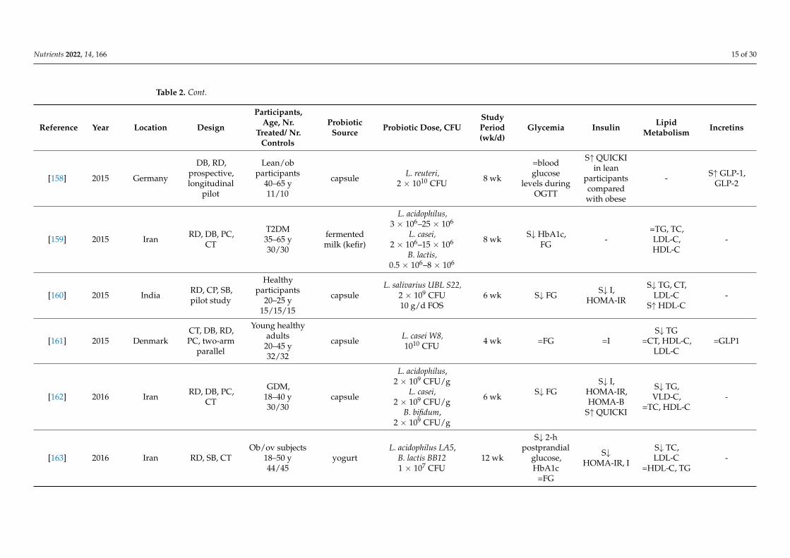
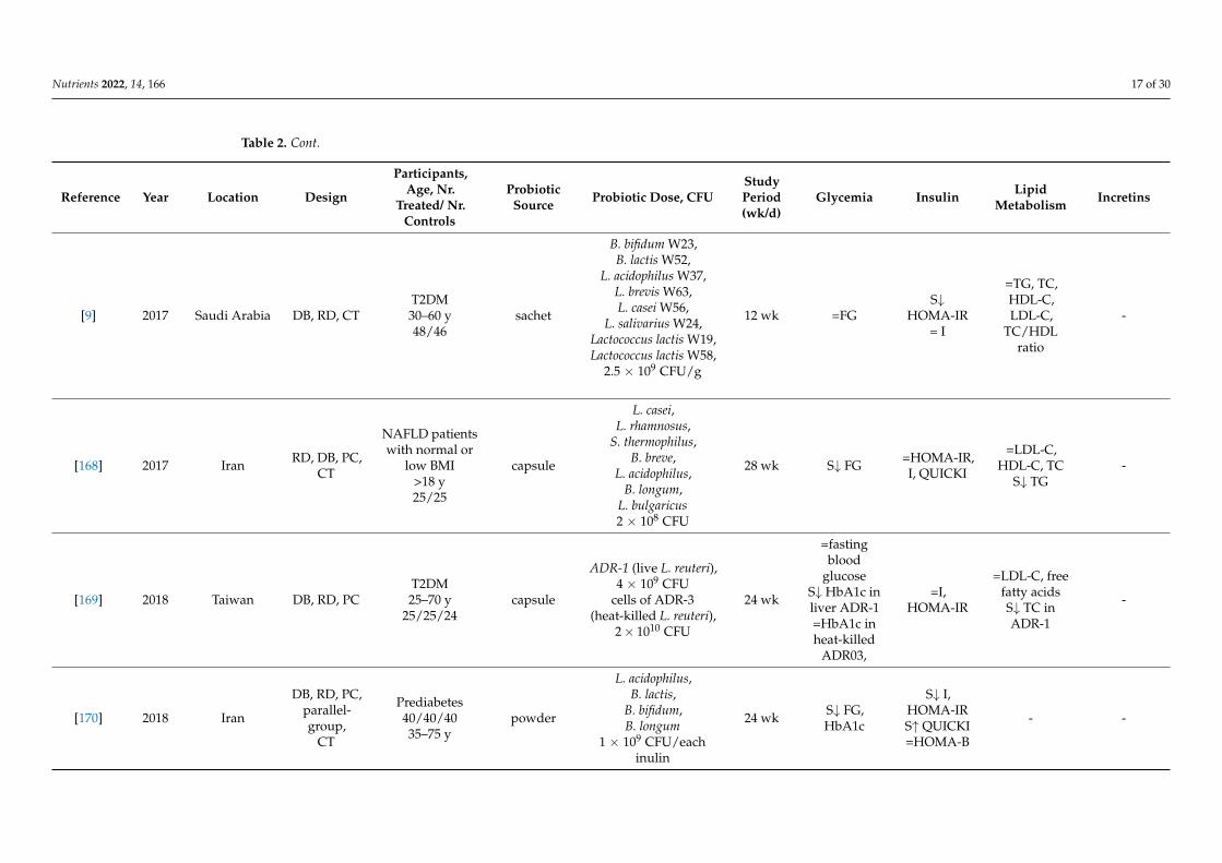
Table 2. Effects of probiotic or synbiotic on glycemia, insulin, lipid metabolism and incretins.
Reference Year Location Design
Participants,Age, Nr.
Treated/ Nr.Controls
ProbioticSource Probiotic Dose, CFU
StudyPeriod(wk/d)
Glycemia Insulin LipidMetabolism Incretins
[135] 2002 Poland RD, DB, CT
Healthyparticipants
35–45 y18/18
rose-hipdrink
L. plantarum 299v,5 × 107 CFU/mL 6 wk =FG =I
=TC, LDL-C,HDL-C, TG,
lipopro-tein(a)
S↓ leptin
[136] 2006 Australia
DB, PC,parallel
design trial,single centre
Healthyvolunteers
30–75 y23/21
capsule L. fermentum,2 × 109 CFU 10 wk =FG - =LDL-C, TC,
HDL-C, TGL -
[137] 2009 Finland
RD,prospective,
parallel-group
Pregnant women29.7/30.1/30.2 y
85/86/85capsule
L. rhamnosus GG,ATCC 53 103,B. lactis Bb12,
1010 CFU/d each
4 wk S↓ FG,=HbA1c
S↓ I, HOMA,S↑ QUICKI - -
[138] 2010 Denmark RD, PC, DB
T2DM/non-diabetic48–66 y24/24
capsule L. acidophilus NCFM,1 g; about 1010 CFU 4 wk - =QUICKI - -
[139] 2012 Iran DB, RD, CTT2DM30–60 y32/32
yogurt
L. acidophilus La5,7.23 × 106–1.85 × 10⁶
CFU/gB. lactis Bb12,
6.04 × 10⁶
CFU/g–1.79 × 106
CFU/g
6 wk S↓ FG,HbA1c =I - -
[140] 2012 Brazil DB, PC, RD
Healthyparticipants
50–65 y10/10
shake
L. acidophillus,4 × 108 CFU/100 mL
B. bifidum4 ×108 CFU/100 mL
1 g/100 mL FOS
30 d S↓ FG -S↑ HDL-C=TC, TG -

Nutrients 2022, 14, 166 11 of 30
Table 2. Cont.
Reference Year Location Design
Participants,Age, Nr.
Treated/ Nr.Controls
ProbioticSource Probiotic Dose, CFU
StudyPeriod(wk/d)
Glycemia Insulin LipidMetabolism Incretins
[141] 2012 CanadaDB, PC,
multi-centerstudy
Healthyhypercholester-olemic human
subjects20–75 y67/64
capsuleL. reuteri NCIMB
30242,2.9 × 109 CFU
9 wk =FG - - -
[142] 2012 Denmark DB, PC, RDOb adolescents
12–15 y27/23
capsuleL. salivarius Ls-33
ATCC SD5208,1010 CFU
12 wk =FG =I,HOMA-IR
=TC, HDL-C,LDL-C, TG -
[143] 2013 Iran RD, DB, PC,CT
T2DM35–70 y27/27 capsule
L. acidophilus,2 × 109 CFU
L. casei,7 × 109 CFUL. rhamnosus,
1.5 × 10⁹ CFUL. bulgaricus,2 × 108 CFU
B. breve,2 × 1010 CFU
B. longum,7 × 109 CFU
S. thermophiles,1.5 ×109 CFU100 mg FOS
8 wk S↓ FG S↑ I,HOMA-IR S↑ LDL-C -

Nutrients 2022, 14, 166 12 of 30
Table 2. Cont.
Reference Year Location Design
Participants,Age, Nr.
Treated/ Nr.Controls
ProbioticSource Probiotic Dose, CFU
StudyPeriod(wk/d)
Glycemia Insulin LipidMetabolism Incretins
[144] 2013 Iran RD, DB, CT
Patients withNASH18–75 y34/36
tablet
L. acidophilus,1 × 108 CFU
L. casei,5 × 108 CFUL. rhamnosus,
7.5 × 10⁷ CFUL. bulgaricus,
1.5 × 10⁸ CFUB. breve,
5 × 10⁷ CFUB. longum,
2.5 × 10⁷ CFUS. thermophilus,5 × 107 CFU350 mg FOS
24 wk S↓ FG - S↓ TC, TG -
[145] 2013 Koreasingle center,RD, DB, PC,
CT
Ob volunteers19–60 y31/31
capsuleL. gasseri BNR17,
1010 CFU25% FOS
12 wk =FG, HbA1c =I=TC, TG,LDL-C,HDL-C,
-
[146] 2013 RussianFederation
RD, DB, PC,parallel pilot
study
Patients withmetabolicsyndrome
30–69 y25/15
cheese L. plantarum TENSIA,1.5 × 1011 CFU/g 3 wk =FG - =TC, LDL-C,
HDL-C, TG -
[147] 2013 Iran RD, SB, CTPregnant women
37/3318–30 y
yogurtL. acidophilus LA5,B. animalis BB12,
1 × 10⁷ CFU9 wk =FG S↓ I, HOMA - -
[148] 2014 IranRD, DB,
cross-overCT
T2DM35–70 y62/62
packageL. sporogenes,
27 × 107 CFU1.08 g inulin
6 wk =FG S↓ I=HOMA-IR
=CT, LDL-C,TG, HDL-C -

Nutrients 2022, 14, 166 13 of 30
Table 2. Cont.
Reference Year Location Design
Participants,Age, Nr.
Treated/ Nr.Controls
ProbioticSource Probiotic Dose, CFU
StudyPeriod(wk/d)
Glycemia Insulin LipidMetabolism Incretins
[149] 2014 Iran RD, DB, CT
T2DM ov/obobese
53.00 ± 5.9/49.00 ± 7.08 y
22/22
yogurt
B. lactis Bb12,L. acidophilus strain
La5,3.7 × 106 CFU/g
8 wk S↓ HbA1c=FG - - -
[150] 2014 Ireland PC, DB, RD
Ob pregnantwomen,
31.4 ± 5.0/31.0± 5.2 y63/75
capsule L. salivarius UCC118,109 CFU 4 wk =FG =I,
HOMA-IR=TC, HDL-C,LDL-C, TG -
[151] 2014 AustraliaRD, DB,parallelstudy
Ov>55 y
40/37/39/40
Yogurt/capsule
L. acidophilus La5,B. lactis Bb12,
3 × 10⁹ CFU/d6 wk S↑ FG
=HbA1c
S↑HOMA-IR
=I- -
[152] 2014 India RD, CT, DB
Ov/ob healthyadults
40–60 y15/15/15/15
capsule
B. longum,B. infantis,
B. breve,L. acidophilus,L. paracasei,
L. bulgaricus,L. plantarum,
S. thermophilus.112.5× 109 CFU/capsule
6 wk S↓ FG S↓ I,HOMA-IR
S↓ TC, TG,LDL-C,
VLDL-CS↑ HDL-C
-
[153] 2014 Japan
SB, PC,within-subject,
repeated-measure
interventiontrial
Adults withhypertriacyl-glycerolemia,51.1 ± 6.6 y
10/10
fermentedmil
L. gasseri SBT2055(LG2055),
5 × 10¹⁰ CFU/100 g4 wk S↑ HbA1c
=FG =I
S↓ NEFA=TG, ApoB-48, TC,LDL-C,HDL-C
-

Nutrients 2022, 14, 166 14 of 30
Table 2. Cont.
Reference Year Location Design
Participants,Age, Nr.
Treated/ Nr.Controls
ProbioticSource Probiotic Dose, CFU
StudyPeriod(wk/d)
Glycemia Insulin LipidMetabolism Incretins
[154] 2014 Iran RD, DB, PC,CT
NAFLD>18 y26/26
capsule
L. casei,L. rhamnosus,
S. thermophilus,B. breve,
L. acidophilus,B. longum,
L. bulgaricus2 × 108 CFU250 mg FOS
28 wk S↓ FG S↓ I,HOMA-IR - -
[155] 2014 Iran RD, DB, PCpilot study
Patients with MS>18 y19/19
capsule
L. casei,L. rhamnosus,
S. thermophilus,B. breve,
L. acidophilus,B. longum,
L. bulgaricus2 × 108 CFU250 mg FOS
28 wk S↓ FGS↓ I,
HOMA-IRS↑ QUICKI
=LDL-CS↓ TG, CTS↑ HDL
-
[156] 2014 Iran RD, PC, CTPregnant women
18–35 y26/26
foodL. sporogenes,1 × 107 CFU0.04 g inulin
9 wk =FG
S↓ I,HOMA-IR,HOMA-B
S↑ QUICKI
- -
[157] 2014 Iran RD, DB, CTT2DM35–70 y
26/26/26bread
L. sporogenes,1 × 108 CFU0.07 g inulin
8 wk =FG -
S↓ TG,VLDL-C,
TC/HDL-CS↑ HDL-C
= TC, LDL-C,HDL-C
-
Nutrients 2022, 14, 166 15 of 30
Table 2. Cont.
Reference Year Location Design
Participants,Age, Nr.
Treated/ Nr.Controls
ProbioticSource Probiotic Dose, CFU
StudyPeriod(wk/d)
Glycemia Insulin LipidMetabolism Incretins
[158] 2015 Germany
DB, RD,prospective,longitudinal
pilot
Lean/obparticipants
40–65 y11/10
capsule L. reuteri,2 × 1010 CFU 8 wk
=bloodglucose
levels duringOGTT
S↑ QUICKIin lean
participantscomparedwith obese
- S↑ GLP-1,GLP-2
[159] 2015 Iran RD, DB, PC,CT
T2DM35–65 y30/30
fermentedmilk (kefir)
L. acidophilus,3 × 106–25 × 106
L. casei,2 × 106–15 × 106
B. lactis,0.5 × 106–8 × 10⁶
8 wk S↓ HbA1c,FG -
=TG, TC,LDL-C,HDL-C
-
[160] 2015 India RD, CP, SB,pilot study
Healthyparticipants
20–25 y15/15/15
capsuleL. salivarius UBL S22,
2 × 10⁹ CFU10 g/d FOS
6 wk S↓ FG S↓ I,HOMA-IR
S↓ TG, CT,LDL-C
S↑ HDL-C-
[161] 2015 DenmarkCT, DB, RD,PC, two-arm
parallel
Young healthyadults
20–45 y32/32
capsule L. casei W8,1010 CFU 4 wk =FG =I
S↓ TG=CT, HDL-C,
LDL-C=GLP1
[162] 2016 Iran RD, DB, PC,CT
GDM,18–40 y30/30
capsule
L. acidophilus,2 × 109 CFU/g
L. casei,2 × 109 CFU/g
B. bifidum,2 × 109 CFU/g
6 wk S↓ FGS↓ I,
HOMA-IR,HOMA-B
S↑ QUICKI
S↓ TG,VLD-C,
=TC, HDL-C-
[163] 2016 Iran RD, SB, CTOb/ov subjects
18–50 y44/45
yogurtL. acidophilus LA5,
B. lactis BB121 × 107 CFU
12 wk
S↓ 2-hpostprandial
glucose,HbA1c
=FG
S↓HOMA-IR, I
S↓ TC,LDL-C
=HDL-C, TG-

Nutrients 2022, 14, 166 16 of 30
Table 2. Cont.
Reference Year Location Design
Participants,Age, Nr.
Treated/ Nr.Controls
ProbioticSource Probiotic Dose, CFU
StudyPeriod(wk/d)
Glycemia Insulin LipidMetabolism Incretins
[164] 2016 Estoniapreliminary,open label
study
Clinicallyhealthy
volunteers50–75 y
capsuleL. fermentum ME-3
(LFME-3),6 × 109 CFU
4 wk S↓ HbA1c S↓HOMA-IR
S↓ LDL-C,oxLDL, TC,
TG,TG/HDL-C
ratioS↑ HDL-C
-
[165] 2017 Sweden RD, PCT2DM50–75 y
15/15/16stick pack
L. reuteri DSM 17938,108 CFU/day
L. reuteri DSM 17938,1010 CFU/day
12 wk =FG=HbA1c S↑ QUICKI =CT, HDL,
LDL, TGL -
[17] 2017 Iran RD, CT
T2DM, ov,CHD patients
40–85 y30/30
capsule
L. acidophilus,2 × 109
L. casei,2 × 109,
B. bifidum,2 × 109 CFU/g800 mg inulin
12 wk S↓ FG
S↓ I,HOMA-B
S↑ QUICKI=HOMA-IR
S↑ HDL-C=TG, TC,LDL-C,
VLDL-C,TC/HDL-C
ratio
-
[166] 2017 Malaysia
RD, DB,parallel-group,
CT
T2DM,30–70 y68/68
sachet
L. acidophilus,L. casei,L. lactis,
B. bifidum,B. longum,B. infantis,
1010 CFU/d each
12 wk S↓ HbA1c=FG
S↓ I=HOMA-IR,
QUICKI
=TC, TG,LDL-C,HDL-C
-
[167] 2017 Brazil DB, RD, PC,CT
T2DM35–60 y25/25
fermentedgoat milk
L. acidophilus La-5,B. lactis BB-12,
109 CFU/d each6 wk S↓ FS
=HbA1c, FG= I,
HOMA-IR
S↓ TC,LDL-C=HDL,
VLDL, TG.CT/HLD-C
ratio
-
Nutrients 2022, 14, 166 17 of 30
Table 2. Cont.
Reference Year Location Design
Participants,Age, Nr.
Treated/ Nr.Controls
ProbioticSource Probiotic Dose, CFU
StudyPeriod(wk/d)
Glycemia Insulin LipidMetabolism Incretins
[9] 2017 Saudi Arabia DB, RD, CTT2DM30–60 y48/46
sachet
B. bifidum W23,B. lactis W52,
L. acidophilus W37,L. brevis W63,L. casei W56,
L. salivarius W24,Lactococcus lactis W19,Lactococcus lactis W58,
2.5 × 10⁹ CFU/g
12 wk =FGS↓
HOMA-IR= I
=TG, TC,HDL-C,LDL-C,
TC/HDLratio
-
[168] 2017 Iran RD, DB, PC,CT
NAFLD patientswith normal or
low BMI>18 y25/25
capsule
L. casei,L. rhamnosus,
S. thermophilus,B. breve,
L. acidophilus,B. longum,
L. bulgaricus2 × 108 CFU
28 wk S↓ FG =HOMA-IR,I, QUICKI
=LDL-C,HDL-C, TC
S↓ TG-
[169] 2018 Taiwan DB, RD, PCT2DM25–70 y
25/25/24capsule
ADR-1 (live L. reuteri),4 × 109 CFU
cells of ADR-3(heat-killed L. reuteri),
2× 10¹⁰ CFU
24 wk
=fastingblood
glucoseS↓ HbA1c inliver ADR-1=HbA1c inheat-killed
ADR03,
=I,HOMA-IR
=LDL-C, freefatty acidsS↓ TC inADR-1
-
[170] 2018 Iran
DB, RD, PC,parallel-group,
CT
Prediabetes40/40/4035–75 y
powder
L. acidophilus,B. lactis,
B. bifidum,B. longum
1 × 10⁹ CFU/eachinulin
24 wk S↓ FG,HbA1c
S↓ I,HOMA-IRS↑ QUICKI=HOMA-B
- -

Nutrients 2022, 14, 166 18 of 30
Table 2. Cont.
Reference Year Location Design
Participants,Age, Nr.
Treated/ Nr.Controls
ProbioticSource Probiotic Dose, CFU
StudyPeriod(wk/d)
Glycemia Insulin LipidMetabolism Incretins
[171] 2018 UkraineDB, singlecenter RD,
CT
T2DM, ov18–75 y31/22
sachet
14 alive probioticstrains of L.+Lactococcus,
6 × 10¹⁰ CFU/gB.,
1 × 10¹⁰ CFU/g,Propionibacterium,3 × 1010 CFU/g,
Acetobacter,1 × 106 CFU/g
8 wk S↓ HbA1c=FG
S↓HOMA-IR
=I- -
[172] 2018 Iran RD, DB, PC,CT
T2DM, CHD45–85 y30/30
capsule
L. acidophilus,B. bifidum,L. reuteri,
L. fermentum8 × 109 CFU/g
12 wk =FGS↓ I,
HOMA-IRS↑ QUICKI
S↑ HDL-C=LDL, TC,
TG, VLDL-C -
[82] 2018 Saudi Arabia DB, RD, CTT2DM,30–60 y30/31
sachet
B. bifidum W23, B.lactis W52,
L. acidophilus W37,L. brevis W63,L. casei W56,
L. salivarius W24,Lactococcus lactis W19,Lactococcus lactis W58
2.5 × 10⁹ CFU/g
24 wk S↓ FG, S↓ I,HOMA-IR,
S↓ TC, TG,total/HDL-cholesterol
ratio
-
[173] 2019 Iranparallel-
group, RD,CT
T2DM,20/20
30–50 ycapsule L. casei,
108 CFU/d 8 wk S↓ FG=HbA1c
S↓ I,HOMA-IR - -

Nutrients 2022, 14, 166 19 of 30
Table 2. Cont.
Reference Year Location Design
Participants,Age, Nr.
Treated/ Nr.Controls
ProbioticSource Probiotic Dose, CFU
StudyPeriod(wk/d)
Glycemia Insulin LipidMetabolism Incretins
[174] 2019 Iran RD, DB, CT
T2DM30–75 y34/34 capsule
L. acidophilus,2 × 109 CFU
L. casei,7 × 109 CFUL. rhamnosus,
1.5 × 10⁹ CFUL. bulgaricus,2 × 108 CFU
B. breve,3 × 1010 CFU
B. longum,7 × 109 CFU
S. thermophilus,1.5 × 109 CFU
100 mg FOS
6 wk S↓ FG =I,HOMA-IR
S↑ HDL-C=TG, TC -
[175] 2019 India RD, DB, CT
T2DM, Ob18–65 y39/40 capsule
L. salivarius,L. casei,
L. plantarum,L. acidophilus,
B. breve,B. coagulans,
30 billion CFU100 mg FOS
12 wk S↓ HbA1c=FG
=I,HOMA-IR
= TC, TG,HDL-C,LDL-C
-
[133] 2019 Belgium RD, DB, PC,pilot study
Ob/ovinsulin-resistant
volunteers18–70 y
14/13/13
sachet
Live/pasteurizedAkkermansiamuniciphila
1010 bacteria/day
12 wk =FG, HbA1cS↑ insulinsensitivity
S↓ I
S↓ TC=LDL-C, TG =GLP-1
Nutrients 2022, 14, 166 20 of 30
Table 2. Cont.
Reference Year Location Design
Participants,Age, Nr.
Treated/ Nr.Controls
ProbioticSource Probiotic Dose, CFU
StudyPeriod(wk/d)
Glycemia Insulin LipidMetabolism Incretins
[176] 2020 Australia RD, DB, CT
T2DMBMI ≥ 25 kg/m2
≥ 18 y30/30
capsule
L. plantarum,6 × 109 CFU,L. bulgaricus,3 × 109 CFU
L. gasseri,18 × 109 CFU
B. breve,7.5 × 109 CFU
B. animalis sbsp.lactis,
8 × 10⁹ CFUB. bifidum,
7 × 10⁹ CFUS. thermophiles,450 × 106 CFU
Saccharomyces boulardii,45 × 106 CFU
12 wk
S↓ FG,HbA1c
(in patientstaking
probioticsand
metformin)
S↓HOMA-IR(in patients
takingprobiotics
andmetformin)
- -
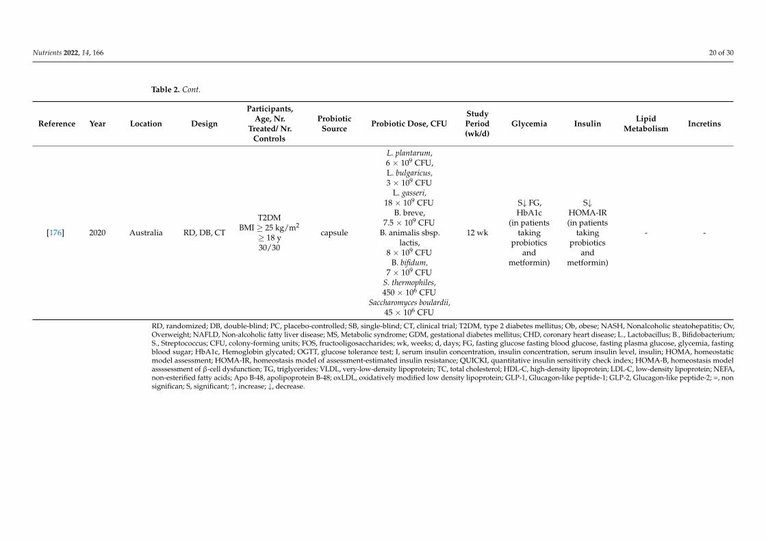
RD, randomized; DB, double-blind; PC, placebo-controlled; SB, single-blind; CT, clinical trial; T2DM, type 2 diabetes mellitus; Ob, obese; NASH, Nonalcoholic steatohepatitis; Ov,Overweight; NAFLD, Non-alcoholic fatty liver disease; MS, Metabolic syndrome; GDM, gestational diabetes mellitus; CHD, coronary heart disease; L., Lactobacillus; B., Bifidobacterium;S., Streptococcus; CFU, colony-forming units; FOS, fructooligosaccharides; wk, weeks; d, days; FG, fasting glucose fasting blood glucose, fasting plasma glucose, glycemia, fastingblood sugar; HbA1c, Hemoglobin glycated; OGTT, glucose tolerance test; I, serum insulin concentration, insulin concentration, serum insulin level, insulin; HOMA, homeostaticmodel assessment; HOMA-IR, homeostasis model of assessment-estimated insulin resistance; QUICKI, quantitative insulin sensitivity check index; HOMA-B, homeostasis modelassssessment of β-cell dysfunction; TG, triglycerides; VLDL, very-low-density lipoprotein; TC, total cholesterol; HDL-C, high-density lipoprotein; LDL-C, low-density lipoprotein; NEFA,non-esterified fatty acids; Apo B-48, apolipoprotein B-48; oxLDL, oxidatively modified low density lipoprotein; GLP-1, Glucagon-like peptide-1; GLP-2, Glucagon-like peptide-2; =, nonsignifican; S, significant; ↑, increase; ↓, decrease.
Nutrients 2022, 14, 166 21 of 30Nutrients 2021, 13, x FOR PEER REVIEW 20 of 30
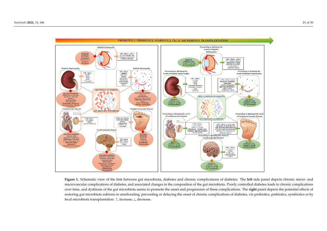
Figure 1. Schematic view of the link between gut microbiota, diabetes and chronic complications of diabetes. The left side panel depicts chronic micro- and macrovascular complications of diabetes, and associated changes in the composition of the gut microbiota. Poorly controlled diabetes leads to chronic complica-tions over time, and dysbiosis of the gut microbiota seems to promote the onset and progression of these complications. The right panel depicts the potential effects of restoring gut microbiota eubiosis in ameliorating, preventing or delaying the onset of chronic complications of diabetes, via probiotics, prebiotics, sym-biotics or by fecal microbiota transplantation. ↑, increase; ↓, decrease.
Figure 1. Schematic view of the link between gut microbiota, diabetes and chronic complications of diabetes. The left side panel depicts chronic micro- andmacrovascular complications of diabetes, and associated changes in the composition of the gut microbiota. Poorly controlled diabetes leads to chronic complicationsover time, and dysbiosis of the gut microbiota seems to promote the onset and progression of these complications. The right panel depicts the potential effects ofrestoring gut microbiota eubiosis in ameliorating, preventing or delaying the onset of chronic complications of diabetes, via probiotics, prebiotics, symbiotics or byfecal microbiota transplantation. ↑, increase; ↓, decrease.

Nutrients 2022, 14, 166 22 of 30
3. Conclusions and Future Perspectives
For the past decade or so, owing to rapid methodological advances in genome sequenc-ing of microbes, an avalanche of studies has rushed to uncover the potential contributionof the so called “forgotten organ” (i.e., gut microbiota) in multiple pathologies, includingmetabolic disorders. While significant strides have been made toward understanding thecomplex interaction between bacteria and the host, particularly at the biochemical, cellularand molecular level, we are still in the early stages when it comes to our understandingof whether gut bacteria play a direct role in prevention, development and treatment ofdiseases. As it is the case with most pathologies in which the effects of gut microbiotahave been studied, the development of diabetes and its complications have been linkedwith the state of dysbiosis of the gut microbiota. This, in and of itself, raises a wide rangeof questions, since “dysbiosis” is a loose term used to characterize a disequilibrium, in agiven organism and time [177]. As noted throughout this review, it is well documented thatdiabetes and its complications are characterized by systemic inflammation, therefore it isnot surprising that numerous studies focused on examining the anti-inflammatory effectsof certain bacteria such as Roseburia in patients with coronary artery disease, Lachnospiraceaein patients at high stroke risk and Faecalibacterium in patients with diabetic nephropathy,diabetic neuropathy, cerebrovascular disease or coronary artery disease. As such, lowabundance of anti-inflammatory bacteria, along with the increased abundance of pro-inflammatory bacteria has been attributed to the onset and progression of complicationsof diabetes. Similarly, bacterial metabolites such as SCFA and TMAO have been shown toinfluence host physiology and improve disease outcome. Notwithstanding such promisingfindings, we are still very much grasping with the demonstration, beyond doubt, of a causalrelationship between gut bacteria and diabetes and its complications. Whereas preclinicalstudies are promising and show direct effect of some bacteria on certain metabolic andclinical parameters of diabetes, the results in humans are less promising, with few clinicaltrials and by and large, have been inconsistent. Thus, for the modulation of gut microbiotavia prebiotics, probiotics, FMT or other means to be part of any therapeutic protocol indiabetes and its complications, its causal effect in these diseases must be defined andclinically demonstrated. Preclinical animal models such as germ free or antibiotic treatedanimals have been useful in examining host-microbiota interactions via controlling theeffects of individual bacteria, through monocolonization or combined bacteria therapy,however, they each come with significant caveats that often preclude generalization offindings to human disease prevention and treatment. Considering that bacterial strainsof the same species may differ in up to 30% of their genomic structure when comparedby taxonomic analysis, it follows that gut microbiota must be viewed and analyzed as asystem. Similarly, microbial metabolites associated with the gut microbiota, type 2 diabetesand associated complications that act synergistically must be analyzed and their effectstested [178]. It is equally important to examine the dynamical changes in the compositionprofile and production of metabolic byproducts of gut microbiota prior, during and afterthe onset of diabetes and its complications in order to determine dynamic changes duringdisease progression. While to date the list of bacteria reported to affect several parameterscharacteristic of diabetes complications is steadily increasing, very few have been studiedas therapeutic approaches in these pathologies. Likewise, efforts should be dedicatedtoward identification of bacteria signatures and metabolites that will allow early detectionof disease risks, and the mechanisms involved, making possible to personalize therapeuticintervention based on individual’s needs, stage and particularities of the disease. Therefore,modulation of the gut microbiota through prebiotics, probiotics, synbiotics or fecal micro-biota transfer may have beneficial effects in the management of diabetes and associatedcomplications; however, further research involving human trials should be high on the list.
Author Contributions: All authors contributed to the conceptualization, writing and article prepara-tion. All authors have read and agreed to the published version of the manuscript.

Nutrients 2022, 14, 166 23 of 30
Funding: This research was funded by the project “The analysis of interrelationship between gutmicrobiota and the host with applications in the prevention and control of type 2 diabetes” co-financed by European Regional Development Fund through Competitiveness Operational Programunder the contract number 120/16.09.2016.
Institutional Review Board Statement: Not applicable.
Informed Consent Statement: Not applicable.
Data Availability Statement: Not applicable.
Conflicts of Interest: The authors declare no conflict of interest.
References1. D’Argenio, V.; Salvatore, F. The role of the gut microbiome in the healthy adult status. Clin. Chim. Acta 2015, 451, 97–102.
[CrossRef] [PubMed]2. Matijasic, M.; Mestrovic, T.; Paljetak, H.C.; Peric, M.; Baresic, A.; Verbanac, D. Gut Microbiota beyond Bacteria-Mycobiome,
Virome, Archaeome, and Eukaryotic Parasites in IBD. Int. J. Mol. Sci. 2020, 21, 2668. [CrossRef]3. Jayasudha, R.; Das, T.; Kalyana Chakravarthy, S.; Sai Prashanthi, G.; Bhargava, A.; Tyagi, M.; Rani, P.K.; Pappuru, R.R.; Shivaji, S.
Gut mycobiomes are altered in people with type 2 Diabetes Mellitus and Diabetic Retinopathy. PLoS ONE 2020, 15, e0243077.[CrossRef]
4. Mazloom, K.; Siddiqi, I.; Covasa, M. Probiotics: How Effective Are They in the Fight against Obesity? Nutrients 2019, 11, 258.[CrossRef]
5. Mar Rodriguez, M.; Perez, D.; Javier Chaves, F.; Esteve, E.; Marin-Garcia, P.; Xifra, G.; Vendrell, J.; Jove, M.; Pamplona, R.; Ricart,W.; et al. Obesity changes the human gut mycobiome. Sci. Rep. 2015, 5, 14600. [CrossRef] [PubMed]
6. Kowalewska, B.; Zorena, K.; Szmigiero-Kawko, M.; Waz, P.; Mysliwiec, M. Higher diversity in fungal species discriminateschildren with type 1 diabetes mellitus from healthy control. Patient Prefer. Adherence 2016, 10, 591–599. [CrossRef]
7. Pataky, Z.; Bobbioni-Harsch, E.; Hadengue, A.; Carpentier, A.; Golay, A. Gut microbiota, responsible for our body weight? Rev.Med. Suisse 2009, 5, 662–664.
8. Marchesi, J.R.; Adams, D.H.; Fava, F.; Hermes, G.D.; Hirschfield, G.M.; Hold, G.; Quraishi, M.N.; Kinross, J.; Smidt, H.; Tuohy,K.M.; et al. The gut microbiota and host health: A new clinical frontier. Gut 2016, 65, 330–339. [CrossRef] [PubMed]
9. Sabico, S.; Al-Mashharawi, A.; Al-Daghri, N.M.; Yakout, S.; Alnaami, A.M.; Alokail, M.S.; McTernan, P.G. Effects of a multi-strainprobiotic supplement for 12 weeks in circulating endotoxin levels and cardiometabolic profiles of medication naive T2DMpatients: A randomized clinical trial. J. Transl. Med. 2017, 15, 249. [CrossRef]
10. Khalili, L.; Alipour, B.; Asghari Jafarabadi, M.; Hassanalilou, T.; Mesgari Abbasi, M.; Faraji, I. Probiotic assisted weightmanagement as a main factor for glycemic control in patients with type 2 diabetes: A randomized controlled trial. Diabetol. Metab.Syndr. 2019, 11, 5. [CrossRef] [PubMed]
11. Saeedi, P.; Petersohn, I.; Salpea, P.; Malanda, B.; Karuranga, S.; Unwin, N.; Colagiuri, S.; Guariguata, L.; Motala, A.A.; Ogurtsova,K.; et al. Global and regional diabetes prevalence estimates for 2019 and projections for 2030 and 2045: Results from theInternational Diabetes Federation Diabetes Atlas, 9(th) edition. Diabetes Res. Clin. Pract. 2019, 157, 107843. [CrossRef]
12. DeFronzo, R.A. Current issues in the treatment of type 2 diabetes. Overview of newer agents: Where treatment is going. Am. J.Med. 2010, 123, S38–S48. [CrossRef]
13. Bekyarova, G.Y.; Ivanova, D.G.; Madjova, V.H. Molecular mechanisms associating oxidative stress with endothelial dysfunctionin the development of various vascular complications in diabetes mellitus. Folia Med. 2007, 49, 13–19.
14. Baig, M.A.; Panchal, S.S. Streptozotocin-Induced Diabetes Mellitus in Neonatal Rats: An Insight into its Applications to InduceDiabetic Complications. Curr. Diabetes Rev. 2019, 16, 26–39. [CrossRef] [PubMed]
15. Gourgari, E.; Dabelea, D.; Rother, K. Modifiable Risk Factors for Cardiovascular Disease in Children with Type 1 Diabetes: CanEarly Intervention Prevent Future Cardiovascular Events? Curr. Diabetes Rep. 2017, 17, 134. [CrossRef] [PubMed]
16. Heianza, Y.; Sun, D.; Ma, W.; Zheng, Y.; Champagne, C.M.; Bray, G.A.; Sacks, F.M.; Qi, L. Gut-microbiome-related LCT genotypeand 2-year changes in body composition and fat distribution: The POUNDS Lost Trial. Int. J. Obes. 2018, 42, 1565–1573. [CrossRef]
17. Tajabadi-Ebrahimi, M.; Sharifi, N.; Farrokhian, A.; Raygan, F.; Karamali, F.; Razzaghi, R.; Taheri, S.; Asemi, Z. A RandomizedControlled Clinical Trial Investigating the Effect of Synbiotic Administration on Markers of Insulin Metabolism and Lipid Profilesin Overweight Type 2 Diabetic Patients with Coronary Heart Disease. Exp. Clin. Endocrinol. Diabetes 2017, 125, 21–27. [CrossRef]
18. Wang, Y.; Branicky, R.; Noe, A.; Hekimi, S. Superoxide dismutases: Dual roles in controlling ROS damage and regulating ROSsignaling. J. Cell Biol. 2018, 217, 1915–1928. [CrossRef]
19. Constantino, M.I.; Molyneaux, L.; Limacher-Gisler, F.; Al-Saeed, A.; Luo, C.; Wu, T.; Twigg, S.M.; Yue, D.K.; Wong, J. Long-termcomplications and mortality in young-onset diabetes: Type 2 diabetes is more hazardous and lethal than type 1 diabetes. DiabetesCare 2013, 36, 3863–3869. [CrossRef] [PubMed]
20. Lee, C.B.; Chae, S.U.; Jo, S.J.; Jerng, U.M.; Bae, S.K. The Relationship between the Gut Microbiome and Metformin as a Key forTreating Type 2 Diabetes Mellitus. Int. J. Mol. Sci. 2021, 22, 3566. [CrossRef]
Nutrients 2022, 14, 166 24 of 30
21. Mardinoglu, A.; Boren, J.; Smith, U. Confounding Effects of Metformin on the Human Gut Microbiome in Type 2 Diabetes. CellMetab. 2016, 23, 10–12. [CrossRef] [PubMed]
22. Wu, H.; Esteve, E.; Tremaroli, V.; Khan, M.T.; Caesar, R.; Manneras-Holm, L.; Stahlman, M.; Olsson, L.M.; Serino, M.; Planas-Felix,M.; et al. Metformin alters the gut microbiome of individuals with treatment-naive type 2 diabetes, contributing to the therapeuticeffects of the drug. Nat. Med. 2017, 23, 850–858. [CrossRef] [PubMed]
23. Fernandes, R.; Viana, S.D.; Nunes, S.; Reis, F. Diabetic gut microbiota dysbiosis as an inflammaging and immunosenescencecondition that fosters progression of retinopathy and nephropathy. Biochim. Biophys. Acta Mol. Basis. Dis. 2019, 1865, 1876–1897.[CrossRef]
24. Chen, W.; Zhang, M.; Guo, Y.; Wang, Z.; Liu, Q.; Yan, R.; Wang, Y.; Wu, Q.; Yuan, K.; Sun, W. The Profile and Function of GutMicrobiota in Diabetic Nephropathy. Diabetes Metab. Syndr. Obes. 2021, 14, 4283–4296. [CrossRef]
25. Gross, J.L.; de Azevedo, M.J.; Silveiro, S.P.; Canani, L.H.; Caramori, M.L.; Zelmanovitz, T. Diabetic nephropathy: Diagnosis,prevention, and treatment. Diabetes Care 2005, 28, 164–176. [CrossRef] [PubMed]
26. Gupta, A.; Gupta, P.; Biyani, M. Targeted therapies in diabetic nephropathy: An update. J. Nephrol. 2011, 24, 686–695. [CrossRef][PubMed]
27. Ritz, E. Nephropathy in type 2 diabetes. J. Intern. Med. 1999, 245, 111–126. [CrossRef] [PubMed]28. Sabatino, A.; Regolisti, G.; Cosola, C.; Gesualdo, L.; Fiaccadori, E. Intestinal Microbiota in Type 2 Diabetes and Chronic Kidney
Disease. Curr. Diabetes Rep. 2017, 17, 16. [CrossRef] [PubMed]29. Jha, V.; Garcia-Garcia, G.; Iseki, K.; Li, Z.; Naicker, S.; Plattner, B.; Saran, R.; Wang, A.Y.; Yang, C.W. Chronic kidney disease:
Global dimension and perspectives. Lancet 2013, 382, 260–272. [CrossRef]30. Tuttle, K.R.; Bakris, G.L.; Bilous, R.W.; Chiang, J.L.; de Boer, I.H.; Goldstein-Fuchs, J.; Hirsch, I.B.; Kalantar-Zadeh, K.; Narva, A.S.;
Navaneethan, S.D.; et al. Diabetic kidney disease: A report from an ADA Consensus Conference. Am. J. Kidney Dis. 2014, 64,510–533. [CrossRef]
31. McMullan, C.J.; Lambers Heerspink, H.J.; Parving, H.H.; Dwyer, J.P.; Forman, J.P.; de Zeeuw, D. Visit-to-visit variability in bloodpressure and kidney and cardiovascular outcomes in patients with type 2 diabetes and nephropathy: A post hoc analysis fromthe RENAAL study and the Irbesartan Diabetic Nephropathy Trial. Am. J. Kidney Dis. 2014, 64, 714–722. [CrossRef]
32. Navarro-Gonzalez, J.F.; Mora-Fernandez, C.; Muros de Fuentes, M.; Garcia-Perez, J. Inflammatory molecules and pathways in thepathogenesis of diabetic nephropathy. Nat. Rev. Nephrol. 2011, 7, 327–340. [CrossRef]
33. Singh, D.K.; Winocour, P.; Farrington, K. Oxidative stress in early diabetic nephropathy: Fueling the fire. Nat. Rev. Endocrinol.2011, 7, 176–184. [CrossRef] [PubMed]
34. Ramezani, A.; Massy, Z.A.; Meijers, B.; Evenepoel, P.; Vanholder, R.; Raj, D.S. Role of the Gut Microbiome in Uremia: A PotentialTherapeutic Target. Am. J. Kidney Dis. 2016, 67, 483–498. [CrossRef] [PubMed]
35. Mahmoodpoor, F.; Rahbar Saadat, Y.; Barzegari, A.; Ardalan, M.; Zununi Vahed, S. The impact of gut microbiota on kidneyfunction and pathogenesis. Biomed. Pharm. 2017, 93, 412–419. [CrossRef] [PubMed]
36. Vaziri, N.D.; Yuan, J.; Nazertehrani, S.; Ni, Z.; Liu, S. Chronic kidney disease causes disruption of gastric and small intestinalepithelial tight junction. Am. J. Nephrol. 2013, 38, 99–103. [CrossRef] [PubMed]
37. Kieffer, D.A.; Piccolo, B.D.; Vaziri, N.D.; Liu, S.; Lau, W.L.; Khazaeli, M.; Nazertehrani, S.; Moore, M.E.; Marco, M.L.; Martin, R.J.;et al. Resistant starch alters gut microbiome and metabolomic profiles concurrent with amelioration of chronic kidney disease inrats. Am. J. Physiol.-Renal Physiol. 2016, 310, F857–F871. [CrossRef]
38. Fukuuchi, F. Intestinal bacteria-derived putrefactants in chronic renal failure. Clin. Exp. Nephrol. 2002, 6, 99–104. [CrossRef]39. Xu, K.Y.; Xia, G.H.; Lu, J.Q.; Chen, M.X.; Zhen, X.; Wang, S.; You, C.; Nie, J.; Zhou, H.W.; Yin, J. Impaired renal function and
dysbiosis of gut microbiota contribute to increased trimethylamine-N-oxide in chronic kidney disease patients. Sci. Rep. 2017, 7,1445. [CrossRef]
40. Jiang, S.; Xie, S.; Lv, D.; Wang, P.; He, H.; Zhang, T.; Zhou, Y.; Lin, Q.; Zhou, H.; Jiang, J.; et al. Alteration of the gut microbiota inChinese population with chronic kidney disease. Sci. Rep. 2017, 7, 2870. [CrossRef]
41. Kanbay, M.; Onal, E.M.; Afsar, B.; Dagel, T.; Yerlikaya, A.; Covic, A.; Vaziri, N.D. The crosstalk of gut microbiota and chronickidney disease: Role of inflammation, proteinuria, hypertension, and diabetes mellitus. Int. Urol. Nephrol. 2018, 50, 1453–1466.[CrossRef]
42. Ranganathan, N.; Friedman, E.A.; Tam, P.; Rao, V.; Ranganathan, P.; Dheer, R. Probiotic dietary supplementation in patients withstage 3 and 4 chronic kidney disease: A 6-month pilot scale trial in Canada. Curr. Med. Res. Opin. 2009, 25, 1919–1930. [CrossRef]
43. Wang, F.; Jiang, H.; Shi, K.; Ren, Y.; Zhang, P.; Cheng, S. Gut bacterial translocation is associated with microinflammation inend-stage renal disease patients. Nephrology 2012, 17, 733–738. [CrossRef] [PubMed]
44. Takayama, F.; Taki, K.; Niwa, T. Bifidobacterium in gastro-resistant seamless capsule reduces serum levels of indoxyl sulfate inpatients on hemodialysis. Am. J. Kidney Dis. 2003, 41, S142–S145. [CrossRef]
45. Cruz-Mora, J.; Martinez-Hernandez, N.E.; Martin del Campo-Lopez, F.; Viramontes-Horner, D.; Vizmanos-Lamotte, B.; Munoz-Valle, J.F.; Garcia-Garcia, G.; Parra-Rojas, I.; Castro-Alarcon, N. Effects of a symbiotic on gut microbiota in Mexican patients withend-stage renal disease. J. Renal Nutr. 2014, 24, 330–335. [CrossRef]
46. Rossi, M.; Johnson, D.W.; Morrison, M.; Pascoe, E.M.; Coombes, J.S.; Forbes, J.M.; Szeto, C.C.; McWhinney, B.C.; Ungerer, J.P.;Campbell, K.L. Synbiotics Easing Renal Failure by Improving Gut Microbiology (SYNERGY): A Randomized Trial. Clin. J. Am.Soc. Nephrol. 2016, 11, 223–231. [CrossRef] [PubMed]

Nutrients 2022, 14, 166 25 of 30
47. Miranda Alatriste, P.V.; Urbina Arronte, R.; Gomez Espinosa, C.O.; Espinosa Cuevas Mde, L. Effect of probiotics on human bloodurea levels in patients with chronic renal failure. Nutr. Hosp. 2014, 29, 582–590. [CrossRef]
48. Simenhoff, M.L.; Dunn, S.R.; Zollner, G.P.; Fitzpatrick, M.E.; Emery, S.M.; Sandine, W.E.; Ayres, J.W. Biomodulation of thetoxic and nutritional effects of small bowel bacterial overgrowith in end-stage kidney disease using freeze-dried Lactobacillusacidophilus. Min. Electrolyte Metab. 1996, 22, 92–96.
49. Dunn, S.R.; Simenhoff, M.L.; Ahmed, K.E.; Gaughan, W.J.; Eltayeb, B.O.; Fitzpatrick, M.E.; Emery, S.M.; Ayres, J.W.; Holt, K.E.Effect of Oral Administration of Freeze-Dried Lactobacillus acidophilus on Small Bowel Bacterial Overgrowith in Patients withEnd Stage Kidney Disease: Reducing Uremic Toxins and Improving Nutrition. Int. Dairy J. 1998, 8, 545–553. [CrossRef]
50. Mafi, A.; Namazi, G.; Soleimani, A.; Bahmani, F.; Aghadavod, E.; Asemi, Z. Metabolic and genetic response to probioticssupplementation in patients with diabetic nephropathy: A randomized, double-blind, placebo-controlled trial. Food Funct. 2018,9, 4763–4770. [CrossRef]
51. Abbasi, B.; Ghiasvand, R.; Mirlohi, M. Kidney Function Improvement by Soy Milk Containing Lactobacillus plantarum A7 inType 2 Diabetic Patients with Nephropathy: A Double-Blinded Randomized Controlled Trial. Iran. J. Kidney Dis. 2017, 11, 36–43.
52. Soleimani, A.; Zarrati Mojarrad, M.; Bahmani, F.; Taghizadeh, M.; Ramezani, M.; Tajabadi-Ebrahimi, M.; Jafari, P.; Esmaillzadeh,A.; Asemi, Z. Probiotic supplementation in diabetic hemodialysis patients has beneficial metabolic effects. Kidney Int. 2017, 91,435–442. [CrossRef]
53. Mazruei Arani, N.; Emam-Djomeh, Z.; Tavakolipour, H.; Sharafati-Chaleshtori, R.; Soleimani, A.; Asemi, Z. The Effects ofProbiotic Honey Consumption on Metabolic Status in Patients with Diabetic Nephropathy: A Randomized, Double-Blind,Controlled Trial. Probiotics Antimicrob. Proteins 2019, 11, 1195–1201. [CrossRef]
54. Miraghajani, M.; Zaghian, N.; Dehkohneh, A.; Mirlohi, M.; Ghiasvand, R. Probiotic Soy Milk Consumption and Renal FunctionAmong Type 2 Diabetic Patients with Nephropathy: A Randomized Controlled Clinical Trial. Probiotics Antimicrob. Proteins 2019,11, 124–132. [CrossRef]
55. Miraghajani, M.; Zaghian, N.; Mirlohi, M.; Feizi, A.; Ghiasvand, R. The Impact of Probiotic Soy Milk Consumption on OxidativeStress Among Type 2 Diabetic Kidney Disease Patients: A Randomized Controlled Clinical Trial. J. Renal Nutr. 2017, 27, 317–324.[CrossRef]
56. Papatheodorou, K.; Papanas, N.; Banach, M.; Papazoglou, D.; Edmonds, M. Complications of Diabetes 2016. J. Diabetes Res. 2016,2016, 6989453. [CrossRef]
57. Lee, R.; Wong, T.Y.; Sabanayagam, C. Epidemiology of diabetic retinopathy, diabetic macular edema and related vision loss. EyeVis. 2015, 2, 17. [CrossRef]
58. Beli, E.; Yan, Y.; Moldovan, L.; Vieira, C.P.; Gao, R.; Duan, Y.; Prasad, R.; Bhatwadekar, A.; White, F.A.; Townsend, S.D.; et al.Restructuring of the Gut Microbiome by Intermittent Fasting Prevents Retinopathy and Prolongs Survival in db/db Mice. Diabetes2018, 67, 1867–1879. [CrossRef] [PubMed]
59. Bader, M. Tissue renin-angiotensin-aldosterone systems: Targets for pharmacological therapy. Annu. Rev. Pharm. Toxicol. 2010, 50,439–465. [CrossRef] [PubMed]
60. Das, A. Diabetic Retinopathy: Battling the Global Epidemic. Investig. Ophthalmol. Vis. Sci. 2016, 57, 6669–6682. [CrossRef][PubMed]
61. Dominguez, J.M., 2nd; Hu, P.; Caballero, S.; Moldovan, L.; Verma, A.; Oudit, G.Y.; Li, Q.; Grant, M.B. Adeno-Associated VirusOverexpression of Angiotensin-Converting Enzyme-2 Reverses Diabetic Retinopathy in Type 1 Diabetes in Mice. Am. J. Pathol.2016, 186, 1688–1700. [CrossRef]
62. Jeganathan, V.S. The therapeutic implications of renin-angiotensin system blockade in diabetic retinopathy. Curr. Pharm. Biotechnol.2011, 12, 392–395. [CrossRef] [PubMed]
63. Perkins, B.A.; Aiello, L.P.; Krolewski, A.S. Diabetes complications and the renin-angiotensin system. N. Engl. J. Med. 2009, 361,83–85. [CrossRef]
64. Sjolie, A.K.; Dodson, P.; Hobbs, F.R. Does renin-angiotensin system blockade have a role in preventing diabetic retinopathy? Aclinical review. Int. J. Clin. Pract. 2011, 65, 148–153. [CrossRef] [PubMed]
65. Verma, A.; Shan, Z.; Lei, B.; Yuan, L.; Liu, X.; Nakagawa, T.; Grant, M.B.; Lewin, A.S.; Hauswirth, W.W.; Raizada, M.K.; et al. ACE2and Ang-(1–7) confer protection against development of diabetic retinopathy. Mol. Ther. 2012, 20, 28–36. [CrossRef] [PubMed]
66. Caspi, R.R. In this issue: Immunology of the eye—Inside and out. Int. Rev. Immunol. 2013, 32, 1–3. [CrossRef]67. Tap, J.; Mondot, S.; Levenez, F.; Pelletier, E.; Caron, C.; Furet, J.P.; Ugarte, E.; Munoz-Tamayo, R.; Paslier, D.L.; Nalin, R.; et al.
Towards the human intestinal microbiota phylogenetic core. Environ. Microbiol. 2009, 11, 2574–2584. [CrossRef]68. Huang, Y.; Yang, B.; Li, W. Defining the normal core microbiome of conjunctival microbial communities. Clin. Microbiol. Infect.
2016, 22, 643.e7–643.e12. [CrossRef]69. Ozkan, J.; Willcox, M.; Wemheuer, B.; Wilcsek, G.; Coroneo, M.; Thomas, T. Biogeography of the human ocular microbiota. Ocul.
Surf. 2019, 17, 111–118. [CrossRef]70. Lu, L.J.; Liu, J. Human Microbiota and Ophthalmic Disease. Yale J. Biol. Med. 2016, 89, 325–330.71. Das, T.; Jayasudha, R.; Chakravarthy, S.; Prashanthi, G.S.; Bhargava, A.; Tyagi, M.; Rani, P.K.; Pappuru, R.R.; Sharma, S.; Shivaji, S.
Alterations in the gut bacterial microbiome in people with type 2 diabetes mellitus and diabetic retinopathy. Sci. Rep. 2021, 11,2738. [CrossRef] [PubMed]

Nutrients 2022, 14, 166 26 of 30
72. Liu, W.; Wang, C.; Xia, Y.; Xia, W.; Liu, G.; Ren, C.; Gu, Y.; Li, X.; Lu, P. Elevated plasma trimethylamine-N-oxide levels areassociated with diabetic retinopathy. Acta Diabetol. 2021, 58, 221–229. [CrossRef] [PubMed]
73. Huang, Y.; Wang, Z.; Ma, H.; Ji, S.; Chen, Z.; Cui, Z.; Chen, J.; Tang, S. Dysbiosis and Implication of the Gut Microbiota in DiabeticRetinopathy. Front. Cell Infect. Microbiol. 2021, 11, 646348. [CrossRef] [PubMed]
74. Verma, A.; Xu, K.; Du, T.; Zhu, P.; Liang, Z.; Liao, S.; Zhang, J.; Raizada, M.K.; Grant, M.B.; Li, Q. Expression of Human ACE2 inLactobacillus and Beneficial Effects in Diabetic Retinopathy in Mice. Mol. Methods Clin. Dev. 2019, 14, 161–170. [CrossRef]
75. LI, Q.; XU, K.; DU, T.; ZHU, P.; VERMA, A. Recombinant Probiotics Expressing Angiotensin-(1-7) Improves Glucose Metabolismand Diabetes-Induced Renal and Retinal Injury. Diabetes 2018, 67, 33-LB. [CrossRef]
76. Petit Homme, R.; George, A.K.; Stanisic, D.N.; Malonee, C.; Molnar, J.; Smolenkova, I.; Sandhu, H.A.S.; Tyagi, S.C.; Singh, M.Effects of Probiotic on the Development of Diabetic Retinopathy. Investig. Ophthalmol. Vis. Sci. 2020, 61, 4961.
77. Vinik, A.I.; Nevoret, M.L.; Casellini, C.; Parson, H. Diabetic neuropathy. Endocrinol. Metab. Clin. Neurol. Am. 2013, 42, 747–787.[CrossRef]
78. Grasset, E.; Burcelin, R. The gut microbiota to the brain axis in the metabolic control. Rev. Endocr. Metab. Disord. 2019, 20, 427–438.[CrossRef]
79. Yagihashi, S.; Mizukami, H.; Sugimoto, K. Mechanism of diabetic neuropathy: Where are we now and where to go? J. DiabetesInvestig. 2011, 2, 18–32. [CrossRef]
80. Rolim, L.C.; da Silva, E.M.; Flumignan, R.L.; Abreu, M.M.; Dib, S.A. Acetyl-L-carnitine for the treatment of diabetic peripheralneuropathy. Cochrane Database Syst. Rev. 2019, 6, CD011265. [CrossRef]
81. Wang, Y.; Ye, X.; Ding, D.; Lu, Y. Characteristics of the intestinal flora in patients with peripheral neuropathy associated with type2 diabetes. J. Int. Med. Res. 2020, 48, 300060520936806. [CrossRef] [PubMed]
82. Sabico, S.; Al-Mashharawi, A.; Al-Daghri, N.M.; Wani, K.; Amer, O.E.; Hussain, D.S.; Ahmed Ansari, M.G.; Masoud, M.S.;Alokail, M.S.; McTernan, P.G. Effects of a 6-month multi-strain probiotics supplementation in endotoxemic, inflammatory andcardiometabolic status of T2DM patients: A randomized, double-blind, placebo-controlled trial. Clin. Nutr. 2019, 38, 1561–1569.[CrossRef] [PubMed]
83. Lin, B.; Wang, Y.; Zhang, P.; Yuan, Y.; Zhang, Y.; Chen, G. Gut microbiota regulates neuropathic pain: Potential mechanisms andtherapeutic strategy. J. Headache Pain 2020, 21, 103. [CrossRef]
84. Defaye, M.; Gervason, S.; Altier, C.; Berthon, J.Y.; Ardid, D.; Filaire, E.; Carvalho, F.A. Microbiota: A novel regulator of pain. J.Neural. Transm. 2020, 127, 445–465. [CrossRef] [PubMed]
85. Yang, N.J.; Chiu, I.M. Bacterial Signaling to the Nervous System through Toxins and Metabolites. J. Mol. Biol. 2017, 429, 587–605.[CrossRef] [PubMed]
86. Blake, K.J.; Baral, P.; Voisin, T.; Lubkin, A.; Pinho-Ribeiro, F.A.; Adams, K.L.; Roberson, D.P.; Ma, Y.C.; Otto, M.; Woolf, C.J.; et al.Staphylococcus aureus produces pain through pore-forming toxins and neuronal TRPV1 that is silenced by QX-314. Nat. Commun.2018, 9, 37. [CrossRef]
87. Feigin, V.L.; Forouzanfar, M.H.; Krishnamurthi, R.; Mensah, G.A.; Connor, M.; Bennett, D.A.; Moran, A.E.; Sacco, R.L.; Anderson,L.; Truelsen, T.; et al. Global and regional burden of stroke during 1990–2010: Findings from the Global Burden of Disease Study2010. Lancet 2014, 383, 245–254. [CrossRef]
88. Stevens, R.J.; Coleman, R.L.; Adler, A.I.; Stratton, I.M.; Matthews, D.R.; Holman, R.R. Risk Factors for Myocardial Infarction CaseFatality and Stroke Case Fatality in Type 2 Diabetes. UKPDS 2004, 27, 201–207. [CrossRef]
89. Clemente, J.C.; Ursell, L.K.; Parfrey, L.W.; Knight, R. The impact of the gut microbiota on human health: An integrative view. Cell2012, 148, 1258–1270. [CrossRef]
90. Carabotti, M.; Scirocco, A.; Maselli, M.A.; Severi, C. The gut-brain axis: Interactions between enteric microbiota, central andenteric nervous systems. Ann. Gastroenterol. 2015, 28, 203–209.
91. Stanley, D.; Moore, R.J.; Wong, C.H.Y. An insight into intestinal mucosal microbiota disruption after stroke. Sci. Rep. 2018, 8, 568.[CrossRef] [PubMed]
92. Yamashiro, K.; Tanaka, R.; Urabe, T.; Ueno, Y.; Yamashiro, Y.; Nomoto, K.; Takahashi, T.; Tsuji, H.; Asahara, T.; Hattori, N.Gut dysbiosis is associated with metabolism and systemic inflammation in patients with ischemic stroke. PLoS ONE 2017, 12,e0171521. [CrossRef]
93. Li, N.; Wang, X.; Sun, C.; Wu, X.; Lu, M.; Si, Y.; Ye, X.; Wang, T.; Yu, X.; Zhao, X.; et al. Change of intestinal microbiota in cerebralischemic stroke patients. BMC Microbiol. 2019, 19, 191. [CrossRef]
94. Karlsson, F.H.; Fak, F.; Nookaew, I.; Tremaroli, V.; Fagerberg, B.; Petranovic, D.; Backhed, F.; Nielsen, J. Symptomatic atherosclerosisis associated with an altered gut metagenome. Nat. Commun. 2012, 3, 1245. [CrossRef]
95. Zeng, X.; Gao, X.; Peng, Y.; Wu, Q.; Zhu, J.; Tan, C.; Xia, G.; You, C.; Xu, R.; Pan, S.; et al. Higher Risk of Stroke Is Correlatedwith Increased Opportunistic Pathogen Load and Reduced Levels of Butyrate-Producing Bacteria in the Gut. Front. Cell. Infect.Microbiol. 2019, 9, 4. [CrossRef] [PubMed]
96. Tang, W.H.W.; Wang, Z.; Levison, B.S.; Koeth, R.A.; Britt, E.B.; Fu, X.; Wu, Y.; Hazen, S.L. Intestinal Microbial Metabolism ofPhosphatidylcholine and Cardiovascular Risk. N. Engl. J. Med. 2013, 368, 1575–1584. [CrossRef]
97. Haghikia, A.; Li, X.S.; Liman, T.G.; Bledau, N.; Schmidt, D.; Zimmermann, F.; Krankel, N.; Widera, C.; Sonnenschein, K.; Haghikia,A.; et al. Gut Microbiota-Dependent Trimethylamine N-Oxide Predicts Risk of Cardiovascular Events in Patients with Stroke andIs Related to Proinflammatory Monocytes. Arterioscler. Thromb. Vasc. Biol. 2018, 38, 2225–2235. [CrossRef]
Nutrients 2022, 14, 166 27 of 30
98. Zhu, W.; Gregory, J.C.; Org, E.; Buffa, J.A.; Gupta, N.; Wang, Z.; Li, L.; Fu, X.; Wu, Y.; Mehrabian, M.; et al. Gut MicrobialMetabolite TMAO Enhances Platelet Hyperreactivity and Thrombosis Risk. Cell 2016, 165, 111–124. [CrossRef]
99. Koeth, R.A.; Wang, Z.; Levison, B.S.; Buffa, J.A.; Org, E.; Sheehy, B.T.; Britt, E.B.; Fu, X.; Wu, Y.; Li, L.; et al. Intestinal microbiotametabolism of L-carnitine, a nutrient in red meat, promotes atherosclerosis. Nat. Med. 2013, 19, 576–585. [CrossRef]
100. Wang, Z.; Klipfell, E.; Bennett, B.J.; Koeth, R.; Levison, B.S.; Dugar, B.; Feldstein, A.E.; Britt, E.B.; Fu, X.; Chung, Y.M.; et al. Gutflora metabolism of phosphatidylcholine promotes cardiovascular disease. Nature 2011, 472, 57–63. [CrossRef] [PubMed]
101. Svingen, G.F.T.; Zuo, H.; Ueland, P.M.; Seifert, R.; Loland, K.H.; Pedersen, E.R.; Schuster, P.M.; Karlsson, T.; Tell, G.S.; Schartum-Hansen, H.; et al. Increased plasma trimethylamine-N-oxide is associated with incident atrial fibrillation. Int. J. Cardiol. 2018, 267,100–106. [CrossRef]
102. Tang, W.H.; Wang, Z.; Li, X.S.; Fan, Y.; Li, D.S.; Wu, Y.; Hazen, S.L. Increased Trimethylamine N-Oxide Portends High MortalityRisk Independent of Glycemic Control in Patients with Type 2 Diabetes Mellitus. Clin. Chem. 2017, 63, 297–306. [CrossRef][PubMed]
103. Nie, J.; Xie, L.; Zhao, B.X.; Li, Y.; Qiu, B.; Zhu, F.; Li, G.F.; He, M.; Wang, Y.; Wang, B.; et al. Serum Trimethylamine N-OxideConcentration Is Positively Associated with First Stroke in Hypertensive Patients. Stroke 2018, 49, 2021–2028. [CrossRef] [PubMed]
104. Yin, J.; Liao, S.X.; He, Y.; Wang, S.; Xia, G.H.; Liu, F.T.; Zhu, J.J.; You, C.; Chen, Q.; Zhou, L.; et al. Dysbiosis of Gut Microbiota withReduced Trimethylamine-N-Oxide Level in Patients with Large-Artery Atherosclerotic Stroke or Transient Ischemic Attack. J. Am.Heart Assoc. 2015, 4, e002699. [CrossRef]
105. Akhoundzadeh, K.; Vakili, A.; Shadnoush, M.; Sadeghzadeh, J. Effects of the Oral Ingestion of Probiotics on Brain Damage in aTransient Model of Focal Cerebral Ischemia in Mice. Iran. J. Med. Sci. 2018, 43, 32–40.
106. Aronson, D.; Edelman, E.R. Coronary artery disease and diabetes mellitus. Cardiol. Clin. 2014, 32, 439–455. [CrossRef] [PubMed]107. Yissachar, N.; Zhou, Y.; Ung, L.; Lai, N.Y.; Mohan, J.F.; Ehrlicher, A.; Weitz, D.A.; Kasper, D.L.; Chiu, I.M.; Mathis, D.; et al.
An Intestinal Organ Culture System Uncovers a Role for the Nervous System in Microbe-Immune Crosstalk. Cell 2017, 168,1135–1148.e12. [CrossRef]
108. Lee, S.H. Update on Familial Hypercholesterolemia: Diagnosis, Cardiovascular Risk, and Novel Therapeutics. Endocrinol. Metab.2017, 32, 36–40. [CrossRef]
109. Jones, B.V.; Begley, M.; Hill, C.; Gahan, C.G.; Marchesi, J.R. Functional and comparative metagenomic analysis of bile salthydrolase activity in the human gut microbiome. Proc. Natl. Acad. Sci. USA 2008, 105, 13580–13585. [CrossRef]
110. Emoto, T.; Yamashita, T.; Kobayashi, T.; Sasaki, N.; Hirota, Y.; Hayashi, T.; So, A.; Kasahara, K.; Yodoi, K.; Matsumoto, T.; et al.Characterization of gut microbiota profiles in coronary artery disease patients using data mining analysis of terminal restrictionfragment length polymorphism: Gut microbiota could be a diagnostic marker of coronary artery disease. Heart Vessel. 2017, 32,39–46. [CrossRef]
111. Zhu, Q.; Gao, R.; Zhang, Y.; Pan, D.; Zhu, Y.; Zhang, X.; Yang, R.; Jiang, R.; Xu, Y.; Qin, H. Dysbiosis signatures of gut microbiotain coronary artery disease. Physiol. Genom. 2018, 50, 893–903. [CrossRef]
112. Cui, L.; Zhao, T.; Hu, H.; Zhang, W.; Hua, X. Association Study of Gut Flora in Coronary Heart Disease through High-ThroughputSequencing. Biomed. Res. Int. 2017, 2017, 3796359. [CrossRef]
113. Lam, V.; Su, J.; Hsu, A.; Gross, G.J.; Salzman, N.H.; Baker, J.E. Intestinal Microbial Metabolites Are Linked to Severity ofMyocardial Infarction in Rats. PLoS ONE 2016, 11, e0160840. [CrossRef]
114. Lam, V.; Su, J.; Koprowski, S.; Hsu, A.; Tweddell, J.S.; Rafiee, P.; Gross, G.J.; Salzman, N.H.; Baker, J.E. Intestinal microbiotadetermine severity of myocardial infarction in rats. FASEB J. 2012, 26, 1727–1735. [CrossRef] [PubMed]
115. Fialho, A.; Fialho, A.; Kochhar, G.; Schenone, A.L.; Thota, P.; McCullough, A.J.; Shen, B. Association Between Small IntestinalBacterial Overgrowith by Glucose Breath Test and Coronary Artery Disease. Dig. Dis. Sci. 2018, 63, 412–421. [CrossRef] [PubMed]
116. Brown, J.M.; Hazen, S.L. Microbial modulation of cardiovascular disease. Nat. Rev. Microbiol. 2018, 16, 171–181. [CrossRef][PubMed]
117. Mayerhofer, C.C.K.; Ueland, T.; Broch, K.; Vincent, R.P.; Cross, G.F.; Dahl, C.P.; Aukrust, P.; Gullestad, L.; Hov, J.R.; Troseid, M.Increased Secondary/Primary Bile Acid Ratio in Chronic Heart Failure. J. Card. Fail. 2017, 23, 666–671. [CrossRef] [PubMed]
118. Akbarzadeh, F.; Homayouni, A. Dairy Probiotic Foods and Coronary Heart Disease: A Review on Mechanism of Action. InProbiotics; Rigobelo, E., Ed.; InTech: London, UK, 2012; pp. 121–128. [CrossRef]
119. Malik, M.; Suboc, T.M.; Tyagi, S.; Salzman, N.; Wang, J.; Ying, R.; Tanner, M.J.; Kakarla, M.; Baker, J.E.; Widlansky, M.E.Lactobacillus plantarum 299v Supplementation Improves Vascular Endothelial Function and Reduces Inflammatory Biomarkersin Men with Stable Coronary Artery Disease. Circ. Res. 2018, 123, 1091–1102. [CrossRef]
120. Raygan, F.; Rezavandi, Z.; Bahmani, F.; Ostadmohammadi, V.; Mansournia, M.A.; Tajabadi-Ebrahimi, M.; Borzabadi, S.; Asemi, Z.The effects of probiotic supplementation on metabolic status in type 2 diabetic patients with coronary heart disease. Diabetol.Metab. Syndr. 2018, 10, 51. [CrossRef]
121. Sawacha, Z.; Guarneri, G.; Avogaro, A.; Cobelli, C. A New Classification of Diabetic Gait Pattern Based on Cluster Analysis ofBiomechanical Data. J. Diabetes Sci. Technol. 2010, 4, 1127–1138. [CrossRef]
122. Karadurmus, N.; Sahin, M.; Tasci, C.; Naharci, I.; Ozturk, C.; Ilbasmis, S.; Dulkadir, Z.; Sen, A.; Saglam, K. Potential benefitsof hyperbaric oxygen therapy on atherosclerosis and glycaemic control in patients with diabetic foot. Endokrynol. Pol. 2010, 61,275–279. [PubMed]
Nutrients 2022, 14, 166 28 of 30
123. Singh, N.; Armstrong, D.G.; Lipsky, B.A. Preventing foot ulcers in patients with diabetes. JAMA 2005, 293, 217–228. [CrossRef][PubMed]
124. Lepantalo, M.; Apelqvist, J.; Setacci, C.; Ricco, J.B.; de Donato, G.; Becker, F.; Robert-Ebadi, H.; Cao, P.; Eckstein, H.H.; De Rango,P.; et al. Chapter V: Diabetic foot. Eur. J. Vasc. Endovasc. Surg. 2011, 42 (Suppl. 2), S60–S74. [CrossRef]
125. Gourgari, E.; Wilhelm, E.E.; Hassanzadeh, H.; Aroda, V.R.; Shoulson, I. A comprehensive review of the FDA-approved labels ofdiabetes drugs: Indications, safety, and emerging cardiovascular safety data. J. Diabetes Complicat. 2017, 31, 1719–1727. [CrossRef]
126. Criqui, M.H.; Aboyans, V. Epidemiology of peripheral artery disease. Circ. Res. 2015, 116, 1509–1526. [CrossRef] [PubMed]127. Boyko, E.J.; Seelig, A.D.; Ahroni, J.H. Limb- and Person-Level Risk Factors for Lower-Limb Amputation in the Prospective Seattle
Diabetic Foot Study. Diabetes Care 2018, 41, 891–898. [CrossRef] [PubMed]128. Nativel, M.; Potier, L.; Alexandre, L.; Baillet-Blanco, L.; Ducasse, E.; Velho, G.; Marre, M.; Roussel, R.; Rigalleau, V.; Mohammedi,
K. Lower extremity arterial disease in patients with diabetes: A contemporary narrative review. Cardiovasc. Diabetol. 2018, 17, 138.[CrossRef] [PubMed]
129. Huseini, H.F.; Rahimzadeh, G.; Fazeli, M.R.; Mehrazma, M.; Salehi, M. Evaluation of wound healing activities of kefir products.Burns 2012, 38, 719–723. [CrossRef]
130. Mohseni, S.; Bayani, M.; Bahmani, F.; Tajabadi-Ebrahimi, M.; Bayani, M.A.; Jafari, P.; Asemi, Z. The beneficial effects of probioticadministration on wound healing and metabolic status in patients with diabetic foot ulcer: A randomized, double-blind,placebo-controlled trial. Diabetes Metab. Res. Rev. 2018, 34, e2970. [CrossRef]
131. Sonal Sekhar, M.; Unnikrishnan, M.K.; Vijayanarayana, K.; Rodrigues, G.S.; Mukhopadhyay, C. Topical application/formulationof probiotics: Will it be a novel treatment approach for diabetic foot ulcer? Med. Hypotheses 2014, 82, 86–88. [CrossRef]
132. Covasa, M.; Stephens, R.W.; Toderean, R.; Cobuz, C. Intestinal Sensing by Gut Microbiota: Targeting Gut Peptides. Front.Endocrinol. 2019, 10, 82. [CrossRef] [PubMed]
133. Depommier, C.; Everard, A.; Druart, C.; Plovier, H.; Van Hul, M.; Vieira-Silva, S.; Falony, G.; Raes, J.; Maiter, D.; Delzenne,N.M.; et al. Supplementation with Akkermansia muciniphila in overweight and obese human volunteers: A proof-of-conceptexploratory study. Nat. Med. 2019, 25, 1096–1103. [CrossRef]
134. Kumari, M.; Singh, P.; Nataraj, B.H.; Kokkiligadda, A.; Naithani, H.; Azmal Ali, S.; Behare, P.V.; Nagpal, R. Fostering next-generation probiotics in human gut by targeted dietary modulation: An emerging perspective. Food Res. Int. 2021, 150, 110716.[CrossRef] [PubMed]
135. Naruszewicz, M.; Johansson, M.L.; Zapolska-Downar, D.; Bukowska, H. Effect of Lactobacillus plantarum 299v on cardiovasculardisease risk factors in smokers. Am. J. Clin. Nutr. 2002, 76, 1249–1255. [CrossRef]
136. Simons, L.A.; Amansec, S.G.; Conway, P. Effect of Lactobacillus fermentum on serum lipids in subjects with elevated serumcholesterol. Nutr. Metab. Cardiovasc. Dis. 2006, 16, 531–535. [CrossRef]
137. Laitinen, K.; Poussa, T.; Isolauri, E. Probiotics and dietary counselling contribute to glucose regulation during and after pregnancy:A randomised controlled trial. Br. J. Nutr. 2009, 101, 1679–1687. [CrossRef]
138. Andreasen, A.S.; Larsen, N.; Pedersen-Skovsgaard, T.; Berg, R.M.; Moller, K.; Svendsen, K.D.; Jakobsen, M.; Pedersen, B.K. Effectsof Lactobacillus acidophilus NCFM on insulin sensitivity and the systemic inflammatory response in human subjects. Br. J. Nutr.2010, 104, 1831–1838. [CrossRef]
139. Ejtahed, H.S.; Mohtadi-Nia, J.; Homayouni-Rad, A.; Niafar, M.; Asghari-Jafarabadi, M.; Mofid, V. Probiotic yogurt improvesantioxidant status in type 2 diabetic patients. Nutrition 2012, 28, 539–543. [CrossRef]
140. Moroti, C.; Souza Magri, L.F.; de Rezende Costa, M.; Cavallini, D.C.; Sivieri, K. Effect of the consumption of a new symbioticshake on glycemia and cholesterol levels in elderly people with type 2 diabetes mellitus. Lipids Health Dis. 2012, 11, 29. [CrossRef]
141. Jones, M.L.; Martoni, C.J.; Di Pietro, E.; Simon, R.R.; Prakash, S. Evaluation of clinical safety and tolerance of a Lactobacillusreuteri NCIMB 30242 supplement capsule: A randomized control trial. Regul. Toxicol. Pharm. 2012, 63, 313–320. [CrossRef]
142. Gobel, R.J.; Larsen, N.; Jakobsen, M.; Molgaard, C.; Michaelsen, K.F. Probiotics to adolescents with obesity: Effects on inflammationand metabolic syndrome. J. Pediatric Gastroenterol. Nutr. 2012, 55, 673–678. [CrossRef] [PubMed]
143. Asemi, Z.; Zare, Z.; Shakeri, H.; Sabihi, S.S.; Esmaillzadeh, A. Effect of multispecies probiotic supplements on metabolic profiles,hs-CRP, and oxidative stress in patients with type 2 diabetes. Ann. Nutr. Metab. 2013, 63, 1–9. [CrossRef] [PubMed]
144. Shavakhi, A.; Minakari, M.; Firouzian, H.; Assali, R.; Hekmatdoost, A.; Ferns, G. Effect of a Probiotic and Metformin on LiverAminotransferases in Non-alcoholic Steatohepatitis: A Double Blind Randomized Clinical Trial. Int. J. Prev. Med. 2013, 4, 531–537.
145. Jung, S.P.; Lee, K.M.; Kang, J.H.; Yun, S.I.; Park, H.O.; Moon, Y.; Kim, J.Y. Effect of Lactobacillus gasseri BNR17 on Overweightand Obese Adults: A Randomized, Double-Blind Clinical Trial. Korean J. Fam. Med. 2013, 34, 80–89. [CrossRef]
146. Sharafedtinov, K.K.; Plotnikova, O.A.; Alexeeva, R.I.; Sentsova, T.B.; Songisepp, E.; Stsepetova, J.; Smidt, I.; Mikelsaar, M.Hypocaloric diet supplemented with probiotic cheese improves body mass index and blood pressure indices of obese hypertensivepatients—A randomized double-blind placebo-controlled pilot study. Nutr. J. 2013, 12, 138. [CrossRef] [PubMed]
147. Asemi, Z.; Samimi, M.; Tabassi, Z.; Naghibi Rad, M.; Rahimi Foroushani, A.; Khorammian, H.; Esmaillzadeh, A. Effect of dailyconsumption of probiotic yoghurt on insulin resistance in pregnant women: A randomized controlled trial. Eur. J. Clin. Nutr.2013, 67, 71–74. [CrossRef]
148. Asemi, Z.; Khorrami-Rad, A.; Alizadeh, S.A.; Shakeri, H.; Esmaillzadeh, A. Effects of synbiotic food consumption on metabolicstatus of diabetic patients: A double-blind randomized cross-over controlled clinical trial. Clin. Nutr. 2014, 33, 198–203. [CrossRef]

Nutrients 2022, 14, 166 29 of 30
149. Mohamadshahi, M.; Veissi, M.; Haidari, F.; Shahbazian, H.; Kaydani, G.A.; Mohammadi, F. Effects of probiotic yogurt consumptionon inflammatory biomarkers in patients with type 2 diabetes. Bioimpacts 2014, 4, 83–88. [CrossRef]
150. Lindsay, K.L.; Kennelly, M.; Culliton, M.; Smith, T.; Maguire, O.C.; Shanahan, F.; Brennan, L.; McAuliffe, F.M. Probiotics in obesepregnancy do not reduce maternal fasting glucose: A double-blind, placebo-controlled, randomized trial (Probiotics in PregnancyStudy). Am. J. Clin. Nutr. 2014, 99, 1432–1439. [CrossRef]
151. Ivey, K.L.; Hodgson, J.M.; Kerr, D.A.; Lewis, J.R.; Thompson, P.L.; Prince, R.L. The effects of probiotic bacteria on glycaemiccontrol in overweight men and women: A randomised controlled trial. Eur. J. Clin. Nutr. 2014, 68, 447–452. [CrossRef]
152. Rajkumar, H.; Mahmood, N.; Kumar, M.; Varikuti, S.R.; Challa, H.R.; Myakala, S.P. Effect of probiotic (VSL#3) and omega-3 onlipid profile, insulin sensitivity, inflammatory markers, and gut colonization in overweight adults: A randomized, controlled trial.Mediat. Inflamm. 2014, 2014, 348959. [CrossRef]
153. Ogawa, A.; Kadooka, Y.; Kato, K.; Shirouchi, B.; Sato, M. Lactobacillus gasseri SBT2055 reduces postprandial and fasting serumnon-esterified fatty acid levels in Japanese hypertriacylglycerolemic subjects. Lipids Health Dis. 2014, 13, 36. [CrossRef] [PubMed]
154. Eslamparast, T.; Poustchi, H.; Zamani, F.; Sharafkhah, M.; Malekzadeh, R.; Hekmatdoost, A. Synbiotic supplementation innonalcoholic fatty liver disease: A randomized, double-blind, placebo-controlled pilot study. Am. J. Clin. Nutr. 2014, 99, 535–542.[CrossRef] [PubMed]
155. Eslamparast, T.; Zamani, F.; Hekmatdoost, A.; Sharafkhah, M.; Eghtesad, S.; Malekzadeh, R.; Poustchi, H. Effects of synbioticsupplementation on insulin resistance in subjects with the metabolic syndrome: A randomised, double-blind, placebo-controlledpilot study. Br. J. Nutr. 2014, 112, 438–445. [CrossRef]
156. Taghizadeh, M.; Asemi, Z. Effects of synbiotic food consumption on glycemic status and serum hs-CRP in pregnant women: Arandomized controlled clinical trial. Hormones 2014, 13, 398–406. [CrossRef] [PubMed]
157. Shakeri, H.; Hadaegh, H.; Abedi, F.; Tajabadi-Ebrahimi, M.; Mazroii, N.; Ghandi, Y.; Asemi, Z. Consumption of synbiotic breaddecreases triacylglycerol and VLDL levels while increasing HDL levels in serum from patients with type-2 diabetes. Lipids 2014,49, 695–701. [CrossRef]
158. Simon, M.C.; Strassburger, K.; Nowotny, B.; Kolb, H.; Nowotny, P.; Burkart, V.; Zivehe, F.; Hwang, J.H.; Stehle, P.; Pacini, G.; et al.Intake of Lactobacillus reuteri improves incretin and insulin secretion in glucose-tolerant humans: A proof of concept. DiabetesCare 2015, 38, 1827–1834. [CrossRef]
159. Ostadrahimi, A.; Taghizadeh, A.; Mobasseri, M.; Farrin, N.; Payahoo, L.; Beyramalipoor Gheshlaghi, Z.; Vahedjabbari, M. Effectof probiotic fermented milk (kefir) on glycemic control and lipid profile in type 2 diabetic patients: A randomized double-blindplacebo-controlled clinical trial. Iran. J. Public Health 2015, 44, 228–237.
160. Rajkumar, H.; Kumar, M.; Das, N.; Kumar, S.N.; Challa, H.R.; Nagpal, R. Effect of Probiotic Lactobacillus salivarius UBL S22 andPrebiotic Fructo-oligosaccharide on Serum Lipids, Inflammatory Markers, Insulin Sensitivity, and Gut Bacteria in Healthy YoungVolunteers: A Randomized Controlled Single-Blind Pilot Study. J. Cardiovasc. Pharm. 2015, 20, 289–298. [CrossRef]
161. Bjerg, A.T.; Kristensen, M.; Ritz, C.; Stark, K.D.; Holst, J.J.; Leser, T.D.; Wellejus, A.; Astrup, A. Four weeks supplementation withLactobacillus paracasei subsp. paracasei L. casei W8(R) shows modest effect on triacylglycerol in young healthy adults. Benef.Microbes. 2015, 6, 29–39. [CrossRef]
162. Karamali, M.; Dadkhah, F.; Sadrkhanlou, M.; Jamilian, M.; Ahmadi, S.; Tajabadi-Ebrahimi, M.; Jafari, P.; Asemi, Z. Effectsof probiotic supplementation on glycaemic control and lipid profiles in gestational diabetes: A randomized, double-blind,placebo-controlled trial. Diabetes Metab. 2016, 42, 234–241. [CrossRef] [PubMed]
163. Madjd, A.; Taylor, M.A.; Mousavi, N.; Delavari, A.; Malekzadeh, R.; Macdonald, I.A.; Farshchi, H.R. Comparison of the effect ofdaily consumption of probiotic compared with low-fat conventional yogurt on weight loss in healthy obese women following anenergy-restricted diet: A randomized controlled trial. Am. J. Clin. Nutr. 2016, 103, 323–329. [CrossRef] [PubMed]
164. Kullisaar, T.; Zilmer, K.; Salum, T.; Rehema, A.; Zilmer, M. The use of probiotic L. fermentum ME-3 containing Reg’ActivCholesterol supplement for 4 weeks has a positive influence on blood lipoprotein profiles and inflammatory cytokines: Anopen-label preliminary study. Nutr. J. 2016, 15, 93. [CrossRef] [PubMed]
165. Mobini, R.; Tremaroli, V.; Stahlman, M.; Karlsson, F.; Levin, M.; Ljungberg, M.; Sohlin, M.; Berteus Forslund, H.; Perkins, R.;Backhed, F.; et al. Metabolic effects of Lactobacillus reuteri DSM 17938 in people with type 2 diabetes: A randomized controlledtrial. Diabetes Obes. Metab. 2017, 19, 579–589. [CrossRef] [PubMed]
166. Firouzi, S.; Majid, H.A.; Ismail, A.; Kamaruddin, N.A.; Barakatun-Nisak, M.Y. Effect of multi-strain probiotics (multi-strainmicrobial cell preparation) on glycemic control and other diabetes-related outcomes in people with type 2 diabetes: A randomizedcontrolled trial. Eur. J. Nutr. 2017, 56, 1535–1550. [CrossRef] [PubMed]
167. Tonucci, L.B.; Olbrich Dos Santos, K.M.; Licursi de Oliveira, L.; Rocha Ribeiro, S.M.; Duarte Martino, H.S. Clinical applicationof probiotics in type 2 diabetes mellitus: A randomized, double-blind, placebo-controlled study. Clin. Nutr. 2017, 36, 85–92.[CrossRef]
168. Mofidi, F.; Poustchi, H.; Yari, Z.; Nourinayyer, B.; Merat, S.; Sharafkhah, M.; Malekzadeh, R.; Hekmatdoost, A. Synbioticsupplementation in lean patients with non-alcoholic fatty liver disease: A pilot, randomised, double-blind, placebo-controlled,clinical trial. Br. J. Nutr. 2017, 117, 662–668. [CrossRef]
169. Hsieh, M.C.; Tsai, W.H.; Jheng, Y.P.; Su, S.L.; Wang, S.Y.; Lin, C.C.; Chen, Y.H.; Chang, W.W. The beneficial effects of Lactobacillusreuteri ADR-1 or ADR-3 consumption on type 2 diabetes mellitus: A randomized, double-blinded, placebo-controlled trial. Sci.Rep. 2018, 8, 16791. [CrossRef] [PubMed]

Nutrients 2022, 14, 166 30 of 30
170. Kassaian, N.; Feizi, A.; Aminorroaya, A.; Jafari, P.; Ebrahimi, M.T.; Amini, M. The effects of probiotics and synbiotic supplementa-tion on glucose and insulin metabolism in adults with prediabetes: A double-blind randomized clinical trial. Acta Diabetol. 2018,55, 1019–1028. [CrossRef]
171. Kobyliak, N.; Falalyeyeva, T.; Mykhalchyshyn, G.; Kyriienko, D.; Komissarenko, I. Effect of alive probiotic on insulin resistance intype 2 diabetes patients: Randomized clinical trial. Diabetes Metab. Syndr. 2018, 12, 617–624. [CrossRef]
172. Raygan, F.; Ostadmohammadi, V.; Bahmani, F.; Asemi, Z. The effects of vitamin D and probiotic co-supplementation on mentalhealth parameters and metabolic status in type 2 diabetic patients with coronary heart disease: A randomized, double-blind,placebo-controlled trial. Prog. Neuropsychopharmacol. Biol. Psychiatry 2018, 84, 50–55. [CrossRef] [PubMed]
173. Khalili, L.; Alipour, B.; Asghari Jafar-Abadi, M.; Faraji, I.; Hassanalilou, T.; Mesgari Abbasi, M.; Vaghef-Mehrabany, E.; AlizadehSani, M. The Effects of Lactobacillus casei on Glycemic Response, Serum Sirtuin1 and Fetuin-A Levels in Patients with Type 2Diabetes Mellitus: A Randomized Controlled Trial. Iran. Biomed. J. 2019, 23, 68–77. [CrossRef] [PubMed]
174. Razmpoosh, E.; Javadi, A.; Ejtahed, H.S.; Mirmiran, P.; Javadi, M.; Yousefinejad, A. The effect of probiotic supplementation onglycemic control and lipid profile in patients with type 2 diabetes: A randomized placebo controlled trial. Diabetes Metab. Syndr.2019, 13, 175–182. [CrossRef] [PubMed]
175. Madempudi, R.S.; Ahire, J.J.; Neelamraju, J.; Tripathi, A.; Nanal, S. Efficacy of UB0316, a multi-strain probiotic formulation inpatients with type 2 diabetes mellitus: A double blind, randomized, placebo controlled study. PLoS ONE 2019, 14, e0225168.[CrossRef] [PubMed]
176. Palacios, T.; Vitetta, L.; Coulson, S.; Madigan, C.D.; Lam, Y.Y.; Manuel, R.; Briskey, D.; Hendy, C.; Kim, J.N.; Ishoey, T.; et al.Targeting the Intestinal Microbiota to Prevent Type 2 Diabetes and Enhance the Effect of Metformin on Glycaemia: A RandomisedControlled Pilot Study. Nutrients 2020, 12, 2041. [CrossRef]
177. Brussow, H. Problems with the concept of gut microbiota dysbiosis. Microb. Biotechnol. 2020, 13, 423–434. [CrossRef]178. Koh, A.; Backhed, F. From Association to Causality: The Role of the Gut Microbiota and Its Functional Products on Host
Metabolism. Mol. Cell 2020, 78, 584–596. [CrossRef]