Growth/Differentiation Factor-15 and its role in peripheral ...
135
Growth/Differentiation Factor-15 and its role in peripheral nervous system lesion and regeneration INAUGURALDISSERTATION zur Erlangung des Doktorgrades der Fakultät für Chemie und Pharmazie der Albert-Ludwigs Universität Freiburg im Breisgau vorgelegt von Xiaolong Wang aus Xi’an Shaanxi China 2014
-
Upload
khangminh22 -
Category
Documents
-
view
1 -
download
0
Transcript of Growth/Differentiation Factor-15 and its role in peripheral ...

Growth/Differentiation Factor-15
and its role in peripheral nervous system
lesion and regeneration
INAUGURALDISSERTATION
zur Erlangung des Doktorgrades
der Fakultät für Chemie und Pharmazie
der Albert-Ludwigs Universität Freiburg im Breisgau
vorgelegt von
Xiaolong Wang
aus Xi’an Shaanxi China
2014


Vorsitzender des Promotionsausschusses: Herr Prof. Dr. Stefan Weber
Referent: Frau Prof. Dr. Irmgard Merfort
Korreferent: Herr Prof. Dr. Klaus Unsicker
Drittprüfer: Herr Prof. Dr. Andreas Bechthold
Datum der mündlichen Prüfung: 05.03.2015


Publication from Ph.D. thesis:
Regulation and effects of GDF-15 in DRG and peripheral nerve after sciatic
nerve injury. Manuscript under preparation.
Publications during Ph.D. study:
Local substitution of GDF-15 improves axonal and sensory recovery after
peripheral nerve injury. Mensching L, Börger AK, Wang X, Charalambous P,
Unsicker K, Haastert-Talini K. Cell Tissue Res. 2012 Nov; 350(2): 225-38.
Regulation and effects of GDF-15 in the retina following optic nerve crush.
Charalambous P, Wang X, Thanos S, Schober A, Unsicker K. Cell Tissue Res.
2013 Jul; 353(1): 1-8.
Poster from Ph.D. thesis:
GROWTH DIFFERENTIATION FACTOR-15 (GDF-15) IN PERIPHERAL
NERVE INJURY. P. Charalambous, W. Xiaolong Wang, A. Schober, J. Strelau,
F. Bosse, H. W. Müller, K. Unsicker. Ninth Göttingen Meeting of the German
Neuroscience Society, Poster.

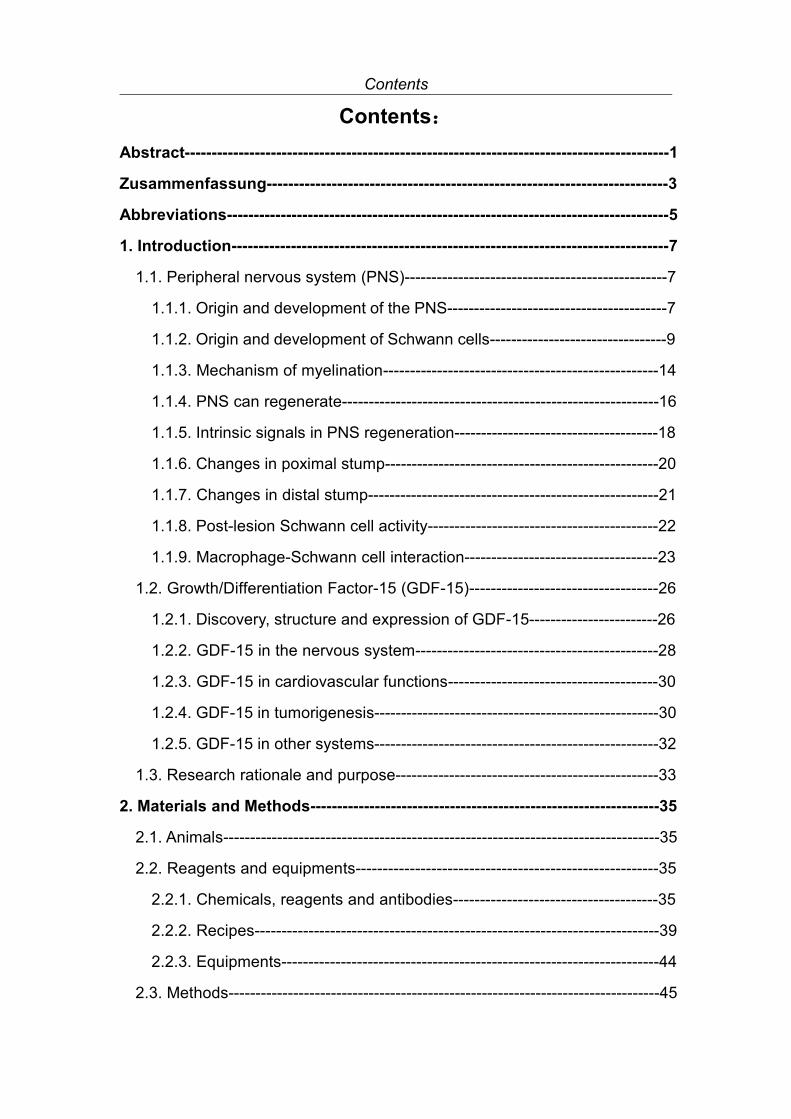
Contents
Contents:Abstract------------------------------------------------------------------------------------------1
Zusammenfassung--------------------------------------------------------------------------3
Abbreviations----------------------------------------------------------------------------------5
1. Introduction---------------------------------------------------------------------------------7
1.1. Peripheral nervous system (PNS)-------------------------------------------------7
1.1.1. Origin and development of the PNS-----------------------------------------7
1.1.2. Origin and development of Schwann cells---------------------------------9
1.1.3. Mechanism of myelination---------------------------------------------------14
1.1.4. PNS can regenerate-----------------------------------------------------------16
1.1.5. Intrinsic signals in PNS regeneration--------------------------------------18
1.1.6. Changes in poximal stump---------------------------------------------------20
1.1.7. Changes in distal stump------------------------------------------------------21
1.1.8. Post-lesion Schwann cell activity-------------------------------------------22
1.1.9. Macrophage-Schwann cell interaction------------------------------------23
1.2. Growth/Differentiation Factor-15 (GDF-15)-----------------------------------26
1.2.1. Discovery, structure and expression of GDF-15------------------------26
1.2.2. GDF-15 in the nervous system---------------------------------------------28
1.2.3. GDF-15 in cardiovascular functions---------------------------------------30
1.2.4. GDF-15 in tumorigenesis-----------------------------------------------------30
1.2.5. GDF-15 in other systems-----------------------------------------------------32
1.3. Research rationale and purpose-------------------------------------------------33
2. Materials and Methods-----------------------------------------------------------------35
2.1. Animals---------------------------------------------------------------------------------35
2.2. Reagents and equipments--------------------------------------------------------35
2.2.1. Chemicals, reagents and antibodies--------------------------------------35
2.2.2. Recipes---------------------------------------------------------------------------39
2.2.3. Equipments----------------------------------------------------------------------44
2.3. Methods--------------------------------------------------------------------------------45

Contents
2.3.1. Genotyping-----------------------------------------------------------------------45
2.3.2. Surgery---------------------------------------------------------------------------47
2.3.3. RNA and protein extraction--------------------------------------------------47
2.3.4. Reverse transcription and Real-time qPCR-----------------------------49
2.3.5. Western blot---------------------------------------------------------------------52
2.3.6. Tissue preparation-------------------------------------------------------------53
2.3.7. Paraffin embedding and sectioning----------------------------------------53
2.3.8. Cryo-embedding and sectioning--------------------------------------------54
2.3.9. DRG neuron quantification---------------------------------------------------55
2.3.10. Immunohistochemistry------------------------------------------------------55
2.3.11. Electron Microscopy---------------------------------------------------------56
2.3.12. Electromyography (EMG)--------------------------------------------------58
2.3.13. Statistical analysis------------------------------------------------------------59
3. Results--------------------------------------------------------------------------------------60
3.1. GDF-15 expression pattern-------------------------------------------------------60
3.1.1. GDF-15 in DRG after sciatic nerve lesion-------------------------------60
3.1.2. GDF-15 in sciatic nerve-------------------------------------------------------61
3.2. DRG neuron survival/death in WT and GDF-15 KO animals-------------66
3.2.1. Survival/death related genes in WT and GDF-15
KO animals----------------------------------------------------------------------66
3.2.2. DRG neuron quantification---------------------------------------------------69
3.3. GDF-15 and axonal regeneration------------------------------------------------70
3.3.1. Demyelination and regeneration related genes
in WT and GDF-15 KO animals--------------------------------------------70
3.3.2. Inflammatory genes and macrophage recruitment
in WT and GDF-15 KO animals--------------------------------------------72
3.3.3. Fibroblast and Schwann cell activation in WT
and GDF-15 KO animals-----------------------------------------------------76
3.3.4. Remyelination analysis of regrowing axons in WT
and GDF-15 KO animals-----------------------------------------------------77

Contents
3.3.5. Regenerating axon quantification in WT
and GDF-15 KO animals-----------------------------------------------------78
3.3.6. Regenerating axon diameter distribution in WT
and GDF-15 KO animals-----------------------------------------------------80
3.4. Functional recovery in WT and GDF-15 KO
animals after SN crush lesion----------------------------------------------------83
4. Discussion---------------------------------------------------------------------------------86
4.1. GDF-15 expression pattern-------------------------------------------------------86
4.2. GDF-15 and DRG neuron survival----------------------------------------------87
4.3. GDF-15 and SN regeneration----------------------------------------------------90
4.3.1. GDF-15 and axon myelination/remyelination----------------------------90
4.3.2. GDF-15 and Schwann cells/fibroblasts/macrophages----------------91
4.3.3. GDF-15 and regenerated axons--------------------------------------------94
5. Conclusion---------------------------------------------------------------------------------97
Acknowledgement--------------------------------------------------------------------------98
References----------------------------------------------------------------------------------100
Curriculum Vitae---------------------------------------------------------------------------125


Abstract
1
Abstract:
Growth/Differentiation Factor-15 (GDF-15) is a distant member of the TGF-β
superfamily, which is widely expressed in the body, most prominently in liver,
lung, kidney and exocrine glands. Functions assigned to GDF-15 to date are
mostly related to cancer biology and progression, inflammation and
cardioprotection. In the field of the nervous system, several studies had shown
that injured cortical neurons, microglia, Schwann cells and activated
macrophages are the main producers of GDF-15. Exogenous GDF-15 has
neurotrophic functions for cultured neurons, and can promote maturation of the
regenerating axons. GDF-15 deficient mice exhibit postnatal progressive
neuron loss. However, the functions and underlying signaling pathways of
GDF-15 in the nervous system are largely unknown.
In this project, crush or transection lesion on the sciatic nerve was performed
on GDF-15 knock out (KO) mice and wild type (WT) littermates. The main
parameters and markers related to GDF-15 expression, neuron survival, nerve
regeneration and functional recovery were analyzed.
GDF-15 mRNA levels in the dorsal root ganglia (DRG) were slightly
upregulated at 3 and 7 days after sciatic nerve lesion, and returned to control
level thereafter. GDF-15 expression in the sciatic nerve segments distal to the
lesion peaked at 7 days after injury, which may be related to dedifferentiation of
Schwann cells and infiltration of macrophages. GDF-15 was then
downregulated after 7 days in the sciatic nerve crush model, but it remained at
high level in the transection model. The potential neurotrophic factor Galanin
was higher expressed in the GDF-15 deficient DRG than in the WT. The
immunoreactivity of the survival-promoting molecule phospho-Bad was found
to be constitutively expressed in the GDF-15 KO DRG, but was only seen in

Abstract
2
the WT DRG after lesion. In the GDF-15 KO mice, the myelination modulator
Krox-20 was maintained at a high level in unlesioned nerve, which might
account for the hypermyelination phenotype. However, it did not alter the
remyelination during nerve regeneration. The macrophage attractor MAC-2,
cytokines IL-1β and IL-6 were found to be more prominently expressed in the
GDF-15 knock out nerve than in WT littermates. As a consequence, the levels
of macrophage markers CD11b and F4/80 in KO mice were higher than in the
WT; this was further substantiated by the staining of F4/80 in the nerve distal to
the lesion. The alternative macrophage activation markers Arginase 1 and
Ym1 were more prominently expressed in lesioned nerves from GDF-15 KO
than in WT mice.
After sciatic nerve lesion, DRG neuron loss and axon remyelination showed no
significant difference between the genotypes. However, regenerated axons in
wild type animals gained a larger diameter at a later time point after sciatic
nerve lesion, and showed a better improvement in terms of conduction velocity
and amplitude in the electromyography test, meaning a better functional
recovery.
In conclusion, GDF-15 does not alter DRG neuron death after sciatic nerve
lesion; GDF-15 suppresses macrophage recruitment and alternative activation;
finally, GDF-15 promotes the maturation of regenerated axons by enlarging the
diameter.

Zusammenfassung
3
Zusammenfassung:
Growth/Differentiation Factor-15 (GDF-15) ist ein entferntes Mitgleid der
TGF-ß Superfamilie und ist bei Säugern weit verbreitet; die höchste
Expression findet man in den Organen Leber, Lunge, Niere und in exokrinen
Drüsen. Bisher meist untersuchte Funktionen von GDF-15 betreffen die
Biologie von Krebszellen, Metastasierung, Entzündung und das pathologische
Herz. Für das Nervensystem ist in mehreren Studien gezeigt worden, dass
GDF-15 im lädierten cerebralen Cortex, in Mikroglia, in Schwannzellen und in
aktivierten Makrophagen synthetisiert und sekretiert wird. Exogen appliziertes
GDF-15 wirkt neurotroph und kann die Regeneration lädierter Axone über
einen Spalt hinweg fördern. GDF-15 Knockout-Mäuse zeigen einen
progredienten postnatalen Verlust motorischer und sensorischer Neurone.
Allerdings sind die meisten Funktionen von GDF-15 im Nervensystem noch
nicht aufgeklärt.
In meiner Dissertationsarbeit habe ich an GDF-15 WIldtyp- und
Knockout-Mäusen Quetschungs- und Durchtrennungs-Experimente am
Nervus Ischiadicus durchgeführt und die morphologischen, molekularen und
biochemischen Konsequenzen sowie die Nervenregeneration (nach
Quetschung) untersucht.
GDF-15 wurde in Spinalganglien (dorsal root Ganglien, DRG) nach 3 – 7
Tagen postläsional hochreguliert und normaliserte sich danach. Distal der
Nervenläsionsstelle erreichte die GDF-15 Expression 7 Tage nach Verletzung
ihren Gipfel; dies steht offenbar im Zusammenhang mit der
Aktivierung/Dedifferenzierung von Schwannzellen und der
Makrophageninfiltration. Nach Quetschung kehrten die GDF-15 Werte jenseits
von Tag 7 auf Kontrollniveau zurück; nach Nerv-Durchtrennung blieben die

Zusammenfassung
4
Werte hoch. Galanin ist ein neurotropher Faktor und war in GDF-15
defizienten DRGs höher exprimiert als in WIldtyp-Ganglien. GDF-15
Knockouts exprimierten phosphoryliertes Bad (das als Überlebenssignal
funktioniert) konstitutiv; in WIldtyp-Tieren wurde es nur nach Läsion
beobachtet. Krox-20 ist ein positiver Regulator der Myelinisierung; es war
prominent heraufreguliert in unlädierten Nerven von Knockout-Tieren; wir
sehen dies im Zusammenhang mit dem Hypermyelinisierungs-Phänotyp
dieser Tiere. Allerdings förderte hochreguliertes Krox-20 nicht die
Remyelinisierung bei der Nervenregeneration. Der Makrophagen-Attractor
MAC-2 sowie die Cytokine IL-1β und IL-6 waren höher exprimiert in GDF-15
knockout-nerven als in Wildtypen. Dies führte in Kockout-Tieren zu einer
höheren Makrophagen-Invasion, erkennbar an hohen Werten der
Makrophagen-Marker CD11b und F4/80. Dies entsprach auch den
immuncytochemischen Resultaten. Arginase 1 und Ym1 sind Marker für den
alternativen (M2) Aktivierungsweg für Makrophagen; beide Marker waren
prominenter exprimiert in läsionierten Nerven von GDF-15 Knockout Tieren als
in Wildtyp-Tieren. Nach Nervenläsion entwickelten sich DRG Neurontod und
Axonremyeliniserung in Knockout- und Wildtyp-Tieren nahezu ohne
Unterschied. Allerdings hatten regenerierte WIldtyp-Axone größere
Durchmesser und eine verbesserte Leitungsgeschwindigkeit, also eine
bessere funktionelle Erholung.
Die Gesamt-Schlussfolgerung ist, dass GDF-15 nach Nervenläsion für das
Überleben sensorischer Neurone in vivo unerheblich ist. Jedoch unterdrückt
GDF-15 die Makrophagen-Aktivierung und ihre alternative (M2) Aktivierung.
Wichtig ist schließlich, dass GDF-15 die Reifung regenerierender Axone durch
Vergrößerung ihrer Durchmesser fördert.

Abbreviations
5
Abbreviations:
Bcl2 B-cell CLL/lymphoma 2Bcl-xl BCL2-like 1BDNF Brain-derived neurotrophic factorcAMP Cyclic adenosine monophosphateCCR2 Chemokine (C-C motif) receptor 2CNS Central nervous systemCNTF Ciliary neurotrophic factorCREB cAMP response element-binding proteinCREM cAMP-responsive element modulatorDRG Dorsal root ganglionEgr-3 Early growth response protein 3EMG ElectromyographyERK Extracellular signal regulated kinasesGALR Galanin receptorGAP-43 Growth associated protein 43GDF-15 Growth/Differentiation Factor-15GDNF Glial cell derived neurotrophic factorGFAP Glial fibrillary acidic proteinGM-CSF Granulocyte-macrophage colony-stimulating factorGSK-3 Glycogen synthase kinase 3HIF Hypoxia inducible factorId2 Inhibitor of DNA binding 2IFN-γ Interferon gammaIGF1 Insulin-like growth factor 1IGF2 Insulin-like growth factor 2IHC ImmunohistochemistryIL-13 Interleukin 13IL-1β Interleukin 1 betaJAK Janus kinaseJNK c-Jun N-terminal kinasesKO Knock outLIF Leukemia inhibitory factorLPC LysophosphatidylcholinesMAG Myelin associated glycoproteinMAPK Mitogen-activated protein kinasesMBP Myelin basic proteinMCP-1 Monocyte chemotactic protein 1

Abbreviations
6
MIC-1 Macrophage inhibitory cytokine 1MIP-1α Macrophage Inflammatory Proteins 1 alphamTOR Mammalian target of rapamycinNAG-1 Nonsteroidal anti-inflammatory drug-activated protein-1NCAM Neural cell adhesion moleculeNF-κB Nuclear factor kappa-light-chain-enhancer of activated B cellsNGF Nerve growth factorNMSC Non-myelinating Schwann cellNPY Neuropeptide YNRG1 Neuregulin 1NT3 Neurotrophin-3Olig1 Oligodendrocyte transcription factor 1OMgP Oligodendrocyte myelin glycoproteinORF Open reading frameP0 Myelin protein zerop75NTR Low-Affinity Nerve Growth Factor ReceptorPDGFB Platelet-derived growth factor subunit BPKA Protein kinase APKB Protein kinase BPLA2 Phospholipase A2PMP22 Peripheral myelin protein 22PNS Peripheral nervous systemSC Schwann cellSCG Superior cervical ganglionSCP Schwann cell precursorSHH Sonic hedgehogSN Sciatic nerveSox10 SRY (sex determining region Y)-box 10Sox2 SRY (sex determining region Y)-box 2STAT3 Signal transducer and activator of transcription 3TGF-β Transforming growth factor betaTLR Toll-like receptorTNF-α Tumor necrosis factor alphaVEGF Vascular endothelial growth factorWT Wild type

Introduction
7
1. Introduction1.1. Peripheral nervous system (PNS)
1.1.1. Origin and development of the PNS
The mammalian nervous system can be divided into two sub-systems: the
central nervous system (CNS) and the peripheral nervous system (PNS). The
CNS consists of the brain and the spinal cord, while the PNS encompasses the
nerves and ganglia external to the CNS, mainly includes spinal nerves which
derive from the spinal cord and cranial nerves which come from the brain. The
PNS contains both the autonomic system and the somatic system, each with
components locating within the CNS and the PNS. The autonomic system
transfers information between the CNS and visceral organs (Irwin, 1993). The
somatic system conducts information between the CNS and the skin, skeleton
muscles, bones, and joints (Elenkov et al., 2000).
As reviewed extensively by numerous articles and books (e.g. Neuroscience,
3rd edition, Dale Purves, et al.; Developmental Biology, 6th edition, Scott F
Gilbert), during embryonic development, the ectoderm overlying the notochord
thickens and becomes a temporary structure, the neural plate (Chang and
Hemmati-Brivanlou, 1998; Duprat et al., 1990). The neural plate then begins to
fold and forms a transitional structure, named neural tube (Bronner-Fraser,
1994). A group of cells locating between the neural plate and the ectoderm
starts to differentiate and migrate laterally and dorsally to the neural tube. This
group of cells is termed the neural crest (Bronner-Fraser, 1995) (Fig 1.1).
The marginal region of the neural tube consists of neural ectodermal cells
which are named the neuroepithelium, and will later give rise to a
subpopulation of cells called neuroblasts (Morest and Silver, 2003). The
neuroblasts form the mantle layer locating peripherally to the neuroepithelial

Introduction
8
cells. The cells in the mantle layer finally become the gray matter of the spinal
cord (McConnell, 1995; Tanabe and Jessell, 1996). Neuroblasts send out
processes peripherally from the mantle layer and form the nerve fibers, which
are then myelinated and become the white matter in the spinal cord
(McConnell, 1995). Neuroepithelial cells also give rise to spongioblasts
(gliablasts), which will finally become the glial cells in the CNS (Morest and
Silver, 2003).
Fig 1.1 Development of spinal cord and peripheral nervous system.
Viewed transversely, the neural tube includes the central canal. The lateral
corners of the canal are called the sulcus limitans (Miller et al., 2010). This

Introduction
9
sulcus forms the boundary between the cells located dorsally from the alar
plate, and the cells located more ventrally, the latter are termed the basal plate.
The cells located in the alar plate are connected to sensory function, while the
cells from the basal plate are related predominently to motor function. During
neural development, the intermediate region between the alar plate and the
basal plate become an intermediolateral region of the gray matter, with the
neurons showing autonomic function (Bronner-Fraser, 1994; McConnell, 1995;
Tanabe and Jessell, 1996).
A group of neural crest cells, which locate dorsolaterally to the neural tube, will
further divided into segments corresponding to the somites which develop in
the mesoderm. Some of the neural crest cells go through
epithelial/mesenchymal transitions and migrate to form ganglion primordial
(finally form the dorsal root ganglia), in which the sensory neuron precursors
undergo terminal differentiation and extend axons centrally to spinal cord and
peripherally to various targets (muscle spindles, cutaneous receptors, sensory
receptors) (Miller et al., 2010). Another group of neural crest cells, located in
the margins of dorsal neural tube, migrate ventrally to a region near the dorsal
aorta. They develop to a column of sympathetic ganglion primordia, and
differentiate to obtain mainly noradrenergic properties. Finally, these cells
become the sympathetic ganglia, some of which migrate rostrally to form, e.g.,
the superior cervical ganglion (SCG) and some migrate ventrally to establish
the prevertebral ganglia, leaving the remaining cells in the column forming the
sympathetic chain (Cowen and Gavazzi, 1998; Francis and Landis, 1999).
1.1.2. Origin and development of Schwann cells
Except neurons, there are two main types of glial cells in the PNS, satellite glia
cells (SGCs) and Schwann cells (SCs). SGCs originate from the early cell
pools, e.g. boundary cap cells in neural crest. They normally surround neuron

Introduction
10
bodies within ganglia, and are considered to have various functions, such as
nutrients supply, protection, and microenvironment control (Fex et al., 2004;
Nascimento et al., 2008; Pannese, 2010).
Besides SGCs, SCs are of particular importance. SCs are the myelinating and
non-myelinating glial cells in the PNS. Depending on the formation of myelin
sheath, SCs can be further divided into two subgroups, myelinating Schwann
cells (produce myelin sheath) and non-myelinating Schwann cells (NMSCs),
both derived from the neural crest (Griffin and Thompson, 2008; Woodhoo and
Sommer, 2008) (Fig 1.1). Three developmental stages are involved in the
formation of SCs: firstly, neural crest cells migrate to the nerve trunks and
become Schwann cell precursors (SCPs) at mouse embryo day (E) 12/13;
secondly, SCPs develop into immature Schwann cells at E15/16; finally,
around birth, the immature Schwann cells differentiate into myelinating
Schwann cells and the non-myelinating Schwann cells (Jessen and Mirsky,
2005; Jessen, 2005) (Fig 1.2).
Fig 1.2 The Schwann cell lineage. Figure obtained from (Jessen and Mirsky, 2005).
Sox10 is thought to be critical for the formation of the glial lineage from the
migrating neural crest cells. Sox10 is expressed in developing SGCs and

Introduction
11
SCPs, but downregulated in immature neurons. In mice lacking Sox10, the
SGCs and SCPs are missing while neurons are in normal number. One
possible function of Sox10 might be maintaining the response to Neuregulin 1
(NRG1) in early glial cells (Garratt et al., 2000; Jagalur et al., 2011; Kuspert et
al., 2012) (Fig 1.3).
Fig 1.3 Factors involved in Schwann cell development and myelination. Figure
adapted from (Jessen and Mirsky, 2005).
Early embryonic nerves are tightly associated with SCPs, forming a column
structure without connective tissue, protective perineurium or blood supply.
Thus SCPs are thought to be essential for trophic support and proper nerve
fasciculation to immature neurons (Jessen and Mirsky, 2005). SCPs, in
contrast of mature SCs, lack autocrine survival loops and largely depend on a

Introduction
12
NRG1 signaling, which is provided by axons, for survival (Chen and Strickland,
2003; Feltri et al., 2002; Meyer and Birchmeier, 1995; Wallquist et al., 2002).
NRG1 is also a major regulator during the transition of SCPs to immature
Schwann cells (Cornejo et al., 2010; Newbern and Birchmeier, 2010).
The formation of immature Schwann cell is, in time course, paralleled with the
increase in vascularization, connective tissue spacing, and the formation of the
perineurium in the peripheral nerve (Jessen and Mirsky, 2005). At this stage,
immature Schwann cell processes surround large bundles of axons. Immature
Schwann cells gain the ability to support their own vitality by autocrine survival
factors, such as leukaemia inhibitory factor (LIF), neurotrophin 3 (NT3),
insulin-like growth factor 2 (IGF2), and platelet-derived growth factor-β
(PDGFB) (Dowsing et al., 1999; Meier et al., 1999; Weiner and Chun, 1999). It
is crucial that the autocrine circle could also promote self-survival after lesion
and the regeneration of axons. On the other hand, excess immature Schwann
cells need to be eliminated, this is probably controlled by the balance between
survival and death factors (Jessen and Mirsky, 2005). Survival factors include
axon associated NRG1, laminin from the basal lamina, as well as the autocrine
factors mentioned above (Chen and Strickland, 2003; Feltri et al., 2002; Meyer
and Birchmeier, 1995; Wallquist et al., 2002). At least two death factors have
been identified: p75 neurotrophin receptor (p75NTR), which is required for SCs
cell death, probably activated by binding of nerve growth factor (NGF) (Syroid
et al., 2000); and TGF-β, as the elevated cell death in neonatal SCs after
lesion is suppressed by injection of TGF-β-blocking antibody (Einheber et al.,
1995; Parkinson et al., 2001) (Fig 1.3).
During late embryo stages, individual immature Schwann cells gradually
associate with single axons only to reach a 1:1 ratio, which is termed radial
sorting, the pre-myelinating step (Webster et al., 1973). Around birth, immature
Schwann cells begin to differentiate into myelinating Schwann cells and,

Introduction
13
subsequently, NMSCs (Jessen and Mirsky, 2005). This reversible process
takes several weeks to complete in rodent nerves and is thought to be
controlled by axonal signals, as random association with axons of different
caliber determines subsequent maturation. Those SCs associated with large
diameter axons (>~1 μm) will bacome myelinating SC and those associated
with small diameter axons become NMSCs (Griffin and Thompson, 2008;
Woodhoo and Sommer, 2008) (Fig 1.2).
After radial sorting, myelinating Schwann cells go through comprehensive
biochemical and morphological changes concerning lipids, protein and myelin
synthesis and wrapping of lamellae to form myelin sheaths (Sherman and
Brophy, 2005). Myelination offers several biological advantages such as a
dramatic increase in conduction velocity, which directly correlates with total
fiber diameter, as well as reduction in energy costs to generate an action
potential (Gillespie and Stein, 1983; Nave, 2010). This structure increases
conduction velocity in two known ways: saltatory conduction and an increase
in axon diameter (Gillespie and Stein, 1983). Each myelinated segment of an
axon is referred to as an internode and internodes are separated by nodes of
Ranvier. Nodes of Ranvier are ~1 μm in length, rich in sodium channels, and
are seperated by the insulating myelin sheath, therefore, the electric signal can
“jump” from one node to the next (Lustig et al., 2001; Sakurai et al., 2001;
Sherman and Brophy, 2005). Myelination can also induce the phosphorylation
of neurofilament M (medium) and H (heavy), which enlarges the interfilament
spacing and consequently, increase the axonal caliber (Hisanaga and
Hirokawa, 1989; Julien and Mushynski, 1983). Myelination also offers axonal
protection and limits collateral sprouting from myelinated shafts, the latter
effect probably results from growth inhibitory molecules in the myelin, such as
netrin, myelin-associated glycoprotein (MAG) and oligodendrocyte myelin
glycoprotein (OMgP) (Gan et al., 1999; Gupta et al., 2006; Ji et al., 2008; Lim
et al., 1999; Pagany et al., 2003; Willison et al., 1988). In the PNS, myelinating

Introduction
14
fibers are mainly A and B group fibers (Navarro et al., 2007).
The non-myelinating Schwann cells (NMSCs) consist of the Schwann cells in
the Remak fibers, the terminal Schwann cells (tSCs) found at neuromuscular
junctions, and specialized sensory transducers, e.g. Pacinian corpuscles and
Meissner ’s corpuscles (Griffin and Thompson, 2008; Vega et al., 2009). All of
these NMSCs keep the ability to myelinate if they receive the appropriate
axonal signal (Navarro et al., 2007; Stoll and Muller, 1999).
In contrast to the CNS, the PNS contains large population of unmyelinated
axons, which are estimated to be 4 times more frequent than myelinated axons
(Holland et al., 1998). In rodents, 85-90% NMSCs usually ensheath more than
one axon, while the remaining 10-15% NMSCs enclose small axons in a 1:1
ratio. Interestingly, in human, all NMSCs associate with small axons in a 1:1
ratio (Sharghi-Namini et al., 2006). Non-myelinating fibers consist of
postganglionic sympathetic fibers, C fiber nociceptors, as well as some
preganglionic sympathetic and parasympathetic fibers (Griffin and Thompson,
2008). NMSCs are highly plastic, and have the ability to sprout out if necessary.
This is due to the absence of myelin, which contains the inhibitory molecules
for sprouting (Murinson and Griffin, 2004).
1.1.3. Mechanism of myelination
Myelination is a complex process and includes signals from microenvironment
and axon (Jessen and Mirsky, 2005; Sherman and Brophy, 2005).
Axon-associated neuregulin-1 controls the formation and thickness of the
myelin sheath (Cornejo et al., 2010; Newbern and Birchmeier, 2010; Stassart
et al., 2013; Wolpowitz et al., 2000). Many neurotrophic factors participate in
the myelination process, including glial cell line-derived neurotrophic factor
(GDNF) (Averill et al., 2004; Hoke et al., 2003; Piirsoo et al., 2010),

Introduction
15
brain-derived neurotrophic factor (BDNF)(Omura et al., 2005; Tolwani et al.,
2004; Xiao et al., 2009; Yamauchi et al., 2004), and insulin-like growth factors
(IGF-1/2)(Cheng et al., 2000; Silva et al., 2009). Schwann cells also produce
myelin structrue proteins to construct myelin sheaths: myelin protein zero
(P0)(Trapp, 1988), myelin basic protein (MBP)(Topilko et al., 1994), peripheral
myelin protein 22 (PMP22)(Snipes et al., 1999), and
myelin-associate-glycoprotein (MAG)(Trapp, 1988). Current evidence has
shown that myelination is regulated by balancing the positive and negative
transcriptional regulators. Positive transcription factors for myelin are normally
upregulated in immature Schwann cells, which are essential for the initiation of
myelination, inter alia, Sox10 (Britsch et al., 2001; Paratore et al., 2001),
Krox-20 (Egr-2)(Topilko et al., 1997; Zorick et al., 1999), octamer-binding
transcription factor 6 (Oct-6)(Ghazvini et al., 2002; Mandemakers et al., 2000;
Smith et al., 2001) and brain 2 class III POU-domain protein (Brn-2) (Jaegle et
al., 2003). Sox10, except its role in the transition from neural crest cells to
SCPs, is critical for Oct-6 expression and the induction of myelin genes in
immature Schwann cells (Jagalur et al., 2011; Paratore et al., 2001). Krox-20
deficient mice show a 1:1 ratio of axon to immature Schwann cells, but fail to
form myelin sheaths, indicating that Krox-20 is crucial for myelination but not
radial sorting (Topilko et al., 1994). However, Krox-20 is shown to be essential
to maintain the myelin sheath in physiological condition (Decker et al., 2006).
Oct-6 has functional overlap with Brn-2 in terms of myelinogenesis, as Oct-6
conditional KO mice showed a delayed initiation of myelination compared with
WT littermates (Ghazvini et al., 2002; Jaegle et al., 2003).
On the other hand, there are negative transcriptional regulators of myelination,
which are generally activated in immature Schwann cells, deactivated in
myelinating Schwann cells, re-activated upon denervating/lesion conditions to
induce Schwann cell dedifferentiation, orchestrating suppression of myelin
protein gene expression. After lesioning the nerve, the negative regulators are

Introduction
16
believed to induce the dedifferentiation program of SCs, which leads to a
phenotype similar to immature Schwann cells and then remyelinate the
regrowed axons (Jessen and Mirsky, 2008). Currently, several candidates of
negative regulators have been documented, including the transcription factors
c-Jun (Parkinson et al., 2008), Pax-3 (Blanchard et al., 1996), Sox2 (Le et al.,
2005; Parrinello et al., 2010), Krox-24 (Grose et al., 2002; Topilko et al., 1997),
Egr-3 (Mercier et al., 2001), Notch (Mirsky et al., 2008; Woodhoo et al., 2009)
and Id2 (Le et al., 2005; Mager et al., 2008). c-Jun is a central member of the
AP-1 transcription factor family and can be phosphorylated by Jun N-terminal
kinases (JNKs) (Raivich et al., 2004). c-Jun is actively expressed in Krox-20
deficient nerves, where axon myelination is restricted (Parkinson et al., 2004;
Pham et al., 2009). c-Jun is essential for SC dedifferentiation: firstly, c-Jun is
upregulated in denervated SCs following nerve lesion; secondly, c-Jun
deficient SCs fail to phagocytose myelin debris; thirdly, c-Jun null SCs are
unable to suppress myelin genes expression (Arthur-Farraj et al., 2012; Pham
et al., 2009). Notch is a transmembrane receptor, and is cleaved after binding
to the ligand. The Notch intra-cellular domain (NICD) is then released and
passes the signal down and regulates downstream transcription (Li et al.,
2004). Notch is also known to control the developmental transition from SCPs
to immature Schwann cells, as well as SC proliferation within embryonic
nerves (Kubu et al., 2002; Morrison et al., 2000). Like c-Jun, Notch also acts as
a negatively regulator of myelination postnatally (Woodhoo et al., 2009)(Fig
1.3).
1.1.4. PNS can regenerate
One of the most significant differences between the PNS and CNS is the fact
that the PNS has the ability to spontaneously regenerate after lesion. Although
in clinical cases in humans, the regenerative results are mostly suboptimal, the
experiments on rodents have produced satisfactory results in many instances

Introduction
17
(Reyes et al., 2005). The major features leading to the regenerative difference
in the PNS and CNS are well documented and include multiple facets of
intrinsic growth programs, the inhibitory molecules associated with myelin,
removal of myelin debris, glial scar formation, activation of phagocytic cells,
and bridging for regeneration in lesion sites (Cafferty et al., 2008; Giger et al.,
2010).
The major events in the PNS after lesion include: axon degradation, collapse
of myelin sheaths, proximal neurite sprouting, Schwann cell proliferation
forming the Bands of Büngner, invasion of circulating macrophages, removal of
myelin debris, elongation of the axon distally, reconstruction of the myelin
sheath, and reinnervation of the targets (Abe and Cavalli, 2008; Chen et al.,
2007; Fawcett and Keynes, 1990; Navarro et al., 2007; Stoll and Muller, 1999)
(Fig 1.4).
Fig 1.4 Major events after PNS lesion. Figure obtained from (Navarro et al., 2007).

Introduction
18
The most widely used methods for studying regeneration in the PNS in rodents
are nerve transection (neurotmesis), crush injury (axonotmesis) and chronic
constrictive injury (neuropraxia) (Chen et al., 2007; Navarro et al., 2007).
Among these, crush injury and chronic constrictive injury, which normally leave
the epineurium intact, allow fast regeneration and fully functional recovery;
while in case of nerve transection, surgical or mechanical repair is necessary
to obtain satisfactory regeneration (Fawcett and Keynes, 1990; Hudson et al.,
2000).
1.1.5. Intrinsic signals in PNS regeneration
Axons are complex structures that require a combined support for the
functional maintainance and regeneration from the neuronal cell bodies and
the attached glial cells. Lesioning an axon leads to the loss of a large amount
of axonplasmic volume and results in chromatolysis (dissolution of the Nissl
bodies) of the neuron soma; furthermore, this intervention causes soma and
nucleolar swelling, as well as nuclear eccentricity (Lieberman, 1971; Navarro
et al., 2007; Stoll and Muller, 1999). The injured neurons switch their metabolic
machinery from generating neural transmitters and nerve impulses to
promoting self-survival and regenerating axons. The percentage of sensory
neuron death in DRG after SN lesion has been reported to amount to between
10 and 30%, affecting preferentially more the small than the large neuron
populations (Groves et al., 1999; Shi et al., 2001; Tandrup et al., 2000). A slight
loss of 0–10% motoneurons has been found after adult SN injury (Lowrie et al.,
1994; Vanden et al., 1993). Neurons in the adult are less vulnerable to die than
immature neurons, whereas lesions close to the cell bodies cause more
neuronal death than distal lesions (Snider et al., 1992; Ygge, 1989).
Nerve injury also results in the activation of an intrinsic growth capacity which
has been extensively studied: several molecules are strongly upregulated or

Introduction
19
reexpressed in neurons after nerve lesion. Cyclic adenosine monophosphate
(cAMP) signaling functions through protein kinase A (PKA) activity and
switches on downstream genes via cAMP response element binding protein
(CREB), to initiate and promote regeneration program (Cai et al., 2002; Gao et
al., 2004). Downstream molecules of cAMP signaling include Arginase 1
(ARG1), neuropeptide Y (NPY), protein kinase A (PKA), CREM (cAMP
response element modulator), and IL-6 (Parlato et al., 2006; Spooren et al.,
2010; Zhou et al., 2012). Lesion also upregulates trophic factors, such as
BDNF, NGF, and NGF receptor (Averill et al., 2004; Delcroix et al., 2003;
Meyer et al., 1992; Micera et al., 2007; Piirsoo et al., 2010; Wilhelm et al.,
2012). c-Jun has been shown to contribute to regeneration, as the c-Jun null
neurons fail to organize a proper regeneration program (Arthur-Farraj et al.,
2012; Fontana et al., 2012; Yuan et al., 2012). c-fos, similar to c-Jun, also
promotes neuron regeneration (Dai et al., 2009; Jergova et al., 2008; Yuan et
al., 2009). ATF3 and signal transducer and activator of transcription 3 (STAT3)
are induced in DRG neurons after peripheral injury, but not central lesion
(Kiryu-Seo et al., 2008; Reid et al., 2010; Saul et al., 2010). STAT3 may
promote the survival of motor neurons after peripheral nerve injury by
activating some motor neuron survival factors, such as Reg-2 and Bcl-xl
(Schwartz et al., 2002; Sekikawa et al., 2008). Other regeneration-associated
genes upregulated in neurons following injury include Galanin, Integrin α7β1,
CD44, and GAP-43 (Frey et al., 2000; Hirata et al., 2002; Lindwall et al., 2004;
Raivich et al., 2004; Son et al., 2007). These genes differentially promote
neuron survival or regeneration by enhancing neurite sprouting and
cytoskeletal reconstruction (Fig 1.5).

Introduction
20
Fig 1.5 Intrinsic signals in PNS regeneration. (Chen et al., 2007).
1.1.6. Changes in poximal stump
Lesion leads to a retrogradely degeneration from the lesion site towards
neuron body, but depends on the degree of injury, only one or a few intermodal
lengths are affected, and certain amount of cytosol outflows before the
opening reseals by the fusion of cellular membrane. Within hours, proximal
axons start to regrow collaterally and distally inside of the basal lamina
(Navarro et al., 2007; Stoll and Muller, 1999). The newly sprouted neurites are
protruding through the injury and are decreased in numbers when they reach
and contact the endoneurium in the distal stump (Fawcett and Keynes, 1990).
This process is strongly dependent on the guidance of SCs which form
physical contact to the regrowing axons (Donnerer, 2003). The regenerating
sprouts will stay unmyelinated until a later stage, regardless of the previous

Introduction
21
myelination status of axons (Flores et al., 2000). Regrowing axons will
preferentially reinnervate the endoneurial tubes in the distal stump over the
neighboring tissues (Son and Thompson, 1995).
1.1.7. Changes in distal stump
Immediately after lesion, the axonal membranes proximal to the injury site fuse
the opening ends to stop the cytosolic content from outflow (Fawcett and
Keynes, 1990). On the contrary, the distal axonal segment will undergo
Wallerian degeneration, a process first described by Augustus Waller in 1850.
Wallerian degeneration involves the granular disintegration of the cytoskeleton
systems and the breakdown of previous myelin sheath which is due to the
calcium-mediated proteolysis triggered by the damage of membrane. Schwann
cells and macrophages are activated and phagocytose the myelin breakdown
products and axonal debris (Fu and Gordon, 1997; Makwana and Raivich,
2005; Navarro et al., 2007). This is a critical step for Wallerian degeneration
that helps to provide a permissive microenvironment for axon regeneration.
SCs form column-shaped structures (Bands of Büngner) which could guide
axons to correct targets. Axons reconnected to the proper distal targets would
benefit from trophic supply and finally enlarge their diameter to normal level in
a late stage. On the contrary, axons that fail to reconnect with correct targets,
may grow into connective tissue, and will later be eliminated (Hall, 1997; Liu,
1996).
Neurotrophic factors, such as NGF, NT-3 and BDNF, have all been shown to
be upregulated after lesion and induce axon growth (Makwana and Raivich,
2005; Snider et al., 2002). Cytokines such as interleukin-6 (IL-6) are related to
nerve regeneration, as mice with conditional knock out of IL-6 exhibit sensory
defects and impaired DRG axons regeneration after lesion (Tofaris et al., 2002;
Zhong et al., 1999). Another immune regulator, leukemia inhibitory factor (LIF)

Introduction
22
also shows its role in the nerve regeneration by studies using LIF knockout
animals (Cafferty et al., 2001; Livesey et al., 1997). STAT3 has been shown to
be not only essential for the survival of spinal motor neurons, but also critical
for the expression of IL-6 and LIF (Dominguez et al., 2010; Haas et al., 1999;
Ohbayashi et al., 2007). Extracellular matrix molecules and cell adhesion
molecules, such as laminin, NCAM, fibronectin and L1 are also necessary for
guiding axons to pass the injury site, to enter the distal stumps and finally to
reach the targets (Chen et al., 2005; Chen and Strickland, 2003; Chernousov
et al., 2008; Parrinello et al., 2010; Hansen et al., 2008; Schäfer and Frotscher,
2012; Schmid and Maness, 2008).
1.1.8. Post-lesion Schwann cell activity
Nerve lesion triggers dramatic morphological and physiological changes in the
nerve segment distal to injury. Schwann cells transit to a status similar to
immature Schwann cells, termed dedifferentiation (Arthur-Farraj et al., 2012).
The SCs phagocytose axon and myelin debris and briefly proliferate, which
peaks at 3-4 days post lesion (Navarro et al., 2007; Stoll and Muller, 1999).
The molecular phenotype of immature Schwann cells and adult
dedifferentiated Schwann cells is different (Arthur-Farraj et al., 2012; Jessen
and Mirsky, 2005). Several molecules are well documented: the lipid antigen
O4 is a marker of immature Schwann cells, also expressed in both myelinating
and non-myelinating adult Schwann cells, but downregulated in
dedifferentiated cells (Mirsky et al., 1990). Conversely, Integrin α1β1 is absent
from immature cells but is expressed in denervated SCs (Stewart et al., 1997).
In vivo, N-cadherin is suppressed to a low level in immature cells, but is
strongly activated in lesioned nerves (Thornton et al., 2005; Wanner et al.,
2006). Furthermore, immature cells in developing nerves are known to be
different from mature NMSCs, because NMSCs are P0 mRNA negative,
galactocerebroside positive and Integrin α1β1, α7β1 positive. The NMSCs also

Introduction
23
differ from dedifferentiated cells, as the latter express P0 mRNA, but lack of
galactocerebroside and O4 (Mirsky et al., 2008).
Schwann cell dedifferentiation is a key step of successful regeneration. At the
dedifferentiation stage, SCs downregulate genes related to myelin production
and maintainance, including myelin structural proteins, P0, MBP, MAG,
periaxin and cholesterol synthesizing enzymes (LeBlanc and Poduslo, 1990;
Scherer et al., 1995; Stoll and Muller, 1999). Dedifferentiated Schwann cells
also reprogram to express molecules normally found in immature SCs, such as
L1, NCAM, p75 low affinity neurotrophin receptor (p75NTR), and glial fibrillary
acidic protein (GFAP) (Johnson et al., 1988; Schmid and Maness, 2008; Tacke
and Martini, 1990; Thornton et al., 2008; Triolo et al., 2006). Furthermore,
dedifferentiated SCs upregulate trophic factors such as NGF, BDNF and GDNF,
and cytokines, such as TNF-α, IL-1β, IL-6, LIF and MCP-1, to facilitate the
regenerative program (Flugel et al., 2001; Nadeau et al., 2011; Piirsoo et al.,
2010; Taskinen and Roytta, 2000; Wilhelm et al., 2012). Although the details of
transcriptional control of the phenotype transition are still unclear, a recent
study has demonstrated that c-Jun is actually the major controller of the
dedifferentiation phenotype (Arthur-Farraj et al., 2012).
1.1.9. Macrophage-Schwann cell interaction
During Wallerian degeneration, products of degenerated neural tissue could
stimulate monocyte chemotactic protein-1 (MCP-1) expression in SCs partially
through Toll-like receptor 2/3 (TLR2/3) (Boivin et al., 2007; Karanth et al., 2006;
Lee et al., 2006). Other cytokines such as tumor necrosis factor alpha (TNF-α),
IL-1α and macrophage inflammatory protein 1 alpha (MIP-1α) are also found in
SCs immediately after SN injury. IL-1β expression is then induced by TLRs and
related to the early response to injury (Taskinen and Roytta, 2000). After
macrophages have infiltrated into the nerve, they also express TNF-α, IL-1α,

Introduction
24
and IL-1β (Gordon and Martinez, 2010; Martini et al., 2008; Tofaris et al., 2002).
Those molecules mentioned above stimulate the expression of the
phospholipase A2 (PLA2) family members, which consist of both intracellular
and secretory forms (Murakami et al., 1997). PLA2s hydrolyze membrane
phosphotidylcholine, which is a main component of the myelin membranes,
resulting in the production of lysophosphotidylcholine (LPC), which has
potential myelinolytic activity. Injection of LPC into the SN could induce
demyelination within short time, which probably results from the activation of
the classic complement pathway, since LPC could bind to C-reactive protein
(Larsen et al., 2003; Ousman and David, 2000). Thus, the PLA2s induced LPC
generation has the ability of stimulating myelin breakdown at the beginning of
Wallerian degeneration. Myelin-derived lipids are also reabsorbed for
regeneration and remyelination. Lipid binding proteins, such as
Apolipoproteins D and E (ApoD, ApoE), accumulate in the distal nerve portion
after injury. ApoD is mainly expressed by endoneural fibroblasts, while ApoE is
primarily produced by infiltrating macrophages (Li et al., 2010; Spreyer et al.,
1990; Stoll and Muller, 1986).
Macrophages play an important role in the processes of removing myelin and
axonal debris in the lesioned nerve, especially the ones from hematogenous
origin (Hu and McLachlan, 2003; Rosenberg et al., 2012; Shibata et al., 2003).
They invade into the lesioned loci within the first 48 hours and spread to the
whole distal part of the nerve by no later than day 4, and their quantity peaks
by 14 days (Bendszus and Stoll, 2003). In vitro studies have demonstrated that
IL-1β and TNF-α promote myelin phagocytosis by recruiting infiltrating
macrophages (Shamash et al., 2002). In vivo evidence shows that functional
blocking antibodies neutralizing MCP-1/MIP-1α or IL-1β dramatically reduce
the number of phagocytic macrophages (Perrin et al., 2005). Macrophage
surface marker, MAC-2 (galactin-3) participates in myelin phagocytosis and
has been reported to be induced by Granulocyte-macrophage

Introduction
25
colony-stimulating factor (GM-CSF) in peripheral nerve (Reichert et al., 1994;
Saada et al., 1996). GM-CSF is found to be expressed predominantly by active
fibroblasts in the lesioned nerve, pointing out the involvement of non-neural
cells in the process of Wallerian degeneration (Mirski et al., 2003).
Nevertheless, fibroblasts can also form a bridge connecting the nerve stumps
in transection lesions and, through ephrin-B/EphB2 signaling, guide the SC
migration and direct the axon regrowth (Parrinello et al., 2010). SCs express
MAC-2 and surprisingly, are able to phagocytose myelin debris; thus, before
macrophages infiltrating into the distal stump, SCs are the main contributor of
phagocytosis (Saada et al., 1996)(Fig 1.6).
Fig 1.6 A pathway map of Schwann cell-Macrophage interaction during PNS
regeneration. Figure obtained from (Martini et al., 2008).
Macrophages exhibit an anti-inflammatory phenotype after they have started
phagocytosing myelin. NgR1, which is found to be a common receptor for all
three axonal regrowth inhibitory molecules in myelin (Nogo-A, MAG and

Introduction
26
OMgp), could lead to the growth cone collapse in the CNS and impair neurite
growth (Martini et al., 2008). NgR2 is also found in peripheral nerve, and has a
higher affinity binding to MAG than to Nogo-A or OMgp (Venkatesh et al.,
2005). NgR1 is downregulated in axons after nerve lesion, but is highly
expressed in phagocytic macrophages. The proportion of all macrophages
expressing NgR1 and NgR2 increases after lesion and reaches a peak by day
7 (Fry et al., 2007). On the contrary, only a few (12%) circulating monocytes
express NgR1 and NgR2 (Fry et al., 2007). Circulating macrophages have to
infiltrate into the basal lamina of SCs for phagocytosis, and afterwards, at the
end stage of Wallerian degeneration, they have to migrate out of the basal
lamina (Martini et al., 2008). NgR plays an essential role on the outward
migration of macrophages after clearance of debris, as NgR expressing
macrophages are pushed away when they contact with the NgR ligands (MAG
and OMgp) in newly synthesized myelin of regenerated axon. This repulsion is
believed to be induced by the Rho-A pathway (David et al., 2008; Fry et al.,
2007).
1.2. Growth/Differentiation Factor-15 (GDF-15)
1.2.1. Discovery, structure and expression of GDF-15
Growth/Differentiation Factor-15 (GDF-15) is a distant member in the
transforming growth factor beta (TGF-β) superfamily and was discovered in
the late 1990s almost simultaneously by several different laboratories (Bootcov
et al., 1997; Böttner et al., 1999; Fairlie et al., 1999). According to the different
screening/cloning methods and functions involved, GDF-15 has received
various names, such as Nonsteroidal anti-inflammatory drug (NSAID)
activated gene-1 (NAG-1)(Baek et al., 2001), macrophage inhibitory cytokine-1
(MIC-1)(Bootcov et al., 1997), placental transformation growth factor-β
(PTGFB)(Lawton et al., 1997), prostate-derived factor (PDF)(Paralkar et al.,
1998) and placental bone morphogenetic protein (PLAB)(Hromas et al., 1997).
Introduction
27
The GDF-15 gene consists of two exons seperated by a single intron. This
pattern resembles closely GDF-9 and inhibin α and β in the TGF-β superfamily.
The rat and mouse GDF-15 preproprotein contains 303 amino acids (aa),
compared to human which has 308 amino acid, including a 29 aa signal
peptide, a 167 aa propeptide and a 112 aa mature protein (Böttner et al.,
1999)(Fig 1.7). The preproprotein contains an RXXR dibasic cleavage site at
the C-terminal. The generation of active mature protein, which is a 25 KDa
secreted dimer, needs the removal of a signal sequence, disulfide-linked
dimerization of GDF-15 monomers, and the furin-like protease cleavage at the
RXXR site (Böttner et al., 1999). Nevertheless, recent evidence also suggests
that GDF-15 can be secreted in both processed and unprocessed form, using
different secretory pathways (Bootcov et al., 1997). The mature peptides from
the three species all contain two cysteine residues in addition to the seven
cysteines required to form the cystine knot, which is also the conserved feature
among TGF-β superfamily members. Unlike the other members, GDF-15
shows the lowest conservation between species, as the mature protein from
human and rat only share 70% conserved residues (Böttner et al., 1999). The
analysis of the promoter region has identified many regulatory sites upstream
of the ORF region, including several AP-1 and SP-1 sites, as well as WT-1,
NF-κB, AP-2, PUR factor, HiNF-c and MIG-1 binding sites, suggesting that
GDF-15 can be regulated by various upstream molecules (Böttner et al.,
1999).

Introduction
28
Fig 1.7 Gene structure of gdf-15. Figure obtained from (Fairlie et al., 1999).
GDF-15 has been shown to be expressed ubiquitously in adult tissues, with
relatively higher expression in prostate, placenta, bronchi, bronchioli, exocrine
glands, macrophages, brain choroid plexus, kidney, lung, liver and spleen, and
with comparably lower expression in heart, gut, skeletal muscle, thymus and
pituitary, suggesting its multiple functionality (Böttner et al., 1999; Kadara et al.,
2006; Nakamura et al., 2003; Unsicker et al., 2013; Xu et al., 2006). GDF-15 is
also found in mouse embryo, e.g. in skin and cartilaginous tissue, indicating
that it has a role in developmental control (Paralkar et al., 1998).
1.2.2. GDF-15 in the nervous system
GDF-15 has been shown to have a neurotrophic function in in vitro
experiments, as it could promote the survival of cultured midbrain
dopaminergic neuron, cerebellar granule neurons, sensory, sympathetic and
spinal cord motor neurons (Strelau et al., 2009). Furthermore, GDF-15 KO

Introduction
29
mice exhibit a postnatal progressive neuron loss phenotype, including spinal
cord, trigeminal, facial motoneurons and DRG sensory neurons, with a
reduction of ~20% in quantity from postnatal day 90 (P90) to P180. Neuron
loss is accompanied by impaired behavioral function and rotarod performance
(Strelau et al., 2009). GDF-15 KO mice also upregulate myelin-related genes,
including MBP, P0 and PMP22, leading to hypermyelination in the PNS
(Strelau et al., unpublished data). SCs have been identified as a producer of
GDF-15, which can be retrogradely transported along the axons (Strelau et al.,
2009). The GDF-15 KO phenotype partially resembles the CNTF KO
phenotype, but exact underlying mechanisms are still uncovered (Sendtner et
al., 1994).
In normal physiological conditions, GDF-15 expression is found ubiquitously in
all regions of rodent CNS and PNS, with the highest mRNA levels in the
choroid plexus, where GDF-15 protein is secreted into the cerebrospinal fluid
(Strelau et al., 2000). Current studies show that GDF-15 is regulated after
lesioning the nervous system. In the cold-lesioned cerebral cortex, GDF-15
mRNA is found to be upregulated in the region adjacent to the lesion and
peaks at 36-48h (Schober et al., 2001). Interestingly the main source of
GDF-15 are the lesioned neurons as well as a moderate number of microglia,
this is in contrast to the notion that activated macrophages are the major
producer of GDF-15 in other tissue (de Jager et al., 2011; Fairlie et al., 1999;
Schlittenhardt et al., 2004). A more recent experiment on mouse optic nerve
lesions has shown GDF-15 to be upregulated post lesion, together with some
important cell-death/survival related molecules, such as ATF-3, Bad, and Bcl-2
(Charalambous et al., 2013; Subramaniam et al., 2003). Galanin and Caspase
8 were shown to be differentially regulated in GDF-15 KO and WT mice.
However, cell death of retinal ganglion cells in GDF-15 KO and WT mice
follows identical quantitative and temporal patterns (Charalambous et al.,
2013).

Introduction
30
Local application of GDF-15 into the gap after a sciatic nerve transection has
been shown to result in an improved sensory, but not motor regeneration.
Additionally, in the GDF-15 treated animal, less but more mature regenerated
axons were found suggesting that GDF-15 may be involved in the scenarios of
PNS regeneration (Mensching et al., 2012).
1.2.3. GDF-15 in cardiovascular functions
The largest portion of all publications related to GDF-15 is concentrated on its
cardiovascular functions. After myocardial infarction, GDF-15 is rapidly
upregulated in the infarcted myocardium in both mouse models and human
patients, and can keep this high expression level for a prolonged period
(Kempf et al., 2006; Kempf et al., 2007; Kempf et al., 2011). GDF-15 has been
reported to be increased in atherosclerotic lesions, colocalized with
macrophages. GDF-15 deficient mice have a decreased macrophage
infiltration, chemokine receptor 2 (CCR2) expression, apoptosis and necrotic
core formation (Bonaterra et al., 2012; de Jager et al., 2011). Increased levels
of GDF-15 in circulating blood have been observed in patients with acute
myocardial infarction (Kempf et al., 2006). GDF-15 also shows a protective
effect on cultured cardiomyocytes in reducing apoptosis (Kempf et al., 2006).
In patients with heart failure, levels of GDF-15 and increases in GDF-15 over
time have been linked to an adverse outcome (Kehl et al., 2012). Collectively,
GDF-15 is viewed as a marker and predictor of cardiometabolic risk and heart
failure. GDF-15 has also been identified as a novel autocrine/endocrine
molecule that antagonizes the hypertrophy and loss of ventricular function,
possibly via the SMAD 2/3 pathway (Xu et al., 2006).
1.2.4. GDF-15 in tumorigenesis
Elevated GDF-15 expression has been characterized as a feature of various

Introduction
31
tumors, including breast, gastric, colon, ovarian and prostate cancer. Serum
GDF-15 levels are significantly increased in metastatic cancers, particularly
colorectal cancer (Baek et al., 2009; Brown et al., 2003; Dubey et al., 2012;
Kim et al., 2005; Welsh et al., 2003). During epithelial neoplastic
transformation, GDF-15 is upregulated in response of many antitumorigenic
stimuli e.g. anti-inflammatory chemicals and gamma irradiation. During the
early stages of carcinogenesis, anti-cancer reagent-induced GDF-15 can
result in tumor cell apoptosis, cell cycle arrest and inhibition of blood vessel
formation (Unsicker et al., 2013).
The specific role of GDF-15 in tumorgenesis is still not fully understood, but
some clues give a preliminary idea about it. GDF-15 is shown to be stimulated
on the activation of the MAPK-ERK1/2 or PKB/Akt pathway by SP-1 family
transcription factors in breast or gastric cancer (Wollmann et al., 2005).
GDF-15 can also activate HIF-1 and VEGF by phosphorylation and activate
the ErbB receptor, mTOR, and ERK1/2 pathways, while downregulating ErbB2
can lead to the suppression of GDF-15-mediated downstream signaling (Kim
et al., 2008). GDF-15 is also found to be induced by hepatitis C virus and in
hepatocellular carcinoma, where the GDF-15 autocrine signaling of infected
hepatocytes mediates GSK-3/β catenin, Akt and Raf phosphorylation (Si et al.,
2011). This is followed by cell-cycle regulation by e.g. cyclins A2, D2 and E1,
or cell adhesion molecules, such as E-cadherin (Si et al., 2011). In malignant
glioblastomas, GDF-15 is upregulated following anoxia, indicating its role in
angiogenesis (Boyle et al., 2009; Huh et al., 2010). GDF-15 is highly
expressed in malignant melanomas and leads to the formation of new vessels
affecting the tumor tissue size (Huh et al., 2010).
The effect of GDF-15 in tumorgenesis is also related to immunoregulation.
Studies based on GDF-15 deletion or knock down show an enhanced natural
killer T cell mediated cytotoxicity, and delayed growth of glioma cells, which

Introduction
32
resembles the phenotype of TGF-β downregulation (Friese et al., 2004; Roth
et al., 2010). p53 has been reported to be necessary for GDF-15 expression.
Tumor cell migration decreased in response to GDF-15, with unaffected
proliferation (Cheng et al., 2011; Song et al., 2012). Furthermore, in a model of
prostate cancer, GDF-15 has been shown to influence tumor invasion through
the Rho GTPase network, however, the underlying mechanisms are still
unclear (Cheng et al., 2011).
1.2.5. GDF-15 in other systems
Because of its ubiquitous tissue distribution, GDF-15 is involved in diverse
functions and in multiple processes. GDF-15 can serve as an
anti-inflammatory cytokine in the model of heart infarction, by inhibiting the
recruitment of macrophage towards the lesion loci (Kempf et al., 2011). This is
achieved by the activation of Cdc42, repression of Rap 1, and clustering of β2
integrin on leukocytes. GDF-15 has also been verified as an acute phase
mediator of the CCR2/TGFβRII dependent inflammation, and as a regulator of
the IL-6 dependent inflammation in response of vascular injury (Kempf et al.,
2011). GDF-15 also has an impact on metabolic control, such as appetite,
obesity, and body weight change (Johnen et al., 2007). Obese patients
express high levels of GDF-15, especially patients suffering from type II
diabetes (Lajer et al., 2010). GDF-15 is associated with keratinocyte
differentiation and several skin diseases, such as skin sclerosis and melanoma
(Ichikawa et al., 2008; Kluger et al., 2011; Yanaba et al., 2012). GDF-15 also
regulates osteoclastic differentiation through inhibiting carbonic anhydrase II
and cathepsin K (Hinoi et al., 2012; Vanhara et al., 2009).

Introduction
33
1.3. Research rationale and purpose
The probably most fascinating feature of the peripheral nervous system is its
ability to spontaneously regenerate after lesion. This regeneration requires a
complex orchestration of different physiological processes, including the
retrograde transfer of lesion signal(s), transcriptional transition, morphological
changes, neuron survival, axon degeneration, Schwann
cell/macrophage/fibroblast activation, neurite regrowth, axon remyelination
and target reinnervation, finally leading to functional recovery. Research on
PNS regeneration has a great and profound significance: firstly, to better
understand the mechanisms of PNS regeneration provides new approaches to
promote CNS regeneration; secondly, research on rodent models may
eventually lead to improvements for the therapy of human patients.
GDF-15 has been shown to have ubiquitous origins and involvement in
different biological processes. Our particular interests focuses on nervous
system regeneration: firstly, GDF-15 may be expressed by macrophages and
Schwann cells, both of which are the most important participants in PNS
regeneration; secondly, GDF-15 is related to the trophic support of cultured
neurons and maintenance of adult neuron in vivo, which might be applied to
the in vivo situation following injury; thirdly, GDF-15 has a role in the regulation
of cell adhesion molecules and recruitment of macrophages, as well as in the
suppression of inflammation. These facts revealed by previous research have
generated the hypothesis that GDF-15 might be involved in the orchestration
of PNS regeneration.
We used GDF-15 knock out mice and respective wild types as model animals,
applied sciatic nerve crush or transection+stump ligation, to investigate: firstly,
whether GDF-15 plays a role in PNS regeneration; secondly, what is the
expression pattern of GDF-15 during PNS regeneration; thirdly, which are the

Introduction
34
targets and interacting molecules within the GDF-15 network.

Materials and Methods
35
2. Materials and Methods2.1. Animals
All animal treatments were performed with permission from the responsible
authority of the University Clinic Freiburg. C57BL6/J WT mice were provided
by the animal facility of the BioMed Zentrum, University Clinic Freiburg. The
GDF-15lacZ/lacZ mouse colony was established in Heidelberg (Strelau et al.,
2009) on a C57BL6/J background and was reestablished in Freiburg by
embryo transfer. The animals were held under standard conditions, with a
12/12 hours day/night cycle. Food and water were provided ad libitum. All
experiments were done on the mice of 5~6 months old.
Fig 2.1 Structure of the GDF-15 gene and strategy for knock out this gene. Figure
obtained from (Strelau et al., 2009).
2.2. Reagents and equipments
2.2.1. Chemicals, reagents and antibodies
All chemicals used in the present study are listed below in Table 2.1.

Materials and Methods
36
Table 2.1 Chemicals
Chemicals Catalog # Company
Acetic acid 20104.334 VWR
Ammonium persulfate (APS) 9592.2 Carl Roth
Bromochloropropane B9673-200ML Sigma
Citric acid monohydrate C7129-500G Sigma
Cresyl echt violet 1A396 Chroma
Glutaraldehyde 1.12179.0025 VWR
Hydrogen chloride 30271-1L Sigma
Isopropanol P/7500/17 Fisher Scientific
K2HPO4 1.05104 Merck
KCl 1.04936.1000 Merck
KH2PO4 3904.1 Carl Roth
Methanol 20847.320 VWR
Na2HPO4 P030.3 Carl Roth
Na2HPO4·2H2O 4984.1 Carl Roth
NaH2PO4·2H2O 6345.1000 Merck
NaCl 9265.1 Carl Roth
Osmium tetraoxide 8088.1 Carl Roth
Paraformaldehyde (PFA) 1.04005.1000 Merck
Propylene oxide 00236-1 Polysciences
SDS pellets CN30.2 Carl Roth
Sodium hydroxide 1.06498.1000 Merck
Sucrose 4621.1 Carl Roth
TEMED T9281-25ML Sigma
Toluidin blue 1B481 Chroma
Triton X-100 X100-100ML Sigma
Trizma base T6066-1KG Sigma
Tween-20 P9416-100ML Sigma

Materials and Methods
37
All reagents and kits used in the present study are listed below in Table 2.2.
Table 2.2 Reagents and Kits
Reagents Catalog # Company
Agarose M3044.0500 GENAXXON
BCA Protein Assay Kit 23227 Thermo Scientific
Blue film 30-100 Genesee Scientific
Bovine serum albumin (BSA) 3737.2 Carl Roth
DNA Extraction Solution QE09050 Epicentre
DNA Ladder SM0241 Thermo Scientific
dNTP Mix U1511 Promega
Durcupan™ ACM A 44611-100ML Sigma
Durcupan™ ACM B 44612-100ML Sigma
Durcupan™ ACM C 44613-100ML Sigma
Durcupan™ ACM D 44614-100ML Sigma
ECL Prime RPN2232 Life Sciences
Entellan 1.07691 VWR
Fluoromount-G 0100-01 SouthernBiotech
GelRed™ Nucleic Acid Gel Stain 41003 Biotium
GoTaq® qPCR Master Mix A6001 Promega
Hyperfilm ECL 28-9068-37 Life Sciences
Inhibitorcocktail Plus 3751.1 Carl Roth
Isofluran CDS019936 Sigma
Ketamine 400203.00.00 Cp-pharma
M-MLV Reverse Transcriptase M1701 Promega
Non-fat milk T145.2 Carl Roth
Paraffin 39502004 McCormick
Protein ladder 7711s NEB
PVDF membrane 0.2 um 39-4010 PeqLab
PVDF membrane 0.45 um NEF1002001PK Pekin Elmer
Materials and Methods
38
Random Primers C1181 Promega
RedTaq Mastermix (2x) M3029.0100 GENAXXON
RNA Clean & Concentrator™-25 R1018 ZYMO Research
RNAlater Stabilization Solution AM7020 Life Technologies
RNasin® Plus RNase Inhibitor N2611 Promega
Rotihistol 6640.1 Carl Roth
Rotiphorese® Gel 30 (37.5:1) 3029.1 Carl Roth
RQ1 RNase-Free DNase M6101 Promega
Tissue freezing medium 020108926 Leica
TriFast 30-2020 PeqLab
Xylazine 6293841.00.00 BAYER
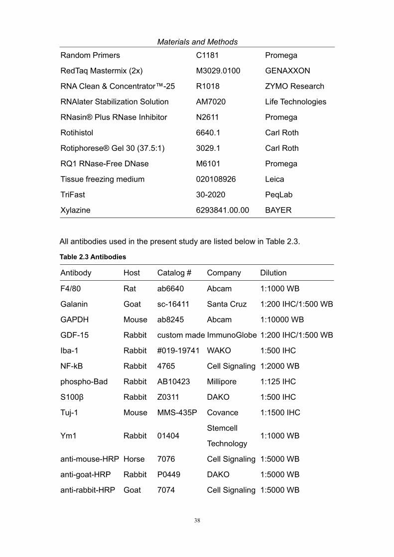
All antibodies used in the present study are listed below in Table 2.3.
Table 2.3 Antibodies
Antibody Host Catalog # Company Dilution
F4/80 Rat ab6640 Abcam 1:1000 WB
Galanin Goat sc-16411 Santa Cruz 1:200 IHC/1:500 WB
GAPDH Mouse ab8245 Abcam 1:10000 WB
GDF-15 Rabbit custom made ImmunoGlobe 1:200 IHC/1:500 WB
Iba-1 Rabbit #019-19741 WAKO 1:500 IHC
NF-kB Rabbit 4765 Cell Signaling 1:2000 WB
phospho-Bad Rabbit AB10423 Millipore 1:125 IHC
S100β Rabbit Z0311 DAKO 1:500 IHC
Tuj-1 Mouse MMS-435P Covance 1:1500 IHC
Ym1 Rabbit 01404Stemcell
Technology1:1000 WB
anti-mouse-HRP Horse 7076 Cell Signaling 1:5000 WB
anti-goat-HRP Rabbit P0449 DAKO 1:5000 WB
anti-rabbit-HRP Goat 7074 Cell Signaling 1:5000 WB

Materials and Methods
39
anti-rat-HRP Goat 7077 Cell Signaling 1:5000 WB
DaM-Dylight405 Donkey 715-475-151 Dianova 1:500 IHC
DaM-Alexa488 Donkey 715-545-151 Dianova 1:500 IHC
DaM-Cy3 Donkey 715-165-151 Dianova 1:500 IHC
DaR-Dylight405 Donkey 711-475-152 Dianova 1:500 IHC
DaR-Alexa488 Donkey 711-545-152 Dianova 1:500 IHC
DaR-Cy3 Donkey 711-165-152 Dianova 1:500 IHC
DaR-Alexa594 Donkey 711-585-152 Dianova 1:500 IHC
DaG-Alexa488 Donkey A-11055Life
Technologies1:500 IHC
DaG-Cy3 Donkey 705-165-147 Dianova 1:500 IHC
DaM-Biotin Donkey 715-065-151 Dianova 1:500 IHC
DaR-Biotin Donkey 711-065-152 Dianova 1:500 IHC
DaG-Biotin Donkey 705-065-147 Dianova 1:500 IHC
Streptavidin-Cy2 016-220-084 Dianova 1:500 IHC
Streptavidin-Cy3 016-160-084 Dianova 1:500 IHC
Donkey Serum Donkey C06SBZ AbD serotec 10% IHC
2.2.2. Recipes
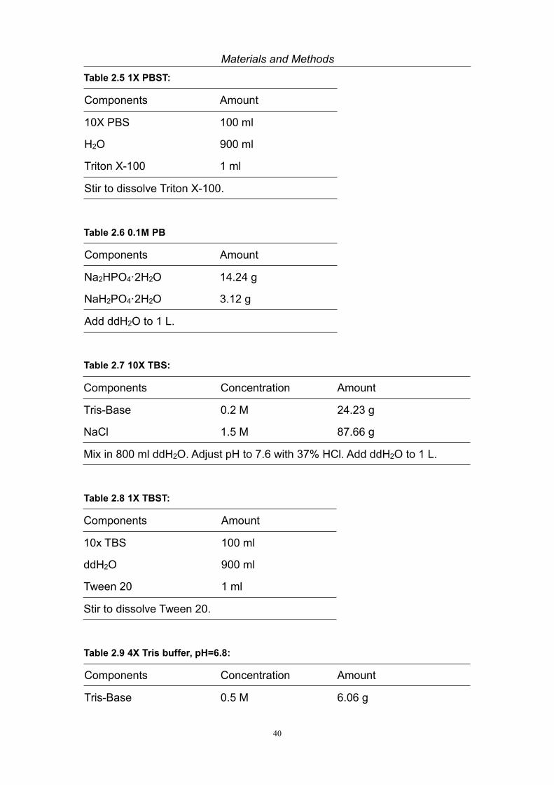
All recipes used in the present study are listed below in Table 2.4—Table 2.20.
Table 2.4 10X PBS:
Components Amount
NaCl 80 g
KCl 2 g
Na2HPO4 14.4 g
KH2PO4 2.4 g
Add ddH2O to 800 ml. Adjust pH to 7.4.
Add ddH2O to 1 L.
Materials and Methods
40
Table 2.5 1X PBST:
Components Amount
10X PBS 100 ml
H2O 900 ml
Triton X-100 1 ml
Stir to dissolve Triton X-100.
Table 2.6 0.1M PB
Components Amount
Na2HPO4·2H2O 14.24 g
NaH2PO4·2H2O 3.12 g
Add ddH2O to 1 L.
Table 2.7 10X TBS:
Components Concentration Amount
Tris-Base 0.2 M 24.23 g
NaCl 1.5 M 87.66 g
Mix in 800 ml ddH2O. Adjust pH to 7.6 with 37% HCl. Add ddH2O to 1 L.
Table 2.8 1X TBST:
Components Amount
10x TBS 100 ml
ddH2O 900 ml
Tween 20 1 ml
Stir to dissolve Tween 20.
Table 2.9 4X Tris buffer, pH=6.8:
Components Concentration Amount
Tris-Base 0.5 M 6.06 g

Materials and Methods
41
ddH2O 80 ml
Adjust pH to 6.8 with 10N or 6N HCl. Add ddH2O to 100 ml.
Table 2.10 4X Tris buffer, pH=8.8:
Components Concentration Amount
Tris-Base 1.5 M 45.4 g
ddH2O 200 ml
Adjust pH to 8.8 with 10 N or 6 N HCl. Add ddH2O to 250 ml.
Table 2.11 10X Western blot electrophoresis buffer:
Components Concentration Amount
Tris-Base 250 mM 30.3 g
Glycine 1.92 M 144.1 g
SDS 1% 10 g
Add ddH2O to 1 L. Do not adjust pH, it should be ~8.3.
Table 2.12 10X Western blot transblot buffer:
Components Concentration Amount
Tris-Base 250 mM 30.3 g
Glycine 1.92 M 144.1 g
Add ddH2O to 1 L. Do not adjust pH, it should be ~8.3.
Table 2.13 Western blot transblot working buffer:
Components Amount
10x Transblot buffer 100 ml
Methanol 200 ml
ddH2O 700 ml
Pre-cool before use.

Materials and Methods
42
Table 2.14 5X Western blot sample loading buffer:
Components Concentration Amount
SDS 1 g
DTT 2.5 M 2 ml
Tris buffer pH=6.8 2 M 1.5 ml
Mix well to dissolve SDS in a 15 ml tube, incubate in 37°C if necessary.
BPB 5% 100 ul
Add ddH2O to 5 ml and mix thoroughly.
Add 5 ml prewarmed glycerol and mix thoroughly.
Table 2.15 Western blot stripping buffer:
Components Amount
Glycine 7.5 g
ddH2O 800 ml
Adjust pH to 2.5 by 6 N or 10 N HCl.
Add ddH2O to 1 L.
Table 2.16 SDS-PAGE separating gel:
Components 8% 12% 15% 18%
4X Tris buffer pH=8.8 5 ml 5 ml 5 ml 5 ml
ddH2O 9.7 ml 7 ml 5 ml 3 ml
30% Acrylamide (37.5:1) 5.3 ml 8 ml 10 ml 12 ml
10% SDS 200 µl 200 µl 200 µl 200 µl
TEMED 25 µl 25 µl 25 µl 25 µl
10% APS 62.5 µl 62.5 µl 62.5 µl 62.5 µl
Table 2.17 SDS-PAGE 4% stacking gel:
Components Amount
4X Tris buffer pH=6.8 1.25 ml

Materials and Methods
43
ddH2O 3.05 ml
30% Acrylamide (37.5:1) 0.65 ml
10% SDS 50 µl
TEMED 15 µl
10% APS 37.5 µl
Table 2.18 0.1M Citric acid buffer:
Components Amount
Citric acid monohydrate 21.01 g
Add ddH2O to 800 ml. Adjust pH to 6.0 by NaOH.
Add ddH2O to 1 L.
Table 2.19 0.5% Cresyl violet for Nissl staining:
Components Amount
1M Na-acetate 40 ml
100% Acetic acid 9.6 ml
Add ddH2O to 1 L.
Cresyl violet 5 g
Stir overnight.
Table 2.20 Resin for Electron Microscope sample embedding:
Components Amount
Durcupan™ ACM A 24 ml
Durcupan™ ACM B 36 ml
Durcupan™ ACM C 0.3 ml
Durcupan™ ACM D 0.9 ml

Materials and Methods
44
2.2.3. Equipments
All equipments used in the present study are listed below in Table 2.21.
Table 2.21 Equipments:
Equipments Catalog # Company
Balance BP4100S Sartorius
CCD camera 2K Sharp-eye Troendle
Centrifuge 5804R Eppendorf
Cryo-microtome cm3050s Leica
DAKO Pen S200230-2 DAKO
Data acquisition device PowerLab26T AD Instruments
DNA electrophoresis chamber Horizo 11.14 Life Technologies
DNA electrophoresis power supply EPS601 Amersham
Electron microscope LEO 906E Carl Zeiss
EMG electrode MLA1204 AD instruments
FC Multiscan plate reader 357 Thermo Fisher
Fluorescence microscope Axioplan 2 Carl Zeiss
Heating rack 2-2515 Neolab
Horizontal shaker KS250 basic IKA
iQ™ 96-Well PCR Plates 223-9441 Bio-Rad
Laboratory temperature controllers LTR 2055 Jucheim Solingen
Michel suture clips 12040-01 F.S.T
Microtome 2050 Reichert Jung
PCR cycler Mastercycler Eppendorf
pH meter FEP20 Mettler Toledo
Precellys Keramik-Kit 1.4 mm 91-PCS-CK14S PeqLab
Precellys 24 homogenisator 91-PCS24 PeqLab
Real-time PCR cycler iQ5 System Bio-Rad
Silk Suture Thread 18020-60 F.S.T

Materials and Methods
45
Stereoscope Stemi DV4 Carl Zeiss
Superfrost Plus slide J1800AMNZ Thermo Scientific
Ultramicrotome EM UC6 Leica
Westernblot chember Mini Trans-Blot Bio-Rad
Westernblot power supply PowerPac HC Bio-Rad
X-Clear Advanced Polyolefin StarSeal E2796-9795 StarLab
2.3. Methods
2.3.1. Genotyping
Total genomic DNA was extracted by the QuickExtractTM DNA Extraction
Solution from Epicenter. Briefly, 50 µl extraction buffer was added into tubes
containing mouse tail tips, vortexed for 15 sec and incubated at 65°C for 6 min.
Then samples were vortexed again for 15 sec and incubated at 98°C for 2 min.
Resulting DNA was processed immediately or stored at -20°C until use.
2 sets of primers for genotyping were used, as listed in Table 2.22 (Strelau et
al., 2009).
Table 2.22 Primers for GDF-15 knock out mice genotyping:
Set Name Sequence 5'--3'
Set 1 F21 GCAGAGAGGCTGAGGAACTT
LacZR4 GTTCTTGTTGGTCAAAGTAAACGAC
F30 TTGGGAAAAGGTTGGAGAGA
R18 GATACAGGTGGGGACACTCG
Set 2 NeoF1 TCGCCTTCTTGACGAGTTCT
R20 CCCAGTCTTGTAGACAGAGCAA
F26 ATGCGCACCCAAGAGACT
R21 GGCCACCAGGTCATCATAAG

Materials and Methods
46
All primers were ordered from Metabion, and diluted to 50 µM with ddH2O.
The RedTaq Mastermix (2x) from Genaxxon was used for PCR. Each reaction
had a total volume of 25 µl, including 8.5 µl H2O, 12.5 µl RedTaq Mastermix
(2x), 3 µl DNA, 1 µl primer set 1 or 2 (0.25 µl of each individual primer). The
programs are listed below (Table 2.23).
Table 2.23 Programs for genotyping:
Reaction for primer Set 1 Reaction for primer Set 2
94°C hot start 94°C hot start
94°C 3 min 94°C 3 min
94°C 1 min 94°C 30 sec
61°C 2 min 59°C 30 sec
72°C 3 min 72°C 1 min
Repeat for another 39 cycles Repeat for another 39 cycles
72°C 10 min 72°C 5 min
8°C 8°C
The PCR products were then loaded onto 1% agarose gels and detected by
Gel Red. The outcome of genotyping was the following (Fig 2.1):
Primer set 1: Primer set 2:
Fig 2.1 Genotyping results using the 2 sets of primers.
690 bp
320 bp
690 bp320 bp
1810 bp
806 bp
1810 bp806 bp

Materials and Methods
47
2.3.2. Surgery
Animals were anesthetized with 2.5% (v/v with oxygen) Isoflurane (Forane)
through inhalation. When the respiration became deep and slow (~1 time per
second), the toe tips were pinched to check the reflection. After reaching deep
anesthesia, the animal was placed onto a heating plate with constant 39°C,
cornea gel was applied, hair on the limb was shaved and the skin was
disinfected by 70% isopropanol. The skin and muscle was opened by scissors
at mid-thigh level, until reaching the sciatic nerve. Lesions were introduced at a
locus 1 cm proximal to the branching point of tibial nerve and common fibular
nerve. For crush lesion, 5/45 forceps, with graphite powder sticking on it, was
used to crush the sciatic nerve for 15 seconds; for transection lesion, 2 knots
were made by 6/0 suture silk with a 1 mm interval, followed by the transection
of nerve between the 2 knots. The wound was then closed by wound clips.
Animals were kept on the heating plate until wakeup, and then put back to the
standard condition mentioned above.
2.3.3. RNA and protein extraction
The animals were sacrificed by cervical translocation. The tissues needed (e.g.
sciatic nerve, DRG) were dissected, briefly washed in pre-cooled PBS, and
then kept in RNALater reagent. After shaking overnight at 4°C, the tissue could
be used for RNA extraction or for long-term storage at -20 or -80°C. Tissue
from 2 animals of same treatment were pooled and transferred from RNALater
to homogenesis tube containing ceramic beads and 400 µl Trifast reagent.
Tissues were smashed by bead-beating in homogeniser (5000 rpm 15 sec x2
for nerve, 5000 rpm 15 sec x3 for DRG). The RNA extraction protocol was
modified from literature (Chey et al., 2011), basically, after homogenesis in 400
µl Trifast, 80 µl bromochloropropane was added and samples were shaked
vigorously by hand for 15 sec, kept at room temperature (RT) for 5 min,

Materials and Methods
48
followed by centrifugation at 12000g 4°C, 15 min. Then the colorless
supernate (~200 µl) was transferred to a new 0.5 ml tube, with 200 µl
isopropanol added, while the remaining intermediate white layer and
red-colored organic layer was for the protein extraction later. The compound
was invert-mixed by hand and kept at -20°C for at least 10 min to precipitate
RNA. The tubes were then centrifuged at 12000g 4°C, 10 min, followed by
discarding the supernate, adding 300 µl 75% ethanol, inverting the tube and
centrifuged at 7500g 4°C, 5 min. The ethanol was completely removed by
pipette, and the remaining RNA pellet at the bottom of tube was air-dried in the
hood for 3~5 min, then 80 ul RNase-free water was added to dissolve the RNA.
RNA solution was then purified by RNA Clean & Concentrator™-25 kit, from
Zymo Research. This procedure was exactly followed the instruction of the kit.
The purified RNA was measured by Nanodrop N-1000 spectrophotometer.
The white layer and red organic phase bearing protein was mixed with 120 µl
ethanol, inverted by hand, kept at RT for 5 min, then centrifugated at 2000g
4°C, 5 min. The supernate was then transferred into a new 2 ml tube, followed
by adding 700 µl ethanol and vortex, then by adding 200 µl
bromochloropropane and vortex, furthermore, by adding 600 µl water and
vortex. The mixture was left at RT for 5 min, followed by 12000g 10 min
centrifugation for phase seperation. The upper colorless layer was discarded
while the intermediate white layer and bottom red organic phase was
transferred into a new 1.5 ml tube. 700 µl ethanol was added to the 1.5 ml tube,
and mixed by vortex, centrifuged at 12000g for 15 min. All the liquid phase was
then discard, the pellet was air-dried in the hood for 10 min. 4% SDS solution
containing proteinase inhibitor (1% inhibitorcocktail plus) was added to
dissolve the pellet, by shaking at RT. The protein concentration was
determined by BCA kit.

Materials and Methods
49
2.3.4. Reverse transcription and Real-time qPCR
Total RNA concentration was determined, and 1 µg RNA was used for reverse
transcription. The reaction was listed below (Table 2.24).
Table 2.24 Reaction for reverse transcription:
Components Volume µl
Total RNA+H2O 20
Ribonuclease Inhibitor (40 u/µl) 0.5
RQ1 DNase 0.5
Incubate at 37°C 20 min, 70°C 10 min
5 x reaction buffer 8
dNTP (10 mM each) 4
Random primers 2
H2O 4.3
M-MLV reverse transcriptase 0.7
Incubate at 37°C 2 hours, 90 °C 5 min
cDNA product could be used for Real-time qPCR immediately or stored at
-20°C.
For Real-time qPCR, each well in 96 wells plate contained 10 µl GoTaq®
qPCR Master Mix, 4.8 µl H2O, 0.1 µl forward primer (50 mM), 0.1 µl reverse
primer (50 mM) and 5 µl cDNA (diluted according to the gene expression). The
reaction was the following (Table 2.25).
Table 2.25 Real-time qPCR reaction:
Components Volume µl
GoTaq® qPCR Master Mix 10
Forward primer (50 µM) 0.1
Reverse primer (50 µM) 0.1
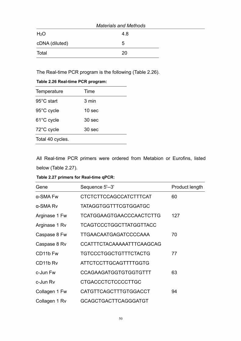
Materials and Methods
50
H2O 4.8
cDNA (diluted) 5
Total 20
The Real-time PCR program is the following (Table 2.26).
Table 2.26 Real-time PCR program:
Temperature Time
95°C start 3 min
95°C cycle 10 sec
61°C cycle 30 sec
72°C cycle 30 sec
Total 40 cycles.
All Real-time PCR primers were ordered from Metabion or Eurofins, listed
below (Table 2.27).
Table 2.27 primers for Real-time qPCR:
Gene Sequence 5'--3' Product length
α-SMA Fw CTCTCTTCCAGCCATCTTTCAT 60
α-SMA Rv TATAGGTGGTTTCGTGGATGC
Arginase 1 Fw TCATGGAAGTGAACCCAACTCTTG 127
Arginase 1 Rv TCAGTCCCTGGCTTATGGTTACC
Caspase 8 Fw TTGAACAATGAGATCCCCAAA 70
Caspase 8 Rv CCATTTCTACAAAAATTTCAAGCAG
CD11b Fw TGTCCCTGGCTGTTTCTACTG 77
CD11b Rv ATTCTCCTTGCAGTTTTGGTG
c-Jun Fw CCAGAAGATGGTGTGGTGTTT 63
c-Jun Rv CTGACCCTCTCCCCTTGC
Collagen 1 Fw CATGTTCAGCTTTGTGGACCT 94
Collagen 1 Rv GCAGCTGACTTCAGGGATGT

Materials and Methods
51
F4/80 Fw ATGGACAAACCAACTTTCAAGGC 95
F4/80 Rv GCAGACTGAGTTAGGACCACAA
Fibronectin Fw CGGAGAGAGTGCCCCTACTA 77
Fibronectin Rv CGATATTGGTGAATCGCAGA
Fizz 1 Fw TCCTGCCCTGCTGGGATGACTGCTA 125
Fizz 1 Rv CAGCGGGCAGTGGTCCAGTCAA
Galanin Fw TTTCTCAGTTTCTTGCACCTTAAA 110
Galanin Rv CAGTGGACATGGTCTCAGGA
GAPDH Fw GGCATTGCTCTCAATGACAA 97
GAPDH Rv ATGTAGGCCATGAGGTCCAC
GDF-15 Fw GAGCTACGGGGTCGCTTC 130
GDF-15 Rv GGGACCCCAATCTCACCT
Iba-1 Fw ATGAGCCAAAGCAGGGATTT 169
Iba-1 Rv ACTCCATGTACTTCACCTTG
IL-1β Fw CGTGGACCTTCCAGGATGAG 111
IL-1β Rv CGTCACACACCAGCAGGTTA
IL-6 Fw GCTGGTGACAACCACGGCCT 158
IL-6 Rv TGCACAACTCTTTTCTCATTTCCACGA
Krox-20 Fw CTACCCGGTGGAAGACCTC 112
Krox-20 Rv AATGTTGATCATGCCATCTCC
MAC-2 Fw GCCTACCCCAGTGCTCCT 82
MAC-2 Rv GGTCATAGGGCACCGTCA
MCP-1 Fw TTAAAAACCTGGATCGGAACCAA 121
MCP-1 Rv GCATTAGCTTCAGATTTACGGGT
NCAM Fw CCAGTGGACAAGCCTCTGA 106
NCAM Rv GGGGGCTCAGACTTTGTTT
NF-κB Fw GGGTCTGGGGATACTGAACA 108
NF-κB Rv GCCTCCATCAGCTCTTTGAT
NGF Fw TATACTGGCCGCAGTGAGGT 109

Materials and Methods
52
NGF Rv GGACATTGCTATCTGTGTACGG
Olig1 Fw CTCGCCCAGGTGTTTTGTTG 149
Olig1 Rv TAAGTCCGAACACCGATGGC
S100β Fw AACAACGAGCTCTCTCACTTCC 66
S100β Rv CTCCATCACTTTGTCCACCA
SHH Fw TGGAAGCAGGTTTCGACTGG 165
SHH Rv CGGGACGTAAGTCCTTCACC
Ym 1 Fw AGACTTGCGTGACTATGAAGCATTG 96
Ym 1 Rv GCAGGTCCAAACTTCCATCCTC
2.3.5. Western blot
25 or 50 µg total proteins (depending on the expression level) were loaded
onto SDS-PAGE gels; the concentration of gel differed according to the
molecular weight of the target protein. Electrophoresis was performed under a
condition of constant 120 V, for 100 min. Then proteins in the gel were
transblotted onto 0.2 µm or 0.45 µm PVDF membrane (depending on the
molecular weight of protein) under 100 V, for 100 min. After a brief washing by
TBST, the membrane was blocked by 5% non-fat milk for 1 hour at RT,
followed by TBST washing and primary antibody (dissolved in 3% BSA)
incubation at 4°C overnight with shaking. Generally, following 3 times 5 min
washing by TBST, the secondary antibody conjugated with HRP (in 5% non-fat
milk) was then applied onto the membrane for 1 h at RT. Subsequently, the
membrane was washed and incubated with the substrate of HRP for enhanced
chemiluminescence (ECL) (500 µl A + 500 µl B /membrane) for 5 min and the
signals were visualized by film. The optical density of the Bands on the film
was measured by Image J software and used for quantification (GAPDH was
used in all cases as a reference gene).

Materials and Methods
53
For stripping the membrane was incubated with stripping buffer at RT for 1.5 h
with shaking, and then further incubated with 1% SDS at RT for 1.5 h with
shaking. To detect proteins, whose molecular weights differed from the
reference protein GAPDH (~37 KDa), the membrane could also be cut and
incubated with different primary antibodies simultaneously, avoiding re-blotting.
2.3.6. Tissue preparation
Animals were anesthetized by an intraperitoneal injection of Ketamine (187.5
mg/kg body weight). Nervous reflexes were checked by pinching the toe tip.
The thorax was opened and animals were transcardially perfused with 20 ml of
pre-cooled 0.1 M PB, followed by 20 ml pre-cooled 4% PFA (in 0.1 M PB), with
a speed of 3~3.5 ml/min. The tissues were then carefully dissected and
immersed in 4% PFA (in 0.1 M PB) at 4°C with shaking for post-fixation (for
DRG and nerve, 4 hours post-fixation).
2.3.7. Paraffin embedding and sectioning
After post-fixation, the tissues underwent a dehydration process as the follows
(Table 2.28).
Table 2.28 Dehydration process for paraffin embedding:
Reagents Sciatic nerve DRG Tibial nerve
50% Ethanol 5 min 3 min 3 min
70% Ethanol 5 min 3 min 3 min
80% Ethanol 5 min 3 min 3 min
90% Ethanol 5 min 3 min 3 min
95% Ethanol 5 min 3 min 3 min
100% Ethanol 5 min X2 3 min X2 3 min X2
Rotihistol 5 min X2 3 min X2 3 min X2

Materials and Methods
54
Paraffin 15 min 15 min 15 min
Paraffin 30 min X2 30 min X2 30 min X2
The tissues were then embedded in paraffin and left at RT overnight for
solidification. After microtome sectioning at the desired thickness, the sections
were mounted onto Superfrost slides and left on the heating plate (~40°C)
overnight. Before staining, sections went through a de-paraffinization
procedure as follows (Table 2.29).
Table 2.29 De-paraffin procedure:
Reagents Time
Rotihistol 5 min X4
100% Ethanol 5 min X2
95% Ethanol 5 min
90% Ethanol 5 min
80% Ethanol 5 min
70% Ethanol 5 min
PBS 5 min
For antigen retrieval, sections were immersed in boiling 0.1 M citric acid buffer
(pH=6.0) for 3 min. Sections could then be used for staining immediately or
stored in PBS at 4°C for 1~2 days if preferred.
2.3.8. Cryo-embedding and sectioning
After post-fixation, DRG and sciatic nerve were immersed in 15% sucrose
(dissolved in PBS) solution at 4°C for 6~8 hours with shaking, and
subsequently transferred to 30% sucrose (in PBS) solution at 4°C overnight
with shaking for cryo-protection. After carefully absorbing the excess sucrose,
with cooling of dry ice and a metal plate, tissues were embedded in tissue

Materials and Methods
55
freezing medium and then stored at -20°C (short term) or -80°C (long term).
Cryo-embedded tissue was cut on a cryotome to the preferred thickness and
mounted onto superfrost slides, then kept at -20°C (short term) or -80°C (long
term). Before staining, sections were brought to RT and re-hydrated in PBS for
3 times 5 min prior to further processing.
2.3.9. DRG neuron quantification
DRG neuron quantification was down as previously described (Strelau et al.,
2009). Briefly, DRGs were cut into serial sections of 8 µm thickness, Nissl
stained (see Table 2.30), and mounted with Entellan. Neuronal profiles with a
visible nucleolus were counted in every fifth section using the Image J program.
Total numbers were corrected by Abercrombie’ s formula.
Table 2.30 Nissl staining for DRG section:
Reagents Time
0.5% Cresyl Echt Violet 7~8 min
H2O + 250 μl acetic acid 5 sec
H2O 5 sec
70% Ethanol 5 sec
80% Ethanol 5 sec
90% Ethanol 5 sec
95% Ethanol 5 sec
100% Ethanol 5 min X2
Rotihistol 5 min X3
2.3.10. Immunohistochemistry
Generally, cryo- or paraffin embedded tissues were cut at 10 µm thickness.

Materials and Methods
56
After pre-staining procedures (re-hydration or de-paraffin) as mentioned above
in 2.3.7. and 2.3.8., the sections were encircled by DAKO pen with its water
repellence property. 10% normal donkey serum in PBST (300 µl per slide) was
used for antigen blocking at RT for 1 hour, followed by 3 times 5 min PBS
washing. Then primary antibody/antibodies (antibodies from different animal
source could be used together) diluted in PBST (200 µl per slide) was/were
applied onto the slides and incubated at 4°C overnight. After 3 times 5 min
PBS washing, secondary antibody/antibodies diluted in PBS (200 µl per slide,
antibodies with different species reactivity could be used together) conjugated
with fluorescent tags was/were incubated at RT for 1 hour. After washing by
PBS, 1:500 Hoechst or 1:1000 DAPI in PBS was utilized to stain the nucleus at
RT for 10 min. Finally, after washing, slides were mounted with fluoromount-G
medium.
For proteins with very low expression, e.g. GDF-15, modification in the
protocol was helpful. Basically, the secondary antibody was not conjugated
with fluorescent group, but with biotin. Subsequently, streptavidin linked with
fluorescent tag was used to visualize the signal.
2.3.11. Electron Microscopy
After perfusion with 4% PFA, the sciatic nerve/tibial nerve was dissected and
attached to a piece of thick filter paper to maintain the shape. Then the nerve
piece with the filter paper was incubated in 2.5% glutaraldehyde solution (in
0.1 M PB) for 2 h at RT. Tissue was then washed 3 times for 5 min each with
0.1 M PB, and incubated in 1% OsO4 in 0.05 M PB in the dark, at RT for 2 h,
followed by 3 times 5 min washing with 0.05 M PB. Samples could then be
stored at 4°C overnight, and were dehydrated as described in Table 2.31.

Materials and Methods
57
Table 2.31 Dehydration procedure for Electron Microscopy embedding:
Reagents Time
50% Ethanol 10 min
70% Ethanol 10 min
80% Ethanol 10 min
90% Ethanol 10 min
95% Ethanol 10 min
100% Ethanol 10 min X3
Propylene oxide 10 min X3
1:1 Propylene oxide:Resin 2 hours
Resin 2 hours
After dehydration, the nerve was carefully seperated from filter paper, and
embedded into Epoxy resin in a rubber mold, polymerized at 58°C in the oven
for ~3 days.
The tissue was then transversely cut into semi-thin sections (500 nm) on an
ultramicrotome and stained with 1% toluidin blue to check the quality of fixation.
To measure the g-ratio, samples were cut into ultra-thin sections (60 nm) at 0.5
cm distal to the lesion site, mounted onto copper slot grids coated with
Formvar, and photos were taken at 1670x fold magnificantion on a Zeiss
electron microscope. Random photos were chosen and all intact myelinated
axons on the screen were measured in two directions (long axis and short axis)
for the diameter of the axon, the axon plus myelin, as shown in Fig 2.2. The
g-ratio was then calculated by the formula: g=((A/B)+(C/D))/2.

Materials and Methods
58
Fig 2.2 g-ratio measurement.
2.3.12. Electromyography (EMG)
5 and 9 weeks after the unilateral sciatic nerve crush, WT or GDF-15 KO mice,
were anesthetized by intraperitoneal injection of 187.5 mg/kg ketamine and
12.5 mg/kg xylazine in physiological saline. Reflexes were checked by
pinching the toe tip. One grounding electrode was subcutaneously injected into
the tail. To stimulate the sciatic nerve, skin and muscles were opened at
mid-thigh level and two stimulation needle electrodes were placed next to the
sciatic nerve. Two recording needle electrodes were injected subcutaneously
above the musculus digitorum brevis. The sciatic nerve was stimulated by
increasing current pulses with a duration of 200 ms, and the resulting EMG
signals were recorded at the musculus digitorum brevis.
First, the left unlesioned control site was measured and then the EMG of the
right lesioned site was recorded. For the analysis of latency and amplitude,
only signals triggered by supramaximal stimuli were evaluated. Amplitude was
defined as voltage interval between minimal and maximal peak of the recorded
signal. The latency was defined as the time interval between the given stimulus
and the maximal peak of the recorded signal. The conduction velocity (CV)
was calculated by distance from stimulation to recording site divided by
C D
A
B
Axon
Myelin
g=((A/B)+(C/D))/2

Materials and Methods
59
latency.
2.3.13. Statistical analysis
Data are shown as mean ± SEM (only in EMG test) or mean ± SD (all the other
tests). Significant differences were determined by unpaired t-test (neuron
quantification, g-ratio, EMG test) or by One-Way ANOVA followed by Tukey
post test using GraphPad software. P value was shown as: ns (p>0.05, not
significant), * (0.05>P>0.01, significant), ** (0.01>P>0.001, very significant), ***
(P<0.001, extremely significant).

Results
60
3. Results3.1. GDF-15 expression pattern
It has previously been shown that GDF-15 is upregulated in cerebral cortical
neurons following a “cold lesion” of the cortex and in retinal ganglion cells
(RGCs) after optic nerve crush (Charalambous et al., 2013; Schober et al.,
2001; Unsicker et al., 2013). One question addressed in the present study
concerns whether in the peripheral nervous system (PNS) GDF-15 is similarly
regulated in lesion paradigms. We used sciatic nerve (SN) crush and
transection lesions.
3.1.1. GDF-15 in DRG after sciatic nerve lesion
Since the two types of lesion to SN (crush and transection) were previously
shown to have different effects on dorsal root ganglia (DRG) neuron survival
(Navarro et al., 2007), both lesion methods were initially applied to wild type
(WT) animals in the present study. At different time points post lesion, animals
were sacrificed and total RNA from DRGs was isolated and GDF-15 mRNA
transcriptional levels were determined by quantitative Real-Time qPCR
(qRT-PCR). After crush lesion, GDF-15 mRNA levels were slightly increased,
reaching the maximum at 3 and 7 days post crush, followed by a decrease to
control levels after 28 days (Fig 3.1 A). In the transection model, GDF-15
showed a similar pattern as in the crush model, however, with a slight increase
in the size of the peaks at 3 and 7 days, respectively (2 fold in crush, 3 fold in
transection) (Fig 3.1 B). Western blots of GDF-15 failed to mimic the mRNA
study (Fig 3.1 C D); possibly, the transcriptional elevation was not high enough
to produce a steady translational change.

Results
61
Fig 3.1 GDF-15 expression in DRG. GDF-15 mRNA levels were quantified by qRT-PCRat different time points after crush (A) or transection (B) lesion of SN (normalized toGAPDH, n=3). Representative Western blots of GDF-15 protein in DRG at different timepoints after crush (C) or transection (D) lesion of SN are shown. (E, F) Schematicdiagrams of lesion and tested tissue showing the different segments of the nerve foranalysis. Data are shown as mean ± SD. P-values were calculated by One-Way ANOVAfollowed by Turkey post-hoc test, *p<0.05, **p<0.01, ***p<0.001.
3.1.2. GDF-15 in sciatic nerve
As shown in previous studies, Schwann cells (SCs) and macrophages
(Charalambous et al., 2013; de Jager et al., 2011; Strelau et al., 2009) can
synthesize and release GDF-15 under certain physiological and pathological
conditions. My specific interest was to determine the GDF-15 expression in the
lesioned SN. Following crush, GDF-15 mRNA in segment B (proximal to lesion)
gradually increased and reached levels ~6 times higher than control at 7 days,
and subsequently returned to levels ~2 times higher than control at 28 days
(Fig 3.2 A). In segment C (distal to lesion), GDF-15 mRNA showed a similar
pattern of up- and downregulation, but surprisingly peaked at 7 days with ~25

Results
62
times higher than basal level, followed by stabilization at 14/28 day with 10~15
times higher than control (Fig 3.2 B). The dramatic increase of GDF-15 mRNA
in the distal part of the nerve was confirmed by Western blot (Fig 3.2 D), which,
after quantification, showed that protein expression was ~8 times higher than
control at 7 days (Fig 3.2 C).
Fig 3.2 GDF-15 expression in sciatic nerve (SN) after crush lesion. GDF-15 mRNAlevels in Segment B (A) or Segment C (B) were shown by qRT-PCR at different timepoints after crush lesion of SN (normalized by GAPDH, n=3). (C) GDF-15 proteinregulation in Segment C after crush was quantified based on Western blot (normalized byGAPDH, n=3). (D) Representative Western blot graphs of GDF-15 protein in Segment Cat different time point after crush lesion of SN are shown. (E) Schematic diagrams oflesion and tested tissue. Data are shown as mean ± SD. P-values were calculated byOne-Way ANOVA followed by Turkey post-hoc test, *p<0.05, **p<0.01, ***p<0.001.
The same measurement was done in the transection model. GDF-15 mRNA in
Segment B slightly increased and reached levels ~3 times higher than control
at 7/14 day, and stayed at a basal level at 28 days (Fig 3.3 A). In Segment C,
GDF-15 mRNA reached a ~12 times increase at 7 days, followed by
stabilization at 14/28 day being ~10 times higher than control levels (Fig 3.3 B).

Results
63
Western blot of GDF-15 in Segment C lent further support to lesion-dependent
regulation, there was a ~8-fold change at 7 days, and 5-fold at 28 days (Fig 3.3
C D).
Fig 3.3 GDF-15 expression in SN after transection. GDF-15 mRNA levels in Segment B(A) or Segment C (B) were shown by qRT-PCR at different time points after transection ofSN (normalized by GAPDH, n=3). (C) GDF-15 protein regulation in Segment C aftertransection was quantified based on Western blot (normalized by GAPDH, n=3). (D)Representative Western blot graphs of GDF-15 protein in Segment C at different timepoints after transection of SN are shown. (E) Schematic diagrams of lesion and testedtissue. Data are shown as mean ± SD. P-values were calculated by One-Way ANOVAfollowed by Turkey post-hoc test, *p<0.05, **p<0.01, ***p<0.001.
GDF-15 expression was also checked at a histological level. For wild type (WT)
and knock out (KO) animals, SN segments both proximal and distal to the
crush lesion, were dissected at 7 days, cryo-sectioned (10 µm), and stained
with GDF-15 and S100β (marking Schwann cells) antibodies. In WT, both
proximal (Fig 3.4 A a-c) and distal (Fig 3.4 A d-f) segments in crushed nerve
exhibited increased GDF-15 immunoreactivity (ir) compared to unlesioned

Results
64
nerves (Fig 3.4 A g-i). In the proximal part, GDF-15 ir displayed a special
pattern approximately vertical to the fibers, pointing out it might locate at the
node of Ranvier. However, the staining with Ankyrin G antibodies (node of
Ranvier marker) failed to show the co-localization of GDF-15 and node region
(data not shown). In the distal part, GDF-15 ir was strongly upregulated, which
further confirmed the above qRT-PCR and Western blot results. For the KO
nerve, no GDF-15 ir was detected in any proximal (Fig 3.4 B a-c), distal (Fig
3.4 B d-f) and unlesioned parts (Fig 3.4 B g-i).

Results
65
Fig 3.4 GDF-15 immunoreaction in SN after crush lesion. GDF-15/S100β

Results
66
immunohistochemistry in WT (A) and GDF-15 KO (B) SN at 7 days post crush lesion. (a-c)nerve proximal to lesion; (d-f) nerve distal to lesion; (g-i) unlesioned nerve. GDF-15: redchannel; S100β: green channel; DAPI: blue channel. Bar=10 μm.
In summary, in both SN crush and transection models, GDF-15 mRNA levels
revealed an increase at 7 days, and subsequent decrease to control levels in
DRG. GDF-15 peaked at 7 days and fell back in SN proximal to lesion, while it
was greatly elevated at 7 days and kept at high levels until 28 days in SN distal
to lesion.
3.2. DRG neuron survival/death in WT and GDF-15 KO animals
3.2.1. Survival/death related genes in WT and GDF-15 KO animals
When injuring the sciatic nerve, due to the depolarization of membrane and
influx of ions, an “injury signal” is generated and retrogradely transported to the
DRG neuron body, where it triggers downstream gene expression controlling
cell survival/death (Abe and Cavalli, 2008; Chen et al., 2007). To investigate
the role of GDF-15 on neuron survival/death after injury, representative
candidate genes were chosen to be studied, including c-Jun for transcriptional
control of regeneration (De Leon et al., 1995; Raivich et al., 2004), Galanin for
neuroprotection (Coronel et al., 2008; Holmes et al., 2000; Raivich et al., 2004),
Caspase 8 for apoptosis (Monnier et al., 2011) and Bad for promoting survival.
After SN crush lesion, DRGs of WT and GDF-15 KO animals were collected,
and underwent either RNA isolation or cryo-sectioning (10 µm). Upregulation
of c-Jun mRNA was found at 1 day after injury, and then decreased, but still
remained higher than control, at 7 days (Fig 3.5 A). There was a trend that KO
DRG had higher c-Jun levels after injury than WT, although this was not
statistically significant (Fig 3.5 A). Galanin mRNA was dramatically elevated
following the lesion (Fig 3.5 C), with significantly higher expression in KO than
in WT animals (almost double level). As Galanin is transcriptionally controlled

Results
67
by c-Jun (Raivich et al., 2004), the higher Galanin level in KO might result from
the higher c-Jun in KO animals. Caspase 8 mRNA was gradually leveled up in
WT, ~3 times higher than control at 7 days (Fig 3.5 B); however, it remained
stable in KO, being not significantly different to WT. Staining of phosphorylated
Bad in WT DRG showed an upregulation of p-Bad ir in the nucleus, while, on
the contrary, KO animals displayed constitutive expression before and after
injury (Fig 3.5 E).

Results
68
Fig 3.5 Neuron survival/death related genes in DRG. Survival-promoting gene c-Jun(A), Galanin (C) and cell-death related gene Caspase 8 (B). mRNA levels weredetermined by qRT-PCR in DRG at different time points after SN crush (normalized toGAPDH, n=3). (D) Schematic diagrams of lesion and tested tissue. (E) Phospho-Badstaining of DRG in WT and KO animals after SN crush lesion (p-Bad: red channel; DPAI:blue channel; arrow head: p-Bad ir in the nucleus). Data are presented as mean ± SD.

Results
69
P-values were calculated by One-Way ANOVA followed by Turkey post-hoc test, *p<0.05,**p<0.01, ***p<0.001; or by unpaired t-test, ##p<0.01.
3.2.2. DRG neuron quantification
To further study whether GDF-15 was involved in neuron survival/death, L4
DRGs from WT and KO animals at 14 days and 28 days after SN transection
were dissected, paraffin sectioned (8 µm), Nissl stained and counted (Fig 3.6
C). In unlesioned conditions (Fig 3.6 A B, wt con/ko con), the KO had fewer
neurons than WT (although not statistically significant), which coincided with
previous observations that KO DRG underwent a mild postnatal neuron loss
(Strelau et al., 2009). At 14 days after SN transection, DRG neuron numbers of
the lesion side revealed a reduction of ~25%, without an imparity between WT
and KO (Fig 3.6 A). At 28 days after transection, the reduction reached
50~60%, however, both genotypes showed no difference (Fig 3.6 B).
Fig 3.6 L4 DRG neuron quantification after SN transection. L4 DRGs from WT andGDF-15 KO animals were sectioned, Nissl stained and analyzed at 14 days (A, n=3) or 28days (B, n=3) after SN transection. (C) Representative DRG Nissl staining. (D) Schematicdiagrams of lesion and tested tissue. Data are shown as mean ± SD. P-values werecalculated by unpaired t-test, *p<0.05, **p<0.01.
In summary, survival-promoting molecules as e.g. c-Jun, Galanin, and Bad
were distinctly expressed in DRGs of WT and KO animals, however, the final
fate of DRG neurons after lesion was similar in the two genotypes. GDF-15 did
not alter neuron survival/death.

Results
70
3.3. GDF-15 and axonal regeneration
3.3.1. Demyelination and regeneration related genes in WT and GDF-15
KO animals
After SN lesion, the distal nerve segment undergoes complex molecular and
morphological changes called Wallerian degeneration. During this process,
myelination related genes are repressed, while cell adhesion related and
neurotrophic genes are promoted (Navarro et al., 2007; Stoll and Muller, 1999).
To investigate whether there was a difference between WT and KO animals in
terms of demyelination and regeneration, representative genes were studied,
including Krox-20 for demyelination (Jessen and Mirsky, 2008), NF-κB for
activation of glial cells (Nickols et al., 2003; Smith et al., 2009), MAC-2 for
macrophage-Schwann cell interaction (Saada et al., 1996; Stoll and Muller,
1999) and NCAM for SC migration (Hansen et al., 2008; Thomaidou et al.,
2001). After SN crush, the distal Segments C from WT and KO animals were
collected at different time points, RNAs and proteins were isolated for
qRT-PCR and Western blot analysis. Krox-20 mRNAs decreased following
crush injury in both genotypes, while under unlesioned conditions and at 1 day
after lesion, the KO showed a significantly higher expression than WT (Fig 3.7
A) (this might be related to the hypermyelination phenotype of KO animals,
unpublished data). MAC-2 mRNA was upregulated after lesion in both
genotypes, however, the KO showed higher expression at all time points
compared to WT (Fig 3.7 B). NCAM mRNA in WT was increased in a
time-dependent manner, in contrast to the KO, which remained stable at the
tested time points (Fig 3.7 C). In control and 1 day after injury, KO had higher
NCAM mRNA levels, but in the late stage, which is more important for the
regrowing axon, it did not show significant disparity to WT. NF-κB mRNA levels
in KO were always higher than in WT, while both decreased over time (Fig 3.7
D). An opposite result was obtained in NF-κB Western blots, which revelaed
that the protein was up-regulated over time, and at 1 and 7 days, the KO

Results
71
tended to express more NF-κB than WT (Fig 3.7 E F).
Fig 3.7 Regeneration-related genes in SN Segment C. (A) Krox-20, (B) MAC-2, (C)NCAM, (D) NF-κB mRNA levels from WT and GDF-15 KO animals were determined byqRT-PCR at different time points after SN crush in Segment C (normalized by GAPDH,n=3). (E) NF-κB (p65) protein regulation in Segment C after SN crush was quantifiedbased on Western blot (normalized by GAPDH, n=3). (F) Representative photograph ofNF-κB Western blot. (G) Schematic diagrams of lesion and tested tissue. Data are shownas mean ± SD. P-values were calculated by One-Way ANOVA followed by Tukey post-hoctest, *p<0.05, **p<0.01, ***p<0.001; or by unpaired t-test, #p<0.05, ##p<0.01,###p<0.001.

Results
72
3.3.2. Inflammatory genes and macrophage recruitment in WT and
GDF-15 KO animals
As the clearance of degraded axon debris after lesion is a critical step for
effective regeneration, inflammation after injury and recruitment of circulating
macrophages is of particular interest (Martini et al., 2008; Stoll and Muller,
1999). To verify whether GDF-15 had an impact on inflammation, immune
mediators such as MCP-1, IL-1β, IL-6 (Shamash et al., 2002; Taskinen and
Roytta, 2000; Ydens et al., 2012), and alternative immune pathway markers
such as Arg 1, Fizz 1 and Ym1 were chosen for analysis (Arora et al., 2006;
Cai et al., 2002; Teng et al., 2003). After SN crush lesion, distal Segments C
from WT and KO animals were collected at different time points, RNAs and
proteins were isolated for qRT-PCR and Western blot analysis. MCP-1 mRNA
followed an up-and-down pattern after lesion, from 1 day to 7 days, there was
a 5~8 fold increase over basal levels, but no difference between the genotypes
was observed (Fig 3.8 A). IL-1β mRNA exhibited two peaks, at 6 h/1 day
(30~40 fold), and at 7 days (10~20 fold)(Fig 3.8 B). At all time points, higher
levels of IL-1β were seen in the KO than in WT, especially at 1 day and 7 days.
IL-6 mRNA also revealed two waves of upregulation, at 6 h/1 day (10~20 fold),
and at 3/7 day (5~15 fold), while levels in the KO significantly surpassed the
WT at 3 and 7 days (Fig 3.8 C). Arg 1 mRNA levels were 150~250 fold
elevated at 1 day, and then started to drop (Fig 3.8 D). Arg 1 mRNA levels in
the KO tended to exceed those in the WT at all time points (not statistically
significant). Fizz 1 mRNA was first decreased and then increased again,
without difference between the genotypes (Fig 3.8 E). The pattern of Ym1
mRNA was similar to Arg 1, levels in the KO exceeded those in the WT at all
time points, especially at 1 day (Fig 3.8 F). The Ym1 result was reinforced by
Western blot (Fig 3.8 H).

Results
73
Fig 3.8 Inflammation related genes in SN Segment C from WT and GDF-15 KOanimals after crush lesion. (A) MCP-1, (B) IL-1β, (C) IL-6, (D) Arg 1, (E) Fizz 1, (F) Ym1mRNA levels were determined by qRT-PCR (normalized by GAPDH, n=3). (G) Schematicdiagrams of lesion and tested tissue. (H) Representative photograph of Ym1 Western blot.Data are shown as mean ± SD. P-values were calculated by One-Way ANOVA followed byTukey post-hoc test, *p<0.05, **p<0.01, ***p<0.001; or by unpaired t-test, #p<0.05,##p<0.01.

Results
74
Macrophage markers CD11b and F4/80 were used to compare the
macrophage recruitment in the nerve distal to the lesion (Mueller et al., 2003;
Murray and Wynn, 2011). CD11b and F4/80 mRNA patterns were similar,
increased during time, indicating accumulation of macrophages distal to the
lesion (Fig 3.9 A B). Both graphs show that expression in the KO was
consistently higher than in WT (especially at 14 days for CD11b, at 1 and 7
days for F4/80). Western blot of F4/80 corroborated the qRT-PCR result (Fig
3.9 C D). IHC of F4/80 in the distal nerve further verified the phenotype
suggesting that more macrophages were present after injury in the KO (Fig 3.9
F).

Results
75
Fig 3.9 Macrophage markers in SN Segment C from WT and GDF-15 KO animalsafter crush. (A) CD11b, (B) F4/80 mRNA levels (normalized to GAPDH, n=3). (C)Regulation of F4/80 protein levels based on Western blot in WT and KO Segment C afterSN crush (normalized to GAPDH, n=3). (D) Representative photograph of F4/80 Westernblot. (E) Schematic diagram of lesion and tested tissue. (F) Immunohistochemistry ofF4/80 in SN Segment C, 7 day post crush lesion (F4/80: red channel; DAPI: blue channel).Data are shown as mean ± SD. P-values were calculated by One-Way ANOVA followed byTukey post-hoc test, ***p<0.001; or by unpaired t-test, #p<0.05.

Results
76
3.3.3. Fibroblast and Schwann cell activation in WT and GDF-15 KO
animals
Fibroblasts and SCs also contribute to the rapid and successful regeneration,
as the former provide favorable extracellular matrix conditions and guide
Schwann cells across the lesion site, while the latter participate in
phagocytosis, guidance of axon and remyelination (Griffin and Thompson,
2008; Parrinello et al., 2010; Stoll and Muller, 1999). To clarify whether deletion
of GDF-15 leads to alterations in fibroblast or SC performance, fibroblast
markers Fibronectin, alpha-smooth muscle actin (α-SMA) and Collagen 1
(Parrinello et al., 2010), as well as SC activation markers sonic hedgehog
(SHH), Olig1 and S100β were tested (Arthur-Farraj et al., 2012). After SN
crush lesion, distal Segments C from WT and KO animals were collected at
different time points, RNAs were isolated for qRT-PCR. Fibronectin mRNA,
after a delay period at 1 day, was upregulated and remained at high levels from
3 to 14 days, without revealing differences between WT and KO (Fig 3.10 A).
All other markers, α-SMA (Fig 3.10 B), Collagen 1 (Fig 3.10 C), SHH (Fig 3.10
D), Olig1 (Fig 3.10 E) and S100β (Fig 3.10 F) shared the same regulatory
mode and were upregulated at 7 days, but showed no difference between WT
and KO.

Results
77
Fig 3.10 Fibroblast and SC markers in SN Segment C from WT and GDF-15 KOanimals after crush. (A) Fibronectin, (B) α-SMA, (C) Collagen 1, (D) SHH, (E) Olig1, (F)S100β mRNA levels are shown (normalized to GAPDH, n=3). (G) Schematic diagram oflesion and tested tissue. Data were shown as mean ± SD. P-values were calculated byOne-Way ANOVA followed by Tukey post-hoc test, *p<0.05, **p<0.01, ***p<0.001.
3.3.4. Remyelination analysis of regrowing axons in WT and GDF-15 KO
animals
Although some regeneration-related genes showed discrepancies in WT and
KO (see above), morphological evaluation was essential to determine the
impact of molecular changes on structural regeneration. To identify the
maturation/remyelination of regenerating axons, unlesioned and crushed
sciatic nerves were dissected at 28 days after injury from both WT and KO
animals. Samples were OsO4 fixed, resin-embedded and cut into 60 nm
ultrathin section to determine the g-ratio (Fig 3.11 A). As shown in Fig 3.11 B,
on the non-lesioned side, the KO nerve had a smaller g-ratio than the WT
indicating a relatively thicker myelin sheath which corrorborated the previously

Results
78
documented hypermyelination phenotype (Strelau et al., unpublished).
However, after lesion, when the original myelin sheath was broken down and
the new axon was enwrapped by myelin, both genotypes showed a similar
g-ratio of the new myelin sheath.
Fig 3.11 Remyelination and g-ratio of regenerating axons. (A) Representativephotograph of ultrathin sections from sciatic nerve of WT and GDF-15 KO animals, at 28days after SN crush. (B) g-ratio of axons is given (n=3, ~100 axons measured per nerve).(C) Schematic diagrams of lesion and tested tissue. Data are shown as mean ± SD.P-values were calculated by unpaired t-test, **p<0.01, ###p<0.001. Bar=5 μm.
3.3.5. Regenerating axon quantification in WT and GDF-15 KO animals
Highly efficient regeneration can be assumed to lead to a faster axon regrowth
from the lesion site towards distal targets. To determine axon regrowth,
unlesioned and crushed sciatic nerves/tibial nerves were collected from WT
and KO animals at 5 and 9 weeks after injury. Samples were PFA fixed,
paraffin embedded and cut into 10 μm sections, at 0.5 cm, 1.2 cm and 2.0 cm
distal to crush site. Sections were stained with antibodies against Tuj1 for
axons and S100β for SCs (Nikolaev et al., 2009; Saijilafu et al., 2011)(Fig 3.12

Results
79
G). Axon numbers in tibial nerve or in the tibial bundle of sciatic nerve were
recorded (Fig 3.12 A-F). As previously reported, KO animals have a
progressive motor-/sensory neuron loss phenotype (Strelau et al., 2009),
which would result in a reduced axon number in KO animals. Thus, as
expected, the KO control group had fewer axons than WT control group
throughout. After lesion, axon numbers on the WT crush side were similar to
the WT control side (only except Fig 3.12 E). However, numbers of axons on
the KO crush side were slightly reduced as compared to the KO control side
(Fig 3.12 B C E F). This reduction was more prominent in the more distal part
of the nerve, suggesting a retarded protrusion of axons towards the distal
nerve end in the KO.
Fig 3.12 Axon quantification of tibial nerve from WT and GDF-15 KO animals at 5

Results
80
and 9 weeks after SN crush. Total axon numbers are shown at 5 weeks after crush intibial nerve at 0.5 cm (A), 1.2 cm (B), 2.0 cm (C) distal to lesion (n=4). Total axon numbersare shown at 9 weeks after crush in tibial nerve at 0.5 cm (D), 1.2 cm (E), 2.0 cm (F) distalto lesion (n=4). (G) Representative photograph of tibial nerve (Tuj1: green channel; S100β:red channel; DAPI: blue channel). (H) Schematic diagrams of lesion and tested tissue.Data are presented as mean ± SD. P-values were calculated by unpaired t-test, *p<0.05,**p<0.01, ***p<0.001; #p<0.05, ##p<0.01.
3.3.6. Regenerating axon diameter distribution in WT and GDF-15 KO
animals
For regeneration, efficiency, speed and maturation/diameter of the regrowing
axon are all essential. Conduction velocity is correlated to the axon diameter
(Kriz et al., 2000; Waxman, 1980). To determine the axon size distribution, an
identical experiment as described in Chapter 3.3.5. was performed, but axon
numbers were recorded and subdivided in three groups: axon diameter larger
than 5 μm; diameter between 2 and 5 μm; diameter less than 2 μm. Two time
points were analyzed, at 5 (Fig 3.13) and 9 weeks after injury (Fig 3.14). In Fig
3.13 A, E, I show overall diameter distribution: under control conditions,
middle-sized (2~5 μm) axons constituted the majority of axons, but after lesion,
small-sized (less than 2 μm) axons formed a larger portion in the most distal
part while middle-sized axons still predominated in the nerve portion close to
the lesion. This may be due to the maturation process, which starts from the
proximal nerve portion. Although a tendency was noted that WT nerves had
more regenerating axons in all groups than KO, only the small diameter
regrown axons 0.5 cm away from the lesion showed statistical significance (Fig
3.13 D).

Results
81
Fig 3.13 Tibial nerve axon diameter distribution in WT and GDF-15 KO animals 5weeks after SN crush. (A-D) Tibial nerve axon diameter distribution at 0.5 cm distal tolesion (n=4). (E-H) Tibial nerve axon diameter distribution at 1.2 cm distal to lesion (n=4).(I-L) Tibial nerve axon diameter distribution at 2.0 cm distal to lesion (n=4). (M) Schematicdiagrams of lesion and tested tissue. Data are shown as mean ± SD. P-values werecalculated by unpaired t-test, **p<0.01, ***p<0.001; #p<0.05, ##p<0.01.
9 weeks after lesion, the axon size distribution was quite similar to that of 5
weeks (Fig 3.14). Of particular interest was that the middle-sized regrown
axons (grey bar) at 9 weeks occupied a slightly larger portion compared to 5
weeks (Fig 3.13 A E I vs Fig 3.14 A E I), suggesting an improved maturation at
a later time point. Another finding was that 9 weeks after lesion, in all three
locations, WT displayed more middle-sized axons than the KOs (Table 3.1). As
axons thicker than 2 μm were mostly myelinated, this category could be the
main contributor to improved impulse conduction (see physiological tests,

Results
82
below).
Fig 3.14 Tibial nerve axon diameter distribution in WT and GDF-15 KO animals 9weeks after SN crush. (A-D) Tibial nerve axon diameter distribution at 0.5 cm distal tolesion (n=4). (E-H) Tibial nerve axon diameter distribution at 1.2 cm distal to lesion (n=4).(I-L) Tibial nerve axon diameter distribution at 2.0 cm distal to lesion (n=4). (M) Schematicdiagram of lesion and tested tissue. Data are shown as mean ± SD. P-values werecalculated by unpaired t-test, ***p<0.001; #p<0.05, ##p<0.01, ###p<0.001.

Results
83
Table 3.1: Percentage of middle size axon (2~5 μm) in WT and KO crushed nerve, 9
weeks after lesion.
Percentage of 2~5μm axon
Distance to crush site0.5cm 1.2cm 2.0cm
WT lesion 66.92% ± 0.64% 45.31% ± 1.55% 17.57% ± 2.60%KO lesion 60.74% ± 2.26% 41.44% ± 2.15% 14.60% ± 1.83%WT vs KO ** * nsn=4. Data are shown as mean ± SD. P-values were calculated by unpaired t-test, *p<0.05;**p<0.01; ns=not significant.
In summary, at molecular levels, GDF-15 KO animals showed higher
expression of axon regeneration related genes, inflammatory genes, and a
tendency towards more macrophage recruitment, but no indication of
increased fibroblast and SC activation. On a morphological level, the myelin
sheath of regrown axons was similar in the 2 genotypes (g-ratio), while at 9
week after crush, WT showed significantly more middle-sized (2~5μm)
regrown axons than the KO animals.
3.4. Functional recovery in WT and GDF-15 KO animals after SN crush
lesion
The above morphological and molecular investigations were supplemented by
electrophysiological studies. To determine the functional outcome of axon
regeneration in WT and KO animals, electromyography (EMG) was performed
at 5 and 9 weeks after SN crush. EMG provided information on velocity and
strength of impulse conduction from nerve to muscle (Pattichis et al., 1999). 5
weeks after crush, both WT and KO showed a significant reduction of
conduction velocity (CV) (Fig 3.15 A). Nevertheless, the amplitude was
significantly decreased when comparing the control side to crush lesion side
(Fig 3.15 C). However, there were no significant differences between WT
control vs KO control or WT lesion vs KO lesion sides. 9 weeks after lesion,
both CV and amplitude patterns resembled the 5 weeks pattern, with

Results
84
significant changes between control and lesion sides, but not between WT and
KO (Fig 3.15 B D).
Fig 3.15 Electromyography analysis of WT and GDF-15 KO animals after sciaticnerve crush lesion. Conduction velocity recovery from sciatic nerve to musculusdigitorum brevis was shown at 5 weeks after SN crush (A)(n=6), and 9 weeks after SNcrush (B)(n=6). Amplitude changes were shown at 5 weeks after SN crush (C)(n=6), and 9weeks after SN crush (D)(n=6). Data are shown as mean ± SEM. P-values werecalculated by unpaired t-test, *p<0.05, **p<0.01, ***p<0.001.
Interestingly, when comparing data between 5 and 9 weeks post-injury, a
significant difference in the lesioned WT EMG was observed, in contrast to the
lesioned GDF-15 KO EMG, where no significant differences were detected
(Table 3.1). In the lesioned WT, the CV was increased after 9 weeks by 20% (p
< 0.05) and the amplitude was increased by 204% (p < 0.05). In the lesioned
GDF-15 KO, the CV was decreased by 6% and the amplitude was increased
by 27%, but no statistical differences between the two time points were
observed. The difference in functional recovery might result from the better
maturation of regenerated axons in WT (Fig 3.14 C G K), which contributed to
better impulse conduction.

Results
85
Table 3.2: Comparison of conduction velocity (CV) and amplitude at 5 and 9 weeks
of lesioned WT and GDF-15 KO mice (unpaired t-test, * p < 0.05).
5 9 weeks wt lesion GDF-15 KO lesion
CV + 20% * - 6%
Amplitude + 204% * + 27%
In summary, at an early time point after lesion (5 weeks), there was no
significant discrepancy between WT and KO animals in terms of conduction
velocity and amplitude. However, at 9 weeks the WT displayed more robust
functional recovery compared to KO.

Discussion
86
4. Discussion4.1. GDF-15 expression pattern
GDF-15 is expressed ubiquitously in animal organs and tissues, as described
previously, including the nervous system (Böttner et al., 1999; Charalambous
et al., 2013; Strelau et al., 2009; Strelau et al., 2000). Here we have shown the
mRNA level of GDF-15 in DRG to be slightly upregulated at 3 and 7 days after
SN crush and transection lesion (Fig 3.1 A B). However, the protein levels do
not follow the same pattern (Fig 3.1 C D), possibly resulting from the mild
change in transcriptional levels. In the brain, GDF-15 is highly expressed
predominantly by the neurons in both cortex cryolesion and cerebral ischemia
models, but also expressed by a small proportion of microglial cells
(Schindowski et al., 2011; Schober et al., 2001). Another study on optic nerve
showed that GDF-15 is mainly expressed in retinal ganglion cells and neurons
in the inner nuclear layer after optic nerve crush (Charalambous et al., 2013). It
remains unclear which type of cell is the main source for GDF-15 in DRG after
sciatic nerve lesion. As active macrophages have a notion of being producers
of GDF-15 (Böttner et al., 1999; de Jager et al., 2011), and macrophages
infiltrate the DRG after lesioning, macrophages might be a source of GDF-15
after lesion (Hu and McLachlan, 2003; Vega-Avelaira et al., 2009). However,
satellite glia cells and DRG neurons should also be considered as production
sites of GDF-15.
A previous article points out that GDF-15 is expressed by SCs (Strelau et al.,
2009), which could help to interpret our findings in the SN lesion models. After
SN lesion, the distal nerve undergoes Wallerian degeneration, myelin sheaths
break down, SCs become dedifferentiated, proliferate and enter a “debris
removing” phase. This active stage starts within 24 h; SCs proliferation peaks
at 3~7 days, can last until they contact the regrowing axons, and finally triples

Discussion
87
SC cell numbers (Griffin and Thompson, 2008; Navarro et al., 2007). In
addition to proliferation, SCs also gain phagocytic ability when being
stimulated by myelin break down products, such as lysophosphotidylcholine
(LPS). Thus, SCs may be considered to be the major executor of phagocytosis,
before macrophage invasion occurs on a large scale (Ousman and David,
2000).
Macrophages also play an important role in cleaning up the debris from
degradation of axon and myelin. Circulating macrophages infiltrate the lesion
and distal nerve, together with the proliferating local resident macrophages,
starting phagocytosis no later than day 2, and reaching their peaks at day 4~7
(Rosenberg et al., 2012; Stoll and Muller, 1999; Tofaris et al., 2002). The
increasing activation of SCs and macrophages could explain the gradual
upregulation of GDF-15 in lesioned SN, as numerical increase and
phagocytotic activity of both cell types match the growth curve of GDF-15 (Fig
3.2, 3.3). At the late time points (14, 28 days, Fig 3.2 B C), the SN crush model
shows a downregulation of GDF-15, which could be a consequence of the
renewed contact of axon and SCs. At a speed of 1~3 mm per day,
regenerating axons reach the distal stump and re-contact the dedifferentiated
SCs. The latter then re-enter the myelinating phase (Arthur-Farraj et al., 2012;
Giger et al., 2010; Napoli et al., 2012; Navarro et al., 2007). However, in the
SN transection model, due to the ligation of the nerve stumps, the contact of
axons and dedifferentiated SCs is forbidden, thus the SCs remain active, and
the GDF-15 expression stays at a relative high level after 7 days (Fig 3.3 B C).
4.2. GDF-15 and DRG neuron survival
Lesion in SN could lead to DRG neuron death. Depending on the distance
between lesion and cell body and the type of injury, the percentage of cell
death varies, from 5~30% in distal crush lesion to ~50% in a more proximal

Discussion
88
transection model (Groves et al., 1999; Shi et al., 2001; Tandrup et al., 2000).
A couple of transcription factors, as well as survival/death mediators
orchestrate for the final outcome. Neurotrophic factors including NT-3, BDNF,
and NGF are known to promote cell survival and induce axonal regrowth, via a
precisely controlled activation of PI3K at the frontier of the proximal axon, and
inactivation of GSK-3β (Chen et al., 2007). c-Jun in the DRG neurons is among
the most important factors in the context of promoting survival, and is also one
of the first responding genes after peripheral nerve lesion (Kenney and Kocsis,
1998). Lesion induces local activation and retrograde transport of several
MAPKs, e.g. Erk, JNK, protein kinase G (Hanz et al., 2003; Lindwall and Kanje,
2005; Perlson et al., 2005; Sung et al., 2006). JNK then phosphorylates c-Jun,
and the latter transcriptionally switches on/promotes the expression of trophic
factors and survival mediators, such as Integrin α7β1, CD44, Galanin, NPY
(Frey et al., 2000; Hirata et al., 2002; Lindwall et al., 2004; Raivich et al., 2004;
Son et al., 2007).
A recent study demonstrated that the survival mechanisms in c-Jun null DRG
are impaired, leading to extensive cell death after SN crush (Arthur-Farraj et al.,
2012). Galanin, a neuropeptide with a protective and pro-regeneration function,
is controlled by c-Jun, and has three G-protein coupled receptors, GALR1,
GALR2, and GALR3, among which, GALR2 is believed to mediate the sensory
neuron survival and regeneration (Burazin and Gundlach, 1998; Webling et al.,
2012; Xu et al., 2012). After axotomy of SN, Galanin-expressing DRG neuron
counts go up to 40~50% of all DRG neurons, compared with 2~3% in
unlesioned DRG. Deletion of Galanin results in a reduction of peripheral nerve
regeneration and long-term functional deficiency (Holmes et al., 2000; Wynick
et al., 2001).
Bad is a pro-apoptotic member in the Bcl-2 family, it can be rapidly
phosphorylated by serine-threonine kinases in non-apoptotic conditions.

Discussion
89
Phospho-Bad then binds to 14-3-3, instead of binding to Bcl-xL and Bcl-2. The
free Bcl-xL and Bcl-2 can then inhibit cytochrome C release and therefore
inhibits the activation of the caspase cascade (Fang et al., 1999; Springer et al.,
2000).
As described in previous paper, GDF-15 KO mice undergo a progressive
postnatal reduction in DRG neuron numbers resulting in the loss of ~20%
neurons between P90 and P180. GDF-15 in cell culture media can promote
the survival of cultured neurons (Strelau et al., 2009). Thus, GDF-15 seems to
be connected to neuron survival. However, GDF-15 knock out animals failed to
show a significant difference to WT animals with regard to selected
survival/death molecules, including c-Jun and Caspase 8 (Fig 3.5 A B).
Phospho-Bad expression differs between genotypes (Fig 3.5 E). Expression of
p-Bad in unlesioned KO DRG, in contrast to WT, may suggest that
endogenous GDF-15 suppresses p-BAD. However, after injury, the KOs and
WTs have a similar p-BAD immunoreactivity suggesting involvement of other
regulatory mechanisms.
GDF-15 KO mice have higher expression levels of Galanin than WT mice (Fig
3.5 C), further supporting the notion that GDF-15 is a physiological regulator of
Galanin (Charalambous et al., 2013). Given that Galanin is a potent
neurotrophic molecule, it was surprising that no survival rate difference could
be seen in the DRG neuron quantifications between genotypes (Fig 3.6).
Although its receptors have been identified, the Galanin-mediated neuronal
protective pathway is largely unknown. Especially the downstream effectors
are not known. It could be speculated that some still unidentified molecule(s)
neutralize(s) the downstream function of Galanin. As a summary to this part of
the discussion, endogenous GDF-15, despite the observed alterations in
Galanin and phospho-Bad, does not play a role in lesion-associated DRG
neuron survival.

Discussion
90
4.3. GDF-15 and SN regeneration
4.3.1. GDF-15 and axon myelination/remyelination
GDF-15 KO mice have a hypermyelination phenotype, and have higher
expression levels of myelin-related genes, e.g. P0, MBP (Strelau et al.,
unpublished data). Zinc finger transcription factor Krox-20 is known to be a key
positive regulator of myelin related genes, which drives the myelination
program and maintains the myelin sheath in a physiological condition (Topilko
et al., 1994; Zorick et al., 1996). In case of nerve lesions, negative regulators,
such as c-Jun and Pax-3, are upregulated, while Krox-20 is downregulated to
suppress myelination. SCs enter the dedifferentiation phase for phagocytosis
(Jessen and Mirsky, 2008; Kioussi et al., 1995; Parkinson et al., 2008; Pham et
al., 2009). Krox-20 is re-expressed again, when the regenerating axons enter
the distal stump and establish contact with local SCs for remyelination
(LeBlanc and Poduslo, 1990; Zorick et al., 1996). In the present study, I could
show that in unlesioned GDF-15 KO mice the background level of Krox-20 is
higher than it in WT mice (Fig 3.7 A). Possibly, this might explain the
hypermyelination phenotype, making GDF-15 a key regulator of Krox-20.
After lesionings, the myelin sheath breaks down and the debris is
phagocytosed by active (dedifferentiated) SCs and macrophages. Active SCs
form the Bands of Büngner guide the axons. When regenerating axons contact
SCs, SCs wrap the axon and start the remyelination process (Navarro et al.,
2007; Stoll and Muller, 1999). NF-κB is a multiple-function transcription factor,
which participates in neuroinflammation, neural plasticity, cell survival and
apoptosis (Pollock et al., 2005). Through TNFR1, NF-κB is activated by TNF-α,
leading to a pro-inflammatory process. NF-κB is a key modulator of Wallerian
degeneration for successful axon regeneration (Smith et al., 2009).
Furthermore, inhibition of NF-κB activation results in a delayed sensory axon
bundle formation and delayed compact remyelination (Morton et al., 2012). In

Discussion
91
our study, the analysis of Krox-20 and NF-κB was terminated at 7 days after
lesion, which leaves the role of these genes at later time points open.
Analysis of the g-ratio at 28 day after crush lesion did not reveal obvious
differences between the two genotypes studied. We could speculate that at
day 28, the Krox-20 levels in both genotypes may be still comparable to each
other, since myelin sheath thickness is similar. Considering that remyelination
after lesion might be a longer process, perhaps at an even later time points,
the small discrepancy in myelin thickness between the genotypes might
become re-established.
4.3.2. GDF-15 and Schwann cells/fibroblasts/macrophages
Fibroblasts are another player during nerve regeneration. They accumulate at
the lesion site, mediate SC sorting via ephrin-B/EphB2 signaling and Sox2,
N-cadherin relocalization, finally orchestrate SC migration and formation of
Bands of Büngner, and eventually guide the regrowing axon (Parrinello et al.,
2010). In our study, markers such as Fibronectin, α-SMA were determined as a
measure of fibroblast activity (Kalluri and Zeisberg, 2006; Parrinello et al.,
2010). However, we did not detect differences between GDF-15 KO and WT
littermates (Fig 3.10 A B C). It is not clear whether fibroblasts are a source of
GDF-15, but apparently their phenotype is not prominently affected in the
GDF-15 gene knock out.
SCs are main players during the PNS lesion and regeneration (Chen et al.,
2007; Navarro et al., 2007; Stoll and Muller, 1999). Immediately after lesion,
they exhibit an early response, including alterations in calcium fluxes. cAMP
conducts the injury signal via ERK/MAPK, JNK/c-Jun, Notch, JAK-STAT to the
nucleus for transcriptional remodulation (Newbern and Snider, 2012).
Increased cAMP levels trigger PKA, which activates CREB and downstream

Discussion
92
genes such as Arginase 1 (Cai et al., 2002; Gao et al., 2004). cAMP also
promotes immune regulators such as IL-6 via the STAT3 pathway, which
further leads to expression of GAP-43, promoting axon regrowth.
c-Jun is a central molecule in the orchestration of axon regeneration (Blom et
al., 2014; Ishii et al., 2013; McDonald et al., 2006; Monje et al., 2010;
Parkinson et al., 2004; Yang et al., 2012). The activation of c-Jun inhibits
Krox-20, thereby suppressing myelination. c-Jun also promotes the level of
transcription factor SHH, trophic factors such as GDNF and BDNF, cell surface
molecules such as NCAM, N-cadherin, and the neurotrophin receptor p75NTR
(Ahmad et al., 2014; Arthur-Farraj et al., 2012; Coulson et al., 2000; Coulson et
al., 1999; Fontana et al., 2012; Hashimoto et al., 2008; Jiang et al., 2005; Klein
et al., 2014; Parkinson et al., 2008; Pham et al., 2009; Shy et al., 1996; Topilko
et al., 1997; Topilko et al., 1994). SCs are then entering a unique active stage
normally named dedifferentiation. As markers for the active SCs, the SHH and
Olig1 levels were not significantly changed when comparing GDF-15 KO to
WT (Fig 3.10 D E). This suggests that the activation of Schwann cells in the
lesion condition is not markedly influenced by GDF-15. However, the
proliferation and phagocytosis of SCs, two additional aspects of SC activity,
were not analyzed in the present study.
SCs are one of the first cells to react to the injury, they detect the axon
degradation products via Toll-like receptors (TLR2/3/4) and MyD88 signaling
(Boivin et al., 2007; Karanth et al., 2006; Lee et al., 2006), and switch on the
expression of pro-inflammatory cytokines including IL-1β, IL-1α, MCP-1 and
TNF-α within hours after lesion, followed by the expression of IL-6 and IL-10
(Bolin et al., 1995; Perrin et al., 2005; Reichert et al., 1996; Saada et al., 1996;
Shamash et al., 2002; Taskinen et al., 2000). Fibroblasts in the lesion site also
secret GM-CSF, which promotes the expression of MAC-2 for the attraction of
phagocytosing macrophages (Saada et al., 1996). The degraded lipid

Discussion
93
components in the myelin sheath and axon membrane need to be recycled,
thus apolipoproteins are upregulated for binding of lipids. ApoD is mainly
expressed by fibroblasts while ApoE is generated by invading macrophages
(Spreyer et al., 1990; Stoll and Muller, 1986b). Schwann cell-derived MCP-1,
IL-1β and MIP-1α contribute to the macrophage recruitment, as neutralizing
these factors leads to a severe reduction of phagocytic macrophages (Perrin et
al., 2005). MAC-1 and MAC-2 are also crucial for the myelin phagocytosis by
macrophages and SCs (Makranz et al., 2004; Makranz et al., 2006; Saada et
al., 1996). In the present study, I observed that MAC-2, IL-1β and IL-6 are
significantly higher expressed in GDF-15 KO nerve, furthermore, MCP-1 levels
in the KO also showed a trend to surpass its expression in WT (Fig 3.7 B, Fig
3.8 A B C). These observations possibly indicate stronger macrophage
recruitment in the KO animals. This was in part supported by the mRNA
analysis and staining of macrophages marker CD11b and F4/80 (Fig 3.9)
(Murray and Wynn, 2011).
A previous publication on an atherosclerosis model had shown that GDF-15
deficiency impairs the expression of CCR2, attenuates MCP-1 mediated
macrophage migration, and as a consequence, leads to reduced macrophage
recruitment (de Jager et al., 2011). Interestingly, another study on the
polymorphonuclear leukocytes (PMNs) demonstrates an increased PMNs
recruitment into the infracted myocardium in GDF-15 deficient animals (Kempf
et al., 2011). This phenotype may result from the activation of small GTPase
Cdc42 and inhibition of the small GTPase Rap1, which leads to the
conformation and clustering change of β2 integrin (Kempf et al., 2011). The
results are contradictory concerning the immunocell response to GDF-15,
however, the underlying mechanism for macrophages and PMNs must not be
the same. A further question concerning macrophages has not been answered
in my study, namely whether the excess macrophages in GDF-15 animals are
a consequence of reinforced recruitment or proliferation.

Discussion
94
Controlled SN lesion is a type of “sterile inflammation”, as there is no pathogen
to be removed by macrophages; instead, a more predominant task for
macrophages is to restructure the damaged tissue. Pro-inflammatory cytokines
recruiting macrophage are necessary, but the magnitude of inflammation
needs to be carefully limited by negative feedback loops, or switched to a T
helper 2 cytokines, such as IL-4 and IL-13, mediated pathway, preventing
excessive tissue damage (Gordon and Martinez, 2010; Murray and Wynn,
2011). Classical activation of macrophages (M1) is triggered by interferon-γ
(IFNγ) and TNF, leading to an enhanced anti-microbe and anti-tumor activity,
as well as reinforced pro-inflammatory cytokine secretion of, e.g. IL-1, IL-6
(O'Shea and Murray, 2008). On the other hand, IL-4 and IL-13 are the most
important cytokines to mediate an alternative activation pathway (Gordon and
Martinez, 2010). Fizz1 and Arginase 1 (Arg 1) have been shown to decrease
the magnitude of inflammation and be beneficial for tissue healing (Herbert et
al., 2010; Sutherland et al., 2009). Although the secondary pathway is still not
fully clarified, several studies demonstrate that the T help 2 cytokine pathway
(alternative activation of macrophage, M2 pathway) promotes nerve
regeneration (Kigerl et al., 2009; Pannu et al., 2005; Ydens et al., 2012). Our
results possibly suggest a higher degree of alternative activation of
macrophages in GDF-15 KO animals, as they expressed higher levels of Arg 1
and Ym1 (Fig 3.8 D F), which are signature molecules for the M2 pathway
(Gordon and Martinez, 2010; Murray and Wynn, 2011). A recent study showed
that in the M2 pathway macrophages undergo massive local proliferation,
rather than infiltration from the circulation (Jenkins et al., 2011). This might also
be applicable to the sciatic nerve lesion issue (Jenkins et al., 2011; Ydens et
al., 2012) (Fig 3.9).
4.3.3. GDF-15 and regenerated axons
The relation between SN lesion and neuron death has been investigated for

Discussion
95
many years, and several factors, including age, severity of injury and distance
of injury to the cell body are believed to influence the neuron fate (Navarro et
al., 2007). In general, the percentage of DRG sensory neuron death varies
between 5 and 30%, and the death rate of motoneurons amounts to 0~10%
(Arvidsson et al., 1986; Groves et al., 2003; Lowrie et al., 1994; Tandrup et al.,
2000; Valero-Cabre et al., 2001; Vanden et al., 1993; Vestergaard et al., 1997).
As graphed in Fig 3.12, there is a difference in axon number in control
conditions between GDF-15 KO and WT, which can be interpreted as resulting
from the progressive postnatal neuron loss (up to 20%) in the KO animals
(Strelau et al., 2009). Interestingly, at 5 weeks or 9 weeks after crush lesion,
the difference in axon number between KO and WT remains, which
demonstrates there is no additional neuron loss after crush lesion; thus,
GDF-15 deficiency does not change the neuron fate after lesion. Nevertheless,
the measurement of axon number in different loci distal to the lesion site shows
that the axonal regrowth velocity towards the distal nerve part is similar in both
genotypes (Fig 3.12).
Regenerated axons, however, normally have a small diameter, which is a
consequence of downregulation and reduced transport of neurofilament
proteins (Fawcett and Keynes, 1990; Fenrich and Gordon, 2004).
Nevertheless, upregulation of tubulin and actin permits a relatively high speed
of neurite outgrowth, but in turn, further reduce the axonal caliber (Gordon et
al., 1991; Tetzlaff et al., 1991; Tetzlaff et al., 1996). At late time points of
regeneration, myelination of newly formed axons could induce the
phosphorylation of neurofilament M and H, which leads to a larger interfilament
space and finally an increase in axonal diameter (Hisanaga and Hirokawa,
1989; Hoffman et al., 1987; Kriz et al., 2000). In the present study I could show
that at a late time point (9 weeks post lesion), WT nerves contain a larger
proportion of mid-caliber (2~5 μm) axons than KO nerves, as well as a lower
proportion of small caliber (smaller than 2 μm) axons than KO nerves (Fig 3.13,

Discussion
96
3.14, Table 3.1). The axon size distribution result is also supported by the
electromyography study, which shows from 5 weeks post lesion to 9 weeks
post lesion, that there is significant improvement in terms of conduction
velocity and amplitude in WT animals, with magnitude of +20% and +204%,
respectively. On the contrary, the change in KO animals is only -6% and +27%
respectively (Table 3.2). This is a proof of better functional recovery in WT
animals (Kriz et al., 2000). Our result is also supported by a previous study on
a local substitution of GDF-15 in a sciatic nerve after gap lesion, where
animals with exogenous GDF-15 applied into the nerve gap exhibited a lower
density of myelinated axons and larger axon diameters, meaning that GDF-15
could promote the maturity of newly regenerated axons (Mensching et al.,
2012). This could be the result of GDF-15-controlled production or
phosphorylation of neurofilaments. However, this issue needs further
investigation.

Conclusion
97
5. Conclusion
In this study, I used GDF-15 KO mice as a model organism, and introduced SN
crush or transection plus stump ligation to analyze the potential role of GDF-15
in peripheral nerve lesion and regeneration. I have found that GDF-15 does not
finally alter the DRG neurons’ fate after SN transection. GDF-15 expression in
the distal nerve after lesion reaches the peak at 7 days post lesion, the putative
source is very likely the activated macrophages and SCs. GDF-15 KO animals
show a stronger expression of inflammatory modulators, M2 pathway markers
and more macrophages in lesioned nerve. However, the activation of
fibroblasts and SCs is not significantly changed between genotypes. The
GDF-15 KO exhibits a higher level of Krox-20 in physiological (unlesioned)
conditions, possibly accounting for the hypermyelination phenotype, but failed
to obtain significant hypermyelination after axonal regeneration. Both KO and
WT animals show a similar axon protruding speed, but WT animals exhibit
better axon maturation after lesion, as they gain a larger axonal caliber. Thus
the WT mice show a better improvement in the conduction velocity and signal
amplitude in the electromyography test.

Acknowledgement
98
Acknowledgement:
I would like to express my sincere appreciation to Prof. Dr. Klaus Unsicker, my
supervisor in the Ph.D. project. I learnt a lot from him about how to do a
research, from logic thinking to solving problem, from experiment planning to
thesis writing. He is not only my teacher in scientific research, but also an elder
taking care of my life in Freiburg.
I would like to sincerely thank Prof. Dr. Irmgard Merfort for giving me chance to
be an external student in her laboratory, and a registered Ph.D. candidate in
the Faculty of Pharmacy, University of Freiburg, and also for scientific support
to the project.
I would like to sincerely thank Prof. Dr. Kerstin Krieglstein, for giving me
permission to work in Institute of Anatomy and Cell Biology, University of
Freiburg, and give me financial support in my hard time.
I would like to sincerely thank Prof. Hans-Werner Müller, Dr. Frank Bosse, and
Dipl. Julia Krebbers for the animal surgery, as well as suggestions and
performance of the electrophysiological experiments.
I would like to sincerely thank Frau. Sigrun Nestel and Frau. Barbara Joch for
their help on the Electron microscopy.
I would like to express my appreciation to Dr. Petar Charalambous, Dr. Xiaolai
Zhou, Dr. Stella Shtukmaster and Venissa Machado, who act as my brothers
and sisters in the scientific field as well as in my daily life.
I would like to express my appreciation to Dr. Andreas Schober, Dr. Attila

Acknowledgement
99
Magyar, Dr. Veronica La Padula, Dr. Xiaolai Zhou, Dr. Björn Spittau and
Venissa Machado, M.Sc., for their technical support and scientific discussions
in the project.
I warmly thank Frau. Ute Baur and Herr. Günter Frank for their kind technical
help in my experiments.
I would also express my gratitude to Herr. Helmut Gerlach for the IT support
and solving problems I met in my daily life.
Of course I am grateful to all the members in our department: Dr. Stella
Shtukmaster, Dr. Amparo Tolosa, Dr. Ahmed Sharaf, Dr. Priyanka Anantha
Narasimhan, Dr. Sopie Koszinowski, Dr. Katalin Zajzon, Dr. Oliver Öhlke, Dr.
Jan Spear, Christian Bohrer, Sabrina Purr, Sagar, Riccardo Vezzali, Nicole
Hellbach, Sebastian Schildge, Shalaka Wahane...... We experienced a great
time together in Freiburg, we are a big family.
I am grateful for the China Scholarship Council (CSC) and Northwest
Agriculture & Forestry Universty, as they give me the chance to perform my
Ph.D. study in Germany.
At last, I would like to show my great appreciation to my parents, my wife Lihui
Yang, and all my family members in China for all types of help to support my
study. I love you all forever.
Xiaolong Wang
November 2014
Freiburg, Germany

References
100
References:
Abe, N., and Cavalli, V. (2008). Nerve injury signaling. Curr Opin Neurobiol 18, 276-283.
Ahmad, I., Yue, W.Y., Fernando, A., Clark, J.J., Woodson, E.A., and Hansen, M.R. (2014).p75NTR is highly expressed in vestibular schwannomas and promotes cell survival byactivating nuclear transcription factor kappaB. Glia 62, 1699-1712.
Arora, M., Chen, L., Paglia, M., Gallagher, I., Allen, J.E., Vyas, Y.M., Ray, A., and Ray, P.(2006). Simvastatin promotes Th2-type responses through the induction of thechitinase family member Ym1 in dendritic cells. Proc Natl Acad Sci U S A 103,7777-7782.
Arthur-Farraj, P.J., Latouche, M., Wilton, D.K., Quintes, S., Chabrol, E., Banerjee, A.,Woodhoo, A., Jenkins, B., Rahman, M., and Turmaine, M., et al. (2012). c-Junreprograms Schwann cells of injured nerves to generate a repair cell essential forregeneration. Neuron 75, 633-647.
Arvidsson, J., Ygge, J., and Grant, G. (1986). Cell loss in lumbar dorsal root ganglia andtransganglionic degeneration after sciatic nerve resection in the rat. Brain Res 373,15-21.
Averill, S., Michael, G.J., Shortland, P.J., Leavesley, R.C., King, V.R., Bradbury, E.J.,McMahon, S.B., and Priestley, J.V. (2004). NGF and GDNF ameliorate the increase inATF3 expression which occurs in dorsal root ganglion cells in response to peripheralnerve injury. Eur J Neurosci 19, 1437-1445.
Baek, K.E., Yoon, S.R., Kim, J.T., Kim, K.S., Kang, S.H., Yang, Y., Lim, J.S., Choi, I., Nam,M.S., Yoon, M., and Lee, H.G. (2009). Upregulation and secretion of macrophageinhibitory cytokine-1 (MIC-1) in gastric cancers. Clin Chim Acta 401, 128-133.
Baek, S.J., Horowitz, J.M., and Eling, T.E. (2001). Molecular cloning and characterizationof human nonsteroidal anti-inflammatory drug-activated gene promoter. Basaltranscription is mediated by Sp1 and Sp3. J Biol Chem 276, 33384-33392.
Bendszus, M., and Stoll, G. (2003). Caught in the act: in vivo mapping of macrophageinfiltration in nerve injury by magnetic resonance imaging. J Neurosci 23,10892-10896.
Blanchard, A.D., Sinanan, A., Parmantier, E., Zwart, R., Broos, L., Meijer, D., Meier, C.,Jessen, K.R., and Mirsky, R. (1996). Oct-6 (SCIP/Tst-1) is expressed in Schwann cellprecursors, embryonic Schwann cells, and postnatal myelinating Schwann cells:comparison with Oct-1, Krox-20, and Pax-3. J Neurosci Res 46, 630-640.
Blom, C.L., Martensson, L.B., and Dahlin, L.B. (2014). Nerve injury-induced c-Junactivation in Schwann cells is JNK independent. Biomed Res Int 2014, 392971.
Boivin, A., Pineau, I., Barrette, B., Filali, M., Vallieres, N., Rivest, S., and Lacroix, S.

References
101
(2007). Toll-like receptor signaling is critical for Wallerian degeneration and functionalrecovery after peripheral nerve injury. J Neurosci 27, 12565-12576.
Bolin, L.M., Verity, A.N., Silver, J.E., Shooter, E.M., and Abrams, J.S. (1995). Interleukin-6production by Schwann cells and induction in sciatic nerve injury. J Neurochem 64,850-858.
Bonaterra, G.A., Zugel, S., Thogersen, J., Walter, S.A., Haberkorn, U., Strelau, J., andKinscherf, R. (2012). Growth differentiation factor-15 deficiency inhibitsatherosclerosis progression by regulating interleukin-6-dependent inflammatoryresponse to vascular injury. J Am Heart Assoc 1, e2550.
Bootcov, M.R., Bauskin, A.R., Valenzuela, S.M., Moore, A.G., Bansal, M., He, X.Y., Zhang,H.P., Donnellan, M., Mahler, S., and Pryor, K., et al. (1997). MIC-1, a novelmacrophage inhibitory cytokine, is a divergent member of the TGF-beta superfamily.Proc Natl Acad Sci U S A 94, 11514-11519.
Böttner, M., Laaff, M., Schechinger, B., Rappold, G., Unsicker, K., and Suter-Crazzolara,C. (1999). Characterization of the rat, mouse, and human genes ofgrowth/differentiation factor-15/macrophage inhibiting cytokine-1 (GDF-15/MIC-1).Gene 237, 105-111.
Böttner, M., Suter-Crazzolara, C., Schober, A., and Unsicker, K. (1999). Expression of anovel member of the TGF-beta superfamily, growth/differentiationfactor-15/macrophage-inhibiting cytokine-1 (GDF-15/MIC-1) in adult rat tissues. CellTissue Res 297, 103-110.
Boyle, G.M., Pedley, J., Martyn, A.C., Banducci, K.J., Strutton, G.M., Brown, D.A., Breit,S.N., and Parsons, P.G. (2009). Macrophage inhibitory cytokine-1 is overexpressed inmalignant melanoma and is associated with tumorigenicity. J Invest Dermatol 129,383-391.
Britsch, S., Goerich, D.E., Riethmacher, D., Peirano, R.I., Rossner, M., Nave, K.A.,Birchmeier, C., and Wegner, M. (2001). The transcription factor Sox10 is a keyregulator of peripheral glial development. Genes Dev 15, 66-78.
Bronner-Fraser, M. (1994). Neural crest cell formation and migration in the developingembryo. Faseb J 8, 699-706.
Bronner-Fraser, M. (1995). Origins and developmental potential of the neural crest. ExpCell Res 218, 405-417.
Brown, D.A., Ward, R.L., Buckhaults, P., Liu, T., Romans, K.E., Hawkins, N.J., Bauskin,A.R., Kinzler, K.W., Vogelstein, B., and Breit, S.N. (2003). MIC-1 serum level andgenotype: associations with progress and prognosis of colorectal carcinoma. ClinCancer Res 9, 2642-2650.
Burazin, T.C., and Gundlach, A.L. (1998). Inducible galanin and GalR2 receptor system inmotor neuron injury and regeneration. J Neurochem 71, 879-882.

References
102
Cafferty, W.B., Gardiner, N.J., Gavazzi, I., Powell, J., McMahon, S.B., Heath, J.K.,Munson, J., Cohen, J., and Thompson, S.W. (2001). Leukemia inhibitory factordetermines the growth status of injured adult sensory neurons. J Neurosci 21,7161-7170.
Cafferty, W.B., McGee, A.W., and Strittmatter, S.M. (2008). Axonal growth therapeutics:regeneration or sprouting or plasticity? Trends Neurosci 31, 215-220.
Cai, D., Deng, K., Mellado, W., Lee, J., Ratan, R.R., and Filbin, M.T. (2002). Arginase Iand polyamines act downstream from cyclic AMP in overcoming inhibition of axonalgrowth MAG and myelin in vitro. Neuron 35, 711-719.
Chang, C., and Hemmati-Brivanlou, A. (1998). Cell fate determination in embryonicectoderm. J Neurobiol 36, 128-151.
Charalambous, P., Wang, X., Thanos, S., Schober, A., and Unsicker, K. (2013).Regulation and effects of GDF-15 in the retina following optic nerve crush. Cell TissueRes 353, 1-8.
Chen, Y.Y., McDonald, D., Cheng, C., Magnowski, B., Durand, J., and Zochodne, D.W.(2005). Axon and Schwann cell partnership during nerve regrowth. J Neuropathol ExpNeurol 64, 613-622.
Chen, Z., Yu, W., and Strickland, S. (2007). Peripheral regeneration. Annu. Rev. Neurosci.30, 209-233.
Chen, Z.L., and Strickland, S. (2003). Laminin gamma1 is critical for Schwann celldifferentiation, axon myelination, and regeneration in the peripheral nerve. J Cell Biol163, 889-899.
Cheng, H.L., Steinway, M., Delaney, C.L., Franke, T.F., and Feldman, E.L. (2000). IGF-Ipromotes Schwann cell motility and survival via activation of Akt. Mol Cell Endocrinol170, 211-215.
Cheng, J.C., Chang, H.M., and Leung, P.C. (2011). Wild-type p53 attenuates cancer cellmotility by inducing growth differentiation factor-15 expression. Endocrinology 152,2987-2995.
Chernousov, M.A., Yu, W.M., Chen, Z.L., Carey, D.J., and Strickland, S. (2008).Regulation of Schwann cell function by the extracellular matrix. Glia 56, 1498-1507.
Chey, S., Claus, C., and Liebert, U.G. (2011). Improved method for simultaneous isolationof proteins and nucleic acids. Anal Biochem 411, 164-166.
Cornejo, M., Nambi, D., Walheim, C., Somerville, M., Walker, J., Kim, L., Ollison, L.,Diamante, G., Vyawahare, S., and de Bellard, M.E. (2010). Effect of NRG1, GDNF,EGF and NGF in the migration of a Schwann cell precursor line. Neurochem Res 35,1643-1651.
Coronel, M.F., Brumovsky, P.R., Hokfelt, T., and Villar, M.J. (2008). Differential galaninupregulation in dorsal root ganglia and spinal cord after graded single ligature nerve

References
103
constriction of the rat sciatic nerve. J Chem Neuroanat 35, 94-100.
Coulson, E.J., Reid, K., Murray, S.S., Cheema, S.S., and Bartlett, P.F. (2000). Role ofneurotrophin receptor p75NTR in mediating neuronal cell death following injury. ClinExp Pharmacol Physiol 27, 537-541.
Coulson, E.J., Reid, K., and Bartlett, P.F. (1999). Signaling of neuronal cell death by thep75NTR neurotrophin receptor. Mol Neurobiol 20, 29-44.
Cowen, T., and Gavazzi, I. (1998). Plasticity in adult and ageing sympathetic neurons.Prog Neurobiol 54, 249-288.
Dai, Y., Sun, X., and Chen, Q. (2009). Differential induction of c-Fos and c-Jun in thelateral geniculate nucleus of rats following unilateral optic nerve injury withcontralateral retinal blockade. Exp Brain Res 193, 9-18.
David, S., Fry, E.J., and Lopez-Vales, R. (2008). Novel roles for Nogo receptor ininflammation and disease. Trends Neurosci 31, 221-226.
de Jager, S.C., Bermudez, B., Bot, I., Koenen, R.R., Bot, M., Kavelaars, A., de Waard, V.,Heijnen, C.J., Muriana, F.J., and Weber, C., et al. (2011). Growth differentiation factor15 deficiency protects against atherosclerosis by attenuating CCR2-mediatedmacrophage chemotaxis. J Exp Med 208, 217-225.
De Leon, M., Nahin, R.L., Molina, C.A., De Leon, D.D., and Ruda, M.A. (1995).Comparison of c-jun, junB, and junD mRNA expression and protein in the rat dorsalroot ganglia following sciatic nerve transection. J Neurosci Res 42, 391-401.
Decker, L., Desmarquet-Trin-Dinh, C., Taillebourg, E., Ghislain, J., Vallat, J.M., andCharnay, P. (2006). Peripheral myelin maintenance is a dynamic process requiringconstant Krox20 expression. J Neurosci 26, 9771-9779.
Delcroix, J.D., Valletta, J.S., Wu, C., Hunt, S.J., Kowal, A.S., and Mobley, W.C. (2003).NGF signaling in sensory neurons: evidence that early endosomes carry NGFretrograde signals. Neuron 39, 69-84.
Dominguez, E., Mauborgne, A., Mallet, J., Desclaux, M., and Pohl, M. (2010).SOCS3-mediated blockade of JAK/STAT3 signaling pathway reveals its majorcontribution to spinal cord neuroinflammation and mechanical allodynia afterperipheral nerve injury. J Neurosci 30, 5754-5766.
Donnerer, J. (2003). Regeneration of primary sensory neurons. Pharmacology 67,169-181.
Dowsing, B.J., Morrison, W.A., Nicola, N.A., Starkey, G.P., Bucci, T., and Kilpatrick, T.J.(1999). Leukemia inhibitory factor is an autocrine survival factor for Schwann cells. JNeurochem 73, 96-104.
Dubey, S., Vanveldhuizen, P., Holzbeierlein, J., Tawfik, O., Thrasher, J.B., and Karan, D.(2012). Inflammation-associated regulation of the macrophage inhibitory cytokine(MIC-1) gene in prostate cancer. Oncol Lett 3, 1166-1170.

References
104
Duprat, A.M., Saint-Jeannet, J.P., Pituello, F., Huang, S., Boudannaoui, S., Kan, P., andGualandris, L. (1990). From presumptive ectoderm to neural cells in an amphibian. IntJ Dev Biol 34, 149-156.
Einheber, S., Hannocks, M.J., Metz, C.N., Rifkin, D.B., and Salzer, J.L. (1995).Transforming growth factor-beta 1 regulates axon/Schwann cell interactions. J CellBiol 129, 443-458.
Elenkov, I.J., Wilder, R.L., Chrousos, G.P., and Vizi, E.S. (2000). The sympatheticnerve--an integrative interface between two supersystems: the brain and the immunesystem. Pharmacol Rev 52, 595-638.
Fairlie, W.D., Moore, A.G., Bauskin, A.R., Russell, P.K., Zhang, H.P., and Breit, S.N.(1999). MIC-1 is a novel TGF-beta superfamily cytokine associated with macrophageactivation. J Leukoc Biol 65, 2-5.
Fang, X., Yu, S., Eder, A., Mao, M., Bast, R.J., Boyd, D., and Mills, G.B. (1999).Regulation of BAD phosphorylation at serine 112 by the Ras-mitogen-activatedprotein kinase pathway. Oncogene 18, 6635-6640.
Fawcett, J.W., and Keynes, R.J. (1990). Peripheral nerve regeneration. Annu RevNeurosci 13, 43-60.
Feltri, M.L., Graus Porta, D., Previtali, S.C., Nodari, A., Migliavacca, B., Cassetti, A.,Littlewood-Evans, A., Reichardt, L.F., Messing, A., and Quattrini, A., et al. (2002).Conditional disruption of beta 1 integrin in Schwann cells impedes interactions withaxons. J Cell Biol 156, 199-209.
Fenrich, K., and Gordon, T. (2004). Canadian Association of Neuroscience review: axonalregeneration in the peripheral and central nervous systems--current issues andadvances. Can J Neurol Sci 31, 142-156.
Fex, S.A., Colman, D.R., and Pedraza, L. (2004). Satellite cells of dorsal root ganglia aremultipotential glial precursors. Neuron Glia Biol 1, 85-93.
Flores, A.J., Lavernia, C.J., and Owens, P.W. (2000). Anatomy and physiology ofperipheral nerve injury and repair. Am J Orthop (Belle Mead NJ) 29, 167-173.
Flugel, A., Hager, G., Horvat, A., Spitzer, C., Singer, G.M., Graeber, M.B., Kreutzberg,G.W., and Schwaiger, F.W. (2001). Neuronal MCP-1 expression in response toremote nerve injury. J Cereb Blood Flow Metab 21, 69-76.
Fontana, X., Hristova, M., Da, C.C., Patodia, S., Thei, L., Makwana, M., Spencer-Dene, B.,Latouche, M., Mirsky, R., and Jessen, K.R., et al. (2012). c-Jun in Schwann cellspromotes axonal regeneration and motoneuron survival via paracrine signaling. J CellBiol 198, 127-141.
Francis, N.J., and Landis, S.C. (1999). Cellular and molecular determinants ofsympathetic neuron development. Annu Rev Neurosci 22, 541-566.
Frey, D., Laux, T., Xu, L., Schneider, C., and Caroni, P. (2000). Shared and unique roles

References
105
of CAP23 and GAP43 in actin regulation, neurite outgrowth, and anatomical plasticity.J Cell Biol 149, 1443-1454.
Friese, M.A., Wischhusen, J., Wick, W., Weiler, M., Eisele, G., Steinle, A., and Weller, M.(2004). RNA interference targeting transforming growth factor-beta enhancesNKG2D-mediated antiglioma immune response, inhibits glioma cell migration andinvasiveness, and abrogates tumorigenicity in vivo. Cancer Res 64, 7596-7603.
Fry, E.J., Ho, C., and David, S. (2007). A role for Nogo receptor in macrophage clearancefrom injured peripheral nerve. Neuron 53, 649-662.
Fu, S.Y., and Gordon, T. (1997). The cellular and molecular basis of peripheral nerveregeneration. Mol Neurobiol 14, 67-116.
Gan, W.B., Wong, V.Y., Phillips, A., Ma, C., Gershon, T.R., and Macagno, E.R. (1999).Cellular expression of a leech netrin suggests roles in the formation of longitudinalnerve tracts and in regional innervation of peripheral targets. J Neurobiol 40, 103-115.
Gao, Y., Deng, K., Hou, J., Bryson, J.B., Barco, A., Nikulina, E., Spencer, T., Mellado, W.,Kandel, E.R., and Filbin, M.T. (2004). Activated CREB is sufficient to overcomeinhibitors in myelin and promote spinal axon regeneration in vivo. Neuron 44,609-621.
Garratt, A.N., Britsch, S., and Birchmeier, C. (2000). Neuregulin, a factor with manyfunctions in the life of a schwann cell. Bioessays 22, 987-996.
Ghazvini, M., Mandemakers, W., Jaegle, M., Piirsoo, M., Driegen, S., Koutsourakis, M.,Smit, X., Grosveld, F., and Meijer, D. (2002). A cell type-specific allele of the POUgene Oct-6 reveals Schwann cell autonomous function in nerve development andregeneration. Embo J 21, 4612-4620.
Giger, R.J., Hollis, E.R., and Tuszynski, M.H. (2010). Guidance molecules in axonregeneration. Cold Spring Harbor perspectives in biology 2.
Gillespie, M.J., and Stein, R.B. (1983). The relationship between axon diameter, myelinthickness and conduction velocity during atrophy of mammalian peripheral nerves.Brain Res 259, 41-56.
Gordon, S., and Martinez, F.O. (2010). Alternative activation of macrophages: mechanismand functions. Immunity 32, 593-604.
Gordon, T., Gillespie, J., Orozco, R., and Davis, L. (1991). Axotomy-induced changes inrabbit hindlimb nerves and the effects of chronic electrical stimulation. J Neurosci 11,2157-2169.
Griffin, J.W., and Thompson, W.J. (2008). Biology and pathology of nonmyelinatingSchwann cells. Glia 56, 1518-1531.
Grose, R., Harris, B.S., Cooper, L., Topilko, P., and Martin, P. (2002). Immediate earlygenes krox-24 and krox-20 are rapidly up-regulated after wounding in the embryonicand adult mouse. Dev Dyn 223, 371-378.

References
106
Groves, M.J., An, S.F., Giometto, B., and Scaravilli, F. (1999). Inhibition of sensory neuronapoptosis and prevention of loss by NT-3 administration following axotomy. ExpNeurol 155, 284-294.
Groves, M.J., Schanzer, A., Simpson, A.J., An, S.F., Kuo, L.T., and Scaravilli, F. (2003).Profile of adult rat sensory neuron loss, apoptosis and replacement after sciatic nervecrush. J Neurocytol 32, 113-122.
Gupta, R., Rummler, L.S., Palispis, W., Truong, L., Chao, T., Rowshan, K., Mozaffar, T.,and Steward, O. (2006). Local down-regulation of myelin-associated glycoproteinpermits axonal sprouting with chronic nerve compression injury. Exp Neurol 200,418-429.
Haas, C.A., Hofmann, H.D., and Kirsch, M. (1999). Expression of CNTF/LIF-receptorcomponents and activation of STAT3 signaling in axotomized facial motoneurons:evidence for a sequential postlesional function of the cytokines. J Neurobiol 41,559-571.
Hall, S. (1997). Axonal regeneration through acellular muscle grafts. J Anat 190 ( Pt 1),57-71.
Hansen, S.M., Berezin, V., and Bock, E. (2008). Signaling mechanisms of neuriteoutgrowth induced by the cell adhesion molecules NCAM and N-cadherin. Cell MolLife Sci 65, 3809-3821.
Hanz, S., Perlson, E., Willis, D., Zheng, J.Q., Massarwa, R., Huerta, J.J., Koltzenburg, M.,Kohler, M., Van-Minnen, J., Twiss, J.L., and Fainzilber, M. (2003). Axoplasmicimportins enable retrograde injury signaling in lesioned nerve. Neuron 40, 1095-1104.
Hashimoto, M., Ishii, K., Nakamura, Y., Watabe, K., Kohsaka, S., and Akazawa, C. (2008).Neuroprotective effect of sonic hedgehog up-regulated in Schwann cells followingsciatic nerve injury. J Neurochem 107, 918-927.
Herbert, D.R., Orekov, T., Roloson, A., Ilies, M., Perkins, C., O'Brien, W., Cederbaum, S.,Christianson, D.W., Zimmermann, N., Rothenberg, M.E., and Finkelman, F.D. (2010).Arginase I suppresses IL-12/IL-23p40-driven intestinal inflammation during acuteschistosomiasis. J Immunol 184, 6438-6446.
Hinoi, E., Ochi, H., Takarada, T., Nakatani, E., Iezaki, T., Nakajima, H., Fujita, H.,Takahata, Y., Hidano, S., and Kobayashi, T., et al. (2012). Positive regulation ofosteoclastic differentiation by growth differentiation factor 15 upregulated in osteocyticcells under hypoxia. J Bone Miner Res 27, 938-949.
Hirata, A., Masaki, T., Motoyoshi, K., and Kamakura, K. (2002). Intrathecal administrationof nerve growth factor delays GAP 43 expression and early phase regeneration ofadult rat peripheral nerve. Brain Res 944, 146-156.
Hisanaga, S., and Hirokawa, N. (1989). The effects of dephosphorylation on the structureof the projections of neurofilament. J Neurosci 9, 959-966.

References
107
Hoffman, P.N., Cleveland, D.W., Griffin, J.W., Landes, P.W., Cowan, N.J., and Price, D.L.(1987). Neurofilament gene expression: a major determinant of axonal caliber. ProcNatl Acad Sci U S A 84, 3472-3476.
Hoke, A., Ho, T., Crawford, T.O., LeBel, C., Hilt, D., and Griffin, J.W. (2003). Glial cellline-derived neurotrophic factor alters axon schwann cell units and promotesmyelination in unmyelinated nerve fibers. J Neurosci 23, 561-567.
Holland, N.R., Crawford, T.O., Hauer, P., Cornblath, D.R., Griffin, J.W., and McArthur, J.C.(1998). Small-fiber sensory neuropathies: clinical course and neuropathology ofidiopathic cases. Ann Neurol 44, 47-59.
Holmes, F.E., Mahoney, S., King, V.R., Bacon, A., Kerr, N.C., Pachnis, V., Curtis, R.,Priestley, J.V., and Wynick, D. (2000). Targeted disruption of the galanin genereduces the number of sensory neurons and their regenerative capacity. Proc NatlAcad Sci U S A 97, 11563-11568.
Hromas, R., Hufford, M., Sutton, J., Xu, D., Li, Y., and Lu, L. (1997). PLAB, a novelplacental bone morphogenetic protein. Biochim Biophys Acta 1354, 40-44.
Hu, P., and McLachlan, E.M. (2003). Distinct functional types of macrophage in dorsalroot ganglia and spinal nerves proximal to sciatic and spinal nerve transections in therat. Exp Neurol 184, 590-605.
Hudson, T.W., Evans, G.R., and Schmidt, C.E. (2000). Engineering strategies forperipheral nerve repair. Orthop Clin North Am 31, 485-498.
Huh, S.J., Chung, C.Y., Sharma, A., and Robertson, G.P. (2010). Macrophage inhibitorycytokine-1 regulates melanoma vascular development. Am J Pathol 176, 2948-2957.
Ichikawa, T., Suenaga, Y., Koda, T., Ozaki, T., and Nakagawara, A. (2008).TAp63-dependent induction of growth differentiation factor 15 (GDF15) plays a criticalrole in the regulation of keratinocyte differentiation. Oncogene 27, 409-420.
Irwin, M. (1993). Stress-induced immune suppression. Role of the autonomic nervoussystem. Ann N Y Acad Sci 697, 203-218.
Ishii, A., Furusho, M., and Bansal, R. (2013). Sustained activation of ERK1/2 MAPK inoligodendrocytes and schwann cells enhances myelin growth and stimulatesoligodendrocyte progenitor expansion. J Neurosci 33, 175-186.
Jaegle, M., Ghazvini, M., Mandemakers, W., Piirsoo, M., Driegen, S., Levavasseur, F.,Raghoenath, S., Grosveld, F., and Meijer, D. (2003). The POU proteins Brn-2 andOct-6 share important functions in Schwann cell development. Genes Dev 17,1380-1391.
Jagalur, N.B., Ghazvini, M., Mandemakers, W., Driegen, S., Maas, A., Jones, E.A., Jaegle,M., Grosveld, F., Svaren, J., and Meijer, D. (2011). Functional dissection of the Oct6Schwann cell enhancer reveals an essential role for dimeric Sox10 binding. JNeurosci 31, 8585-8594.

References
108
Jenkins, S.J., Ruckerl, D., Cook, P.C., Jones, L.H., Finkelman, F.D., van Rooijen, N.,MacDonald, A.S., and Allen, J.E. (2011). Local macrophage proliferation, rather thanrecruitment from the blood, is a signature of TH2 inflammation. Science 332,1284-1288.
Jergova, S., Kolesar, D., and Cizkova, D. (2008). Expression of c-Fos in the parabrachialnucleus following peripheral nerve injury in rats. Eur J Pain 12, 172-179.
Jessen, K.R., and Mirsky, R. (2005). The origin and development of glial cells in peripheralnerves. Nat Rev Neurosci 6, 671-682.
Jessen, K.R., and Mirsky, R. (2008). Negative regulation of myelination: relevance fordevelopment, injury, and demyelinating disease. Glia 56, 1552-1565.
Ji, B., Case, L.C., Liu, K., Shao, Z., Lee, X., Yang, Z., Wang, J., Tian, T.,Shulga-Morskaya, S., and Scott, M., et al. (2008). Assessment of functional recoveryand axonal sprouting in oligodendrocyte-myelin glycoprotein (OMgp) null mice afterspinal cord injury. Mol Cell Neurosci 39, 258-267.
Jiang, Y., Zhang, J.S., and Jakobsen, J. (2005). Differential effect of p75 neurotrophinreceptor on expression of pro-apoptotic proteins c-jun, p38 and caspase-3 in dorsalroot ganglion cells after axotomy in experimental diabetes. Neuroscience 132,1083-1092.
Johnen, H., Lin, S., Kuffner, T., Brown, D.A., Tsai, V.W., Bauskin, A.R., Wu, L., Pankhurst,G., Jiang, L., and Junankar, S., et al. (2007). Tumor-induced anorexia and weight lossare mediated by the TGF-beta superfamily cytokine MIC-1. Nat Med 13, 1333-1340.
Johnson, E.J., Taniuchi, M., and DiStefano, P.S. (1988). Expression and possible functionof nerve growth factor receptors on Schwann cells. Trends Neurosci 11, 299-304.
Julien, J.P., and Mushynski, W.E. (1983). The distribution of phosphorylation sites amongidentified proteolytic fragments of mammalian neurofilaments. J Biol Chem 258,4019-4025.
Kadara, H., Schroeder, C.P., Lotan, D., Pisano, C., and Lotan, R. (2006). Induction ofGDF-15/NAG-1/MIC-1 in human lung carcinoma cells by retinoid-related moleculesand assessment of its role in apoptosis. Cancer Biol Ther 5, 518-522.
Kalluri, R., and Zeisberg, M. (2006). Fibroblasts in cancer. Nat Rev Cancer 6, 392-401.
Karanth, S., Yang, G., Yeh, J., and Richardson, P.M. (2006). Nature of signals that initiatethe immune response during Wallerian degeneration of peripheral nerves. Exp Neurol202, 161-166.
Kehl, D.W., Iqbal, N., Fard, A., Kipper, B.A., De La Parra, L.A., and Maisel, A.S. (2012).Biomarkers in acute myocardial injury. Transl Res 159, 252-264.
Kempf, T., Bjorklund, E., Olofsson, S., Lindahl, B., Allhoff, T., Peter, T., Tongers, J.,Wollert, K.C., and Wallentin, L. (2007). Growth-differentiation factor-15 improves riskstratification in ST-segment elevation myocardial infarction. Eur Heart J 28,

References
109
2858-2865.
Kempf, T., Eden, M., Strelau, J., Naguib, M., Willenbockel, C., Tongers, J., Heineke, J.,Kotlarz, D., Xu, J., and Molkentin, J.D., et al. (2006). The transforming growthfactor-beta superfamily member growth-differentiation factor-15 protects the heartfrom ischemia/reperfusion injury. Circ Res 98, 351-360.
Kempf, T., von Haehling, S., Peter, T., Allhoff, T., Cicoira, M., Doehner, W., Ponikowski, P.,Filippatos, G.S., Rozentryt, P., and Drexler, H., et al. (2007). Prognostic utility ofgrowth differentiation factor-15 in patients with chronic heart failure. J Am Coll Cardiol50, 1054-1060.
Kempf, T., Zarbock, A., Widera, C., Butz, S., Stadtmann, A., Rossaint, J., Bolomini-Vittori,M., Korf-Klingebiel, M., Napp, L.C., and Hansen, B., et al. (2011). GDF-15 is aninhibitor of leukocyte integrin activation required for survival after myocardial infarctionin mice. Nat Med 17, 581-588.
Kenney, A.M., and Kocsis, J.D. (1998). Peripheral axotomy induces long-term c-Junamino-terminal kinase-1 activation and activator protein-1 binding activity by c-Junand junD in adult rat dorsal root ganglia In vivo. J Neurosci 18, 1318-1328.
Kigerl, K.A., Gensel, J.C., Ankeny, D.P., Alexander, J.K., Donnelly, D.J., and Popovich,P.G. (2009). Identification of two distinct macrophage subsets with divergent effectscausing either neurotoxicity or regeneration in the injured mouse spinal cord. JNeurosci 29, 13435-13444.
Kim, J.S., Baek, S.J., Sali, T., and Eling, T.E. (2005). The conventional nonsteroidalanti-inflammatory drug sulindac sulfide arrests ovarian cancer cell growth via theexpression of NAG-1/MIC-1/GDF-15. Mol Cancer Ther 4, 487-493.
Kim, K.K., Lee, J.J., Yang, Y., You, K.H., and Lee, J.H. (2008). Macrophage inhibitorycytokine-1 activates AKT and ERK-1/2 via the transactivation of ErbB2 in humanbreast and gastric cancer cells. Carcinogenesis 29, 704-712.
Kioussi, C., Gross, M.K., and Gruss, P. (1995). Pax3: a paired domain gene as a regulatorin PNS myelination. Neuron 15, 553-562.
Kiryu-Seo, S., Kato, R., Ogawa, T., Nakagomi, S., Nagata, K., and Kiyama, H. (2008).Neuronal injury-inducible gene is synergistically regulated by ATF3, c-Jun, andSTAT3 through the interaction with Sp1 in damaged neurons. J Biol Chem 283,6988-6996.
Klein, D., Groh, J., Wettmarshausen, J., and Martini, R. (2014). Nonuniform molecularfeatures of myelinating Schwann cells in models for CMT1: distinct disease patternsare associated with NCAM and c-Jun upregulation. Glia 62, 736-750.
Kluger, H.M., Hoyt, K., Bacchiocchi, A., Mayer, T., Kirsch, J., Kluger, Y., Sznol, M., Ariyan,S., Molinaro, A., and Halaban, R. (2011). Plasma markers for identifying patients withmetastatic melanoma. Clin Cancer Res 17, 2417-2425.

References
110
Kriz, J., Zhu, Q., Julien, J.P., and Padjen, A.L. (2000). Electrophysiological properties ofaxons in mice lacking neurofilament subunit genes: disparity between conductionvelocity and axon diameter in absence of NF-H. Brain Res 885, 32-44.
Kubu, C.J., Orimoto, K., Morrison, S.J., Weinmaster, G., Anderson, D.J., and Verdi, J.M.(2002). Developmental changes in Notch1 and numb expression mediated by localcell-cell interactions underlie progressively increasing delta sensitivity in neural creststem cells. Dev Biol 244, 199-214.
Kuspert, M., Weider, M., Muller, J., Hermans-Borgmeyer, I., Meijer, D., and Wegner, M.(2012). Desert hedgehog links transcription factor Sox10 to perineurial development.J Neurosci 32, 5472-5480.
Lajer, M., Jorsal, A., Tarnow, L., Parving, H.H., and Rossing, P. (2010). Plasma growthdifferentiation factor-15 independently predicts all-cause and cardiovascular mortalityas well as deterioration of kidney function in type 1 diabetic patients with nephropathy.Diabetes Care 33, 1567-1572.
Larsen, P.H., Wells, J.E., Stallcup, W.B., Opdenakker, G., and Yong, V.W. (2003). Matrixmetalloproteinase-9 facilitates remyelination in part by processing the inhibitory NG2proteoglycan. J Neurosci 23, 11127-11135.
Lawton, L.N., Bonaldo, M.F., Jelenc, P.C., Qiu, L., Baumes, S.A., Marcelino, R.A., deJesus, G.M., Wellington, S., Knowles, J.A., and Warburton, D., et al. (1997).Identification of a novel member of the TGF-beta superfamily highly expressed inhuman placenta. Gene 203, 17-26.
Le, N., Nagarajan, R., Wang, J.Y., Araki, T., Schmidt, R.E., and Milbrandt, J. (2005).Analysis of congenital hypomyelinating Egr2Lo/Lo nerves identifies Sox2 as aninhibitor of Schwann cell differentiation and myelination. Proc Natl Acad Sci U S A 102,2596-2601.
LeBlanc, A.C., and Poduslo, J.F. (1990). Axonal modulation of myelin gene expression inthe peripheral nerve. J Neurosci Res 26, 317-326.
Lee, H., Jo, E.K., Choi, S.Y., Oh, S.B., Park, K., Kim, J.S., and Lee, S.J. (2006). Necroticneuronal cells induce inflammatory Schwann cell activation via TLR2 and TLR3:implication in Wallerian degeneration. Biochem Biophys Res Commun 350, 742-747.
Li, F.Q., Fowler, K.A., Neil, J.E., Colton, C.A., and Vitek, M.P. (2010). An apolipoproteinE-mimetic stimulates axonal regeneration and remyelination after peripheral nerveinjury. J Pharmacol Exp Ther 334, 106-115.
Li, Y., Rao, P.K., Wen, R., Song, Y., Muir, D., Wallace, P., van Horne, S.J., Tennekoon,G.I., and Kadesch, T. (2004). Notch and Schwann cell transformation. Oncogene 23,1146-1152.
Lieberman, A.R. (1971). The axon reaction: a review of the principal features of perikaryalresponses to axon injury. Int Rev Neurobiol 14, 49-124.

References
111
Lim, Y.S., Mallapur, S., Kao, G., Ren, X.C., and Wadsworth, W.G. (1999). Netrin UNC-6and the regulation of branching and extension of motoneuron axons from the ventralnerve cord of Caenorhabditis elegans. J Neurosci 19, 7048-7056.
Lindwall, C., Dahlin, L., Lundborg, G., and Kanje, M. (2004). Inhibition of c-Junphosphorylation reduces axonal outgrowth of adult rat nodose ganglia and dorsal rootganglia sensory neurons. Mol Cell Neurosci 27, 267-279.
Lindwall, C., and Kanje, M. (2005). Retrograde axonal transport of JNK signalingmolecules influence injury induced nuclear changes in p-c-Jun and ATF3 in adult ratsensory neurons. Mol Cell Neurosci 29, 269-282.
Liu, H.M. (1996). Growth factors and extracellular matrix in peripheral nerve regeneration,studied with a nerve chamber. J Peripher Nerv Syst 1, 97-110.
Livesey, F.J., O'Brien, J.A., Li, M., Smith, A.G., Murphy, L.J., and Hunt, S.P. (1997). ASchwann cell mitogen accompanying regeneration of motor neurons. Nature 390,614-618.
Lowrie, M.B., Lavalette, D., and Davies, C.E. (1994). Time course of motoneurone deathafter neonatal sciatic nerve crush in the rat. Dev Neurosci 16, 279-284.
Lustig, M., Zanazzi, G., Sakurai, T., Blanco, C., Levinson, S.R., Lambert, S., Grumet, M.,and Salzer, J.L. (2001). Nr-CAM and neurofascin interactions regulate ankyrin G andsodium channel clustering at the node of Ranvier. Curr Biol 11, 1864-1869.
Mager, G.M., Ward, R.M., Srinivasan, R., Jang, S.W., Wrabetz, L., and Svaren, J. (2008).Active gene repression by the Egr2.NAB complex during peripheral nerve myelination.J Biol Chem 283, 18187-18197.
Makranz, C., Cohen, G., Baron A, Levidor, L., Kodama, T., Reichert, F., and Rotshenker,S. (2004). Phosphatidylinositol 3-kinase, phosphoinositide-specificphospholipase-Cgamma and protein kinase-C signal myelin phagocytosis mediatedby complement receptor-3 alone and combined with scavenger receptor-AI/II inmacrophages. Neurobiol Dis 15, 279-286.
Makranz, C., Cohen, G., Reichert, F., Kodama, T., and Rotshenker, S. (2006). cAMPcascade (PKA, Epac, adenylyl cyclase, Gi, and phosphodiesterases) regulatesmyelin phagocytosis mediated by complement receptor-3 and scavengerreceptor-AI/II in microglia and macrophages. Glia 53, 441-448.
Makwana, M., and Raivich, G. (2005). Molecular mechanisms in successful peripheralregeneration. FEBS J 272, 2628-2638.
Mandemakers, W., Zwart, R., Jaegle, M., Walbeehm, E., Visser, P., Grosveld, F., andMeijer, D. (2000). A distal Schwann cell-specific enhancer mediates axonal regulationof the Oct-6 transcription factor during peripheral nerve development andregeneration. Embo J 19, 2992-3003.
Martini, R., Fischer, S., Lopez-Vales, R., and David, S. (2008). Interactions between

References
112
Schwann cells and macrophages in injury and inherited demyelinating disease. Glia56, 1566-1577.
McConnell, S.K. (1995). Strategies for the generation of neuronal diversity in thedeveloping central nervous system. J Neurosci 15, 6987-6998.
McDonald, D., Cheng, C., Chen, Y., and Zochodne, D. (2006). Early events of peripheralnerve regeneration. Neuron Glia Biol 2, 139-147.
Meier, C., Parmantier, E., Brennan, A., Mirsky, R., and Jessen, K.R. (1999). DevelopingSchwann cells acquire the ability to survive without axons by establishing an autocrinecircuit involving insulin-like growth factor, neurotrophin-3, and platelet-derived growthfactor-BB. J Neurosci 19, 3847-3859.
Mensching, L., Borger, A.K., Wang, X., Charalambous, P., Unsicker, K., andHaastert-Talini, K. (2012). Local substitution of GDF-15 improves axonal and sensoryrecovery after peripheral nerve injury. Cell Tissue Res 350, 225-238.
Mercier, G., Turque, N., and Schumacher, M. (2001). Early activation of transcriptionfactor expression in Schwann cells by progesterone. Brain Res Mol Brain Res 97,137-148.
Meyer, D., and Birchmeier, C. (1995). Multiple essential functions of neuregulin indevelopment. Nature 378, 386-390.
Meyer, M., Matsuoka, I., Wetmore, C., Olson, L., and Thoenen, H. (1992). Enhancedsynthesis of brain-derived neurotrophic factor in the lesioned peripheral nerve:different mechanisms are responsible for the regulation of BDNF and NGF mRNA. JCell Biol 119, 45-54.
Micera, A., Lambiase, A., Stampachiacchiere, B., Bonini, S., and Levi-Schaffer, F. (2007).Nerve growth factor and tissue repair remodeling: trkA(NGFR) and p75(NTR), tworeceptors one fate. Cytokine Growth Factor Rev 18, 245-256.
Miller, L.C., Freter, S., Liu, F., Taylor, J.S., Patient, R., and Begbie, J. (2010). Separatingearly sensory neuron and blood vessel patterning. Dev Dyn 239, 3297-3302.
Mirski, R., Reichert, F., Klar, A., and Rotshenker, S. (2003). Granulocyte macrophagecolony stimulating factor (GM-CSF) activity is regulated by a GM-CSF bindingmolecule in Wallerian degeneration following injury to peripheral nerve axons. JNeuroimmunol 140, 88-96.
Mirsky, R., Dubois, C., Morgan, L., and Jessen, K.R. (1990). 04 and A007-sulfatideantibodies bind to embryonic Schwann cells prior to the appearance ofgalactocerebroside; regulation of the antigen by axon-Schwann cell signals andcyclic AMP. Development 109, 105-116.
Mirsky, R., Woodhoo, A., Parkinson, D.B., Arthur-Farraj, P., Bhaskaran, A., and Jessen,K.R. (2008). Novel signals controlling embryonic Schwann cell development,myelination and dedifferentiation. J Peripher Nerv Syst 13, 122-135.

References
113
Monje, P.V., Soto, J., Bacallao, K., and Wood, P.M. (2010). Schwann cell dedifferentiationis independent of mitogenic signaling and uncoupled to proliferation: role of cAMP andJNK in the maintenance of the differentiated state. J Biol Chem 285, 31024-31036.
Monnier, P.P., D'Onofrio, P.M., Magharious, M., Hollander, A.C., Tassew, N., Szydlowska,K., Tymianski, M., and Koeberle, P.D. (2011). Involvement of caspase-6 andcaspase-8 in neuronal apoptosis and the regenerative failure of injured retinalganglion cells. J Neurosci 31, 10494-10505.
Morest, D.K., and Silver, J. (2003). Precursors of neurons, neuroglia, and ependymal cellsin the CNS: what are they? Where are they from? How do they get where they aregoing? Glia 43, 6-18.
Morrison, S.J., Perez, S.E., Qiao, Z., Verdi, J.M., Hicks, C., Weinmaster, G., andAnderson, D.J. (2000). Transient Notch activation initiates an irreversible switch fromneurogenesis to gliogenesis by neural crest stem cells. Cell 101, 499-510.
Morton, P.D., Johnstone, J.T., Ramos, A.Y., Liebl, D.J., Bunge, M.B., and Bethea, J.R.(2012). Nuclear factor-kappaB activation in Schwann cells regulates regeneration andremyelination. Glia 60, 639-650.
Mueller, M., Leonhard, C., Wacker, K., Ringelstein, E.B., Okabe, M., Hickey, W.F., andKiefer, R. (2003). Macrophage response to peripheral nerve injury: the quantitativecontribution of resident and hematogenous macrophages. Lab Invest 83, 175-185.
Murakami, M., Nakatani, Y., Atsumi, G., Inoue, K., and Kudo, I. (1997). Regulatoryfunctions of phospholipase A2. Crit Rev Immunol 17, 225-283.
Murinson, B.B., and Griffin, J.W. (2004). C-fiber structure varies with location in peripheralnerve. J Neuropathol Exp Neurol 63, 246-254.
Murray, P.J., and Wynn, T.A. (2011). Protective and pathogenic functions of macrophagesubsets. Nat Rev Immunol 11, 723-737.
Nadeau, S., Filali, M., Zhang, J., Kerr, B.J., Rivest, S., Soulet, D., Iwakura, Y., de Rivero,V.J., Keane, R.W., and Lacroix, S. (2011). Functional recovery after peripheral nerveinjury is dependent on the pro-inflammatory cytokines IL-1beta and TNF: implicationsfor neuropathic pain. J Neurosci 31, 12533-12542.
Nakamura, T., Scorilas, A., Stephan, C., Yousef, G.M., Kristiansen, G., Jung, K., andDiamandis, E.P. (2003). Quantitative analysis of macrophage inhibitory cytokine-1(MIC-1) gene expression in human prostatic tissues. Br J Cancer 88, 1101-1104.
Napoli, I., Noon, L.A., Ribeiro, S., Kerai, A.P., Parrinello, S., Rosenberg, L.H., Collins, M.J.,Harrisingh, M.C., White, I.J., Woodhoo, A., and Lloyd, A.C. (2012). A central role forthe ERK-signaling pathway in controlling Schwann cell plasticity and peripheral nerveregeneration in vivo. Neuron 73, 729-742.
Nascimento, R.S., Santiago, M.F., Marques, S.A., Allodi, S., and Martinez, A.M. (2008).Diversity among satellite glial cells in dorsal root ganglia of the rat. Braz J Med Biol

References
114
Res 41, 1011-1017.
Navarro, X., Viv O, M., and Valero-Cabr E, A. (2007). Neural plasticity after peripheralnerve injury and regeneration. Prog Neurobiol 82, 163-201.
Nave, K.A. (2010). Myelination and the trophic support of long axons. Nat Rev Neurosci11, 275-283.
Newbern, J., and Birchmeier, C. (2010). Nrg1/ErbB signaling networks in Schwann celldevelopment and myelination. Semin Cell Dev Biol 21, 922-928.
Newbern, J.M., and Snider, W.D. (2012). Bers-ERK Schwann cells coordinate nerveregeneration. Neuron 73, 623-626.
Nickols, J.C., Valentine, W., Kanwal, S., and Carter, B.D. (2003). Activation of thetranscription factor NF-kappaB in Schwann cells is required for peripheral myelinformation. Nat Neurosci 6, 161-167.
Nikolaev, A., McLaughlin, T., O Leary, D.D.M., and Tessier-Lavigne, M. (2009). APP bindsDR6 to trigger axon pruning and neuron death via distinct caspases. Nature 457,981-989.
Ohbayashi, N., Ikeda, O., Taira, N., Yamamoto, Y., Muromoto, R., Sekine, Y., Sugiyama,K., Honjoh, T., and Matsuda, T. (2007). LIF- and IL-6-induced acetylation of STAT3 atLys-685 through PI3K/Akt activation. Biol Pharm Bull 30, 1860-1864.
Omura, T., Sano, M., Omura, K., Hasegawa, T., Doi, M., Sawada, T., and Nagano, A.(2005). Different expressions of BDNF, NT3, and NT4 in muscle and nerve aftervarious types of peripheral nerve injuries. J Peripher Nerv Syst 10, 293-300.
O'Shea, J.J., and Murray, P.J. (2008). Cytokine signaling modules in inflammatoryresponses. Immunity 28, 477-487.
Ousman, S.S., and David, S. (2000). Lysophosphatidylcholine induces rapid recruitmentand activation of macrophages in the adult mouse spinal cord. Glia 30, 92-104.
Pagany, M., Jagodic, M., Schubart, A., Pham-Dinh, D., Bachelin, C., Baron vanEvercooren, A., Lachapelle, F., Olsson, T., and Linington, C. (2003). Myelinoligodendrocyte glycoprotein is expressed in the peripheral nervous system ofrodents and primates. Neurosci Lett 350, 165-168.
Pannese, E. (2010). The structure of the perineuronal sheath of satellite glial cells (SGCs)in sensory ganglia. Neuron Glia Biol 6, 3-10.
Pannu, R., Barbosa, E., Singh, A.K., and Singh, I. (2005). Attenuation of acuteinflammatory response by atorvastatin after spinal cord injury in rats. J Neurosci Res79, 340-350.
Paralkar, V.M., Vail, A.L., Grasser, W.A., Brown, T.A., Xu, H., Vukicevic, S., Ke, H.Z., Qi,H., Owen, T.A., and Thompson, D.D. (1998). Cloning and characterization of a novelmember of the transforming growth factor-beta/bone morphogenetic protein family. J

References
115
Biol Chem 273, 13760-13767.
Paratore, C., Goerich, D.E., Suter, U., Wegner, M., and Sommer, L. (2001). Survival andglial fate acquisition of neural crest cells are regulated by an interplay between thetranscription factor Sox10 and extrinsic combinatorial signaling. Development 128,3949-3961.
Parkinson, D.B., Bhaskaran, A., Arthur-Farraj, P., Noon, L.A., Woodhoo, A., Lloyd, A.C.,Feltri, M.L., Wrabetz, L., Behrens, A., Mirsky, R., and Jessen, K.R. (2008). c-Jun is anegative regulator of myelination. J Cell Biol 181, 625-637.
Parkinson, D.B., Bhaskaran, A., Droggiti, A., Dickinson, S., D'Antonio, M., Mirsky, R., andJessen, K.R. (2004). Krox-20 inhibits Jun-NH2-terminal kinase/c-Jun to controlSchwann cell proliferation and death. J Cell Biol 164, 385-394.
Parkinson, D.B., Dong, Z., Bunting, H., Whitfield, J., Meier, C., Marie, H., Mirsky, R., andJessen, K.R. (2001). Transforming growth factor beta (TGFbeta) mediates Schwanncell death in vitro and in vivo: examination of c-Jun activation, interactions withsurvival signals, and the relationship of TGFbeta-mediated death to Schwann celldifferentiation. J Neurosci 21, 8572-8585.
Parlato, R., Rieker, C., Turiault, M., Tronche, F., and Schutz, G. (2006). Survival of DAneurons is independent of CREM upregulation in absence of CREB. Genesis 44,454-464.
Parrinello, S., Napoli, I., Ribeiro, S., Digby, P.W., Fedorova, M., Parkinson, D.B., Doddrell,R.D., Nakayama, M., Adams, R.H., and Lloyd, A.C. (2010). EphB signaling directsperipheral nerve regeneration through Sox2-dependent Schwann cell sorting. Cell143, 145-155.
Pattichis, C.S., Schofield, I., Merletti, R., Parker, P.A., and Middleton, L.T. (1999).Introduction to this special issue. Intelligent data analysis in electromyography andelectroneurography. Med Eng Phys 21, 379-388.
Perlson, E., Hanz, S., Ben-Yaakov, K., Segal-Ruder, Y., Seger, R., and Fainzilber, M.(2005). Vimentin-dependent spatial translocation of an activated MAP kinase ininjured nerve. Neuron 45, 715-726.
Perrin, F.E., Lacroix, S., Aviles-Trigueros, M., and David, S. (2005). Involvement ofmonocyte chemoattractant protein-1, macrophage inflammatory protein-1alpha andinterleukin-1beta in Wallerian degeneration. Brain 128, 854-866.
Pham, K., Nassiri, N., and Gupta, R. (2009). c-Jun, krox-20, and integrin beta4 expressionfollowing chronic nerve compression injury. Neurosci Lett 465, 194-198.
Piirsoo, M., Kaljas, A., Tamm, K., and Timmusk, T. (2010). Expression of NGF and GDNFfamily members and their receptors during peripheral nerve development anddifferentiation of Schwann cells in vitro. Neurosci Lett 469, 135-140.
Pollock, G., Pennypacker, K.R., Memet, S., Israel, A., and Saporta, S. (2005). Activation

References
116
of NF-kappaB in the mouse spinal cord following sciatic nerve transection. Exp BrainRes 165, 470-477.
Raivich, G., Bohatschek, M., Da, C.C., Iwata, O., Galiano, M., Hristova, M., Nateri, A.S.,Makwana, M., Riera-Sans, L., and Wolfer, D.P., et al. (2004). The AP-1 transcriptionfactor c-Jun is required for efficient axonal regeneration. Neuron 43, 57-67.
Reichert, F., Levitzky, R., and Rotshenker, S. (1996). Interleukin 6 in intact and injuredmouse peripheral nerves. Eur J Neurosci 8, 530-535.
Reichert, F., Saada, A., and Rotshenker, S. (1994). Peripheral nerve injury inducesSchwann cells to express two macrophage phenotypes: phagocytosis and thegalactose-specific lectin MAC-2. J Neurosci 14, 3231-3245.
Reid, A.J., Welin, D., Wiberg, M., Terenghi, G., and Novikov, L.N. (2010). Peripherin andATF3 genes are differentially regulated in regenerating and non-regenerating primarysensory neurons. Brain Res 1310, 1-7.
Reyes, O., Sosa, I., and Kuffler, D.P. (2005). Promoting neurological recovery following atraumatic peripheral nerve injury. P R Health Sci J 24, 215-223.
Rosenberg, A.F., Wolman, M.A., Franzini-Armstrong, C., and Granato, M. (2012). In vivonerve-macrophage interactions following peripheral nerve injury. J Neurosci 32,3898-3909.
Roth, P., Junker, M., Tritschler, I., Mittelbronn, M., Dombrowski, Y., Breit, S.N., Tabatabai,G., Wick, W., Weller, M., and Wischhusen, J. (2010). GDF-15 contributes toproliferation and immune escape of malignant gliomas. Clin Cancer Res 16,3851-3859.
Saada, A., Reichert, F., and Rotshenker, S. (1996). Granulocyte macrophage colonystimulating factor produced in lesioned peripheral nerves induces the up-regulationof cell surface expression of MAC-2 by macrophages and Schwann cells. J Cell Biol133, 159-167.
Saijilafu, Hur, E.M., and Zhou, F.Q. (2011). Genetic dissection of axon regeneration via invivo electroporation of adult mouse sensory neurons. Nat Commun 2, 543.
Sakurai, T., Lustig, M., Babiarz, J., Furley, A.J., Tait, S., Brophy, P.J., Brown, S.A., Brown,L.Y., Mason, C.A., and Grumet, M. (2001). Overlapping functions of the cell adhesionmolecules Nr-CAM and L1 in cerebellar granule cell development. J Cell Biol 154,1259-1273.
Saul, K.E., Koke, J.R., and Garcia, D.M. (2010). Activating transcription factor 3 (ATF3)expression in the neural retina and optic nerve of zebrafish during optic nerveregeneration. Comp Biochem Physiol A Mol Integr Physiol 155, 172-182.
Schäfer, M.K., and Frotscher, M. (2012). Role of L1CAM for axon sprouting and branching.Cell Tissue Res 349, 39-48.
Scherer, S.S., Xu, Y.T., Bannerman, P.G., Sherman, D.L., and Brophy, P.J. (1995).

References
117
Periaxin expression in myelinating Schwann cells: modulation by axon-glialinteractions and polarized localization during development. Development 121,4265-4273.
Schindowski, K., von Bohlen, U.H.O., Strelau, J., Ridder, D.A., Herrmann, O., Schober, A.,Schwaninger, M., and Unsicker, K. (2011). Regulation of GDF-15, a distant TGF-betasuperfamily member, in a mouse model of cerebral ischemia. Cell Tissue Res 343,399-409.
Schlittenhardt, D., Schober, A., Strelau, J., Bonaterra, G.A., Schmiedt, W., Unsicker, K.,Metz, J., and Kinscherf, R. (2004). Involvement of growth differentiationfactor-15/macrophage inhibitory cytokine-1 (GDF-15/MIC-1) in oxLDL-inducedapoptosis of human macrophages in vitro and in arteriosclerotic lesions. Cell TissueRes 318, 325-333.
Schmid, R.S., and Maness, P.F. (2008). L1 and NCAM adhesion molecules as signalingcoreceptors in neuronal migration and process outgrowth. Curr Opin Neurobiol 18,245-250.
Schober, A., Böttner, M., Strelau, J., Kinscherf, R., Bonaterra, G.A., Barth, M., Schilling, L.,Fairlie, W.D., Breit, S.N., and Unsicker, K. (2001). Expression of growth differentiationfactor-15/ macrophage inhibitory cytokine-1 (GDF-15/MIC-1) in the perinatal, adult,and injured rat brain. J Comp Neurol 439, 32-45.
Schwartz, S., Maquet, P., and Frith, C. (2002). Neural correlates of perceptual learning: afunctional MRI study of visual texture discrimination. Proc Natl Acad Sci U S A 99,17137-17142.
Sekikawa, A., Fukui, H., Fujii, S., Ichikawa, K., Tomita, S., Imura, J., Chiba, T., andFujimori, T. (2008). REG Ialpha protein mediates an anti-apoptotic effect of STAT3signaling in gastric cancer cells. Carcinogenesis 29, 76-83.
Sendtner, M., Carroll, P., Holtmann, B., Hughes, R.A., and Thoenen, H. (1994). Ciliaryneurotrophic factor. J Neurobiol 25, 1436-1453.
Shamash, S., Reichert, F., and Rotshenker, S. (2002). The cytokine network of Walleriandegeneration: tumor necrosis factor-alpha, interleukin-1alpha, and interleukin-1beta. JNeurosci 22, 3052-3060.
Sharghi-Namini, S., Turmaine, M., Meier, C., Sahni, V., Umehara, F., Jessen, K.R., andMirsky, R. (2006). The structural and functional integrity of peripheral nerves dependson the glial-derived signal desert hedgehog. J Neurosci 26, 6364-6376.
Sherman, D.L., and Brophy, P.J. (2005). Mechanisms of axon ensheathment and myelingrowth. Nat Rev Neurosci 6, 683-690.
Shi, T.J., Tandrup, T., Bergman, E., Xu, Z.Q., Ulfhake, B., and Hokfelt, T. (2001). Effect ofperipheral nerve injury on dorsal root ganglion neurons in the C57 BL/6J mouse:marked changes both in cell numbers and neuropeptide expression. Neuroscience105, 249-263.

References
118
Shibata, A., Zelivyanskaya, M., Limoges, J., Carlson, K.A., Gorantla, S., Branecki, C.,Bishu, S., Xiong, H., and Gendelman, H.E. (2003). Peripheral nerve inducesmacrophage neurotrophic activities: regulation of neuronal process outgrowth,intracellular signaling and synaptic function. J Neuroimmunol 142, 112-129.
Shy, M.E., Shi, Y., Wrabetz, L., Kamholz, J., and Scherer, S.S. (1996). Axon-Schwanncell interactions regulate the expression of c-jun in Schwann cells. J Neurosci Res 43,511-525.
Si, Y., Liu, X., Cheng, M., Wang, M., Gong, Q., Yang, Y., Wang, T., and Yang, W. (2011).Growth differentiation factor 15 is induced by hepatitis C virus infection and regulateshepatocellular carcinoma-related genes. PLoS One 6, e19967.
Silva, D., Dikkes, P., Barnes, M., and Lopez, M.F. (2009). Decreased motoneuron survivalin Igf2 null mice after sciatic nerve transection. Neuroreport 20, 1414-1418.
Smith, D., Tweed, C., Fernyhough, P., and Glazner, G.W. (2009). Nuclear factor-kappaBactivation in axons and Schwann cells in experimental sciatic nerve injury and its rolein modulating axon regeneration: studies with etanercept. J Neuropathol Exp Neurol68, 691-700.
Smith, P.M., Sim, F.J., Barnett, S.C., and Franklin, R.J. (2001). SCIP/Oct-6, Krox-20, anddesert hedgehog mRNA expression during CNS remyelination by transplantedolfactory ensheathing cells. Glia 36, 342-353.
Snider, W.D., Elliott, J.L., and Yan, Q. (1992). Axotomy-induced neuronal death duringdevelopment. J Neurobiol 23, 1231-1246.
Snider, W.D., Zhou, F.Q., Zhong, J., and Markus, A. (2002). Signaling the pathway toregeneration. Neuron 35, 13-16.
Snipes, G.J., Orfali, W., Fraser, A., Dickson, K., and Colby, J. (1999). The anatomy andcell biology of peripheral myelin protein-22. Ann N Y Acad Sci 883, 143-151.
Son, S.J., Lee, K.M., Jeon, S.M., Park, E.S., Park, K.M., and Cho, H.J. (2007). Activationof transcription factor c-jun in dorsal root ganglia induces VIP and NPY upregulationand contributes to the pathogenesis of neuropathic pain. Exp Neurol 204, 467-472.
Son, Y.J., and Thompson, W.J. (1995). Schwann cell processes guide regeneration ofperipheral axons. Neuron 14, 125-132.
Song, H., Yin, D., and Liu, Z. (2012). GDF-15 promotes angiogenesis through modulatingp53/HIF-1alpha signaling pathway in hypoxic human umbilical vein endothelial cells.Mol Biol Rep 39, 4017-4022.
Spooren, A., Kooijman, R., Lintermans, B., Van Craenenbroeck, K., Vermeulen, L.,Haegeman, G., and Gerlo, S. (2010). Cooperation of NFkappaB and CREB to inducesynergistic IL-6 expression in astrocytes. Cell Signal 22, 871-881.
Spreyer, P., Schaal, H., Kuhn, G., Rothe, T., Unterbeck, A., Olek, K., and Muller, H.W.(1990). Regeneration-associated high level expression of apolipoprotein D mRNA in

References
119
endoneurial fibroblasts of peripheral nerve. Embo J 9, 2479-2484.
Springer, J.E., Azbill, R.D., Nottingham, S.A., and Kennedy, S.E. (2000).Calcineurin-mediated BAD dephosphorylation activates the caspase-3 apoptoticcascade in traumatic spinal cord injury. J Neurosci 20, 7246-7251.
Stassart, R.M., Fledrich, R., Velanac, V., Brinkmann, B.G., Schwab, M.H., Meijer, D.,Sereda, M.W., and Nave, K.A. (2013). A role for Schwann cell-derived neuregulin-1 inremyelination. Nat Neurosci 16, 48-54.
Stewart, H.J., Turner, D., Jessen, K.R., and Mirsky, R. (1997). Expression and regulationof alpha1beta1 integrin in Schwann cells. J Neurobiol 33, 914-928.
Stoll, G., and Muller, H.W. (1986). Macrophages in the peripheral nervous system andastroglia in the central nervous system of rat commonly express apolipoprotein Eduring development but differ in their response to injury. Neurosci Lett 72, 233-238.
Stoll, G., and Muller, H.W. (1999). Nerve injury, axonal degeneration and neuralregeneration: basic insights. Brain Pathol 9, 313-325.
Strelau, J., Strzelczyk, A., Rusu, P., Bendner, G., Wiese, S., Diella, F., Altick, A.L., vonBartheld, C.S., Klein, R., Sendtner, M., and Unsicker, K. (2009). Progressive postnatalmotoneuron loss in mice lacking GDF-15. J Neurosci 29, 13640-13648.
Strelau, J., Sullivan, A., Böttner, M., Lingor, P., Falkenstein, E., Suter-Crazzolara, C.,Galter, D., Jaszai, J., Krieglstein, K., and Unsicker, K. (2000). Growth/differentiationfactor-15/macrophage inhibitory cytokine-1 is a novel trophic factor for midbraindopaminergic neurons in vivo. J Neurosci 20, 8597-8603.
Subramaniam, S., Strelau, J., and Unsicker, K. (2003). Growth differentiation factor-15prevents low potassium-induced cell death of cerebellar granule neurons bydifferential regulation of Akt and ERK pathways. J Biol Chem 278, 8904-8912.
Sung, Y.J., Chiu, D.T., and Ambron, R.T. (2006). Activation and retrograde transport ofprotein kinase G in rat nociceptive neurons after nerve injury and inflammation.Neuroscience 141, 697-709.
Sutherland, T.E., Maizels, R.M., and Allen, J.E. (2009). Chitinases and chitinase-likeproteins: potential therapeutic targets for the treatment of T-helper type 2 allergies.Clin Exp Allergy 39, 943-955.
Syroid, D.E., Maycox, P.J., Soilu-Hanninen, M., Petratos, S., Bucci, T., Burrola, P., Murray,S., Cheema, S., Lee, K.F., Lemke, G., and Kilpatrick, T.J. (2000). Induction ofpostnatal schwann cell death by the low-affinity neurotrophin receptor in vitro andafter axotomy. J Neurosci 20, 5741-5747.
Tacke, R., and Martini, R. (1990). Changes in expression of mRNA specific for celladhesion molecules (L1 and NCAM) in the transected peripheral nerve of the adultrat. Neurosci Lett 120, 227-230.
Tanabe, Y., and Jessell, T.M. (1996). Diversity and pattern in the developing spinal cord.

References
120
Science 274, 1115-1123.
Tandrup, T., Woolf, C.J., and Coggeshall, R.E. (2000). Delayed loss of small dorsal rootganglion cells after transection of the rat sciatic nerve. J Comp Neurol 422, 172-180.
Taskinen, H.S., Olsson, T., Bucht, A., Khademi, M., Svelander, L., and Roytta, M. (2000).Peripheral nerve injury induces endoneurial expression of IFN-gamma, IL-10 andTNF-alpha mRNA. J Neuroimmunol 102, 17-25.
Taskinen, H.S., and Roytta, M. (2000). Increased expression of chemokines (MCP-1,MIP-1alpha, RANTES) after peripheral nerve transection. J Peripher Nerv Syst 5,75-81.
Teng, X., Li, D., Champion, H.C., and Johns, R.A. (2003). FIZZ1/RELMalpha, a novelhypoxia-induced mitogenic factor in lung with vasoconstrictive and angiogenicproperties. Circ Res 92, 1065-1067.
Tetzlaff, W., Alexander, S.W., Miller, F.D., and Bisby, M.A. (1991). Response of facial andrubrospinal neurons to axotomy: changes in mRNA expression for cytoskeletalproteins and GAP-43. J Neurosci 11, 2528-2544.
Tetzlaff, W., Leonard, C., Krekoski, C.A., Parhad, I.M., and Bisby, M.A. (1996).Reductions in motoneuronal neurofilament synthesis by successive axotomies: apossible explanation for the conditioning lesion effect on axon regeneration. ExpNeurol 139, 95-106.
Thomaidou, D., Coquillat, D., Meintanis, S., Noda, M., Rougon, G., and Matsas, R. (2001).Soluble forms of NCAM and F3 neuronal cell adhesion molecules promote Schwanncell migration: identification of protein tyrosine phosphatases zeta/beta as theputative F3 receptors on Schwann cells. J Neurochem 78, 767-778.
Thornton, M.R., Mantovani, C., Birchall, M.A., and Terenghi, G. (2005). Quantification ofN-CAM and N-cadherin expression in axotomized and crushed rat sciatic nerve. JAnat 206, 69-78.
Thornton, M.R., Shawcross, S.G., Mantovani, C., Kingham, P.J., Birchall, M.A., andTerenghi, G. (2008). Neurotrophins 3 and 4 differentially regulate NCAM, L1 andN-cadherin expression during peripheral nerve regeneration. Biotechnol ApplBiochem 49, 165-174.
Tofaris, G.K., Patterson, P.H., Jessen, K.R., and Mirsky, R. (2002). Denervated Schwanncells attract macrophages by secretion of leukemia inhibitory factor (LIF) andmonocyte chemoattractant protein-1 in a process regulated by interleukin-6 and LIF. JNeurosci 22, 6696-6703.
Tolwani, R.J., Cosgaya, J.M., Varma, S., Jacob, R., Kuo, L.E., and Shooter, E.M. (2004).BDNF overexpression produces a long-term increase in myelin formation in theperipheral nervous system. J Neurosci Res 77, 662-669.
Topilko, P., Levi, G., Merlo, G., Mantero, S., Desmarquet, C., Mancardi, G., and Charnay,

References
121
P. (1997). Differential regulation of the zinc finger genes Krox-20 and Krox-24 (Egr-1)suggests antagonistic roles in Schwann cells. J Neurosci Res 50, 702-712.
Topilko, P., Schneider-Maunoury, S., Levi, G., Baron-Van, E.A., Chennoufi, A.B.,Seitanidou, T., Babinet, C., and Charnay, P. (1994). Krox-20 controls myelination inthe peripheral nervous system. Nature 371, 796-799.
Trapp, B.D. (1988). Distribution of the myelin-associated glycoprotein and P0 proteinduring myelin compaction in quaking mouse peripheral nerve. J Cell Biol 107,675-685.
Triolo, D., Dina, G., Lorenzetti, I., Malaguti, M., Morana, P., Del Carro, U., Comi, G.,Messing, A., Quattrini, A., and Previtali, S.C. (2006). Loss of glial fibrillary acidicprotein (GFAP) impairs Schwann cell proliferation and delays nerve regeneration afterdamage. J Cell Sci 119, 3981-3993.
Unsicker, K., Spittau, B., and Krieglstein, K. (2013). The multiple facets of the TGF-βfamily cytokine growth/differentiation factor-15/macrophage inhibitory cytokine-1.Cytokine Growth F R 24, 373-384.
Valero-Cabre, A., Tsironis, K., Skouras, E., Perego, G., Navarro, X., and Neiss, W.F.(2001). Superior muscle reinnervation after autologous nerve graft orpoly-L-lactide-epsilon-caprolactone (PLC) tube implantation in comparison to siliconetube repair. J Neurosci Res 63, 214-223.
Vanden, N.S., Wallace, N., Muccio, D., Turtz, A., and Pinter, M.J. (1993). Adult spinalmotoneurons remain viable despite prolonged absence of functional synaptic contactwith muscle. Exp Neurol 123, 147-156.
Vanhara, P., Lincova, E., Kozubik, A., Jurdic, P., Soucek, K., and Smarda, J. (2009).Growth/differentiation factor-15 inhibits differentiation into osteoclasts--a novel factorinvolved in control of osteoclast differentiation. Differentiation 78, 213-222.
Vega, J.A., Garcia-Suarez, O., Montano, J.A., Pardo, B., and Cobo, J.M. (2009). TheMeissner and Pacinian sensory corpuscles revisited new data from the last decade.Microsc Res Tech 72, 299-309.
Vega-Avelaira, D., Geranton, S.M., and Fitzgerald, M. (2009). Differential regulation ofimmune responses and macrophage/neuron interactions in the dorsal root ganglion inyoung and adult rats following nerve injury. Mol Pain 5, 70.
Venkatesh, K., Chivatakarn, O., Lee, H., Joshi, P.S., Kantor, D.B., Newman, B.A., Mage,R., Rader, C., and Giger, R.J. (2005). The Nogo-66 receptor homolog NgR2 is a sialicacid-dependent receptor selective for myelin-associated glycoprotein. J Neurosci 25,808-822.
Vestergaard, S., Tandrup, T., and Jakobsen, J. (1997). Effect of permanent axotomy onnumber and volume of dorsal root ganglion cell bodies. J Comp Neurol 388, 307-312.
Wallquist, W., Patarroyo, M., Thams, S., Carlstedt, T., Stark, B., Cullheim, S., and

References
122
Hammarberg, H. (2002). Laminin chains in rat and human peripheral nerve:Distribution and regulation during development and after axonal injury. The Journal ofComparative Neurology 454, 284-293.
Wanner, I.B., Guerra, N.K., Mahoney, J., Kumar, A., Wood, P.M., Mirsky, R., and Jessen,K.R. (2006). Role of N-cadherin in Schwann cell precursors of growing nerves. Glia 54,439-459.
Waxman, S.G. (1980). Determinants of conduction velocity in myelinated nerve fibers.Muscle Nerve 3, 141-150.
Webling, K.E., Runesson, J., Bartfai, T., and Langel, U. (2012). Galanin receptors andligands. Front Endocrinol (Lausanne) 3, 146.
Webster, H.D., Martin, R., and O'Connell, M.F. (1973). The relationships betweeninterphase Schwann cells and axons before myelination: a quantitative electronmicroscopic study. Dev Biol 32, 401-416.
Weiner, J.A., and Chun, J. (1999). Schwann cell survival mediated by the signalingphospholipid lysophosphatidic acid. Proc Natl Acad Sci U S A 96, 5233-5238.
Welsh, J.B., Sapinoso, L.M., Kern, S.G., Brown, D.A., Liu, T., Bauskin, A.R., Ward, R.L.,Hawkins, N.J., Quinn, D.I., and Russell, P.J., et al. (2003). Large-scale delineation ofsecreted protein biomarkers overexpressed in cancer tissue and serum. Proc NatlAcad Sci U S A 100, 3410-3415.
Wilhelm, J.C., Xu, M., Cucoranu, D., Chmielewski, S., Holmes, T., Lau, K.S., Bassell, G.J.,and English, A.W. (2012). Cooperative roles of BDNF expression in neurons andSchwann cells are modulated by exercise to facilitate nerve regeneration. J Neurosci32, 5002-5009.
Willison, H.J., Trapp, B.D., Bacher, J.D., and Quarles, R.H. (1988). The expression ofmyelin-associated glycoprotein in regenerating cat sciatic nerve. Brain Res 444,10-16.
Wollmann, W., Goodman, M.L., Bhat-Nakshatri, P., Kishimoto, H., Goulet, R.J., Mehrotra,S., Morimiya, A., Badve, S., and Nakshatri, H. (2005). The macrophage inhibitorycytokine integrates AKT/PKB and MAP kinase signaling pathways in breast cancercells. Carcinogenesis 26, 900-907.
Wolpowitz, D., Mason, T.B., Dietrich, P., Mendelsohn, M., Talmage, D.A., and Role, L.W.(2000). Cysteine-rich domain isoforms of the neuregulin-1 gene are required formaintenance of peripheral synapses. Neuron 25, 79-91.
Woodhoo, A., Alonso, M.B., Droggiti, A., Turmaine, M., D'Antonio, M., Parkinson, D.B.,Wilton, D.K., Al-Shawi, R., Simons, P., and Shen, J., et al. (2009). Notch controlsembryonic Schwann cell differentiation, postnatal myelination and adult plasticity. NatNeurosci 12, 839-847.
Woodhoo, A., and Sommer, L. (2008). Development of the Schwann cell lineage: from the

References
123
neural crest to the myelinated nerve. Glia 56, 1481-1490.
Wynick, D., Thompson, S.W., and McMahon, S.B. (2001). The role of galanin as amulti-functional neuropeptide in the nervous system. Curr Opin Pharmacol 1, 73-77.
Xiao, J., Wong, A.W., Willingham, M.M., Kaasinen, S.K., Hendry, I.A., Howitt, J., Putz, U.,Barrett, G.L., Kilpatrick, T.J., and Murray, S.S. (2009). BDNF exerts contrastingeffects on peripheral myelination of NGF-dependent and BDNF-dependent DRGneurons. J Neurosci 29, 4016-4022.
Xu, J., Kimball, T.R., Lorenz, J.N., Brown, D.A., Bauskin, A.R., Klevitsky, R., Hewett, T.E.,Breit, S.N., and Molkentin, J.D. (2006). GDF15/MIC-1 functions as a protective andantihypertrophic factor released from the myocardium in association with SMADprotein activation. Circ Res 98, 342-350.
Xu, X., Jiang, H., Liu, H., Zhang, W., Xu, X., and Li, Z. (2012). The effects of galanin ondorsal root ganglion neurons with high glucose treatment in vitro. Brain Res Bull 87,85-93.
Yamauchi, J., Chan, J.R., and Shooter, E.M. (2004). Neurotrophins regulate Schwann cellmigration by activating divergent signaling pathways dependent on Rho GTPases.Proc Natl Acad Sci U S A 101, 8774-8779.
Yanaba, K., Asano, Y., Tada, Y., Sugaya, M., Kadono, T., and Sato, S. (2012). Clinicalsignificance of serum growth differentiation factor-15 levels in systemic sclerosis:association with disease severity. Mod Rheumatol 22, 668-675.
Yang, D.P., Kim, J., Syed, N., Tung, Y.J., Bhaskaran, A., Mindos, T., Mirsky, R., Jessen,K.R., Maurel, P., Parkinson, D.B., and Kim, H.A. (2012). p38 MAPK activationpromotes denervated Schwann cell phenotype and functions as a negative regulatorof Schwann cell differentiation and myelination. J Neurosci 32, 7158-7168.
Ydens, E., Cauwels, A., Asselbergh, B., Goethals, S., Peeraer, L., Lornet, G.,Almeida-Souza, L., Van Ginderachter, J.A., Timmerman, V., and Janssens, S. (2012).Acute injury in the peripheral nervous system triggers an alternative macrophageresponse. J Neuroinflammation 9, 176.
Ygge, J. (1989). Neuronal loss in lumbar dorsal root ganglia after proximal compared todistal sciatic nerve resection: a quantitative study in the rat. Brain Res 478, 193-195.
Yuan, Q., Su, H., Guo, J., Tsang, K.Y., Cheah, K.S., Chiu, K., Yang, J., Wong, W.M., So,K.F., and Huang, J.D., et al. (2012). Decreased c-Jun expression correlates withimpaired spinal motoneuron regeneration in aged mice following sciatic nerve crush.Exp Gerontol 47, 329-336.
Yuan, Z., Gong, S., Luo, J., Zheng, Z., Song, B., Ma, S., Guo, J., Hu, C., Thiel, G., andVinson, C., et al. (2009). Opposing roles for ATF2 and c-Fos in c-Jun-mediatedneuronal apoptosis. Mol Cell Biol 29, 2431-2442.
Zhong, J., Dietzel, I.D., Wahle, P., Kopf, M., and Heumann, R. (1999). Sensory

References
124
impairments and delayed regeneration of sensory axons in interleukin-6-deficientmice. J Neurosci 19, 4305-4313.
Zhou, X., Spittau, B., and Krieglstein, K. (2012). TGFbeta signalling plays an importantrole in IL4-induced alternative activation of microglia. J Neuroinflammation 9, 210.
Zorick, T.S., Syroid, D.E., Arroyo, E., Scherer, S.S., and Lemke, G. (1996). TheTranscription Factors SCIP and Krox-20 Mark Distinct Stages and Cell Fates inSchwann Cell Differentiation. Mol Cell Neurosci 8, 129-145.
Zorick, T.S., Syroid, D.E., Brown, A., Gridley, T., and Lemke, G. (1999). Krox-20 controlsSCIP expression, cell cycle exit and susceptibility to apoptosis in developingmyelinating Schwann cells. Development 126, 1397-1406.

Curriculum Vitae
125
Curriculum Vitae
Basic Information:
Name: Xiaolong Wang
Gender: Male
Birthday: Feb, 1982
Place of Birth: Xi'an, Shaanxi, China
Nationality: China
Marital status: Married
E-mail: [email protected]
Educational Experience:
Since 11/2009 Ph.D. candidate in Department of Pharmazia, Department
of Molecular Embryology, Institute of Anatomy and Cell Biology, University of
Freiburg, Germany.
09/2006-07/2009 M.Sc. in Biochemistry and Molecular Biology. College of
Life Sciences, Northwest A&F University, China.
09/2000-07/2004 B.Sc. in Biotechnology. College of Life Sciences, Zhejiang
University, China.
Work Experience:
2005, Research assistant in Prof. Ming Zhang's lab, Department of Cell
Biology, Zhejiang University. Work topic: Establishment and differentiation of
mouse embryo stem cell.