Obturation of the Root Canal System: cold and warm techniques
Upload
khangminh22Category
view
4download
0

EDITORS
B. Suresh Chandra, MDS
Dean/Director-ResearchDepartment of Conservative Dentistry & EndodonticsAJ Institute of Dental Sciences Mangalore, India
V. Gopikrishna, MDS, FISDR Professor Department of Conservative Dentistry & EndodonticsThai Moogambigai Dental College & HospitalDr MGR Educational & Research Institute University Chennai, IndiaandFounder-Director Root Canal CentreChennai, India
Grossman’s
ENDODONTIC PRACTICE13TH Edition
FM_GEP.indd 3 12/08/14 8:40 PM

Dr. Louis I. Grossman ( )
FM_GEP.indd 7 12/08/14 8:40 PM

xi
He who studies Medicine without books sails an uncharted sea, but he who studies Medicine without patients does not go to sea at all.
—Sir William Osler
Preface to Thirteenth Edition
It has personally been an intellectual evolution in bringing out this thirteenth edition of the evergreen classic Grossman’s Endodontic Practice. The process necessitates oneself to be a student in assimilating the sweeping changes that are happening in the specialty of endodontics. It was as much a learning and enrich-ing process as it was enlightening.
The twelfth edition brought out by us in 2010 re-established this textbook as the premier teaching and clinical textbook for students across South Asia. The current edition builds up on this platform by updating and revising concepts, materials, and techniques. The increased awareness and research in biological concepts of treating the pulp tissue has made us revisit the chapter on vital pulp therapy, thereby updating it according to the current clinical guidelines. We have incorporated two new chapters into this edition: Chapter 7, Endodontic Emergencies, and Chapter 11, Regenerative Endodontics. We have also included “Clinical Notes” in each chapter that highlight the pertinent important clinical aspects of the topic being discussed. This book contains over 1100 figures, radiographs, and illustrations, many of which are contributions from clinicians and academicians from across the world. The format and style of presentation has also been changed to make it reader friendly. Accompanying the text is a “Visual Masterclass” DVD presenting videos of important clinical procedures.
We have strived to live up to the legacy of Louis I. Grossman by ensuring that this edition of Grossman’s Endodontic Practice continues to be an evidence-based resource for students and practitioners in the field of endodontics.
FM_GEP.indd 11 12/08/14 8:40 PM

xix
Contents
Preface to Thirteenth Edition xiPreface to Twelfth Edition xiiPreface to First Edition xiiiAcknowledgments xvContributors xvii
CHAPTER 1 The Dental Pulp and
Periradicular Tissues 1
Part 1: Embryology 1Development of the Dental Lamina
and Dental Papilla 1Dentinogenesis 9Amelogenesis 10Development of the Root 13Development of the Periodontal
Ligament and Alveolar Bone 15Circulation and Innervation
of Developing Tooth 16Part 2: Normal Pulp 17Functions of the Pulp 17Zones of Pulp 17Mineralizations 32Effects of Aging on Pulp 34Part 3: Normal Periradicular Tissues 35Cementum 35Periodontal Ligament 38Alveolar Process 40Bibliography 41
CHAPTER 2 Microbiology 43
Historical Background 43Bacterial Pathways into the Pulp 43Terminologies 44Endodontic Microbiota 44Types of Endodontic Infections 45Biofilms 47Methods of Microbial Identification 48Post-Treatment Sequelae 50Bibliography 50
CHAPTER 3 Clinical Diagnostic
Methods 53
History and Record 53Symptoms 56Subjective Symptoms 56Objective Symptoms 57Bibliography 77
CHAPTER 4 Rationale of
Endodontic Treatment 79
Inflammation 79Endodontic Implications 85Bibliography 87
FM_GEP.indd 19 12/08/14 8:41 PM

xx Contents
CHAPTER 5 Diseases of the
Dental Pulp 89
Causes of Pulp Disease 89Diseases of the Pulp 96Bibliography 109
CHAPTER 6 Diseases of the
Periradicular Tissues 112
Periradicular Diseases 112Bibliography 143
CHAPTER 7 Endodontic
Emergencies 146
Classification 146Endodontic Emergencies Presenting
Before Treatment 146Endodontic Emergencies During Treatment 149Endodontic Emergencies After Treatment 151Clinical Management of Endodontic
Emergencies 152Bibliography 159
CHAPTER 8 Selection of Cases
for Treatment 160
Assessment of the Patient’s Systemic Status 161Case Difficulty Assessment Form 165Endodontic Treatment Outcomes 168Success and Failure in Endodontics 172Considerations Warranting Removal of Tooth 173Endodontics and Prosthodontic Treatment 174Endodontics and Orthodontic Treatment 174Endodontics and Single-Tooth Implants 175Informed Consent 175General Guidelines 175Bibliography 177
CHAPTER 9 Principles of
Endodontic Treatment 178
Local Anesthesia 178Rubber Dam Isolation 182Techniques of Rubber Dam Application 188
Sterilization of Instruments 194Cold Sterilization 198Biological Monitoring 200Bibliography 200
CHAPTER 10 Vital Pulp Therapy,
Pulpotomy, and Apexification 202
Historical Perspective 202Materials Used for Vital Pulp Therapy 202Vital Pulp Therapy 207Clinical Management of Pulpal Exposure 210Apexification 221Bibliography 227
CHAPTER 11 Regenerative
Endodontics 230
Components of Regenerative Endodontics 231Mechanism of Revascularization 232Clinical Protocol 233Conclusion 236Bibliography 236
CHAPTER 12 Anatomy of Pulp
Cavity and Its Access Opening 237
Pulp Cavity 237Pulp Chamber 237Root Canals 238Isthmus 241Apical Foramen 242Lateral Canals and Accessory Foramina 243Influence of Aging on Pulp Cavity 243Tooth Anatomy and Its Relation
to the Preparation of Access Opening 244Goals of Access Cavity Preparation 244Clinical Guidelines for Access Cavity
Preparation 244Maxillary Central Incisor 249Maxillary Lateral Incisor 252Maxillary Canine 252Maxillary First Premolar 253Maxillary Second Premolar 256Maxillary First Molar 258Maxillary Second Molar 261Maxillary Third Molar 266
FM_GEP.indd 20 12/08/14 8:41 PM

Contents xxi
Mandibular Central Incisor 267Mandibular Lateral Incisor 269Mandibular Canine 270Mandibular First Premolar 270Mandibular Second Premolar 273Mandibular First Molar 274Mandibular Second Molar 278Mandibular Third Molar 280Anomalies of Pulp Cavities 281Dens in Dente 282Dens Evaginatus 282Palato-Gingival Developmental Groove 285Bibliography 285
CHAPTER 13 Shaping and Cleaning
of the Radicular Space: Instruments
and Techniques 287
Shaping and Cleaning of Radicular Space 288Guidelines for Shaping of a Root Canal 301Instrumentation Guidelines 312Bibliography 321
CHAPTER 14 Irrigants and
Intracanal Medicaments 324
Irrigants 327Intracanal Medicaments 336Temporary Filling Materials 338Bibliography 341
CHAPTER 15 Obturation of the
Radicular Space 343
When to Obturate the Root Canal 343Solid Core Obturating Materials 344Gutta-Percha Obturation Techniques 347Root Canal Sealers 367Single-Visit Endodontics 370Bibliography 371
CHAPTER 16 Procedural Errors:
Prevention and Management 374
Clinical Guidelines 374Procedural Errors 375Bibliography 396
CHAPTER 17 Prosthodontic
Considerations in Endodontically
Treated Teeth 398
Assessment of Restorability 398Anatomical, Biological, and Mechanical
Considerations in Restoring Endodontically Treated Teeth 401
Restorative Treatment Planning of Nonvital Teeth 404
Core 404Evaluation of Teeth 405Factors Determining Post Selection 407Clinical Recommendations 414Bibliography 417
CHAPTER 18 Treatment
of Traumatized Teeth 421
Causes and Incidence of Dental Injuries 421Fractures of Teeth 422Traumatic Dental Injuries 422Response of Pulp to Trauma 442Effect of Trauma on Supporting Tissues 445Bibliography 446
CHAPTER 19 Endodontic–Periodontic
Interrelationship 449
Pulpoperiodontal Pathways 449Etiology of Endo–Perio Lesions 449Classification 452Sequence of Treatment 459Differentiation of a Sinus Tract from
an Infrabony Pocket 460Bibliography 460
CHAPTER 20 Endodontic Surgery 462
Objectives and Rationale for Surgery 463Indications 463Contraindications 464Treatment Planning and Presurgical
Notes for Periradicular Surgery 464Stages in Surgical Endodontics 466Microsurgery 466Classification 468
FM_GEP.indd 21 12/08/14 8:41 PM

xxii Contents
Local Anesthesia and Hemostasis for a Bloodless Operation Field 468
Soft-Tissue Management 472Hard Tissue Considerations 475Postsurgical Care 486Repair 486Additional Surgical Procedures 487Bibliography 496
CHAPTER 21 Bleaching
of Discolored Teeth 499
Classification of Tooth Discoloration 499Causes of Intrinsic Tooth Discoloration 501Bleaching 504Management of Tetracycline-Stained Teeth 517Microabrasion 517Macroabrasion 518Bibliography 519
CHAPTER 22 Lasers in
Endodontics 521
Chronology of Laser Development 521Basics of Laser Physics 521Characteristics of a Laser Beam 522Dental Laser Delivery Systems 522Tissue Response to Lasers 523Laser Wavelengths Used in Dentistry 525Applications of Lasers in Endodontics 526Bibliography 528
Appendix A Radiographic Technique for Endodontics 531
Appendix B Root Canal Configuration 541
Index 547
FM_GEP.indd 22 12/08/14 8:41 PM

112
Pulpal disease is only one of the several possible causes of diseases of the periradicular tissues. Because of the inter-relationship between the pulp and the perira-dicular tissues, pulpal inflammation causes inflam-matory changes in the periodontal ligament even before the pulp becomes totally necrotic. Bacteria and their toxins, immunologic agents, tissue debris, and products of tissue necrosis from the pulp reach the periradicular area through the various foram-ina of the root canals and give rise to inflammatory and immunologic reactions. Neoplastic disorders, periodontal conditions, developmental factors, and trauma can also cause periradicular diseases. The sequelae of periradicular diseases is given in Box 6.1 while the post-treatment sequelae of periradicular diseases is given in Box 6.2.
The diseases of periradicular tissues can be classi-fied on the basis of the etiology, symptoms, and histo-pathological features. The clinical classification of the diseases of the periradicular tissues is given in Box 6.3.
PERIRADICULAR DISEASES
I. SYMPTOMATIC PERIRADICULAR
DISEASES
These disorders include symptomatic apical peri-odontitis, acute alveolar abscess, and acute exacer-bation of a chronic lesion (Phoenix abscess).
A. Symptomatic Apical Periodontitis
(Previously known as acute apical periodontitis)
Definition: Symptomatic apical periodontitis is a painful inflammation of the periodontium as a result of trauma, irritation, or infection through the root canal, regardless of whether the pulp is vital or non-vital, producing clinical symptoms including painful response to biting and percussion.
Life tells you nothing … it shows you everything.
—Richard Bach
Chapter 6
Diseases of the Periradicular Tissues
Ch_06_GEP.indd 112 07/08/14 7:43 PM

CHAPTER 6 Diseases of the Periradicular Tissues 113
Symptomatic periradicular diseases(a) Symptomatic apical periodontitis (previously
known as acute apical periodontitis) (i) Vital tooth(ii) Nonvital tooth
(b) Acute alveolar abscess (c) Acute exacerbation of asymptomatic apical
periodontitis (phoenix abscess)Asymptomatic periradicular diseases (a) Asymptomatic apical periodontitis (previously
known as chronic apical periodontitis)(b) Chronic alveolar abscess(c) Radicular cyst(d) Condensing osteitisExternal root resorptionPersistent apical periodontitis Diseases of the periradicular tissues of nonendodontic origin
CausesSymptomatic apical periodontitis may occur in a vital tooth that has experienced occlusal trauma caused by
– Abnormal occlusal contacts – Recently inserted restoration extending
beyond the occlusal plane
Acute Chronic Condensing
Phoenix abscess
Radicular cyst
Periapical Periapical pocket true cyst cyst
Periradicular diseases
treatment
Ch_06_GEP.indd 113 07/08/14 7:43 PM
202
The unaffected, exposed vital pulp possesses an inherent capacity for healing through cell reor-ganization and bridge formation when a proper biological seal is provided and maintained against microbial leakage. Throughout the life of a tooth, vital pulp tissue contributes to the production of secondary dentin, peritubular dentin, and reparative dentin in response to biological and pathological stimuli. The pulp tissue with its cir-culation extending into the tubular dentin keeps the dentin moist, which in turn ensures that the dentin maintains its resilience and toughness (Fig. 10.1).
HISTORICAL PERSPECTIVE
The earliest account of vital pulp therapy was in 1756, when Phillip Pfaff packed a small piece of gold over an exposed vital pulp to promote heal-ing. By 1922, in the light of his experiences with similar antiseptic treatments, Rebel summarized his thoughts in the expression, “the exposed pulp is a doomed organ.” He concluded that recovery of
the vital unaffected pulp when exposed to the oral environment was invariably doomed and that one must consider it as a lost organ. Despite Rebel’s much-quoted statements, the realization gradually evolved that the dental pulp did at times possess definite powers of recuperation and repair. Major advances in the practice of vital pulp therapy have been made and the emphasis has shifted from the “doomed organ” concept of an exposed pulp to one of “predictable repair and recovery.”
MATERIALS USED FOR
VITAL PULP THERAPY
Cohen and Combe have given the requirements of an ideal pulp capping agent (Fig. 10.2):
It should maintain pulp vitality.It should stimulate reparative dentin formation.It should be either bactericidal or bacteriostatic in nature and should be able to provide bacte-rial seal.
Never must the physician say the disease is incurable. By that admission he denies God, our Creator; he doubts Nature with her profuseness of hidden powers and mysteries.
—Paracelsus
Chapter 10
Vital Pulp Therapy, Pulpotomy, and Apexification
Ch_10_GEP.indd 202 07/08/14 7:46 PM

210 Grossman’s Endodontic Practice
CLINICAL MANAGEMENT
OF PULPAL EXPOSURE
The clinician has to decide upon one of the follow-ing treatment options when faced with an exposed pulp:
I. Direct pulp cappingII. Pulpotomy
A. Partial/Cvek pulpotomyB. Full pulpotomy
III. Pulpectomy
The following sections would elaborate on the above-mentioned treatment options except pulpectomy, which is discussed in Chapters 12 and 13.
Factors Affecting Prognosis of Pulpal Exposures (Fig. 10.12)According to Seltzer and Bender, carious pulpal exposure is normally associated with inflammation and subsequent necrosis. Hence, mechanical expo-sures always have a better prognosis than a carious exposure. The next most important prognostic factor is the sizes of exposure, with larger expo-sures having lower healing potential than smaller pinpoint exposures.
The time gap between the exposure and the pulp capping procedure is critical, as the longer the time gap, the higher the chances of bacte-rial microleakage and contamination of the pulp space. Mechanical exposures should be pulp-capped immediately. Care should be taken to ensure that the bleeding is controlled before the pulp is capped.
The flowchart depicting the clinical manage-ment of pulpal exposure is given in Box 10.3.
nd
Ch_10_GEP.indd 210 07/08/14 7:46 PM

211CHAPTER 10 Vital Pulp Therapy, Pulpotomy, and Apexification
I. DIRECT PULP CAPPING
Definition: Direct pulp capping is defined as a pro-cedure in which the exposed vital pulp is covered with a protective dressing or base placed directly over the site of exposure in an attempt to preserve pulpal vitality.
IndicationsAsymptomatic (no spontaneous pain, normal response to thermal testing, and pulp is vital before the operative procedure)
Small exposure, less than 0.5 mm in diameterHemorrhage from the exposure site is easily controlled (within 10 minutes)The exposure occurred is clean and uncontami-nated (rubber dam isolation)Atraumatic exposure and little desiccation of the tooth with no evidence of aspiration of blood into the dentin (dentin blushing)
Techniques of Direct Pulp CappingTwo techniques have demonstrated success with direct pulp capping: calcium hydroxide technique and MTA technique. Caries removal is accom-plished with the #2 carbide bur (Fig. 10.13) and spoon excavators.
The flowchart for the clinical protocol for direct pulp capping is given in Box 10.4.
Figure 10.14 demonstrates a case report of direct pulp capping using Biodentine.
Courtesy: Dentsply Caulk.
Courtesy: Dentsply DeTrey.)
(a)
(b)
Courtesy: Hu-Friedy Mfg. Co., USA.)
Area of theexposure
Bacterialcontamination
Microleakage Duration ofexposure before
treatment
Size of theexposure
Local factors
Carious vs. mechanicalexposure
Ch_10_GEP.indd 211 07/08/14 7:47 PM

212 Grossman’s Endodontic Practice
This procedure is similar in concept to direct pulp capping except in the amount and extent of pulp tissue removal.
ObjectivesPreservation of vitality of the radicular pulp: Through the surgical excision of the coro-nal pulp, the infected and inflamed area is removed, leaving vital, uninfected pulpal tissue in the root canal.Relief of pain in patients with acute pulpal-gia and inflammatory changes in the tissue: Removal of the inflamed portion of the pulp affords temporary, rapid relief of pulpalgia.Ensuring the continuation of normal apexogen-esis in immature permanent teeth by retaining the vitality of the radicular pulp: The remaining pulp may undergo repair while completing apexogen-esis, i.e., root-end development and calcification.
RationaleThe inflamed coronal portion of the pulp is removed and a dressing is placed over the pulp stump to protect it and to promote healing. The two most commonly used dressings contain either Ca(OH)2 or MTA.
II. PULPOTOMY
Definition: Pulpotomy is defined as a procedure in which a portion of the exposed coronal vital pulp is surgically removed as a means of preserving the vital-ity and function of the remaining radicular portion.
Ch_10_GEP.indd 212 07/08/14 7:47 PM

230
Current endodontic therapy aims to maintain the health of the pulp in cases of inflammation, but a much-desired objective is the regeneration of a healthy pulp–dentin complex. The management of immature permanent teeth with open api-ces and pulpal necrosis is a significant challenge. Apexification procedures have been used tradition-ally for the management of these teeth. However, regenerative endodontic procedures have, of late, emerged as valuable alternatives. The significant contributions in the evolution of regenerative end-odontic procedures are listed in Box 11.1.
Concept: Normal, sterile granulation tissue should be developed within the root canal for revascularization. This will stimulate the cement-oblasts or the undifferentiated mesenchymal cells at the periapex and lead to formation of a calcific material at the apex and lateral dentinal walls. Conventional calcium hydroxide or min-eral trioxide aggregate (MTA)–induced apexifica-tion resulted in the formation of a calcific barrier at the apex. On the contrary, regenerative proce-dures showed normal maturation of root in the radiograph.Definitions:
Regenerative endodontics are biologically based procedures designed to replace damaged
structures, including dentin and root structures, as well as cells of the pulp–dentin complex.Revascularization, as defined by Andreasen, is the restoration of the vascularity to a tissue or organ.Repair is the restoration of tissue continuity without the loss of original architecture and function.Revitalization is described as an in-growth of vital tissue that does not resemble the original lost tissue.
The goals of regenerative endodontic procedures are as follows:
Primary goal: Elimination of symptoms and the evidence of bony healing
Two roads diverged in a wood, I took the one less traveled by, And that has made all the difference.
—Robert Frost
Chapter 11
Regenerative Endodontics
Nygaard–Ostby, 1961: Use of a revascularization procedure for regeneration of the pulp–dentin com-plex in immature teeth with pulpal necrosis Rule DC, 1966: Use of double antibiotic pasteHoshino, 1993: Use of triple antibiotic pasteIwaya, 2001: Evoked intracanal bleeding step Banchs and Trope, 2004: Case reports on immature mandibular premolars
Ch_11_GEP.indd 230 08/08/14 2:21 PM

CHAPTER 11 Regenerative Endodontics 233
of the root canal to induce bleeding can also transplant mesenchymal stem cells from the bone into the canal lumen. These cells have extensive proliferating capacity. The blood clot is a rich source of growth factors such as platelet-derived growth factor, vascu-lar endothelial growth factor, platelet-derived epithelial growth factor, and tissue growth factor. These could play an important role in regeneration.
CLINICAL PROTOCOL
INDICATIONS
Teeth with necrotic pulp and an immature apexPulp space not needed for post/core, final restorationPatient complianceNo allergy to the medicaments to be used
Boxes 11.4 and 11.5 depict the protocol for regen-erative endodontic procedures.
Stem cells in the periodontal ligament can prolif-erate and grow into the apical end and within the root canal. They may deposit hard tissue both at the apical end and on the lateral root walls. The evidence in support of this hypothe-sis is presented by documentation of cementum and Sharpey’s fibers in the newly formed tissues.The fourth possible mechanism of root develop-ment could be attributed to SCAP or to the bone marrow. Instrumentation beyond the confines
Appointment)
Dry the canal with paper points
Placement of intracanal medicament
Temporary seal with
Calcium hydroxide
o tep n o e in t e ep on o te et i i in )
Acellular plasma
Fibrin clot (PRF)
Red corpuscles base
The following three layers are formed: (a) Upper straw-colored acellular plasma
The upper straw-colored layer is then removed
Ch_11_GEP.indd 233 08/08/14 2:21 PM

234 Grossman’s Endodontic Practice
ROLE OF ANTIBIOTIC PASTE
The success of the regenerative endodontic proce-dure depends on the effective disinfection of the canal. Antibiotic pastes are a combination of more than one antibiotic mixed into a consistency of a paste (Table 11.1). They are advocated as an effec-tive alternative to calcium hydroxide that has been traditionally used for intracanal disinfection.
The triple antibiotic paste is the most commonly advocated type and the following guidelines have to be ensured when employing an antibiotic paste:
It remains below CEJ (minimize crown staining).Concentration is adjusted to 0.1 mg/mL (100 μg of each drug/mL).The pulp chamber is sealed with a dentin-bonding agent to avoid the risk of staining.
Clinical Note
-cal disinfection rather than mechanical instrumenta-
Aggressive shaping and cleaning procedures could damage the fragile and relatively thin root canals walls of immature incompletely developed
acid (EDTA) is recommended during irrigation as it is found to promote the bioavailability of growth factors
-tives should be considered in teeth where there is an esthetic concern
For anterior and premolar teethcollaplug followed by placement of 3 mm of resin-
restoration
Figure 11.3 represents a case of regenerative endodontics on an immature central incisor.
o e ene a e n o on c e ap econ Appointment)
without a vasoconstrictor)
EDTA, drying the canal with paper points
extended 2 mm past the apical foramen)
level of the CEJ
Stop the bleeding at a level that allows for 3–4 mm of the
Placement of a resorbable matrix over the blood clot
2 as capping material covered with a 3–4 mm layer of GIC
Ch_11_GEP.indd 234 08/08/14 2:21 PM

237
The external morphologic features of the crowns of teeth vary according to the shape and size of the head. The length of the crown differs with the size and gender of the person and is generally shorter in females than in males. As the external morphology of the tooth varies from person to person, so does the internal morphology of the crown and root. Changes in pulp cavity anatomy result from age, disease, and trauma. Although morphologic variations occur, clinical experience indicates that these changes usu-ally follow a general pattern, and thus the study of pulp cavity morphology is an important undertaking.
PULP CAVITY
The pulp cavity is the central cavity within a tooth and is entirely enclosed by dentin except at the apical fora-men (Fig. 12.1). The pulp cavity may be divided into the following:
A coronal portion pulp chamberA radicular portion root canal
PULP CHAMBER
In anterior teeth, the pulp chamber gradu-ally merges into the root canal, and this division becomes indistinct. In multirooted teeth, the pulp cavity consists of a single pulp chamber and usually three root canals, although the number of canals can vary from one to four or more.
Roof of the pulp chamber consists of dentin covering the pulp chamber occlusally or incis-ally (Fig. 12.1).Pulp horn is an accentuation of the roof of the pulp chamber directly under a cusp or develop-mental lobe. The term refers more commonly to the prolongation of the pulp itself directly under a cusp. Floor of the pulp chamber runs parallel to the roof and consists of dentin bounding the pulp chamber near the cervical area of the tooth, particularly dentin forming the furcation area.
Of all the phases of anatomic study in the human system,one of the most complex is the pulp cavity morphology.
—M.T. Barrett
The journey of a thousand miles begins with a single small step.
—Lao Tzu
Chapter 12
Anatomy of Pulp Cavity and Its Access Opening
Ch_12_GEP.indd 237 08/08/14 5:03 PM
CHAPTER 12 Anatomy of Pulp Cavity and Its Access Opening 245
SecondPremolar
FirstPremolar
CanineLateralIncisor
CentralIncisor
SecondMolar
FirstMolar
Maxillary Teeth
Mandibular Teeth
ThirdMolar
SecondPremolar
FirstPremolar
CanineLateralIncisor
CentralIncisor
SecondMolar
FirstMolar
ThirdMolar
MB
MB MB MB
MBMB
DB
DBPP P P
DB
B
DB
DB DB
PP
PP
B
D
M
ML MB
Fron
tal V
iew
Dis
tal
Late
ral V
iew
Pala
tal
Late
ral V
iew
Ling
ual
Fron
tal V
iew
Dis
tal
Mes
ial
Bucc
alBu
ccal
Mes
ial
i e -
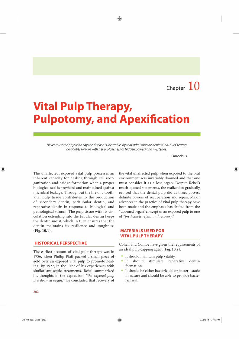
molar, and the mesial root of mandibular molars, which have two root canals. B, buccal; D, distal; DB, distobuccal; M, mesial; MB, mesiobuccal; P, palatal. (Courtesy: Marco Versiani, Pecora and Sousa-Neto, Brazil.)
Ch_12_GEP.indd 245 08/08/14 5:03 PM

246 Grossman’s Endodontic Practice
structure immaterial of its location. This would invariably lead into the pulp chamber. Hence, in case of a tooth with distal carious tooth structure, the access opening commences from the distal side towards the mesial pulp chamber.
B. Complete De-Roofing and
Removal of Dentinal ShouldersThe overhanging roof of the pulp chamber misdirects the instrument, which results in ledge formation in the canal. Hence, complete de- roofing must be done to obtain unrestricted access to the canals. Removing the roof completely from the pulp chamber will bring canal orifices into view and allow immediate access to each orifice. Using a round bur and working from inside out will accom-plish this end.
Removal of the dentinal shoulders present between root canal orifices will help in achieving straight line access and improve the clinical access to the root canals (Fig. 12.15).
i e (Courtesy: Dentsply Maillefer.)
i e Courtesy: Dentsply Maillefer.)
i e Courtesy: Dentsply Maillefer.)
(a)
(b)
i e Courtesy: Hu-Friedy Mfg Co., USA.)
II. CLINICAL CONSIDERATIONS
A. Complete Removal of Carious
Tooth Structure and Other Restorative
MaterialWhile preparing the access cavity in a cariously involved tooth, start removing the carious tooth
Ch_12_GEP.indd 246 08/08/14 5:03 PM
287
Endodontic treatment can be divided into three main phases:
Proper access preparation into the pulp space Shaping and cleaning of the root canal Obturation
The initial step for shaping and cleaning the root canal is proper access to the chamber that leads to straight-line penetration of the root canal orifices. The concepts of achieving proper access into the pulp space are elaborated in Chapter 12. The next step is exploration of the root canal, extirpation of the remaining pulp tissue or gross debridement of the necrotic tissue, and verification of the working length. This step is followed by proper instrumen-tation, irrigation, debridement, and disinfection of the root canal. Obturation completes the procedure.
Definitions: Shaping and cleaning of the root canal consists of removing the pulp tissue and debris from the canal and shaping the canal to receive an obtu-rating material.
Pulpectomy, or pulp extirpation, is the complete removal of a normal or diseased pulp from the pulp cavity of the tooth. The operation is some-times inappropriately referred to as devitalization.When food or other debris have accumulated in the pulp cavity, in addition to the residual necrotic pulpal debris, the removal of this material from the pulp cavity is referred to as debridement.
Using sequentially larger sizes of files and irrigat-ing and disinfecting the canal to clear it of debris, one shapes the canal to receive a well-compacted filling that seals the root canal apically and laterally to prevent any leakage.
The importance of adequate canal shaping and cleaning, rather than reliance on antiseptics, cannot be overemphasized. Histologic examination of pulpless teeth in which root canal therapy has failed often shows that the canals were not completely cleaned. Obturation of an improperly cleaned canal would still lead to an endodontic failure irrespective of the quality of obturation (Figs 13.1 and 13.2).
What we remove from the pulp space, is far more important than what we replace it with...
Chapter 13
Shaping and Cleaning of the Radicular Space:
Instruments and Techniques
Ch_13_GEP.indd 287 08/08/14 3:03 PM

310 Grossman’s Endodontic Practice
o n le a io ap ic et o o o in en t ete mina on
510
1520
25m
m
2520
1510
5m
m
Workinglength
0.5–1.0 mm
o in len t e ta li e )
o t o t e a io ap ic ape mo e t an mm
Add
e on t e a io ap ic ape
Reduce
Ch_13_GEP.indd 310 08/08/14 3:04 PM

314 Grossman’s Endodontic Practice
Table 13.8 presents the summary of principle techniques of root canal instrumentation.
a. Step-Back Technique
Conventional Step-Back (Telescopic) Technique In the step-back preparation of the root canal, the canal is enlarged first in the apical third to at least
a le
A t o Year Name o ec ni e
o nc onal o on o n tr menta on
Reaming:Fig. 13.32
Filing:
Watch winding:
Circumferential filing:
Anticurvature filing: Abou Rass and Jastrab
pulled intopushed away
Fig re 13.32
(a) (b)
o 13.3 ec ni e o aping an Cleaning
Ch_13_GEP.indd 314 08/08/14 3:04 PM

CHAPTER 13 Shaping and Cleaning of the Radicular Space: Instruments and Techniques 315
a le 13.
Feat re tep ac tep o n ri
III
II
I III
II
I I
II
III
Ch_13_GEP.indd 315 08/08/14 3:04 PM
343
Definition: According to the American Association of Endodontists “Obturation is the method used to fill and seal a cleaned and shaped root canal using a root canal sealer and core filling material.”
The function of a root canal filling is to obtu-rate the canal and eliminate all portals of entry between the periodontium and the root canal. The better the seal, the better the prognosis of the tooth. Achieving the ideal seal, however, is as complex as the anatomy of the root canal system itself. Because all root canal fillings must seal all foramina leading into the periodontium, an ideal filling must be well compacted, must conform and adhere to the shaped canal walls, and must end at the juncture of the root canal and the periodon-tium (Box 15.1).
Clinical Note
Naidorf has stated that inadequate obturation of the root canal exposes it to periradicular tissue fluids, which provide material for growth of microorganisms or localization of bacteria in such dead spaces. According to a study by Ingle and Beveridge, 58% of endodontic failures can be attributed to incomplete obturation of root canals (Fig. 15.1).
Perfection is not attainable, but if we chase perfection we can catch excellence.
—Vince Lombardi
Chapter 15
Obturation of the Radicular Space
o 15.1 Grossman’s Requirements for an Ideal Root Canal Filling Material
The material should be easily introduced into the root canal.It should seal the canal laterally as well as apically.It should not shrink after being inserted.It should set slowly.It should be impervious to moisture.It should be bactericidal or, at least, should discour-age the growth of bacteria.It should be radiopaque.It should not stain the tooth structure.It should not irritate periradicular tissues or affect the tooth structure.It should be sterile, or easily and quickly sterilized immediately before insertion.It should be easily removable from the root canal if necessary.
WHEN TO OBTURATE THE ROOT CANAL
A root canal may be obturated when the tooth is asymptomatic and the root canal is reasonably dry. Obturation after obtaining a negative culture and closure of an existing sinus tract have been suggested in the past. However, this concept is no longer valid.
Ch_15_GEP.indd 343 08/08/14 5:09 PM

Grossman’s Endodontic Practice 348
Figure 15. Resilon (Real Seal System). (Courtesy: SybronEndo.)
I. COLD LATERAL
COMPACTION TECHNIQUE
This has been one of the most commonly prac-ticed obturation techniques (Fig. 15.5). However, in contemporary endodontics, it is not the best technique to achieve a three-dimensional seal. The stepwise technique is given in Box 15.5.
Clinical Considerations1. Sealer considerations: Sealer application on
the canal walls can also be performed using a lentulo sprial (Fig. 15.7) or with the master gutta-percha cone itself.
2. Spreader considerations (Figs 15.8 and 15.9): The size of the spreader is determined by the width of the prepared canal and the lat-eral fit of the primary cone; the greater the space between the canal wall and the butt end of the gutta-percha, the larger (wider) the spreader used. The spreader size should reach within 1–2 mm of the working length in order to obtain optimal apical compaction. This can be ensured by placing a silicon stopper on the spreader.
3. Master cone considerations: Selection of the master cone should be sim-ilar to the master apical file size. Minimal judicious force should be used on the spreader during the compaction process in order to avoid root fractures.
defined as the ability to deform and to flow away from a force directed at its mass.
Each technique is designed to force the gutta-percha filling to flow into the root canal, compress against its walls, fill fine tortuous canals, seal the var-ious foramina exiting into the periodontium, and finally, compact into a solid core filling. The cold lateral compaction method of filling uses spreaders by inserting these instruments alongside the gutta-percha and compressing them laterally and apically.
Clinical Note
The vertical compaction technique uses vertical force combined with applied heat to drive the gutta-percha apically and laterally. Thermoplastic techniques use more heat to increase the plasticity of gutta-percha and thereby enable the operator to fill the root canal by using less pressure.
o 15. ec niques of tura on
Cold lateral compactionWarm compaction (warm gutta-percha)(a) Vertical(b) LateralContinuous wave compaction techniqueThermoplasticized gutta-percha injectionCarrier-based gutta-percha(a) Thermafil thermoplasticized(b) SimpliFill sectional obturationMcSpadden thermomechanical compactionChemically plasticized gutta-perchaCustom cone
Ch_15_GEP.indd 348 08/08/14 5:09 PM

Grossman’s Endodontic Practice 350
Figure 15. Lentulo spiral.
o 15.5 ec nique of Cold ateral Compac on
canal with paper points master cone “TUG BACK” (Fig. 15.6) (same size as Master (Fig. 15.5) Apical File)
ort of t e ape
working length, then patency has to be established to the corrected length followed
the canal to the master apical
corrected working length for
Be ond t e ape If the master cone extends
beyond the working length, the
snugly at the working length or
radiographically.
At or ing lengt
and coat the canal with sealerusing the master cone or with
a lentulo spiral (Fig. 15.7)
(Fig. 15.8) is inserted alongside the master cone to a level 1 mm short
of the working length
The spreader is disengaged from
in an arc
radicular pulp space (Fig. 15.5)
(Fig. 15.9)
Ch_15_GEP.indd 350 08/08/14 5:09 PM

CHAPTER 15 Obturation of the Radicular Space 357
Figure 15.1-
B unit is set to 200°C and the heated plugger is moved rapidly (1–2 seconds) to within 3 mm of the binding point. The heat is
removed. (f)–(i) The remaining canal space is obturated using a
(a) (b)(i) (b)(iv)(b)(iii)(b)(ii)
(c) (e)(d) (f) (g)
(h) (i)
Ch_15_GEP.indd 357 08/08/14 5:09 PM
398
A successful endodontic treatment has to be complemented with an adequate postendodontic restoration to make the pulpless tooth function indefinitely as an integral part of the oral mastica-tory apparatus. Endodontically treated teeth fail principally due to one of the following two reasons:
Persistent intraradicular infectionPostendodontic restorative difficulties
Careful postendodontic restoration is required, as the cumulative loss of tooth structure due to car-ies, trauma, and endodontic procedures combined with the loss of structural integrity contributes to the fracture of the tooth. Ideally, the final restora-tion should be planned before the root canal treat-ment is begun, though the restorative plan may be modified as the treatment progresses.
ASSESSMENT OF RESTORABILITY
An endodontically treated tooth must be evalu-ated before definitive restorative procedures are
initiated. Evaluation factors (Fig. 17.1) are used to determine whether the endodontically treated tooth is restorable, unrestorable, or restorable after successful retreatment. Definitive restorative treat-ment should not be initiated if the treated tooth exhibits any of the following:
Poor root canal fillingActive inflammationPressure sensitivityExudateFistula (or parulis)Periodontal disease (moderate or severe periodontitis)Severe loss of sound tooth structure (tooth would not benefit from crown lengthening or orthodontic extrusion)
In short, seven categories of infection, trauma, inflammation, unacceptable endodontics, or lack of restorability, as listed, can delay or end up in no definitive restorative treatment (Figs 17.2 and 17.3).
Our objective should be the perpetual preservation of what remains than the meticulous restoration of what is missing.
—M. M. De Van
Chapter 17
Prosthodontic Considerations in Endodontically Treated Teeth
Ch_17_GEP.indd 398 08/08/14 3:12 PM

399 CHAPTER 17 Prosthodontic Considerations in Endodontically Treated Teeth
Figure 17.1
Endodonticallytreated tooth
Significant coronal damageMinimal coronal damage
with no color change)
oor root filling
ST r
tooth
Moderate coronaldamage
Minimal coronaldamage
e
Significant coronaldamage
ee
Moderate coronaldamage
e
crowne
treatment
canal
a
crown
a
crown
n
eea
Anteriortooth
Ch_17_GEP.indd 399 08/08/14 3:12 PM

403 CHAPTER 17 Prosthodontic Considerations in Endodontically Treated Teeth
The amount of coronal tooth structure, along with the position of the tooth in the arch, will dictate the type of core indicated; whether a prefabricated post or a cast post and core is indicated; and whether a crown is needed.
PROTECTING THE REMAINING CORONAL
TOOTH TISSUE—CREATING THE FERRULE
A ferrule is defined as a band of extracoronal mate-rial at the cervical margin of a crown preparation that encompassess the tooth and provides resistance form to the tooth. This is usually provided by the crown that is placed over the post and core system. It is of paramount importance that as much coronal or supragingival tooth tissue is preserved as possi-ble, as this significantly improves the prognosis of the tooth and restoration. One to two millimeters of tooth tissue coronal to the finish line of the crown preparation significantly improves the fracture resistance of the tooth and is more important than the type of core and post material (Fig. 17.6).
The word ferrule is thought to be derived from the Latin word ferrum, meaning iron, and viriola, meaning bracelet. Thus, the ferrule effect occurs because of the crown bracing against the remaining supragingival tooth tissue (Fig. 17.7). Some authors have questioned the benefit of the ferrule; however, majority of the literature would support its impor-tance in reducing the probability of tooth fracture.
Clinical Note
Clinical Note
Figure 17.6
Minimumthickness = 1 mm
Ferrule wallsalmost parallel
2.0
Figure 17.7
Ch_17_GEP.indd 403 08/08/14 3:12 PM
407 CHAPTER 17 Prosthodontic Considerations in Endodontically Treated Teeth
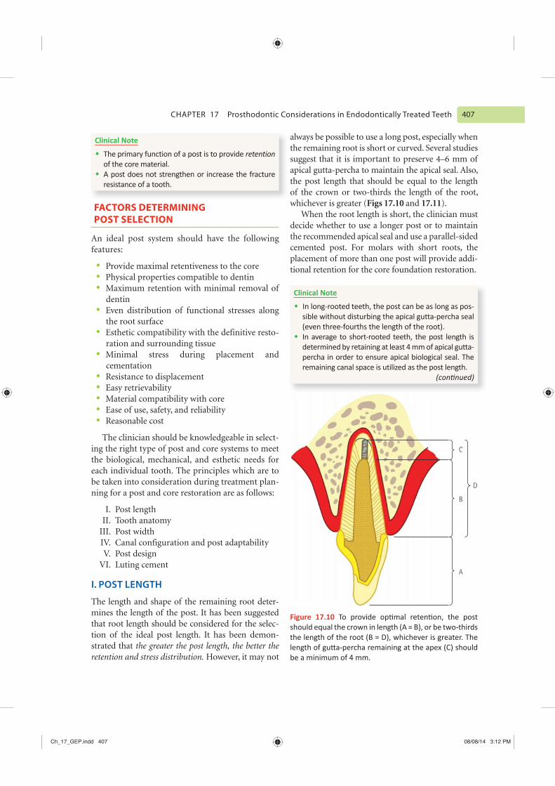
always be possible to use a long post, especially when the remaining root is short or curved. Several studies suggest that it is important to preserve 4–6 mm of apical gutta-percha to maintain the apical seal. Also, the post length that should be equal to the length of the crown or two-thirds the length of the root, whichever is greater (Figs 17.10 and 17.11).
When the root length is short, the clinician must decide whether to use a longer post or to maintain the recommended apical seal and use a parallel-sided cemented post. For molars with short roots, the placement of more than one post will provide addi-tional retention for the core foundation restoration.
Clinical Note
Clinical Note
retention
FACTORS DETERMINING
POST SELECTION
An ideal post system should have the following features:
Provide maximal retentiveness to the corePhysical properties compatible to dentinMaximum retention with minimal removal of dentinEven distribution of functional stresses along the root surfaceEsthetic compatibility with the definitive resto-ration and surrounding tissueMinimal stress during placement and cementationResistance to displacementEasy retrievabilityMaterial compatibility with coreEase of use, safety, and reliabilityReasonable cost
The clinician should be knowledgeable in select-ing the right type of post and core systems to meet the biological, mechanical, and esthetic needs for each individual tooth. The principles which are to be taken into consideration during treatment plan-ning for a post and core restoration are as follows:
I. Post length II. Tooth anatomyIII. Post width IV. Canal configuration and post adaptability V. Post design VI. Luting cement
I. POST LENGTH
The length and shape of the remaining root deter-mines the length of the post. It has been suggested that root length should be considered for the selec-tion of the ideal post length. It has been demon-strated that the greater the post length, the better the retention and stress distribution. However, it may not
C
B
D
A
Figure 17.1
Ch_17_GEP.indd 407 08/08/14 3:12 PM
421
Trauma of the oral and maxillofacial region occurs frequently and comprises 5% of all inju-ries for which people seek dental treatment. Among all facial injuries, dental injuries are the most common, of which crown fractures and lux-ations occur most frequently. Trauma to the teeth may result either in injury of the pulp, with or without damage to the crown or root, or in dis-placement of the tooth from its socket. When the crown or root is fractured, the pulp may recover and survive the injury, it may succumb immedi-ately, or it may undergo progressive degeneration and ultimately die.
CAUSES AND INCIDENCE
OF DENTAL INJURIES
Traumatic injuries to the teeth can occur at any age. Young children learning to walk or falling from a chair are subject to anterior tooth injuries. Frequently, child abuse results in facial and dental trauma. Sports accidents and fights affect teenagers and young adults, whereas automobile accidents
affect all age groups. As many dental accidents are sports related, every precaution should be taken to protect the teeth of children and teenagers from such accidents by conducting educational pro-grams in addition to mouth guards.
The common causes of traumatic injuries to the teeth include the following:
Sports accidentsAutomobile accidentsFights and assaultsDomestic violenceInappropriate use of teethBiting hard items
Clinical Note
The incidence of tooth fractures is about 5%.The following age groups are most prone to dental accidents:
-tured teeth as girls
Healing is a matter of time, but it is sometimes also a matter of opportunity.
—Hippocrates
Chapter 18
Treatment of Traumatized Teeth
Ch_18_GEP.indd 421 08/08/14 2:59 PM

425 CHAPTER 18 Treatment of Traumatized Teeth
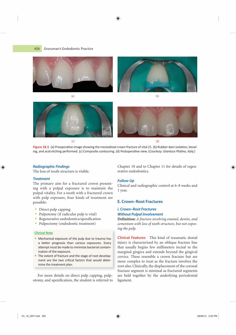
cases, the fractured segments can be approximated and bonded back with the help of dentin bonding agents and composite resins (Fig. 18.3). The use of indirect veneering procedures at a later date is another approach to improve the esthetics.
Follow-Up Clinical and radiographic control at 6–8 weeks and 1 year. The tooth should be periodically tested with the electric pulp tester or Endo Ice. If the pulp con-tinues to respond normally during this time, the pulp can be assumed to have recovered. If progres-sively more current is necessary to elicit a vitality response, the pulpal prognosis is unfavorable, and the pulp will probably become necrotic necessitat-ing endodontic treatment.
D. Enamel–Dentin Fracture
with Pulpal Exposure Definition: A fracture involving the enamel and dentin with loss of tooth structure and exposure of pulp.
Clinical FeaturesNormal mobility. The tooth is not tender on percussion, however if tenderness is observed, eval-uate for possible luxation or root fracture injury. Exposed pulp is sensitive to stimuli.
The objective in treating a tooth with a fractured crown without pulp exposure is threefold:
Elimination of discomfort Preservation of the vital pulpRestoration of the fractured crown
Clinical Features The tooth is not tender on percussion. If tenderness is observed, evaluate the tooth for possible luxation or root fracture injury. Normal mobility is observed and pulp sensibility test is usually positive.
Radiographic FindingsThe enamel–dentin loss is visible. Radiographs recommended: periapical, occlusal, and eccentric exposure to rule out tooth displacement or pos-sible presence of root fracture. Radiograph of lip or cheek lacerations suggested to search for tooth frag-ments or foreign materials.
Treatment In an uncomplicated fracture of the crown with-out pulpal exposure, a remaining dentinal thickness of 2 mm is sufficient to shield the pulp and ensure a good prognosis. Inflammatory response in the form of pain on percussion is usually transient as long as the vascular supply to the pulp remains intact. Composite resin restoration is the preferred restorative procedure in such cases. In certain
Figure 18.2
(a) (b)
Ch_18_GEP.indd 425 08/08/14 2:59 PM
426 Grossman’s Endodontic Practice
Chapter 10 and to Chapter 11 for details of regen-erative endodontics.
Follow-UpClinical and radiographic control at 6–8 weeks and 1 year.
E. Crown–Root Fractures
i. Crown–Root Fractures Without Pulpal InvolvementDefinition: A fracture involving enamel, dentin, and cementum with loss of tooth structure, but not expos-ing the pulp.
Clinical Features This kind of traumatic dental injury is characterized by an oblique fracture line that usually begins few millimeters incisal to the marginal gingiva and extends beyond the gingival crevice. These resemble a crown fracture but are more complex to treat as the fracture involves the root also. Clinically, the displacement of the coronal fracture segment is minimal as fractured segments are held together by the underlying periodontal ligament.
Radiographic Findings The loss of tooth structure is visible.
Treatment The primary aim for a fractured crown present-ing with a pulpal exposure is to maintain the pulpal vitality. For a tooth with a fractured crown with pulp exposure, four kinds of treatment are possible:
Direct pulp cappingPulpotomy (if radicular pulp is vital)Regenerative endodontics/apexificationPulpectomy (endodontic treatment)
Clinical Note
Mechanical exposure of the pulp due to trauma has
-ination of the exposure.
-ment are the two critical factors that would deter-mine the treatment plan.
For more details on direct pulp capping, pulp-otomy, and apexification, the student is referred to
(c) (d)
(a) (b)
Figure 18.3 -)
Ch_18_GEP.indd 426 08/08/14 2:59 PM

441 CHAPTER 18 Treatment of Traumatized Teeth
(a) (b)
(c) (d)
(e) (f) (g)
Figure 18.13 was
)
Ch_18_GEP.indd 441 08/08/14 3:00 PM

442 Grossman’s Endodontic Practice
Bo 18.3 Management of A ulsed Teet it an traoral r Time of ess T an 6 Minutes
T AN
Administer local anesthesia and irrigate the socket with saline. Examine the socket for possible fracture and
N AC A
for 5 minutes
( )
unless there are signs of pulp
necrosis-
tooth will appear to be in an infraocclusal position. Radiographic evidence of resorption (inflammatory, infection-related resorption, or ankylosis-related replacement resorption).
RESPONSE OF PULP TO TRAUMA
The pulpal response to dental trauma is dependent on three critical factors:
Intensity of the trauma Stage of root developmentPresence or absence of bacteria
Bo 18.2 Transport Medium for an A ulsed Toot
follows:
Milk
Propolis
Ch_18_GEP.indd 442 08/08/14 3:00 PM

462
The scope of endodontic surgery has extended beyond root-end resection to include other forms of periradicular surgery, fistulative surgery, correc-tive surgery, and intentional replantation. Root-end resection is still the most common form of perira-dicular surgery.
In general practice, the number of cases being indicated for root-end surgery has drastically reduced over a period of time. This may be due to the fact that today the science of endodontics has a better understanding of the biological prin-ciples of shaping and cleaning. In the last few decades, endodontics was more of a biological sci-ence than mere chemomechanical debridement. There has been a tremendous improvement in the available materials and instruments for shaping and cleaning. With the present knowledge of internal anatomy of pulp space, microbiology, disinfection of the pulp space, and also with the introduction of rotary and micro endodontic instrumentation, clinicians are now better equipped to produce a more predictable disinfec-tion of the pulp space.
There has been a gradual paradigm shift from surgical to nonsurgical treatment over the past few decades. However, nonsurgical management
may not be always successful. Even if nonsurgical treatment is unsuccessful, the current concept is to do an introspection of the quality of nonsurgical treatment before selecting surgical intervention. If the initial endodontic treatment of a tooth is not satisfactory, then one should attempt nonsurgical retreatment of that tooth first.
The view that endodontic surgery is the last resort is based on past experience with instru-ments that were unsuitable. Also, the vision available at the surgical site was inadequate and incidence of postoperative complications was high. Fortunately, today the endodontist is equipped with better magnification, illumination, and instruments. The present era of microsurgery is with surgical operating microscopes, ultrasonic tips for retropreparation, low-speed high-torque motors, and miniaturized surgical instruments for root-end surgery, and all these have resulted in better success rates.
Clinical Note
success.
We are what we repeatedly do. Excellence, then, is not an act, but a habit.
—Aristotle
Chapter 20
Endodontic Surgery
Ch_20_GEP.indd 462 08/08/14 11:40 AM

463 CHAPTER 20 Endodontic Surgery
isthmus, and trace accessory canals in nonsur-gical endodontic cases that are clinically failing.
INDICATIONS
Evidence-based endodontic literature has led to substantial reduction in the indications for root-end surgery. It is recognized that nonsurgical treatment is the choice in most cases. However, the following indications may have to be considered (Fig. 20.1):
Failure of nonsurgical endodontic treatment: Per-sistence of symptoms in teeth in which radio-graphically adequate nonsurgical endodontic
OBJECTIVES AND
RATIONALE FOR SURGERY
Curettage: Effective curettage of the pathologi-cally affected periradicular tissue which cannot be accessed in an orthograde approach. This includes therapy-resistant granuloma, true cysts, and foreign body reactions.Resection: Surgical resection of root apex in cases where the apical ramifications cannot be eliminated in a nonsurgical endodontic treat-ment or surgical resection of a root in cases of poor periodontal support.Inspection: Inspection of the periradicular area to ascertain causes of failure, inspection of
Figure 2 .1
(a) (b)
(c) (d)
Ch_20_GEP.indd 463 08/08/14 11:40 AM
Grossman’s Endodontic Practice 470
Bo 2 .2 Gutmann’s Classi ca on of ndodon c urger
Bo 2 .3 Kim’s Classi ca on of Microsurgical Cases (Fig. 20.8)
Class A -
Class B
Class C --
Class D
Class E --
Class F
-X C -C M
D U -
-
-
Effective hemostasis is critically important dur-ing endodontic microsurgery because uncontrolled bleeding in the surgical site obscures the anatomi-cal landmarks guiding the surgeon. It is therefore not surprising that one of the most frequently asked questions about endodontic microsurgery is on effective management of bleeding in the osteotomy site and inside the bony crypt.
Effective hemostasis is a prerequisite for end-odontic microsurgery and successful hemostasis begins with effective local anesthesia. Surgeons must understand the normal clotting mechanism and normal clotting time of human blood; it takes several minutes for the blood to begin clotting.
VASOCONSTRICTORS
To obtain hemostasis, vasoconstrictors are always a constituent of local anesthetics used in perira-dicular surgeries. The vasoconstrictor agent used in local anesthetics will have an effect in both the duration of anesthesia and the quality of hemor-rhage control in the surgical area.
Clinical Note
-
effects of the agents.-
(continued)
Clinical Note
X
Ch_20_GEP.indd 470 08/08/14 11:40 AM

471 CHAPTER 20 Endodontic Surgery
It is also to be well understood that infiltration sites for periradicular surgery are always multiple and involve deposition of anesthetic throughout the entire surgical area in the alveolar mucosa just super-ficial to periosteum at the level of root apices. Apart from the routine anesthetic techniques of nerve block anesthesia, other standard techniques may be observed to obtain profound local anesthesia.
CLINICAL MANAGEMENT OF
HEMORRHAGE IN A NORMAL PATIENT
Incision planningUse of hemostatsHemostasis through application of pressureHemostatic agents (Fig. 20.9)Hypotensive anesthesia and vasoconstrictors
(continued)
-
-
local infiltration of local anesthetic with a higher
-
Safety limit:
-
rine containing 2% lidocaine to reach the danger
Class A Class B Class C
Class D Class E Class F
Figure 20.8 Courtesy: Syngcuk Kim, University of Pennsylvania, USA
Ch_20_GEP.indd 471 08/08/14 11:40 AM

475 CHAPTER 20 Endodontic Surgery
(a) (b)
(c)
C
B
A
(d)
Figure 20.12
Technique1. Hemostasis is the primary issue at this stage of
surgery.2. In most of the clinical cases, there is a breach in
the cortical plate and this can be located around the root apex by gently probing with a DG16 ex-plorer, and if the breach is located, the explorer will sink and this could be the starting point for an efficient osteotomy. However, in most cystic pathosis, the cortical plate is thinned out due to the growth of the cyst and has an egg-shell crackling appearance. In these situations, the cortical plate can be peeled off leaving the
retractors are available and are designed to have wider and thinner working ends than standard retractors.
HARD TISSUE CONSIDERATIONS
OSTEOTOMY
Osteotomy involves the removal of cortical plate to expose the root end in microendodontic sur-gical procedures. Once the flap has been elevated and placed in retracted position, the surgical area is taken into control.
Ch_20_GEP.indd 475 08/08/14 11:41 AM

Grossman’s Endodontic Practice 480
(a) (b)
(c)
Figure 20.19Courtesy: Arnaldo Castelluci, Italy
(a) (b)
Figure 20.20
Ch_20_GEP.indd 480 08/08/14 11:41 AM

499
Esthetics is an important factor in a patient’s decision to undergo endodontic treatment. A fre-quent question is, “Will my tooth turn black?” The usual response is a “qualified no,” with the expla-nation that modern treatment and procedures are designed to avoid crown staining and tooth discolor-ation. Nevertheless, teeth can and do discolor, some-times before endodontic treatment and sometimes afterward, in spite of all precautions taken to prevent color changes. When teeth discolor, bleaching should be considered as a means of restoring tooth esthetics.
The color of teeth is determined by the translu-cency and thickness of the enamel, the thickness and color of the underlying dentin, and the color of the pulp. Alterations in the color may be physiologic or pathologic and endogenous or exogenous in nature.
With age, the enamel becomes thinner because of abrasion and erosion, and the dentin becomes thicker because of the deposition of secondary and reparative dentin, which produce color changes in teeth during one’s life.
Clinical Note
The normal color of primary teeth is bluish white.The color of permanent teeth is grayish yellow, grayish white, or yellowish white.Teeth of elderly persons are usually more yellow or grayish yellow than those of younger persons.
CLASSIFICATION OF
TOOTH DISCOLORATION
Tooth discoloration can be classified as either extrinsic or intrinsic.
I. EXTRINSIC DISCOLORATIONS
Extrinsic discolorations are found on the outer surface of teeth and are usually of local origin, such as tobacco stains. Some extrinsic discolor-ation, such as the green discoloration associated with the Nasmyth’s membrane in children and tea and tobacco stains (Fig. 21.1), can be removed by scaling and polishing during tooth prophylaxis.
Chapter 21
Bleaching of Discolored Teeth
We live only to discover beauty. All else is a form of waiting.
—Kahlil Gibran
Figure 21.1 Extrinsic tobacco stains. (Courtesy: Priya Ramani, India.)
Ch_21_GEP.indd 499 08/08/14 11:42 AM

502 Grossman’s Endodontic Practice
Ta le 21.1
Cause of Toot iscolora on Color
trinsic discolora on
marijuana
Yellow-brown to black Dark brown to black ringsBrown to blackYellow or brown shades
trinsic and intrinsic discolora on
White, yellow, brown, gray, black
Intrinsic discolora on
With hemorrhageWithout hemorrhage
Brown, blackBrown, blueBlue-green, brown, purple-brown
Brown, gray, black
Blue-green, brown
Yellow
Yellow, gray-brown
Iatrogenic causes
Brown, gray, blackBrown, gray, black
V. FILLING MATERIALS
Discoloration from filling materials depends on the kind of filling used. Silver amalgam produces a stain ranging from slate gray to dark gray; copper amalgam produces a bluish black to black stain; stains from amalgam are likely to occur when the
dentinal wall is thin, and the filling material almost shimmers through the enamel. Microleakage of the old resin composite restorations might cause dark discoloration of the margins and may stain the dentin over time. Metal post can be seen through the translucent enamel or may release
Ch_21_GEP.indd 502 08/08/14 11:42 AM

508 Grossman’s Endodontic Practice
Bleaching
agent
Undercut
Protective
base
Temporary
fillingComposite
entrance filling
Permanent
restoration
Obturation
Pulp
horn
(a)
(d) (e)
(b) (c)
Figure 21.9
-
materials from the chamber with solvents, a paste composed of sodium perborate and water (mixed to the con-
(e) At a subsequent appointment, when the desired shade has been reached, a permanent
and to support the incisal edge.
Ch_21_GEP.indd 508 08/08/14 11:43 AM
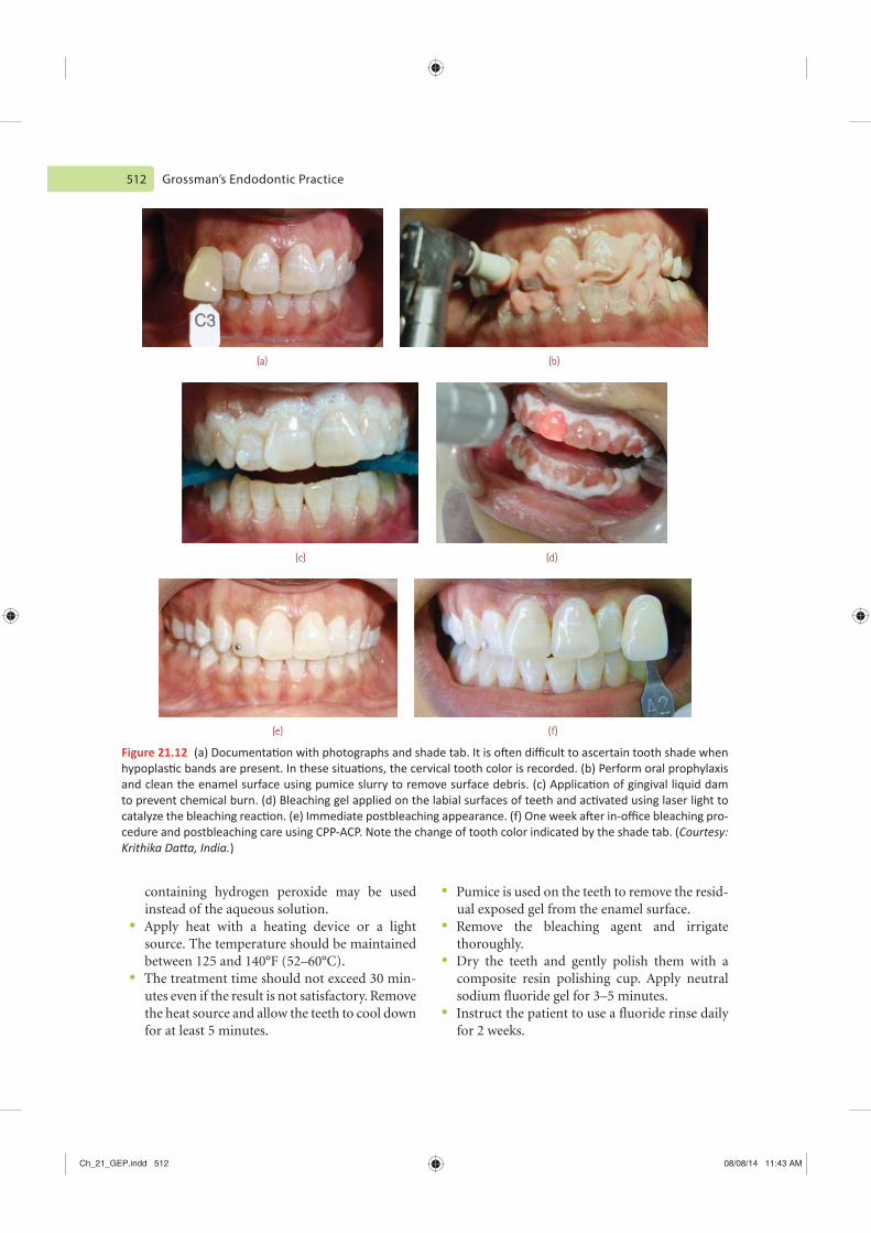
512 Grossman’s Endodontic Practice
Pumice is used on the teeth to remove the resid-ual exposed gel from the enamel surface. Remove the bleaching agent and irrigate thoroughly. Dry the teeth and gently polish them with a composite resin polishing cup. Apply neutral sodium fluoride gel for 3–5 minutes.Instruct the patient to use a fluoride rinse daily for 2 weeks.
containing hydrogen peroxide may be used instead of the aqueous solution. Apply heat with a heating device or a light source. The temperature should be maintained between 125 and 140°F (52–60°C). The treatment time should not exceed 30 min-utes even if the result is not satisfactory. Remove the heat source and allow the teeth to cool down for at least 5 minutes.
Figure 21.12
-Courtesy:
)
(a) (b)
(d)(c)
(f)(e)
Ch_21_GEP.indd 512 08/08/14 11:43 AM
531
Radiographs are indispensable diagnostic and prognostic aids in endodontics and are one of the most reliable methods of monitoring endodon-tic treatment. They provide an important visual method of gaining clinical knowledge of teeth and periradicular tissues; therefore, they are essential to the practice of endodontics.
Proper positioning and stabilization of the radiographic film during endodontic procedures becomes difficult because of the interference from the protruding rubber dam clamp or root canal instruments or interference from the obturating material protruding from the access cavity. The visualization of the tooth for proper film position-ing and cone angulation is impeded by the presence of the rubber dam. This makes the process of taking a radiograph a difficult proposition.
RADIOGRAPHIC
TECHNICAL REQUIREMENTS
1. The image of the tooth being evaluated or undergoing endodontic therapy should be in the center of the radiograph.
2. Radiographs should show at least 5 mm of bone surrounding the apex of the tooth being evalu-ated or undergoing endodontic therapy.
3. If a periradicular lesion is too large to fit in one periapical film, supplemental diagnostic radio-graphs must be made.
4. A single radiograph taken from one direction only may not provide sufficient diagnostic information in multirooted teeth or in teeth with curved roots. Under these circumstances, at least two periapical radiographs should be taken to help gain a three-dimensional per-spective. One radiograph should be taken at normal vertical and horizontal angulation, while the other should be taken at a 20° change in the horizontal angle from either the mesial or the distal direction (Fig. A.l).
5. If a sinus tract is present, a tracing radio-graph should be taken. This procedure is accomplished by carefully threading a gutta-percha cone into the tract and by taking a radiograph to identify the origin of the tract. This technique is also useful for localization and depth marking of certain periodontal defects.
The most pathetic person in the world is someone who has sight but no vision.
—Helen Keller
Appendix A
Radiographic Technique for Endodontics
App_A_GEP.indd 531 07/08/14 2:29 PM

APPENDIX A Radiographic Technique for Endodontics 535
Figure A.8
Figure A.9 Figure A.11
Note.(e) Make the exposure.(f) Replace the rubber dam frame.
RADIOGRAPHIC TECHNIQUE
FOR POSTERIOR TEETH
1. Assembly of the endodontic film holder(a) Select an appropriate film holder and pos-
terior rod assembly.(b) Slide the beam-alignment ring onto the
rod and push it within 2 inches of the film-holding portion of the instrument (Fig. A.10). Be sure that the film is centered in the ring and the long axis of the film is parallel to the posterior rod (Fig. A.11).
2. Taking the radiograph(a) Remove the rubber dam frame.(b) Insert the assembled instrument and make
sure that the tooth is in the center of the film and the film is parallel to the long axis of the tooth (Fig. A.12).
(c) For mandibular radiographs, position the film between the teeth and the tongue and make sure that the lower edge of the film does not impinge on the muscle attachments in the floor of the mouth. Care should be taken that the patient does not displace the film by moving the
Figure A.10
App_A_GEP.indd 535 07/08/14 2:29 PM

APPENDIX A Radiographic Technique for Endodontics 537
(a)
(b)
(a)
(b)
Figure A.1
Figure A.15
manipulated in different dimensions with the help of software, which includes enhancement, contrast, magnification, colorize, and reversing (Fig. A.16). The image is stored in the computer for records.
App_A_GEP.indd 537 07/08/14 2:29 PM

541
Ta le B.1 Root Canals and Apical Foramina in Maxillary First Premolars
In es gator
One Canal and One
Foramen ( )
One Canal and T o
Foramina ( )
T o Canalsand One
Foramen ( )
T o Canalsand T o
Foramina ( )T ree Canals
( )
26.2 7.7 23.9 41.7 0.5
Green 8.0 – 26.0 66.0 –
Cams and Skidmore 9.0 – 13.0 72.0 6.0
8.0 7.0 18.0 62.0 5.0
Bellizzi and Hartwell 6.2 – – 90.5 3.3
Ta le B.2 Root Canals and Apical Foramina in Maxillary Second Premolars
In es gator
One Canal and One
Foramen ( )
One Canal and T o
Foramina ( )
T o Canalsand One
Foramen ( )
T o Canalsand T o
Foramina ( )T ree Canals
( )
62.8 8.9 19.0 9.3 –
Green 72.0 – 24.0 4.0 –
Vertucci and colleagues 48.0 – 27.0 24.0 1.0
Bellizzi and Hartwell 40.3 – – 58.6 1.1
The palest ink is better than the best memory.
—Chinese Proverb
Appendix B
Root Canal Configuration
App_B_GEP.indd 541 07/08/14 2:30 PM
542 Grossman’s Endodontic Practice
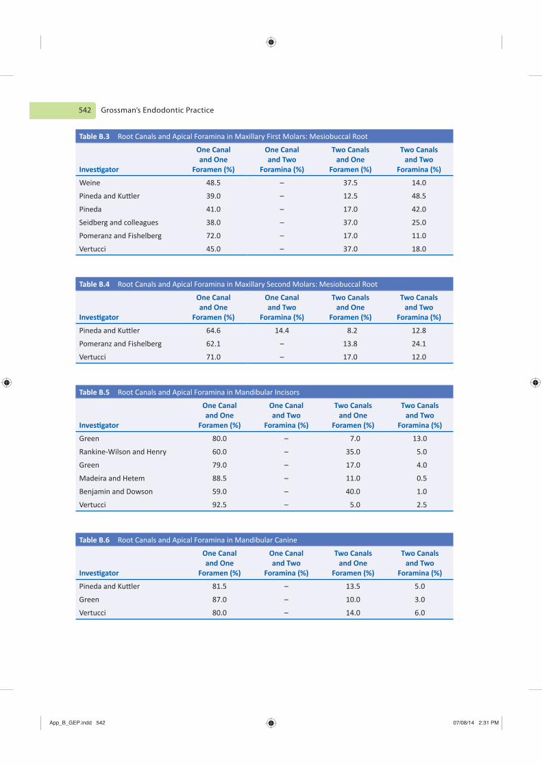
Ta le B.3 Root Canals and Apical Foramina in Maxillary First Molars: Mesiobuccal Root
In es gator
One Canal and One
Foramen ( )
One Canal and T o
Foramina ( )
T o Canals and One
Foramen ( )
T o Canals and T o
Foramina ( )
Weine 48.5 – 37.5 14.0
39.0 – 12.5 48.5
Pineda 41.0 – 17.0 42.0
Seidberg and colleagues 38.0 – 37.0 25.0
Pomeranz and Fishelberg 72.0 – 17.0 11.0
Vertucci 45.0 – 37.0 18.0
Ta le B. Root Canals and Apical Foramina in Maxillary Second Molars: Mesiobuccal Root
In es gator
One Canal and One
Foramen ( )
One Canal and T o
Foramina ( )
T o Canals and One
Foramen ( )
T o Canals and T o
Foramina ( )
64.6 14.4 8.2 12.8
Pomeranz and Fishelberg 62.1 – 13.8 24.1
Vertucci 71.0 – 17.0 12.0
Ta le B.5 Root Canals and Apical Foramina in Mandibular Incisors
In es gator
One Canal and One
Foramen ( )
One Canal and T o
Foramina ( )
T o Canals and One
Foramen ( )
T o Canals and T o
Foramina ( )
Green 80.0 – 7.0 13.0
Rankine-Wilson and Henry 60.0 – 35.0 5.0
Green 79.0 – 17.0 4.0
Madeira and Hetem 88.5 – 11.0 0.5
Benjamin and Dowson 59.0 – 40.0 1.0
Vertucci 92.5 – 5.0 2.5
Ta le B.6 Root Canals and Apical Foramina in Mandibular Canine
In es gator
One Canal and One
Foramen ( )
One Canal and T o
Foramina ( )
T o Canals and One
Foramen ( )
T o Canals and T o
Foramina ( )
81.5 – 13.5 5.0
Green 87.0 – 10.0 3.0
Vertucci 80.0 – 14.0 6.0
App_B_GEP.indd 542 07/08/14 2:31 PM

Copyright © 2022 FDOKUMEN