GPSM2 Mutations Cause the Brain Malformations and Hearing Loss in Chudley-McCullough Syndrome
6
REPORT GPSM2 Mutations Cause the Brain Malformations and Hearing Loss in Chudley-McCullough Syndrome Dan Doherty, 1, * Albert E. Chudley, 2 Gail Coghlan, 3 Gisele E. Ishak, 4 A. Micheil Innes, 5 Edmond G. Lemire, 6 R. Curtis Rogers, 7 Aizeddin A. Mhanni, 2 Ian G. Phelps, 1 Steven J.M. Jones, 8 Shing H. Zhan, 8 Anthony P. Fejes, 8 Hashem Shahin, 9 Moien Kanaan, 9 Hatice Akay, 10 Mustafa Tekin, 11,12 FORGE Canada Consortium, 13 Barbara Triggs-Raine, 2 and Teresa Zelinski 2,3, * Autosomal-recessive inheritance, severe to profound sensorineural hearing loss, and partial agenesis of the corpus callosum are hall- marks of the clinically well-established Chudley-McCullough syndrome (CMS). Although not always reported in the literature, frontal polymicrogyria and gray matter heterotopia are uniformly present, whereas cerebellar dysplasia, ventriculomegaly, and arachnoid cysts are nearly invariant. Despite these striking brain malformations, individuals with CMS generally do not present with significant neuro- developmental abnormalities, except for hearing loss. Homozygosity mapping and whole-exome sequencing of DNA from affected individuals in eight families (including the family in the first report of CMS) revealed four molecular variations (two single-base dele- tions, a nonsense mutation, and a canonical splice-site mutation) in the G protein-signaling modulator 2 gene, GPSM2, that underlie CMS. Mutations in GPSM2 have been previously identified in people with profound congenital nonsyndromic hearing loss (NSHL). Subsequent brain imaging of these individuals revealed frontal polymicrogyria, abnormal corpus callosum, and gray matter heterotopia, consistent with a CMS diagnosis, but no ventriculomegaly. The gene product, GPSM2, is required for orienting the mitotic spindle during cell division in multiple tissues, suggesting that the sensorineural hearing loss and characteristic brain malformations of CMS are due to defects in asymmetric cell divisions during development. The autosomal-recessively inherited disorder, Chudley- McCullough Syndrome (CMS [MIM 604213]), was first described 1 in Canadian siblings of Dutch-German Menno- nite (sometimes referred to as Old Colony or Chortitza Mennonite) ancestry, who presented with hydrocephalus and profound sensorineural hearing loss. Several subse- quent reports 2–8 have expanded the clinical phenotype to include partial agenesis of the corpus callosum, frontal polymicrogyria, gray matter heterotopia, cerebellar dysplasia, and arachnoid cysts. This combination of brain malformations is highly distinctive and not seen in any other genetic syndrome. In an effort to identify mutations that cause CMS, we re- cruited individuals with CMS from centers in Canada and the United States. Study subjects were enrolled with informed consent under protocols approved by the health research ethics boards of the participating academic insti- tutions. All affected individuals had severe or profound sensorineural hearing loss and ventriculomegaly (Table 1). Brain imaging revealed additional findings characteristic of the syndrome, including posterior agenesis of the corpus callosum, frontal polymicrogyria, frontal heterotopia, cere- bellar dysplasia, and arachnoid cysts (Figure 1, Table 2). The affected individuals were nondysmorphic, except for 5B (Table 1), who had downslanting palpebral fissures and low-set, posteriorly rotated ears. 1 Only subject 3B had developmental issues beyond what is typically seen in individuals with severe hearing loss (Table 1). Perhaps most surprising given the polymicrogyria and heterotopia in all individuals, seizures were present in only two subjects (3B and 7B), and they were well controlled with medication. In four Mennonite families, genomic DNA from six affected individuals and their unaffected relatives was gen- otyped with the Affymetrix GeneChip Human Mapping 250K NspI SNP array. Loss of heterozygosity on chromo- some 1p was observed in all six affected individuals, but not in any member of their extended families. Four of the six individuals studied were identically homozygous for a 5.8 Mb region (range: 8.3 Mb to 76.5 Mb). The re- maining two individuals (a sister and brother) also shared a homozygous interval on 1p, but their haplotype differed from the other four affected individuals. Overlap between the two unique haplotypes was approximately 2.9 Mb (from rs2863991 to rs402684), a chromosomal segment within 1p13.3 containing 42 known and putative genes (Genome Reference Consortium human genome build 37 [GRCh37]/hg19). 1 Department of Pediatrics, University of Washington, Seattle Children’s Hospital, Seattle, WA 98105, USA; 2 Department of Pediatrics and Child Health and Department of Biochemistry and Medical Genetics, University of Manitoba, Winnipeg, MB R3T 2N2, Canada; 3 Rh Laboratory, Department of Pediatrics and Child Health, University of Manitoba, Winnipeg, MB R3E 0L8, Canada; 4 Department of Radiology, University of Washington, Seattle Children’s Hospital, Seattle, WA 98105, USA; 5 Department of Pediatrics, University of Calgary, Calgary, AB T3B 6A8, Canada; 6 Department of Pediatrics, University of Saskatch- ewan, Saskatoon, SK S7N 0W8, Canada; 7 Greenwood Genetic Center, Greenville, SC 29605, USA; 8 Canada’s Michael Smith Genome Sciences Centre, BC Cancer Agency, Vancouver, BC V5Z 1L3, Canada; 9 Department of Life Sciences, Bethlehem University, Bethlehem, Palestine; 10 Memorial Hospital, Diyar- bakir 21070, Turkey; 11 Department of Human Genetics, Dr. John T. Macdonald Foundation, and John P. Hussman Institute for Human Genomics, Miller School of Medicine, University of Miami, Miami, FL 33136, USA; 12 Division of Pediatric Genetics, Ankara University School of Medicine, Ankara 06100, Turkey; 13 A full list of FORGE Canada Consortium members may be found in the Acknowledgments *Correspondence: [email protected] (D.D.), [email protected] (T.Z.) DOI 10.1016/j.ajhg.2012.04.008. Ó2012 by The American Society of Human Genetics. All rights reserved. The American Journal of Human Genetics 90, 1–6, June 8, 2012 1 Please cite this article in press as: Doherty et al., GPSM2 Mutations Cause the Brain Malformations and Hearing Loss in Chudley-McCullough Syndrome, The American Journal of Human Genetics (2012), doi:10.1016/j.ajhg.2012.04.008
-
Upload
independent -
Category
Documents
-
view
1 -
download
0
Transcript of GPSM2 Mutations Cause the Brain Malformations and Hearing Loss in Chudley-McCullough Syndrome

Please cite this article in press as: Doherty et al., GPSM2Mutations Cause the Brain Malformations and Hearing Loss in Chudley-McCulloughSyndrome, The American Journal of Human Genetics (2012), doi:10.1016/j.ajhg.2012.04.008
REPORT
GPSM2 Mutations Cause the Brain Malformationsand Hearing Loss in Chudley-McCullough Syndrome
Dan Doherty,1,* Albert E. Chudley,2 Gail Coghlan,3 Gisele E. Ishak,4 A. Micheil Innes,5
Edmond G. Lemire,6 R. Curtis Rogers,7 Aizeddin A. Mhanni,2 Ian G. Phelps,1 Steven J.M. Jones,8
Shing H. Zhan,8 Anthony P. Fejes,8 Hashem Shahin,9 Moien Kanaan,9 Hatice Akay,10
Mustafa Tekin,11,12 FORGE Canada Consortium,13 Barbara Triggs-Raine,2 and Teresa Zelinski2,3,*
Autosomal-recessive inheritance, severe to profound sensorineural hearing loss, and partial agenesis of the corpus callosum are hall-
marks of the clinically well-established Chudley-McCullough syndrome (CMS). Although not always reported in the literature, frontal
polymicrogyria and gray matter heterotopia are uniformly present, whereas cerebellar dysplasia, ventriculomegaly, and arachnoid cysts
are nearly invariant. Despite these striking brain malformations, individuals with CMS generally do not present with significant neuro-
developmental abnormalities, except for hearing loss. Homozygosity mapping and whole-exome sequencing of DNA from affected
individuals in eight families (including the family in the first report of CMS) revealed four molecular variations (two single-base dele-
tions, a nonsense mutation, and a canonical splice-site mutation) in the G protein-signaling modulator 2 gene, GPSM2, that underlie
CMS. Mutations in GPSM2 have been previously identified in people with profound congenital nonsyndromic hearing loss (NSHL).
Subsequent brain imaging of these individuals revealed frontal polymicrogyria, abnormal corpus callosum, and gray matter heterotopia,
consistent with a CMS diagnosis, but no ventriculomegaly. The gene product, GPSM2, is required for orienting the mitotic spindle
during cell division in multiple tissues, suggesting that the sensorineural hearing loss and characteristic brain malformations of CMS
are due to defects in asymmetric cell divisions during development.
The autosomal-recessively inherited disorder, Chudley-
McCullough Syndrome (CMS [MIM 604213]), was first
described1 in Canadian siblings of Dutch-German Menno-
nite (sometimes referred to as Old Colony or Chortitza
Mennonite) ancestry, who presented with hydrocephalus
and profound sensorineural hearing loss. Several subse-
quent reports2–8 have expanded the clinical phenotype
to include partial agenesis of the corpus callosum, frontal
polymicrogyria, gray matter heterotopia, cerebellar
dysplasia, and arachnoid cysts. This combination of brain
malformations is highly distinctive and not seen in any
other genetic syndrome.
In an effort to identify mutations that cause CMS, we re-
cruited individuals with CMS from centers in Canada and
the United States. Study subjects were enrolled with
informed consent under protocols approved by the health
research ethics boards of the participating academic insti-
tutions. All affected individuals had severe or profound
sensorineural hearing loss and ventriculomegaly (Table 1).
Brain imaging revealed additional findings characteristic of
the syndrome, including posterior agenesis of the corpus
callosum, frontal polymicrogyria, frontal heterotopia, cere-
bellar dysplasia, and arachnoid cysts (Figure 1, Table 2).
The affected individuals were nondysmorphic, except for
1Department of Pediatrics, University of Washington, Seattle Children’s Hospit
Department of Biochemistry andMedical Genetics, University ofManitoba,Wi
Child Health, University of Manitoba, Winnipeg, MB R3E 0L8, Canada; 4Depa
Seattle, WA 98105, USA; 5Department of Pediatrics, University of Calgary, Calg
ewan, Saskatoon, SK S7N 0W8, Canada; 7Greenwood Genetic Center, Greenvi
Cancer Agency, Vancouver, BC V5Z 1L3, Canada; 9Department of Life Science
bakir 21070, Turkey; 11Department of Human Genetics, Dr. John T. Macdonal
School of Medicine, University of Miami, Miami, FL 33136, USA; 12Division o
Turkey; 13A full list of FORGE Canada Consortium members may be found in
*Correspondence: [email protected] (D.D.), [email protected]
DOI 10.1016/j.ajhg.2012.04.008. �2012 by The American Society of Human
5B (Table 1), who had downslanting palpebral fissures
and low-set, posteriorly rotated ears.1 Only subject 3B
had developmental issues beyond what is typically seen
in individuals with severe hearing loss (Table 1). Perhaps
most surprising given the polymicrogyria and heterotopia
in all individuals, seizures were present in only two
subjects (3B and 7B), and they were well controlled with
medication.
In four Mennonite families, genomic DNA from six
affected individuals and their unaffected relatives was gen-
otyped with the Affymetrix GeneChip Human Mapping
250K NspI SNP array. Loss of heterozygosity on chromo-
some 1p was observed in all six affected individuals, but
not in any member of their extended families. Four of
the six individuals studied were identically homozygous
for a 5.8 Mb region (range: 8.3 Mb to 76.5 Mb). The re-
maining two individuals (a sister and brother) also shared
a homozygous interval on 1p, but their haplotype differed
from the other four affected individuals. Overlap between
the two unique haplotypes was approximately 2.9 Mb
(from rs2863991 to rs402684), a chromosomal segment
within 1p13.3 containing 42 known and putative genes
(Genome Reference Consortium human genome build
37 [GRCh37]/hg19).
al, Seattle, WA 98105, USA; 2Department of Pediatrics and Child Health and
nnipeg,MB R3T 2N2, Canada; 3Rh Laboratory, Department of Pediatrics and
rtment of Radiology, University of Washington, Seattle Children’s Hospital,
ary, AB T3B 6A8, Canada; 6Department of Pediatrics, University of Saskatch-
lle, SC 29605, USA; 8Canada’s Michael Smith Genome Sciences Centre, BC
s, Bethlehem University, Bethlehem, Palestine; 10Memorial Hospital, Diyar-
d Foundation, and John P. Hussman Institute for Human Genomics, Miller
f Pediatric Genetics, Ankara University School of Medicine, Ankara 06100,
the Acknowledgments
a (T.Z.)
Genetics. All rights reserved.
The American Journal of Human Genetics 90, 1–6, June 8, 2012 1

Table 1. Clinical Features in Subjects with GPSM2-Related Chudley-McCullough Syndrome
Indiv.a Sex AgeDNAChange
ProteinChange Ethnicity
HearingLoss
Aids/Implants
MotorDelay
Comm.Delayb
CognitiveImpairment
OtherFeatures
1A M 1 yr c.1471delG p.G491GfsX6 Mennonite severe cochlearimplant
mild mild no none
2A F 25 yr c.1471delG p.G491GfsX6 Mennonite severe cochlearimplant
no no no none
3A M 12 yr c.1471delG p.G491GfsX6 Mennonite severe none no yes mild none
3B F 15 yr c.1471delG p.G491GfsX6 Mennonite severe none yes yes mild tomoderate ID
h/o seizures,now offmedications
4A F 4 yr c.1471delG p.G491GfsX6 Mennonite profound cochlearimplant
mild mild mild none
5A F 17 yr c.741delC p.N247NfsX34 Mennonite severe-profound
cochlearimplant
no no no none
5B M 21 yr c.741delC p.N247NfsX34 Mennonite profound cochlearimplant
no mild,resolved
no downslantingpalpebral fissures,rotated ears,nasal voice
6A F 2 yr c.741delC p.N247NfsX34 European-American
profound cochlearimplant
mild no no none
7A F 10 yr c.741delC p.N247NfsX34 Dutch profound cochlearimplant
no no mild,resolved
none
c.1661C>A p.S554X
7B F 4 yr c.741delC p.N247NfsX34 Dutch severe-profound
cochlearimplant
mild, mildlyincreasedtone
mild,resolving
mild,resolved
controlledseizures, breathholdingc.1661C>A p.S554X
8A F 7 yr c.1062þ1G>T p.R318RfsX8 Mexican-American
profound cochlearimplant
mild no no none
8B M 6 yr c.1062þ1G>T p.R318RfsX8 Mexican-American
profound cochlearimplant
mild no no none
Previously Published Subjects with GPSM2 Mutations9,10
9A, Walsh9 CG6 M 26 yr c.379C>T p.R127X Palestinian severe-profound
none no no no none
10A, Yariz10 IV-1 F 16 yr c.1684C>T p.Q562X Turkish severe-profound
none no no no none
10B, Yariz10 IV-2 M 12 yr c.1684C>T p.Q562X Turkish severe-profound
none no no no none
11A,c Yariz10 IV-3 M 16 yr c.1684C>T p.Q562X Turkish severe-profound
none no no no none
Indiv., individual; Comm., communication; M, male; F, female; yr, years of age; ID, intellectual disability; h/o, ‘‘history of.’’aA and B indicate siblings.bMore than expected for severe to profound hearing loss.c11A is a first cousin of 10A and 10B.
Please cite this article in press as: Doherty et al., GPSM2Mutations Cause the Brain Malformations and Hearing Loss in Chudley-McCulloughSyndrome, The American Journal of Human Genetics (2012), doi:10.1016/j.ajhg.2012.04.008
As part of a cross-Canada initiative known as FORGE
(Finding of Rare Disease Genes), genomic DNA from two
Mennonite individuals (one of each unique haplotype
described above) and four non-Mennonite affected indi-
viduals from other parts of Canada and the United States
were subjected to whole-exome sequencing. Details of
exome-capture-library preparation, sequencing, and bioin-
formatics analysis can be found in Supplemental Data
(available online). In brief, the span of the human genome
covered by at least one qualified aligned read (total
sequence yield) averaged 1.77 Gb per subject (Table S1).
More than 23,000 sequence variants were identified in
each subject with > 12,000 of these being nonsynony-
2 The American Journal of Human Genetics 90, 1–6, June 8, 2012
mous variants. Although more than 2,500 identified vari-
ants per subject were not cataloged in dbSNP129 or
dbSNP130, only about 200 per subject were novel; that
is, not listed in the 1000 Genomes Project database or
the noncancer genome database compiled at the Michael
Smith Genome Sciences Centre (Vancouver, BC, Canada).
Of these 200 novel variants, the only gene within the
identified homozygous SNP interval that carried biallelic
mutations in all six sequenced subjects was the G pro-
tein-signaling modulator 2 gene (GPSM2 [MIM 609245]).
The mutations identified with exome sequencing were
verified with Sanger sequencing in all affected subjects
and, when available, in the extended family of each
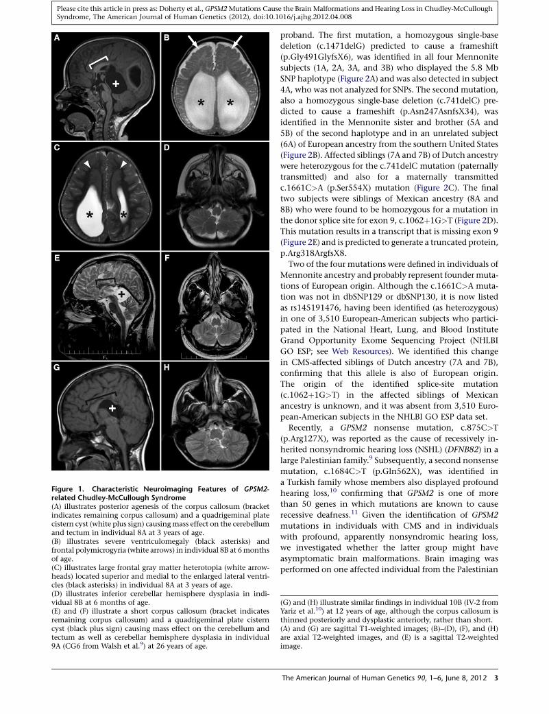
Figure 1. Characteristic Neuroimaging Features of GPSM2-related Chudley-McCullough Syndrome(A) illustrates posterior agenesis of the corpus callosum (bracketindicates remaining corpus callosum) and a quadrigeminal platecistern cyst (white plus sign) causingmass effect on the cerebellumand tectum in individual 8A at 3 years of age.(B) illustrates severe ventriculomegaly (black asterisks) andfrontal polymicrogyria (white arrows) in individual 8B at 6monthsof age.(C) illustrates large frontal gray matter heterotopia (white arrow-heads) located superior and medial to the enlarged lateral ventri-cles (black asterisks) in individual 8A at 3 years of age.(D) illustrates inferior cerebellar hemisphere dysplasia in indi-vidual 8B at 6 months of age.(E) and (F) illustrate a short corpus callosum (bracket indicatesremaining corpus callosum) and a quadrigeminal plate cisterncyst (black plus sign) causing mass effect on the cerebellum andtectum as well as cerebellar hemisphere dysplasia in individual9A (CG6 from Walsh et al.9) at 26 years of age.
Please cite this article in press as: Doherty et al., GPSM2Mutations Cause the Brain Malformations and Hearing Loss in Chudley-McCulloughSyndrome, The American Journal of Human Genetics (2012), doi:10.1016/j.ajhg.2012.04.008
proband. The first mutation, a homozygous single-base
deletion (c.1471delG) predicted to cause a frameshift
(p.Gly491GlyfsX6), was identified in all four Mennonite
subjects (1A, 2A, 3A, and 3B) who displayed the 5.8 Mb
SNP haplotype (Figure 2A) and was also detected in subject
4A, who was not analyzed for SNPs. The second mutation,
also a homozygous single-base deletion (c.741delC) pre-
dicted to cause a frameshift (p.Asn247AsnfsX34), was
identified in the Mennonite sister and brother (5A and
5B) of the second haplotype and in an unrelated subject
(6A) of European ancestry from the southern United States
(Figure 2B). Affected siblings (7A and 7B) of Dutch ancestry
were heterozygous for the c.741delC mutation (paternally
transmitted) and also for a maternally transmitted
c.1661C>A (p.Ser554X) mutation (Figure 2C). The final
two subjects were siblings of Mexican ancestry (8A and
8B) who were found to be homozygous for a mutation in
the donor splice site for exon 9, c.1062þ1G>T (Figure 2D).
This mutation results in a transcript that is missing exon 9
(Figure 2E) and is predicted to generate a truncated protein,
p.Arg318ArgfsX8.
Two of the four mutations were defined in individuals of
Mennonite ancestry and probably represent foundermuta-
tions of European origin. Although the c.1661C>A muta-
tion was not in dbSNP129 or dbSNP130, it is now listed
as rs145191476, having been identified (as heterozygous)
in one of 3,510 European-American subjects who partici-
pated in the National Heart, Lung, and Blood Institute
Grand Opportunity Exome Sequencing Project (NHLBI
GO ESP; see Web Resources). We identified this change
in CMS-affected siblings of Dutch ancestry (7A and 7B),
confirming that this allele is also of European origin.
The origin of the identified splice-site mutation
(c.1062þ1G>T) in the affected siblings of Mexican
ancestry is unknown, and it was absent from 3,510 Euro-
pean-American subjects in the NHLBI GO ESP data set.
Recently, a GPSM2 nonsense mutation, c.875C>T
(p.Arg127X), was reported as the cause of recessively in-
herited nonsyndromic hearing loss (NSHL) (DFNB82) in a
large Palestinian family.9 Subsequently, a second nonsense
mutation, c.1684C>T (p.Gln562X), was identified in
a Turkish family whose members also displayed profound
hearing loss,10 confirming that GPSM2 is one of more
than 50 genes in which mutations are known to cause
recessive deafness.11 Given the identification of GPSM2
mutations in individuals with CMS and in individuals
with profound, apparently nonsyndromic hearing loss,
we investigated whether the latter group might have
asymptomatic brain malformations. Brain imaging was
performed on one affected individual from the Palestinian
(G) and (H) illustrate similar findings in individual 10B (IV-2 fromYariz et al.10) at 12 years of age, although the corpus callosum isthinned posteriorly and dysplastic anteriorly, rather than short.(A) and (G) are sagittal T1-weighted images; (B)–(D), (F), and (H)are axial T2-weighted images, and (E) is a sagittal T2-weightedimage.
The American Journal of Human Genetics 90, 1–6, June 8, 2012 3

Table 2. Neuroimaging Features in Subjects with GPSM2-Related Chudley-McCullough Syndrome
Individuala Ventriculomegaly CC Heterotopia Frontal PMG Cerebellar Dysplasia Arachnoid Cyst
1A yes posterior agenesis small moderate unable to scoredue to mass effect
large bilateral CPA cysts
2A yes, right > left posterior agenesis moderate presentb yes ND
3A shunted HC posterior agenesis small extensive yes no, right ventricle herniationinto midline
3B shunted HC posterior agenesis small moderate yes left CPA
4A yes posterior agenesis extensive extensive no right CPA
5Ac HC, foramen ofMonro fenestration
probable partialagenesis
ND ND ND ND
5Bc shunted HC probable partialagenesis
ND ND ND ND
6A shunted HC posterior agenesis small extensive yes large interhemispheric andsmall left CPA cysts
7A yes posterior agenesis small moderate yes moderate interhemispheric cyst
7B shunted HC posterior agenesis small moderate yes small pineal cyst
8A yes posterior agenesis large extensive yes large interhemispheric andsmall bilateral CPA cysts
8B shunted HC posterior agenesis moderate extensive yes moderate interhemisphericand small right CPA cysts
Previously Published Subjects with GPSM2 Mutations9,10
9A, Walsh9 CG6 no short and thin moderate moderate yes moderate interhemispheric cyst
10A, Yariz10 IV-1 no short and thin small subtle no moderate interhemispheric cyst
10B, Yariz10 IV-2 no short, severelythin posteriorly
moderate subtle yes small interhemispheric cyst
11A,d Yariz10 IV-3 no short and thin small subtle mild on right moderate interhemispheric cyst
CC, corpus callosum; CPA, cerebellopontine angle; HC, hydrocephalus; ND, no data; PMG, polymicrogyria.aA and B indicate siblings.bUnable to determine the extent due to technical limitations of the available images.cOnly computed tomography scans available.d11A is a first cousin of 10A and 10B.
Please cite this article in press as: Doherty et al., GPSM2Mutations Cause the Brain Malformations and Hearing Loss in Chudley-McCulloughSyndrome, The American Journal of Human Genetics (2012), doi:10.1016/j.ajhg.2012.04.008
family and all three affected individuals from the Turkish
family. Despite the fact that study subjects 9A, 10A, 10B,
and 11A did not exhibit any neurological deficits (Table 1),
all four individuals displayed imaging features consistent
with CMS (Table 2 and Figure 1E–1H). In contrast to the
CMS subjects, none of the individuals studied solely
because of hearing loss had ventriculomegaly, and the
corpus callosum abnormalities tended to be less severe.
Genotype-phenotype correlations were not apparent, in
that the individuals (5A, 5B, 6A, and 9A) with GPSM2
mutations predicted to result in the shortest proteins
(p.Asn247AsnfsX34 and p.Arg127X) did not have more
severe brain malformations or clinical features than the
other subjects studied.
The GPSM2 mutations identified in individuals with
CMS highlight the role of GPSM2 in normal brain develop-
ment and in mechanisms that underlie common brain
malformations, such as partial agenesis of the corpus cal-
losum, heterotopia, and polymicrogyria. During early neu-
rogenesis in the mouse cerebral cortex, Gpsm2 is required
for planar orientation of the mitotic spindle in apical
4 The American Journal of Human Genetics 90, 1–6, June 8, 2012
progenitor cells (radial glia).12,13 In mice, an engineered
variant (DC) very similar to the human p.Gly491GlyfsX6
variant (Figure 2F) results in abnormally localized apical
progenitors but does not seem to radically affect the
number or organization of cortical neurons, although
phenotypes in mature mouse brain have not been pub-
lished.13,14 It is tempting to speculate that the ectopic
neuronal precursors could result in heterotopic neurons
analogous to the heterotopia observed in individuals
with CMS. GPSM2 is also required for correct spindle orien-
tation in keratinocyte progenitors,15 T cells,16 oocytes,17
and epithelial cells,18 as well as for neurotransmitter local-
ization in mature neurons.19 Given this central role of
GPSM2 (also known as LGN [Leu-Gly-Asn repeat-enriched
protein] and Pins [Partner of Inscuteable, homolog of
Drosophila]) in cell division, it is surprising that truncating
mutations do not cause more widespread defects in indi-
viduals with CMS and in the mouse model.
In conclusion, we provide compelling evidence that
GPSM2 mutations account for CMS in most, if not all,
affected individuals, confirming a role for GPSM2 in
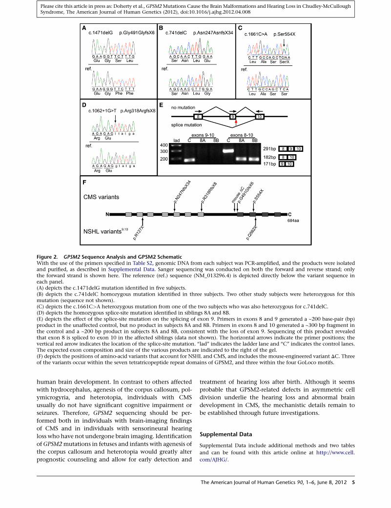
Figure 2. GPSM2 Sequence Analysis and GPSM2 SchematicWith the use of the primers specified in Table S2, genomic DNA from each subject was PCR-amplified, and the products were isolatedand purified, as described in Supplemental Data. Sanger sequencing was conducted on both the forward and reverse strand; onlythe forward strand is shown here. The reference (ref.) sequence (NM_013296.4) is depicted directly below the variant sequence ineach panel.(A) depicts the c.1471delG mutation identified in five subjects.(B) depicts the c.741delC homozygous mutation identified in three subjects. Two other study subjects were heterozygous for thismutation (sequence not shown).(C) depicts the c.1661C>A heterozygous mutation from one of the two subjects who was also heterozygous for c.741delC.(D) depicts the homozygous splice-site mutation identified in siblings 8A and 8B.(E) depicts the effect of the splice-site mutation on the splicing of exon 9. Primers in exons 8 and 9 generated a ~200 base-pair (bp)product in the unaffected control, but no product in subjects 8A and 8B. Primers in exons 8 and 10 generated a ~300 bp fragment inthe control and a ~200 bp product in subjects 8A and 8B, consistent with the loss of exon 9. Sequencing of this product revealedthat exon 8 is spliced to exon 10 in the affected siblings (data not shown). The horizontal arrows indicate the primer positions; thevertical red arrow indicates the location of the splice-site mutation. ‘‘lad’’ indicates the ladder lane and ‘‘C’’ indicates the control lanes.The expected exon composition and size of the various products are indicated to the right of the gel.(F) depicts the positions of amino-acid variants that account for NSHL and CMS, and includes the mouse-engineered variant DC. Threeof the variants occur within the seven tetratricopeptide repeat domains of GPSM2, and three within the four GoLoco motifs.
Please cite this article in press as: Doherty et al., GPSM2Mutations Cause the Brain Malformations and Hearing Loss in Chudley-McCulloughSyndrome, The American Journal of Human Genetics (2012), doi:10.1016/j.ajhg.2012.04.008
human brain development. In contrast to others affected
with hydrocephalus, agenesis of the corpus callosum, pol-
ymicrogyria, and heterotopia, individuals with CMS
usually do not have significant cognitive impairment or
seizures. Therefore, GPSM2 sequencing should be per-
formed both in individuals with brain-imaging findings
of CMS and in individuals with sensorineural hearing
loss who have not undergone brain imaging. Identification
ofGPSM2mutations in fetuses and infants with agenesis of
the corpus callosum and heterotopia would greatly alter
prognostic counseling and allow for early detection and
treatment of hearing loss after birth. Although it seems
probable that GPSM2-related defects in asymmetric cell
division underlie the hearing loss and abnormal brain
development in CMS, the mechanistic details remain to
be established through future investigations.
Supplemental Data
Supplemental Data include additional methods and two tables
and can be found with this article online at http://www.cell.
com/AJHG/.
The American Journal of Human Genetics 90, 1–6, June 8, 2012 5

Please cite this article in press as: Doherty et al., GPSM2Mutations Cause the Brain Malformations and Hearing Loss in Chudley-McCulloughSyndrome, The American Journal of Human Genetics (2012), doi:10.1016/j.ajhg.2012.04.008
Acknowledgments
We are grateful to the family members who participated in this
study, to Sunita Khatkar for conducting candidate gene analysis,
to Kirk McManus for preparing Figure 2, and to Tom Walsh and
Mary-Claire King for critically reading the manuscript. The
research was supported by grants from the National Institutes of
Health, KL2-RR025015 (to D.D.), R01DC009645 (to M.T.), and
R01DC011835 (to M.K.); the Manitoba Institute of Child Health
(to A.E.C. and B.T.R.); and the Winnipeg Rh Institute Foundation
(to T.Z.). FORGE (Finding of Rare Disease Genes) Canada funding
was provided by the government of Canada through Genome
Canada, the Canadian Institutes of Health Research, and the On-
tario Genomics Institute (OGI-049). Additional funding was
provided by Genome Quebec and Genome British Columbia.
FORGE Canada Consortium Steering Committee: Kym Boycott
(leader; University of Ottawa), Jan Friedman (coleader; University
of British Columbia), Jacques Michaud (coleader; Universite de
Montreal), Francois Bernier (University of Calgary), Michael
Brudno (University of Toronto), Bridget Fernandez (Memorial
University), Bartha Knoppers (McGill University), Mark Samuels
(Universite de Montreal), Steve Scherer (University of Toronto).
Received: November 24, 2011
Revised: April 12, 2012
Accepted: April 16, 2012
Published online: May 10, 2012
Web Resources
The URLs for data presented herein are as follows:
dbSNP, http://ncbi.nlm.nih.gov/projects/SNP
NHLBI Grand Opportunity Exome Sequencing Project (NHLBI
GO ESP) Exome Variant Server (EVS), http://evs.gs.washington.
edu/EVS
Online Mendelian Inheritance in Man (OMIM), http://www.
omim.org
UCSC Genome Browser, http://genome.ucsc.edu
References
1. Chudley, A.E., McCullough, C., and McCullough, D.W.
(1997). Bilateral sensorineural deafness and hydrocephalus
due to foramen of Monro obstruction in sibs: a newly
described autosomal recessive disorder. Am. J. Med. Genet.
68, 350–356.
2. Hendriks, Y.M.C., Laan, L.A.E.M., Vielvoye, G.J., and van
Haeringen, A. (1999). Bilateral sensorineural deafness, partial
agenesis of the corpus callosum, and arachnoid cysts in two
sisters. Am. J. Med. Genet. 86, 183–186.
3. Lemire, E.G., and Stoeber, G.P. (2000). Chudley-McCullough
syndrome: bilateral sensorineural deafness, hydrocephalus,
and other structural brain abnormalities. Am. J. Med. Genet.
90, 127–130.
4. Welch, K.O., Tekin, M., Nance, W.E., Blanton, S.H., Arnos,
K.S., and Pandya, A. (2003). Chudley-McCullough syndrome:
expanded phenotype and review of the literature. Am. J. Med.
Genet. A. 119A, 71–76.
6 The American Journal of Human Genetics 90, 1–6, June 8, 2012
5. Østergaard, E., Pedersen, V.F., Skriver, E.B., and Brøndum-
Nielsen, K. (2004). Brothers with Chudley-McCullough
syndrome: sensorineural deafness, agenesis of the corpus
callosum, and other structural brain abnormalities. Am. J.
Med. Genet. A. 124A, 74–78.
6. Matteucci, F., Tarantino, E., Bianchi, M.C., Cingolani, C.,
Fattori, B., Nacci, A., and Ursino, F. (2006). Sensorineural deaf-
ness, hydrocephalus and structural brain abnormalities in two
sisters: the Chudley-McCullough syndrome. Am. J. Med.
Genet. A. 140, 1183–1188.
7. Alrashdi, I., Barker, R., and Patton, M.A. (2011). Chudley-
McCullough syndrome: another report and a brief review of
the literature. Clin. Dysmorphol. 20, 107–110.
8. Kau, T., Veraguth, D., Schiegl, H., Scheer, I., and Boltshauser, E.
(2012). Chudley-McCullough syndrome: case report and review
of the neuroimaging spectrum. Neuropediatrics 43, 44–47.
9. Walsh, T., Shahin, H., Elkan-Miller, T., Lee, M.K., Thornton,
A.M., Roeb, W., Abu Rayyan, A., Loulus, S., Avraham, K.B.,
King, M.-C., and Kanaan, M. (2010). Whole exome
sequencing and homozygosity mapping identify mutation
in the cell polarity protein GPSM2 as the cause of nonsyn-
dromic hearing loss DFNB82. Am. J. Hum. Genet. 87, 90–94.
10. Yariz, K.O., Walsh, T., Akay, H., Duman, D., Akkaynak, A.C.,
King, M.-C., and Tekin, M. (2012). A truncating mutation in
GPSM2 is associated with recessive non-syndromic hearing
loss. Clin. Genet. 81, 289–293.
11. Dror, A.A., and Avraham, K.B. (2009). Hearing loss: mecha-
nisms revealed by genetics and cell biology. Annu. Rev. Genet.
43, 411–437.
12. Morin, X., Jaouen, F., and Durbec, P. (2007). Control of planar
divisions by theG-protein regulator LGNmaintains progenitors
in the chick neuroepithelium. Nat. Neurosci. 10, 1440–1448.
13. Konno, D., Shioi, G., Shitamukai, A., Mori, A., Kiyonari, H.,
Miyata, T., and Matsuzaki, F. (2008). Neuroepithelial progeni-
tors undergo LGN-dependent planar divisions to maintain
self-renewability during mammalian neurogenesis. Nat. Cell
Biol. 10, 93–101.
14. Shioi, G., Konno, D., Shitamukai, A., andMatsuzaki, F. (2009).
Structural basis for self-renewal of neural progenitors in
cortical neurogenesis. Cereb. Cortex 19 (Suppl 1 ), i55–i61.
15. Williams, S.E., Beronja, S., Pasolli, H.A., and Fuchs, E. (2011).
Asymmetric cell divisions promote Notch-dependent
epidermal differentiation. Nature 470, 353–358.
16. Oliaro, J., Van Ham, V., Sacirbegovic, F., Pasam, A., Bomzon,
Z., Pham, K., Ludford-Menting, M.J., Waterhouse, N.J., Bots,
M., Hawkins, E.D., et al. (2010). Asymmetric cell division of
T cells upon antigen presentation uses multiple conserved
mechanisms. J. Immunol. 185, 367–375.
17. Guo, X., and Gao, S. (2009). Pins homolog LGN regulates
meiotic spindle organization in mouse oocytes. Cell Res. 19,
838–848.
18. Zheng, Z., Zhu, H., Wan, Q., Liu, J., Xiao, Z., Siderovski, D.P.,
and Du, Q. (2010). LGN regulates mitotic spindle orientation
during epithelial morphogenesis. J. Cell Biol. 189, 275–288.
19. Sans, N., Wang, P.Y., Du, Q., Petralia, R.S., Wang, Y.X., Nakka,
S., Blumer, J.B., Macara, I.G., andWenthold, R.J. (2005). mPins
modulates PSD-95 and SAP102 trafficking and influences
NMDA receptor surface expression. Nat. Cell Biol. 7, 1179–
1190.