Golgi fragmentation during Fas-mediated apoptosis is associated with the rapid loss of GM130
6
Golgi fragmentation during Fas-mediated apoptosis is associated with the rapid loss of GM130 q Annemieke Walker, * Carol Ward, Tara A. Sheldrake, Ian Dransfield, Adriano G. Rossi, James G. Pryde, and Christopher Haslett Centre for Inflammation Research, The University of Edinburgh, Medical School, Teviot Place, Edinburgh EH8 9AG, UK Received 2 February 2004 Abstract During apoptosis, the Golgi complex becomes fragmented and key proteins (e.g., GRASP65 and p115) are targets for caspase cleavage. GM130, an integral membrane protein, contributes to the maintenance of Golgi structure and facilitates membrane fusion with secretory vesicles. We show that GM130 levels decrease during Fas-induced apoptosis but not during staurosporine-in- duced apoptosis while in both models p115 levels remain unaffected. We conclude that GM130 is rapidly diminished during Fas-mediated apoptosis associated with Golgi fragmentation in contrast to previous studies which have suggested that loss of GM130 during apoptosis is a late event. Ó 2004 Elsevier Inc. All rights reserved. Keywords: Golgi; Apoptosis; p115; GM130; Staurosporine; Fas The Golgi complex is the ‘organising centre’ for protein cargo en route from the endoplasmic reticulum to the plasma membrane. During mitosis, this structure becomes fragmented with a concomitant arrest in ves- icle trafficking [1]. During apoptosis the Golgi complex also fragments [2–4] and it is possible that the secretory pathway is similarly inhibited, which would in part explain why apoptotic cells fail to respond to various external stimuli [5]. Mitotic Golgi fragmentation has been demonstrated to be dependent on cyclin-depen- dent-kinase-1 (CDK1) and later GM130 was identified as a CDK1 substrate [6]. GM130, a cis-Golgi matrix protein, is associated with the cytoplasmic face of the Golgi via GRASP65 [7], a N-myristoylated peripheral membrane protein [8], and binds the p115 tethering protein present on incoming vesicles [9,10]. Following phosphorylation, GM130 is no longer able to bind to p115 and dissociation of p115–GM130 is thought to contribute to fragmentation of the Golgi complex during mitosis [9]. GM130, GRASP65, and p115 have already been shown to be cleaved during apoptosis in a caspase-dependent manner facilitating Golgi fragmen- tation during apoptosis [2,3]. These studies concluded that GM130 cleavage during apoptosis is secondary to GRASP65 cleavage and a relatively late event [2,3]. However, these conclusions were based on stauro- sporine (broad-range kinase inhibitor) -induced apop- tosis and work by Cha et al. [11] demonstrates phosphorylation of Golgi-associated targets may modulate cleavage. To address this issue, we followed the fate of GM130 during apoptosis in human J.CaM1.6 cells, a derivative of Jurkat T lymphocyte cells by triggering the physiologically relevant Fas death pathway [12] and for comparison by the broad spectrum kinase inhibitor staurosporine. Materials and methods Cell line. J.CaM1.6 cells were obtained from the European Col- lection of Animal Cell Cultures (Porton Down, UK) and routinely maintained in RPMI-1640 containing 10% FCS, 100 U/ml penicillin/ streptomycin, 2 mM L-glutamine, and 1% non-essential amino acids. Cells were grown in a 5% CO 2 humidified incubator at 37 °C. q Abbreviations: z-VAD-fmk, benzylocarbonyl-Val-Ala-Asp-flou- romethylketone; PI, propidium iodide; SSP, staurosporine. * Corresponding author. Fax: +44-131-650-4384. E-mail address: awalker4@staffmail.ed.ac.uk (A. Walker). 0006-291X/$ - see front matter Ó 2004 Elsevier Inc. All rights reserved. doi:10.1016/j.bbrc.2004.02.015 Biochemical and Biophysical Research Communications 316 (2004) 6–11 BBRC www.elsevier.com/locate/ybbrc
-
Upload
independent -
Category
Documents
-
view
0 -
download
0
Transcript of Golgi fragmentation during Fas-mediated apoptosis is associated with the rapid loss of GM130
Biochemical and Biophysical Research Communications 316 (2004) 6–11
BBRCwww.elsevier.com/locate/ybbrc
Golgi fragmentation during Fas-mediated apoptosis is associatedwith the rapid loss of GM130q
Annemieke Walker,* Carol Ward, Tara A. Sheldrake, Ian Dransfield, Adriano G. Rossi,James G. Pryde, and Christopher Haslett
Centre for Inflammation Research, The University of Edinburgh, Medical School, Teviot Place, Edinburgh EH8 9AG, UK
Received 2 February 2004
Abstract
During apoptosis, the Golgi complex becomes fragmented and key proteins (e.g., GRASP65 and p115) are targets for caspase
cleavage. GM130, an integral membrane protein, contributes to the maintenance of Golgi structure and facilitates membrane fusion
with secretory vesicles. We show that GM130 levels decrease during Fas-induced apoptosis but not during staurosporine-in-
duced apoptosis while in both models p115 levels remain unaffected. We conclude that GM130 is rapidly diminished during
Fas-mediated apoptosis associated with Golgi fragmentation in contrast to previous studies which have suggested that loss of
GM130 during apoptosis is a late event.
� 2004 Elsevier Inc. All rights reserved.
Keywords: Golgi; Apoptosis; p115; GM130; Staurosporine; Fas
The Golgi complex is the ‘organising centre’ for
protein cargo en route from the endoplasmic reticulum
to the plasma membrane. During mitosis, this structure
becomes fragmented with a concomitant arrest in ves-
icle trafficking [1]. During apoptosis the Golgi complex
also fragments [2–4] and it is possible that the secretory
pathway is similarly inhibited, which would in part
explain why apoptotic cells fail to respond to variousexternal stimuli [5]. Mitotic Golgi fragmentation has
been demonstrated to be dependent on cyclin-depen-
dent-kinase-1 (CDK1) and later GM130 was identified
as a CDK1 substrate [6]. GM130, a cis-Golgi matrix
protein, is associated with the cytoplasmic face of the
Golgi via GRASP65 [7], a N-myristoylated peripheral
membrane protein [8], and binds the p115 tethering
protein present on incoming vesicles [9,10]. Followingphosphorylation, GM130 is no longer able to bind to
p115 and dissociation of p115–GM130 is thought to
contribute to fragmentation of the Golgi complex
qAbbreviations: z-VAD-fmk, benzylocarbonyl-Val-Ala-Asp-flou-
romethylketone; PI, propidium iodide; SSP, staurosporine.* Corresponding author. Fax: +44-131-650-4384.
E-mail address: [email protected] (A. Walker).
0006-291X/$ - see front matter � 2004 Elsevier Inc. All rights reserved.
doi:10.1016/j.bbrc.2004.02.015
during mitosis [9]. GM130, GRASP65, and p115 have
already been shown to be cleaved during apoptosis in a
caspase-dependent manner facilitating Golgi fragmen-
tation during apoptosis [2,3]. These studies concluded
that GM130 cleavage during apoptosis is secondary to
GRASP65 cleavage and a relatively late event [2,3].
However, these conclusions were based on stauro-
sporine (broad-range kinase inhibitor) -induced apop-tosis and work by Cha et al. [11] demonstrates
phosphorylation of Golgi-associated targets may
modulate cleavage. To address this issue, we followed
the fate of GM130 during apoptosis in human
J.CaM1.6 cells, a derivative of Jurkat T lymphocyte
cells by triggering the physiologically relevant Fas
death pathway [12] and for comparison by the broad
spectrum kinase inhibitor staurosporine.
Materials and methods
Cell line. J.CaM1.6 cells were obtained from the European Col-
lection of Animal Cell Cultures (Porton Down, UK) and routinely
maintained in RPMI-1640 containing 10% FCS, 100U/ml penicillin/
streptomycin, 2mM LL-glutamine, and 1% non-essential amino acids.
Cells were grown in a 5% CO2 humidified incubator at 37 �C.
A. Walker et al. / Biochemical and Biophysical Research Communications 316 (2004) 6–11 7
Induction of apoptosis. J.CaM1.6 cells were incubated in 96-well
plates at 5� 106 cells/ml. Cells were incubated with either the broad-
range kinase inhibitor staurosporine (2lM) (Calbiochem–Novabio-
chem, UK) or the anti-Fas monoclonal antibody CH11 (500 ng/ml)
(TCS Biologicals, UK) to induce apoptosis. One hundred micromolar
of the pan-caspase inhibitor z-VAD-fmk (Bachem, UK) was added to
appropriate wells in both staurosporine and CH11 assays and ZB4
(Fas blocking monoclonal antibody, 500 ng/ml, TCS Biologicals, UK)
was added to appropriate wells in CH11 assays.
Flow cytometry. Externalisation of phosphatidylserine was moni-
tored by FITC-labelled recombinant human Annexin-V binding. Stock
Annexin-V-FITC (Bender MedSystems, Austria) was diluted 1/3000
with Annexin-V-binding buffer (500ml Hanks’ Balanced Salt Solution
containing 5lM CaCl2). Cells were incubated on ice for 10min with
Annexin-V-FITC and 1min with propidium iodide (PI). Annexin-V-
FITC/PI binding was assessed by flow cytometry on a Coulter EPICS
(Beckman Coulter, UK) and analysed on associated EXPO software.
All experiments were carried out in triplicate (n¼ 3).
Immunofluorescence and image acquisition. The indirect immuno-
fluorescence protocol was as described [13]. Cells were cytocentrifuged
(300 rpm for 3min) onto 1.5� 22� 22-mm glass coverslips and fixed in
methanol-free 3% (w/v) para-formaldehyde/PBS containing 1mM
CaCl2 and 1mM MgCl2 for 20min at 20 �C. Cells were permeabilised
with 0.1% (w/v) Triton X-100 and non-specific binding was blocked
with 10% (v/v) sheep serum in 0.2% (w/v) fish skin gelatin (Sigma,
UK). Primary antibodies used were: anti-active-caspase-3 rabbit
polyclonal antibody (BD Pharmingen, UK) and mouse anti-rat
GM130 monoclonal antibody (BD Transduction Laboratories, UK).
Secondary antibodies used were Alexa 488 (green) goat anti-mouse
IgG (highly cross-adsorbed) and Alexa 568 (red) goat anti-rabbit
(highly cross-adsorbed) (Molecular Probes, Netherlands). Nuclei were
stained with TO-PRO3 (Molecular Probes). Images for each channel
were captured separately and assembled into a single file with TCS-
MERGE software (Leica Microsystems, Heidelberg, Germany) prior
to analysis. All confocal images were processed digitally using Adobe
Photoshop 5.02 and Paint Shop Pro 4.
Western blotting. J.CaM1.6 cells (2.5� 106) were harvested per
sample and lysed as described [13]. Each sample was loaded onto a
10% SDS–polyacrylamide gel and proteins were separated and elec-
trophoretically transferred to nitrocellulose. The nitrocellulose
membranes were probed with mouse monoclonal antibodies to pro-
caspase-3 (BD Pharmingen, UK), GM130, p115 (BD Transduction
Laboratories, UK), and actin (Sigma, UK) followed by HRP-Goat
anti mouse IgG (Dako, UK). The membranes were then incubated
with ECL reagent (Amersham Biosciences, UK), placed under BioMax
MS-1 X-ray sensitive film, and processed through an X-ray developer
(X-Ograph Imaging Systems, Wilts, UK).
Statistical analysis. Results are reported either as pooled data from
a series of n separate experiments (mean� SEM) or as individual
representative experiments (mean� SD). Statistical significance was
assessed by one-way analysis of variance with comparisons between
groups made using the Tukey procedure.
Results
Induction of apoptosis in J.CaM1.6 cells
To examine whether GM130 was affected during Fas-
mediated apoptosis, we used J.CaM1.6 cells, a derivative
of Jurkat cells, which constitutively expresses Fas.J.CaM1.6 cells were treated with 500 ng/ml CH11 in the
presence of FCS, over 6 h and the proportion of apop-
totic cells, as assessed by Annexin-V-FITC binding, was
monitored at 1, 2, 4, and 6 h (Fig. 1A). At 1 h, thepercentage of apoptotic cells was �12%, significantly
(p < 0:05) higher than present in control populations
(�2%), increasing up to 4 h, where the amount of ap-
optosis was �60%, after which the rate of apoptosis
began to decline. To ensure that induction of apoptosis
occurred via the Fas pathway, control samples were pre-
incubated with 500 ng/ml of antagonist Fas antibody
ZB4 for 30min, prior to addition of CH11. ZB4 abol-ished the apoptotic effect of CH11 upon J.CaM1.6 cells,
decreasing the proportion of cells which bound Ann-
exin-V-FITC (�5% not significant (p < 0:05) from
untreated control levels). In addition, z-VAD-fmk
completely inhibited Annexin-V-FITC binding, which
was not significantly different from control levels.
Fig. 1B shows representative flow cytometry profiles
demonstrating that the amount of PI positive cells neverincreases significantly above control levels.
To follow the kinetics of apoptosis induction in
J.CaM1.6 cells by staurosporine, cells were incubated in
the presence of 2 lM staurosporine over 4 h (Fig. 2A)
and apoptosis was assessed by Annexin-V-FITC binding
and exclusion of PI. The results from three separate
experiments show that at 1 h, the number of apoptotic
cells present, �14%, was already significantly (p < 0:05)increased from control cells �2%. This trend continued
up to 4 h, where �80% of cells bound Annexin-V-FITC
and excluded PI. Apoptosis was inhibited by z-VAD-
fmk, demonstrating that the effects of staurosporine
were due to caspase activity. Fig. 2B shows representa-
tive flow cytometry profiles revealing that staurosporine
does not induce significant amounts of necrosis over this
time frame.
Immunofluorescence of Golgi fragmentation in apoptotic
J.CaM1.6 cells
To examine the fragmentation of the Golgi in apop-totic J.CaM1.6 cells, cells were treated with 500 ng/ml
CH11 or 2 lM staurosporine over 4 h to induce apop-
tosis. Apoptotic cells were identified by their fragmented
nuclei and by positive staining with the polyclonal an-
tibody raised against active-caspase-3 (Fig. 3). Many of
the cells treated with 500 ng/ml CH11 had caspase-3
activity throughout the cytoplasm and in those cells the
nuclei and Golgi were fragmented. In cells that had beenpre-treated with the neutralising antibody ZB4 against
Fas or the caspase inhibitor z-VAD-fmk, caspase-3 ac-
tivation, and Golgi fragmentation were prevented. This
demonstrated that Golgi fragmentation and nuclear
“pebbling” were a direct consequence of signalling
through the Fas death pathway in CH11-treated cells
and that the morphological changes observed were
caspase-dependent. To compare the fragmentation ofthe Golgi complex during staurosporine-induced
apoptosis, J.CaM1.6 cells were treated with 2 lM
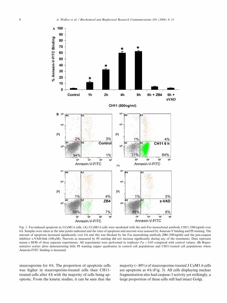
Fig. 1. Fas-induced apoptosis in J.CaM1.6 cells. (A) J.CaM1.6 cells were incubated with the anti-Fas monoclonal antibody CH11 (500 ng/ml) over
6 h. Samples were taken at the time points indicated and the rates of apoptosis and necrosis were assessed by Annexin-V binding and PI staining. The
amount of apoptosis increased significantly over 6 h and this was blocked by the Fas neutralising antibody ZB4 (500 ng/ml) and the pan-caspase
inhibitor z-VAD-fmk (100 lM). Necrosis as measured by PI staining did not increase significantly during any of the treatments. Data represent
means�SEM of three separate experiments. All experiments were performed in triplicate (*p < 0:05 compared with control values). (B) Repre-
sentative scatter plots demonstrating little PI staining (upper quadrants) in control cell populations and CH11-treated cell populations where
Annexin-FITC binding is increased.
8 A. Walker et al. / Biochemical and Biophysical Research Communications 316 (2004) 6–11
staurosporine for 4 h. The proportion of apoptotic cells
was higher in staurosporine-treated cells than CH11-
treated cells after 4 h with the majority of cells being ap-
optotic. From the kinetic studies, it can be seen that the
majority (�80%) of staurosporine-treated J.CaM1.6 cells
are apoptotic at 4 h (Fig. 3). All cells displaying nuclear
fragmentation also had caspase-3 activity yet strikingly, a
large proportion of these cells still had intact Golgi.
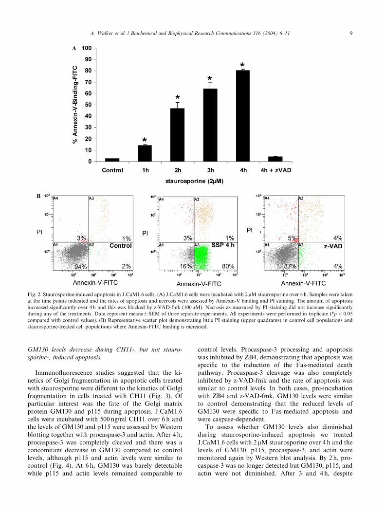
Fig. 2. Staurosporine-induced apoptosis in J.CaM1.6 cells. (A) J.CaM1.6 cells were incubated with 2lM staurosporine over 4 h. Samples were taken
at the time points indicated and the rates of apoptosis and necrosis were assessed by Annexin-V binding and PI staining. The amount of apoptosis
increased significantly over 4 h and this was blocked by z-VAD-fmk (100lM). Necrosis as measured by PI staining did not increase significantly
during any of the treatments. Data represent means� SEM of three separate experiments. All experiments were performed in triplicate (*p < 0:05
compared with control values). (B) Representative scatter plot demonstrating little PI staining (upper quadrants) in control cell populations and
staurosporine-treated cell populations where Annexin-FITC binding is increased.
A. Walker et al. / Biochemical and Biophysical Research Communications 316 (2004) 6–11 9
GM130 levels decrease during CH11-, but not stauro-
sporine-, induced apoptosis
Immunofluorescence studies suggested that the ki-
netics of Golgi fragmentation in apoptotic cells treated
with staurosporine were different to the kinetics of Golgi
fragmentation in cells treated with CH11 (Fig. 3). Ofparticular interest was the fate of the Golgi matrix
protein GM130 and p115 during apoptosis. J.CaM1.6
cells were incubated with 500 ng/ml CH11 over 6 h and
the levels of GM130 and p115 were assessed by Western
blotting together with procaspase-3 and actin. After 4 h,
procaspase-3 was completely cleaved and there was a
concomitant decrease in GM130 compared to control
levels, although p115 and actin levels were similar tocontrol (Fig. 4). At 6 h, GM130 was barely detectable
while p115 and actin levels remained comparable to
control levels. Procaspase-3 processing and apoptosis
was inhibited by ZB4, demonstrating that apoptosis was
specific to the induction of the Fas-mediated death
pathway. Procaspase-3 cleavage was also completely
inhibited by z-VAD-fmk and the rate of apoptosis was
similar to control levels. In both cases, pre-incubation
with ZB4 and z-VAD-fmk, GM130 levels were similarto control demonstrating that the reduced levels of
GM130 were specific to Fas-mediated apoptosis and
were caspase-dependent.
To assess whether GM130 levels also diminished
during staurosporine-induced apoptosis we treated
J.CaM1.6 cells with 2 lM staurosporine over 4 h and the
levels of GM130, p115, procaspase-3, and actin were
monitored again by Western blot analysis. By 2 h, pro-caspase-3 was no longer detected but GM130, p115, and
actin were not diminished. After 3 and 4 h, despite
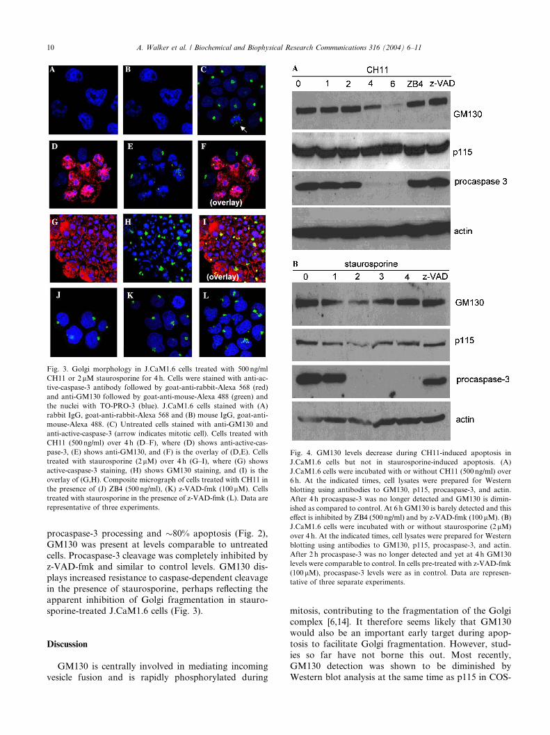
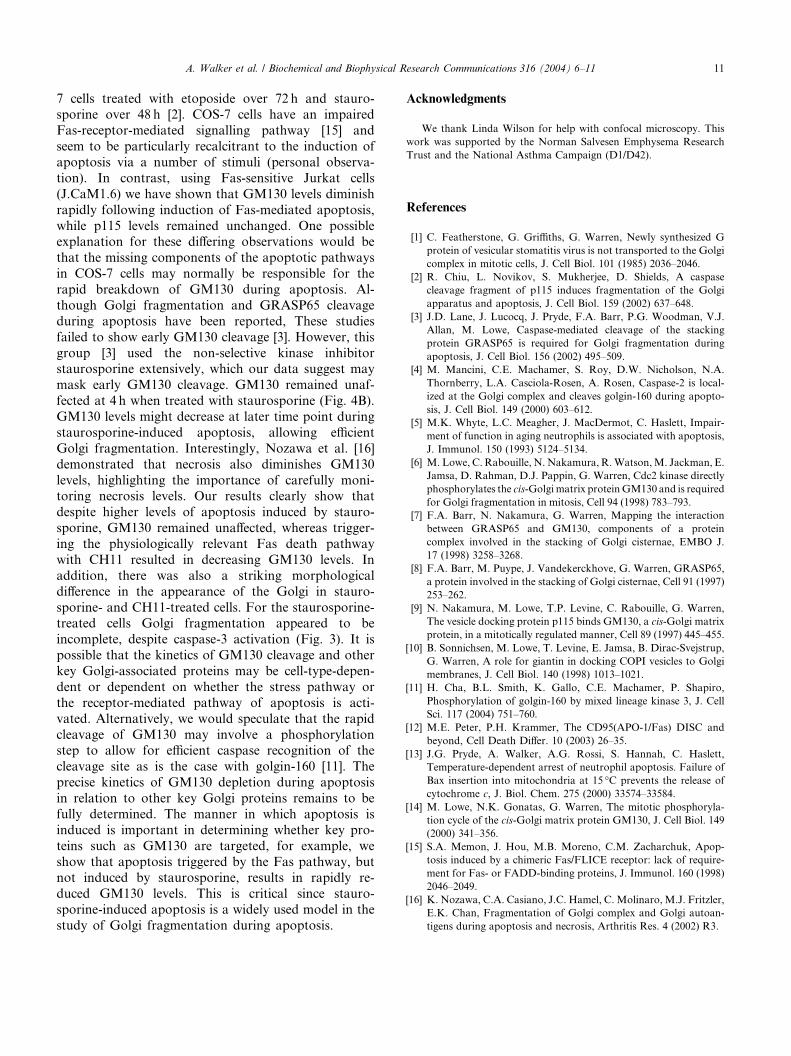
Fig. 4. GM130 levels decrease during CH11-induced apoptosis in
J.CaM1.6 cells but not in staurosporine-induced apoptosis. (A)
J.CaM1.6 cells were incubated with or without CH11 (500 ng/ml) over
6 h. At the indicated times, cell lysates were prepared for Western
blotting using antibodies to GM130, p115, procaspase-3, and actin.
After 4 h procaspase-3 was no longer detected and GM130 is dimin-
ished as compared to control. At 6 h GM130 is barely detected and this
effect is inhibited by ZB4 (500 ng/ml) and by z-VAD-fmk (100 lM). (B)
J.CaM1.6 cells were incubated with or without staurosporine (2lM)
over 4 h. At the indicated times, cell lysates were prepared for Western
blotting using antibodies to GM130, p115, procaspase-3, and actin.
After 2 h procaspase-3 was no longer detected and yet at 4 h GM130
levels were comparable to control. In cells pre-treated with z-VAD-fmk
(100 lM), procaspase-3 levels were as in control. Data are represen-
tative of three separate experiments.
Fig. 3. Golgi morphology in J.CaM1.6 cells treated with 500 ng/ml
CH11 or 2lM staurosporine for 4 h. Cells were stained with anti-ac-
tive-caspase-3 antibody followed by goat-anti-rabbit-Alexa 568 (red)
and anti-GM130 followed by goat-anti-mouse-Alexa 488 (green) and
the nuclei with TO-PRO-3 (blue). J.CaM1.6 cells stained with (A)
rabbit IgG, goat-anti-rabbit-Alexa 568 and (B) mouse IgG, goat-anti-
mouse-Alexa 488. (C) Untreated cells stained with anti-GM130 and
anti-active-caspase-3 (arrow indicates mitotic cell). Cells treated with
CH11 (500 ng/ml) over 4 h (D–F), where (D) shows anti-active-cas-
pase-3, (E) shows anti-GM130, and (F) is the overlay of (D,E). Cells
treated with staurosporine (2lM) over 4 h (G–I), where (G) shows
active-caspase-3 staining, (H) shows GM130 staining, and (I) is the
overlay of (G,H). Composite micrograph of cells treated with CH11 in
the presence of (J) ZB4 (500 ng/ml), (K) z-VAD-fmk (100lM). Cells
treated with staurosporine in the presence of z-VAD-fmk (L). Data are
representative of three experiments.
10 A. Walker et al. / Biochemical and Biophysical Research Communications 316 (2004) 6–11
procaspase-3 processing and �80% apoptosis (Fig. 2),
GM130 was present at levels comparable to untreated
cells. Procaspase-3 cleavage was completely inhibited by
z-VAD-fmk and similar to control levels. GM130 dis-
plays increased resistance to caspase-dependent cleavage
in the presence of staurosporine, perhaps reflecting theapparent inhibition of Golgi fragmentation in stauro-
sporine-treated J.CaM1.6 cells (Fig. 3).
Discussion
GM130 is centrally involved in mediating incoming
vesicle fusion and is rapidly phosphorylated during
mitosis, contributing to the fragmentation of the Golgi
complex [6,14]. It therefore seems likely that GM130
would also be an important early target during apop-
tosis to facilitate Golgi fragmentation. However, stud-ies so far have not borne this out. Most recently,
GM130 detection was shown to be diminished by
Western blot analysis at the same time as p115 in COS-
A. Walker et al. / Biochemical and Biophysical Research Communications 316 (2004) 6–11 11
7 cells treated with etoposide over 72 h and stauro-sporine over 48 h [2]. COS-7 cells have an impaired
Fas-receptor-mediated signalling pathway [15] and
seem to be particularly recalcitrant to the induction of
apoptosis via a number of stimuli (personal observa-
tion). In contrast, using Fas-sensitive Jurkat cells
(J.CaM1.6) we have shown that GM130 levels diminish
rapidly following induction of Fas-mediated apoptosis,
while p115 levels remained unchanged. One possibleexplanation for these differing observations would be
that the missing components of the apoptotic pathways
in COS-7 cells may normally be responsible for the
rapid breakdown of GM130 during apoptosis. Al-
though Golgi fragmentation and GRASP65 cleavage
during apoptosis have been reported, These studies
failed to show early GM130 cleavage [3]. However, this
group [3] used the non-selective kinase inhibitorstaurosporine extensively, which our data suggest may
mask early GM130 cleavage. GM130 remained unaf-
fected at 4 h when treated with staurosporine (Fig. 4B).
GM130 levels might decrease at later time point during
staurosporine-induced apoptosis, allowing efficient
Golgi fragmentation. Interestingly, Nozawa et al. [16]
demonstrated that necrosis also diminishes GM130
levels, highlighting the importance of carefully moni-toring necrosis levels. Our results clearly show that
despite higher levels of apoptosis induced by stauro-
sporine, GM130 remained unaffected, whereas trigger-
ing the physiologically relevant Fas death pathway
with CH11 resulted in decreasing GM130 levels. In
addition, there was also a striking morphological
difference in the appearance of the Golgi in stauro-
sporine- and CH11-treated cells. For the staurosporine-treated cells Golgi fragmentation appeared to be
incomplete, despite caspase-3 activation (Fig. 3). It is
possible that the kinetics of GM130 cleavage and other
key Golgi-associated proteins may be cell-type-depen-
dent or dependent on whether the stress pathway or
the receptor-mediated pathway of apoptosis is acti-
vated. Alternatively, we would speculate that the rapid
cleavage of GM130 may involve a phosphorylationstep to allow for efficient caspase recognition of the
cleavage site as is the case with golgin-160 [11]. The
precise kinetics of GM130 depletion during apoptosis
in relation to other key Golgi proteins remains to be
fully determined. The manner in which apoptosis is
induced is important in determining whether key pro-
teins such as GM130 are targeted, for example, we
show that apoptosis triggered by the Fas pathway, butnot induced by staurosporine, results in rapidly re-
duced GM130 levels. This is critical since stauro-
sporine-induced apoptosis is a widely used model in the
study of Golgi fragmentation during apoptosis.
Acknowledgments
We thank Linda Wilson for help with confocal microscopy. This
work was supported by the Norman Salvesen Emphysema Research
Trust and the National Asthma Campaign (D1/D42).
References
[1] C. Featherstone, G. Griffiths, G. Warren, Newly synthesized G
protein of vesicular stomatitis virus is not transported to the Golgi
complex in mitotic cells, J. Cell Biol. 101 (1985) 2036–2046.
[2] R. Chiu, L. Novikov, S. Mukherjee, D. Shields, A caspase
cleavage fragment of p115 induces fragmentation of the Golgi
apparatus and apoptosis, J. Cell Biol. 159 (2002) 637–648.
[3] J.D. Lane, J. Lucocq, J. Pryde, F.A. Barr, P.G. Woodman, V.J.
Allan, M. Lowe, Caspase-mediated cleavage of the stacking
protein GRASP65 is required for Golgi fragmentation during
apoptosis, J. Cell Biol. 156 (2002) 495–509.
[4] M. Mancini, C.E. Machamer, S. Roy, D.W. Nicholson, N.A.
Thornberry, L.A. Casciola-Rosen, A. Rosen, Caspase-2 is local-
ized at the Golgi complex and cleaves golgin-160 during apopto-
sis, J. Cell Biol. 149 (2000) 603–612.
[5] M.K. Whyte, L.C. Meagher, J. MacDermot, C. Haslett, Impair-
ment of function in aging neutrophils is associated with apoptosis,
J. Immunol. 150 (1993) 5124–5134.
[6] M. Lowe, C. Rabouille, N. Nakamura, R.Watson,M. Jackman, E.
Jamsa, D. Rahman, D.J. Pappin, G. Warren, Cdc2 kinase directly
phosphorylates the cis-Golgimatrix proteinGM130 and is required
for Golgi fragmentation in mitosis, Cell 94 (1998) 783–793.
[7] F.A. Barr, N. Nakamura, G. Warren, Mapping the interaction
between GRASP65 and GM130, components of a protein
complex involved in the stacking of Golgi cisternae, EMBO J.
17 (1998) 3258–3268.
[8] F.A. Barr, M. Puype, J. Vandekerckhove, G. Warren, GRASP65,
a protein involved in the stacking of Golgi cisternae, Cell 91 (1997)
253–262.
[9] N. Nakamura, M. Lowe, T.P. Levine, C. Rabouille, G. Warren,
The vesicle docking protein p115 binds GM130, a cis-Golgi matrix
protein, in a mitotically regulated manner, Cell 89 (1997) 445–455.
[10] B. Sonnichsen, M. Lowe, T. Levine, E. Jamsa, B. Dirac-Svejstrup,
G. Warren, A role for giantin in docking COPI vesicles to Golgi
membranes, J. Cell Biol. 140 (1998) 1013–1021.
[11] H. Cha, B.L. Smith, K. Gallo, C.E. Machamer, P. Shapiro,
Phosphorylation of golgin-160 by mixed lineage kinase 3, J. Cell
Sci. 117 (2004) 751–760.
[12] M.E. Peter, P.H. Krammer, The CD95(APO-1/Fas) DISC and
beyond, Cell Death Differ. 10 (2003) 26–35.
[13] J.G. Pryde, A. Walker, A.G. Rossi, S. Hannah, C. Haslett,
Temperature-dependent arrest of neutrophil apoptosis. Failure of
Bax insertion into mitochondria at 15 �C prevents the release of
cytochrome c, J. Biol. Chem. 275 (2000) 33574–33584.
[14] M. Lowe, N.K. Gonatas, G. Warren, The mitotic phosphoryla-
tion cycle of the cis-Golgi matrix protein GM130, J. Cell Biol. 149
(2000) 341–356.
[15] S.A. Memon, J. Hou, M.B. Moreno, C.M. Zacharchuk, Apop-
tosis induced by a chimeric Fas/FLICE receptor: lack of require-
ment for Fas- or FADD-binding proteins, J. Immunol. 160 (1998)
2046–2049.
[16] K. Nozawa, C.A. Casiano, J.C. Hamel, C.Molinaro,M.J. Fritzler,
E.K. Chan, Fragmentation of Golgi complex and Golgi autoan-
tigens during apoptosis and necrosis, Arthritis Res. 4 (2002) R3.