Recent Advancements in the Nanomaterial Application ... - MDPI
Upload
independentCategory
view
2download
0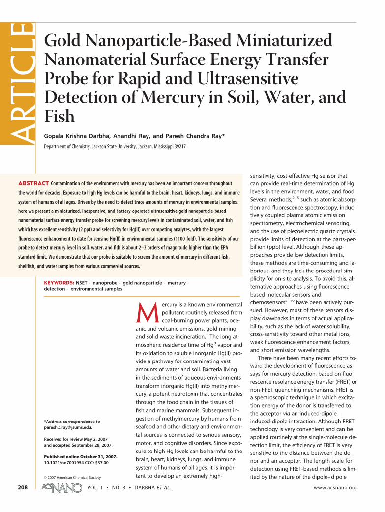
Gold Nanoparticle-Based MiniaturizedNanomaterial Surface Energy TransferProbe for Rapid and UltrasensitiveDetection of Mercury in Soil, Water, andFishGopala Krishna Darbha, Anandhi Ray, and Paresh Chandra Ray*
Department of Chemistry, Jackson State University, Jackson, Mississippi 39217
Mercury is a known environmentalpollutant routinely released fromcoal-burning power plants, oce-
anic and volcanic emissions, gold mining,
and solid waste incineration.1 The long at-
mospheric residence time of Hg0 vapor and
its oxidation to soluble inorganic Hg(II) pro-
vide a pathway for contaminating vast
amounts of water and soil. Bacteria living
in the sediments of aqueous environments
transform inorganic Hg(II) into methylmer-
cury, a potent neurotoxin that concentrates
through the food chain in the tissues of
fish and marine mammals. Subsequent in-
gestion of methylmercury by humans from
seafood and other dietary and environmen-
tal sources is connected to serious sensory,
motor, and cognitive disorders. Since expo-
sure to high Hg levels can be harmful to the
brain, heart, kidneys, lungs, and immune
system of humans of all ages, it is impor-
tant to develop an extremely high-
sensitivity, cost-effective Hg sensor thatcan provide real-time determination of Hglevels in the environment, water, and food.Several methods,2–5 such as atomic absorp-tion and fluorescence spectroscopy, induc-tively coupled plasma atomic emissionspectrometry, electrochemical sensoring,and the use of piezoelectric quartz crystals,provide limits of detection at the parts-per-billion (ppb) level. Although these ap-proaches provide low detection limits,these methods are time-consuming and la-borious, and they lack the procedural sim-plicity for on-site analysis. To avoid this, al-ternative approaches using fluorescence-based molecular sensors andchemosensors5–10 have been actively pur-sued. However, most of these sensors dis-play drawbacks in terms of actual applica-bility, such as the lack of water solubility,cross-sensitivity toward other metal ions,weak fluorescence enhancement factors,and short emission wavelengths.
There have been many recent efforts to-ward the development of fluorescence as-says for mercury detection, based on fluo-rescence resolance energy transfer (FRET) ornon-FRET quenching mechanisms. FRET isa spectroscopic technique in which excita-tion energy of the donor is transferred tothe acceptor via an induced-dipole–induced-dipole interaction. Although FRETtechnology is very convenient and can beapplied routinely at the single-molecule de-tection limit, the efficiency of FRET is verysensitive to the distance between the do-nor and an acceptor. The length scale fordetection using FRET-based methods is lim-ited by the nature of the dipole– dipole
*Address correspondence [email protected].
Received for review May 2, 2007and accepted September 28, 2007.
Published online October 31, 2007.10.1021/nn7001954 CCC: $37.00
© 2007 American Chemical Society
ABSTRACT Contamination of the environment with mercury has been an important concern throughout
the world for decades. Exposure to high Hg levels can be harmful to the brain, heart, kidneys, lungs, and immune
system of humans of all ages. Driven by the need to detect trace amounts of mercury in environmental samples,
here we present a miniaturized, inexpensive, and battery-operated ultrasensitive gold nanoparticle-based
nanomaterial surface energy transfer probe for screening mercury levels in contaminated soil, water, and fish
which has excellent sensitivity (2 ppt) and selectivity for Hg(II) over competing analytes, with the largest
fluorescence enhancement to date for sensing Hg(II) in environmental samples (1100-fold). The sensitivity of our
probe to detect mercury level in soil, water, and fish is about 2–3 orders of magnitude higher than the EPA
standard limit. We demonstrate that our probe is suitable to screen the amount of mercury in different fish,
shellfish, and water samples from various commercial sources.
KEYWORDS: NSET · nanoprobe · gold nanoparticle · mercurydetection · environmental samples
ART
ICLE
VOL. 1 ▪ NO. 3 ▪ DARBHA ET AL. www.acsnano.org208
mechanism, which effectively constrains the length
scales to distances on the order of �100 Å (R0 � 60 Å).
The limitations of FRET can be overcome with a dy-
namic molecular ruler based on the distance-
dependent plasmon coupling of metal
nanoparticles.22–30 Recently, several groups, including
ours,22–30 have reported that nanomaterial surface en-
ergy transfer (NSET) is a technique capable of measur-
ing distances nearly twice as far as FRET in which energy
transfer from a donor molecule to a nanoparticle sur-
face follows a predictable distance dependence. In this
article, we present a gold nanoparticle-based miniatur-
ized, inexpensive, and battery-operated ultrasensitive
NSET probe for screening mercury levels in water and
fish, which has excellent sensitivity (2 ppt) and selectiv-
ity for Hg(II) over competing analytes, including com-
mon metal ion contaminants such as Cu2� and Pb2�,
with the largest fluorescence enhancement to date for
sensing Hg(II) in water (1100-fold). A battery-operated
laser pointer induced the fluorescence from rhodamine
B. The compact architecture of the designed sensor,
along with the carefully aligned optics, offers a cost-
effective way to detect mercury in water with a detec-
tion limit at the parts-per-trillion (ppt) level. Experi-
ments with contaminated soils and fish collected from
the field show that our nanoparticle probe is capable of
reliably detecting mercury levels in environmental
samples at the parts-per-trillion level.
RESULTS AND DISCUSSIONSOur mercury detection approach is based on the
use of gold nanoparticle-based NSET11–20 (where the
gold nanoparticle surface acts as an acceptor and or-
ganic dye acts as a donor) which provides high sensitiv-
ity for the detection of metal ions because of their
unique property of superquenching chromophores
through both energy-transfer and electron-transfer pro-
cesses. Rhodamine B molecules, which are highly fluo-
rescent (fluorescence quantum yield 0.70) in aqueous
solution, were self-adsorbed onto the surface of gold
nanoparticles (see Figure 1).
Our experimental data showed a quenching effi-
ciency of nearly 100% when the fluorophore was stati-
cally adsorbed on the particle (static quenching). The
fluorophore is completely quenched by efficient nonra-
diative energy transfer to the gold particle, and the
strong quenching abilities of colloidal gold are related
to its large molar extinction coefficients (�1010 cm�1
M�1 for 20 –30 nm particles) and its nanomolar bind-
ing affinities for organic dyes. Like normal FRET, the in-
teraction for gold nanomaterial-based surface energy
transfer is dipole– dipole in nature but is geometrically
different because the acceptor nanoparticle has a sur-
face and an isotropic distribution of dipole vectors to
accept energy from the donor. This arrangement in-
creases the probability of energy transfer and accounts
for the enhanced efficiency for gold nanomaterial-based NSET over normal FRET.
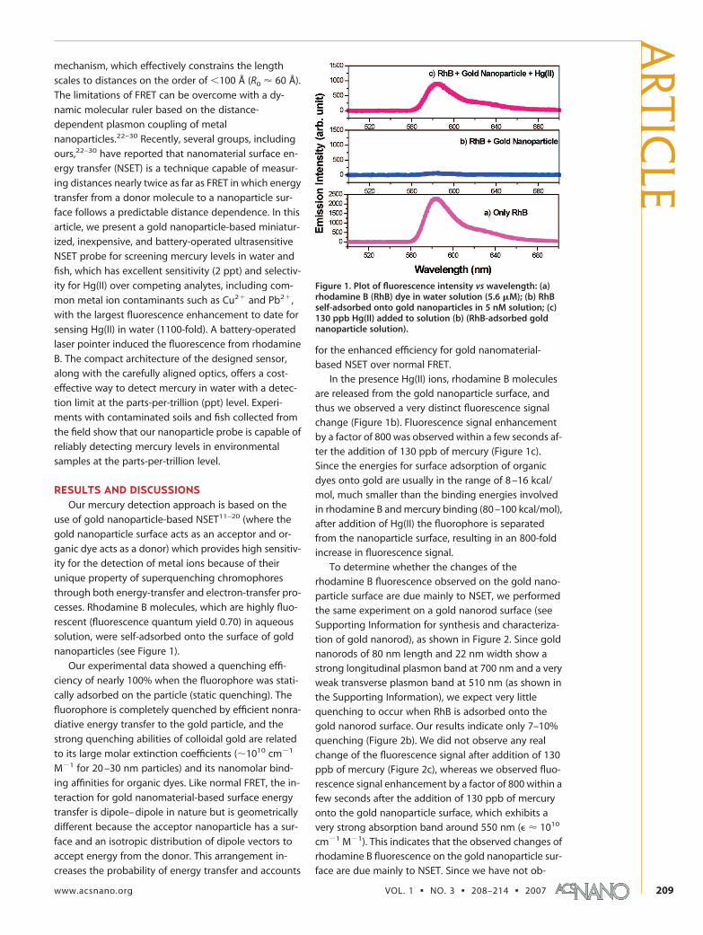
In the presence Hg(II) ions, rhodamine B moleculesare released from the gold nanoparticle surface, andthus we observed a very distinct fluorescence signalchange (Figure 1b). Fluorescence signal enhancementby a factor of 800 was observed within a few seconds af-ter the addition of 130 ppb of mercury (Figure 1c).Since the energies for surface adsorption of organicdyes onto gold are usually in the range of 8 –16 kcal/mol, much smaller than the binding energies involvedin rhodamine B and mercury binding (80 –100 kcal/mol),after addition of Hg(II) the fluorophore is separatedfrom the nanoparticle surface, resulting in an 800-foldincrease in fluorescence signal.
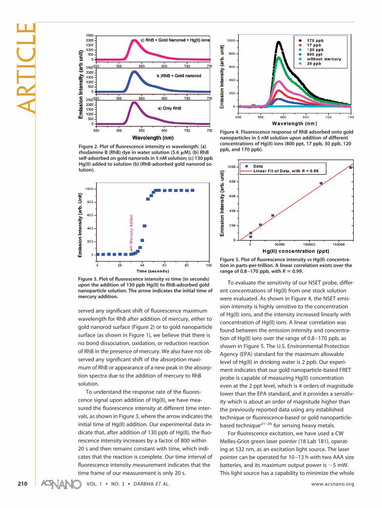
To determine whether the changes of therhodamine B fluorescence observed on the gold nano-particle surface are due mainly to NSET, we performedthe same experiment on a gold nanorod surface (seeSupporting Information for synthesis and characteriza-tion of gold nanorod), as shown in Figure 2. Since goldnanorods of 80 nm length and 22 nm width show astrong longitudinal plasmon band at 700 nm and a veryweak transverse plasmon band at 510 nm (as shown inthe Supporting Information), we expect very littlequenching to occur when RhB is adsorbed onto thegold nanorod surface. Our results indicate only 7–10%quenching (Figure 2b). We did not observe any realchange of the fluorescence signal after addition of 130ppb of mercury (Figure 2c), whereas we observed fluo-rescence signal enhancement by a factor of 800 within afew seconds after the addition of 130 ppb of mercuryonto the gold nanoparticle surface, which exhibits avery strong absorption band around 550 nm (� � 1010
cm�1 M�1). This indicates that the observed changes ofrhodamine B fluorescence on the gold nanoparticle sur-face are due mainly to NSET. Since we have not ob-
Figure 1. Plot of fluorescence intensity vs wavelength: (a)rhodamine B (RhB) dye in water solution (5.6 �M); (b) RhBself-adsorbed onto gold nanoparticles in 5 nM solution; (c)130 ppb Hg(II) added to solution (b) (RhB-adsorbed goldnanoparticle solution).
ARTIC
LE
www.acsnano.org VOL. 1 ▪ NO. 3 ▪ 208–214 ▪ 2007 209
served any significant shift of fluorescence maximum
wavelength for RhB after addition of mercury, either to
gold nanorod surface (Figure 2) or to gold nanoparticle
surface (as shown in Figure 1), we believe that there is
no bond dissociation, oxidation, or reduction reaction
of RhB in the presence of mercury. We also have not ob-
served any significant shift of the absorption maxi-
mum of RhB or appearance of a new peak in the absorp-
tion spectra due to the addition of mercury to RhB
solution.
To understand the response rate of the fluores-
cence signal upon addition of Hg(II), we have mea-
sured the fluorescence intensity at different time inter-
vals, as shown in Figure 3, where the arrow indicates the
initial time of Hg(II) addition. Our experimental data in-
dicate that, after addition of 130 ppb of Hg(II), the fluo-
rescence intensity increases by a factor of 800 within
20 s and then remains constant with time, which indi-
cates that the reaction is complete. Our time interval of
fluorescence intensity measurement indicates that the
time frame of our measurement is only 20 s.
To evaluate the sensitivity of our NSET probe, differ-
ent concentrations of Hg(II) from one stock solution
were evaluated. As shown in Figure 4, the NSET emis-
sion intensity is highly sensitive to the concentration
of Hg(II) ions, and the intensity increased linearly with
concentration of Hg(II) ions. A linear correlation was
found between the emission intensity and concentra-
tion of Hg(II) ions over the range of 0.8 –170 ppb, as
shown in Figure 5. The U.S. Environmental Protection
Agency (EPA) standard for the maximum allowable
level of Hg(II) in drinking water is 2 ppb. Our experi-
ment indicates that our gold nanoparticle-based FRET
probe is capable of measuring Hg(II) concentration
even at the 2 ppt level, which is 4 orders of magnitude
lower than the EPA standard, and it provides a sensitiv-
ity which is about an order of magnitude higher than
the previously reported data using any established
technique or fluorescence-based or gold nanoparticle-
based technique21–26 for sensing heavy metals.
For fluorescence excitation, we have used a CW
Melles-Griot green laser pointer (18 Lab 181), operat-
ing at 532 nm, as an excitation light source. The laser
pointer can be operated for 10 –13 h with two AAA size
batteries, and its maximum output power is �5 mW.
This light source has a capability to minimize the whole
Figure 2. Plot of fluorescence intensity vs wavelength: (a)rhodamine B (RhB) dye in water solution (5.6 �M); (b) RhBself-adsorbed on gold nanorods in 5 nM solution; (c) 130 ppbHg(II) added to solution (b) (RhB-adsorbed gold nanorod so-lution).
Figure 3. Plot of fluorescence intensity vs time (in seconds)upon the addition of 130 ppb Hg(II) to RhB-adsorbed goldnanoparticle solution. The arrow indicates the initial time ofmercury addition.
Figure 4. Fluorescence response of RhB adsorbed onto goldnanoparticles in 5 nM solution upon addition of differentconcentrations of Hg(II) ions (800 ppt, 17 ppb, 30 ppb, 120ppb, and 170 ppb).
Figure 5. Plot of fluorescence intensity vs Hg(II) concentra-tion in parts-per-trillion. A linear correlation exists over therange of 0.8 –170 ppb, with R � 0.99.
ART
ICLE
VOL. 1 ▪ NO. 3 ▪ DARBHA ET AL. www.acsnano.org210
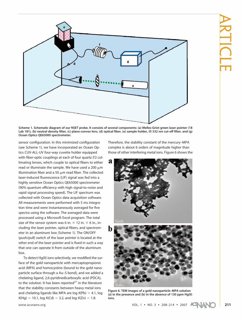
sensor configuration. In this minimized configuration
(see Scheme 1), we have incorporated an Ocean Op-
tics CUV-ALL-UV four-way cuvette holder equipped
with fiber-optic couplings at each of four quartz f/2 col-
limating lenses, which couple to optical fibers to either
read or illuminate the sample. We have used a 200 �m
illumination fiber and a 50 �m read fiber. The collected
laser-induced fluorescence (LIF) signal was fed into a
highly sensitive Ocean Optics QE65000 spectrometer
(90% quantum efficiency with high signal-to-noise and
rapid signal processing speed). The LIF spectrum was
collected with Ocean Optics data acquisition software.
All measurements were performed with 5 ms integra-
tion time and were instantaneously averaged for five
spectra using the software. The averaged data were
processed using a Microsoft Excel program. The total
size of the sensor system was 6 in. � 12 in. � 6 in., in-
cluding the laser pointer, optical fibers, and spectrom-
eter in an aluminum box (Scheme 1). The ON/OFF
(push/pull) switch of the laser pointer is located at the
other end of the laser pointer and is fixed in such a way
that one can operate it from outside of the aluminum
box.
To detect Hg(II) ions selectively, we modified the sur-
face of the gold nanoparticle with mercaptopropionic
acid (MPA) and homocystine (bound to the gold nano-
particle surface through a Au–S bond), and we added a
chelating ligand, 2,6-pyridinedicarboxylic acid (PDCA),
to the solution. It has been reported27 in the literature
that the stability constants between heavy metal ions
and chelating ligands like MPA are log K(Pb) 4.1, log
K(Hg) 10.1, log K(Cd) 3.2, and log K(Zn) 1.8.
Therefore, the stability constant of the mercury–MPA
complex is about 6 orders of magnitude higher than
those of other interfering metal ions. Figure 6 shows the
Scheme 1. Schematic diagram of our NSET probe. It consists of several components: (a) Melles-Griot green laser pointer (18Lab 181), (b) neutral density filter, (c) plano-convex lens, (d) optical fiber, (e) sample holder, (f) 532 nm cut-off filter, and (g)Ocean Optics QE65000 spectrometer.
Figure 6. TEM images of a gold nanoparticle–MPA solution(a) in the presence and (b) in the absence of 130 ppm Hg(II)ions.
ARTIC
LE
www.acsnano.org VOL. 1 ▪ NO. 3 ▪ 208–214 ▪ 2007 211
TEM images of a gold nanoparticle–MPA solution in
the presence and in the absence of Hg(II) ions. Due to
strong binding of Hg(II) with chelating ligands like MPA,
aggregation of gold nanoparticles in the presence of
Hg(II) ions was observed in our TEM image (Figure 6a).
Our results indicate that, just by modifying the gold
nanoparticle surface with MPA, our sensor has 6 –10
times higher sensitivity toward mercury ions than that
toward Cd, Pb, and Zn ions.
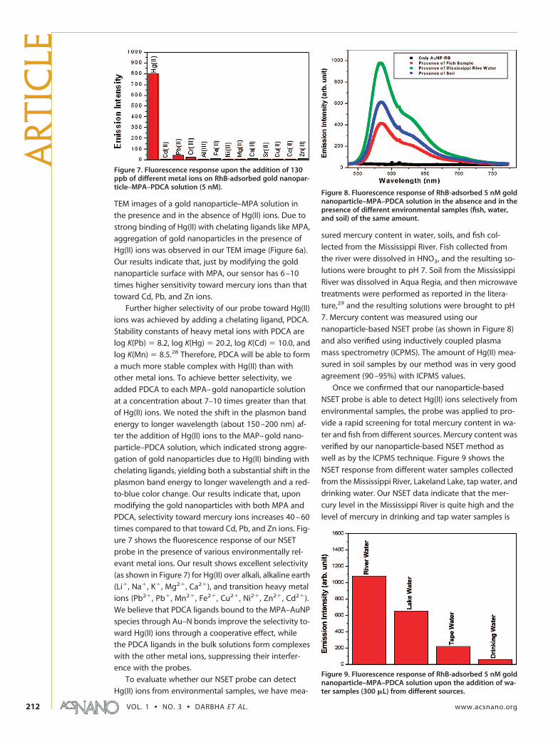
Further higher selectivity of our probe toward Hg(II)
ions was achieved by adding a chelating ligand, PDCA.
Stability constants of heavy metal ions with PDCA are
log K(Pb) 8.2, log K(Hg) 20.2, log K(Cd) 10.0, and
log K(Mn) 8.5.28 Therefore, PDCA will be able to form
a much more stable complex with Hg(II) than with
other metal ions. To achieve better selectivity, we
added PDCA to each MPA– gold nanoparticle solution
at a concentration about 7–10 times greater than that
of Hg(II) ions. We noted the shift in the plasmon band
energy to longer wavelength (about 150 –200 nm) af-
ter the addition of Hg(II) ions to the MAP– gold nano-
particle–PDCA solution, which indicated strong aggre-
gation of gold nanoparticles due to Hg(II) binding with
chelating ligands, yielding both a substantial shift in the
plasmon band energy to longer wavelength and a red-
to-blue color change. Our results indicate that, upon
modifying the gold nanoparticles with both MPA and
PDCA, selectivity toward mercury ions increases 40 – 60
times compared to that toward Cd, Pb, and Zn ions. Fig-
ure 7 shows the fluorescence response of our NSET
probe in the presence of various environmentally rel-
evant metal ions. Our result shows excellent selectivity
(as shown in Figure 7) for Hg(II) over alkali, alkaline earth
(Li�, Na�, K�, Mg2�, Ca2�), and transition heavy metal
ions (Pb2�, Pb�, Mn2�, Fe2�, Cu2�, Ni2�, Zn2�, Cd2�).
We believe that PDCA ligands bound to the MPA–AuNP
species through Au–N bonds improve the selectivity to-
ward Hg(II) ions through a cooperative effect, while
the PDCA ligands in the bulk solutions form complexes
with the other metal ions, suppressing their interfer-
ence with the probes.
To evaluate whether our NSET probe can detect
Hg(II) ions from environmental samples, we have mea-
sured mercury content in water, soils, and fish col-
lected from the Mississippi River. Fish collected from
the river were dissolved in HNO3, and the resulting so-
lutions were brought to pH 7. Soil from the Mississippi
River was dissolved in Aqua Regia, and then microwave
treatments were performed as reported in the litera-
ture,29 and the resulting solutions were brought to pH
7. Mercury content was measured using our
nanoparticle-based NSET probe (as shown in Figure 8)
and also verified using inductively coupled plasma
mass spectrometry (ICPMS). The amount of Hg(II) mea-
sured in soil samples by our method was in very good
agreement (90 –95%) with ICPMS values.
Once we confirmed that our nanoparticle-based
NSET probe is able to detect Hg(II) ions selectively from
environmental samples, the probe was applied to pro-
vide a rapid screening for total mercury content in wa-
ter and fish from different sources. Mercury content was
verified by our nanoparticle-based NSET method as
well as by the ICPMS technique. Figure 9 shows the
NSET response from different water samples collected
from the Mississippi River, Lakeland Lake, tap water, and
drinking water. Our NSET data indicate that the mer-
cury level in the Mississippi River is quite high and the
level of mercury in drinking and tap water samples is
Figure 7. Fluorescence response upon the addition of 130ppb of different metal ions on RhB-adsorbed gold nanopar-ticle–MPA–PDCA solution (5 nM).
Figure 8. Fluorescence response of RhB-adsorbed 5 nM goldnanoparticle–MPA–PDCA solution in the absence and in thepresence of different environmental samples (fish, water,and soil) of the same amount.
Figure 9. Fluorescence response of RhB-adsorbed 5 nM goldnanoparticle–MPA–PDCA solution upon the addition of wa-ter samples (300 �L) from different sources.
ART
ICLE
VOL. 1 ▪ NO. 3 ▪ DARBHA ET AL. www.acsnano.org212

much less than the EPA limit of 2 ppb. ICPMS data for
Mississippi River water and Lakeland lake water match
our NSET data to within 95%.
To determine the concentration of Hg(II) in fish
samples, we used a standard addition method. We ob-
served a good linear emission response, and the total
mercury content was 0.1– 0.005 ppm, which indicates
that our NSET probe is capable of distinguishing safe
and toxic levels of mercury in fish samples according to
the 0.55 ppm U.S. EPA standard. The Hg(II) content mea-
sured in fish samples by our method was in very good
agreement (92–98%) with ICPMS values. Figure 10
shows the fluorescence response of our NSET probefrom samples of different fish. Our NSET probe data in-dicate that swordfish contain mercury at levels a littlehigher than the EPA limit of 0.55 ppm, whereas theamounts of mercury in other fish and shellfish, e.g., lob-ster, crab, catfish, and tilapia, are lower than the EPAlimit. Our data match within 85–92% the reported dataavailable in the literature.30
CONCLUSIONIn conclusion, in this article we have reported a min-
iaturized, inexpensive, and battery-operated ultrasensi-tive gold nanoparticle-based NSET probe for screeningmercury levels in soil, fish, and water with excellent sen-sitivity (2 ppt) and selectivity for Hg(II) over competinganalytes. Our probe exhibits the largest fluorescenceenhancement to date for sensing Hg(II) in water. Fur-thermore, the sensitivity of our probe to detect mer-cury levels in soil, water, and fish is about 2–3 orders ofmagnitude higher than the EPA standard limit. Wehave shown that our probe is suitable to screen theamount of mercury in different fish, shellfish, and wa-ter samples from various commercial sources. Thoughwe have demonstrated this only for soil, water, and fishsamples, we believe that our probe provides a usefulstarting point for the development of a practicalnanosensor for screening mercury from a wide rangeof biological, toxicological, and environmental samples.
MATERIALS AND EXPERIMENTSHydrogen tetrachloroaurate (HAuCl4 · 3H2O), NaBH4,
rhodamine B dye, mercaptopropionic acid (MPA), homocystine(HCys), 2,6-pyridinedicarboxylic acid, buffer solution, sodiumchloride, and sodium citrate were purchased from Sigma-Aldrichand used with out further purification.
Gold Nanoparticle Synthesis. Gold nanoparticles of 15 nm orlarger diameters were synthesized using reported methods.11–13
Gold nanoparticles of different sizes and shapes were synthe-sized by controlling the ratio of HAuCl4 · 3H2O and sodium cit-rate concentration as we reported recently.11–13 For smaller goldnanoparticles, we used a sodium borohydride method as re-ported previously. A 0.5 mL amount of 0.01 M HAuCl4 · 3H2O inwater and 0.5 mL of 0.01 M sodium citrate in water were addedto 18 mL of deionized H2O and stirred. Next, 0.5 mL of freshlyprepared 0.1 M NaBH4 was added, and the solution changedfrom colorless to orange. Stirring was stopped, and the solutionwas left undisturbed for 2 h. The resulting spherical gold nano-particles were 4 nm in diameter. Transmission electron micros-copy (TEM) and UV–visible absorption spectroscopy were usedto characterize the nanoparticles. The particle concentration wasmeasured by UV–visible spectroscopy using the molar extinc-tion coefficients at the wavelength of the maximum absorptionof each gold colloid, as reported recently11 [�(15)528nm 3.6 �108 cm�1 M�1, �(30)530nm 3.0 � 109 cm�1 M�1, �(40)533nm 6.7� 109 cm�1 M�1, �(50)535nm 1.5 � 1010 cm�1 M�1, �(60)540nm
2.9 � 1010 cm�1 M�1, and �(80)550nm 6.9 � 1010 cm�1 M�1].Gold Nanoparticle Surface Modification. MPA and HCys were at-
tached to the gold nanoparticle surface through –SH bonds us-ing a method similar to one we have described before. We added10 mM MPA (10 �L) and 10 mM HCys (10 �L) to the gold nano-particle solution (15 nM, 10 mL) with stirring. After 2 h, 5– 8 mMNaBH4 was added. Finally, 1 mL of 5 � 10�4 M aqueousrhodamine B solution was added to 9 mL of the 15 nM gold
nanoparticle solution, and the mixture was left for a few hourswithout disturbance.
Fish Assay. We have used a method similar to the one de-scribed before.10 OmniTrace Ultra-grade nitric acid (EM Science)was used for digestion experiments. All glassware was rinsedwith dilute nitric acid and Millipore water before use. Samplesof fish tissue (0.2 g) were dissected from frozen whole specimensafter scale removal and digested in nitric acid (5 mL) at 180 °Cfor 5–10 min. The resulting solutions were neutralized withNaOH and HEPES to pH 7.
Acknowledgment. P.C.R. thanks NSF-PREM grant no. DMR-0611539, NSF-CRIFMU grant no. 0443547, and NSF-MRI grantno. 0421406 for generous funding. We thank Sara H. Bayley, In-strumentation Facilities Coordinator, University of Southern Mis-sissippi, for helping to acquire TEM data. We also thank review-ers whose valuable suggestions improved the quality of themanuscript.
Supporting Information Available: Synthesis and characteriza-tion of gold nanorods. This material is available free of chargevia the Internet at http://pubs.acs.org.
REFERENCES AND NOTES1. U.S. EPA. Regulatory Impact Analysis of the Clean Air
Mercury; EPA-452/R-05-003; U.S. Government PrintingOffice: Washington, DC, 2005.
2. Butler, O. T.; Cook, J. M.; Harrington, C. F.; Hill, S. J.;Rieuwerts, J.; Miles, D. L. J. Atomic Spectrometry Update.Environmental Analysis. Anal. At. Spectrom. 2006, 21, 217–243.
3. Li, Y.; Chen, C.; Li, B.; Sun, J.; Wang, J.; Gao, Y.; Zhao, Y.;Chai, Z. J. Elimination Efficiency of Different Reagents for
Figure 10. Fluorescence response of RhB-adsorbed 5 nMgold nanoparticle–MPA–PDCA solution upon the additionof different fish samples in the same amounts (100 �g).
ARTIC
LE
www.acsnano.org VOL. 1 ▪ NO. 3 ▪ 208–214 ▪ 2007 213
the Memory Effect of Mercury Using ICP-MS. Anal. At.Spectrom. 2006, 21, 94–96.
4. Leermakers, M.; Baeyens, W.; Quevauviller, P.; Horvat, M.Mercury in Environmental Samples: Speciation, Artifactsand Validation. Trends Anal. Chem. 2005, 24, 383–393.
5. Molecular Fluorescence: Principles and Applications; Valeur,B., Ed.; Wiley-VCH: Weinheim, 2002.
6. Komatsu, H.; Miki, T.; Citterio, D.; Kubota, T.; Shindo, Y.;Kitamura, Y.; Oka, K.; Suzuki, K. Single MolecularMultianalyte (Ca2�, Mg2�) Fluorescent Probe andApplications to Bioimaging. J. Am. Chem. Soc. 2005, 127,10798–10799.
7. Zhu, X.-J.; Fu, S.-T.; Wong, W.-K.; Guo, J.-P.; Wong, W.-Y. ANear-Infrared-Fluorescent Chemodosimeter for MercuricIon Based on an Expanded Porphyrin. Angew. Chem., Int.Ed. 2005, 45, 3150–3154.
8. Mello, J. V.; Finney, N. S. Reversing the DiscoveryParadigm: A New Approach to the CombinatorialDiscovery of Fluorescent Chemosensors. J. Am. Chem. Soc.2005, 127, 10124–10125.
9. Nolan, E. M.; Lippard, S. J. A ”Turn-On” Fluorescent Sensorfor the Selective Detection of Mercuric Ion in AqueousMedia. J. Am. Chem. Soc. 2003, 125, 14270–14271.
10. Caballero, A.; Martínez, R.; Lioveras, V.; Tatera, I.; Vidal-Gancedo, J.; Wurst, K.; Tárraga, A.; Molina, P.; Veciana, J.Highly Selective Chromogenic and Redox or FluorescentSensors of Hg2� in Aqueous Environment Based on 1,4-Disubstituted Azines. J. Am. Chem. Soc. 2005, 127, 15666–15667.
11. Ray, P. C.; Fortner, A.; Darbha, G. K. Gold NanoparticleBased FRET Asssay for the Detection of DNA Cleavage. J.Phys. Chem. B 2006, 110, 20745–20748.
12. Ray, P. C. Label-Free Diagnostics of Single Base-MismatchDNA Hybridization on Gold Nano-Particles Using Hyper-Rayleigh Scattering Technique. Angew. Chem. 2006, 45,1151–1154.
13. Kim, C. K.; Kalluru, R. R.; Singh, J. P.; Fortner, A.; Griffin, J.;Darbha, G. K.; Ray, P. C. Gold Nanoparticle BasedMiniaturized Laser Induced Fluorescence Probe forSpecific DNA Hybridization Detection: Studies on SizeDependent Optical Properties. Nanotechnology 2006, 17,3085.
14. Jennings, T. L.; Singh, M. P.; Strouse, G. F. FluorescentLifetime Quenching near d 1.5 nm Gold Nanoparticles:Probing NSET Validity. J. Am. Chem. Soc. 2006, 128, 5462.
15. Dubertret, B.; Calame, M.; Libchaber, A. J. Single-MismatchDetection Using Gold-Quenched FluorescentOligonucleotides. Nat. Biotechnol. 2001, 19, 365–370.
16. Thaxton, C. S.; Mirkin, C. A. Plasmon Coupling MeasuresUp. Nature Biotechnol. 2005, 23, 681–682.
17. Sonnichsen, C.; Reinhard, B. M.; Liphardt, J.; Alivisatos, A. P.A Molecular Ruler Based on Plasmon Coupling of SingleGold and Silver Nanoparticles. Nature Biotechnol. 2005, 23,741–745.
18. Famulok, M.; Mayer, G. Aptamers in Nanoland. Nature2006, 439, 666–669.
19. Maxwell, D. J.; Taylor, J. R.; Nie, S. Self-AssembledNanoparticle Probes for Recognition and Detection ofBiomolecules. J. Am. Chem. Soc. 2002, 124, 9602.
20. Fan, C.; Wang, S.; Hong, J. W.; Bazan, G. C.; Plaxco, K. W.;Heeger, A. J. Beyond Superquenching: Hyper-EfficientEnergy Transfer from Conjugated Polymers to GoldNanoparticles. Proc. Natl. Acad. Sci. U.S.A. 2003, 100, 6297–6301.
21. Rex, M.; Hernandez, F. E.; Campiglia, A. D. Pushing theLimits of Mercury Sensors with Gold Nanorods. Anal.Chem. 2006, 78, 445–451.
22. Kim, Y.; Johnson, R. C.; Hupp, J. T. Gold Nanoparticle-BasedSensing of ”Spectroscopically Silent” Heavy Metal Ions.Nano Lett. 2001, 1, 165–167.
23. Liu, J.; Lu, Y. Stimuli-Responsive Disassembly ofNanoparticle Aggregates for Light-Up ColorimetricSensing. J. Am. Chem. Soc. 2005, 127, 12677–12683.
24. Huang, C.-C.; Chang, H.-T. Selective Gold-Nanoparticle-Based “Turn-On” Fluorescent Sensors for Detection of
Mercury(II) in Aqueous Solution. Anal. Chem. 2006, 78,8332–8338.
25. Wanunu, M.; Popovitz-Biro, R.; Cohen, H.; Vaskevich, A.;Rubinstein, I. Coordination-Based Gold NanoparticleLayers. J. Am. Chem. Soc. 2005, 127, 9207–9215.
26. He, X.; Liu, H.; Li, Y.; Wang, S.; Li, Y.; Wang, N.; Xiao, J.; Xu,X.; Zhu, D. Gold Nanoparticle-Based Fluorometric andColorimetric Sensing of Copper(II) Ions. Adv. Mater. 2005,17, 2811–2815.
27. Morel, F. M. M. Principles of Aquatic Chemistry;Wiley-Interscience: New York, 1983; 237–310.
28. Norkus, E; Stalnioniene, I.; Crans, D. C. Interaction ofPyridine- and 4-Hydroxypyridine-2,6-Dicarboxylic Acidswith Heavy Metal Ions in Aqueous Solutions. HeteroatomChem. 2003, 14, 625–632.
29. Bontidean, I; Mortari, A.; Leth, S. M.; Larsen, M. M.;Corbisher, P.; Csoregi, E. Biosensors for Detection ofMercury in Contaminated Soils. Environ. Pollut. 2004, 131,255.
30. Mercury in fish: FDA monitoring, www.cfsan.fda.gov.
ART
ICLE
VOL. 1 ▪ NO. 3 ▪ DARBHA ET AL. www.acsnano.org214
Copyright © 2022 FDOKUMEN