Composite Fiber-Resin Lamina, Compared by Finite Element Analysis and Analytical Solution
Upload
jagiellonianCategory
view
1download
0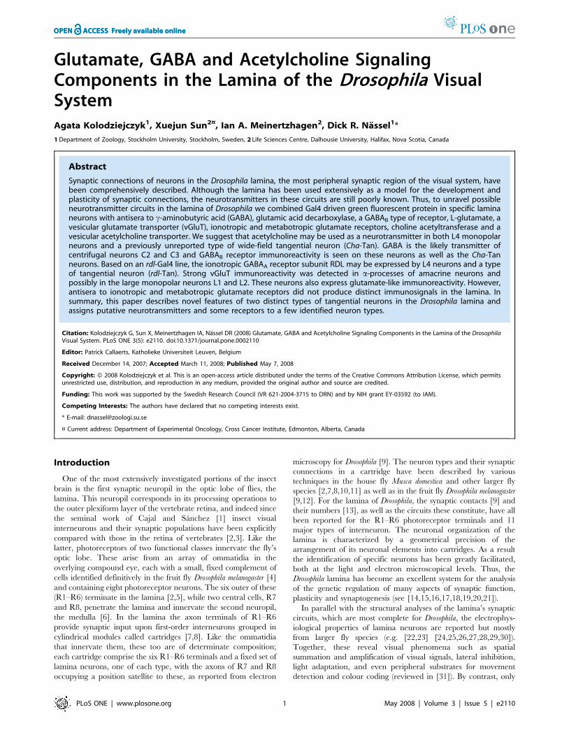
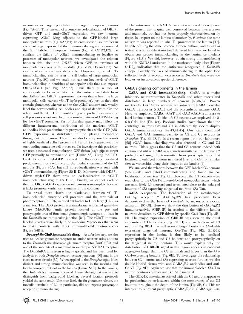
Glutamate, GABA and Acetylcholine SignalingComponents in the Lamina of the Drosophila VisualSystemAgata Kolodziejczyk1, Xuejun Sun2¤, Ian A. Meinertzhagen2, Dick R. Nassel1*
1Department of Zoology, Stockholm University, Stockholm, Sweden, 2 Life Sciences Centre, Dalhousie University, Halifax, Nova Scotia, Canada
Abstract
Synaptic connections of neurons in the Drosophila lamina, the most peripheral synaptic region of the visual system, havebeen comprehensively described. Although the lamina has been used extensively as a model for the development andplasticity of synaptic connections, the neurotransmitters in these circuits are still poorly known. Thus, to unravel possibleneurotransmitter circuits in the lamina of Drosophila we combined Gal4 driven green fluorescent protein in specific laminaneurons with antisera to c-aminobutyric acid (GABA), glutamic acid decarboxylase, a GABAB type of receptor, L-glutamate, avesicular glutamate transporter (vGluT), ionotropic and metabotropic glutamate receptors, choline acetyltransferase and avesicular acetylcholine transporter. We suggest that acetylcholine may be used as a neurotransmitter in both L4 monopolarneurons and a previously unreported type of wide-field tangential neuron (Cha-Tan). GABA is the likely transmitter ofcentrifugal neurons C2 and C3 and GABAB receptor immunoreactivity is seen on these neurons as well as the Cha-Tanneurons. Based on an rdl-Gal4 line, the ionotropic GABAA receptor subunit RDL may be expressed by L4 neurons and a typeof tangential neuron (rdl-Tan). Strong vGluT immunoreactivity was detected in a-processes of amacrine neurons andpossibly in the large monopolar neurons L1 and L2. These neurons also express glutamate-like immunoreactivity. However,antisera to ionotropic and metabotropic glutamate receptors did not produce distinct immunosignals in the lamina. Insummary, this paper describes novel features of two distinct types of tangential neurons in the Drosophila lamina andassigns putative neurotransmitters and some receptors to a few identified neuron types.
Citation: Kolodziejczyk G, Sun X, Meinertzhagen IA, Nassel DR (2008) Glutamate, GABA and Acetylcholine Signaling Components in the Lamina of the DrosophilaVisual System. PLoS ONE 3(5): e2110. doi:10.1371/journal.pone.0002110
Editor: Patrick Callaerts, Katholieke Universiteit Leuven, Belgium
Received December 14, 2007; Accepted March 11, 2008; Published May 7, 2008
Copyright: ! 2008 Kolodziejczyk et al. This is an open-access article distributed under the terms of the Creative Commons Attribution License, which permitsunrestricted use, distribution, and reproduction in any medium, provided the original author and source are credited.
Funding: This work was supported by the Swedish Research Council (VR 621-2004-3715 to DRN) and by NIH grant EY-03592 (to IAM).
Competing Interests: The authors have declared that no competing interests exist.
* E-mail: [email protected]
¤ Current address: Department of Experimental Oncology, Cross Cancer Institute, Edmonton, Alberta, Canada
Introduction
One of the most extensively investigated portions of the insectbrain is the first synaptic neuropil in the optic lobe of flies, thelamina. This neuropil corresponds in its processing operations tothe outer plexiform layer of the vertebrate retina, and indeed sincethe seminal work of Cajal and Sanchez [1] insect visualinterneurons and their synaptic populations have been explicitlycompared with those in the retina of vertebrates [2,3]. Like thelatter, photoreceptors of two functional classes innervate the fly’soptic lobe. These arise from an array of ommatidia in theoverlying compound eye, each with a small, fixed complement ofcells identified definitively in the fruit fly Drosophila melanogaster [4]and containing eight photoreceptor neurons. The six outer of these(R1–R6) terminate in the lamina [2,5], while two central cells, R7and R8, penetrate the lamina and innervate the second neuropil,the medulla [6]. In the lamina the axon terminals of R1–R6provide synaptic input upon first-order interneurons grouped incylindrical modules called cartridges [7,8]. Like the ommatidiathat innervate them, these too are of determinate composition;each cartridge comprise the six R1–R6 terminals and a fixed set oflamina neurons, one of each type, with the axons of R7 and R8occupying a position satellite to these, as reported from electron
microscopy for Drosophila [9]. The neuron types and their synapticconnections in a cartridge have been described by varioustechniques in the house fly Musca domestica and other larger flyspecies [2,7,8,10,11] as well as in the fruit fly Drosophila melanogaster[9,12]. For the lamina of Drosophila, the synaptic contacts [9] andtheir numbers [13], as well as the circuits these constitute, have allbeen reported for the R1–R6 photoreceptor terminals and 11major types of interneuron. The neuronal organization of thelamina is characterized by a geometrical precision of thearrangement of its neuronal elements into cartridges. As a resultthe identification of specific neurons has been greatly facilitated,both at the light and electron microscopical levels. Thus, theDrosophila lamina has become an excellent system for the analysisof the genetic regulation of many aspects of synaptic function,plasticity and synaptogenesis (see [14,15,16,17,18,19,20,21]).In parallel with the structural analyses of the lamina’s synaptic
circuits, which are most complete for Drosophila, the electrophys-iological properties of lamina neurons are reported but mostlyfrom larger fly species (e.g. [22,23] [24,25,26,27,28,29,30]).Together, these reveal visual phenomena such as spatialsummation and amplification of visual signals, lateral inhibition,light adaptation, and even peripheral substrates for movementdetection and colour coding (reviewed in [31]). By contrast, only
PLoS ONE | www.plosone.org 1 May 2008 | Volume 3 | Issue 5 | e2110
limited electrophysiological data are available for lamina neuronsin Drosophila ([32,33]).In contrast to the extensive anatomical and electrophysiological
investigations, we have little information about the neurotrans-mitters in the lamina of flies (see [34,35]). It is clear that flyphotoreceptors use histamine as their neurotransmitter[34,36,37,38,39,40]. When released from photoreceptor synapseshistamine acts as a fast neurotransmitter at ligand-gated chloridechannels on postsynapic lamina interneurons [36], which includeL1–L3 [41]. There is also immunocytochemical evidence forGABA in two types of small field centrifugal interneurons, C2 andC3 [42,43,44,45]. This evidence is based on several antisera toGABA and antisera to the biosynthetic enzyme glutamic aciddecarboxylase (GAD). Some reports indicate the immunolabelingof lamina monopolar cells (first-order interneurons) with antiserato glutamate, in flies [46,47] and honeybees [48]. In Drosophila,these cells also label with an antibody against choline acetyltrans-ferase (ChAT), the biosynthetic enzyme of acetylcholine [49],which is encoded by the gene Cha [50]. Cha transcript has alsobeen found by in situ hybridization in cell bodies of laminamonopolar neurons [51]. Finally, fly amacrine cells are reported toexpress glutamate immunoreactivity [47]. Clearly there is someuncertainty in these reports. Some describe tentative identifica-tions of lamina neurons, while in others the antisera used mayidentify a substance (e.g. glutamate) that is present only as ametabolic intermediate; some studies also do not includeDrosophila. Thus, for Drosophila there is a need to investigate thelamina further with respect to these classical neurotransmitters.Here we applied immunocytochemistry to the lamina of
Drosophila to identify neurotransmitters or associated moleculesimportant for neurotransmitter function, including correspondingreceptors proteins. Examination of these markers was combinedwith use of the Gal4-UAS system [52] to drive expression of greenfluorescent protein (GFP) in specific neuron populations of thelamina. The focus of our investigation is on neurons expressingmarkers for acetylcholine, glutamate, GABA, and some of theirreceptors.
Materials and Methods
Fly strainsWe used adult wild type Drosophila melanogaster (Oregon R or
w1118 strains) for basic immunocytochemistry. For correlation withvarious neuronal phenotypes we performed immunocytochemistryon a variety of Gal4 lines crossed with UAS-GFP, as specifiedbelow. L2 monopolar interneurons were visualized by the 21D-Gal4 driver [53] (from Tomas Raabe, University Wurtzburg,Germany). C3 neurons were identified by 5-6-8/CyO;TM2/TM6B-Gal4 (abbreviated 5-6-8-Gal4; from Larry Zipursky,Howard Hughes Medical Institute at UCLA, Los Angeles, CA).Other Gal-4 lines used were: rdl-Gal4 (4.7 kb upstream rdl-gene;from Julie Simpson, Howard Hughes Medical Institute, JaneliaFarm, VA), Cha-Gal4 [54] (from Bloomington Stock Center atIndiana University, Bloomington, IN), and OK371-Gal4 ([55],from Hermann Aberle, University of Munster, Germany). Thesewere used to visualize expression of the GABAA receptor subunitRDL, choline acetyltransferase (Cha), and vesicular glutamatetransporter (vGluT) gene products, respectively. To visualize Gal4-expression with GFP, we crossed these lines with flies expressingUAS-mCD8-GFP (from Bloomington Stock Center). Presynapticsites were visualized by driving a neuronal synaptobrevin-GFP fusion line (w[*];P{w[!mC] # UAS-nsyb.egfp}2; Bloomingtonstock center) with either the OK371- or 21D-Gal4 lines (see[56,57]).
AntiseraSeveral antisera were used to detect neurotransmitters and other
signaling components in the lamina. The antisera and theircorresponding antigens are listed separately (Table 1). Antisrumspecificities have been carried out for all antisera in earlierpublications (listed in Table 1). A comprehensive description ofantiserum production and specificity tests is given below.
DmGluRA. The mouse monoclonal antibody to DmGluRA,7G11 ([58]; purchased from European Molecular BiologyLaboratory, Heidelberg, Germany) was raised againstrecombinant receptor protein that was purified to homogeneity[58,59]. Specificity of 7G11 was tested by expressing DmGluRA ina baculovirus-insect cell system and testing cell extract by westernblotting [59]. The 7G11 antibody was also tested on Western blotsof head extracts of Drosophila controls (2b) and DmGluRA mutants(112b) showing loss of staining in mutants [60].
DvGluT. The Rabbit anti-DvGluT (Drosophila vesicularglutamate transporter; kind gift from Dr. A. DiAntonio,Washington University School of Medicine, St. Louis, MO; [61])was raised against a C-terminal peptide (CQMPSYDPQGYQQQ)of the Drosophila vGluT, affinity purified and characterized bywestern blotting and by its detection of transgenically expressedvGluT [61].Two other polyclonal rabbit antisera to the DrosophilavGluT were raised against the C-terminus (amino acids 561–632)and N-terminus (amino acids 21–87) of the transporter protein,respectively. The C-terminus antiserum was affinity purified. Bothantisera were kindly provided by Dr. H. Aberle (University ofMunster, Germany; [55]). In Drosophila embryos homozygous for asmall deficiency that removes the vGluT gene Mahr and Aberle [55]did not observe immunolabeling. They also found a good matchbetween the immunolabeling obtained with the two vGluT antisera(indistinguishable from each other) to in situ hybridization and theOK371 (vGluT-Gal4) expression pattern.
Glutamate. We used two antibodies both raised against L-glutamate conjugated to keyhole limpet hemocyanin (KLH) withglutaraldehyde: a rabbit polyclonal (Cat. no. 1766; ArnelProducts, New York, NY) raised by Hepler et al. [62]; and amouse monoclonal (Cat. no. G9282; Sigma, St. Louis, MO) raisedby Madl et al. [63]. The specificity of both antibodies in fly tissuesis revealed because both gave similar labeling patterns in Drosophilato an antibody against vGluT (above), and both immunolabeledthe same cells in two other species of fly (Musca, Calliphora) that theylabeled in Drosophila.
GABA. We used a commercial antiserum to GABA (Sigma;Cat. No. A2052) that was raised to GABA-bovine serum albumin(BSA) conjugate and then affinity immunopurified by themanufacturers. The GABA antiserum was characterized by dot-blot immunoassay by the manufacturers and was previouslyapplied to Drosophila brain [64,65].
GAD-1. Antiserum to full-length gel-purified Drosophila GAD1protein was raised in rabbit (kind gift from Dr. F.R. Jackson;[66,67]. This antiserum has been previously characterized byFeatherstone et al. [67] by Western blotting (recognizes a 57-kDaband, as expected) and by demonstrating the absence of labeling oftissue in a homozygous mutant lacking the gad1 gene.
GABABR2. Production of antisera to GABABR2 wasdescribed previously [65]. In brief, three antisera were raised inrabbits against a sequence (CLNDDIVRLSAPPVRREMPS) ofthe C-terminus of the receptor protein conjugated to KLH. Theseantisera were characterized by ELISA, Western blotting and withstandard pre-adsorption tests [65]. In addition, preimmune serafrom the rabbits were collected prior to immunization and used forimmunocytochemistry and Western blotting as controls. The bestantiserum (code B7873/3) was used here.
Transmitters in Fly Lamina
PLoS ONE | www.plosone.org 2 May 2008 | Volume 3 | Issue 5 | e2110
RDL. For The GABAA receptor subunit RDL we synthesizeda C-terminal peptide sequence: CLHVSDVVADDLVLLGEE,which was coupled to Limulus hemocyanine (LPH) via the N-terminal cysteine. The best RDL antiserum (code 7385)producedin rabbit was characterized by Western blotting andimmunocytochemistry, by pre-adsorption with peptide used forimmunization, and by tests of preimmune serum [68].
ChAT. A mouse monoclonal antibody to recombinant ChATprotein (Code 4B1; [69]) was purchased from the DevelopmentalStudy Hybridoma Bank. This antibody was characterized by pre-adsortption with crude recombinant ChAT [69,70] and thelabeling pattern in Drosophila confirmed by in situ hydridizationand LacZ expression (see [49]).
NMDAR1. Mouse monoclonal antibodies raised against the ratNMDAR1 (mab363) were purchased from Chemicon (Temecula,CA) who performed specificity test of the antibodies (no crossreactivity with other NMDA receptors). The antiserum was raisedagainst a recombinant fusion protein containing the amino acids660–811 of rat NMDAR1 [71]. The NMDAR1 protein displays46% amino acid identity to a D. melanogaster NMDA-like protein[72]. This antibody was previously utilized on the lamina of flies andhoneybees by Sinakevitch and Strausfeld [47].
vAChT. A rabbit polyclonal antiserum to vAChT was raisedagainst amino acids 441–546 of the protein [73]. The antiserumwas characterized in western blots of extract from wild type (bandwith Mr of 65 kD) as well as vacht- (vesicular acetylcholinetransporter) and Cha-mutant flies [73,74].
vGAT. Antiserum was raised in rabbits to a peptide sequence(N-terminal amino acids 24–38) of the putative Drosophila vesicularGABA transporter (vGAT; CG8394). The peptide was synthesizedwith a cysteine CQTARQQIPERKDYEQamide for directedconjugation to maleimid-coupled KLH at the N-terminal. Thebest vGAT antiserum (code 1061) was affinity immunopurified.This antiserum was characterized by Western blotting, pre-adsorption with the peptide used for immunization, and tests ofpreimmune serum [68].
GFP. A mouse monoclonal antibody to GFP (mAb 3E6; code#A-11120; Molecular Probes, Leiden, Netherlands) was used at1:1000 for amplifying the GFP signal in some specimens. Thisantibody was raised against GFP purified from the jellyfish Aequoreavictoria and characterized by the manufacturer; it produces noimmunolabeling in wild type Drosophila CNS and thus onlyamplifies the GFP fluorescence.
ImmunocytochemistryFor glutamate immunolabeling, brains were dissected out of the
head capsule in modified Zamboni’s fixative (4% paraformalde-hyde, 1.6% glutaraldehyde, 0.2% saturated picric acid, in 0.1Msodium phosphate buffer, pH 7.4) and left for between 1 h andovernight at 4uC. They were washed in sodium phosphatebuffered saline (PBS), and then sectioned at 50–80 mm slices ona Vibratome. The sections were washed in PBS, blocked withnormal goat serum (NGS), transferred to 0.5% Triton X-100 inPBS for 30 min prior to primary antibody incubation, and then
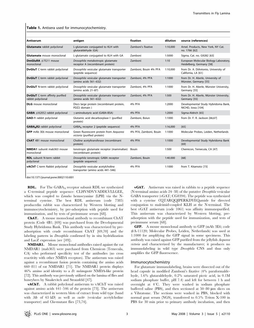
Table 1. Antisera used for immunocytochemistry.
Antiserum antigen fixation dilution source (references)
Glutamate rabbit polyclonal L-glutamate conjugated to KLH withglutaraldehyde (GA)
Zamboni’s fixative 1:10,000 Arnel. Products, New York, NY Cat.no. 1766 [62]
Glutamate mouse monoclonal L-glutamate conjugated to KLH with GA Zamboni 1:5000 Sigma, Cat. no. G9282 [63]
DmGluRA #7G11 mousemonoclonal
Drosophila metabotropic glutamatereceptor A (recombinant protein)
Zamboni 1:10 European Molecular Biology Laboratory,Heidelberg, Germany [58]
DvGluT C-term rabbit polyclonal Drosophila vesicular glutamate transporter(peptide sequence)
Zamboni, Bouin 4% PFA 1:10,000 from Dr. A. DiAntonio, University ofCalifornia, LA [61]
DvGluT C-term rabbit polyclonal Drosophila vesicular glutamate transporter(amino acids 561–632)
Zamboni, 4% PFA 1:1000 from Dr. H. Aberle, University ofMunster, Germany [55]
DvGluT N-term rabbit polyclonal Drosophila vesicular glutamate transporter(amino acids 21–87)
Zamboni, 4% PFA 1:1000 from Dr. H. Aberle, Munster University,Germany [55]
DvGluT C-term affinity purifiedrabbit polyclonal
Drosophila vesicular glutamate transporter(amino acids 561–632)
Zamboni, 4% PFA 1:500 from Dr. H. Aberle, Munster University,Germany [55]
DLG mouse monoclonal Discs large protein (recombinant protein,PDZ2 domain )
4% PFA 1:2000 Developmental Study Hybridoma Bank,NICHD, Iowa [104]
GABA #A2052 rabbit polyclonal c-aminobutyric acid (GABA-BSA) 4% PFA 1:2000 Sigma-Aldrich [65]
GAD-1 rabbit polyclonal Glutamic acid decarboxylase-1 (purifiedprotein)
Zamboni, Boiun 1:1000 from Dr. F. R. Jackson [66,67]
GABABR2 rabbit polyclonal GABAB receptor 2 (peptide sequence) 4% PFA 1:16,000 [65]
GFP mAb 3E6 mouse monoclonal Green fluorescent protein from Aequoreavictoria (purified protein)
4% PFA, Zamboni, Bouin 1:1000 Molecular Probes, Leiden, Netherlands
ChAT 4B1 mouse monoclonal Choline acetyltransferase (recombinantprotein)
4% PFA 1:1000 Developmental Study Hybridoma Bank[69]
NMDA1 subunit mab363 mousemonoclonal
Ionotropic glutamate receptor (mammalian)(recombinant protein)
Bouin 1:500 Chemicon, Temecula, CA [47]
RDL subunit N-term rabbitpolyclonal
Drosophila ionotropic GABA receptor(peptide sequence)
Zamboni, Bouin 1:40.000 [68]
vAChT C-term Rabbit polyclonal Drosophila vesicular acetylcholinetransporter (amino acids 441–546)
4% PFA 1:1000 from T. Kitamoto [73]
doi:10.1371/journal.pone.0002110.t001
Transmitters in Fly Lamina
PLoS ONE | www.plosone.org 3 May 2008 | Volume 3 | Issue 5 | e2110
and incubated overnight at 8uC in one of two glutamate antisera(Table 1), a rabbit polyclonal antibody at a dilution of 1:10,000, ora monoclonal antibody at a dilution of 1:5000. After the primaryantibody, the tissue was washed several times in PBS, and thenincubated with the corresponding secondary antibody (goat anti-rabbit, goat anti-mouse: Jackson ImmunoResearch Labs, WestGrove, PA) conjugated to a fluorochrome (Cy3 or FITC).For all other immunocytochemistry the adult fly heads were
dissected in PBS-TX (0.01M phosphate-buffered saline with 0.5%Triton X-100, pH 7.2) before fixation. For DmGluRA, DvGluTand DLG immunolabeling, opened heads were fixed 2 h in freshlyprepared ice-cold Zamboni’s fixative (4% paraformaldehyde and0.5% picric acid in 0.1 M phosphate buffer, pH 7.2) or 4%paraformaldehyde in 0.1 M sodium phosphate buffer (PB) atpH 7.4. For GABABR2 immunolabeling, fly heads were fixed for2 h in ice-cold 4% paraformaldehyde in 0.1 M PB (pH 7.2). ForGABA and GAD immunolabeling, the heads were fixed in freshlyprepared ice-cold Bouin’s fixative for 30 min (tissues were alsofixed in Zamboni’s fixative to obtain better GFP preservation). ForChAT and vAChT immunolabeling, adult fly heads were fixed for2 h in ice-cold 4% paraformaldehyde (pH 7.4) or in Zamboni’sfixative. Additional labeling with anti-GFP was necessary toamplify GFP fluorescence partly quenched after Bouin fixation.Tissues were thoroughly washed with 0.1 M PB and incubated
overnight at 4uC in 20% sucrose in 0.1 M PB. 20 mm thick sectionsof the head were cut on a Leitz 1720 Cryostat at 223uC andcollected on chromalum-gelatin–covered microscope slides. Afterwashing in PBS-TX, tissues were incubated with primary antibodiesin 4uC overnight or for 48 h. Brains were washed in PBS-TX atroom temperature (about 22uC) and incubated with fluorophore-tagged secondary antibodies (Cy2- or Cy3-tagged IgGs, raised ingoat; Jackson ImmunoResearch) diluted 1:1000, either overnight at4uC or 2 h at room temperature (around 22uC). After washing inPBS-TX and rinsing in 0.01 M PBS, tissue was mounted under acoverslip in 20% glycerol in 0.01 M PBS.
Pre-embedding immuno-electron microscopyWe also used both wild-type and white eye mutants for electron
microscopy of preparations immunolabeled for glutamate by thepre-embedding method using the polyclonal rabbit anti-glutamate[62]. Tissue incubated as above in this primary antibody was nextincubated in a biotinylated goat anti-rabbit antibody (Vector Labs)and then in a solution of peroxidase conjugated Avidin BiotinComplex (ABC complex, Vector Labs). Labeling was detectedwith 3,39-diaminobenzidine (DAB) as the substrate. The sectionswere then osmicated, dehydrated in graded ethanol series,changed into propylene oxide and then flat-embedded betweenAclar sheets (Ted Pella Inc, Redding, CA) in Poly/Bed 812 resin.The tissue was sliced at 80 mm using a Vibratome and selectedslices subsequently resectioned at 60 nm for electron microscopy.Ultrathin sections were then viewed at 60 kV with a Philips 201 Celectron microscope, photographed on 35 mm film at primarymagnifications of between 5,000 and 20,000, and the prints thenlabeled and scanned.
ImagingFor glutamate-immunolabeled specimens, Vibratome slices
were mounted in Vectashield (Vector Labs, Burlingame, CA)and viewed with a Zeiss LSM 410 confocal microscope (Zeiss,Jena, Germany). All other specimens were imaged with a ZeissLSM 510 confocal microscope. Confocal images were obtained atan optical section thickness from 0.1–0.35 mm and were processedwith Zeiss LSM software and edited for contrast and brightness inAdobe Photoshop CS3 Extended version 10.0.
Results
The optic lobe of Drosophila consists of four neuropil regionslocated beneath the retina: the lamina, medulla, lobula and lobulaplate (Fig. 1A). Each of these neuropils exhibits a columnarorganization that derives from the pattern of photoreceptorinnervation from the ommatidia of the overlying retina. R1–R6,the six outer of eight photoreceptor neurons in each ommatidium,terminate in the lamina in columnar modules termed cartridges[5,7], while the two inner neurons, R7 and R8, penetrate thelamina and innervate the distal strata of columns in the medulla(see Fig. 2B). The optic lobe neuropils are also stratified(Figs. 1A,D, and 2B), the result of overlap between stratum-specific terminals of (1) columnar centripetal neurons (thoserunning from periphery to center), (2) columnar centrifugalneurons (those running in the opposite direction); and lateralarborizations (dendrites or collaterals) of (3) various columnarneuronal elements and (4) tangentially oriented wide-fieldbranches of non-columnar neurons (see [12,75]. Some neuronsdo not display a pronounced stratified organization within thelamina. For example the L2 monopolar neurons form uniformarrangements of short, radially-directed dendritic spines through-out the depth of the lamina neuropil (Figs. 1B,C, and 2B). Incontrast to their processing counterparts in the vertebrate retina, adistinctive feature of insect neurons is that they have their cellbodies located in a cortex surrounding the synaptic neuropil(Fig. 1C). Thus all interneurons referred to in this report have cellbodies in the lamina cortex, or in a cortex of the deeper optic lobe.To facilitate interpretation of the immunolabeling and GFP
expression patterns in the lamina and distal medulla we first brieflypresent the neuron types of the Drosophila lamina. The neuronalmorphologies depicted in Fig. 2 are based on analyses of a largenumber of Golgi impregnations of Drosophila [12]. There are 3types of photoreceptor axon and 11 types of interneuronassociated with the lamina. Most of these are columnar, with anaxon oriented parallel to the main axis of the visual columns, thusestablishing the retinotopic organization of the optic lobe. All theinterneurons, except the wide-field elements (amacrines andtangential neurons), are readily distinguished and morphologicalcounterparts have been identified in other fly species [2,12,76] thathave been reasoned to be evolutionary homologues [77].Together, the columnar elements form a bundle of invariantpattern and composition, the axon of each contributing a distinctprofile to the cartridge cross section (Fig. 3A, 4A,B).There is, however, some ambiguity with respect to amacrine
and tangential neurons. This is important to point out in order toaccurately interpret our immunolabeling and Gal4-GFP expres-sion patterns (see later sections). Fig. 2A depicts one type ofamacrine (Am) and one of two types of tangential neurons (5-HT-IR Tan). The other (Tan; designated La wf1 by Fischbach andDittrich [12]) has arborizations in the distal synaptic layer of thelamina (Fig. 2B), while 5-HT-IR Tan (designated Lat by [12]), hasall its varicose processes in a layer distal to the lamina neuropil,and is known in Drosophila and larger flies to react with antisera toserotonin (see [35]). In the paper by Fischbach and Dittrich [12]apossible third type of tangential neuron (La wf2) is depicted (intheir Fig. 24F). This also has processes reaching into the distallamina, but its morphology differs from that of Tan (their La wf1).La wf2 has tangential branches with large boutons hanging downinto the lamina neuropil. Only one type of amacrine (Am;designated Lai by Fischbach and Dittrich [12]) was described inDrosophila, with tangential processes sprouting characteristic a-processes running between the R1-R6 terminals in the cartridges.These make many synapses [13]. However, in other flies a second
Transmitters in Fly Lamina
PLoS ONE | www.plosone.org 4 May 2008 | Volume 3 | Issue 5 | e2110
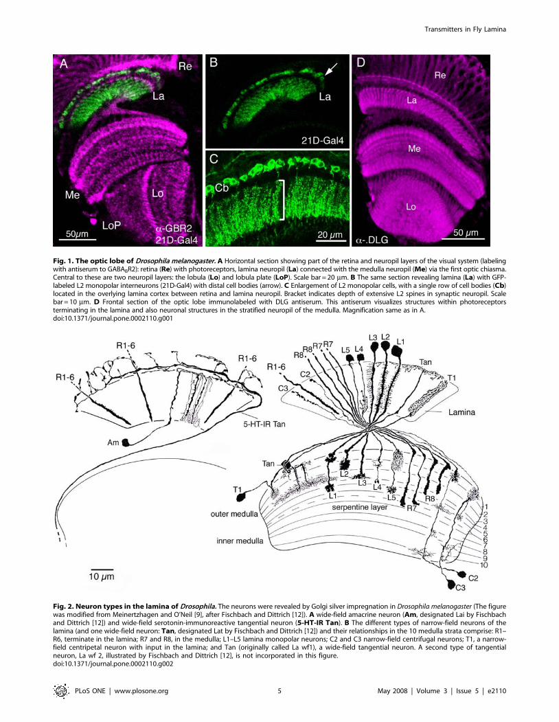
Fig. 2. Neuron types in the lamina of Drosophila. The neurons were revealed by Golgi silver impregnation in Drosophila melanogaster (The figurewas modified from Meinertzhagen and O’Neil [9], after Fischbach and Dittrich [12]). A wide-field amacrine neuron (Am, designated Lai by Fischbachand Dittrich [12]) and wide-field serotonin-immunoreactive tangential neuron (5-HT-IR Tan). B The different types of narrow-field neurons of thelamina (and one wide-field neuron: Tan, designated Lat by Fischbach and Dittrich [12]) and their relationships in the 10 medulla strata comprise: R1–R6, terminate in the lamina; R7 and R8, in the medulla; L1–L5 lamina monopolar neurons; C2 and C3 narrow-field centrifugal neurons; T1, a narrow-field centripetal neuron with input in the lamina; and Tan (originally called La wf1), a wide-field tangential neuron. A second type of tangentialneuron, La wf 2, illustrated by Fischbach and Dittrich [12], is not incorporated in this figure.doi:10.1371/journal.pone.0002110.g002
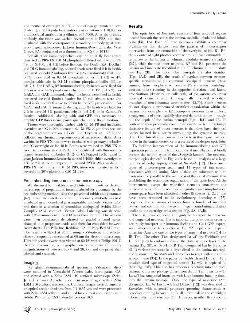
Fig. 1. The optic lobe of Drosophila melanogaster. A Horizontal section showing part of the retina and neuropil layers of the visual system (labelingwith antiserum to GABABR2): retina (Re) with photoreceptors, lamina neuropil (La) connected with the medulla neuropil (Me) via the first optic chiasma.Central to these are two neuropil layers: the lobula (Lo) and lobula plate (LoP). Scale bar = 20 mm. B The same section revealing lamina (La) with GFP-labeled L2 monopolar interneurons (21D-Gal4) with distal cell bodies (arrow). C Enlargement of L2 monopolar cells, with a single row of cell bodies (Cb)located in the overlying lamina cortex between retina and lamina neuropil. Bracket indicates depth of extensive L2 spines in synaptic neuropil. Scalebar = 10 mm. D Frontal section of the optic lobe immunolabeled with DLG antiserum. This antiserum visualizes structures within photoreceptorsterminating in the lamina and also neuronal structures in the stratified neuropil of the medulla. Magnification same as in A.doi:10.1371/journal.pone.0002110.g001
Transmitters in Fly Lamina
PLoS ONE | www.plosone.org 5 May 2008 | Volume 3 | Issue 5 | e2110
type (Am2) has been noted [30,76], which seems to lack the a -processes and have all its processes distal to the lamina neuropil.It is noteworthy that the distinguishing morphology of many
types of lamina neuron can best be detected in the medulla(Fig. 2B), which is also modular in organization (Fig. 4A,B). Thus,for example, L1–L5 and C2 and C3, each have a characteristicterminal or arborization in a distinct set of medulla strata.In the following sections we describe the different neurotrans-
mitter systems as revealed by different markers for acetylcholine-,glutamate- and GABA-associated molecules. For coherence wehave organized the figures according to the major neuron types inthe lamina, with the result that a few figures fall out of numericalorder in the text.
Acetylcholine signaling components in the laminaAcetylcholine is a major excitatory neurotransmitter in
Drosophila and other insects [49,78,79]. Choline acetyltransferase(ChAT) and the vesicular acetylcholine transporter (vAChT) areessential for cholinergic neurotransmission and antisera to theseproteins are phenotypic markers for cholinergic neurons [54,73].Several papers have used ChAT antisera or in situ hybridization forCha transcript to localize presumed cholinergic neurons to theDrosophila visual system [49,51,70,80], but to our knowledge none
has yet employed vAChT antiserum to this part of the brain. Weexamined the Drosophila lamina with antisera to both proteins.ChAT-immunolabeling reveals several types of lamina neuron(Fig. 5A). The cell bodies of large and small monopolar neuronsare ChAT-immunolabeled (Fig. 5A,E), and what appear to be theaxons and tripartite lamina collaterals of L4 monopolar neuronsalso react with ChAT antiserum (Fig 5A, see also 5B). The axonsof other monopolar neurons were not seen. Using 21D-Gal4 todrive GFP in L2 cells we could also show that anti-ChAT labeledL2 cell bodies, but no immunolabeling was visible in theirdendritic processes in the lamina (Fig. 5E).Additional to the cell bodies and presumed L4 processes, the
ChAT antiserum also labeled enlarged boutons at the level of theC2 terminals (Fig. 5A, 6A,C). These structures seem to beassociated with tangential neuronal elements having boutons inthe distal lamina neuropil. Using Cha-Gal4 to drive GFP weobtained strong fluorescence in tangential neurons with similarboutons (Fig. 6A), but no labeling of any monopolar neurons. It isnot clear whether these tangential processes seen with Cha-Gal4are derived from the Tan tangential neurons (see Fig. 2B) or anovel type of tangential neurons (or even new amacrine neurons,like Am2 of other flies), both with more pronounced varicositiesdistal to the lamina neuropil than Tan. Arguing against theamacrine neuron possibility, the Cha-Gal4 expressing neuronsappear to derive from neurons with axons projecting towards oreven connecting to the medulla (Fig. 6A). Thus they are most likelyto be a form of wide-field tangential neuron. For simplicity we will
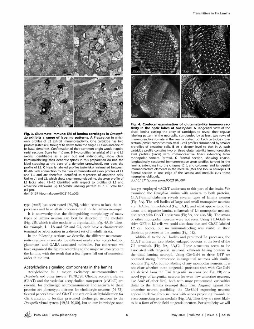
Fig. 3. Glutamate immuno-EM of lamina cartridges in Drosoph-ila exhibits a range of labeling patterns. A Preparation in whichonly profiles of L2 exhibit immunoreactivity. One cartridge has twoprofiles (asterisks), thought to derive from the single L2 axon and one ofits basal dendrites. Confirmation of their common origin would requireserial sections. Scale bar: 1.0 mm. B Two profiles (asterisks) of L1 and L2axons, identifiable as a pair but not individually, show clearimmunolabeling; their dendritic spines in this preparation do not, thelabel stopping at the base of a dendrite (arrowhead), nor does theprofile of L3. C Heavily labeled profiles (asterisks), insinuated betweenR1–R6, lack connection to the two immunolabeled axon profiles of L1and L2, and are therefore identified as a-process of amacrine cells.Unlike L1 and L2, which show clear immunolabeling, the axon profile ofL3 lacks label. R1–R6 identified with respect to profiles of L3 andamacrine cell axons (a). D Similar labeling pattern as in C. Scale bar:0.5 mm.doi:10.1371/journal.pone.0002110.g003
Fig. 4. Confocal examination of glutamate-like immunoreac-tivity in the optic lobes of Drosophila. A Tangential view of thedistal lamina cutting the array of cartridges to reveal their regularlabeling pattern in the neuropile, surrounded by at least two rows ofimmunoreactive somata in the lamina cortex (Lc). Each cartridge cross-section (circle) comprises two axial L-cell profiles surrounded by smallera-profiles of amacrine cells. B At a deeper level to that in A, eachcartridge profile contains two or three glutamate-like immunoreactiveaxial profiles (circle) with immunoreactive fibers extending frommonopolar somata (arrow). C Frontal section, showing coarse,longitudinally sectioned immunoreactive axon profiles (arrow) in thelamina, extending into the chiasma (Ch), and columnar and tangentialimmunoreactive elements in the medulla (Me) and lobula neuropils. DFrontal section at one edge of the lamina and medulla cuts theseneuropiles obliquely.doi:10.1371/journal.pone.0002110.g004
Transmitters in Fly Lamina
PLoS ONE | www.plosone.org 6 May 2008 | Volume 3 | Issue 5 | e2110
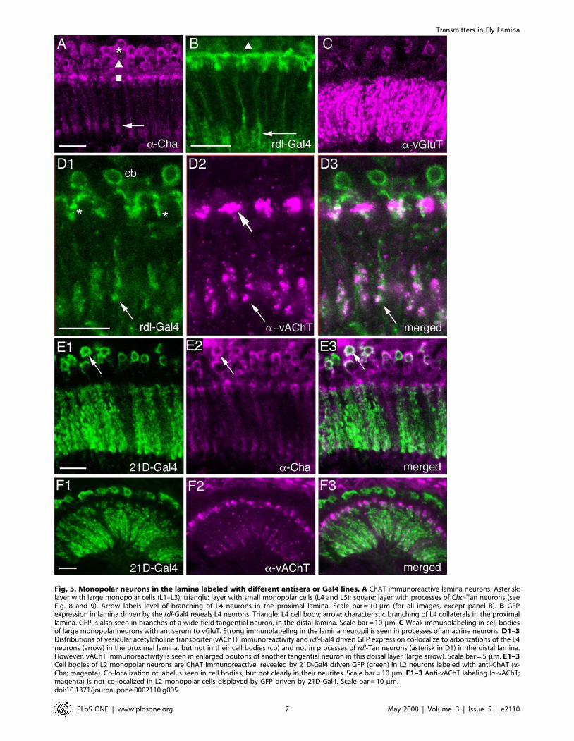
Fig. 5. Monopolar neurons in the lamina labeled with different antisera or Gal4 lines. A ChAT immunoreactive lamina neurons. Asterisk:layer with large monopolar cells (L1–L3); triangle: layer with small monopolar cells (L4 and L5); square: layer with processes of Cha-Tan neurons (seeFig. 8 and 9). Arrow labels level of branching of L4 neurons in the proximal lamina. Scale bar = 10 mm (for all images, except panel B). B GFPexpression in lamina driven by the rdl-Gal4 reveals L4 neurons. Triangle: L4 cell body; arrow: characteristic branching of L4 collaterals in the proximallamina. GFP is also seen in branches of a wide-field tangential neuron, in the distal lamina. Scale bar = 10 mm. C Weak immunolabeling in cell bodiesof large monopolar neurons with antiserum to vGluT. Strong immunolabeling in the lamina neuropil is seen in processes of amacrine neurons. D1–3Distributions of vesicular acetylcholine transporter (vAChT) immunoreactivity and rdl-Gal4 driven GFP expression co-localize to arborizations of the L4neurons (arrow) in the proximal lamina, but not in their cell bodies (cb) and not in processes of rdl-Tan neurons (asterisk in D1) in the distal lamina.However, vAChT immunoreactivity is seen in enlarged boutons of another tangential neuron in this dorsal layer (large arrow). Scale bar = 5 mm. E1–3Cell bodies of L2 monopolar neurons are ChAT immunoreactive, revealed by 21D-Gal4 driven GFP (green) in L2 neurons labeled with anti-ChAT (a-Cha; magenta). Co-localization of label is seen in cell bodies, but not clearly in their neurites. Scale bar = 10 mm. F1–3 Anti-vAChT labeling (a-vAChT;magenta) is not co-localized in L2 monopolar cells displayed by GFP driven by 21D-Gal4. Scale bar = 10 mm.doi:10.1371/journal.pone.0002110.g005
Transmitters in Fly Lamina
PLoS ONE | www.plosone.org 7 May 2008 | Volume 3 | Issue 5 | e2110
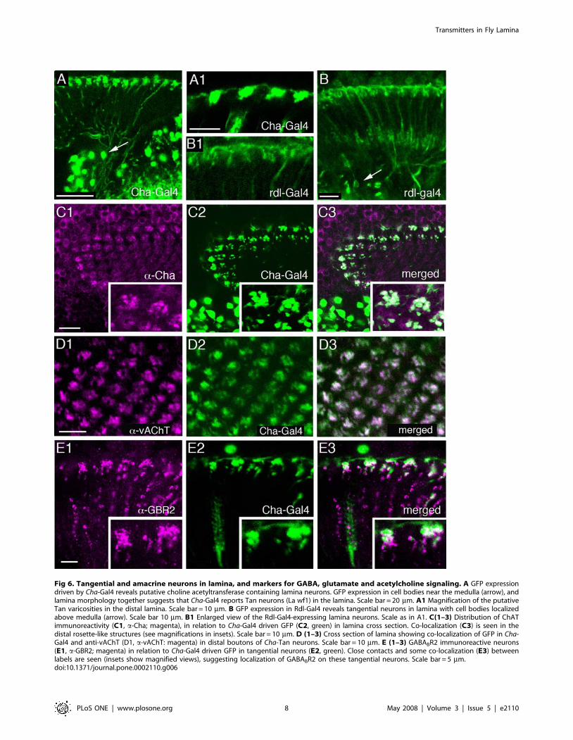
Fig 6. Tangential and amacrine neurons in lamina, and markers for GABA, glutamate and acetylcholine signaling. A GFP expressiondriven by Cha-Gal4 reveals putative choline acetyltransferase containing lamina neurons. GFP expression in cell bodies near the medulla (arrow), andlamina morphology together suggests that Cha-Gal4 reports Tan neurons (La wf1) in the lamina. Scale bar = 20 mm. A1 Magnification of the putativeTan varicosities in the distal lamina. Scale bar = 10 mm. B GFP expression in Rdl-Gal4 reveals tangential neurons in lamina with cell bodies localizedabove medulla (arrow). Scale bar 10 mm. B1 Enlarged view of the Rdl-Gal4-expressing lamina neurons. Scale as in A1. C(1–3) Distribution of ChATimmunoreactivity (C1, a-Cha; magenta), in relation to Cha-Gal4 driven GFP (C2, green) in lamina cross section. Co-localization (C3) is seen in thedistal rosette-like structures (see magnifications in insets). Scale bar = 10 mm. D (1–3) Cross section of lamina showing co-localization of GFP in Cha-Gal4 and anti-vAChT (D1, a-vAChT: magenta) in distal boutons of Cha-Tan neurons. Scale bar = 10 mm. E (1–3) GABABR2 immunoreactive neurons(E1, a-GBR2; magenta) in relation to Cha-Gal4 driven GFP in tangential neurons (E2, green). Close contacts and some co-localization (E3) betweenlabels are seen (insets show magnified views), suggesting localization of GABABR2 on these tangential neurons. Scale bar = 5 mm.doi:10.1371/journal.pone.0002110.g006
Transmitters in Fly Lamina
PLoS ONE | www.plosone.org 8 May 2008 | Volume 3 | Issue 5 | e2110
therefore refer to these neurons henceforth as Cha-Tan neurons.We found co-localization of Cha-Gal4 driven GFP and anti-ChATimmunolabeling in the tangential processes and enlarged boutonsof these cells (Fig. 6C), but ChAT-immunolabeling was detectedmainly in the GFP-labeled processes in the distal lamina, not intheir cell bodies or axons (data not shown). Our immunocyto-chemistry thus confirms that the lamina neurons seen in the Cha-Gal4 reporter line actually express ChAT-immunoreactivity. Wecould exclude that the ChAT-immunolabeling in this distal layer isderived from C2 neurons, although, as we show later, C2 neuronsmay contact the Cha-expressing tangential neurons on theseenlarged boutons. Seen in cross section, it appears that theoverlapping Cha-Tan neurons form aggregates of boutons, eachaggregate associated with an underlying cartridge (see section onGABA receptors).The antiserum to the vAChT confirmed most of the ChAT-
immunolabeling in the lamina. We could detect vAChT-immuno-labeling in basal processes likely to represent collaterals of L4neurons (Fig. 5D) and in the dilated boutons of the Cha-Tan neurons(Fig. 6D). Double-labeling with rdl-Gal4 and vAChT antiserumshowed the close match between the two in the morphology of theL4-like profiles (Fig. 5D). This double-labeling also clearly showedthe distinction between the rdl-Gal4 (described below) and Cha-Gal4expressing tangential processes in the distal lamina. Whereas theCha-Gal4 tangential profiles co-localize another acetylcholinemarker, vAChT (Fig. 6D), the rdl-Gal4 processes did not (seeFig. 5D). Furthermore, the vAChT antiserum did not label neuronalcell bodies in the lamina cortex (Fig. 5D), and as a result we could notmatch the rdl-Gal4 signal with ChAT-immunolabeling in monopolarcell bodies, even those of L4 cells.
Glutamate signaling components in the laminaImmunocytochemistry has previously suggested the presence of
glutamate in the large lamina monopolar neurons, L1 and L2, in thefliesDrosophila,Musca, Calliphora and Phaenicia sericata [46,47] as well asin type 1 amacrine neurons of the latter fly [47]. Here, we examinedthe Drosophila lamina for evidence of glutamate neurotransmission byapplying antisera to two essential molecules, the neurotransmitterglutamate and theDrosophila vesicular glutamate transporter (vGluT).The presence of glutamate is a requirement for its candidacy as aneurotransmitter, but given the widespread availability of glutamateas an intermediary metabolite, this evidence alone is unacceptablyweak. On the other hand, vGluT is required to load synaptic vesicleswith glutamate and is a highly specific marker for sites of glutamateneurotransmission so that, for example, vGluT antisera labelmotoneuron varicosities [55,61] that are known to utilize glutamateas a neurotransmitter [81].
Glutamate immunolabeling. To seek the presence ofglutamate in the lamina, we examined the lamina frompreparations sectioned in either a tangential (Fig. 4A,B) orfrontal plane (Fig. 4C,D) or, in immuno-EM preparations, in aplane cut at a tangent to the lamina’s surface (Fig. 3A), to revealcross-sections of individual cartridges (Fig. 3B). Strong glutamateimmunolabeling of monopolar cell profiles was apparent in allcartridges, and the corresponding terminals in the medulla.The labeling pattern in Drosophila visible by confocal microscopy
was substantially similar to, but varied in details from, that seen intwo other fly species, the housefly Musca domestica, and the blowflyCalliphora erythrocepha (Figures S1, S2, and S3). A number ofimmunoreactive profiles were visible in single cross-sections of thecartridge, but the slender axon size Drosophila lamina cells gavesome uncertainty in the exact determination of which profiles wereaxons and which dendrites. The small cartridge diameter relativeto the somata of monopolar cells in the lamina cortex, and the
short axon path between cortex and neuropil, made it particularlyeasy to identify the cell body fiber of immunoreactive monopolarcells (Fig. 4B). There were two rows of such somata above thecartridge (Fig. 4A). Similarly, it was easy to see the axons ofmonopolar cells extending into the chiasma (Fig. 4C). The deeperneuropiles showed qualitatively similar labeling patterns to those inthe larger flies, but were not examined further.For immuno-EM studies, we used a pre-embedding method
with the polyclonal antiserum [62] This revealed a clear pattern oflabeling that confirmed at higher resolution much of what wasseen by confocal microscopy, and resolving the pattern of labelingof tiny profiles in Drosophila. From the enhanced resolution of thepreparations we could also demonstrate that there was nodifference in the labeling patterns in the lamina betweenpreparations from wild-type flies, with red eyes, and mutant withwhite eyes. The consensus pattern was also highly consistent in allthree fly species examined (Figures S1A–E).The pattern of immuno-EM labeling in individual preparations
varied somewhat. In some only a single monopolar cell axonprofile, probably of L2 (Fig. 3A), was labeled. The basis for thisidentification was twofold. First, it was generally the larger of theaxial monopolar cell profiles, as previously reported in a statisticalsense [82,83]. The same profile was labeled in surroundingcartridges, even if such a size difference was not seen in all.Second, we identified the profile by virtue of its position withrespect to those of L3, between R5 and R6, and of a bundle ofsmall amacrine cell fibers near R4 [9]. Such profiles did notaccompany all cartridges however and were sometimes ambigu-ous, leaving some residual doubt about the identity of the labeledprofile. Other preparations had the profiles of both L1 and L2labeled (Fig. 3B), as was also seen in Musca (Figures S2, S3). Unlikethe two other fly species, L3 was apparently not labeled inDrosophila. Cartridge profiles in the same preparation had the sameimmunolabeling patterns, so that variation was mostly betweenspecimens.In addition to axon profiles of L-cells, small immunoreactive
profiles were visible between profiles of the R1–R6 terminals.These were especially clear in the cartridges of Drosophila(Fig. 3C,D) when labeled heavily with the pre-embedding method,compared with those ofMusca (e.g. Figure S3B). Such locations areoccupied by dendrites of both L1–L3 and amacrine cell alpha-processes that approach tetrad photoreceptor synapses [9]. Insome preparations it was clear that immunolabel in the L-cell axonprofiles disappeared at the base of the dendrites that arose fromthese (e.g. Fig. 3B: Drosophila; Figure S2A: Musca). The smalllabeled profiles between R1–R6 also never connected with theaxon profiles of L1 and L2 (Fig. 3C,D). Both observations providestrong evidence that the immunolabeled profiles were insteadthose of the alpha-processes from amacrine cells. Correspondingsomata of the amacrine cells were not examined.
Drosophila vGluT immunolabeling. To confirm thatimmunoreactivity to glutamate signified a capacity forglutamatergic transmission in the monopolar cells, we alsoapplied four different antisera to the Drosophila vGluT andobtained identical labeling with each (Fig. 7). Strong vGluTimmunolabeling was detected in profiles similar to a-processes ofthe amacrine neurons or possibly like b-processes of T1 neurons(Fig. 7; Figure S4). Weak vGluT immunolabeling of cell bodieswas seen in the chiasma between the lamina and medulla, in aposition corresponding to those of amacrine cells (Fig. 2A), but itwas not possible to connect these to lamina processes (Figure S4A).The vGluT immunosignal in the lamina was mostly distinct fromthat seen with the OK371-Gal4 [55], representing vGluTpromoter expression (Fig. 7A,B). The OK371-Gal4 drove GFP
Transmitters in Fly Lamina
PLoS ONE | www.plosone.org 9 May 2008 | Volume 3 | Issue 5 | e2110
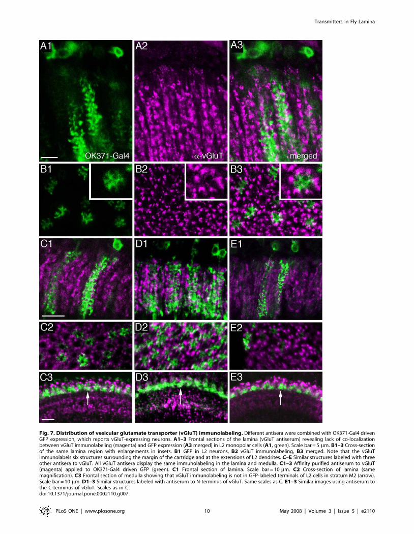
Fig. 7. Distribution of vesicular glutamate transporter (vGluT) immunolabeling. Different antisera were combined with OK371-Gal4 drivenGFP expression, which reports vGluT-expressing neurons. A1–3 Frontal sections of the lamina (vGluT antiserum) revealing lack of co-localizationbetween vGluT immunolabeling (magenta) and GFP expression (A3merged) in L2 monopolar cells (A1, green). Scale bar = 5 mm. B1–3 Cross-sectionof the same lamina region with enlargements in insets. B1 GFP in L2 neurons, B2 vGluT immunolabeling, B3 merged. Note that the vGluTimmunolabels six structures surrounding the margin of the cartridge and at the extensions of L2 dendrites. C–E Similar structures labeled with threeother antisera to vGluT. All vGluT antisera display the same immunolabeling in the lamina and medulla. C1–3 Affinity purified antiserum to vGluT(magenta) applied to OK371-Gal4 driven GFP (green). C1 Frontal section of lamina. Scale bar = 10 mm. C2 Cross-section of lamina (samemagnification). C3 Frontal section of medulla showing that vGluT immunolabeling is not in GFP-labeled terminals of L2 cells in stratum M2 (arrow).Scale bar = 10 mm. D1–3 Similar structures labeled with antiserum to N-terminus of vGluT. Same scales as C. E1–3 Similar images using antiserum tothe C-terminus of vGluT. Scales as in C.doi:10.1371/journal.pone.0002110.g007
Transmitters in Fly Lamina
PLoS ONE | www.plosone.org 10 May 2008 | Volume 3 | Issue 5 | e2110
in smaller or larger populations of large monopolar neurons(Fig. 7A–E). Thus, instead of a complete co-localization of OK371driven GFP and anti-vGluT expression, we saw neuronsexpressing vGluT lying adjacent to the GFP-labeled largemonopolar neurons (Fig. 6A3,B3). In cross-section, six profiles ineach cartridge expressed vGluT immunolabeling and surroundedthe GFP labeled monopolar neurons (Fig. 7B,C2,D2,E2). Toconfirm the failure of vGluT immunolabeling to localize toprocesses of monopolar neurons, we investigated the relationbetween this label and OK371-driven GFP in terminals ofmonopolar neurons in the medulla (Fig. 7C3, D3 and E3). Noclear co-localization was detectable. However, some vGluT-immunolabeling can be seen in cell bodies of large monopolarneurons (Fig. 5C) and we could not rule out low levels of vGluTimmunolabeling in dendrites of monopolar cells that also expressOK371-Gal4 (see Fig. 7A3,B3). Thus there is a lack ofcorrespondence between data from the antisera and data fromthe Gal4 driver. OK371 expression indicates that at least the largemonopolar cells express vGluT (vglut-promoter), just as they alsocontain glutamate, whereas at best the vGluT antisera only weaklylabel the corresponding cell bodies and tips of dendrites. On theother hand the vGluT immunolabeling seen probably in amacrinecell processes is not matched by a similar pattern of GFP-labelingfor the vGluT promoter. Part of this discrepancy may reflect thedifferent intraneuronal distribution of the markers: vGluTantibodies label predominantly presynaptic sites while GFP (cd8-GFP) expression is distributed in the plasma membranethroughout the neuron. There may also be very small amountsof highly localized vGluT protein in L1 and L2 compared with thesurrounding amacrine cell processes. To investigate this possibilitywe used a neuronal synaptobrevin-GFP fusion (nsyb-gfp) to targetGFP primarily to presynaptic sites (Figure S5). Using the 21D-Gal4 to drive nsyb-GFP resulted in fluorescence localizedpredominantly or exclusively to the medulla terminals of the L2neurons (Figure S5A), but still no co-localization was seen withvGluT immunolabeling (Figure S5 B–D). Moreover with OK371-driven nsyb-GFP there was no co-localization to vGluTimmunolabeling (Figure S5 E1–3). Finally, we cannot excludethat the OK371-Gal4 expression in neurons is incomplete becauseit lacks promoter/enhancer elements in the construct.To reveal more clearly the relationship between vGluT-
immunolabeled amacrine cell processes and the terminals ofphotoreceptors R1–R6, we used antibodies to Discs large (DLG) asa marker. The DLG protein is a membrane associated guanylatekinase (MAGUK) family protein located at the pre andpostsynaptic area of functional glutamatergic synapses, at least inthe Drosophila neuromuscular junction [84]. The vGluT immuno-labeled structures are likely to be amacrine a-processes that seemto make contacts with DLG immunolabeled photoreceptors(Figure S4B1).
DrosophilaGluR immunolabeling. As a further step, we alsotried to localize glutamate receptors to lamina neurons using antiserato the Drosophila metabotropic glutamate receptor DmGluRA andone of the subunits of a mammalian ionotropic NMDA1 receptor.The DmGluRA antiserum is highly specific and has been used foranalysis of both Drosophila neuromuscular junctions [60] and in theclock neuron circuits [85]. When applied to the Drosophila optic lobesdistinct and strong immunolabeling was seen in the medulla andlobula complex, but not in the lamina (Figure S4C). In the lamina,the DmGluRA antiserum produced diffuse labeling that was hard todistinguish from background labeling. Several fixation protocolsyielded the same result. The most likely site for glutamate release, themedulla terminals of L2, in particular, did not express presynapticreceptor immunolabeling.
The antiserum to the NMDA1 subunit was raised to a sequenceof the protein that is quite well conserved between invertebratesand mammals, but has not been properly characterized on flytissue. In a report on the lamina of another fly, P. sericata, the sameantiserum was reported to label T1 processes in the lamina [47].In spite of using the same protocol as these authors, and as well astesting several modifications (and different fixatives), we failed toobtain any proper immunolabeling in the lamina or medulla(Figure S4D1). We did, however, obtain strong immunolabelingwith this NMDA1 antiserum in the mushroom body lobes (FigureS4D2), indicating that the antiserum recognized a Drosophilaepitope. Possibly the lack of immunolabeling in the optic lobereflected levels of receptor expression in Drosophila that were toolow, or an inconvenient species difference.
GABA signaling components in the laminaGABA and GAD immunolabeling. GABA is a major
inhibitory neurotransmitter in Drosophila and other insects anddistributed in large numbers of neurons [68,86,87]. Provenmarkers for GABAergic neurons are antisera to GABA, vesicularGABA transporter (vGAT) and the biosynthetic enzyme GAD.Here we employed GABA, vGAT and GAD (GAD-1) antisera tolabel lamina neurons. To identify C3 neurons we employed the 5-6-8-Gal4 line (Fig. 8A). Previous studies have shown that thecentrifugal neurons C2 and C3 in different fly species displayGABA immunoreactivity [42,43,44,45]. Our study confirmedGABA and GAD immunoreactivity in C2 and C3 neurons inDrosophila (Fig. 8B–D, J). In a recent report from our laboratory[68] vGAT immunolabeling was also detected in C2 and C3neurons. This suggests that the C2 and C3 neurons indeed bothcontain and utilize GABA as a neurotransmitter in the lamina: C2probably releasing the transmitter from presynaptic sites thatlocalized to enlarged boutons in a distal layer and C3 from similarsites at varicosities along their length in the lamina [9].We analyzed the relations between the GFP-labeled C3 neurons
(5-6-8-Gal4) and ChAT-immunolabeling and found no co-localization of markers (Fig. 8I). However, the C3 neurons wereseen close to the ChAT-immunolabeled monopolar axons (whichare most likely L4 neurons) and terminated close to the enlargedboutons of Cha-expressing tangential neurons, Cha-Tan.
GABA receptors. The localization of the metabotropicGABAB receptor 2 (GABABR2) has previously beendemonstrated in the brain of Drosophila by means of a specificantiserum [65,68]. Here we show the distribution of GABABR2immunoreactivity (GBR-IR) in relation to the different laminaneurons visualized by GFP driven by specific Gal4-lines (Fig. 8E–H). The major expression of GBR-IR was seen on the distalvaricosities of C2 neurons (Fig. 8F–H) and in boutons of C3neurons (Fig. 8E–H), as well as on enlarged boutons of Cha-Gal4-expressing tangential neurons, Cha-Tan (Fig. 6E). GBR-IRexpression in the lamina is thus likely to be localizedpresynaptically in C2 and C3 boutons and postsynaptically onthe tangential neuron boutons. This would explain why thedistribution of GBR-IR signal in this region appears in coherentaggregates larger than the C2 terminals and larger than the Cha-Gal4-expressing boutons (Fig. 6E). To investigate the relationshipbetween C2 neurons and Cha-expressing neurons further, we alsodouble-labeled tissues with anti-GABABR2 antibodies and anti-ChAT (Fig. 9D). Again we saw that the immunolabeled Cha-Tanneuron boutons co-expressed GBR-IR material.The GBR-IR material associated with the C3 neurons appear to
be predominantly co-localized within the membranes of the C3boutons throughout the depth of the lamina (Fig. 8F, G). This weinterpret to represent presynaptic GABABR2 in GABAergic C3s.
Transmitters in Fly Lamina
PLoS ONE | www.plosone.org 11 May 2008 | Volume 3 | Issue 5 | e2110
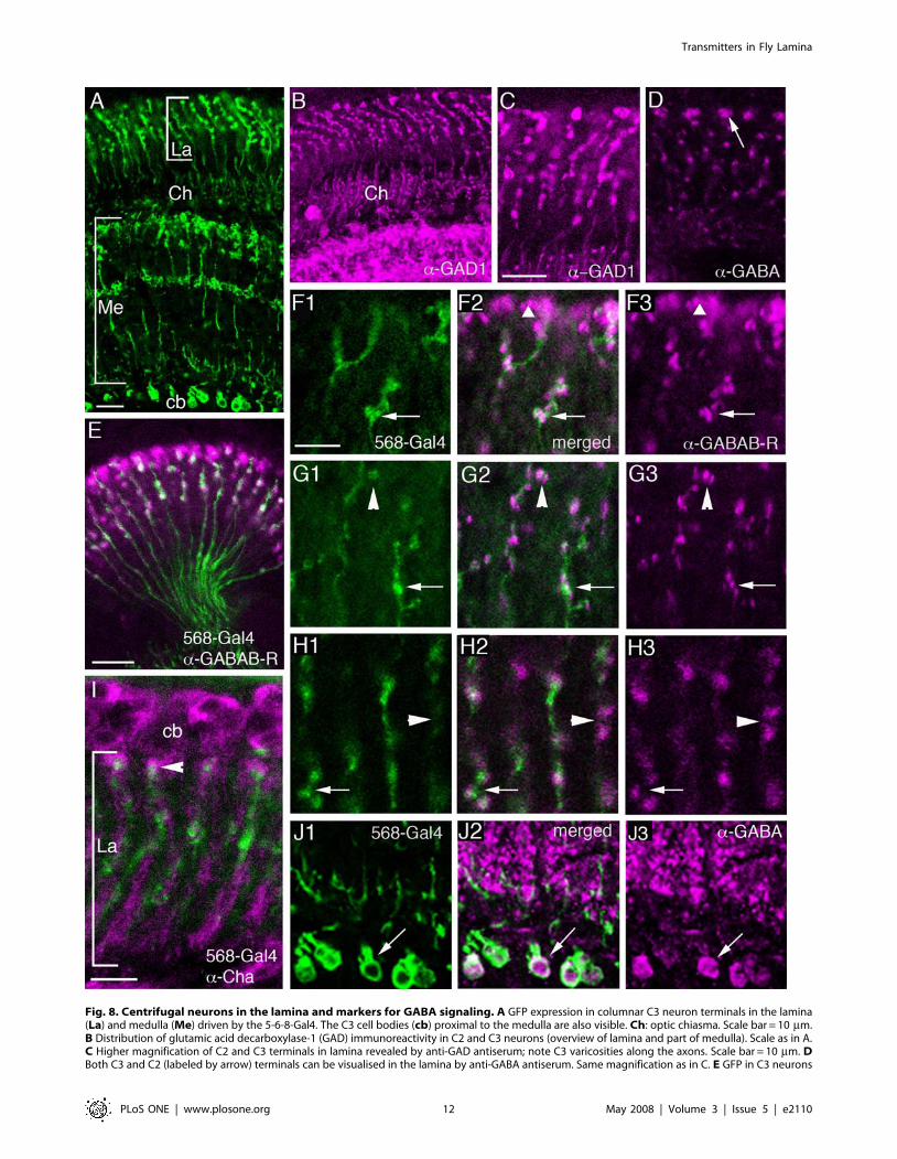
Fig. 8. Centrifugal neurons in the lamina and markers for GABA signaling. A GFP expression in columnar C3 neuron terminals in the lamina(La) and medulla (Me) driven by the 5-6-8-Gal4. The C3 cell bodies (cb) proximal to the medulla are also visible. Ch: optic chiasma. Scale bar = 10 mm.B Distribution of glutamic acid decarboxylase-1 (GAD) immunoreactivity in C2 and C3 neurons (overview of lamina and part of medulla). Scale as in A.C Higher magnification of C2 and C3 terminals in lamina revealed by anti-GAD antiserum; note C3 varicosities along the axons. Scale bar = 10 mm. DBoth C3 and C2 (labeled by arrow) terminals can be visualised in the lamina by anti-GABA antiserum. Same magnification as in C. E GFP in C3 neurons
Transmitters in Fly Lamina
PLoS ONE | www.plosone.org 12 May 2008 | Volume 3 | Issue 5 | e2110
However, some immunoreactivity seemed to lie in structures veryclose to these boutons, not co-localized to them (Fig. 8G, H). Thus,there is a possibility that a neuron postsynaptic to C3 alsoexpresses GABABR2 at sites located close to the contacts with C3neurons. Given the restricted distribution of the immunoreactivityhowever we could not identify this neuron type. From EManalysis, possible candidates postsynaptic to C3 would be L1–L3and amacrine cell processes [9,13].Ionotropic GABAA type receptors are the major receptors
mediating fast inhibitory transmission in the insect brain (see [87]).We tried to analyze the distribution of this type of receptor in theDrosophila visual system, but an antiserum to one of the DrosophilaGABAA receptor subunits, RDL, failed to produce distinctimmunolabeling in the lamina, even though the medulla displayedlayers of strong RDL-immunoreactivity (Figure S4E). An earlierreport indicated diffuse immunolabeling possibly associated in partwith large monopolar neuron dendrites [68]. Given thisuncertainty, we resorted to using an rdl-promotor-Gal4 to driveGFP expression in neurons and reveal lamina expression ofpossible GABAA receptors.In the fly visual system, rdl-Gal4 expression (amplified by anti-
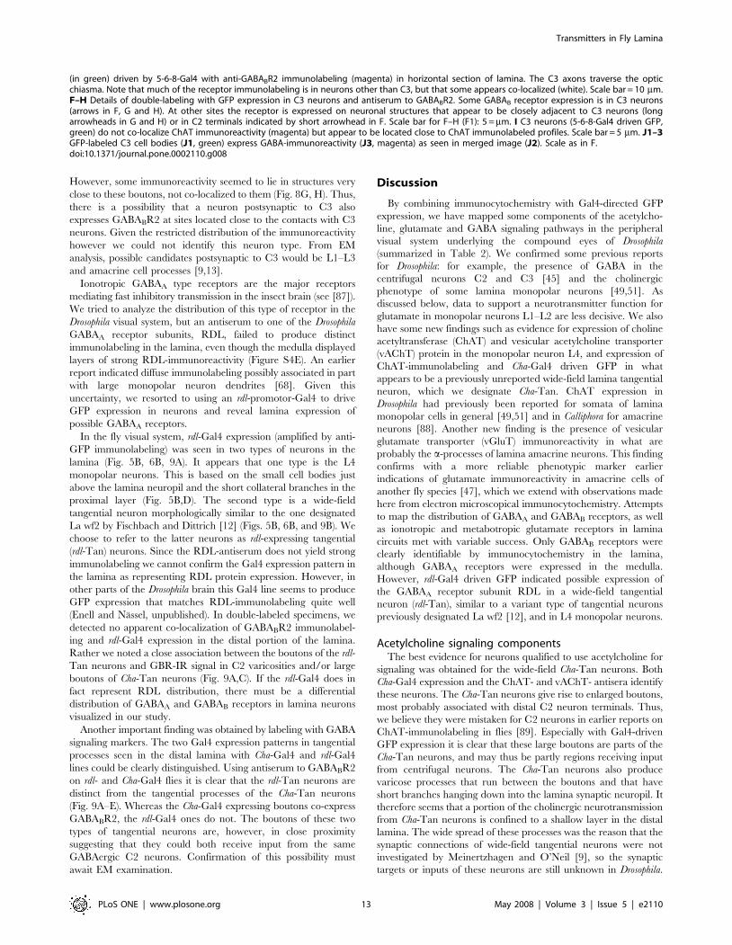
GFP immunolabeling) was seen in two types of neurons in thelamina (Fig. 5B, 6B, 9A). It appears that one type is the L4monopolar neurons. This is based on the small cell bodies justabove the lamina neuropil and the short collateral branches in theproximal layer (Fig. 5B,D). The second type is a wide-fieldtangential neuron morphologically similar to the one designatedLa wf2 by Fischbach and Dittrich [12] (Figs. 5B, 6B, and 9B). Wechoose to refer to the latter neurons as rdl-expressing tangential(rdl-Tan) neurons. Since the RDL-antiserum does not yield strongimmunolabeling we cannot confirm the Gal4 expression pattern inthe lamina as representing RDL protein expression. However, inother parts of the Drosophila brain this Gal4 line seems to produceGFP expression that matches RDL-immunolabeling quite well(Enell and Nassel, unpublished). In double-labeled specimens, wedetected no apparent co-localization of GABABR2 immunolabel-ing and rdl-Gal4 expression in the distal portion of the lamina.Rather we noted a close association between the boutons of the rdl-Tan neurons and GBR-IR signal in C2 varicosities and/or largeboutons of Cha-Tan neurons (Fig. 9A,C). If the rdl-Gal4 does infact represent RDL distribution, there must be a differentialdistribution of GABAA and GABAB receptors in lamina neuronsvisualized in our study.Another important finding was obtained by labeling with GABA
signaling markers. The two Gal4 expression patterns in tangentialprocesses seen in the distal lamina with Cha-Gal4 and rdl-Gal4lines could be clearly distinguished. Using antiserum to GABABR2on rdl- and Cha-Gal4 flies it is clear that the rdl-Tan neurons aredistinct from the tangential processes of the Cha-Tan neurons(Fig. 9A–E). Whereas the Cha-Gal4 expressing boutons co-expressGABABR2, the rdl-Gal4 ones do not. The boutons of these twotypes of tangential neurons are, however, in close proximitysuggesting that they could both receive input from the sameGABAergic C2 neurons. Confirmation of this possibility mustawait EM examination.
Discussion
By combining immunocytochemistry with Gal4-directed GFPexpression, we have mapped some components of the acetylcho-line, glutamate and GABA signaling pathways in the peripheralvisual system underlying the compound eyes of Drosophila(summarized in Table 2). We confirmed some previous reportsfor Drosophila: for example, the presence of GABA in thecentrifugal neurons C2 and C3 [45] and the cholinergicphenotype of some lamina monopolar neurons [49,51]. Asdiscussed below, data to support a neurotransmitter function forglutamate in monopolar neurons L1–L2 are less decisive. We alsohave some new findings such as evidence for expression of cholineacetyltransferase (ChAT) and vesicular acetylcholine transporter(vAChT) protein in the monopolar neuron L4, and expression ofChAT-immunolabeling and Cha-Gal4 driven GFP in whatappears to be a previously unreported wide-field lamina tangentialneuron, which we designate Cha-Tan. ChAT expression inDrosophila had previously been reported for somata of laminamonopolar cells in general [49,51] and in Calliphora for amacrineneurons [88]. Another new finding is the presence of vesicularglutamate transporter (vGluT) immunoreactivity in what areprobably the a-processes of lamina amacrine neurons. This findingconfirms with a more reliable phenotypic marker earlierindications of glutamate immunoreactivity in amacrine cells ofanother fly species [47], which we extend with observations madehere from electron microscopical immunocytochemistry. Attemptsto map the distribution of GABAA and GABAB receptors, as wellas ionotropic and metabotropic glutamate receptors in laminacircuits met with variable success. Only GABAB receptors wereclearly identifiable by immunocytochemistry in the lamina,although GABAA receptors were expressed in the medulla.However, rdl-Gal4 driven GFP indicated possible expression ofthe GABAA receptor subunit RDL in a wide-field tangentialneuron (rdl-Tan), similar to a variant type of tangential neuronspreviously designated La wf2 [12], and in L4 monopolar neurons.
Acetylcholine signaling componentsThe best evidence for neurons qualified to use acetylcholine for
signaling was obtained for the wide-field Cha-Tan neurons. BothCha-Gal4 expression and the ChAT- and vAChT- antisera identifythese neurons. The Cha-Tan neurons give rise to enlarged boutons,most probably associated with distal C2 neuron terminals. Thus,we believe they were mistaken for C2 neurons in earlier reports onChAT-immunolabeling in flies [89]. Especially with Gal4-drivenGFP expression it is clear that these large boutons are parts of theCha-Tan neurons, and may thus be partly regions receiving inputfrom centrifugal neurons. The Cha-Tan neurons also producevaricose processes that run between the boutons and that haveshort branches hanging down into the lamina synaptic neuropil. Ittherefore seems that a portion of the cholinergic neurotransmissionfrom Cha-Tan neurons is confined to a shallow layer in the distallamina. The wide spread of these processes was the reason that thesynaptic connections of wide-field tangential neurons were notinvestigated by Meinertzhagen and O’Neil [9], so the synaptictargets or inputs of these neurons are still unknown in Drosophila.
(in green) driven by 5-6-8-Gal4 with anti-GABABR2 immunolabeling (magenta) in horizontal section of lamina. The C3 axons traverse the opticchiasma. Note that much of the receptor immunolabeling is in neurons other than C3, but that some appears co-localized (white). Scale bar = 10 mm.F–H Details of double-labeling with GFP expression in C3 neurons and antiserum to GABABR2. Some GABAB receptor expression is in C3 neurons(arrows in F, G and H). At other sites the receptor is expressed on neuronal structures that appear to be closely adjacent to C3 neurons (longarrowheads in G and H) or in C2 terminals indicated by short arrowhead in F. Scale bar for F–H (F1): 5 =mm. I C3 neurons (5-6-8-Gal4 driven GFP,green) do not co-localize ChAT immunoreactivity (magenta) but appear to be located close to ChAT immunolabeled profiles. Scale bar = 5 mm. J1–3GFP-labeled C3 cell bodies (J1, green) express GABA-immunoreactivity (J3, magenta) as seen in merged image (J2). Scale as in F.doi:10.1371/journal.pone.0002110.g008
Transmitters in Fly Lamina
PLoS ONE | www.plosone.org 13 May 2008 | Volume 3 | Issue 5 | e2110
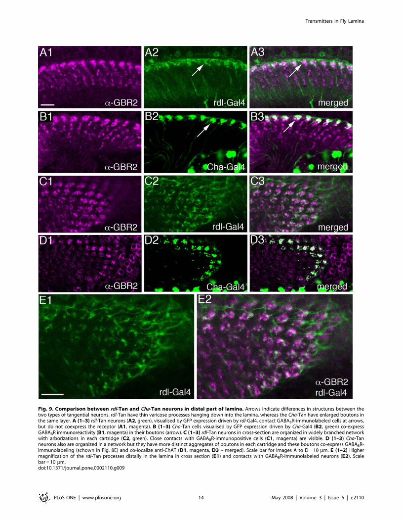
Fig. 9. Comparison between rdl-Tan and Cha-Tan neurons in distal part of lamina. Arrows indicate differences in structures between thetwo types of tangential neurons. rdl-Tan have thin varicose processes hanging down into the lamina, whereas the Cha-Tan have enlarged boutons inthe same layer. A (1–3) rdl-Tan neurons (A2, green), visualised by GFP expression driven by rdl-Gal4, contact GABABR-immunolabeled cells at arrows,but do not coexpress the receptor (A1, magenta). B (1–3) Cha-Tan cells visualised by GFP expression driven by Cha-Gal4 (B2, green) co-expressGABABR immunoreactivity (B1, magenta) in their boutons (arrow). C (1–3) rdl-Tan neurons in cross-section are organized in widely branched networkwith arborizations in each cartridge (C2, green). Close contacts with GABABR-immunopositive cells (C1, magenta) are visible. D (1–3) Cha-Tanneurons also are organized in a network but they have more distinct aggregates of boutons in each cartridge and these boutons co-express GABABR-immunolabeling (schown in Fig. 8E) and co-localize anti-ChAT (D1, magenta, D3 – merged). Scale bar for images A to D= 10 mm. E (1–2) Highermagnification of the rdl-Tan processes distally in the lamina in cross section (E1) and contacts with GABABR-immunolabeled neurons (E2). Scalebar = 10 mm.doi:10.1371/journal.pone.0002110.g009
Transmitters in Fly Lamina
PLoS ONE | www.plosone.org 14 May 2008 | Volume 3 | Issue 5 | e2110
Another layer of cholinergic neurotransmission may occur in theproximal portion of the lamina, by means of collaterals of the L4monopolar neurons.Published reports on the immunocytochemical localization of
acetylcholine receptors in the CNS of Drosophila have also shedsome light on the lamina circuitry. Two nicotinic receptor subunitsand the muscarinic receptor have previously been detected in thelamina [90,91,92]. Whereas the muscarinic receptor [92] and theALS subunit were only seen weakly and diffusely distributed in thisneuropil, the ARD subunit was revealed distally in the lamina inbouton-like clusters [90]. Thus, the ARD distribution closelymatches that of the boutons of the cha-Tan neurons, but it is notclear what neuron type(s) expresses the receptor.L4 monopolar neurons have three collateral processes in the
basal portion of the lamina [12]. These interconnect the L4neurons in adjacent cartridges, as well as the L2 cell andphotoreceptor terminals within the neighboring and nativecartridges, and appear to be the major outputs from the L4swithin the lamina [9]. At intermediate pattern contrasts, L2 inDrosophila recruits L4 as the substrate for detection of front-to-backmotion [93]. We find that the collateral branches of L4s stronglyexpress ChAT and vAChT immunoreactivities, suggesting that acholinergic pathway may be responsible for this recruitment in thelamina. In two earlier reports on ChAT-immunoreactivity inDrosophila [49,89] the L4 collaterals are visible in the figures, butdid not receive specific comment. Interestingly the rdl-Gal4 drivesGFP in what appears to be L4 neurons (and a set of tangentialneurons, rdl-Tan). The failure of our antiserum to provide
matching immunocytochemical evidence for RDL expression inthese neurons, means that it is not clear whether the neuronsexpress this GABAA receptor subunit, or – more likely – whetherthey may merely do so at levels too low to detect immunocyto-chemically. Overall, it is tempting to speculate that acetylcholine isused for lateral connections over larger or smaller areas of thelamina mosaic (respectively: Cha-Tan distally and L4 proximally).As reported previously (see [49]), and confirmed in our study,
the cell bodies of the large monopolar neurons L1 and L2 alsoexpress ChAT-immunoreactivity, and Cha-transcript [51] al-though we could not detect vAChT immunoreactivity in theseneurons. Thus, it is not clear whether the large monopolarneurons utilize acetylcholine as a neurotransmitter, even thoughthey may have a capacity to synthesize it, or whether the vesiculartransporter is expressed at too low levels to detect.
Glutamate signaling componentsWe validated previously published data, including our own [46]
on glutamate signaling components by using different antisera tothe Drosophila vesicular glutamate transporter (vGluT), as well asanalyzing vGluT-Gal4 expression. Glutamate signaling seems to beperformed at two main candidate sites in the lamina, largemonopolar cells and amacrine neurons.We obtained clear evidence for glutamate-like immunoreactivity
in the large monopolar cells L1–L3 in the lamina and medulla oftwo fly species (Musca and Calliphora), whereas only two of theseneurons, L1 and L2, were detected in Drosophila. There was,however, some variation in the latter species, L2 alone being
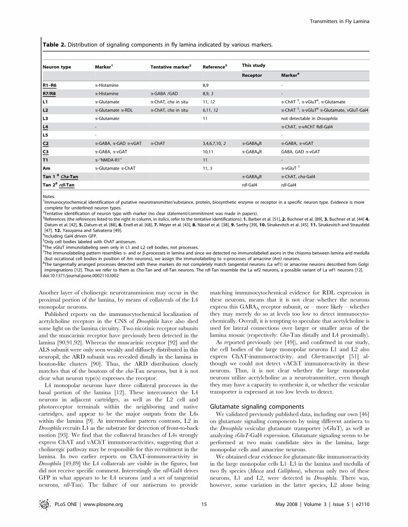
Table 2. Distribution of signaling components in fly lamina indicated by various markers.
Neuron type Marker1 Tentative marker2 Reference3 This study
Receptor Marker4
R1–R6 a-Histamine 8,9 -
R7/R8 a-Histamine a-GABA /GAD 8,9, 3 -
L1 a-Glutamate a-ChAT, cha in situ 11, 12 a-ChAT 5, a-vGluT6, a-Glutamate
L2 a-Glutamate a-RDL a-ChAT, cha in situ 6,11, 12 a-ChAT 5, a-vGluT6 a-Glutamate, vGluT-Gal4
L3 a-Glutamate 11 not detectable in Drosophila
L4 - a-ChAT, a-vAChT Rdl-Gal4
L5 - -
C2 a-GABA, a-GAD a-vGAT a-ChAT 3,4,6,7,10, 2 a-GABABR a-GABA, a-vGAT
C3 a-GABA, a-vGAT 10,11 a-GABABR GABA, GAD a-vGAT
T1 a-‘‘NMDA-R1’’ 11 -
Am a-Glutamate a-ChAT 11, 5 a-vGluT 7
Tan 1 8 Cha-Tan a-GABABR a-ChAT, cha-Gal4
Tan 28 rdl-Tan rdl-Gal4 rdl-Gal4
Notes.1Immunocytochemical identification of putative neurotransmitter/substance, protein, biosynthetic enzyme or receptor in a specific neuron type. Evidence is morecomplete for underlined neuron types.
2Tentative identification of neuron type with marker (no clear statement/commitment was made in papers).3References (the references listed to the right in column, in italics, refer to the tentative identifications): 1. Barber et al. [51], 2. Buchner et al. [89], 3. Buchner et al. [44] 4.Datum et al. [42], 5. Datum et al. [88], 6. Enell et al. [68], 7. Meyer et al. [43], 8. Nassel et al. [38], 9. Sarthy [39], 10. Sinakevitch et al. [45]. 11. Sinakevitch and Strausfeld[47]. 12. Yasuyama and Salvaterra [49].
4Including Gal4 driven GFP.5Only cell bodies labeled with ChAT antiserum.6The vGluT immunolabeling seen only in L1 and L2 cell bodies, not processes.7The immunolabeling pattern resembles a- and or b-processes in lamina and since we detected no immunolabeled axons in the chiasma between lamina and medulla(but occational cell bodies in position of Am neurons), we assign the immunolabeling to a-processes of amacrine (Am) neurons.
8The tangentially arranged processes detected with these markers do not completely match tangential neurons (La wf1) or amacrine neurons described from Golgiimpregnations [12]. Thus we refer to them as Cha-Tan and rdl-Tan neurons. The rdl-Tan resemble the La wf2 neurons, a possible variant of La wf1 neurons [12].doi:10.1371/journal.pone.0002110.t002
Transmitters in Fly Lamina
PLoS ONE | www.plosone.org 15 May 2008 | Volume 3 | Issue 5 | e2110
invariably labeled. It most plausible to attribute this variation todifferent levels of cytoplasmic glutamate that could have existedunder different functional states prior to preparation forimmunolabeling. Compatible with these neurons having thecapacity to store vesicular glutamate, the OK371-Gal4 line,specific for vglut expression, also drives GFP in the largemonopolar neurons, but we did not detect clear vGluTimmunolabeling in monopolar neurons. However, low levels ofvGluT immunolabeling were seen in cell bodies of the largemonopolar neurons and immunolabeling in dendrites of theseneurons may be masked by the stronger immunolabeling seen inamacrine processes. Another more likely possibility is that theamount of vesicle-bound glutamate (and vGluT) is simply too lowto detect. We thus do not have conclusive evidence that L1 and L2have the capacity to store vesicular glutamate, and consequentlythat they are glutamatergic in Drosophila. These neurons have mostof their synaptic output in the medulla and either no (L1) or alimited number (L2) of output synapses in the lamina [9,13]. Insofaras L1 sometimes clearly expresses a glutamate phenotype but lacksoutput synapses in the lamina, we would predict the absence ofglutamate containing vesicles and corresponding vGluT in thelamina, at least in L1. On the other hand, as revealed by the 21D-Gal4 line, we did not detect vGluT immunolabeling in the L2medulla terminals either, again possibly because there wasinsufficient protein to yield a clear immuno signal. Thus it stillcannot be entirely excluded that the glutamate immunoreactivityseen previously [46,47] may represent non-vesicular amino acidstored as a metabolic intermediate. On the other hand, the possibilitythat these monopolar neurons, the major output neurons of the fly’slamina, might use two fast neurotransmitters, glutamate andacetylcholine, may not be unprecedented [94]. However, ourevidence provides no support for the possibility that the cells mightrelease these at different sites, or even in different neuropils (L1 in themedulla, and L2 also in the lamina).While this paper was in revision an elegant study appeared on
the distribution of a vesicular glutamate transporter in Drosophila[95]. The authors of that report used a different vglut promoterGal4, but one of the vGluT antisera [61] also used in ourinvestigation. Although the paper did not report on vGluTdistribution in the lamina, the authors report expression in themedulla, where, as in our study, they found no conclusive evidencefor vglut or vGlut expression in the terminals of L1 to L3.In addition to the monopolar neurons, strong vGluT immuno-
labeling was seen in structures resembling the a-processes ofamacrine neurons, and this could be correlated with immunoreac-tivity to glutamate seen by electron microscopical analysis.Sinakevitch and Strausfeld [47] also detected such immunoreactivityin the fly Phaenicia sericata, thus providing somemeasure of support fora glutamatergic phenotype in the lamina amacrine cells.Overall, there are some incongruencies in the data for
glutamate signaling: the processes of monopolar neurons expressimmunoreactivity for glutamate but not the vesicular transporter,while the amacrine cells express immunolabeling for thetransporter but not the expected Gal4 expression. To resolvesome of these issues, we had hoped to see DmGluRA expression inlamina circuits, but very weak labeling was seen and this could notreadily be assigned to any specific neuron type. Since we detectedvery strong DmGluRA immunolabeling in neurons of the medulla,we presume the expression level is just very low in the laminaneurons. It was therefore surprising that the antiserum to theNMDA1 receptor subunit used in a previous study [47] labeledneither neither in the lamina nor elsewhere in the visual system.The antiserum was raised against a sequence of a mammalianNMDA1 receptor protein with limited similarities to that in
Drosophila and thus not likely to display much cross-reactivity inDrosophila. However, we could show rather strong immunolabelingof neurons in the mushroom body lobes, suggesting that again thelack of signal could be a matter of low levels of expression in thevisual system of Drosophila.Adopting cautious criteria, we can summarize the positive findings
on glutamate signaling components in the lamina as follows. We findevidence that the a-processes of lamina amacrine neurons expressvGluT, and glutamate. These neurons, which we might thereforepredict to be glutamatergic, have many outputs onto b-profiles of T1neurons, and onto R1–R6 and L1–L3 neurons [9]. Compatible withthis suggestion, Sinakevitch and Strausfeld [47] reported NMDA1receptor-like immunoreactivity on T1 neurons in P. sericata.Glutamate may thus be used as a transmitter in amacrine neuronsfor wide-field interconnections (see also [29,30]). We also entertainthe possibility that lamina monopolar neuron L2 may use glutamatefor signaling within the lamina at some of its manyminority classes ofsynapses, but that neither L1 nor L2 shows clear evidence of doing soat their chief output terminals in the medulla.
GABA signalingOur immunocytochemical data show that C2 and C3 neurons
(identified by Gal4-driven GFP) express both GABA and GAD.Neither of these neurons was detected using a GAD1-Gal4 line[96] tested here, and no other lamina neuron clearly expressedGAD1 or GABA immunoreactivity. An exclusive GABA pheno-type among centrifugal neurons is confirms earlier reports onDrosophila and other flies [42,43,44,45]. Previously we have alsoshown that the C2 and C3 neurons express the Drosophila vesicularGABA transporter [68], further suggesting that these neuronssignal by means of GABA.We localized GABABR immunoreactivity in relation to various
identified neurons. For this we used an antiserum to theGABABR2, a G-protein coupled receptor known to dimerize withthe GABABR1, to form a functional receptor complex [97,98].Thus our observations are likely to reveal functional GABAB
receptor sites (see [68]). At least three neuron types seem to expressGABABRs: C2, C3, and the tangential neuron Cha-Tan. Possiblythere is an additional neuron type not identified that may bepostsynaptic to the C3 neurons that express GABABRs, since wealso see immunoreactivity adjacent to C3’s boutons. The likelycontacts between GABAergic C2 neurons and large boutons ofCha-Tan neurons are quite distinct and express high levels ofGABAB receptor immunoreactivity. The presence of GABABR onC2 terminals in the distal lamina indicates the presence ofpresynaptic GABA receptors at a GABA output site of theseneurons. Similarly GABABR immunoreactivity is associated withthe varicosities of the GABAergic C3 neurons. These varicositiescan be assumed to be GABA release sites, and are known toprovide input to L1–L3 and amacrine cell processes and to receiveno inputs themselves [9,13]. Thus the GABAB receptor may bepresynaptic in both the C2 and C3 neurons. Both pre- andpostsynaptic locations of GABABRs have in fact been identified inmammals (see [99]). There, presynaptic GABABR activationinhibits transmitter release by inhibiting voltage-gated Ca2+
channels via the b/c subunit of the G-protein, or by inhibitingadenylate cyclase via Gi/o proteins [99]. In this way, GABA releasefrom C2 or C3 may be negatively regulated.The distribution of GABAA type receptors in the lamina is still
not clear, because the antiserum to the Drosophila GABAA receptorsubunit RDL failed to produce distinct lamina immunolabeling.An earlier study suggested that at least part of the RDL-immunolabeling may be localized to L2 monopolar cells [68].Here we utilized an rdl-Gal4 line to drive GFP, and although we
Transmitters in Fly Lamina
PLoS ONE | www.plosone.org 16 May 2008 | Volume 3 | Issue 5 | e2110
were unable to demonstrate that lamina neurons revealed by rdl-Gal4 actually produce RDL, a good match between the markershas been seen in many parts of the larval CNS (Enell and Nassel,unpublished). The rdl-Gal4 labels L4 monopolar neurons and rdl-Tan neurons. At least rdl-Tan neurons may be targets ofGABAergic C2 neurons as seen in our study, whereas the L4neurons are not known to be postsynaptic to either C2 or C3neurons [9], so that receptor expression on these monopolar cellsis unexplained and may be targeted to the medulla terminals.In summary, GABA seems to be primarily (or exclusively) used
by centrifugal neurons from the medulla with outputs in thelamina, one of which (C2) may signal to wide-field tangentialneurons of the lamina.
ConclusionsThis study has increased the number of lamina neurons for
which a putative neurotransmitter has now been identified and hasalso localized GABAB receptors to identified neurons (see Table 2).There are, of course, still many neuron types for whichtransmitters remain unknown. Perhaps the greatest mystery ofall remains whether the large monopolar neurons utilize glutamateor acetylcholine as neurotransmitters, or whether they maypossibly release both. They appear qualified to use either, but itis neither clear which they actually use, nor whether release is thesame at sites in the lamina and medulla, or in different strata ofthese neuropils. It is also evident that glutamate receptors andRDL subunits of GABAA receptors are expressed at levels too lowto be reliably detected in the lamina. Our study now prompts thecomplete morphological characterization of the possibly noveltypes of tangential neurons Cha-Tan and rdl-Tan. These areperhaps variants of the La wf1 and 2 neurons already described. Itis also urgent to determine the neurotransmitter of the L1 and L2neurons and to localize ionotropic receptors for acetylcholine,GABA and glutamate in the lamina circuits.Since, in contrast to the lamina of locusts, cockroaches or other
non-dipteran insects [100,101,102], it appears that laminainterneurons in flies express neither monoamines such ashistamine, dopamine, octopamine or serotonin (see [35,103]) noridentified neuropeptides, further work will be required to screenfor small-molecule neurotransmitters in those neurons not yetassigned a signal molecule. Co-expression of yet unidentifiedneuromodulators clearly remains an additional possibility, re-vealed for example by dense-core vesicles in C2 ([9]: their Fig.36A), alongside the clear vesicles which we may now presume tocontain GABA. Thus, the complete neurotransmitter repertoire ofeven the tiny constituency of neurons in the fly’s lamina cartridgestill awaits final identification.
Supporting Information
Figure S1 Confocal examination of glutamate-like immunore-activity in the optic lobes of Musca and Calliphora. A–D: Musca.A: Tangential section of the lamina, revealing the array ofcartridges, and the repeated pattern of immunoreactive profiles. B:Horizontal section, showing longitudinally sectioned axon profilesin the lamina, and medulla, and the heavy labeling in the externalchiasma between the two neuropils. C: At higher magnification,each cartridge is revealed by large immunoreactive profiles at itscore (small circle) circumscribed by a ring of small profiles (withinthe large circle) contributed by a-processes of amacrine cells. Theperikarya of some monopolar cell somata in the lamina cortex alsoexhibit faint immunoreactivity. D: Paired axon profiles (circles) areespecially clear deep in the proximal lamina, in a section planethat cuts the adjacent chiasma. E: Calliphora. Tangential section
of the medulla reveals not only immunoreactive chiasmal fibers, asseen in Musca (B,D) but also their axon profiles and terminals inthe array of medulla columns, and tangential fibers. Scale bar:1 mm.iles and terminals in the array of medulla columns, andtangential fibres. Scale bar: 1 mm.Found at: doi:10.1371/journal.pone.0002110.s001 (8.83 MB TIF)
Figure S2 Immuno-EM labeling of lamina cartridges in Muscais localized to L1–L3. A: Immuno-labeled profiles in threecartridges exhibit darkened cytoplasm and microtubules in L1and L2 (L) and illustrate the excellent state of ultrastructuralpreservation of surrounding elements of the cartridge. Immuno-signal stops at the base of a labeled axon, probably L1 (arrow).Scale bar: 1.0 mm. B The profiles of monopolar cells L1–L3 areimmunolabeled, L2 possibly more darkly. The axons of the longvisual fibres (7,8) are also faintly immunoreactive. Scale bar:1.0 mm. C Profiles of a single cartridge. Surrounding photorecep-tor terminals are well preserved, with clear profiles of tetradsynapses (arrowheads), in one case at a site providing input uponL2 (arrowhead). Scale bar: 1.0 mm.Found at: doi:10.1371/journal.pone.0002110.s002 (5.19 MB TIF)
Figure S3 Immuno-EM labeling of lamina cartridges atproximal depths in the lamina (Musca). A Example of L2 feedingsynaptic input back upon surrounding R1–R6 terminals, in thiscase at the unusually large number of three profiles of such sites(arrowheads) on two terminals in this single section. Scale bar:0.5 mm. B Single lamina cartridge revealing immuno-labeledprofiles of L1–L3. L2 has the profile of a single presynaptic site(arrowhead). Scale bar: 1.0 mm. C Enlarged L2 feedback synapticprofile shown in B, upon the profile (asterisk) of what probablyderives from the basket endings of the medulla cell T1, the normalpostsynaptic partner to a receptor terminal profile, such as thatfrom the nearby R1. Note increased density of the presynapticribbon, and of the surrounding synaptic vesicles. Scale bar:0.25 mm. D Profile of L2 feedback synapse, similar to that in C,but in which the receptor terminal (R) and likely T1 profile(asterisk) share the postsynaptic locations. Scale bar: 0.25 mm.Found at: doi:10.1371/journal.pone.0002110.s003 (4.90 MBDOC)
Figure S4 Distribution of vGluT immunolabeling in processes ofamacrine neurons in lamina of Drosophila. Strong immunolabel-ing is seen in a-processes in the lamina and weaker label in cellbodies in the chiasma (arrow). Scale bar = 10 mm. B1 Cross sectionof the lamina reveals topology of vesicular glutamate transporter(vGluT) immunoreactive processes (in magenta) in relation tophotoreceptors labellabeled with antiserum to discs large, DLG(green). The vGluT expression is seen in processes in positions likea-processes of amacrine cells (or b-processes of T1 neurons). B2Anti-vGluT labeling in relation to monopolar neurons revealed byOK371-Gal4 driven GFP (green). B3 Anti-vGluT labeling inrelation to monopolar cells revealed by 21D-Gal4. Scalebar = 5 mm. C1 Metabotropic glutamate receptor A (DmGluRA)immunolabeling is seen in medulla layers (Me) but not in laminaneuropil (La). Scale bar = 20 mm. C2 Higher magnification andincreased imaging intensity reveals strong labeling in medullaneurons (including cell bodies), but very weak and diffuse labelingin the lamina. Scale bar = 10 mm. D (1–2) NMDAR1 immuno-labellabeling in optic lobe is weak and diffuse (D1) whereas in thecentral brain (D2) it was possible to detect NMDAR1 expression inthe mushroom body lobes (MB). Scale bar = 10 mm. E Distribu-tion of RDL-immunolabeling in medulla and lobula (Lo). In thelamina only weak and diffuse labeling was seen (not shown). Scalebar = 20 mm.Found at: doi:10.1371/journal.pone.0002110.s004 (4.78 MB TIF)
Transmitters in Fly Lamina
PLoS ONE | www.plosone.org 17 May 2008 | Volume 3 | Issue 5 | e2110
Figure S5 Attempts to correlate the distribution of vesicularglutamate transporter (vGluT) immunolabeling with structuresrevealead by Gal4-driven GFP. Here we used an n-synaptobrevin-GFP fusion (nsyb-egfp) to direct GFP mainly to synaptic terminals(green). A The 21D-Gal4 drives nsybGFP primarily in the medulla(Me) terminals of the L2 neurons. Commensurate with the 10-foldfewer presynaptic sites in the lamina (13), than the medulla [1],almost no GFP is visible in the lamina (La). B Details of nsyb-eGFP expressing L2 terminals in the medulla in oblique crosssection. C The same terminals seen with vGluT immunolabeling.The two labels do not co-localize, indicating that the L2 neuronsdo not express vGluT in the medulla. D1–D3 Frontal sections ofthe medulla showing L2 terminals displayed by 21D-Gal4 crossedto UAS-nsyb-eGFP and vGluT immunolabeling (magenta). Againthere is no co-localization of labels. E1–E3 Frontal sections ofmedulla comparing the distribution of OK371-Gal4-driven nsyb-eGFP and vGluT immunolabeling. In contrast to many otherparts of the brain the two markers do not co-localize in moststructures, except partly in the inner medulla layers (IL). Inparticular, clear-cut labeling was seen in neither the L1 nor L2
terminals. Reference 1. Takemura S, Lu Z, Meinerzhagen IA(2008) Synaptic circuits of the Drosophila optic lobe: the inputterminals to the medulla. J Comp Neurol (in press)Found at: doi:10.1371/journal.pone.0002110.s005 (12.08 MBTIF)
Acknowledgments
Various antisera were kindly provided by Drs Hermann Aberle, AroonDiAntonio, Rob Jackson, Toshihiro Kitamoto and the DevelopmentalStudy Hybridoma Bank, NICHD at University of Iowa. DifferentDrosophila lines were generously provided by Drs Hermann Aberle, ElzbietaPyza, Tomas Raabe, Julie Simpson, Larry Zipursky and the BloomingtonStock Center at Indiana University, Bloomington, IN. We also thank Dr.K.-F. Fischbach for permission to use of material for a figure re-used in [9]and in altered form here in Fig. 2.
Author Contributions
Conceived and designed the experiments: IM DN AK. Performed theexperiments: AK XS. Analyzed the data: IM DN AK. Contributedreagents/materials/analysis tools: DN. Wrote the paper: IM DN.
References
1. Cajal SR, Sanchez DS (1915) Contribucion al conocimiento de los centrosnerviosos de los insectos. Parte I. Retina y centros opticos. Trab Lab Invest BiolUniv Madrid 13: 1–168.
2. Strausfeld NJ (1971) The organization of the insect visual system (lightmicroscopy). I. Projections and arrangements of neurons in the laminaganglionaris of Diptera. Z Zellforsch Mikrosk Anat 121: 377–441.
3. Meinertzhagen IA (1993) The synaptic populations of the fly’s optic neuropiland their dynamic regulation: parallels with the vertebrate retina. Progr RetinalRes 12: 13–39.
4. Ready DF, Hanson TE, Benzer S (1976) Development of the Drosophila retina,a neurocrystalline lattice. Dev Biol 53: 217–240.
5. Braitenberg V (1967) Patterns of projection in the visual system of the fly. I.Retina-lamina projections. Exp Brain Res 3: 271–298.
6. Melamed J, Trujillo-Cenoz O (1968) The fine structure of the central cells inthe ommatidia of dipterans. J Ultrastruct Res 21: 313–334.
7. Trujillo-Cenoz O (1965) Some aspects of the structural organization of theintermediate retina of dipterans. J Ultrastruct Res 13: 1–33.
8. Boschek CB (1971) On the fine structure of the peripheral retina and laminaganglionaris of the fly, Musca domestica. Z Zellforsch Mikrosk Anat 118:369–409.
9. Meinertzhagen IA, O’Neil SD (1991) Synaptic organization of columnarelements in the lamina of the wild type in Drosophila melanogaster. J Comp Neurol305: 232–263.
10. Strausfeld NJ, Campos-Ortega JA (1977) Vision in insects: pathways possiblyunderlying neural adaptation and lateral inhibition. Science 195: 894–897.
11. Nicol D, Meinertzhagen IA (1982) An analysis of the number and compositionof the synaptic populations formed by photoreceptors of the fly. J Comp Neurol207: 29–44.
12. Fischbach KF, Dittrich A (1989) The optic lobe of Drosophila melanogaster. I. AGolgi analysis of wild-type structure. Cell Tissue Res 258: 441–475.
13. Meinertzhagen IA, Sorra KE (2001) Synaptic organization in the fly’s opticlamina: few cells, many synapses and divergent microcircuits. Prog Brain Res131: 53–69.
14. Meinertzhagen IA, Hanson TE (1993) The development of the optic lobe. In:Bate M, Martinez-Arias A, eds. The Development of Drosophila melanogaster.Cold Spring Harbor, NY: Cold Spring Harbor Press. pp 1363–1491.
15. Meinertzhagen IA, Pyza E (1999) Neurotransmitter regulation of circadianstructural changes in the fly’s visual system. Microsc Res Tech 45: 96–105.
16. Pyza E, Meinertzhagen IA (1995) Monopolar cell axons in the first opticneuropil of the housefly, Musca domestica L., undergo daily fluctuations indiameter that have a circadian basis. J Neurosci 15: 407–418.
17. Meinertzhagen IA, Piper ST, Sun XJ, Frohlich A (2000) Neurite morphogen-esis of identified visual interneurons and its relationship to photoreceptorsynaptogenesis in the flies, Musca domestica and Drosophila melanogaster. EuropeanJournal of Neuroscience 12: 1342–1356.
18. Lee RC, Clandinin TR, Lee CH, Chen PL, Meinertzhagen IA, et al. (2003)The protocadherin Flamingo is required for axon target selection in theDrosophila visual system. Nat Neurosci 6: 557–563.
19. Clandinin TR, Zipursky SL (2002) Making connections in the fly visual system.Neuron 35: 827–841.
20. Hiesinger PR, Zhai RG, Zhou Y, Koh TW, Mehta SQ, et al. (2006) Activity-independent prespecification of synaptic partners in the visual map ofDrosophila. Curr Biol 16: 1835–1843.
21. Prokop A, Meinertzhagen IA (2006) Development and structure of synapticcontacts in Drosophila. Semin Cell Dev Biol 17: 20–30.
22. Jarvilehto M, Zettler F (1973) Electrophysiological-histological studies on somefunctional properties of visual cells and second order neurons of an insectretina. Z Zellforsch Mikrosk Anat 136: 291–306.
23. Shaw SR (1981) Anatomy and physiology of identified non-spiking cells in thephtoreceptor-lamina complex of the compound eye of insects, especiallyDiptera. In: Roberts A, Bush BMH, eds. Neurones without Impulses.Cambridge: Cambridge University Press. pp 61–116.
24. Laughlin SB, Osorio D (1989) Mechanisms for neural signal enhancement inthe blowfly compound eye. J Exp Biol 144: 113–146.
25. Jansonius MM, van Hateren JH (1993) On spiking units int the first opticchiasm of the blowfly. III. The sustaining unit. J Comp Physiol A 173:187–192.
26. Uusitalo RO, Juusola M, Weckstrom M (1995) Graded responses and spikingproperties of identified first-order visual interneurons of the fly compound eye.J Neurophysiol 73: 1782–1792.
27. Juusola M, French AS, Uusitalo RO, Weckstrom M (1996) Informationprocessing by graded-potential transmission through tonically active synapses.Trends Neurosci 19: 292–297.
28. Douglass JK, Strausfeld NJ (2007) Diverse speed response properties of motionsensitive neurons in the fly’s optic lobe. J Comp Physiol A Neuroethol SensNeural Behav Physiol 193: 233–247.
29. Douglass JK, Strausfeld NJ (2005) Sign-conserving amacrine neurons in thefly’s external plexiform layer. Vis Neurosci 22: 345–358.
30. Douglass JK, Strausfeld NJ (2003) Anatomical organization of retinotopicmotion-sensitive pathways in the optic lobes of flies. Microsc Res Tech 62:132–150.
31. Warrant E, Nilsson DE (2006) Invertebrate Vision Cambridge CambridgeUniversity Press. 542 p.
32. Coombe PE, Srinivasan MV, Guy RG (1989) Are the large monopolar cells ofthe insect lamina on the optomotor pathway? J Comp Physiol [a] 166: 23–35.
33. Zheng Y, Hirschberg B, Yuan J, Wang AP, Hunt DC, et al. (2002)Identification of two novel Drosophila melanogaster histamine-gated chloridechannel subunits expressed in the eye. J Biol Chem 277: 2000–2005.
34. Hardie RC (1989) Neurotransmitters in compound eyes. In: Stavenga DG,Hardie RC, eds. Facets of vision. Berlin, Heidelberg: Springer-Verlag. pp235–256.
35. Nassel DR (1991) Neurotransmitters and neuromodulators in the insect visualsystem. Prog Neurobiol 37: 179–254.
36. Hardie RC (1989) A histamine-activated chloride channel involved inneurotransmission at a photoreceptor synapse. Nature 339: 704–706.
37. Hardie RC (1987) Is histamine a neurotransmitter in insect photoreceptors?J Comp Physiol [A] 161: 201–213.
38. Nassel DR, Holmqvist MH, Hardie RC, Hakanson R, Sundler F (1988)Histamine-like immunoreactivity in photoreceptors of the compound eyes andocelli of the flies Calliphora erythrocephala andMusca domestica. Cell Tissue Res 253:639–646.
39. Sarthy PV (1991) Histamine: a neurotransmitter candidate for Drosophilaphotoreceptors. J Neurochem 57: 1757–1768.
40. Stuart AE, Borycz J, Meinertzhagen IA (2007) The dynamics of signaling at thehistaminergic photoreceptor synapse of arthropods. Prog Neurobiol 82:202–227.
Transmitters in Fly Lamina
PLoS ONE | www.plosone.org 18 May 2008 | Volume 3 | Issue 5 | e2110
41. Witte I, Kreienkamp HJ, Gewecke M, Roeder T (2002) Putative histamine-gated chloride channel subunits of the insect visual system and thoracicganglion. J Neurochem 83: 504–514.
42. Datum KH, Weiler R, Zettler F (1986) Immunocytochemical demonstration ofc-aminobutyric acid and glutamic acid decarboxylase in R7 photoreceptorsand C2 centrifugal fibres in the blowfly visual system. J Comp Physiol A 159:241–249.
43. Meyer EP, Matute C, Streit P, Nassel DR (1986) Insect optic lobe neuronsidentifiable with monoclonal antibodies to GABA. Histochem 84: 207–216.
44. Buchner E, Bader R, Buchner S, Cox J, Emson PC, et al. (1988) Cell-specificimmuno-probes for the brain of normal and mutant Drosophila melanogaster. I.Wildtype visual system. Cell Tissue Res 253: 357–370.
45. Sinakevitch I, Douglass JK, Scholtz G, Loesel R, Strausfeld NJ (2003)Conserved and convergent organization in the optic lobes of insects andisopods, with reference to other crustacean taxa. J Comp Neurol 467: 150–172.
46. Meinertzhagen IA, Sun XJ (1996) Monopolar cell axons in the lamina cartridgeof the flies Drosophila, Musca and Calliphora exhibit glutamate-like immunore-activity. Soc Neurosci Abstr 22: 655.
47. Sinakevitch I, Strausfeld NJ (2004) Chemical neuroanatomy of the fly’smovement detection pathway. J Comp Neurol 468: 6–23.
48. Bicker G, Schafer S, Ottersen OP, Storm-Mathisen J (1988) Glutamate-likeimmunoreactivity in identified neuronal populations of insect nervous systems.J Neurosci 8: 2108–2122.
49. Yasuyama K, Salvaterra PM (1999) Localization of choline acetyltransferase-expressing neurons in Drosophila nervous system. Microsc Res Tech 45: 65–79.
50. Itoh N, Slemmon JR, Hawke DH, Williamson R, Morita E, et al. (1986)Cloning of Drosophila choline acetyltransferase cDNA. Proc Natl Acad Sci U S A83: 4081–4085.
51. Barber RP, Sugihara H, Lee M, Vaughn JE, Salvaterra PM (1989) Localizationof Drosophila neurons that contain choline acetyltransferase messenger RNA: anin situ hybridization study. J Comp Neurol 280: 533–543.
52. Brand AH, Perrimon N (1993) Targeted gene expression as a means of alteringcell fates and generating dominant phenotypes. Development 118: 401–415.
53. Gorska-Andrzejak J, Keller A, Raabe T, Kilianek L, Pyza E (2005) Structuraldaily rhythms in GFP-labelled neurons in the visual system of Drosophilamelanogaster. Photochem Photobiol Sci 4: 721–726.
54. Salvaterra PM, Kitamoto T (2001) Drosophila cholinergic neurons and processesvisualized with Gal4/UAS-GFP. Brain Res Gene Expr Patterns 1: 73–82.
55. Mahr A, Aberle H (2006) The expression pattern of the Drosophila vesicularglutamate transporter: a marker protein for motoneurons and glutamatergiccenters in the brain. Gene Expr Patterns 6: 299–309.
56. Estes PS, Ho GL, Narayanan R, Ramaswami M (2000) Synaptic localizationand restricted diffusion of a Drosophila neuronal synaptobrevin–greenfluorescent protein chimera in vivo. J Neurogenet 13: 233–255.
57. Hiesinger PR, Scholz M, Meinertzhagen IA, Fischbach KF, Obermayer K(2001) Visualization of synaptic markers in the optic neuropils of Drosophilausing a new constrained deconvolution method. J Comp Neurol 429: 277–288.
58. Eroglu C, Brugger B, Wieland F, Sinning I (2003) Glutamate-binding affinity ofDrosophila metabotropic glutamate receptor is modulated by association withlipid rafts. Proc Natl Acad Sci U S A 100: 10219–10224.
59. Panneels V, Eroglu C, Cronet P, Sinning I (2003) Pharmacologicalcharacterization and immunoaffinity purification of metabotropic glutamatereceptor from Drosophila overexpressed in Sf9 cells. Protein Expr Purif 30:275–282.
60. Bogdanik L, Mohrmann R, Ramaekers A, Bockaert J, Grau Y, et al. (2004)The Drosophila metabotropic glutamate receptor DmGluRA regulates activity-dependent synaptic facilitation and fine synaptic morphology. J Neurosci 24:9105–9116.
61. Daniels RW, Collins CA, Gelfand MV, Dant J, Brooks ES, et al. (2004)Increased expression of the Drosophila vesicular glutamate transporter leads toexcess glutamate release and a compensatory decrease in quantal content.J Neurosci 24: 10466–10474.
62. Hepler JR, Toomim CS, McCarthy KD, Conti F, Battaglia G, et al. (1988)Characterization of antisera to glutamate and aspartate. J HistochemCytochem 36: 13–22.
63. Madl JE, Larson AA, Beitz AJ (1986) Monoclonal antibody specific forcarbodiimide-fixed glutamate: immunocytochemical localization in the ratCNS. J Histochem Cytochem 34: 317–326.
64. Wilson RI, Laurent G (2005) Role of GABAergic inhibition in shaping odor-evoked spatiotemporal patterns in the Drosophila antennal lobe. J Neurosci 25:9069–9079.
65. Hamasaka Y, Wegener C, Nassel DR (2005) GABA modulates Drosophilacircadian clock neurons via GABAB receptors and decreases in calcium.J Neurobiol 65: 225–240.
66. Jackson FR, Newby LM, Kulkarni SJ (1990) Drosophila GABAergic systems:Sequence and expression of glutamic acid decarboxylase. J Neurochem 54:1068–1078.
67. Featherstone DE, Rushton EM, Hilderbrand-Chae M, Phillips AM,Jackson FR, et al. (2000) Presynaptic glutamic acid decarboxylase is requiredfor induction of the postsynaptic receptor field at a glutamatergic synapse.Neuron 27: 71–84.
68. Enell L, Hamasaka Y, Kolodziejczyk A, Nassel DR (2007) gamma-Aminobutyric acid (GABA) signaling components in Drosophila: immunocyto-chemical localization of GABA(B) receptors in relation to the GABA(A)
receptor subunit RDL and a vesicular GABA transporter. J Comp Neurol 505:18–31.
69. Takagawa K, Salvaterra P (1996) Analysis of choline acetyltransferase proteinin temperature sensitive mutant flies using newly generated monoclonalantibody. Neurosci Res 24: 237–243.
70. Yasuyama K, Kitamoto T, Salvaterra PM (1995) Differential regulation ofcholine acetyltransferase expression in adult Drosophila melanogaster brain.J Neurobiol 30: 205–218.
71. Okabe S, Miwa A, Okado H (1999) Alternative splicing of the C-terminaldomain regulates cell surface expression of the NMDA receptor NR1 subunit.J Neurosci 19: 7781–7792.
72. Ultsch A, Schuster CM, Laube B, Betz H, Schmitt B (1993) Glutamatereceptors of Drosophila melanogaster. Primary structure of a putative NMDAreceptor protein expressed in the head of the adult fly. FEBS Lett 324:171–177.
73. Kitamoto T, Wang W, Salvaterra PM (1998) Structure and organization of theDrosophila cholinergic locus. J Biol Chem 273: 2706–2713.
74. Kitamoto T, Xie XJ, Wu CF, Salvaterra PM (2000) Isolation andcharacterization of mutants for the vesicular acetylcholine transporter genein Drosophila melanogaster. Journal of Neurobiology 42: 161–171.
75. Strausfeld NJ (1976) Atlas of an insect brain. Berlin: Springer Verlag.76. Strausfeld NJ, Nassel DR (1980) Neuroarchitecture of brain regions that
subserve the compound eyes of Crustacea and insects. In: Autrum H, ed.Handbook of sensory physiology VII/6B. Berlin, Heidelberg, New York:Springer Verlag. pp 1–132.
77. Shaw SR, Meinertzhagen IA (1986) Evolution of synaptic connections amonghomologous neurons. Proc Natl Acad Sci USA 83: 7961–7965.
78. Klemm N (1976) Histochemistry of putative transmitter substances in the insectbrain. Prog Neurobiol 7: 99–169.
79. Sattelle DB, Jones AK, Sattelle BM, Matsuda K, Reenan R, et al. (2005) Edit,cut and paste in the nicotinic acetylcholine receptor gene family of Drosophilamelanogaster. Bioessays 27: 366–376.
80. Yasuyama K, Kitamoto T, Salvaterra PM (1995) Immunocytochemical studyof choline acetyltransferase in Drosophila melanogaster: an analysis of cis-regulatory regions controlling expression in the brain of cDNA-transformedflies. J Comp Neurol 361: 25–37.
81. Jan LY, Jan YN (1976) L-glutamate as an excitatory transmitter at theDrosophila larval neuromuscular junction. J Physiol 262: 215–236.
82. Hauser H (1975) Vergleichend quantitative Untersuchungen an den Sehgan-glien der Fliegen Musca domestica und Drosophila melanogaster [PhD Thesis]. PhDThesis, Tubingen: Eberhard-Karls-Universitat.
83. Barth M, Hirsch H, Meinertzhagen IA, Heisenberg M (1997) Experience-dependent developmental plasticity in the optic lobe of Drosophila melanogaster.J Neurosci 17: 1493–1504.
84. Chen K, Featherstone DE (2005) Discs-large (DLG) is clustered by presynapticinnervation and regulates postsynaptic glutamate receptor subunit compositionin Drosophila. BMC Biol 3: 1.
85. Hamasaka Y, Rieger D, Parmentier ML, Grau Y, Helfrich-Forster C, et al.(2007) Glutamate and its metabotropic receptor in Drosophila clock neuroncircuits. J Comp Neurol 505: 32–45.
86. Sattelle DB, Lummis SC, Wong JF, Rauh JJ (1991) Pharmacology of insectGABA receptors. Neurochem Res 16: 363–374.
87. Hosie AM, Aronstein K, Sattelle DB, ffrench CR (1997) Molecular biology ofinsect neuronal GABA receptors. Trends Neurosci 20: 578–583.
88. Datum KH, Rambolt I, Zettler F (1989) Cholinergic neurons in the laminaganglionaris of the blowfly Calliphora erythrocephala. Cell Tissue Res 256:153–158.
89. Buchner E, Buchner S, Crawford G, Mason WT, Salvaterra PM, et al. (1986)Choline acetyltransferase-like immunoreactivity in the brain of Drosophilamelanogaster. Cell Tissue Res 246: 57–62.
90. Schuster R, Phannavong B, Schroder C, Gundelfinger ED (1993) Immuno-histochemical localization of a ligand-binding and a structural subunit ofnicotinic acetylcholine receptors in the central nervous system of Drosophilamelanogaster. J Comp Neurol 335: 149–162.
91. Jonas PE, Phannavong B, Schuster R, Schroder C, Gundelfinger ED (1994)Expression of the ligand-binding nicotinic acetylcholine receptor subunit Dalpha 2 in the Drosophila central nervous system. J Neurobiol 25: 1494–1508.
92. Harrison JB, Chen HH, Blake AD, Huskisson NS, Barker P, et al. (1995)Localization in the nervous system of Drosophila melanogaster of a C-terminusanti-peptide antibody to a cloned Drosophila muscarinic acetylcholine receptor.J Neuroendocrinol 7: 347–352.
93. Rister J, Pauls D, Schnell B, Ting CY, Lee CH, et al. (2007) Dissection of theperipheral motion channel in the visual system of Drosophila melanogaster. Neuron56: 155–170.
94. Jonas P, Bischofberger J, Sandkuhler J (1998) Corelease of two fastneurotransmitters at a central synapse. Science 281: 419–424.
95. Daniels RW, Gelfand MV, Collins CA, Diantonio A (2008) Visualizingglutamatergic cell bodies and synapses in Drosophila larval and adult CNS.J Comp Neurol 508: 131–152.
96. Ng M, Roorda RD, Lima SQ, Zemelman BV, Morcillo P, et al. (2002)Transmission of olfactory information between three populations of neurons inthe antennal lobe of the fly. Neuron 36: 463–474.
Transmitters in Fly Lamina
PLoS ONE | www.plosone.org 19 May 2008 | Volume 3 | Issue 5 | e2110

97. Kaupmann K, Malitschek B, Schuler V, Heid J, Froestl W, et al. (1998)GABA(B)-receptor subtypes assemble into functional heteromeric complexes.Nature 396: 683–687.
98. Mezler M, Muller T, Raming K (2001) Cloning and functional expression ofGABA(B) receptors from Drosophila. Eur J Neurosci 13: 477–486.
99. Bettler B, Kaupmann K, Mosbacher J, Gassmann M (2004) Molecularstructure and physiological functions of GABA(B) receptors. Physiol Rev 84:835–867.
100. Nassel DR (2002) Neuropeptides in the nervous system of Drosophila and otherinsects: multiple roles as neuromodulators and neurohormones. Prog Neurobiol68: 1–84.
101. Homberg U (2002) Neurotransmitters and neuropeptides in the brain of thelocust. Microsc Res Tech 56: 189–209.
102. Nassel DR, Homberg U (2006) Neuropeptides in interneurons of the insectbrain. Cell Tissue Res 326: 1–24.
103. Monastirioti M (1999) Biogenic amine systems in the fruit fly Drosophilamelanogaster. Microsc Res Tech 45: 106–121.
104. Parnas D, Haghighi AP, Fetter RD, Kim SW, Goodman CS (2001) Regulationof postsynaptic structure and protein localization by the Rho-type guaninenucleotide exchange factor dPix. Neuron 32: 415–424.
Transmitters in Fly Lamina
PLoS ONE | www.plosone.org 20 May 2008 | Volume 3 | Issue 5 | e2110
Copyright © 2022 FDOKUMEN