giomer composite system - Shofu
8
cosmetic dentistry 1_2012 I technique_ giomers Author_Jack D. Griffin Jr., DMD, MAGD Esthetics, caries control and gingival health with a versatile giomer composite system _There are many direct composite materials that have the strength to be used in the posterior and a level of esthetics acceptable in the anterior dentition. 1 Direct anterior composites must meet the minimum cosmetic demands of the patient while posterior restorations must provide resistance to mechanical forces. 2–5 In patients with questionable hygiene, dietary habits and a history of caries, we must also choose materials that have properties such as fluoride re- lease and high polishablility to decrease the effects of a less than ideal oral environment. 6 A composite system has been developed with a surface pre-reacted glass ionomer (S-PRG) that has shown to have high esthetic properties as well as significant release of fluoride ions into the dentin and re-chargeability. 7,8 “Beautifil II and Beautifil Flow Plus (Shofu, San Marcos, Calif.) are universal nano-hybrid compos- ites with the durability and esthetics to satisfy the demands of the profession while also providing long- term tissue health by sustained fluoride release from the S-PRG filler particles.” 9 A potential reduction of secondary caries and maintenance of surface luster has been shown long-term with this material. 10,11 The small nano-sized filler has a mean particle size of just 0.8 μm, along with a filler load by weight of 83 percent, making it suitable for almost any clini- cal situation, including incisal edge replacement and posterior restorations in occlusion. 12 The versatility of these products, coupled with the giomer technology, make them unique in the dental marketplace and a strong choice in almost any clini- cal situation. The flowable comes in two viscosities with a vary- ing degree of flowability, each with similar physical properties strong enough to withstand occlusal forces in posterior restorations. 13 The zero flow, F00, is a stackable flow with almost no movement or slump when syringing. The low flow, F03, is a great universal flow material with handling similar to other more viscous flowables on the market. 32 I Fig. 1_Patient concerned with swollen gums and holes in teeth. Fig. 2_He had braces removed the previous year and said he never smiled. Fig. 3_He had generalized sensitivity to sweets and cold. Composites were to be done on 23 teeth over three appointments. Fig. 4_He did not brush “very often” and really liked soda and energy drinks. He seemed committed to improving things in his mouth. Fig. 5_Diode laser gingivoplasty was done after local anesthetic using low wattage and brush strokes. Fig. 6_Preparations were done as conservatively as possible with a 330 bur, finish diamond and slow-speed round bur. Caries indicator was used to verify decay removal. Fig. 1 Fig. 2 Fig. 3 Fig. 4 Fig. 5 Fig. 6
-
Upload
khangminh22 -
Category
Documents
-
view
2 -
download
0
Transcript of giomer composite system - Shofu
cosmetic dentistry 1_2012
I technique_ giomers
Author_Jack D. Griffin Jr., DMD, MAGD
Esthetics, caries control and gingival health with a versatile giomer composite system
_There are many direct composite materials that have the strength to be used in the posterior and a level of esthetics acceptable in the anterior dentition.1 Direct anterior composites must meet the minimum cosmetic demands of the patient while posterior restorations must provide resistance to mechanical forces.2–5
In patients with questionable hygiene, dietary habits and a history of caries, we must also choose materials that have properties such as fluoride re-lease and high polishablility to decrease the effects of a less than ideal oral environment.6
A composite system has been developed with a surface pre-reacted glass ionomer (S-PRG) that has shown to have high esthetic properties as well as significant release of fluoride ions into the dentin and re-chargeability.7,8
“Beautifil II and Beautifil Flow Plus (Shofu, San Marcos, Calif.) are universal nano-hybrid compos-ites with the durability and esthetics to satisfy the demands of the profession while also providing long-
term tissue health by sustained fluoride release from the S-PRG filler particles.”9 A potential reduction of secondary caries and maintenance of surface luster has been shown long-term with this material.10,11
The small nano-sized filler has a mean particle size of just 0.8 μm, along with a filler load by weight of 83 percent, making it suitable for almost any clini-cal situation, including incisal edge replacement and posterior restorations in occlusion.12
The versatility of these products, coupled with the giomer technology, make them unique in the dental marketplace and a strong choice in almost any clini-cal situation.
The flowable comes in two viscosities with a vary-ing degree of flowability, each with similar physical properties strong enough to withstand occlusal forces in posterior restorations.13 The zero flow, F00, is a stackable flow with almost no movement or slump when syringing. The low flow, F03, is a great universal flow material with handling similar to other more viscous flowables on the market.
32 I
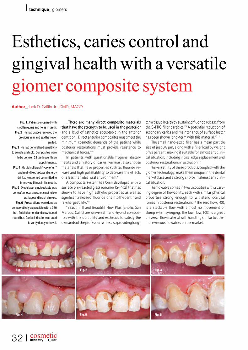
Fig. 1_Patient concerned with
swollen gums and holes in teeth.
Fig. 2_He had braces removed the
previous year and said he never
smiled.
Fig. 3_He had generalized sensitivity
to sweets and cold. Composites were
to be done on 23 teeth over three
appointments.
Fig. 4_He did not brush “very often”
and really liked soda and energy
drinks. He seemed committed to
improving things in his mouth.
Fig. 5_Diode laser gingivoplasty was
done after local anesthetic using low
wattage and brush strokes.
Fig. 6_Preparations were done as
conservatively as possible with a 330
bur, finish diamond and slow-speed
round bur. Caries indicator was used
to verify decay removal.
Fig. 1 Fig. 2 Fig. 3
Fig. 4 Fig. 5 Fig. 6

cosmetic dentistry 1_2012
technique_ giomers I
Fig. 7_Contour matrices were placed
and a BAC containing 37 percent
phosphoric acid etch was placed and
rinsed.
Fig. 8_Several coats of a universal
bonding agent were applied and air
thinned.
Fig. 9_Beautifil Flow Plus was
applied to cover all dentin and then
cured.
Fig. 10_The contour matricies form
a good gingival seal, keeping the
crevicular fluids out while giving
anatomical form to the material.
Fig. 11_The remaining cavities were
restored with the same flowable
material. Notice how the material
holds its shape without running, even
before curing.
Fig. 12_Light polymerization was
done from facial, lingual and incisal
to insure a high level of conversion.
(Images/Provided by Dr. Jack D.
Griffin, Jr.)
Will these flowable materials replace each of the more viscous composite materials? No. The sculpt-ability and layering potential of conventional com-posites will always have a place in esthetic dentistry. Another advantage of a non-flowable material is void reduction in posterior composites as uncured flow-able is followed by a more viscous material pushing out and displacing the flowable as the viscous com-pule material is injected.
_Patient exam and planning
A 15-year-old male came to the office with an “unpleasant” smile after having orthodontic treat-ment the previous year (Fig. 1). His primary concern was the hypertrophic tissue around his incisors and his cold and sweet sensitivity (Fig. 2). There was ram-pant decay, enlarged gingival tissue, poor hygiene and decalcification areas (Figs. 3, 4).
A full series of radiographs and photographic im-ages were taken for treatment planning, marketing and case documentation. These images were studied, along with clinical exam notes, before treatment so that a basic plan was formulated.14
A treatment plan was made to do 23 direct com-posite restorations over three appointments after a prophy, oral hygiene education and tray-delivered home topical fluoride delivery. The plan included laser gingivoplasty followed by restorations with Beautifil II because of its esthetics, ease of use, fluo-ride release and versatility.
After several weeks of maintained oral hygiene improvement, the surface of the anterior teeth would be re-contoured and enhanced at no additional charge.
_Soft-tissue enhancement
Lasers have become a critical component of smile rehabilitations, and if done with respect to peri-odontal tissues and biologic width, results can be a great enhancement to cosmetic treatment.15,16 Diode lasers offer excellent control of tissue sculpting with very predictable healing and tissue tolerance as long as sound biologic principles are followed.17–19 These principles must be understood during treatment in order to prevent possible chronic periodontal inflam-mation and unwanted gingival responses, such as redness, bleeding and irritation.20,21
On the first restorative appointment, a local an-esthetic (Septocaine, Septodont, Lancaster, Pa.) was given and retractors (See More, Discus, Culver City, Calif.) were placed to keep the lips out of the way and to provide some isolation from saliva. An 810 mm diode laser (Odyssey, Ivolcar Vivadent, Amherst, N.Y.) was used on a relatively low wattage, 2.0, to sculpt the tissues and remove hyperplastic gingival tissue (Fig. 5).22
Clean up and removal of the charred tissue was done with a microbrush and hydrogen peroxide. It is expected that the new soft-tissue location would be maintained or even improve with properly contoured restorations, good surface polish and continued plaque control.23
_Giomer composite technique: maxillary
The goal of this one-hour restorative appoint-ment was to provide improved esthetics and an envi-ronment to promote tissue recovery on the maxillary anterior. Tooth preparation was done with a 330
I 33
Fig. 7 Fig. 8 Fig. 9
Fig. 10 Fig. 11 Fig. 12
cosmetic dentistry 1_2012
I technique_ giomers
bur, finish diamond and a #2 slow-speed round bur (Fig. 6). Caries indicator (Sable Seek, Ultradent, South Jordan, Utah) was used to verify complete caries elimination and a long irregular bevel was placed on all enamel margins. Two teeth were done at a time.
A contoured anatomical matrix (Contour Matrix, Ivoclar Vivadent, Amherst, N.Y.) was placed and wedged loosely. The matrix extends slightly, provid-ing a “sulcular seal” aiding in marginal integrity. These matrices not only increase restoration longev-ity, but also greatly increase placement efficiency.24
The teeth were etched with 37 percent phosphoric acid (Etch 37, Bisco, Schaumburg, Ill.) for 10 seconds, rinsed thoroughly and left damp (Fig. 7). This etch contains benzalkonium chloride (BAC) for a contin-ued antimicrobial effect. A universal bonding agent (All Bond Universal, Bisco, Schaumburg, Ill.) was ap-plied in several layers, air thinned and cured (Fig. 8).25
Beautifil Flow Plus, low flow, was placed into the preps, covering all dentin, and cured for 15 seconds (Fig. 10). The non-runny nature of this material and the great adaptability make this a great choice for dentin replacement. The remaining preparation was restored with the same material so that the entire restoration was done in this flowable nano-hybrid composite in a single shade of A2 (Fig. 11).26
Curing was then done for 20 seconds from the facial, lingual and incisal to insure complete polym-erization (Fig. 12). Initial contouring was done quickly with a finishing diamond (Diatech Direct, Charleston, S.C.) to provide basic anatomical shaping.27
Tooth #8 was prepped, caries removed and re-stored in a similar fashion (Fig. 14). Because of the size and depth of this restoration, the flowable material was placed and cured in three different increments
(Fig. 15). The remaining anterior teeth were restored and shaped (Fig. 16). Minimal polishing was done at this time because of time constraints.
Alginate impressions were taken and flexible bleach-type trays were made. The high fluoride re-lease and ability to be recharged make this giomer an ideal product in less than ideal oral environments such as this.28
The potential of recurrent decay and the plaque formation that may compromise the gingival recon-touring may be lessened by the sustained antimicro-bial effect of this material.
The patient was given stannous fluoride at 0.4 percent with instructions to place it in the mouth overnight two to three times per week until all restor-ative work was completed. At that time, re-charging of the giomer material would continue between prophy appointments at once a week (Fig. 17).
_Second appointment: mandibular restorations
Two weeks later, a two-hour appointment was made to restore the mandibular teeth. The gingival health on the maxilla was consistent with better hygiene on restored teeth and bacterial control (Figs. 18, 19). The patient claimed to use the fluoride trays two to three times a week and was brushing at least once a day.
The patient’s right side was done, first focusing on facial decay initially (Fig. 19). Cavity preparation, car-ies removal and enamel beveling as described above was completed with various burs. On the right bicus-pid, a pinkish blush was noticed after decay removal with no obvious pulpal exposure (Fig. 20).
34 I
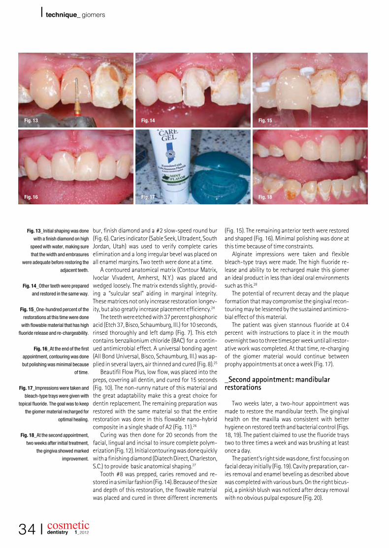
Fig. 13_Initial shaping was done
with a finish diamond on high
speed with water, making sure
that the width and embrasures
were adequate before restoring the
adjacent teeth.
Fig. 14_Other teeth were prepared
and restored in the same way.
Fig. 15_One-hundred percent of the
restorations at this time were done
with flowable material that has high
fluoride release and re-chargeability.
Fig. 16_At the end of the first
appointment, contouring was done
but polishing was minimal because
of time.
Fig. 17_Impressions were taken and
bleach-type trays were given with
topical fluoride. The goal was to keep
the giomer material recharged for
optimal healing.
Fig. 18_At the second appointment,
two weeks after initial treatment,
the gingiva showed marked
improvement.
Fig. 13 Fig. 15Fig. 14
Fig. 16 Fig. 18Fig. 17
cosmetic dentistry 1_2012
technique_ giomers I
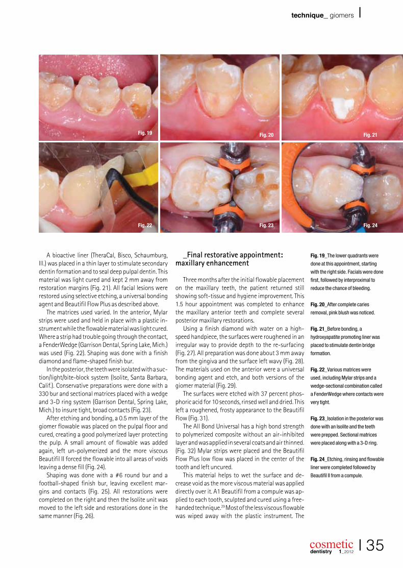
Fig. 19_The lower quadrants were
done at this appointment, starting
with the right side. Facials were done
first, followed by interproximal to
reduce the chance of bleeding.
Fig. 20_After complete caries
removal, pink blush was noticed.
Fig. 21_Before bonding, a
hydroxyapatite promoting liner was
placed to stimulate dentin bridge
formation.
Fig. 22_Various matrices were
used, including Mylar strips and a
wedge-sectional combination called
a FenderWedge where contacts were
very tight.
Fig. 23_Isolation in the posterior was
done with an Isolite and the teeth
were prepped. Sectional matrices
were placed along with a 3-D ring.
Fig. 24_Etching, rinsing and flowable
liner were completed followed by
Beautifil II from a compule.
A bioactive liner (TheraCal, Bisco, Schaumburg, Ill.) was placed in a thin layer to stimulate secondary dentin formation and to seal deep pulpal dentin. This material was light cured and kept 2 mm away from restoration margins (Fig. 21). All facial lesions were restored using selective etching, a universal bonding agent and Beautifil Flow Plus as described above.
The matrices used varied. In the anterior, Mylar strips were used and held in place with a plastic in-strument while the flowable material was light cured. Where a strip had trouble going through the contact, a FenderWedge (Garrison Dental, Spring Lake, Mich.) was used (Fig. 22). Shaping was done with a finish diamond and flame-shaped finish bur.
In the posterior, the teeth were isolated with a suc-tion/light/bite-block system (Isolite, Santa Barbara, Calif.). Conservative preparations were done with a 330 bur and sectional matrices placed with a wedge and 3-D ring system (Garrison Dental, Spring Lake, Mich.) to insure tight, broad contacts (Fig. 23).
After etching and bonding, a 0.5 mm layer of the giomer flowable was placed on the pulpal floor and cured, creating a good polymerized layer protecting the pulp. A small amount of flowable was added again, left un-polymerized and the more viscous Beautifil II forced the flowable into all areas of voids leaving a dense fill (Fig. 24).
Shaping was done with a #6 round bur and a football-shaped finish bur, leaving excellent mar-gins and contacts (Fig. 25). All restorations were completed on the right and then the Isolite unit was moved to the left side and restorations done in the same manner (Fig. 26).
_Final restorative appointment: maxillary enhancement
Three months after the initial flowable placement on the maxillary teeth, the patient returned still showing soft-tissue and hygiene improvement. This 1.5 hour appointment was completed to enhance the maxillary anterior teeth and complete several posterior maxillary restorations.
Using a finish diamond with water on a high-speed handpiece, the surfaces were roughened in an irregular way to provide depth to the re-surfacing (Fig. 27). All preparation was done about 3 mm away from the gingiva and the surface left wavy (Fig. 28). The materials used on the anterior were a universal bonding agent and etch, and both versions of the giomer material (Fig. 29).
The surfaces were etched with 37 percent phos-phoric acid for 10 seconds, rinsed well and dried. This left a roughened, frosty appearance to the Beautifil Flow (Fig. 31).
The All Bond Universal has a high bond strength to polymerized composite without an air-inhibited layer and was applied in several coats and air thinned. (Fig. 32) Mylar strips were placed and the Beautifil Flow Plus low flow was placed in the center of the tooth and left uncured.
This material helps to wet the surface and de-crease void as the more viscous material was applied directly over it. A1 Beautifil from a compule was ap-plied to each tooth, sculpted and cured using a free-handed technique.29 Most of the less viscous flowable was wiped away with the plastic instrument. The
I 35
Fig. 19 Fig. 20 Fig. 21
Fig. 22 Fig. 23 Fig. 24

cosmetic dentistry 1_2012
I technique_ giomers
lighter surface shade brightens the smile while giv-ing vitality to the restorations that were placed over the darker A2 shade from the first restorative ap-pointment. Initial contouring was done with a finish diamond on high speed with water.30–32
Shaping and polishing was completed with a flexible disk system (Super Snap Rainbow, Shofu, San Marcos, Calif.). This system features a sequence of very thin, flexible disks that are very efficient at shap-ing embrasures, final shaping and high polish with-out metal in the center that may gouge or scratch the restoration surface (Fig. 34). The giomer material is easily polished and rivals many nano-hybrids on the market today.33
_Results
Obviously, a great improvement was realized for a boy who said he “never smiled until now” (Fig. 37). It would be naïve to think that these restora-tions will last him his entire life without the need for more definitive porcelain restorations or other cosmetic procedure. However, the improvement in self-esteem, the decrease in sensitivity and the feel-ing of better oral health may help to stimulate him to be committed to better oral care. After six months, the improvement in soft- and hard-tissue health is undeniable (Fig. 38). The attached tissue stippling and lack of bleeding clearly shows how well the soft tissues tolerate these materials.
The giomer materials have excellent esthetics and strength, which combined with the high long-term fluoride release make these materials a strong consideration in most all direct restorative cases. The patient has continued fluoride treatments at home
on average about every one to two weeks, as often as he can remember. Now, if we can just keep him from losing his trays for the third time._
_References
1. Vargas M. Conservative aesthetic enhance-ment of the anterior dentition using a predict-able direct resin protocol. Prac Proced Aesthet Dent.2006;18(8):501–507.
2. Fahl, N. The direct/indirect composite resin veneers: a case report. Pract Periodont Aesthet Dent. 1996;8(7):627–638.
3. Milnar F. A minimal intervention approach to the treatment of a class IV fratcture. J of Cosmet Dent 21(4):106–112;2006.
4. Christensen GJ. Bonding to dentin and enamel where does it stand in 2005? J Am Dent Assoc. 136(9):1299–1302;2005
5. Terry DA. Direct composite resin restoration of adolescent class IV tooth fracture: a case report. Prac Perio Aesthet Dent. 12(1):23–29;2000.
6. Wiegand A, Buchalla W, Attin T. Review on flu-oride-releasing restorative materials—fluoride release and uptake characteristics, antibacterial activity and influence on caries formation. Dent Mater. 2007 Mar;23(3):343–362. Epub 2006 Apr 17.
7. Ikemura K, Tay FR, Endo T, Pashley DH. A review of chemical-approach and ultramorphological studies on the development of fluoride-re-leasing dental adhesives comprising new pre-reacted glass ionomer (PRG) fillers. Dent Mater J. 2008 May;27(3):315–339.
8. Itota T, Carrick TE, Yoshiyama M, McCabe JF.
36 I
Fig. 25_The restorations show
excellent margins with broad, deep,
tight contacts.
Fig. 26_Shaping was completed
with a finish diamond, finish bur and
disks.
Fig. 27_At the third and final
restorative appointment, the
maxillary anteriors were enhanced.
A finish diamond was used with
water to roughen the surface of the
Beautifil Flow Plus from the first
appointment.
Fig. 28_The surface was irregular
and kept 3 mm from the gingiva.
Fig. 29_The materials used during
this enhancement included 37
percent etch, a universal bonding
agent, flowable and more viscous
composite.
Fig. 30_The teeth were isolated with
retractors, etched for 10 seconds
and rinsed thoroughly.
Fig. 25 Fig. 26 Fig. 27
Fig. 28 Fig. 29 Fig. 30

cosmetic dentistry 1_2012
I technique_ giomers
Fluoride release and recharge in giomer, com-pomer and resin composite. Dent Mater. 2004 Nov;20(9):789–795.
9. Gordan VV, Mondragon E, Watson RE, Garvan C, Mjör IA. A clinical evaluation of a self-etching primer and a giomer restorative material: re-sults at eight years. J Am Dent Assoc. 2007 May;138(5):621–627.
10. Valeria V. Gordan, DDS, MS; Eduardo Mondragon; Ronald E. Watson, DDS, MAE; Cyndi Garvan, PhD; Ivar A.Mjör, BDS, MSD, MS, Dr.odont. JADA, Vol. 138, May 2007.
11. Jyothi K, Annapurna S, Kumar AS, Venugopal P, Jayashankara C. Clinical evaluation of giomer- and resin-modified glass ionomer cement in class V noncarious cervical lesions: An in vivo study. J Conserv Dent. 2011 Oct;14(4):409–413.
12. Company data, www.shofu.com.13. Beautifil Flow Plus. Inside Dentistry. Tech
profile;February, 2011(108).14. Griffin JD Jr. Assessing aesthetic composite
veneer placement via digital photography. Pract Proced Aesthet Dent. 19(5):2889–2894;2007.
15. Adams TC, Pang PK. Lasers in aesthetic dentistry. Dent Clin North Am. 2004;48(4):833–860.
16. Rice JH. Laser use in fixed, removable, and implant
dentistry. Dent Clin North Am. 2000;44(4):767–777.
17. Coluzzi DJ. Fundamentals of dental lasers: sci-ence and instruments. Dent Clin North Am. 2004;48:751–770.
18. Kokich VG, et al. Gigival contour and clinical crown length: their effect on the esthetic ap-pearance of maxillary anterior teeth. Amer J Orthod. 1994;86(2):89–94.
19. Yeh S, Andreana S. Crown lengthening: basic principles, indications, techniques and clinical case reports. NY State Dental J. 2004;70(8):30–36.
20. Padbury A Jr, Eber R, Wang HL. Interactions between the gingival and the margin of restora-tions. J Clin Peridontol. 2003;30(5):379–385.
21. Nemcovsky CE, Artzi Z, Moses O. Preprosthetic clinical crown lengthening procedures in the anterior maxilla. Prac Proced Aesthetic Dent. 2001;13(7):581–588.
22. Press J. Effective use of the 810 nm diode laser within the wellness model. Prac Proced Asthet Dent, 2006. Oct;18(9):suppl, 18–21.
23. Cunliffe J, Grey N. Crown lengthening sur-gery-indications and techniques. Dent Update. 2008;35(1):29–35.
38 I
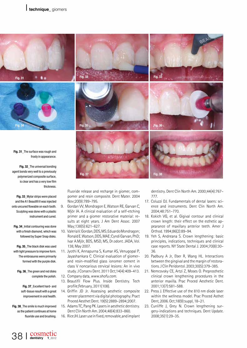
Fig. 31_The surface was rough and
frosty in appearance.
Fig. 32_The universal bonding
agent bonds very well to a previously
polymerized composite surface,
is clear and has a very low film
thickness.
Fig. 33_Mylar strips were placed
and the A1 Beautifil II was injected
onto uncured flowable on each tooth.
Sculpting was done with a plastic
instrument and cured.
Fig. 34_Initial contouring was done
with a finish diamond, which was
followed by Super Snap disks.
Fig. 35_The black disk was used
with light pressure to improve form.
The embrasures were primarily
formed with the purple disk.
Fig. 36_The green and red disks
complete the polish.
Fig. 37_Excellent hard- and
soft-tissue result with a great
improvement in oral health.
Fig. 38_The smile is much improved
as the patient continues at home
fluoride use and brushing.
Fig. 31 Fig. 32 Fig. 33
Fig. 34 Fig. 35
Fig. 37
Fig. 36
Fig. 38

cosmetic dentistry 1_2012
technique_ giomers I
24. Belvedere, PC. Direct bulk placement for posterior composites using an anatomically shaped clear matris creating true anatomic interproximal sur-faces. J Indiana Dent Assoc. 2006;85(1):14–18.
25. Vargas M. Conservative aesthetic enhance-ment of the anterior dentition using a predict-able direct resin protocol. Prac Proced Aesthet Dent.2006;18(8):501–507.
26. Chyz G. Postorthodontic restoration of worn in-cisal edges. Contemp Esthet.10(4):36–39;2006
27. Rosenthal L. The art of tooth shaping and recon-touring. Dent Today. 16(4):1997.
28. Dhull KS, Nandlal B. Effect of low-concentration daily topical fluoride application on fluoride release of giomer and compomer: an in vitro study. J Indian Soc Pedod Prev Dent. 2011 Jan–Mar;29(1):39–45.
29. Dietschi D. Free-hand composite resin restora-tions: a key to anterior aesthetics. Pract Peri-odontics Aesthet Dent. 7(7):15–25;1995.
30. Jackson RD. Understanding the characteristics of naturally shaded composite resins. Pract Proced Aesthet Dent. 15(8):577–585;2003.Christensen GJ. Bonding to dentin and enamel
where does it stand in 2005? J Am Dent Assoc. 136(9):1299–1302;2005
31. Terry DA. Direct composite resin restoration of adolescent class IV tooth fracture: a case report. Prac Perio Aesthet Dent. 12(1):23–29;2000.
32. Fahl, N. The direct/indirect composite resin veneers: a case report. Pract Periodont Aesthet Dent. 1996;8(7):627–638.
I 39
AD
Jack D. Griffin Jr., DMD, MAGD, has a practice in St. Louis county, Miss., where he and his staff have maintained a 50 to 55 percent overhead for 20 years while doing all phases of general dentistry, from high-end cosmetic procedures to everyday restorative and preventive care. Griffin, who has a passion for sharing what he has learned, was awarded diplomat status with the American Board of Aesthetic Dentistry (ABAD), accreditation with the American Academy of Cosmetic Dentistry (AACD) and mastership in the Academy of General Dentistry (AGD). He has published many arti-cles in professional journals and lectured for a variety of dental groups. You may reach him at [email protected] or online at www.eurekasmile.com.
_about the author cosmetic
dentistry


![[Composite Cultures] - CORE](https://static.fdokumen.com/doc/165x107/6325e67de491bcb36c0a86c0/composite-cultures-core.jpg)