
Geographic distribution and evolution of Sindbis virus in Australia
10
Journal of General Virology (1999), 80, 739–748. Printed in Great Britain ................................................................................................................................................................................................................................................................................... Geographic distribution and evolution of Sindbis virus in Australia Leanne M. Sammels, Michael D. Lindsay, Michael Poidinger,† Robert J. Coelen‡ and John S. Mackenzie† Department of Microbiology, The University of Western Australia, Nedlands, Western Australia 6907, Australia The molecular epidemiology and evolution of Sindbis (SIN) virus in Australia was examined. Several SIN virus strains isolated from other countries were also included in the analysis. Two regions of the virus genome were sequenced including a 418 bp region of the E2 gene and a 484 bp region containing part of the junction region and the 5« end of the C gene. Analysis of the nucleotide and deduced amino acid sequence data from 40 SIN virus isolates clearly separated the Paleoarctic/ Ethiopian and Oriental/Australian genetic types of SIN virus. Examination of the Australian strains showed a temporal rather than geographic relationship. This is consistent with the virus having migratory birds as the major vertebrate host, as it allows for movement of virus over vast areas of the continent over a relatively short period of time. The results suggest that the virus is being periodically redistributed over the continent from an enzootic focus of evolving SIN virus. However, SIN virus strains isolated from mosquitoes collected in the south-west of Australia appear to represent a new SIN virus lineage, which is distinct from the Paleoarctic/Ethiopian and Oriental/Australian lineages. Given the widespread geographic dispersal of the Paleoarctic/ Ethiopian and Oriental/Australian lineages, it is surprising that the South-west genetic type is so restricted in its area of circulation. Nucleotide sequence data from the C gene of the prototype strain of the alphavirus Whataroa were also determined. This virus was found to be genetically distinct from the SIN virus isolates included in the present study ; however, it is clearly SIN-like and appears to have evolved from a SIN-like ancestral virus. Introduction Sindbis (SIN) and SIN-like viruses are the most widely distributed of all known arboviruses, with regular isolations occurring in four of the six zoogeographic regions of the world. Human SIN virus infection was considered a minor medical problem from a public health point of view until epidemics in South Africa in 1974 (McIntosh et al., 1976) and Author for correspondence : Leanne Sammels. Fax ›61 8 9346 2912. e-mail lsammels!cyllene.uwa.edu.au † Present address : Department of Microbiology, The University of Queensland, Brisbane, Queensland 4072, Australia. ‡ Present address : Department of Biomedical and Tropical Veterinary Sciences, The James Cook University, Townsville, Queensland 4811, Australia. The GenBank accession numbers of the sequences reported in this paper are AF061205–AF061239 and AF061684–AF071715. later in areas of Northern Europe in the mid 1980s (Skogh & Espmark, 1982 ; Lvov et al., 1984), often involving hundreds of cases, were reported. SIN virus is the most commonly isolated arbovirus from mosquitoes in Australia (Mackenzie et al., 1994). Despite this, human disease has only been reported on two occasions in Australia (Doherty et al., 1969; Guard et al., 1982), although seroepidemiological studies suggest that frequent subclinical infections do occur (Boughton et al., 1984 ; Doherty, 1973 ; Kanamitsu et al., 1979; Liehne et al., 1976). Strain variation or the presence of clinically similar diseases or antigenically related alphaviruses may explain the low in- cidence of clinical infections in humans in Australia. In a study of genetic identity among 15 strains of SIN virus using RNA–RNA hybridizations, Rentier-Delrue & Young (1980) found that two distinct geographic types of SIN virus could be distinguished. These authors suggested that divergent evolution had occurred between European}African and Indian}Far Eastern}Australian types. A subsequent study of antigenic variation among SIN virus isolates from Paleoarctic, 0001-5904 # 1999 SGM HDJ
Transcript of Geographic distribution and evolution of Sindbis virus in Australia

Journal of General Virology (1999), 80, 739–748. Printed in Great Britain. . . . . . . . . . . . . . . . . . . . . . . . . . . . . . . . . . . . . . . . . . . . . . . . . . . . . . . . . . . . . . . . . . . . . . . . . . . . . . . . . . . . . . . . . . . . . . . . . . . . . . . . . . . . . . . . . . . . . . . . . . . . . . . . . . . . . . . . . . . . . . . . . . . . . . . . . . . . . . . . . . . . . . . . . . . . . . . . . . . . . . . . . . . . . . . . . . . . . . . . . . . . . . . . . . . . . . . . . . . . . . . . . . . . . . . . . . . . . . . . . . . . . . . . . . . . . . . . . . . . . . . . . . . . . . . . . . .
Geographic distribution and evolution of Sindbis virus inAustralia
Leanne M. Sammels, Michael D. Lindsay, Michael Poidinger,† Robert J. Coelen‡
and John S. Mackenzie†
Department of Microbiology, The University of Western Australia, Nedlands, Western Australia 6907, Australia
The molecular epidemiology and evolution of Sindbis (SIN) virus in Australia was examined. SeveralSIN virus strains isolated from other countries were also included in the analysis. Two regions of thevirus genome were sequenced including a 418 bp region of the E2 gene and a 484 bp regioncontaining part of the junction region and the 5« end of the C gene. Analysis of the nucleotide anddeduced amino acid sequence data from 40 SIN virus isolates clearly separated the Paleoarctic/Ethiopian and Oriental/Australian genetic types of SIN virus. Examination of the Australian strainsshowed a temporal rather than geographic relationship. This is consistent with the virus havingmigratory birds as the major vertebrate host, as it allows for movement of virus over vast areas ofthe continent over a relatively short period of time. The results suggest that the virus is beingperiodically redistributed over the continent from an enzootic focus of evolving SIN virus. However,SIN virus strains isolated from mosquitoes collected in the south-west of Australia appear torepresent a new SIN virus lineage, which is distinct from the Paleoarctic/Ethiopian andOriental/Australian lineages. Given the widespread geographic dispersal of the Paleoarctic/Ethiopian and Oriental/Australian lineages, it is surprising that the South-west genetic type is sorestricted in its area of circulation. Nucleotide sequence data from the C gene of the prototypestrain of the alphavirus Whataroa were also determined. This virus was found to be geneticallydistinct from the SIN virus isolates included in the present study; however, it is clearly SIN-like andappears to have evolved from a SIN-like ancestral virus.
IntroductionSindbis (SIN) and SIN-like viruses are the most widely
distributed of all known arboviruses, with regular isolationsoccurring in four of the six zoogeographic regions of theworld. Human SIN virus infection was considered a minormedical problem from a public health point of view untilepidemics in South Africa in 1974 (McIntosh et al., 1976) and
Author for correspondence: Leanne Sammels.
Fax 61 8 9346 2912. e-mail lsammels!cyllene.uwa.edu.au
†Present address: Department of Microbiology, The University of
Queensland, Brisbane, Queensland 4072, Australia.
‡Present address: Department of Biomedical and Tropical Veterinary
Sciences, The James Cook University, Townsville, Queensland 4811,
Australia.
The GenBank accession numbers of the sequences reported in this
paper are AF061205–AF061239 and AF061684–AF071715.
later in areas of Northern Europe in the mid 1980s (Skogh &Espmark, 1982 ; Lvov et al., 1984), often involving hundreds ofcases, were reported. SIN virus is the most commonly isolatedarbovirus from mosquitoes in Australia (Mackenzie et al.,1994). Despite this, human disease has only been reported ontwo occasions in Australia (Doherty et al., 1969 ; Guard et al.,1982), although seroepidemiological studies suggest thatfrequent subclinical infections do occur (Boughton et al., 1984 ;Doherty, 1973 ; Kanamitsu et al., 1979 ; Liehne et al., 1976).Strain variation or the presence of clinically similar diseases orantigenically related alphaviruses may explain the low in-cidence of clinical infections in humans in Australia.
In a study of genetic identity among 15 strains of SIN virususing RNA–RNA hybridizations, Rentier-Delrue & Young(1980) found that two distinct geographic types of SIN viruscould be distinguished. These authors suggested that divergentevolution had occurred between European}African andIndian}Far Eastern}Australian types. A subsequent study ofantigenic variation among SIN virus isolates from Paleoarctic,
0001-5904 # 1999 SGM HDJ

L. M. Sammels and othersL. M. Sammels and others
Ethiopian, Oriental and Australian areas demonstrated anti-genic diversion between viruses from the Paleoarctic}Ethiopian and Oriental}Australian regions (Olson & Trent,1985). Genetic studies, by RNase T1 mapping, indicated thatthe primary structures of RNA from viruses isolated in the fourzoogeographic regions were distinct from one another (Olson& Trent, 1985). An RNase T1 mapping study of a limitednumber of Australian SIN virus isolates indicated that the virusmay exist throughout Australia as a single topotype (Brand,1989).
The aim of this study was to examine the geographicdistribution and evolution of SIN virus in Australia bydetermining the nucleotide sequence of regions of the E2 andC genes of a number of SIN virus strains isolated in variousgeographic locations in Australia. Molecular epidemiologicalstudies of another Australian alphavirus, Ross River (RR) virus,have shown that distinct genetic types of the virus areassociated with geographic origin (Faragher et al., 1985 ;Lindsay et al., 1993 ; Sammels et al., 1995). Thus, this study wasundertaken to better understand the genetic variation inalphaviruses and to identify potential mechanisms of spread ofthese viruses in Australia. A limited number of SIN virus strainsisolated in other countries, including Egypt, South Africa,Malaysia and Papua New Guinea (PNG), was also included inthe study to examine the genetic variation of SIN virus indifferent zoogeographic regions of the world. An isolate ofWhataroa (WHA) virus, an alphavirus belonging to theWestern equine encephalitis antigenic complex, which isenzootic in parts of New Zealand (Miles, 1973), was alsoincluded. Neutralization tests have indicated that WHA virusis antigenically related to SIN and SIN-like viruses (Calisher etal., 1988 ; Karabatsos, 1975). Recently, nucleotide sequenceanalysis has shown that WHA virus is SIN-like, although itssequence diverges by about 20% from SIN virus (Weaver et al.,1997).
Methods+ Virus stocks. All SIN virus strains used in this study are listed inTable 1. Virus stocks were prepared on Vero cell (Monkey kidney ; ATCCCCL 81) monolayers at 37 °C at an m.o.i. of ! 0±1 p.f.u. per cell. At 48 hpost-infection, cell culture supernatants were harvested, clarified bycentrifugation at 4000 g for 30 min and stored at ®70 °C.
+ Design and synthesis of oligonucleotide primers. All oligo-nucleotide primers used in this study were synthesized on an automatedDNA synthesizer (380B ; Applied Biosystems) to hybridize to specificlocations of the SIN virus genome, according to the reported sequence(Strauss et al., 1984). The primers are identified according to thenucleotide site at which the 3« end of the primer was designed to anneal.All primers have complementary-sense orientation except those markedwith an asterisk which have plus-sense orientation.
Initially, the E2 gene was chosen for sequencing because the structuralgenes are known to be more variable than the non-structural genes inalphaviruses and also because the E2 gene carries the major antigenicdeterminants. However, numerous attempts to produce cDNA of theregion for several SIN virus isolates failed, suggesting considerable
nucleotide variability. Therefore, a second, more highly conserved regionof the SIN virus genome was targeted for sequencing (Strauss et al., 1984).Sequence data of the 3« end of the highly conserved junction region andthe 5« end of the C gene were obtained for all the selected SIN virusisolates.
+ RT–PCR of cDNA. Details of the methods used to amplify a 482 bpcDNA fragment of the SIN virus E2 gene and a 510 bp cDNA fragmentcontaining part of the junction region and the 5« end of the C gene havebeen described previously (Sellner et al., 1992). Briefly, a 0±5 µl aliquot oftissue culture supernatant was digested with 5 µg proteinase K in a totalvolume of 12±5 µl for 30 min at 42 °C. Proteinase K was inactivated byheating to 94 °C for 5 min. The proteinase K reaction mix was thencombined with the reagents for a one-step RT–PCR reaction in a totalvolume of 25 µl. Each reaction contained 20 pmoles each primer(SIN9170* and SIN9652 for the E2 gene or SIN7575* and SIN8086 forthe C gene), 2 U Taq DNA polymerase, 0±5 U AMV reverse transcriptase,100 µM dNTPs, 2 mM MgCl
#, 2 µg yeast tRNA and 5 µl 5¬ reaction
buffer resulting in final concentrations of 67 mM Tris–HCl (pH 8±8),50 mM KCl, 6 µM EDTA and 0±1% Triton X-100, 0±5 mM DTT. Thetubes were overlaid with 60 µl paraffin oil and incubated in a thermalcycler for 30 min at 42 °C followed by 94 °C for 5 min (to activate andthen inactivate the AMV reverse transcriptase) and then 35 cycles of94 °C for 30 s, 60 °C for 30 s and 72 °C for 60 s, with a final extensionat 72 °C for 7 min.
+ Nucleic acid sequencing and analysis. Nucleic acid sequenceanalysis was performed on an automated Applied Biosystems 373ADNA sequencer. Both strands of the amplified cDNA fragments weresequenced using primers that were identical to those used in the RT–PCRreaction. The purified dsDNA was sequenced on an automated sequencerusing the Prism DyeDeoxy Terminator Cycle Sequencing kit (AppliedBiosystems) and the cycle sequencing parameters used were as describedin the manufacturer’s protocol.
Multiple nucleotide and amino acid sequence alignments wereperformed with the CLUSTAL V program (Higgins & Sharp, 1989).Aligned sequences underwent phylogenetic analysis using programsfrom the PHYLIP package (Felsenstein, 1989). Briefly, distances werecalculated using the Kimura two-parameter method and trees werecalculated using the neighbour-joining method. Bootstrap resampling of1000 replicates was used to place confidence values on groupings withintrees.
ResultsThe 41 SIN virus strains and the WHA isolate (M78)
included in the analysis, with their location and host of origin,are listed in Table 1. The SIN virus strains were isolated fromfour different zoogeographic regions of the world over a 40year period. Thirty-five of these strains were isolated frommosquitoes trapped in various geographic locations inAustralia over a 33 year period. The published sequence of theprototype strain of Ockelbo virus, Edsbyn82 (Shirako et al.,1991), was obtained from GenBank and included in theanalysis.
Nucleotide sequencing studies in the E2 gene
The nucleotide sequences of a 418 bp region of the E2 genewere determined for 32 SIN virus isolates. Five strains of virus,
HEA

Evolutionof
Sindbisvirus
Evolutionof
Sindbisvirus
Table 1. Details of Sindbis and SIN-like virus isolatesAbbreviations used : PNG, Papua New Guinea ; QLD, Queensland, Australia ; NSW, New South Wales, Australia ; Vic, Victoria, Australia ; WA, Western Australia, Australia ; NT, NorthernTerritory, Australia.
No. Isolate name Location Date collected Source (vector or host) Passage history* Supplier†
1 EgAR339 Cairo, Egypt 1953 Culex univittatus 14¬smb, 1¬Vero IM2 EgAR338 Cairo, Egypt 1953 Unavailable 12¬smb, 1¬Vero IM3 SAAR86 South Africa 1954 Culex species 4¬smb, 1¬Vero IM4 M78‡ New Zealand 1962 Culex pervigilans Unavailable DP5 MRE16 Sarawak, Malaysia 1975 Unavailable 6¬mosq, 1¬smb, 1¬Vero IM6 MK6962 Joinjakaka, northern PNG Feb. 1966 Ficalbia flavens 3¬smb, 1¬Vero IM7 M78 New Zealand 1962 Culex pervigilans 3¬smb, 1¬BHK DP8 MRM39 Kowanyama, Cape York Peninsula, far north QLD Apr. 1960 Culex annulirostris 1¬smb, 1¬Vero DP9 16700 Charleville, south-central QLD 1974 Unavailable 3¬smb, 1¬Vero DP
10 RRD764 Ross River Dam, north coast QLD Feb. 1991 Culex annulirostris 1¬C6}36, 1¬BHK BK11 RRD800 Ross River Dam, north coast QLD Feb. 1991 Culex annulirostris 1¬C6}36, 1¬BHK BK12 AS19016 Lachlan River, south-central NSW Feb. 1979 Culex annulirostris 1¬smb, 1¬Vero IM13 BB19153 Griffith, south-central NSW Feb. 1979 Culex annulirostris 1¬smb, 1¬Vero IM14 NL26287 Narran Lake, north-central NSW Apr. 1981 Anopheles amictus 1¬smb, 1¬Vero IM15 RB27266 Redbank Weir, south-west NSW Nov. 1981 Culex annulirostris 1¬smb, 1¬Vero IM16 BH2907 Barmah Forest, north-west Vic Feb. 1974 Culex annulirostris 1¬smb, 1¬Vero IM17 MM840 Lake Moodemere, Vic Feb. 1974 Culex annulirostris 1¬smb, 1¬Vero IM18 BH40503 Barmah Forest, north-west Vic Jan. 1984 Culex annulirostris 1¬smb, 1¬Vero IM19 OR6 Ord River, NE Kimberley, WA 6}6}72 Culex annulirostris 2¬smb, 1¬Vero UWA20 OR339 Ord River, NE Kimberley, WA 30}3}74 Culex annulirostris 2¬smb, 1¬Vero UWA21 OR374 Ord River, NE Kimberley, WA 2}4}74 Culex annulirostris 2¬smb, 1¬Vero UWA22 OR405 Ord River, NE Kimberley, WA 6}4}74 Culex annulirostris 2¬smb, 1¬Vero UWA23 OR437 Ord River, NE Kimberley, WA Mar.}Apr. 1974 Culex annulirostris 2¬smb, 1¬Vero UWA24 OR499 Ord River, NE Kimberley, WA Mar.}Apr. 1974 Aedes normanensis 2¬smb, 1¬Vero UWA25 OR922 Ord River, NE Kimberley, WA June}July 1976 Culex annulirostris 2¬smb, 1¬Vero UWA26 WK465 Camballin, W Kimberley, WA 3}8}79 Culex annulirostris 2¬C6}36, 2¬Vero UWA27 K2673 Lake Argyle, NE Kimberley, WA 22}4}82 Culex annulirostris 2¬C6}36, 2¬Vero UWA28 CX184 Ord River, NE Kimberley, WA 13}6}82 Culex annulirostris 2¬C6}36, 2¬Vero UWA29 K2588 Billiluna, SE Kimberley, WA 21}4}89 Culex annulirostris 2¬C6}36, 2¬Vero UWA30 K1961 Billiluna, SE Kimberley, WA 24}4}89 Aedes eidsvoldensis 2¬C6}36, 2¬Vero UWA31 K2924 Parrys Ck, NE Kimberley, WA 25}4}89 Culex annulirostris 2¬C6}36, 2¬Vero UWA32 K3141 Parrys Ck, NE Kimberley, WA 25}4}89 Culex annulirostris 2¬C6}36, 2¬Vero UWA33 K3028 Nullagine, Pilbara, WA 17}2}90 Aedes pseudonormanensis 2¬C6}36, 2¬Vero UWA34 K3057 Nullagine, Pilbara, WA 19}2}90 Aedes pseudonormanensis 2¬C6}36, 2¬Vero UWA35 K10390 Derby, SW Kimberley, WA 6}4}93 Culex annulirostris 2¬C6}36, 2¬Vero UWA36 K12710 Billiluna, SE Kimberley, WA 20}4}93 Culex annulirostris 2¬C6}36, 2¬Vero UWA37 SW6562 Southern suburbs, Perth, south-west WA 6}2}90 Culex annulirostris 1¬C6}36, 3¬Vero UWA38 SW20055 Peel region, south-west WA 20}1}92 Culex annulirostris 1¬C6}36, 3¬Vero UWA39 SW25713 Leschenault region, south-west WA 13}10}92 Aedes camptorhynchus 1¬C6}36, 3¬Vero UWA40 SW33029 Peel region, south-west WA 5}10}93 Aedes camptorhynchus 1¬C6}36, 3¬BHK UWA41 V620 Darwin, north-central coast NT 6}3}84 Culex annulirostris 1¬C6}36, 3¬BHK RW42 V634 Maratanka, north-central NT 15}3}84 Aedes normanensis 1¬C6}36, 3¬BHK RW
* Passage history, history of each isolate upon arrival in this laboratory. C6}36, C6}36 clone of Aedes albopictus cells ; smb, suckling mouse brain ; Vero, Vero (African green monkey kidney) cells ; and BHK, baby hamster kidney cells.† UWA, Arbovirus Laboratory, Department of Microbiology, University of Western Australia, Nedlands, Australia ; RW, Richard Weir, Berrimah Agricultural Centre, NT, Australia ; IM, Ian Marshall, Department of Biochemistry,Australian National University, Canberra, ACT, Australia ; DP, Debbie Phillips, Laboratory of Microbiology and Pathology, State Health Laboratory, Department of Health, QLD, Australia ; and BK, Brian Kay, Queensland Institute ofMedical Research, QLD, Australia.‡ M78 is a strain of WHA virus.
HEB
L. M. Sammels and othersL. M. Sammels and others
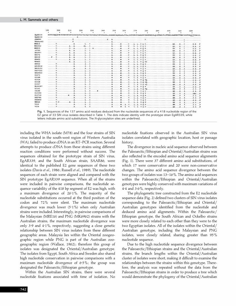
Fig. 1. Sequences of the 137 amino acid residues deduced from the nucleotide sequences of a 418 nucleotide region of theE2 gene of 33 SIN virus isolates described in Table 1. The dots indicate identity with the prototype strain EgAR339, whileletters indicate amino acid substitutions. The N-glycosylation sites are underlined.
including the WHA isolate (M78) and the four strains of SINvirus isolated in the south-west region of Western Australia(WA), failed to produce cDNA in an RT–PCR reaction. Severalattempts to produce cDNA from these strains using differentreaction conditions were performed without success. Thesequences obtained for the prototype strain of SIN virus,EgAR339, and the South African strain, SAAR86, wereidentical to the published E2 gene sequences of these twoisolates (Davis et al., 1986 ; Russell et al., 1989). The nucleotidesequences of each strain were aligned and compared with theSIN prototype EgAR339 sequence. When all of the strainswere included in pairwise comparisons, the nucleotide se-quence variability of the 418 bp segment of E2 was high, witha maximum divergence of 26±1%. The majority of thenucleotide substitutions occurred at the third position of thecodon and 72% were silent. The maximum nucleotidedivergence was much lower (5±1%) when only Australianstrains were included. Interestingly, in pairwise comparisons ofthe Malaysian (MRE16) and PNG (MK6962) strains with theAustralian strains, the maximum nucleotide divergence wasonly 3±9 and 4±1%, respectively, suggesting a close geneticrelationship between SIN virus isolates from these differentgeographic areas. Malaysia lies within the Oriental zoogeo-graphic region while PNG is part of the Australian zoo-geographic region (Wallace, 1962) ; therefore this group ofisolates was designated the Oriental}Australian genotype.The isolates from Egypt, South Africa and Sweden also sharedhigh nucleotide conservation in pairwise comparisons with amaximum nucleotide divergence of 6±5%; the group wasdesignated the Paleoarctic}Ethiopian genotype.
Within the Australian SIN strains, there were severalnucleotide fixations associated with time of isolation. No
nucleotide fixations observed in the Australian SIN virusisolates correlated with geographic location, host or passagehistory.
The divergence in nucleic acid sequence observed betweenthe Paleoarctic}Ethiopian and Oriental}Australian strains wasalso reflected in the encoded amino acid sequence alignments(Fig. 1). There were 37 different amino acid substitutions, ofwhich 17 were conservative and 20 were non-conservativechanges. The amino acid sequence divergence between thetwo groups of isolates was 13–16%. The amino acid sequenceswithin the Paleoarctic}Ethiopian and Oriental}Australiangenotypes were highly conserved with maximum variations of4±4 and 3±6%, respectively.
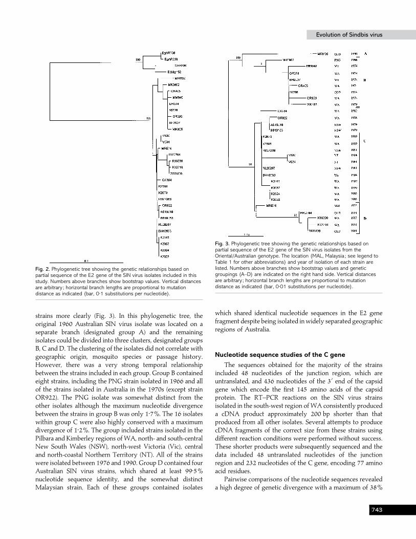
The phylogenetic tree constructed from the E2 nucleotidesequence data (Fig. 2) defined two clusters of SIN virus isolatescorresponding to the Paleoarctic}Ethiopian and Oriental}Australian genotypes identified from the nucleotide anddeduced amino acid alignments. Within the Paleoarctic}Ethiopian genotype, the South African and Ockelbo strainswere more closely related to each other than they were to thetwo Egyptian isolates. All of the isolates within the Oriental}Australian genotype, including the Malaysian and PNGisolates, were closely related, sharing greater than 95%nucleotide sequence.
Due to the high nucleotide sequence divergence betweenthe Paleoarctic}Ethiopian strains and the Oriental}Australianstrains, the branch lengths within the Oriental}Australiancluster of isolates were short, making it difficult to examine therelationships between the strains within this genotype. There-fore, the analysis was repeated without the data from thePaleoarctic}Ethiopian strains in order to produce a tree whichwould demonstrate the phylogeny of the Oriental}Australian
HEC
Evolution of Sindbis virusEvolution of Sindbis virus
Fig. 2. Phylogenetic tree showing the genetic relationships based onpartial sequence of the E2 gene of the SIN virus isolates included in thisstudy. Numbers above branches show bootstrap values. Vertical distancesare arbitrary ; horizontal branch lengths are proportional to mutationdistance as indicated (bar, 0±1 substitutions per nucleotide).
strains more clearly (Fig. 3). In this phylogenetic tree, theoriginal 1960 Australian SIN virus isolate was located on aseparate branch (designated group A) and the remainingisolates could be divided into three clusters, designated groupsB, C and D. The clustering of the isolates did not correlate withgeographic origin, mosquito species or passage history.However, there was a very strong temporal relationshipbetween the strains included in each group. Group B containedeight strains, including the PNG strain isolated in 1966 and allof the strains isolated in Australia in the 1970s (except strainOR922). The PNG isolate was somewhat distinct from theother isolates although the maximum nucleotide divergencebetween the strains in group B was only 1±7%. The 16 isolateswithin group C were also highly conserved with a maximumdivergence of 1±2%. The group included strains isolated in thePilbara and Kimberley regions of WA, north- and south-centralNew South Wales (NSW), north-west Victoria (Vic), centraland north-coastal Northern Territory (NT). All of the strainswere isolated between 1976 and 1990. Group D contained fourAustralian SIN virus strains, which shared at least 99±5%nucleotide sequence identity, and the somewhat distinctMalaysian strain. Each of these groups contained isolates
Fig. 3. Phylogenetic tree showing the genetic relationships based onpartial sequence of the E2 gene of the SIN virus isolates from theOriental/Australian genotype. The location (MAL, Malaysia ; see legend toTable 1 for other abbreviations) and year of isolation of each strain arelisted. Numbers above branches show bootstrap values and geneticgroupings (A–D) are indicated on the right hand side. Vertical distancesare arbitrary ; horizontal branch lengths are proportional to mutationdistance as indicated (bar, 0±01 substitutions per nucleotide).
which shared identical nucleotide sequences in the E2 genefragment despite being isolated inwidely separated geographicregions of Australia.
Nucleotide sequence studies of the C gene
The sequences obtained for the majority of the strainsincluded 48 nucleotides of the junction region, which areuntranslated, and 436 nucleotides of the 3« end of the capsidgene which encode the first 145 amino acids of the capsidprotein. The RT–PCR reactions on the SIN virus strainsisolated in the south-west region of WA consistently produceda cDNA product approximately 200 bp shorter than thatproduced from all other isolates. Several attempts to producecDNA fragments of the correct size from these strains usingdifferent reaction conditions were performed without success.These shorter products were subsequently sequenced and thedata included 48 untranslated nucleotides of the junctionregion and 232 nucleotides of the C gene, encoding 77 aminoacid residues.
Pairwise comparisons of the nucleotide sequences revealeda high degree of genetic divergence with a maximum of 38%
HED

L. M. Sammels and othersL. M. Sammels and others
Fig. 4. Sequences of the 145 amino acid residues deduced from the nucleotide sequences of a 436 nucleotide region of the Cgene from 37 SIN virus isolates and the WHA virus isolate (M78) described in Table 1. The dots indicate identity with theprototype strain EgAR339, while letters indicate amino acid substitutions.
between the Australian 1960 strain MRM39 and the WHAisolate (M78). The sequences in the untranslated junctionregion were highly variable, although the three in-phasetermination codons were maintained in all strains except thefour south-west WA isolates, which have only two in-phasestop codons.
Pairwise comparisons clearly distinguished thePaleoarctic}Ethiopian and Oriental}Australian isolates. Inaddition, a third genotype of strains comprising the fourisolates from the south-west of WA (designated the South-west genotype) could be distinguished in the pairwisecomparisons. The nucleotide sequence of the WHA (M78)virus isolate was substantially different to the sequence of allthe other strains. The level of nucleotide divergence betweenthe Paleoarctic}Ethiopian and the Oriental}Australian isolateswas 24–26±5%. The ranges of divergence between thenucleotide sequences of strains in the South-west genotypeand isolates of the Paleoarctic}Ethiopian and Oriental}Australian genotypes were 16±8–19±4 and 25±4–28±9%, re-spectively, suggesting that the South-west genotype is moreclosely related to the Paleoarctic}Ethiopian isolates than to theOriental}Australian isolates. The WHA virus isolate (M78)appeared to be equally divergent from the Paleoarctic}Ethiopian, Oriental}Australian and South-west isolates withsequence divergence values of 33–34, 33±5–34±9 and 35–35±4%, respectively.
The level of nucleotide divergence observed betweenisolates within each of the three main genotypes was very lowindicating high nucleotide conservation in this region of thecapsid gene. The maximum divergence between the four
strains included in the Paleoarctic}Ethiopian genotype was4%. The Oriental}Australian isolates showed a maximumdivergence of 5±7%. The South-west isolates, which were allobtained from mosquitoes trapped in the south-west region ofWA between 1990 and 1993, shared greater than 99%nucleotide sequence identity.
The deduced amino acid sequence alignments (Fig. 4)included 143 residues of the capsid protein of all the SIN virusstrains except those belonging to the South-west genotype,from which only 77 amino acids were identified. There was atotal of 92 amino acid substitutions, of which 28 wereconservative and 64 were non-conservative changes. Thealignment indicated a highly variable region between residues57 and 96 with numerous substitutions, although there weresubstitutions scattered throughout the entire length of thecapsid fragment. The amino acid divergence values betweenM78 and the Paleoarctic}Ethiopian, Oriental}Australian andSouth-west genotypes were 31±2–32±6, 28–29±4 and 36%,respectively. The maximum amino acid variation between theisolates of the Oriental}Australian genotype was 6±9%. Thevariations between the Oriental}Australian and Paleoarctic}Ethiopian genotypes and between the Oriental}Australian andSouth-west genotypes were 16–21±7 and 20±8–26%, respect-ively. The amino acid variation between the Paleoarctic}Ethiopian and South-west genotypes was somewhat lower at9–11±7%.
The phylogenetic tree constructed from the nucleotidesequence data (Fig. 5) defined three clusters of SIN virusisolates corresponding to the Paleoarctic}Ethiopian, Oriental}Australian and South-west genotypes. The WHA virus isolate
HEE

Evolution of Sindbis virusEvolution of Sindbis virus
Fig. 5. Phylogenetic tree showing the genetic relationships based onpartial sequence of the C gene of the SIN virus isolates and the WHA virusprototype strain included in this study. Numbers above branches showbootstrap values. Vertical distances are arbitrary ; horizontal branch lengthsare proportional to mutation distance (bar, 0±1 substitutions pernucleotide).
(M78), with its unique nucleotide and amino acid sequences,was located on a separate branch of the tree. The tree alsoindicated that isolates belonging to the South-west genotypewere more closely related to strains from the Paleoarctic}Ethiopian genotype than they were to those of the Oriental}Australian genotype. All of the remaining Australian SIN virusisolates were grouped within the Oriental}Australian geno-type. The analysis was repeated without data from thePaleoarctic}Ethiopian and South-west genotypes to produce atree which would demonstrate the phylogeny of the Oriental}Australian strains more clearly (Fig. 6). The analysis placed the1960 Australian SIN virus isolate MRM39 on a separatebranch of the tree (designated group A) and the remainingisolates were divided into three clusters, groups B, C and D.Group B contained the 1966 PNG isolate and nine strainsisolated from various geographic locations in Australia duringthe 1970s. The nucleotide sequences of the Australian isolateswithin this group were highly conserved with a maximumnucleotide divergence of less than 0±5%. The PNG strain wasapproximately 1±5% divergent from the Australian isolates.Group C contained 15 isolates, most of which were isolated inthe late 1970s and the 1980s from widely separated geographic
Fig. 6. Phylogenetic tree showing the genetic relationships based onpartial sequence of the C gene of the SIN virus isolates from theOriental/Australian genotype. The location (MAL, Malaysia ; see legend toTable 1 for other abbreviations) and year of isolation of each strain arelisted. Numbers above branches show bootstrap values and geneticgroupings (A–D) are indicated on the right hand side. Vertical distancesare arbitrary ; horizontal branch lengths are proportional to mutationdistance (bar, 0±01 substitutions per nucleotide).
locations within Australia. The Malaysian strain (MRE16) wasmost closely related to these isolates although it was locatedon a separate branch of the tree. The maximum divergencebetween the isolates in group C was 1±9%. The four strainsincluded in group D, two from northern Queensland (QLD) in1991 (RRD764 and RRD800) and two isolated in Kimberley(WA) in 1993, shared 99% nucleotide sequence identity. Thephylogeny inferred from the C gene sequences (Fig. 6) and E2nucleotide sequences (Fig. 3) of the Oriental}Australian SINvirus isolates was very similar.
DiscussionOur results show that at least two distinct genotypes of
SIN virus are circulating in Australia. One of these, theOriental}Australian genotype, circulates throughout most ofAustralia and in PNG and Sarawak, Malaysia, whereas asecond, previously unrecognized genotype appears to beendemic in a distinct geographic region located in thetemperate south-west of Australia.
The results confirm earlier reports that significant geneticdiversity exists between isolates of SIN virus from thePaleoarctic}Ethiopian and Oriental}Australian zoogeographicregions (Norder et al., 1996 ; Olson & Trent, 1985 ; Rentier-
HEF

L. M. Sammels and othersL. M. Sammels and others
Delrue & Young, 1980). The isolates contained within each ofthe two genotypes were closely related genetically. Onegenotype included most of the Australian isolates and thesingle strains from PNG and Malaysia. These were isolatedover a 33 year period and were obtained from widelydistributed geographic locations including many areas of theAustralian continent. Although all of the isolates within thisgenotype were genetically similar, four genetic subtypes(groups A–D) were identified within the panel of isolates.There is an obvious temporal relationship between isolates inthe different groups which suggests that SIN virus strainscirculating across most of the Australian continent have beenreplaced at various points through time. The first strain of SINvirus isolated in Australia, MRM39, was obtained frommosquitoes trapped in far northern Queensland in 1960. Thegenotype of MRM39 was not observed in any other SIN virusisolates examined, although no Australian strains isolatedbetween 1960 and 1973 were available for inclusion in thisstudy. The results indicated that the group B genetic typepredominated across most of Australia during the 1970s. Theclose genetic relationship observed between these strains andthe 1966 PNG isolate suggests that this genetic type couldhave been introduced into Australia from PNG. Virus strainsisolated in the late 1970s to 1990, from widely separatedgeographic locations in Australia, were identified as membersof group C. Similarly, the most recent isolates included in thestudy, collected in 1991 and 1993, were separated into groupD. The strain isolated in Malaysia in 1975, MRE16, was closelyrelated to the Australian strains in groups C and D. Theseresults suggest that the genetic type represented by group Cmay have been introduced into Australia from the Orientalregion in the 1970s, when group B was still the predominantgenetic type, and subsequently replaced group B across theAustralian continent. The emergence of group D as thedominant genetic type may have occurred as a result of geneticdrift leading to a significant growth advantage or by thereintroduction of a new strain from the Oriental region. Thetemporal clustering of the isolates in the different genetictypes, represented by groups A–D, indicates that the virusmay be maintained in an enzootic focus from where it is rapidlyspread to other regions of Australia. This study could notdetermine whether the enzootic focus is located withinAustralia or another geographic location, such as PNG orMalaysia. However, it appears that the virus is disseminatedover vast geographic areas of Australia with isolates fromwidely separated locations sharing identical nucleotidesequences. Virus isolations and antibody studies have es-tablished that birds are the major vertebrate host in Australia(Doherty et al., 1966). Many species of birds migrate annuallywithin the Australian continent between the north-west andsouth-east regions and north-east and south-east regions ofAustralia (Williams & Williams, 1990). Thus, it seems likelythat such species may play an important role in widelydisseminating SIN virus in Australia.
The similarity of the Malaysian SIN virus isolate to theAustralian isolates in this study is of interest, as Malaysia is inthe Oriental zoogeographic region. Rentier-Delrue & Young(1980) and Olson & Trent (1985), using a different Malaysianisolate of SIN virus, have also demonstrated that the Malaysianisolate was more closely related to Australian isolates than toother isolates from the Oriental zoogeographic region. TheOriental and Australasian zoogeographic regions are separatedby the Wallace Line in the Indo–Australian archipelago, andseroepidemiological studies have indicated that the alphavirusChikungunya virus and the flavivirus Japanese encephalitis (JE)virus may be restricted to the Oriental zoogeographic region,whereas the alphavirus RR virus and flavivirus Murray Valleyencephalitis (MVE) virus are restricted to the Australianzoogeographic region (Mackenzie et al., 1994). Thus, thefinding that SIN virus isolates from Malaysia and Australia aregenetically similar may indicate that the apparent restricteddistribution of these arboviruses (Chikungunya, RR, JE andMVE viruses) may be less rigid than previously believed.Indeed, KUN virus, a flavivirus which is widely distributed inAustralia, has also been isolated from mosquitoes collected inMalaysia (Bowen et al., 1970) and JE virus has recently beenisolated in the Torres Strait of north-eastern Australia (Hannaet al., 1995). Many species of birds migrate annually betweensouth-east Asia and northern Australia (Williams & Williams,1990) providing an opportunity for virus spread between thesegeographic regions by movement of viraemic birds.
The identification of a new distinct genetic type of SINvirus circulating in the south-west region of WA was anunexpected result. At present, we have determined thenucleotide sequence of 48 bases of the junction region and 232bases of the C gene from these strains. Nevertheless, enoughdata were obtained to indicate that these isolates representedan independent enzootic focus in south-west Australia.Analysis of the nucleotide sequences indicated that strainsfrom the South-west genotype are more closely related toPaleoarctic}Ethiopian strains of SIN virus isolates than tothose from the Oriental}Australian region. This findingsuggests that SIN virus circulating in the south-west region ofAustralia may have been introduced, perhaps by a viraemicmigratory bird or travelling human, and evolved under theselective pressures of the ecological conditions in this region.Many of the endemic species of birds in the south-west of WAare territorial and do not move over very large distances(Slater, 1974a, b). It is possible that SIN virus circulating in thesouth-west of Australia has adapted to and is being maintainedin the region by these more sedentary bird species.Alternatively, the virus may have adapted to a new trans-mission cycle utilizing vertebrate hosts with limited mobility,such as small mammals including marsupials. A survey of avianand mammalian species common to the south-west region forantibody to SIN virus may help to identify potential majorhosts of the virus in this region. Surveillance of arbovirusactivity in the south-west region of WA has been undertaken
HEG

Evolution of Sindbis virusEvolution of Sindbis virus
since May 1987 and has involved regular collections ofmosquitoes at various locations. In that time, there have beenover 200 isolations of RR virus but only twelve isolations ofSIN virus. All SIN virus isolates were obtained from mos-quitoes trapped between February 1990 and December 1996(M. D. Lindsay & C. A. Johansen, unpublished results). Thefocus of this program is the surveillance of saltmarsh-breedingmosquito species located in coastal environments where largeoutbreaks of RR virus disease occur. Consequently, saltmarshmosquitoes in the genus Aedes comprise over 80% of the adultmosquito population in the region in which surveillance iscarried out. The low number of isolates of SIN virus comparedto RR virus may indicate that these species are not majorvectors of SIN virus in the region. More extensive genetic andantigenic characterization of these unique SIN strains and otherSIN strains isolated from mosquitoes collected in the south-west region of WA is currently in progress.
The genetic information obtained from the WHA virusprototype strain (M78) confirms previous antigenic and geneticstudies which indicated that the virus was distantly related toSIN and SIN-like viruses (Calisher et al., 1988 ; Weaver et al.,1997). The results of phylogenetic analysis suggest that WHAmay have evolved in isolation from an ancestral SIN-like viruswhich may have originally been introduced into New Zealandby the arrival of viraemic birds.
We thank those researchers who provided many of the virus isolatesused in the study (listed in Table 1). Thanks also to Ms Cheryl Johansenand Dr Annette Broom for their assistance with isolation and identi-fication of the Western Australian SIN virus strains. This work wasfunded by the National Health and Medical Research Council of Australiaand the Health Department of Western Australia.
ReferencesBoughton, C. R., Hawkes, R. A., Naim, H. M., Wild, J. & Chapman, B.(1984). Arbovirus infections in humans in New South Wales. Sero-epidemiology of the alphavirus group of togaviruses. Medical Journal ofAustralia 141, 700–704.
Bowen, T. T. W., Simpson, D. I. H., Platt, G. S., Way, H. J., Smith,C. E. G., Ching, C. Y. & Casals, J. (1970). Arbovirus infections inSarawak : the isolation of Kunjin virus from mosquitoes of the Culexpseudovishnui group. Annals of Tropical Medicine and Parasitology 64,263–268.
Brand, T. N. (1989). Molecular epidemiology of Sindbis virus. Honoursthesis, University of Western Australia.
Calisher, C. H., Karabatsos, N., Lazuick, J. S., Monath, T. P. & Wolff,K. L. (1988). Re-evaluation of the western equine encephalitis antigeniccomplex of alphaviruses (family Togaviridae) as determined byneutralization tests. American Journal of Tropical Medicine and Hygiene 38,447–452.
Davis, N. L., Fuller, F. J., Dougherty, W. G., Olmsted, R. A. & Johnston,R. E. (1986). A single nucleotide change in the E2 glycoprotein gene ofSindbis virus affects penetration rate in cell culture and virulence inneonatal mice. Proceedings of the National Academy of Sciences, USA 83,6771–6775.
Doherty, R. L. (1973). Surveys of haemagglutination-inhibiting anti-
body to arboviruses in Aboriginal and other population groups innorthern and eastern Australia, 1968–1971. Transactions of the RoyalSociety of Tropical Medicine and Hygiene 67, 197–205.
Doherty, R. L., Gorman, B. M., Whitehead, R. H. & Carley, J. G. (1966).Studies of arthropod-borne virus infections in Queensland V. Survey ofantibodies to group A arboviruses in man and other animals. AustralianJournal of Experimental Biology and Medical Science 44, 365–378.
Doherty, R. L., Bodey, A. S. & Carew, J. W. (1969). Sindbis virusinfection in Australia. Medical Journal of Australia 2, 1016–1017.
Faragher, S. G., Marshall, I. D. & Dalgarno, L. (1985). Ross River virusgenetic variants in Australia and the Pacific Islands. Australian Journal ofExperimental Biology and Medical Science 63, 473–488.
Felsenstein, J. (1989). PHYLIP : phylogeny inference package (version3.2). Cladistics 5, 164–166.
Guard, R. W., McAuliffe, M. J., Stallman, N. D. & Bramston, B. A.(1982). Haemorrhagic manifestations with Sindbis infection. CaseReport. Pathology 14, 89–90.
Hanna, J. N., Ritchie, S. A., Phillips, D. A., Shield, J., Bailey, M. C.,Mackenzie, J. S., Poidinger, M., McCall, B. J. & Mills, P. J. (1995). Anoutbreak of Japanese encephalitis in the Torres Strait, Australia. MedicalJournal of Australia 165, 256–60.
Higgins, D. G. & Sharp, P. M. (1989). Fast and sensitive multiplesequence alignments on a microcomputer. Computer Applications in theBiosciences 5, 151–153.
Kanamitsu, M., Taniguchi, K., Urasawa, S., Ogata, T., Wada, Y., Wada,Y. & Saroso, J. S. (1979). Geographic distribution of arbovirusantibodies in indigenous human populations in the Indo–Australianarchipelago. American Journal of Tropical Medicine and Hygiene 28,351–363.
Karabatsos, N. (1975). Antigenic relationships of group A arbovirusesby plaque reduction neutralization testing. American Journal of TropicalMedicine and Hygiene 24, 527–532.
Liehne, C. G., Stanley, N. F., Alpers, M. P., Paul, S., Liehne, P. F. S. &Chan, K. H. (1976). Ord River arboviruses – serological epidemiology.Australian Journal of Experimental Biology and Medical Science 54, 505–512.
Lindsay, M. D. A., Coelen, R. J. & Mackenzie, J. S. (1993). Geneticheterogeneity among isolates of Ross River virus from differentgeographical regions. Journal of Virology 67, 3576–3585.
Lvov, D. K., Berezina, L. K., Yakovlev, B. I., Aristova, V. A., Gushchina,E. L., Lvov, S. D., Myasnikova, I. A., Skvortsova, T. M., Gromashevsky,V. L., Gushchin, B. V., Sidorova, G. A., Klimenko, S. M., Khutoretskaya,N. V. & Khizhnyakova, T. M. (1984). Isolation of Karelian fever agentfrom Aedes communis mosquitoes. Lancet 2, 399.
McIntosh, B. M., Jupp, P. G., Dos Santos, I. & Meenehan, G. M. (1976).Epidemics of West Nile and Sindbis viruses in South Africa with Culexunivittatus Theobold as vector. South African Journal of Science 72, 295.
Mackenzie, J. S., Smith, D. W., Ellis, T. M., Lindsay, M. D., Broom,A. K., Coelen, R. J. & Hall, R. A. (1994). Human and animal arboviraldiseases in Australia. Recent Advances in Microbiology 2, 1–91.
Miles, J. A. R. (1973). The ecology of Whataroa virus, an alphavirus, inSouth Westland, New Zealand. Journal of Hygiene 71, 701–713.
Norder, H., Lundstrom, J. O., Kozuch, O. & Magnius, L. O. (1996).Genetic relatedness of Sindbis virus strains from Europe, Middle East, andAfrica. Virology 222, 440–445.
Olson, K. & Trent, D. W. (1985). Genetic and antigenic variationsamong geographical isolates of Sindbis virus. Journal of General Virology66, 797–810.
Rentier-Delrue, F. & Young, N. A. (1980). Genomic divergence amongSindbis virus strains. Virology 106, 59–70.
HEH

L. M. Sammels and othersL. M. Sammels and others
Russell, D. L., Dalrymple, J. M. & Johnston, R. E. (1989). Sindbis virusmutations which coordinately affect glycoprotein processing,penetration, and virulence in mice. Journal of Virology 63, 1619–1629.
Sammels, L. M., Coelen, R. J., Lindsay, M. D. & Mackenzie, J. S.(1995). Geographic distribution and evolution of Ross River virus inAustralia and the Pacific Islands. Virology 212, 20–29.
Sellner, L. N., Coelen, R. J. & Mackenzie, J. S. (1992). A one-tube, onemanipulation RT–PCR reaction for detection of Ross River virus. Journalof Virological Methods 40, 255–264.
Shirako, Y., Niklasson, B., Dalrymple, J. M., Strauss, E. G. & Strauss,J. H. (1991). Structure of the Ockelbo genome and its relationship toother Sindbis viruses. Virology 182, 753–764.
Skogh, M. & Espmark, A. (1982). Ockelbo disease : epidemic arthritis-exanthema syndrome in Sweden caused by Sindbis-virus like agent.Lancet 1, 795.
Slater, P. (1974a). A Field Guide to Australian Birds. Volume I. Non-passerines. Australia : Rigby.
Slater, P. (1974b). A Field Guide to Australian Birds. Volume II. Passerines.Australia : Rigby.
Strauss, E. G., Rice, C. M. & Strauss, J. H. (1984). Complete nucleotidesequence of the genomic RNA of Sindbis virus. Virology 133, 92–110.
Wallace, A. R. (1962). On zoological regions. In The GeographicalDistribution of Animals, vol. 1, pp. 50–82. New York : Hafner Publishing.
Weaver, S. C., Kang, W., Shirako, Y., Rumenapf, T., Strauss, E. G. &Strauss, J. H. (1997). Recombinational history and molecular evolutionof Western equine encephalomyelitis complex alphaviruses. Journal ofVirology 71, 613–623.
Williams, T. C. & Williams, M. C. (1990). The orientation of transoceanicmigrants. In Bird Migration : Physiology and Ecophysiology, pp. 7–21. Editedby E. Gwinner. Berlin : Springer-Verlag.
Received 14 August 1998; Accepted 4 November 1998
HEI