Genotype-phenotype study of familial haemophagocytic lymphohistiocytosis type 3
33
GENOTYPE-PHENOTYPE STUDY OF FAMILIAL HEMOPHAGOCYTIC LYMPHOHISTIOCYTOSIS TYPE 3 Elena Sieni 1 , Valentina Cetica 1 , Alessandra Santoro 2 , Karin Beutel 1,8 , Elena Mastrodicasa 3 , Marie Meeths 4,5 , Benedetta Ciambotti 1 , Francesca Brugnolo 1 , Udo zur Stadt 6 , Daniela Pende 7 , Lorenzo Moretta 8 , Gillian M. Griffiths 9 , Jan-Inge Henter 4 , Gritta Janka 10 , Maurizio Aricò 1 1. Department Pediatric Hematology Oncology, Azienda Ospedaliero-Universitaria Meyer, Florence, Italy 2. U.O. Ematologia I, A.O. Ospedali Riuniti Villa Sofia-Cervello, Palermo, Italy 3. S.C. di Oncoematologia Pediatrica con Trapianto di CSE, Ospedale “S.M. della Misericordia” A.O. Perugia, Italy. 4. Childhood Cancer Research Unit, Department of Women´s and Children´s Health, Karolinska Institutet, Karolinska University Hospital, Stockholm, Sweden; 5. Clinical Genetics Unit, Department of Molecular Medicine and Surgery, Karolinska Institutet, Karolinska University Hospital, Stockholm, Sweden 6. Research Institute Children’s Cancer Center, Hamburg, Germany 7. Istituto Nazionale per la Ricerca sul Cancro, Genoa, Italy 8. IRCCS Istituto Giannina Gaslini, Genoa, Italy 9. Cambridge Institute for Medical Research, Addenbrooke’s Hospital, Cambridge, UK CB2 0XY 10. Pediatric Hematology and Oncology, University Medical Center Hamburg- Eppendorf, Hamburg, Germany Word count: 4479 Abstract word count 253 Corresponding author: Maurizio Aricò Direttore Dipartimento Oncoematologia Pediatrica Azienda Ospedaliero-Universitaria Meyer Viale Pieraccini, 24 50139 Firenze tel. +39 055 5662739 Fax +39 055 5662746 [email protected] peer-00601562, version 1 - 19 Jun 2011 Author manuscript, published in "Journal of Medical Genetics 48, 5 (2011) 343" DOI : 10.1136/jmg.2010.085456
-
Upload
independent -
Category
Documents
-
view
0 -
download
0
Transcript of Genotype-phenotype study of familial haemophagocytic lymphohistiocytosis type 3

1
GENOTYPE-PHENOTYPE STUDY OF FAMILIAL HEMOPHAGOCYTIC LYMPHOHISTIOCYTOSIS TYPE 3
Elena Sieni1, Valentina Cetica1, Alessandra Santoro2, Karin Beutel1,8, Elena
Mastrodicasa3, Marie Meeths4,5, Benedetta Ciambotti1, Francesca Brugnolo1, Udo zur
Stadt6, Daniela Pende7, Lorenzo Moretta8, Gillian M. Griffiths9, Jan-Inge Henter4, Gritta
Janka10, Maurizio Aricò1
1. Department Pediatric Hematology Oncology, Azienda Ospedaliero-Universitaria
Meyer, Florence, Italy 2. U.O. Ematologia I, A.O. Ospedali Riuniti Villa Sofia-Cervello, Palermo, Italy 3. S.C. di Oncoematologia Pediatrica con Trapianto di CSE, Ospedale “S.M. della
Misericordia” A.O. Perugia, Italy. 4. Childhood Cancer Research Unit, Department of Women´s and Children´s Health,
Karolinska Institutet, Karolinska University Hospital, Stockholm, Sweden; 5. Clinical Genetics Unit, Department of Molecular Medicine and Surgery, Karolinska
Institutet, Karolinska University Hospital, Stockholm, Sweden 6. Research Institute Children’s Cancer Center, Hamburg, Germany 7. Istituto Nazionale per la Ricerca sul Cancro, Genoa, Italy 8. IRCCS Istituto Giannina Gaslini, Genoa, Italy 9. Cambridge Institute for Medical Research, Addenbrooke’s Hospital, Cambridge, UK
CB2 0XY 10. Pediatric Hematology and Oncology, University Medical Center Hamburg-
Eppendorf, Hamburg, Germany
Word count: 4479
Abstract word count 253
Corresponding author: Maurizio Aricò Direttore Dipartimento Oncoematologia Pediatrica Azienda Ospedaliero-Universitaria Meyer Viale Pieraccini, 24 50139 Firenze tel. +39 055 5662739 Fax +39 055 5662746 [email protected]
peer
-006
0156
2, v
ersi
on 1
- 19
Jun
201
1Author manuscript, published in "Journal of Medical Genetics 48, 5 (2011) 343"
DOI : 10.1136/jmg.2010.085456

2
ABSTRACT (253 words) Background: Mutations of UNC13D are causative for FHL3 (OMIM 608898). We present a genotype-phenotype study of 84 FHL3 patients. Methods: A consortium of 3 countries planned to pool in a common database data on presenting features and mutations from individual patients with biallelic UNC13D mutations. Results: 84 FHL3 patients (median age: 4.1 months) were reported from Florence, Italy (n=54), Hamburg, Germany (n=18), Stockholm, Sweden (n=12). Their ethnic origin was: Caucasian, n=57, Turkish, n=10, Asian, n=7, Hispanic, n=4, African, n=3 (not reported, n=3). Thrombocytopenia was present in 96%, splenomegaly in 95%, fever in 89%. Central nervous system was involved in 49/81 (60%) patients versus 36% in FHL2 (p=0.001). The combination of fever, splenomegaly, thrombocytopenia and hyperferritinemia was present in 71%. CD107a expression, NK activity and Munc13-4 protein expression were absent or reduced in all but one of the evaluated patients. We observed 54 different mutations, including 15 novel ones: 19 missense, 14 deletions or insertions, 12 nonsense, 9 splice errors. None was specific for ethnic groups. Patients with two disruptive mutations were younger than patients with two missense mutations (p<0.001), but older than comparable FHL2 patients (p=0.001). Conclusion. UNC13D mutations are scattered over the gene. Ethnic-specific mutations were not identified. CNS involvement is more frequent than in FHL2; in patients with FHL3 and disruptive mutations, age at diagnosis is significantly higher than in FHL2. The combination of fever, splenomegaly, thrombocytopenia and hyperferritinemia appears to be the most easily and frequently recognized clinical pattern and their association with defective granule release assay may herald FHL3.
peer
-006
0156
2, v
ersi
on 1
- 19
Jun
201
1

3
INTRODUCTION
Hemophagocytic lymphohistiocytosis (HLH) is a genetically heterogeneous disorder
characterized by a hyperinflammatory syndrome with fever, hepatosplenomegaly,
cytopenia, and frequently also central nervous system involvement.1 Bone marrow
aspiration shows haemophagocytosis by activated macrophages. In most cases the
natural course of HLH is rapidly fatal within a few weeks, unless appropriate immune
suppressive and cytoreductive treatment by agents including corticosteroids, cyclosporine,
etoposide, anti-thymocyte globuline, can obtain transient disease control.2,3 So far,
haematopoietic stem cell transplantation appears to be the only curative treatment. 4-8
Differential diagnosis of HLH may be difficult.9 To this purpose, diagnostic
guidelines for HLH have been established by the Histiocyte Society.10,11 In particular,
demonstration of frequent association with common pathogens, together with evidence of
impaired natural killer cytotoxic activity, provided the rationale for considering HLH as a
selective immune deficiency.12-14 Starting from the original report by Farquhar et al. in
1952,15 autosomal recessive inheritance was proposed and then confirmed as the
common mode of inheritance for the familial form of HLH (FHL).
Linkage analysis identified a candidate genomic region on chromosome 9q21.3–22
(FHL1, OMIM 603552).16 However, the causative gene responsible for the disease has not
yet been identified. A simultaneous report established a linkage with another region,
10q21–22,17 in which the perforin (PRF1) gene was identified as responsible for a relevant
proportion of cases of FHL (FHL2, OMIM 603553).18 In patients with FHL2, PRF1
mutations reduce or abolish the synthesis of the perforin protein, resulting in an
impairment of the granule-mediated cytotoxic machinery of NK and T cells.19–22 In 2003 a
third locus, 17q25, was reported in linkage with FHL (FHL3, OMIM 608898).23 The
involved gene UNC13D encodes a protein named Munc13-4 which is thought to contribute
to the priming of the secretory granules before they fuse into the plasma cell membrane.
peer
-006
0156
2, v
ersi
on 1
- 19
Jun
201
1

4
Mutations in this gene impair the delivery of the effector proteins perforin and granzymes
into the target cells, resulting in defective cellular cytotoxicity and a clinical picture that
appears identical to that associated with PRF1 mutations. On the basis of a genome-wide
screening in a highly consanguineous Kurdish family with FHL, a fourth chromosomal
region (6q24) has been reported (FHL4, OMIM 603552).24 Mutations of the syntaxin 11
gene, mapped in this region, are thought to alter intracellular vesicle trafficking of the
phagocytic system.25 Very recently, zur Stadt et al. have allocated a novel FHL type, FHL-
5 (OMIM 613101), to a 1 Mb region on chromosome 19p using high resolution SNP
genotyping in eight unrelated FHL patients from consanguineous families. They have
identified mutations in STXBP2, encoding syntaxin binding protein 2 (Munc18-2) a protein
involved in the regulation of vesicle transport to the plasma membrane. The 12 patients
with FHL-5 originated from Turkey, Saudi Arabia, and Central Europe.26 Almost
simultaneously, a similar report was provided by Côte et al.27
The exact contribution of the various mutations in FHL-related genes can only be
evaluated in large cohorts. A genotype-phenotype study of FHL2, due to PRF1 mutations,
was reported a few years ago by an international consortium for the Histiocyte Society.28
Nothing comparable has been performed so far for FHL3, the other large subgroup of this
disease. For this purpose, we pooled data from three European referral centres, allowing
collection of the largest series of patients with FHL3.
peer
-006
0156
2, v
ersi
on 1
- 19
Jun
201
1

5
METHODS
Data collection
The consortium established between Italy, Germany and Sweden pooled in a common
database data on ethnicity, family history, presenting features, mutations and cytotoxic
function from individual patients with FHL3 diagnosed on the basis of documented biallelic
UNC13D mutations. Hemophagocytic Lymphohistiocytosis was defined by the diagnostic
criteria established by the Histiocyte Society.10,11 Central nervous system (CNS)
involvement was defined as the presence of at least one of the following items: neurologic
symptoms, CSF pleocytosis (≥5 cells/mm3), elevated CSF protein (≥30 mg/dl), MRI
alteration. All data were stored in a common database and analysed. The completeness of
the data was: ethnicity, 95%; consanguinity, 95%; age at diagnosis, 98%; persistent fever,
96%; splenomegaly, 96%; central nervous system (CNS) disease, 95%; haemoglobin
85%; neutrophils, 89%; platelets, 92%; triglycerids, 75%; fibrinogen, 70%; ferritin, 67%;
haemophagocytosis, 96%; CD107 expression, 36%; Munc protein expression, 16%; NK
activity, 53%.
Informed consent for the genetic study and the data collection was obtained from the
parents or legal guardian at the participating centres. The study was approved by the local
IRB at all the participating centres.
UNC13D gene analysis
Genomic DNA was isolated from peripheral blood samples using BioRobot® EZ1
Workstation (Qiagen, Jesi Italy). Some samples were retrieved from our DNA library of
patients previously diagnosed.1,19,28-30 The 32 coding exons and exon-intron boundaries of
Munc13-4 gene were amplified and directly sequenced, in both directions, with the
BigDye® Terminator Cycle Sequencing Ready Reaction Kit (Applied Biosystems, Foster
City, CA, USA). Amplification reactions were performed with 60 ng of DNA, 10 ng of each
peer
-006
0156
2, v
ersi
on 1
- 19
Jun
201
1
6
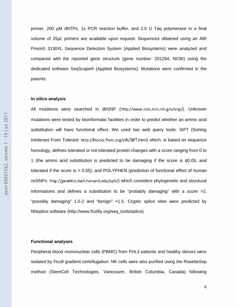
primer, 200 µM dNTPs, 1x PCR reaction buffer, and 2.5 U Taq polymerase in a final
volume of 25µl; primers are available upon request. Sequences obtained using an ABI
Prism® 3130XL Sequence Detection System (Applied Biosystems) were analyzed and
compared with the reported gene structure (gene number: 201294, NCBI) using the
dedicated software SeqScape® (Applied Biosystems). Mutations were confirmed in the
parents.
In silico analysis
All mutations were searched in dbSNP (http://www.ncbi.nlm.nih.gov/snp/). Unknown
mutations were tested by bioinformatic facilities in order to predict whether an amino acid
substitution will have functional effect. We used two web query tools: SIFT (Sorting
Intolerant From Tolerant: http://blocks.fhcrc.org/sift/SIFT.html) which, is based on sequence
homology, defines tolerated or not tolerated protein changes with a score ranging from 0 to
1 (the amino acid substitution is predicted to be damaging if the score is ≤0.05, and
tolerated if the score is > 0.05); and POLYPHEN (prediction of functional effect of human
nsSNPs: http://genetics.bwh.harvard.edu/pph/) which considers phylogenetic and structural
informations and defines a substitution to be “probably damaging” with a score >2,
“possibly damaging” 1.5-2 and “benign” <1.5. Cryptic splice sites were predicted by
NNsplice software (http://www.fruitfly.org/seq_tools/splice).
Functional analyses
Peripheral blood mononuclear cells (PBMC) from FHL3 patients and healthy donors were
isolated by Ficoll gradient centrifugation. NK cells were also purified using the RosetteSep
method (StemCell Technologies, Vancouver, British Columbia, Canada) following
peer
-006
0156
2, v
ersi
on 1
- 19
Jun
201
1

7
manufacturer’s instructions. NK cells were cultured on irradiated feeder cells in the
presence of 2 μg/mL phytohemagglutinin (Sigma-Aldrich, Irvine, UK) and 100U/mL rIL-2
(Proleukin, Chiron Corp., Emeryville, USA) to obtain high numbers of polyclonal activated
NK cell populations. To analyse the cytolytic activity in 4-h 51Cr-release assays, PBMC
were tested against K562, while activated NK cells were tested against the HLA-class I- B-
EBV cell line 721.221, demonstrated to be suitable effector/target combinations to reveal
cytolytic defect of FHL3 patients.30 E:T ratios ranging from 100:1 to 1:1 were used for
PBMC as effector cells, while from 8:1 to 0.5:1 for activated NK. Lytic units (LU) at 30%
lysis were calculated. PBMC and activated NK cells were also tested in degranulation
assay quantifying cell surface CD107a expression upon co-culture with K562, as
previously described.30 All reagents were from BD Biosciences (Oxford, UK). Briefly, anti-
CD107a-PE mAb was added prior to incubation of 3 hours at 37° C in 5% CO2.
Thereafter, the cells were stained with anti-CD56-APC and anti-CD3-PerCP mAb and
analysed by flow cytometry (FACSCalibur, Becton Dickinson). Surface expression of
CD107a was assessed in the CD3- CD56+ cells. Results are reported as ΔCD107a (i.e. %
CD107a+ cells of stimulated - % CD107a+ cells of unstimulated sample).
Western blot analysis
Western blot analysis of Munc13-4 protein was performed as previously described.29
Genotype-phenotype correlations
To explore the correlations between different mutations and clinical, laboratory and
functional parameters, UNC13D mutations were classified according to their functional
impact. The group of disruptive mutations included the nonsense, frameshift and all splice
errors, except c.952-1G>A and c.2626-1G>A (Santoro A, Aricò M., data not shown), that
did not alter the frame; furthermore, the single nucleotide substitution c.1847A>G, based
on our previous findings of its capacity to induce a splicing error only in selected clones,31
peer
-006
0156
2, v
ersi
on 1
- 19
Jun
201
1

8
was not included in the group of disruptive mutations. Based on this, the 84 patients were
classified into 3 groups: group 1, defined by missense mutations only (n=17); group 2,
including patients with one disruptive and one missense mutation (n=18); group 3,
including patients with biallelic disruptive mutations (n=49).
In order to try to define differences between FHL2 and FHL3, data from the current cohort
of patients with FHL3 were compared with available data from the previously reported
cohort with FHL2.28
Statistical analysis
Statistical significance of the differences between the ages at the disease onset and the
quantitative evaluation of the cytoxicity (in lytic units) between groups of patients with
different mutations, was calculated by Student t test; to compare the differences in the
proportions of CNS involvement, Chi-Square test was used. P value ≤0.05 was considered
as significant. Statistical analysis of the functional data comparing FHL3 and healthy
donors was performed using one-way ANOVA followed by Bonferroni’s Multiple
Comparison test, by GraphPad Prism 5 Software.
peer
-006
0156
2, v
ersi
on 1
- 19
Jun
201
1

9
RESULTS
Study population
A total of 84 patients from 69 unrelated families were reported from the following reference
centres: Florence, Italy, n=54; Hamburg, Germany, n=18; Stockholm, Sweden, n=12. They
had been diagnosed between 1981 and 2009. Some of these patients had been previously
reported as part of single-centre series.29-33 The ethnic origin was as follows: Caucasian,
n=58; Turkish, n=10; Asian, n=7; Hispanic, n=4; African, n=3 (not reported, n=2). Male:
Female ratio was 1.2:1.
Presenting features
Main presenting features are summarized in Table 1.
The median age at the diagnosis was 4.1 months, with a range of 1 day to 18.8 years
(quartiles: 2.5, 4.1 and 18.4 months). Consanguinity was reported in 28 of 80 (35%)
patients.
The current diagnostic criteria for HLH were fulfilled – i.e. they had at least five of the eight
items proposed 11 – by 58 of the 84 (69%) patients; of the remaining 26 patients, 15 had a
positive family history, 4 had related parents, 4 had incomplete data, and 3 fulfilled less
than 5/8 criteria.
In particular, among those for whom the information was available, fever was present in
89% of patients, splenomegaly in 96%, bi-cytopenia in 88%, hypertriglyceridemia and/or
hypofibrinogenemia in 95%, haemophagocytosis in 78%, hyperferritinemia in 77%, and
low or absent NK cell activity in 98% (table 1).
The combination of fever, splenomegaly and thrombocytopenia was present in 65 of 77
patients (84%) evaluable for these three parameters; of them, 55 had information on
ferritin level, which turned to be elevated in 39 (71%); otherwise, 59 of the 78 had
peer
-006
0156
2, v
ersi
on 1
- 19
Jun
201
1

10
information on fibrinogen level, which turned to be reduced in 33 (56%); finally, all of the
78 had information on hemophagocytosis, which turned to be present in 54 (69%) (Figure
1).
CNS involvement was reported in 60% of the patients: 30/80 (37%) had neurologic
symptoms, 35/51 (69%) had ≥5 cells in the CSF, 22/31 (71%) elevated CSF protein, and
15/25 (60%) MRI alterations.
UNC13D mutations
A total of 54 different mutations were observed in the 84 patients from 69 families.
Nineteen were missense mutations, 14 deletions or insertions, 12 nonsense and 9 splice
errors (Fig. 2).
A total of 23 different mutations were observed at the homozygous state in 42 individuals
from 36 unrelated families; their presenting features are summarized in table 2. The
remaining 42 patients from 33 unrelated families were compound heterozygous. The most
frequent mutations are described below.
The c.2346_2349delGGAG mutation, causing a frame shift at p.R782, was the most
common mutation; it was identified in a total of 19 patients from 15 families, all Caucasian
but one Turkish (not reported, n=1). In three homozygous patients the ages at the
diagnosis were 2, 3 and 3 months; only one of them had CNS involvement. NK activity
was reduced in the only patient analysed.
The c.753+1G�T mutation, resulting in a splice error, was found in 19 patients from 11
families (15%), 9 Caucasian, one Asian and one Hispanic. Nine homozygous patients had
a median age at the diagnosis of 3 months (range, 2 to 12); 4 of them had CNS
involvement. CD107a expression was absent in two and reduced in two patients analysed,
NK activity was absent in three and reduced in three patients tested.
peer
-006
0156
2, v
ersi
on 1
- 19
Jun
201
1

11
The p.E616G missense mutation, resulting from the c.1847A>G nucleotide change, was
found in 8 individuals from 6 families of Caucasian origin. Four patients carried the
mutation at the homozygous state: age at diagnosis was 99 and 139 months in two
unrelated subjects, and 6 and 226 months in two siblings. Three of the four had CNS
involvement. CD107a expression was reduced in the only patient tested; NK activity was
reduced in all the three patients tested.
The p.R928C missense mutation (c.2782C>T) was identified in 8 patients from 7 unrelated
families, 6 Caucasian and one Hispanic. This mutation falls in the C2B domain; its
predicted impact is controversial, i.e. it may be tolerated according to SIFT, or potentially
deleterious according to POLYPHEN. It was observed in three patients who also carried
two additional mutations; one patient with this as second mutation has defective NK
activity.
The nonsense mutation p.R273X (c.817C>T) was identified in six patients, including one
pair of homozygous Caucasian siblings, both diagnosed at 2 months, and two additional
heterozygous pairs, one Hispanic and one Caucasian. NK activity was absent in one
patient tested.
The splice error c.1389+1G>A was identified in 4 unrelated patients (3 Caucasian, 1 origin
not reported). All were compound heterozygous with different frameshift (n=3) or nonsense
(n=1) mutations.
Novel mutations
Fifteen novel mutations were identified in 15 patients; three were nonsense and five
deletion/insertion mutations (Figure 2, tables 2 and 3), while the remaining seven were
missense mutations. The p.I140T missense mutation (c.419T>C) falls in the C2A domain
and is predicted to be damaging. The p.S398L missense mutation, resulting from the
c.1193C>T nucleotide change, falling outside the functional domains and predicted to be
peer
-006
0156
2, v
ersi
on 1
- 19
Jun
201
1

12
not tolerated, was identified in a 70-month Caucasian girl (UPN 257), with reduced NK
activity. The p.L647P missense mutation (c.1940T>C) falls in the MHD1 domain and is
predicted to be damaging. It was found in a 168-month male (UPN 397) presenting with
typical FHL, reduced CD107 expression and NK-activity. The p.R727Q missense mutation
(c.2180G>A) falls outside any functional domain and is predicted to be tolerated by SIFT
and benign by Polyphen (score 0.353). It was observed in a compound heterozygous
patient with defective degranulation. The p.A859T missense mutation (c.2575G>A) falls in
the MHD2 domain and is predicted to be not tolerated. The p.E1017K missense mutation
(c.3049G>A) falls in the C2B domain and is predicted to be not tolerated. The p.L1058P
missense mutation (c.3173T>C) falls within the C-terminal domain and is predicted to be
damaging. None of the missense mutations resulted in cryptic splice sites according to
NNsplice software.
Analysis of control subjects
None of the following novel mutations were found in 100 healthy Caucasian control
subjects: c.403insC, c.419T>C, c.1193C>T, c.1387C>T, c.1822del12, c.1940T>C,
c.2057C>A, c.2180G>A, c.2212C>T, c.2437_2439delAACinsT, c.2477_2480delTCAC,
c.2575G>A, c.3049G>A, c.3082delC, c.3173T>C.
Functional study
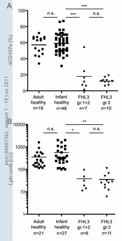
Granule release capacity was significantly reduced in 29 of the 30 patients tested. Of them
17 had quantitative evaluation, analysing activated NK cells upon co-culture with K562,
with a mean value of 14.5% of ΔCD107a+ cells (SD 11.7, SE 2.8). This value was
significantly inferior to that of healthy controls, both adults and infants (p<0.0001).
Reduced or absent NK cytolytic activity was found in 44 of the 45 patients analysed (98%).
Of them 17 had quantitative evaluation of the lytic units (LU), analyzing activated NK cells
peer
-006
0156
2, v
ersi
on 1
- 19
Jun
201
1

13
against 721.221, with a mean value of 36.4 (SD 30.4, SE 7.3). This value was significantly
inferior to that of healthy infant controls (p<0.005).
Missense mutations falling in the functional domains
Overall, one third of the mutations were missense: of them, 11 within the functional
domains of the protein, while the remaining 8 fall outside. Only three patients had biallelic
missense mutations falling outside the domains: UPN 257 homozygous for c.1193C>T,
UPN GE095 homozygous for c.1208C>T, and UPN 483 heterozygous for c.175G>A and
c.2180G>A. Their ages at diagnosis were 69.6, 2.6 and 0.7 months respectively, with no
significant difference compared to the remaining patients with missense mutations.
Genotype-phenotype correlations
Patients in group 1 (missense mutations, n=17), in group 2 (mixed mutations, n=18) and
patients in group 3 (nonsense mutations, n=49) were compared to identify possible
differences in their disease manifestations. Patients in group 3 were diagnosed at a
significantly younger age than patients in groups 1 and 2 (p<0.0001) (Fig. 3); moreover
group 2 developed the disease at a significantly younger age than patients in group 1
(p=0.01).
No major differences in the symptoms present at diagnosis were detected between the
patient groups. The combination of fever + splenomegaly + thrombocytopenia + ferritin
elevation was present in 8 of the 13 (61%) evaluable patients in group 1 versus 27 of 33
(81%) in group 3 (p=0.28). The combination of fever + splenomegaly + thrombocytopenia
was present in 13 of the 16 (81%) evaluable patients in group 1 versus 39 of 43 (90%) in
group 3 (p=0.58). The frequency of CNS involvement also was not statistically different
between the two groups (29/46, 63% in group 3 vs. 10/17 59% in group 1; p=0.98).
peer
-006
0156
2, v
ersi
on 1
- 19
Jun
201
1

14
Granule release capacity was significantly reduced in 29 of the 30 patients tested. Of them
17 had quantitative evaluation, analyzing activated NK cells upon co-culture with K562,
with a mean value of 14.5% of ΔCD107a+ cells (SD 11.7, SE 2.8). This value was
significantly inferior to that of healthy controls, both adults and infants (p<0.0001).
Analysing separately patients of group 3 from those of groups 1 and 2, they did not differ
from each other and both were significantly lower than healthy controls (Figure 4, panel A).
Reduced or absent NK cytolytic activity was found in 44 of the 45 patients analysed
(98%). Of them 17 had quantitative evaluation of the lytic units (LU), analyzing activated
NK cells against 721.221, with a mean value of 36.4 (SD 30.4, SE 7.3). This value was
significantly inferior to that of healthy infant controls (p<0.005). Mean LU value was not
significantly lower in patients from group 3 than those in groups 1 and 2 (Figure 4, panel
B).
Comparison between FHL2 and FHL3
The median age at the diagnosis of patients with FHL3 (4.1 months) was not significantly
different from that previously observed in patients with FHL2 (3 months).28 When the
patients are classified according to the functional impact of the mutations, the age at
diagnosis also is not significantly different for patients with missense (group 1) or with
mixed mutations (group 2). Yet, when we focused on patients with biallelic disruptive
mutations only, patients with FHL2 had a significantly younger age at the diagnosis, with a
median age of 2 months compared with 3 months in patients with FHL3 (Figure 5;
p=0.001).
CNS involvement was found in 49/81 (60%) patients, a frequency which is significantly
higher than that reported in FHL2 (n=31/86; 36%; p=0.001).28
peer
-006
0156
2, v
ersi
on 1
- 19
Jun
201
1

15
DISCUSSION
This is the largest series of patients with FHL3 reported so far. Since UNC13D mutations
have been found worldwide, the predominance of Caucasian patients in this series only
reflects the reporting bias of the European location of the contributing centres. However,
due to increasing immigrations, a minority of families originating from Asia and Africa are
also included.
FHL is usually considered a disease of early infancy. In this series, the median age at the
diagnosis was 4.1 months, three-quarters of the cases being diagnosed within 18 months.
When the patients are classified according to the functional impact of the mutations,
patients with disruptive mutations developed the disease at a significantly younger age
than patients with at least one allele bearing a non-disruptive mutation. This finding is in
keeping with what previously observed in FHL2, in which the median age at diagnosis (3
months) was slightly younger.28 The age at diagnosis of patients with FHL2 or FHL3
having at least one missense mutation is comparable. Yet, when we focus on patients with
biallelic disruptive mutations, those with FHL2 have a significantly younger median age at
the diagnosis than those with FHL3 (2 vs. 3 months). This might suggest that absence of
perforin induces a disruption of the cytotoxic machinery which is even more deleterious
than the priming impairment induced by a Munc13-4 defect. The onset of FHL is a
conditional disease,34 triggered by a viral pathogen. Since we have no evidence that
patients with FHL3 had a later exposure to community acquired pathogens, this might
support the hypothesis that patients with complete perforin defect behave like being fully
unable to cope with any common pathogen, whereas the degranulation defect induced by
defective Munc13-4 may have some residual/redundant function, allowing residual
defensive activity, at least against selected pathogens. Indeed, analysis of cytotoxic
activity of activated NK cells, revealed a more striking defect of FHL2 than FHL3 NK cells
peer
-006
0156
2, v
ersi
on 1
- 19
Jun
201
1

16
when tested against various tumor cell lines or EBV-infected cells.30 However, FHL3 NK
cells, also tested upon activation, displayed a significantly lower degranulation capacity
and cytotoxic activity as compared to healthy controls (Fig. 4), while for other diseases
(e.g. FHL4, FHL5 and GS2) the defects can be prevalently detected using resting NK
cells.25,26 This allows to perform functional assays even using NK cells derived from
patients studied many years ago, since these cells are polyclonal, activated, expanded in
vitro and cryopreserved.
The age at the diagnosis of HLH has been reported to be usually consistent within
individual families, although some exceptions are known.1 Yet, in this series, two siblings
were found to be homozygous for the c.1847A>G (p.E616G), a missense mutation which
we previously documented to disrupt splicing and result in absence of the Munc13-4
protein.31 Both ultimately developed FHL3, although at a different age, i.e. 6 months and
18.8 years. This is the most striking case of age discrepancy at the diagnosis of FHL3 in
this series (Figure 6). This observation further supports the hypothesis that an external
trigger may be necessary to induce symptoms on the basis of a genetic predisposition.
Whether or not additional disease-modifier genes are involved, we are not able to
document, at present. All the above should be taken into account when counseling
asymptomatic family members with documented genetic defect.
The diagnostic criteria for HLH have been originally defined by the Histiocyte Society in
1994, and then revised in 2004.10,11 In the present series, the combination of 5 of the 8
items, i.e. the required standard for the diagnosis of HLH, was fulfilled in 69% of the
patients. It was recently proposed that these criteria might be simplified.35 In the attempt to
contribute to this debate, we have explored different combinations. The combination of
fever, splenomegaly and thrombocytopenia, three clinical findings immediately available,
was present in 84% of our patients. If, in addition, bone marrow aspiration, which is
indicated to rule out the more frequent differential diagnosis “leukemia”, demonstrates
peer
-006
0156
2, v
ersi
on 1
- 19
Jun
201
1

17
hemophagocytosis, the diagnosis is even more supported. In this series, the combination
of these 4 findings was present in 69% of the patients evaluated also for bone marrow
morphology. Alternatively, when the level of ferritin -- an acute phase reactant widely
available bedside – was investigated, it turned to be elevated in 71% of patients with fever,
splenomegaly and thrombocytopenia. Thus, we suggest that the combination of fever,
splenomegaly, and thrombocytopenia represents the initial clinical background to raise the
suspicion of FHL3; when associated with evidence of increased ferritin level, these
features may be considered as a very sensitive tool to address the diagnostic work-up
already during the first few hours from admission. The sensitivity of this combination of
clinical features for the diagnosis of other genotypic subsets remains to be evaluated.
It has been proposed that Munc13-4 deficiency may be associated with a higher rate of
CNS manifestations. Although many proteins of the Munc family have specific roles in the
central nervous system, Munc 13-4 is not expressed in the brain36 rendering a specific
impact of the defective protein unlikely in the pathogenesis of CNS disease in FHL3. CNS
involvement was found in 60% of the patients. While only one third of the cases had
neurologic symptoms at the diagnosis, 69% had ≥5 cells in the CSF, 71% had elevated
CSF protein, and 60% had MR alterations. These data suggest that a Munc13-4 defect
induces a higher proportion of CNS involvement than a perforin defect, with only 36% of
FHL2 patients showing CNS involvement.28,37
UNC13D mutations are scattered over the entire gene, without any apparent
clustering. This confirms that the strategy of analysis cannot rely on any preferential
region(s).38 A total of 54 different mutations were collected, suggesting that no founder
effect is evident in FHL3. In contrast to FHL2, we did not observe any correlation between
ethnic groups and specific mutations. It may be worth mentioning that the c.1596+1G>C
mutation was reported to be the most common UNC13D mutation in Japan, accounting for
70% of the patients.38 In addition, recently Yoon et al. reported that c.754-1G>C accounts
peer
-006
0156
2, v
ersi
on 1
- 19
Jun
201
1

18
for the majority of UNC13D mutations (58%) in Korean patients with FHL3.40 These mutations
were not found in our series, which yet did not include any Japanese or Korean patient.In
this series we report 15 novel mutations. Two mutations, c.2346_2349delGGAG and
c.753+1G>T, accounted for 26 of the 69 unrelated families (38%). As already reported,
46% of patients in FHL3 have at least one mutation responsible for a splicing error,31 as
expected because of the complex structure of UNC13D, with its high number of exons,
which is a well-known predisposing factor for the occurrence of this type of aberrations.
Overall, one third of the mutations were missense, including 7 novel ones. Their
pathogenic role may be questionable. The missense mutation c.1193C>T (p.S398L) was
found at the homozygous state in a Caucasian patient who presented at the age of 70
months. Of the four patients with homozygous p.E616G mutations, three presented at 99,
140 and 226 months. We have previously proven that this mutation is associated with a
splice error and protein defect.31 Missense mutations p.L647P, p.R608P and p.R928P
were associated with reduced cytotoxic function, protein expression and later onset,
suggesting a hypomorphic effect.
In patients with suspected FHL appropriate treatment should be started promptly, since
about 20% of the patients may die within the first few weeks even despite specific
treatment. Although mutation analysis remains the gold standard for diagnosis of FHL,
results are not readily available. We have found defective degranulation in almost all of the
evaluated FHL3 patients. We therefore recommend that patients with a robust clinical
suspicion based on evidence of fever, splenomegaly, thrombocytopenia and
haemophagocytosis, or high ferritin level, should have a functional screening including
perforin expression and cytotoxic lymphocyte degranulation assays by flowcytometry30,41,42
at national reference laboratories. All patients fulfilling the diagnostic pattern and with
degranulation defect should be considered eligible for immediate empiric treatment,
followed by directed mutation analysis to support indication to HSCT as a curative
peer
-006
0156
2, v
ersi
on 1
- 19
Jun
201
1

19
therapeutic program. Given the high frequency of mutations leading to splicing errors, the
strategy of analysis must include the RNA study.
ACKNOWLEDGEMENTS
This work was partly supported by the following sources: European Union 7th Framework
Program under Grant agreement No. HEALTH-F2-2008-201461; "Antonio Pinzino -
Associazione per la Ricerca sulle Sindromi Emofagocitiche (ARSE); “Noi per Voi per il
Meyer Onlus”; Italian Ministry of Health, Bando “Malattie Rare 2008”; A.O.U. Meyer;
Swedish Children's Cancer Foundation, the Swedish Research Council, the Cancer and
Allergy Foundation of Sweden, the Swedish Cancer Foundation, the Karolinska Institutet,
and the Stockholm County Council.
peer
-006
0156
2, v
ersi
on 1
- 19
Jun
201
1

20
REFERENCES
1. Aricò M, Janka G, Fischer A, Henter JI, Blanche S, Elinder G, Martinetti M, Rusca MP. Hemophagocytic lymphohistiocytosis. Report of 122 children from the International Registry. FHL Study Group of the Histiocyte Society. Leukemia. 1996;10:197-203.
2. Henter JI, Samuelsson-Horne A, Aricò M, Egeler RM, Elinder G, Filipovich AH, Gadner H, Imashuku S, Komp D, Ladisch S, Webb D, Janka G; Histocyte Society. Treatment of hemophagocytic lymphohistiocytosis with HLH-94 immunochemotherapy and bone marrow transplantation. Blood. 2002;100:2367-73.
3. Mahlaoui N, Ouachee-Chardin M, de Saint Basile G, Neven B, Picard C, Blanche S, Fischer A. Immunotherapy of familial hemophagocytic lymphohistiocytosis with antithymocyte globulins: a single-center retrospective report of 38 patients. Pediatrics. 2007;120:e622-8.
4. Jabado N, de Graeff-Meeder ER, Cavazzana-Calvo M, Haddad E, Le Deist F, Benkerrou M, Dufourcq R, Caillat S, Blanche S, Fischer A. Treatment of familial hemophagocytic lymphohistiocytosis with bone marrow transplantation from HLA genetically nonidentical donors. Blood. 1997;90:4743-8.
5. Horne A, Janka G, Egeler MR, Gadner H, Imashuku S, Ladisch S, Locatelli F, Montgomery SM, Webb D, Winiarski J, Filipovich AH, Henter JI; Histiocyte Society. Haematopoietic stem cell transplantation in haemophagocytic lymphohistiocytosis. Br J Haematol. 2005;129:622-30.
6. Baker KS, Filipovich AH, Gross TG, Grossman WJ, Hale GA, Hayashi RJ, Kamani NR, Kurian S, Kapoor N, Ringdén O, Eapen M. Unrelated donor hematopoietic cell transplantation for hemophagocytic lymphohistiocytosis. Bone Marrow Transplant. 2008;42:175-80.
7. Cooper N, Rao K, Goulden N, Webb D, Amrolia P, Veys P. The use of reduced-intensity stem cell transplantation in haemophagocytic lymphohistiocytosis and Langerhans cell histiocytosis. Bone Marrow Transplant. 2008;42 Suppl 2:S47-50..
8. Cesaro S, Locatelli F, Lanino E, Porta F, Di Maio L, Messina C, Prete A, Ripaldi M, Maximova N, Giorgiani G, Rondelli R, Aricò M, Fagioli F. Hematopoietic stem cell transplantation for hemophagocytic lymphohistiocytosis: a retrospective analysis of data from the Italian Association of Pediatric Hematology Oncology (AIEOP). Haematologica 2008;93:1694-701.
9. Aricò M, Allen M, Brusa S, Clementi R, Pende D, Maccario R, Moretta L, Danesino C. Haemophagocytic lymphohistiocytosis: proposal of a diagnostic algorithm based on perforin expression. Br J Haematol. 2002;119:180-8.
10. Henter JI, Elinder G, Ost A. Diagnostic guidelines for hemophagocytic lymphohistiocytosis. The FHL Study Group of the Histiocyte Society. Semin Oncol. 1991;18:29-33.
11. Henter JI, Horne A, Aricò M, Egeler RM, Filipovich AH, Imashuku S, Ladisch S, McClain K, Webb D, Winiarski J, Janka G. HLH-2004: Diagnostic and therapeutic guidelines for Hemophagocytic lymphohistiocytosis. Pediatr Blood Cancer. 2007;48:124-31.
12. Perez N, Virelizier JL, Arenzana-Seisdedos F, Fischer A, Griscelli C. Impaired natural killer activity in lymphohistiocytosis syndrome. J Pediatr. 1984;104:569-73.
13. Aricò M, Nespoli L, Maccario R, Montagna D, Bonetti F, Caselli D, Burgio GR. Natural cytotoxicity impairment in familial haemophagocytic lymphohistiocytosis. Arch Dis Child. 1988;63:292-6.
peer
-006
0156
2, v
ersi
on 1
- 19
Jun
201
1

21
14. Schneider EM, Lorenz I, Muller-Rosenberger M, Steinbach G, Kron M, Janka-Schaub GE. Hemophagocytic lymphohistiocytosis is associated with deficiencies of cellular cytolysis but normal expression of transcripts relevant to killer-cell-induced apoptosis. Blood. 2002;100:2891-8.
15. Farquhar J & Claireaux A Familial haemophagocytic reticulosis. Archives of Diseases in Childhood, 1952;27:519–25.
16. Ohadi M, Lalloz MR, Sham P, Zhao J, Dearlove AM, Shiach C, Kinsey S, Rhodes M, Layton DM. Localization of a gene for familial hemophagocytic lymphohistiocytosis at chromosome 9q21.3-22 by homozygosity mapping. Am J Hum Genet. 1999;64:165-71.
17. Dufurcq-Lagelouse R, Jabado N, Le Deist F, Stéphan JL, Souillet G, Bruin M, Vilmer E, Schneider M, Janka G, Fischer A, de Saint Basile G. Linkage of familial hemophagocytic lymphohistiocytosis to 10q21-22 and evidence for heterogeneity. Am J Hum Genet. 1999; 64:172-79.
18. Stepp SE, Dufourcq-Lagelouse R, Le Deist F, Bhawan S, Certain S, Mathew PA, Henter JI, Bennett M, Fischer A, de Saint Basile G, Kumar V. Perforin gene defects in familial hemophagocytic lymphohistiocytosis. Science. 1999;286:1957-9.
19. Clementi R, zur Stadt U, Savoldi G, Varotto S, Conter V, De Fusco C, Notarangelo LD, Schneider M, Klersy C, Janka G, Danesino C, Aricò M. Six novel mutations in the PRF1 gene in children with haemophagocytic lymphohistiocytosis. J Med Genet. 2001;38:643-6.
20. Risma KA, Frayer RW, Filipovich AH, Sumegi J. Aberrant maturation of mutant perforin underlies the clinical diversity of hemophagocytic lymphohistiocytosis. J Clin Invest. 2006;116:182-92.
21. Ueda I, Morimoto A, Inaba T, Yagi T, Hibi S, Sugimoto T, Sako M, Yanai F, Fukushima T, Nakayama M, Ishii E, Imashuku S. Characteristic perforin gene mutations of haemophagocytic lymphohistiocytosis patients in Japan. Br J Haematol. 2003;121:503-10.
22. Feldmann J, Le Deist F, Ouachée-Chardin M, Certain S, Alexander S, Quartier P, Haddad E, Wulffraat N, Casanova JL, Blanche S, Fischer A, de Saint Basile G. Functional consequences of perforin gene mutations in 22 patients with familial haemophagocytic lymphohistiocytosis. Br J Haematol. 2002;117:965-72.
23. Feldmann J, Callebaut I, Raposo G, Certain S, Bacq D, Dumont C, Lambert N, Ouachee-Chardin M, Chedeville G, Tamary H, Minard-Colin V, Vilmer E, Blanche S, Le Deist F, Fischer A, de Saint Basile G. Munc13-4 is essential for cytolytic granules fusion and is mutated in a form of familial hemophagocytic lymphohistiocytosis (FHL3). Cell. 2003;115:461-73.
24. zur Stadt U, Schmidt S, Kasper B, Beutel K, Diler AS, Henter JI, Kabisch H, Schneppenheim R, Nürnberg P, Janka G, Hennies HC. Linkage of familial hemophagocytic lymphohistiocytosis (FHL) type-4 to chromosome 6q24 and identification of mutations in syntaxin 11. Hum Mol Genet. 2005;14:827-34.
25. Bryceson YT, Rudd E, Zheng C, Edner J, Ma D, Wood SM, Bechensteen AG, Boelens JJ, Celkan T, Farah RA, Hultenby K, Winiarski J, Roche PA, Nordenskjöld M, Henter JI, Long EO, Ljunggren HG. Defective cytotoxic lymphocyte degranulation in syntaxin-11 deficient familial hemophagocytic lymphohistiocytosis (FHL4) patients. Blood. 2007;110:1906-15.
26. zur Stadt U, Rohr J, Seifert W, Koch F, Grieve S, Pagel J, Strauss J, Kasper B, Nürnberg G, Becker C, Maul-Pavicic A, Beutel K, Janka G, Griffiths G, Ehl S, Hennies HC. Familial Hemophagocytic Lymphohistiocytosis Type 5 (FHL-5) Is
peer
-006
0156
2, v
ersi
on 1
- 19
Jun
201
1

22
Caused by Mutations in Munc18-2 and Impaired Binding to Syntaxin 11. Am J Hum Genet. 2009;85:482-92.
27. Côte M, Ménager MM, Burgess A, Mahlaoui N, Picard C, Schaffner C, Al-Manjomi F, Al-Harbi M, Alangari A, Le Deist F, Gennery AR, Prince N, Cariou A, Nitschke P, Blank U, El-Ghazali G, Ménasché G, Latour S, Fischer A, de Saint Basile G. Munc18-2 deficiency causes familial hemophagocytic lymphohistiocytosis type 5 and impairs cytotoxic granule exocytosis in patient NK cells. J Clin Invest. 2009;119:3765-73.
28. Trizzino A, zur Stadt U, Ueda I, Risma K, Janka G, Ishii E, Beutel K, Sumegi J, Cannella S, Pende D, Mian A, Henter JI, Griffiths G, Santoro A, Filipovich A, Aricò M; Histiocyte Society HLH Study group. Genotype-phenotype study of familial haemophagocytic lymphohistiocytosis due to perforin mutations. J Med Genet. 2008;45:15-21.
29. Santoro A, Cannella S, Bossi G, Gallo F, Trizzino A, Pende D, Dieli F, Bruno G, Stinchcombe JC, Micalizzi C, De Fusco C, Danesino C, Moretta L, Notarangelo LD, Griffiths GM, Aricò M. Novel Munc13-4 mutations in children and young adult patients with haemophagocytic lymphohistiocytosis. J Med Genet. 2006;43:953-60.
30. Marcenaro S, Gallo F, Martini S, Santoro A, Griffiths GM, Aricò M, Moretta L, Pende D. Analysis of natural killer-cell function in familial Hemophagocytic lymphohistiocytosis (FHL): defective CD107a surface expression heralds Munc13-4 defect and discriminates between genetic subtypes of the disease. Blood. 2006;108:2316-23.
31. Santoro A, Cannella S, Trizzino A, Bruno G, De Fusco C, Notarangelo LD, Pende D, Griffiths GM, Aricò M. Mutations affecting mRNA splicing are the most common molecular defect in patients with familial hemophagocytic lymphohistiocytosis type 3. Haematologica 2008;93:1086-90.
32. Zur Stadt U, Beutel K, Kolberg S, Schneppenheim R, Kabisch H, Janka G, Hennies HC. Mutation spectrum in children with primary hemophagocytic lymphohistiocytosis: molecular and functional analyses of PRF1, UNC13D,STX11,and RAB27A. Hum Mutat. 2006;27:62-8.
33. Rudd E, Bryceson YT, Zheng C, Edner J, Wood SM, Ramme K, Gavhed S, Gürgey A, Hellebostad M, Bechensteen AG, Ljunggren HG, Fadeel B, Nordenskjöld M, Henter JI. Spectrum, and clinical and functional implications of UNC13D mutations in familial haemophagocytic lymphohistiocytosis. J Med Genet. 2008;45:134-41.
34. Crozat K, Hoebe K, Ugolini S, Hong NA, Janssen E, Rutschmann S, Mudd S, Sovath S, Vivier E, Beutler B. Jinx, an MCMV susceptibility phenotype caused by disruption of Unc13d: a mouse model of type 3 familial hemophagocytic lymphohistiocytosis. J Exp Med. 2007;204:853-63.
35. Filipovich AH. Hemophagocytic lymphohistiocytosis (HLH) and related disorders. Hematology Am Soc Hematol Educ Program. 2009:127-31.
36. Ménasché G, Feldmann J, Fischer A, de Saint Basile G. Primary hemophagocytic syndromes point to a direct link between lymphocyte cytotoxicity and homeostasis. Immunol Rev. 2005 Feb;203:165-79.
37. Horne AC, Trottestam H, Aricò M, Egeler RM, Filipovich AH, Gadner H, Imashuku S, Ladisch S, Webb D, Janka G, Henter JI; Histiocyte Society. Frequency and spectrum of central nervous system involvement in 193 children with haemophagocytic lymphohistiocytosis. Br J Haematol. 2008;140:327-35.
38. Cetica V, Pende D, Griffiths GM, Aricò M. Molecular basis of familial hemophagocytic lymphohistiocytosis. Haematologica. 2010;95:538-41.
peer
-006
0156
2, v
ersi
on 1
- 19
Jun
201
1

23
39. Ueda I, Ishii E, Morimoto A, Ohga S, Sako M, Imashuku S. Correlation between phenotypic heterogeneity and gene mutational characteristics in familial hemophagocytic lymphohistiocytosis (FHL). Pediatr Blood Cancer. 2006;46:482-8.
40. Yoon HS, Kim HJ, Yoo KH, Sung KW, Koo HH, Kang HJ, Shin HY, Ahn HS, Kim JY, Lim YT, Bae KW, Lee KO, Shin JS, Lee ST, Chung HS, Kim SH, Park CJ, Chi HS, Im HJ, Seo JJ. UNC13D is the predominant causative gene with recurrent splicing mutations in Korean patients with familial hemophagocytic lymphohistiocytosis. Haematologica. 2010;95:622-6.
41. Wheeler RD, Cale CM, Cetica V, Aricò M, Gilmour KC. A novel assay for investigation of suspected familial haemophagocytic lymphohistiocytosis. Br J Haematol. 2010 Jul 7. [Epub ahead of print]
42. Kogawa K, Lee SM, Villanueva J, Marmer D, Sumegi J, Filipovich AH. Perforin expression in cytotoxic lymphocytes from patients with hemophagocytic lymphohistiocytosis and their family members. Blood. 2002;99:61-6.
peer
-006
0156
2, v
ersi
on 1
- 19
Jun
201
1

24
Table 1. Presenting features in 84 patients with familial hemophagocytic lymphohistiocytosis and UNC13D mutations
Number / total (%)
Age
• Median 4.1 months
• Range at birth to 18.8 years
• Quartiles 2.5, 4.1, 18.4 months
Consanguinity 28/80 (35)
Fever 72/81 (89)
Splenomegaly 78/81 (96)
Central nervous system disease 49/81 (60)
Anaemia 55/71 (77)
Neutropenia 53/75 (71)
Thrombocytopenia 72/77 (94)
Hypofibrinogenemia 37/60 (62)
Hypertriglyceridemia 49/63 (78)
Hyperferritinemia 43/56 (77)
Hemophagocytosis 63/81 (78)
Defective NK activity 44/45 (98)
peer
-006
0156
2, v
ersi
on 1
- 19
Jun
201
1

25
Table 2 . Main clinical features and initial laboratory evaluation of 42 patients with familial hemophagocytic lymphohistiocytosis type 3 and homozygous mutations
Mutation UPN
Ethnic group
Age at diagnosis
(months)
Fever
Splenomegaly
CNS disease
Anemia
Neutropenia
Thrombocytopenia
Hyperferritinaemia
Hypertrigliceridemi
Hypofirinogenemia
CD107 expression
a
NK activity
b
MUNC 13-4
expression
c
c.214delC (p.P72fs) 423 Asian 3 - - - NA NA NA NA NA NA Reduced NA NA
c.247C>T (p.R83X) SWE01 Caucasian 0 - + - + + + + - - NA NA NA
c.441delA (p.P147fs) 336* Caucasian 4 + + + + + + + + + Absent Absent Absent
c.532delC (p.Q178fs) 3 Caucasian 3 + + - - + + NA + + NA Reduced NA
c.627delT (p.T209fs) GE123 Turkish 21 + + + NA + + + + + NA NA NA
c.640C>T (p.R214X)
SWE06** Turkish 2 + + - + + + + + - NA NA NA
SWE07** Turkish 2 + + - + + + + + NA NA NA NA
c.753+1G>T
173 Caucasian 6 + + - + + + - - + NA Absent NA
180*,** Caucasian 3 + + - + - + - - + Absent Absent Absent
229*,** Caucasian 4 + + - + + + NA NA + NA NA NA
285 Caucasian 3 + + + - - + NA NA NA Absent Absent Absent
363 Hispanic 12 + + + + + + + + - NA NA NA
419 Caucasian 11 + + - NA NA NA NA NA NA Reduced Reduced NA
GE085 3 + + + + - + + - + NA Reduced NA
SWE12 Caucasian 7 + + + + + + + + + Reduced Reduced NA
SWE13 Asian NA + + - NA NA NA - + - NA NA NA
c.817C>T (p.R273X)
46** Caucasian 2 - + - NA + + NA - + NA NA NA
159** Caucasian 2 + + - + + + - + - NA Absent NA
c.952-1G>A 390 Asian 3 + + - + + + + + - Reduced NA NA
c.1055+1G>A GE047 African 19 + + + + - + + + - NA NA NA
c.1145G>A (p.W382X) SWE08 Asian 2 + + + + + + + + + Reduced NA NA
c.1193C>T (p.S398L) 257 Caucasian 70 + + - - - + NA NA
- NA Reduced NA
c.1208T>C (p.L403P) GE095 Turkish 3 + + - + + + + + + NA Absent NA
c.1225delC (p.L409fs) 187 Caucasian 3 + + - + + + NA + + NA Absent NA
c.1822_1833del12 (p.V608Fs)
293 African 2 - + - + + + + + + Absent Absent Reduced
c.1847A>G (p.E616G) 197** Caucasian 6 + + - + - - NA NA NA NA NA NA
198** Caucasian 226 + + + + - + + + + NA Absent NA
249 Caucasian 99 + + + - + + - + - NA Reduced Absent
484 Caucasian 140 + + + + + + NA NA NA Reduced Reduced Absent
c.1940T>C (p.L647P) 397 Caucasian 168 + + + + + + + + - Reduced Absent NA
c.2039C>G (p.R680P) 132 Caucasian 83 + + + + + + + + + NA Reduced NA
c.2057C>A (p.S686X )
442 African 5 + + - + + + + + + Reduced Reduced NA
c.2346_2349delGGAG (p.R782fs)
GE242 Caucasian 2 + + - + + + + + - NA NA NA
GE516 Turkish 3 + + - + - + + NA NA NA NA NA
SWE04 Caucasian 3 + + + + NA + NA NA NA NA Reduced NA
c.2626-1G>A SWE02** Asian 168 - + - + + + + + NA Reduced Reduced NA
SWE03** Asian 120 + + + + + + + - - NA Reduced NA
c.2783G>C (p.R928P) SWE05** Turkish 1 + + - NA NA + + + + NA NA NA
SWE09** Turkish 36 + + - + - + - + NA NA NA NA
c.3193C>T (p.R1065X) 520 Indian 6 + + + + + + + + - NA NA NA
GE101 Caucasian 17 + + + + + + + + - NA Reduced NA
GE105 Turkish 6 + + + + + + + + + NA Absent NA
NA: Not Available. UPN: unique patient number; Novel mutations are in bold. ** Siblings * These patients also carried the c.175G>A (p.A59T) mutation. a In the degranulation assays, CD107 expression was considered “absent” if below 5% or 10% in resting or activated NK cells, respectively; “reduced” if significantly (p<0.05) lower than controls. b In the cytotoxicity assays, NK activity was considered “absent” if below 5% or 30% lysis at the highest E:T ratio examined using either resting or activated NK cells, respectively; “reduced” if significantly (p<0.05) lower than controls. cMunc13-4 was tested by western blot.
peer
-006
0156
2, v
ersi
on 1
- 19
Jun
201
1

26
Table 3. Main clinical features and initial laboratory evaluation of 11 patients with familial hemophagocytic lymphohistiocytosis with compound heterozygous novel mutations
Allele 1 Mutation
Allele 2 Mutation
UPN
Eth
nic
gro
up
Age
at d
iagn
osis
(m
onth
s)
Fev
er
Spl
enom
ega
ly
CN
S d
isea
se
Ane
mia
Neu
trop
eni
a
Thr
om
bocy
tope
nia
Hyp
erfe
rriti
nem
ia
Hyp
ertr
iglic
erid
em
ia
Hyp
ofib
rino
gene
mia
CD107 expression
a
NK activity
b
MUNC 13-4
expression
c
c.403insC (p.Y135Fs)
c.627delT (p.T209Fs) 481 Caucasian 28 + + + + + + + + - Reduced Reduced NA
c.419T>C (p.l140T)
c.2782C>T (p.R928C) 349 Hispanic 106 + + - + + NA + + NA NA NA NA
c.1387C>T (p.Q463X) c.753+1G>T 183 Caucasian 3 + + - +
- + NA NA NA NA NA NA
c.1387C>T (p.Q463X) c.753+1G>T 182 Caucasian 1 + + - + - + NA NA NA NA NA NA
c.2180G>A (p.R727Q)
c.175G>A (p.A59T) 483 Caucasian 1 - - - - + + - - + Reduced NA NA
c.2212C>T (p.Q738X)
c.3173T>C (p.L1058P)
GE 268 Caucasian 6 + + - + + + + + - Reduced Reduced NA
c.2437-2439delAACinsT (p.N813Fs)
c.2346-2349delGGAG (p.R782Fs) 472 Caucasian 1 + + - - - + + + NA NA NA NA
c.2477-2480delTCAC (p.L826Fs)
c.2346-2349delGGAG (p.R782Fs) 532 NA 3 NA NA NA NA NA NA NA NA NA NA NA NA
c.2575G>A (p.A859T)
c.1225delC (p.L409Fs) 245* Caucasian 96 - - + + - + NA NA NA Absent Absent NA
c.3049G>A (p.E1017K)
c.2782C>T (p.R928C) 208 Caucasian 58 + + NA + - + + + + NA Absent NA
c.3082delC (p.L1028Fs)
c.2782C>T (p.R928C) 277 Caucasian 119 + + - - + + + NA NA Reduced Absent Absent
NA: Not Available. Novel mutations are in bold. * This patient also carried the c.2782C>T (p.R928C) mutation. a In the degranulation assays, CD107 expression was considered “absent” if below 5% or 10% in resting or activated NK cells, respectively; “reduced” if significantly (p<0.05) lower than controls. b In the cytotoxicity assays, NK activity was considered “absent” if below 5% or 30% lysis at the highest E:T ratio examined using either resting or activated NK cells, respectively; “reduced” if significantly (p<0.05) lower than controls.. c Munc13-4 was tested by western blot.
peer
-006
0156
2, v
ersi
on 1
- 19
Jun
201
1

27
LEGEND TO FIGURES Figure 1. Distribution of the frequency of some diagnostic parameters for HLH and pattern of their association. Figure 2. Details of 54 different UNC13D mutations observed in 84 patients with FHL3 from 69 families. Mutations are spread over the entire gene. Disruptive mutations are indicated in bold. Figure 3. Age at onset of FHL3 depends on the functional impact of the UNC13D mutations. Patients with two disruptive mutations had a significantly younger age at diagnosis than patients with two missense mutations. Each point represents one person, lines indicate mean values. Figure 4. Impaired degranulation and cytotoxicity by activated NK cells from FHL3 patients. Polyclonal activated NK cell populations derived from healthy adult and infant donors, and from FHL3 patients, considered separately as group 1+2 and group 3, were analysed for surface expression of CD107a after stimulation with K562 (data are expressed as % ΔCD107a, panel A) and for cytolytic activity against 721.221 (data are expressed as LU at 30% lysis, panel B). Each point represents one person, lines indicate mean values. *P<0.05; **P<0.001; ***P<0.001; n.s., not significant. Figure 5. Age at onset of FHL in patients with two disruptive mutations is significantly younger in patients with FHL2 than in patients with FHL3. Each point represents one person, lines indicate mean values. Figure 6. Comparison of age at onset in 14 families with FHL3 and at least two affected siblings.
peer
-006
0156
2, v
ersi
on 1
- 19
Jun
201
1

peer
-006
0156
2, v
ersi
on 1
- 19
Jun
201
1

peer
-006
0156
2, v
ersi
on 1
- 19
Jun
201
1

peer
-006
0156
2, v
ersi
on 1
- 19
Jun
201
1
peer
-006
0156
2, v
ersi
on 1
- 19
Jun
201
1

peer
-006
0156
2, v
ersi
on 1
- 19
Jun
201
1

peer
-006
0156
2, v
ersi
on 1
- 19
Jun
201
1