Genomic responses of the brain to ischemic stroke, intracerebral haemorrhage, kainate seizures,...
16
Genomic responses of the brain to ischemic stroke, intracerebral haemorrhage, kainate seizures, hypoglycemia, and hypoxia Yang Tang, 1 Aigang Lu, 1 Bruce J. Aronow, 2 Kenneth R. Wagner 1 and Frank R. Sharp 1 1 Department of Neurology and Neuroscience Program, University of Cincinnati, 3125 Eden Avenue, Cincinnati, OH 45267-0536, USA 2 Divisions of Molecular Developmental Biology and Informatics, Children’s Hospital Research Foundation 1 , University of Cincinnati, Cincinnati, Ohio 45267, USA Keywords: brain, excitotoxicity, gene regulation, genomics, haemorrhage, hypoglycemia, hypoxia, immune reactions, stroke Abstract RNA expression profiles in rat brain were examined 24 h after ischemic stroke, intracerebral haemorrhage, kainate-induced seizures, insulin-induced hypoglycemia, and hypoxia and compared to sham- or untouched controls. Rat oligonucleotide microarrays were used to compare expression of over 8000 transcripts from three subjects in each group (n = 27). Of the somewhat less than 4000 transcripts called ‘present’ in normal or treated cortex, 5–10% of these were up-regulated 24 h after ischemia (415), haemorrhage (205), kainate (187), and hypoglycemia (302) with relatively few genes induced by 6 h of moderate (8% oxygen) hypoxia (15). Of the genes induced 24 h after ischemia, haemorrhage, and hypoglycemia, approximately half were unique for each condition suggesting unique components of the responses to each of the injuries. A significant component of the responses involved immune-process related genes likely to represent responses to dying neurons, glia and vessels in ischemia; to blood elements in haemorrhage; and to the selectively vulnerable neurons that die after hypoglycemia. All of the genes induced by kainate were also induced either by ischemia, haemorrhage or hypoglycemia. This strongly supports the concept that excitotoxicity not only plays an important role in ischemia, but is an important mechanism of brain injury after intracerebral haemorrhage and hypoglycemia. In contrast, there was only a single gene that was down-regulated by all of the injury conditions suggesting there is not a common gene down-regulation response to injury. Introduction During cerebral ischemia there is decreased delivery of glucose and oxygen to the brain, which if sustained, will eventually result in tissue infarction called a stroke. The relative role of decreased glucose and oxygen delivery on the damage is unsettled, though sustained severe hypoglycemia can produce diffuse neuronal death without infarction (Auer et al., 1985). With DNA microarray technology and the availability of several eukaryotic genomes, it is possible to monitor gene expression patterns on a global scale (Brown & Botstein, 1999; Lipshutz et al., 1999; Lockhart & Barlow, 2001) and study genomic responses to a variety of diseases (Whitney et al., 1999; Mirnics et al., 2000; Soriano et al., 2000). In this report, we adopt an approach of assessing the relative roles of hypoxia and low glucose on stroke by examining gene expression using microarrays in the ischemic brain and compare this to the patterns of gene expression after hypoxia alone or insulin-induced hypoglycemia alone. Moreover, though glutamate-induced excitotoxic injury is thought to play a central role in ischemia-induced brain injury (Choi, 1988), the role of excitotoxic injury in human stroke is less clear because of the failure of glutamate receptor antagonists to improve stroke outcome in the clinical setting (Choi, 1998; Dirnagl et al., 1999; Doble, 1999; Lee et al., 1999). Therefore, we also assessed the role of excitotoxicity in ischemic brain by comparing the patterns of gene expression in the ischemic brain to that after kainic acid induced excitotoxic damage (Meldrum, 1994). All of the genes induced by kainate-induced seizures were induced after ischemic stroke and/or after hypoglycemia and intracerebral haemorrhage. The finding that there were no genes specifically induced by kainate suggests that excitotoxicity mediated injury is common to all of these acute injuries (Bittigau & Ikonomidou, 1997; Choi, 1998; Doble, 1999; Kaul et al., 2001). We extended this approach of comparative gene expression to an injury state where the mechanisms of tissue injury are unclear – intracerebral haemorrhage. Though there is decreased blood flow (Yang et al., 1994), marked oedema (Wagner et al., 1996) and apoptotic cell death around intracerebral haemorrhages (Matsushita et al., 2000), the mechanisms of injury are unknown. Therefore, the brain genomic response around an intracerebral haemorrhage was compared to that after ischemic stroke, hypoglycemia, kainate- induced seizures and hypoxia. As there were a substantial number of genes induced around haemorrhages that were also induced after hypoglycemia and kainate, this suggests similar mechanisms of injury around haemorrhage related to altered glucose delivery and excitotoxicity. Correspondence: Dr Frank R Sharp, as above. E-mail: [email protected] Received 11 January 2002, revised 9 April 2002, accepted 17 April 2002 doi:10.1046/j.1460-9568.2002.02030.x European Journal of Neuroscience, Vol. 15, pp. 1937–1952, 2002 ª Federation of European Neuroscience Societies
-
Upload
independent -
Category
Documents
-
view
2 -
download
0
Transcript of Genomic responses of the brain to ischemic stroke, intracerebral haemorrhage, kainate seizures,...

Genomic responses of the brain to ischemic stroke,intracerebral haemorrhage, kainate seizures,hypoglycemia, and hypoxia
Yang Tang,1 Aigang Lu,1 Bruce J. Aronow,2 Kenneth R. Wagner1 and Frank R. Sharp1
1Department of Neurology and Neuroscience Program, University of Cincinnati, 3125 Eden Avenue, Cincinnati, OH 45267-0536,
USA2Divisions of Molecular Developmental Biology and Informatics, Children's Hospital Research Foundation1, University of
Cincinnati, Cincinnati, Ohio 45267, USA
Keywords: brain, excitotoxicity, gene regulation, genomics, haemorrhage, hypoglycemia, hypoxia, immune reactions, stroke
Abstract
RNA expression pro®les in rat brain were examined 24 h after ischemic stroke, intracerebral haemorrhage, kainate-induced
seizures, insulin-induced hypoglycemia, and hypoxia and compared to sham- or untouched controls. Rat oligonucleotidemicroarrays were used to compare expression of over 8000 transcripts from three subjects in each group (n = 27). Of the
somewhat less than 4000 transcripts called `present' in normal or treated cortex, 5±10% of these were up-regulated 24 h after
ischemia (415), haemorrhage (205), kainate (187), and hypoglycemia (302) with relatively few genes induced by 6 h of moderate
(8% oxygen) hypoxia (15). Of the genes induced 24 h after ischemia, haemorrhage, and hypoglycemia, approximately half wereunique for each condition suggesting unique components of the responses to each of the injuries. A signi®cant component of the
responses involved immune-process related genes likely to represent responses to dying neurons, glia and vessels in ischemia;
to blood elements in haemorrhage; and to the selectively vulnerable neurons that die after hypoglycemia. All of the genesinduced by kainate were also induced either by ischemia, haemorrhage or hypoglycemia. This strongly supports the concept that
excitotoxicity not only plays an important role in ischemia, but is an important mechanism of brain injury after intracerebral
haemorrhage and hypoglycemia. In contrast, there was only a single gene that was down-regulated by all of the injury conditionssuggesting there is not a common gene down-regulation response to injury.
Introduction
During cerebral ischemia there is decreased delivery of glucose
and oxygen to the brain, which if sustained, will eventually result
in tissue infarction called a stroke. The relative role of decreased
glucose and oxygen delivery on the damage is unsettled, though
sustained severe hypoglycemia can produce diffuse neuronal death
without infarction (Auer et al., 1985). With DNA microarray
technology and the availability of several eukaryotic genomes, it
is possible to monitor gene expression patterns on a global scale
(Brown & Botstein, 1999; Lipshutz et al., 1999; Lockhart &
Barlow, 2001) and study genomic responses to a variety of
diseases (Whitney et al., 1999; Mirnics et al., 2000; Soriano et al.,
2000). In this report, we adopt an approach of assessing the
relative roles of hypoxia and low glucose on stroke by examining
gene expression using microarrays in the ischemic brain and
compare this to the patterns of gene expression after hypoxia
alone or insulin-induced hypoglycemia alone.
Moreover, though glutamate-induced excitotoxic injury is thought
to play a central role in ischemia-induced brain injury (Choi, 1988),
the role of excitotoxic injury in human stroke is less clear because of
the failure of glutamate receptor antagonists to improve stroke
outcome in the clinical setting (Choi, 1998; Dirnagl et al., 1999;
Doble, 1999; Lee et al., 1999). Therefore, we also assessed the role of
excitotoxicity in ischemic brain by comparing the patterns of gene
expression in the ischemic brain to that after kainic acid induced
excitotoxic damage (Meldrum, 1994). All of the genes induced by
kainate-induced seizures were induced after ischemic stroke and/or
after hypoglycemia and intracerebral haemorrhage. The ®nding that
there were no genes speci®cally induced by kainate suggests that
excitotoxicity mediated injury is common to all of these acute injuries
(Bittigau & Ikonomidou, 1997; Choi, 1998; Doble, 1999; Kaul et al.,
2001).
We extended this approach of comparative gene expression to an
injury state where the mechanisms of tissue injury are unclear ±
intracerebral haemorrhage. Though there is decreased blood ¯ow
(Yang et al., 1994), marked oedema (Wagner et al., 1996) and
apoptotic cell death around intracerebral haemorrhages (Matsushita
et al., 2000), the mechanisms of injury are unknown. Therefore, the
brain genomic response around an intracerebral haemorrhage was
compared to that after ischemic stroke, hypoglycemia, kainate-
induced seizures and hypoxia. As there were a substantial number of
genes induced around haemorrhages that were also induced after
hypoglycemia and kainate, this suggests similar mechanisms of injury
around haemorrhage related to altered glucose delivery and
excitotoxicity.
Correspondence: Dr Frank R Sharp, as above.E-mail: [email protected]
Received 11 January 2002, revised 9 April 2002, accepted 17 April 2002
doi:10.1046/j.1460-9568.2002.02030.x
European Journal of Neuroscience, Vol. 15, pp. 1937±1952, 2002 ã Federation of European Neuroscience Societies

A surprising ®nding was that though there were many genes that
are down-regulated after each of the conditions, there was only a
single gene that was down-regulated after all of the injury conditions.
This suggests that decreased gene expression is unlikely to be a
regulated process common to many injury conditions. More import-
antly it suggests that searching for speci®cally down-regulated genes
is unlikely to provide therapeutic gene targets that would be generally
useful.
Materials and methods
Animal models
Adult male Sprague±Dawley rats (Harlan, Indianapolis, Indiana,
USA) weighing 250±300 g were used. Animals were acclimated to
the animal quarters at least 3 days prior to study. Six rats were used
for each group. Three were used for microarray studies and three
were used for RT-PCR studies. Brain samples from each animal in
each group were taken from the same region of parietal neocortex or
striatum. Therefore, there were three separate RNA samples placed
on three separate microarrays. We used triplicate microarrays because
of recent reports of greater reliability with triplicate samples (Lee
et al., 2000). We did not do replicates of the same samples of RNA
because of the manufacturer's data on good reliability of the
Affymetrix chips when analyzes are performed on separate
Affymetrix chips with the same sample of RNA (Affymetrix
Microarray Suite User Guide, 2000).
Brain ischemia
To produce brain ischemia, rats (n = 6) were anaesthetized with
iso¯urane (3% in 21% oxygen and 76% nitrogen), the neck skin and
muscle incised, and the left common carotid artery isolated. Body
temperature was maintained at 37.0 6 0.2 °C with a rectal thermistor
connected to a feedback controller-driven heating pad. The external
carotid and pterygopalatine arteries were ligated. A 3±0 mono®la-
ment nylon suture was threaded through the external carotid artery
stump into the internal carotid artery and up to the stem of the middle
cerebral artery (MCA). The suture was then anchored in place with 4±
0 silk to produce a permanent MCA occlusion. The muscle and skin
were sutured, and once animals recovered, were returned to their
home cages with food and water available ad libitum. This `suture' or
`thread' model of MCA occlusion produces reliable infarction in the
distribution of the MCA artery (Zarow et al., 1997), and has been
used by many groups for a variety of gene regulation studies in rat
and mouse brain (Rajdev et al., 2000).
Sham-ischemia
These animals (n = 6) were treated similarly to those animals that had
the brain ischemia performed. They were anaesthetized and neck and
muscle incisions performed. The common carotid was isolated, but
no suture was inserted into the carotid and no vessels were occluded.
The wounds were sutured. Once animals recovered from anaesthesia,
they were returned to their home cages where food and water were
available ad libitum.
Intracerebral haemorrhage
To produce brain haemorrhage, adult rats (n = 6) were anaesthetized
with iso¯urane using methods identical to those used for the stroke
group. The scalp was incised and a burr hole drilled 0.5 mm anterior
and 4 mm lateral to bregma. A 25 gauge needle was used to deliver
50 mL of lysed autologous blood 4 mm deep into the right striatum.
The wound was cleaned and sutured. Animals were allowed to
recover in their home cages with food and water ad libitum for a
period of 24 h. This haemorrhage model results in moderate, isolated
cell death around the margins of the haemorrhage (Matsushita et al.,
2000).
Sham-haemorrhage
These animals (n = 6) were treated similarly to those animals that had
the ICH performed. They were anaesthetized, the scalp incised and a
burr hole drilled. Instead of being injected with blood, they were
injected with 50 mL of saline. The wounds were sutured. Once
animals recovered from anaesthesia, they were returned to their home
cages where food and water were available ad libitum.
Kainate-induced seizures
Rats (n = 6) were injected subcutaneously with 10 mg/kg of kainic
acid dissolved in 0.9% sterile saline (Sigma). Only animals that
demonstrated evidence of severe, and prolonged generalized seizures
were studied (Zhang et al., 1997). The seizures generally occur
repeatedly for several hours and then stop spontaneously. Animals
were injected in their home cages and remained there for 24 h (the
entire experiment) with food and water available ad libitum. The
prolonged seizures result in reproducible injury to large numbers of
neurons in hippocampus, cortex, entorhinal cortex and many other
brain regions (Zhang et al., 1996, 1997).
Insulin and glucose
Adult rats (n = 6) were injected with 10 U/kg regular insulin
subcutaneously. One of the three subjects became obtunded, and
the other two became obtunded and had repeated seizures.
Approximately 4±6 h after the insulin all animals were given 20 cc
of 20% sterile glucose intraperitoneally that was repeated every hour
for two hours. All animals were allowed to survive for 24 h after the
insulin injections. Though neuronal injury was not assessed, animals
that have an isoelectric EEG for over one hour after insulin
administration generally have signi®cant neuronal injury (Auer
et al., 1984).
Hypoxia
Adult rats (n = 6), while still in their home cages, were placed in a
large plexiglass chamber (Reming Bioinstruments, Red®eld, New
York, USA) through which 8% oxygen was circulated for a period of
6 h. The oxygen concentration was monitored continuously in the
chamber during that time, as was the carbon dioxide concentration.
After 6 h of hypoxia, the animals while still in their home cages were
returned to the animal room and normoxia (20.8% oxygen in room
air) for a period of 18 h. This degree and duration of hypoxia is not
known to produce any injury in brain, but is known to induce the
hypoxia-inducible factor (HIF-1) in brain (Bergeron et al., 1999).
Untouched controls
Rats that had not been handled in any way were used as untouched
controls (n = 6). These animals were allowed access to food and
water ad libitum and were exposed to a 12-h light and 12-h dark cycle
just as was the case for the all of the other animals in this study. In
addition, all experimental and control animals were housed in the
same room prior to and at the conclusion of the study.
RNA isolation from brain
At one day (24 h) after brain ischemia, intracerebral haemorrhage,
sham ischemia surgery, sham haemorrhage surgery, kainic acid
injection, hypoxia, insulin/glucose injection, or being assigned as an
1938 Y. Tang et al.
ã 2002 Federation of European Neuroscience Societies, European Journal of Neuroscience, 15, 1937±1952
untouched control, all subjects were anaesthetized with ketamine
(100 mg/kg) and xylazine (20 mg/kg). Immediately after this, the
animal was decapitated and the brain removed as rapidly as possible.
A coronal section of the brain was made at the level of bregma. For
ischemia, sham-ischemia, haemorrhage, kainate, insulin-glucose,
hypoxia and untouched animals, parietal cortex was taken for
microarray or RT-PCR analysis. For sham-haemorrhage animals,
striatum was taken and for haemorrhage animals, striatum and the
over-lying cortex were dissected separately and both taken for
microarray or RT-PCR analysis.
Total RNA was isolated with TRIZOL Reagent (Invitrogen,
California, USA). Brie¯y, the brain tissue pellets were lysed in
Trizol Reagent using a homogenizer or repetitive pipetting. After
extraction with chloroform, RNA was precipitated by isopropyl
alcohol and subjected to further puri®cation using a RNeasy mini kit
(Qiagen, Germany).
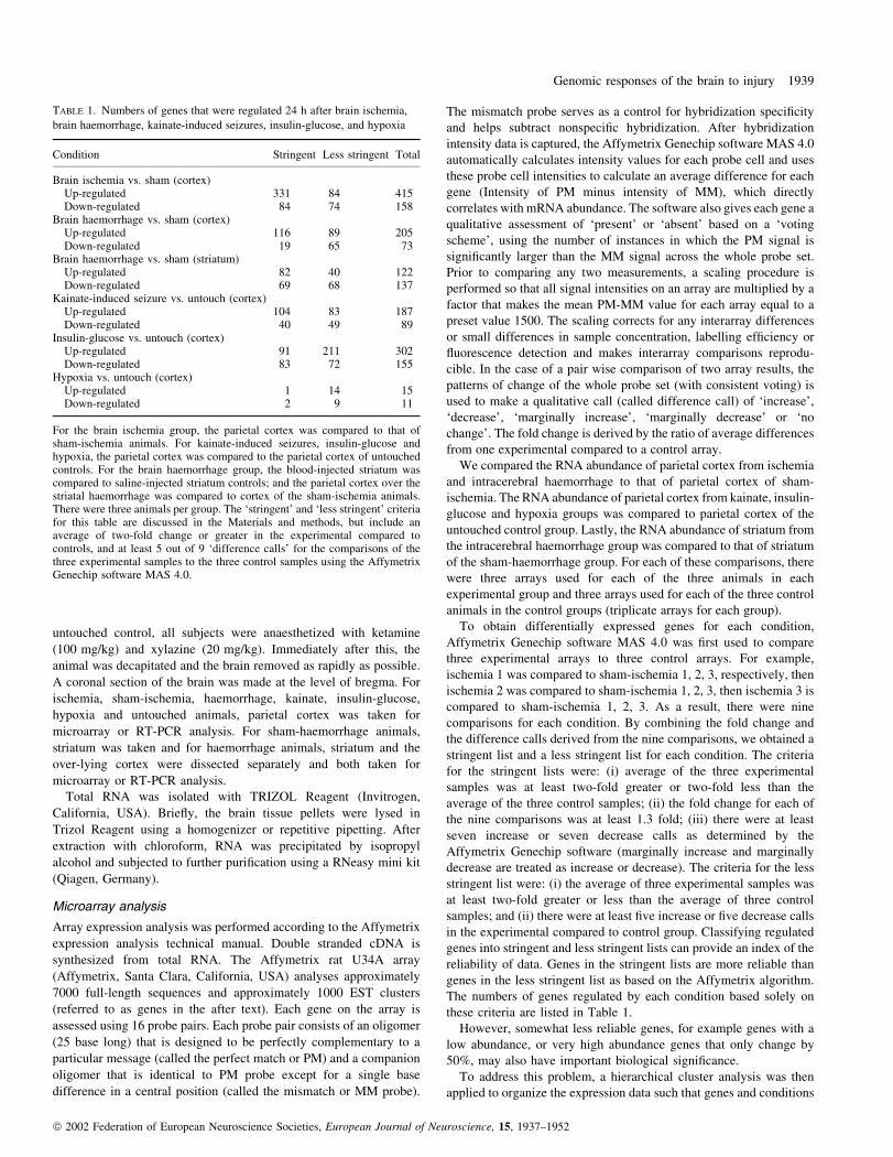
Microarray analysis
Array expression analysis was performed according to the Affymetrix
expression analysis technical manual. Double stranded cDNA is
synthesized from total RNA. The Affymetrix rat U34A array
(Affymetrix, Santa Clara, California, USA) analyses approximately
7000 full-length sequences and approximately 1000 EST clusters
(referred to as genes in the after text). Each gene on the array is
assessed using 16 probe pairs. Each probe pair consists of an oligomer
(25 base long) that is designed to be perfectly complementary to a
particular message (called the perfect match or PM) and a companion
oligomer that is identical to PM probe except for a single base
difference in a central position (called the mismatch or MM probe).
The mismatch probe serves as a control for hybridization speci®city
and helps subtract nonspeci®c hybridization. After hybridization
intensity data is captured, the Affymetrix Genechip software MAS 4.0
automatically calculates intensity values for each probe cell and uses
these probe cell intensities to calculate an average difference for each
gene (Intensity of PM minus intensity of MM), which directly
correlates with mRNA abundance. The software also gives each gene a
qualitative assessment of `present' or `absent' based on a `voting
scheme', using the number of instances in which the PM signal is
signi®cantly larger than the MM signal across the whole probe set.
Prior to comparing any two measurements, a scaling procedure is
performed so that all signal intensities on an array are multiplied by a
factor that makes the mean PM-MM value for each array equal to a
preset value 1500. The scaling corrects for any interarray differences
or small differences in sample concentration, labelling ef®ciency or
¯uorescence detection and makes interarray comparisons reprodu-
cible. In the case of a pair wise comparison of two array results, the
patterns of change of the whole probe set (with consistent voting) is
used to make a qualitative call (called difference call) of `increase',
`decrease', `marginally increase', `marginally decrease' or `no
change'. The fold change is derived by the ratio of average differences
from one experimental compared to a control array.
We compared the RNA abundance of parietal cortex from ischemia
and intracerebral haemorrhage to that of parietal cortex of sham-
ischemia. The RNA abundance of parietal cortex from kainate, insulin-
glucose and hypoxia groups was compared to parietal cortex of the
untouched control group. Lastly, the RNA abundance of striatum from
the intracerebral haemorrhage group was compared to that of striatum
of the sham-haemorrhage group. For each of these comparisons, there
were three arrays used for each of the three animals in each
experimental group and three arrays used for each of the three control
animals in the control groups (triplicate arrays for each group).
To obtain differentially expressed genes for each condition,
Affymetrix Genechip software MAS 4.0 was ®rst used to compare
three experimental arrays to three control arrays. For example,
ischemia 1 was compared to sham-ischemia 1, 2, 3, respectively, then
ischemia 2 was compared to sham-ischemia 1, 2, 3, then ischemia 3 is
compared to sham-ischemia 1, 2, 3. As a result, there were nine
comparisons for each condition. By combining the fold change and
the difference calls derived from the nine comparisons, we obtained a
stringent list and a less stringent list for each condition. The criteria
for the stringent lists were: (i) average of the three experimental
samples was at least two-fold greater or two-fold less than the
average of the three control samples; (ii) the fold change for each of
the nine comparisons was at least 1.3 fold; (iii) there were at least
seven increase or seven decrease calls as determined by the
Affymetrix Genechip software (marginally increase and marginally
decrease are treated as increase or decrease). The criteria for the less
stringent list were: (i) the average of three experimental samples was
at least two-fold greater or less than the average of three control
samples; and (ii) there were at least ®ve increase or ®ve decrease calls
in the experimental compared to control group. Classifying regulated
genes into stringent and less stringent lists can provide an index of the
reliability of data. Genes in the stringent lists are more reliable than
genes in the less stringent list as based on the Affymetrix algorithm.
The numbers of genes regulated by each condition based solely on
these criteria are listed in Table 1.
However, somewhat less reliable genes, for example genes with a
low abundance, or very high abundance genes that only change by
50%, may also have important biological signi®cance.
To address this problem, a hierarchical cluster analysis was then
applied to organize the expression data such that genes and conditions
TABLE 1. Numbers of genes that were regulated 24 h after brain ischemia,
brain haemorrhage, kainate-induced seizures, insulin-glucose, and hypoxia
Condition Stringent Less stringent Total
Brain ischemia vs. sham (cortex)Up-regulated 331 84 415Down-regulated 84 74 158
Brain haemorrhage vs. sham (cortex)Up-regulated 116 89 205Down-regulated 19 65 73
Brain haemorrhage vs. sham (striatum)Up-regulated 82 40 122Down-regulated 69 68 137
Kainate-induced seizure vs. untouch (cortex)Up-regulated 104 83 187Down-regulated 40 49 89
Insulin-glucose vs. untouch (cortex)Up-regulated 91 211 302Down-regulated 83 72 155
Hypoxia vs. untouch (cortex)Up-regulated 1 14 15Down-regulated 2 9 11
For the brain ischemia group, the parietal cortex was compared to that ofsham-ischemia animals. For kainate-induced seizures, insulin-glucose andhypoxia, the parietal cortex was compared to the parietal cortex of untouchedcontrols. For the brain haemorrhage group, the blood-injected striatum wascompared to saline-injected striatum controls; and the parietal cortex over thestriatal haemorrhage was compared to cortex of the sham-ischemia animals.There were three animals per group. The `stringent' and `less stringent' criteriafor this table are discussed in the Materials and methods, but include anaverage of two-fold change or greater in the experimental compared tocontrols, and at least 5 out of 9 `difference calls' for the comparisons of thethree experimental samples to the three control samples using the AffymetrixGenechip software MAS 4.0.
Genomic responses of the brain to injury 1939
ã 2002 Federation of European Neuroscience Societies, European Journal of Neuroscience, 15, 1937±1952

with similar expression pro®les were grouped together. This process
resulted in a phylogenetic tree, the branch lengths of which re¯ect the
degree of similarity between genes or treatments. The cluster analysis
was performed on all the genes that were regulated by any of the
conditions (the sum of the genes under the total column in Table 1,
which totalled 1120). First, the triplicate data for each condition were
averaged and measurements lower than 25 were rounded to 25 using
Excel (Microsoft, Redmond, Washington, USA). Then all of the
conditions were normalized to untouched controls and the normalized
values were graphed in log scale and subjected to a hierarchical
cluster algorithm using GeneSpring software (Silicon Genetics,
Redwood City, California, USA) and a standard correlation coef®-
cient of 0.95 used as the measure for signi®cant statistical similarity.
Genes having similar expression patterns across the seven groups
were grouped while conditions causing similar genomic responses
were clustered together. The branching behaviour of the tree was
controlled using a separation ratio setting of 0.5 and a minimum
distance setting of 0.001.
Quantitative RT-PCR
Real-time Taqman RT-PCR was performed on ®ve selected genes
using the 5700 Sequence Dectection System (PE Biosystems, Foster
City, California, USA). All primers and probes were designed using
Primer Express 2.0 (PE Biosystems, Foster City, California, USA). A
one-step reverse transcription PCR was performed according to
Taqman One-Step RT-PCR Master Mix Reagents Kit protocol (PE
Biosystems). Primers and probes were added at 900 nM and 200 nM for
Narp (neuronal activity-regulated protein, GenBank # S82649), spr
(small proline-rich protein, GenBank # L46593), Spin2c (contrapsin-
like protease inhibitor related protein, GenBank # NM_031531), Arg1
(liver arginase 1, GenBank # NM_017134) and Lbp (lipopolysacchar-
ide binding protein, GenBank # NM_017208). After reverse tran-
scription at 48 °C for 30 min, AmpliTaq Gold was activated at 95 °C
for 10 min thermal cycling proceeded with 40 cycles at 95 °C for 15 s
and 1 min at 60 °C. Input RNA amounts were calculated with relative
standard curves for all mRNAs of interest and GAPDH. Normalization
to GAPDH was performed to account for variability in the initial
concentration and quality of total RNA, and in the conversion
ef®ciency of the reverse transcription reaction.
Results
Gene expression pro®le of cortex and striatum
Only part of the genome is expressed in a speci®c tissue, with the
makeup of a given tissue being determined by the spectrum of
FIG. 1. Diagrams (Venn) showing the numbers of genes up-regulated (a and b) or down-regulated (c and d) in several groups. (a) Numbers of genes that wereup-regulated 24 h after brain ischemia, kainate-induced seizures and brain haemorrhage. (b) Numbers of genes that were up-regulated 24 h after brainischemia, kainate-induced seizures and insulin-glucose treatment. (c) Numbers of genes that were down-regulated 24 h after brain ischemia, kainate-inducedseizures and brain haemorrhage. (d) Numbers of genes that were down-regulated 24 h after brain ischemia, kainate-induced seizures and brain haemorrhage.
1940 Y. Tang et al.
ã 2002 Federation of European Neuroscience Societies, European Journal of Neuroscience, 15, 1937±1952

expressed genes. Of the 8740 genes surveyed by the Affymetrix rat
U34A chips, transcripts of 3869 genes (44.3%) were detected in
parietal cortex and transcripts of 4121 genes were detected in striatum
as de®ned by criteria using the Affymetrix software (with three
present calls in parietal cortex of untouched controls and three present
calls in striatum of sham-haemorrhage, respectively). The most
abundant genes, including mitochondrial cytochrome oxidase subunit
(J01435), 14-3-3 protein eta-subtype (D17445), Thy-1 (EST
AA874848), cyclophilin A (EST AI228674), Hsc-70 (EST
AI234604), alpha-tubulin (EST AI169370), aldolase A (M12919),
ribosomal protein L9 (X51706), and Hsp-90 (S45392), were
expressed at similar levels in cortex and striatum. Of interest, there
were a signi®cant number of genes that were differentially expressed
between cortex and striatum, including such genes as oxytocin/
neurophysin (K01701), melanin concentrating hormone (M62641),
and many others that were expressed at much higher levels in
striatum compared to cortex.
Numbers of regulated genes for each condition
Each condition addressed in this study produced a distinct genomic
response in brain. Table 1 shows the numbers of genes that were up-
regulated or down-regulated for each condition. For example, if
stringent criteria were used 331 transcripts were up-regulated by
brain ischemia and if less stringent criteria were used 84 additional
transcripts were up-regulated by brain ischemia. Similarly, 82 genes
were up-regulated by brain haemorrhage if stringent criteria were
used compared to 40 additional genes if less stringent criteria were
used (Table 1).
Brain ischemia regulated more genes than any other condition (415
up-regulated genes and 158 down-regulated genes), possibly because
ischemia/stroke damages all cellular elements including neurons,
glia, axons/white matter and the vessels. Of note, exposure of animals
to 8% hypoxia for 6 h produced relatively few changes of gene
expression assessed 18 h later (Table 1: 15 up-regulated genes and 11
down-regulated genes). This suggests that any effects of systemic
stress as would be produced by global hypoxia resulted in relatively
few changes of gene expression in brain.
Genes up-regulated by multiple conditions
Many genes that were up-regulated by each experimental condition
were modulated in two or more of the groups. For example, of 122
genes up-regulated by intracerebral haemorrhage, 82 were also
induced by brain ischemia and 53 were induced by kainate-induced
seizures (Fig. 1a). Similarly, of 302 genes up-regulated by insulin-
glucose, 152 are also up-regulated by ischemia and 111 are up-
regulated by kainate (Fig. 1b).
The genes up-regulated in common by the various conditions
might provide an index of common mechanisms of injury. For
example, there were 52 genes induced in common by ischemia,
kainate and haemorrhage (Fig. 1a). There were 98 genes induced in
common by ischemia, kainate and hypoglycemia (Fig. 1b). Of the
genes up-regulated by ischemia (415), kainate (187), haemorrhage
(122), and by insulin-glucose (302) (Fig. 1a and b), there were 45
genes that were up-regulated by all of these injury conditions
(Table 2). As these genes were not up-regulated by hypoxia, they
could serve as markers of neuronal injury, indices of common
mechanisms of injury and/or common responses to injury. For
example, the up-regulation of several of these genes including GFAP,
vimentin, Hsp27, heme oxygenase, and S-100 would be expected to
occur mainly in astrocytes and microglia after ischemia, haemor-
rhage, kainate and hypoglycemia (Dirnagl et al., 1999; Sharp et al.,
2000; Streit, 2000). This would suggest that these are glial responses
that are common to all of these injury conditions.
Cluster analysis of gene regulation
As the gene lists in Table 1 and those tabulated in Fig. 1 were
obtained by applying arbitrary criteria (difference calls), they may not
accurately re¯ect the expression pro®le of each condition. In other
words, some genes, not listed under each condition in Table 1, might
still be regulated by that condition and just did not meet the arbitrary
criteria. To address this problem, hierarchical cluster analysis was
performed using the absolute RNA expression abundance without
considering difference calls (Fig. 2). As expected, ischemia, kainate,
cortex haemorrhage, striatum haemorrhage, insulin-glucose and
striatal sham were clustered together, because these six conditions
all cause injuries (needle injury in striatum of sham group). This
cluster was furthered divided into three subclusters. Ischemia and
kainate were grouped, while cortex haemorrhage, striatal haemor-
rhage and insulin-glucose treatments were clustered.
There were two major groups of clustered genes. One group
included genes up-regulated by at least one injury condition (Fig. 2,
on the left side, red colour) while the other group consisted of genes
down-regulated by one or more conditions (Fig. 2, on the right side,
green colour). Some of the more interesting gene clusters are shown
in Fig. 3.
Figure 3a represents a cluster of genes that are up-regulated mainly
after ischemia (I), kainate (K), insulin-glucose (IG) and striatal
haemorrhage (SH) (Fig. 3a). This cluster of genes should be
considered as those that correspond with the conditions producing
the greatest injury. Though some of these genes are also listed in
Table 2, a signi®cant number of additional genes were identi®ed.
These included ras-related protein (U12187), cytochrome P450
(U09540), platelet activating factor acetylhydrolase alpha 1 subunit
(AF016047), thyrotropin releasing hormone (M23643), lysyl hydro-
lase (S66184), transcriptional repressor CREM (S66024), small
proline-rich protein (L46593), brain glucose transporter protein 1
(M13979), plasminogen activator inhibitor-1 (M24067), and chemo-
kine ST38 precursor (AF053312) (Fig. 3a). Genes that are listed in
Table 2, and were detected in this cluster analysis included Hsp70
heat shock protein and GADD45. These two genes would appear to
be among the most reliable predictors of severe neuronal injury as
suggested from previous studies because they met the stringent
criteria for Table 2 and they were also detected in the cluster analysis
(Figs 2 and 3) (Chen et al., 1998; Schmidt-Kastner et al., 1998; Sharp
et al., 1999).
Figure 3b represents a second cluster of genes that were induced by
all of the injury conditions and not by hypoxia (H). Though there is
marked brain injury after ischemia (I), kainate (K), insulin glucose
(IG), and striatal haemorrhage (SH), some degree of injury would be
expected from needle insertions in striatal sham (SS) as well as in the
cortex (cortex haemorrhage, CH) overlying the striatal haemorrhage
when compared to normal brain of untouched controls (U). Note that
the large majority of the genes in this cluster are also listed in
Table 2. Genes that were identi®ed according to the criteria in
Table 2 and that were also identi®ed by cluster analysis to be induced
by all injury conditions included but were not limited to: CELF,
SOCS-3, GFAP, ASM15, CD44, immediate-early serum-responsive
JE, Hsp27, heme oxygenase (Hsp32), alpha-2u globulin-related
protein, and TIMP-1. These would appear to be among the most
reliable, and yet sensitive injury-related genes as they were detected
using two completely different methods of analysis. It seems likely
that many if not most of the genes identi®ed in the clusters in Fig. 3a
Genomic responses of the brain to injury 1941
ã 2002 Federation of European Neuroscience Societies, European Journal of Neuroscience, 15, 1937±1952

and b are genes induced in response to some aspect of brain injury
and could be induced in one or more cell types in the injured brain.
The genes induced by all of the injury conditions (Table 2) are of
particular interest as they suggest common pathways mediating injury
or repair. CELF is a family of RNA binding proteins that are
implicated in cell-speci®c alternative splicing, which may be
important for brain-speci®c alternative splicing (Ladd et al., 2001).
Heparin-binding EGF-like growth factor is an anti-apoptotic factor
that promotes neuronal and glial survival (Xian & Zhou, 2000), and
has been shown to be induced in ischemic brain (Tanaka et al., 1999).
Annexin II, one of a family of calcium binding proteins found in a
variety of tissues, is induced in astrocytes and endothelial cells after
ischemia (Eberhard et al., 1994). P41-Arc is part of a complex
involved in cross-linking of actin ®laments (Zhao et al., 2001) with
TABLE 2. Genes induced/up-regulated to the greatest extent by brain ischemia, brain haemorrhage, kainate-induced seizures and hypoglycemia
Classi®cation/ID number Name
Fold change
Ischemia Haemorrhage Kainate Hypoglycemia
Heat shock proteinM86389 Heat shock protein (Hsp27) ~946.6 11.4 ~734.3 ~362.7J02722 Heme oxygenase ~267.7 25 ~39.8 ~111.7Z75029 Heat shock protein 70 (HSP70) 50.5 3.2 5.2 2.2
DNA damage repairing proteinL32591 GADD45 7.3 2.5 3.1 1.4
Metal ion chelatorL33869 Ceruloplasmin 7.2 2.5 4 2.4M11794 Metallothionein-1 and metallothionein-2 6.3 3.1 4.3 4.5
Protease and protease inhibitorD00753 Contrapsin-like protease inhibitor related protein (CPi-26) ~164.8 18.1 ~99.7 ~101.8AI169327 Tissue inhibitor of metalloproteinase-1 (TIMP1) 88.7 6.4 255.9 170.5D90404 Cathepsin C 4.3 3 3.1 1.8AA892775 Lysozyme 2.8 3.2 2.4 3
Matrix modelling moleculesM14656 Osteopontin 56.5 5.7 41.3 37.4AI012030 Matrix Gla protein (Mgp) 3.1 2.8 4.5 2.6S61865 Syndecan 5.3 1.5 2.8 1.8S61868 Ryudecan 2.1 1.1 2 1.6
CytoskeletonAF028784 Glial ®brillary acidic protein alpha (GFAP) ~71.5 5.8 ~71.5 ~83.8AA892333 Tubulin alpha 6 (Tuba6) 15 6 10.5 9.4X62952 Vimentin 3.3 3.3 4 4.1AF004811 Moesin 20.7 2.3 ~25.8 ~9.0
ReceptorM61875 CD44 53.7 10.8 ~93 ~45.9AF087943 CD14 16.6 6.6 4.7 3.5J05122 Peripheral-type benzodiazepine receptor (PKBS) 9 3.8 4.3 5.6
Molecules with immune functionAF075383 Suppressor of cytokine signalling-3 (SOCS-3) ~55.5 9.2 ~13.4 ~13.7X17053 Immediate-early serum-responsive JE gene 24.4 14.4 15.5 8.3J02962 IgE binding protein 21.4 6.1 48.8 49.3X13044 MHC-associated invariant chain gamma 11.7 13.5 5 33X73371 Fc gamma receptor 8.1 10.3 5.5 6.2U31599 MHC class II-like beta chain (RT1.DMb) 3.4 2 6.8 5.4
OthersAA946503 Alpha-2u globulin-related protein ~52.1 20.5 ~18.6 ~147.4S74141 Hck = tyrosine kinase ~8.3 5.2 ~13.6 ~26.9U18729 Cytochrome b558 alpha-subunit 29.3 3.6 ~29.1 ~64.6L05489 Heparin-binding EGF-like growth factor 11.4 2.3 7 4.5M65149 CELF 10.9 3.4 8.1 8.4U92081 Epithelial cell transmembrane protein antigen precursor (RTI40) 10.9 3.2 10.2 6.5X59864 ASM15 9 4.3 7.3 2.2D10729 Proteasome subunit RC1 5 2.5 6.1 3.7L13039 Annexin II 4.9 3 2.5 2.3J03627 S-100 related protein 4.7 3.6 4.7 2.7X06916 Protein p9Ka homologous to calcium-binding protein 4.1 5.8 3.6 4.7AF083269 P41-Arc 3.8 3.4 1.9 1.6J04792 Ornithine decarboxylase 3.4 2 2.1 2.2S69874 C-FABP = cutaneous fatty acid-binding protein 2.7 1.8 2.5 2.1X59375 Ribosomal protein S27 2.6 2.2 2.5 2.4M24604 Proliferating cell nuclear antigen (PCNA/cyclin) 2.5 1.9 1.9 1.7AA944422 Acidic calponin 2.5 1.6 2.9 2.6AF036537 Homocysteine respondent protein HCYP2 2 2 4.1 4
The fold change was obtained by comparing ischemia to cortex-sham, haemorrhage to striatum-sham, and kainate and hypoglycemia to untouched controls. Whenthe baseline level was < 25, it was rounded to 25 to facilitate the computation of fold change, which was then preceded by `~'.
1942 Y. Tang et al.
ã 2002 Federation of European Neuroscience Societies, European Journal of Neuroscience, 15, 1937±1952

an unknown function in brain. Ornithine decarboxylase (ODC) is the
rate-limiting enzyme involved in the synthesis of polyamines. ODC is
found in neurons and is induced in glia after ischemia, seizures, and
other stressful conditions (Bernstein & Muller, 1999). Though the
polyamines appear to modulate NMDA and other receptors (Paschen,
1992; Johnson, 1998), their precise role in brain injury has not been
elucidated. Cutaneous fatty acid-binding protein up-regulates VEGF
in tumour cells (Leo et al., 2001; Jing et al., 2001) but had not been
reported in brain. The ribosomal protein S27 is found throughout
normal brain, particularly in hypothalamus, is induced during the
visual critical period, and has zinc ®nger motifs (Chan et al., 1993;
Wong et al., 1993; Prasad & Cynader, 1994; Thomas et al., 2000).
PCNA is induced in all dividing cells after all types of injury
(Lehrmann et al., 1997; Liu et al., 1998; Dirnagl et al., 1999; Liu
et al., 2001). Acidic calponin, is an actin-, tropomyosin- and Ca2+
calmodulin-binding protein that inhibits MgATPase, is a calpain
substrate, and is expressed in neurons, synapses and some glia in
brain (Ferhat et al., 1996; Plantier et al., 1999; Agassandian et al.,
2000; Yoshimoto et al., 2000). Nothing is reported on the
homocysteine respondent protein HCYP2 other than its sequence
and homocysteine inducibility. Homocysteine metabolism is of
interest because homocysteinemia is associated with increased stroke
and myocardial infarction risk (Hankey & Eikelboom, 2001).
Proteases and their inhibitors are also induced by all of these acute
injury conditions (Table 2) including tissue inhibitor of metellopro-
teinase-1 (TIMP-1), cathepsin C, lysozyme, and contrapsin-like
protease inhibitor related protein (Cpi-26). TIMP-1 (Wang et al.,
1998a) and cathepsin C (Yamashima, 2000) are induced after brain
ischemia. Cpi-26, an inhibitor of proteases including trypsin
(Potempa et al., 1995) and lysozyme, a white blood cell lysozomal
enzyme that increases after myocardial infarction (Welman et al.,
1980), had not been studied in brain.
P8 is a notable gene induced by most of the injury conditions. P8
appears to play a role in signalling TGF activation via p8 to Smad
proteins (Garcia-Montero et al., 2001). The Smad proteins are
transcription factors that modulate a number of target genes,
particularly those involved with immune regulation that may play
an important role in a variety of tissue injuries (Miyazono et al.,
2000). TGF and Smads are increased during scar formation after
myocardial infarction (Hao et al., 1999). Moreover, TGF is induced
after stroke and may serve to protect or enhance recovery after brain
ischemia (Ali et al., 2001; Pang et al., 2001). It is possible that
different TGF and p8 mediated Smads are induced with different
types of injury, and with induction of different down-stream genes in
the different types of injury (Itoh et al., 2000; Miyazono et al., 2000;
Schiffer et al., 2000). For example, TGF signalling through Smad3
may be important for induction of TIMP-1, c-jun and Fos (all induced
by all acute injury conditions examined here), whereas signalling
through other Smads might induce other target genes (Roberts et al.,
2001; Verrecchia et al., 2001).
The RET ligand was also induced most of the injury conditions.
RET receptor tyrosine kinase is a functional receptor for GDNF (glial
FIG. 2. Hierarchical clustering of differentially expressed genes. The clustered genes were derived from those in the Table 1 total column. Genes that showsimilar expression patterns across different treatments cluster together. The red colour indicates up-regulation, the yellow green colour represents little change,and the deep green colour indicates down-regulation (see Materials and methods for details).
Genomic responses of the brain to injury 1943
ã 2002 Federation of European Neuroscience Societies, European Journal of Neuroscience, 15, 1937±1952

cell derived neurotrophic factor) that requires calcium (Anders et al.,
2001). GDNF binds to GDNF family receptor alpha (GFR alpha)
which stimulates autophosphorylation of RET and downstream
signalling (Saarma, 2000), where the docking protein FRS2 links
RET with the mitogen-activated protein kinase signalling cascade
(Melillo et al., 2001). GDNF is a potent survival factor for speci®c
neurons including dopamine, noradrenaline and sympathetic neurons
and acts in concert with TGF to support survival of other neurons
(Saarma, 2000). The current study suggests that GDNF-Ret signalling
could be important for survival of cells after many types of acute
neuronal injury.
Genes induced by kainate and haemorrhage andhypoglycemia but not by ischemia
Though the large majority of the genes induced by kainate were also
induced by focal ischemia, there were a few genes induced by kainate
that were also induced by haemorrhage or hypoglycemia and not
by ischemia. These included C4 complement protein (U42719),
macrophage metalloelastase (X98517) and proteasome activator
PA28 subunit beta (D45250) (see Fig. 1). These genes are of interest
because they may point to excitotoxic mechanisms speci®c for
haemorrhage and hypoglycemia injury that may not necessarily be
shared with ischemia.
For example, the macrophage metalloelastase was induced by
kainate and hypoglycemia but not ischemia. The macrophage
metalloelastase is a matrix metalloproteinase, MMP12, one of a
family of metalloproteinases that can degrade all components of the
extracellular matrix (Werner et al., 2000; Warner et al., 2001).
MMP12 appears to exacerbate lung injury (Warner et al., 2001), is
not expressed in normal astrocytes (Agapova et al., 2001), but is
expressed to high levels in astrocytic brain tumours (Kachra et al.,
1999). TGF beta inhibition of MMP12 is mediated by Smad3
(Werner et al., 2000). The role of macrophage metalloelastase in
kainate and hypoglycemic injury is not clear, but could be related to
macrophage removal of dying neurons in speci®c regions of brain in
both conditions.
0.01
0.1
1
10
100
1000
0.01
0.1
1
10
100
1000
0.01
0.1
1
10
100
1000
U CS I CH K H IG SS SH
U CS I CH K H IG SS SH
U CS I CH K H IG SS SH
heat shock protein 70, ras-relatedprotein (rad), Cytochrome P450,platelet-activating factoracetylhydrolase alpha 1 subunit,thyrotropin releasing hormone, lysyloxidase, GADD45, transcriptionalrepressor CREM, small proline-richprotein, brain glucose-transporterprotein, alpha B-crystallin, plasminogenactivator inhibitor-1, CC chemokineST38 precursor
complement component C3, cdc2,macrophage inflammatory protein-2precursor, alpha-1-acid glycoprotein,liver arginase, Glycam 1, ADP-ribosylcyclase (CD38), intercellular calcium-binding protein (MRP8), cyclin D1,DORA protein
NGFI-A, NGFI-B, NGF-I C, NGF-inducible anti-proliferative putativesecreted protein (PC3), Krox-24, Arc,protein tyrosine phosphatase, activityand neurotransmitter-induced early gene3, Vesl
0.01
0.1
1
10
100
1000
0.01
0.1
1
10
100
1000
0.01
0.1
1
10
100
1000
U CS I CH K H IG SS SH
U CS I CH K H IG SS SH
U CS I CH K H IG SS SH
CELF, Suppressor of cytokine signaling-3(SOCS-3), GFAP, ASM15, TGF-beta 1,CD44, PHAS-I, IgE binding protein,thyrotropin-releasing hormone (TRH)precursor, contrapsin-like proteaseinhibitor related protein (CPi-26), MHCclass Ib antigen (RT1.Cl), immediate-early serum-responsive JE, Hsp27, hemeoxygenase, alpha-2u globulin-relatedprotein, TIMP-1
type II hexokinase, JAK2,lipopolysaccharide binding protein,interleukin 6, decay accelerating factorGPI-form precursor (DAF), brain-derivedneurotrophic factor (BDNF), c-fos,cyclooxygenase-2, major acute phasealpha-1 protein, ICE-like cysteineprotease (Lice), leucine zipper protein(LRF-I), growth arrest and DNA-damage-inducible protein GADD153
brain finger protein (BFP), braindigoxin carrier protein, GABA-Areceptor delta subunit, non-receptorprotein kinase (batk), m3 muscarinicacetylcholine receptor, alpha 1 subunitof soluble guanylyl cyclase,phospholipase C-1, glutathione S-transferase Yc1 subunit,Ca2+/calmodulin-dependent proteinkinase, glutamate receptor (GluR-C),NMDAR1 glutamate receptor subunit,Shal 1 (potassium channel polypeptide)
Inte
nsity
norm
aliz
edto
unto
uche
dco
ntro
l(lo
gsc
ale)
a b
c d
e f
FIG. 3. Relative expression of genes (log scale of normalized to untouched control) for the different conditions studied here: untouched control (U), cortexsham (CS), brain ischemia (I), cortex haemorrhage (CH), kainate-induced seizures (K), hypoxia (H), insulin-glucose (IG), striatum sham (SS) and striatumhaemorrhage (SH). (a)Genes that were up-regulated by brain ischemia (I), kainate-induced seizures (K), insulin-glucose (IG) and striatum haemorrhage (SH).(b) Genes that were up-regulated by brain ischemia (I), cortex haemorrhage (CH), kainate-induced seizures (K), insulin-glucose (IG), and striatumhaemorrhage (SH). (c) Genes that were up-regulated most by striatum haemorrhage (SH) and cortex haemorrhage (CH), and occasionally in other conditions.(d) Genes that were speci®cally up-regulated after brain ischemia (I). (e) Genes that were up-regulated by ischemia (I) and tended to be down-regulated afterkainate-induced seizures (K). (f) Genes that were down-regulated by one or multiple conditions.
1944 Y. Tang et al.
ã 2002 Federation of European Neuroscience Societies, European Journal of Neuroscience, 15, 1937±1952

The proteasome activator PA28 was also induced by kainate and
hypoglycemia, and not by ischemia. Proteasome activator PA28 is a
gamma interferon inducible complex (Fabunmi et al., 2001). PA28
(also called 11S REG) is one of two protein complexes that have been
found to bind the ends of the proteasome and activate it. There are
three PA28 subunits, alpha, beta and gamma; alpha and beta being
found in the cytosol, and gamma in the nucleus (Yawata et al., 2001).
PA28 is required for the presentation of certain major histocompat-
ibility (MHC) class I antigens that allows cytotoxic T lymphocytes to
lyse the cells expressing cell-surface MHC Class I molecules
complexed with foreign peptides (Rechsteiner et al., 2000;
Stohwasser et al., 2000). Again, PA28 up-regulation by kainate and
hypoglycemia suggests speci®c mechanisms of cell mediated immune
injury in these conditions. However, it has also been found that
Hsc70, Hsp40 and PA28 are necessary and suf®cient to fully
reconstitute Hsp90-mediated refolding of partially denatured ®re¯y
luciferase (Minami et al., 2000), suggesting a possible protective role
for PA28.
Genes speci®cally up-regulated by each condition
The data also show that though there were many genes induced in
common by ischemia, haemorrhage, hypoglycemia, and kainate,
there were a number of genes induced that were speci®c for each of
these conditions except for kainate. Of the 415 genes induced by
ischemia, 236 were only induced by ischemia and not by kainate or
haemorrhage (Fig. 1a). Of the 122 genes induced by haemorrhage, 39
were induced only by haemorrhage and not induced by ischemia or
kainate (Fig. 1a). Of the 302 genes induced by insulin, 132 were only
induced by insulin and not ischemia or kainate (Fig. 1b). Kainate,
however, was the only condition in which there were no speci®cally
up-regulated genes. Of the 187 genes induced by kainate, 150 were
induced by ischemia and/or haemorrhage (Fig. 1a). Of the same 187
genes induced by kainate, 162 were induced by ischemia and/or
insulin-glucose (Fig. 1b). All of the 187 genes induced by kainate
were induced either by ischemia, haemorrhage and/or insulin. As
discussed below, this data suggests that excitotoxicity is an important
mechanism of injury in ischemia, haemorrhage and hypoglycemia
and that there is not likely to be a mechanism of injury speci®c for
kainate.
Genes that are markedly regulated at 24 h after ischemia but not
after the other conditions are presented in Fig. 3d. These genes
include in¯ammatory, immune and signalling molecules like JAK2
(U13396), MAP-kinase phosphatase (cpg-21) (AF013144), dual
speci®city phosphatase (U42627), lipopolysaccaride binding protein
(L32132), interleukin-6 (M26745), decay accelerating factor GPI-
form precursor (DAF) (AF039583), major acute phase alpha-1
protein (K02814), eNOS (AJ011116), CC chemokine ST38 precursor
(AF053312), intercellular adhesion molecule-1 (ICAM-1) (D00913),
interleukin 1 receptor antagonist (M63101), and integrin-alpha 1
(X52140). Hypoxia-inducible factor 1 (Y09507) and some of its
target genes like hexokinase II (S56464), VEGF (L20913),
angiopoietin (AF030378), adrenomedullin (D14069), and brain
0 .02 .04 .06 .08 .0
10 .012 .0
U C S I C H K H IG S S S H0
10
20
30
40
U CS I CH K H IG SS SH0
50
100
150
200
U C S I C H K H IG S S SH
0
10
20
30
U C S I C H K H IG S S S H012345
U CS I CH K H IG S S S H
Array PCR
ca b
d e
FIG. 4. Microarray results compared to real-time RT-PCR results for different conditions: untouched control (U), cortex sham (CS), brain ischemia (I), cortexhaemorrhage (CH), kainate-induced seizures (K), hypoxia (H), insulin-glucose (IG), striatum sham (SS) and striatum haemorrhage (SH). The fold change,plotted on the y-axis, was calculated as the average expression of the gene in each condition vs. the untouched controls, so that the fold change for untouchedcontrol in every case is 1. Error bar indicates standard error (n = 3). (a) Neuronal activity-regulated protein (GenBank # S82649). (b) Small proline-richprotein (GenBank # L46593). (c) contrapsin-like protease inhibitor related protein (GenBank # NM_031531). (d)lipopolysaccharide binding protein (GenBank# NM_017208). (e) liver arginase 1 (GenBank # NM_017134).
Genomic responses of the brain to injury 1945
ã 2002 Federation of European Neuroscience Societies, European Journal of Neuroscience, 15, 1937±1952
glucose-transporter protein (M13979) were induced (Fig. 3d). Of
note is the ®nding that the DNA-damaging inducible protein
GADD153 (U30186) is induced at 24 h after ischemia but not in
the other conditions examined here.
There were a number of genes that were induced mainly after
haemorrhage (Figs 1a and 3c). Many of these genes were related to
in¯ammatory or immune responses, such as prepro- complement C3
(X52477), macrophage in¯ammatory protein-2 precursor (U45965),
CD38 (D29646), intercellular calcium binding protein (macrophage-
related protein 8) (EST AA957003), alpha-1 acid glycoprotein
(V01216) and Glycam1 (glycosylation-dependent cell adhesion
molecule 1 L08100) (Fig. 3c). Others included MHC class II MHC
RT1-B region Ia antigen (M15562), leucocyte antigen MRC-OX44
(M57276), MRC OX-45 surface antigen (X13016), and interleukin-1
receptor type 2 (Z22812). Additional genes up-regulated by
haemorrhage included CC chemokine receptor (E13732), DORA
protein (AJ223184), apolipoprotein B mRNA editing protein
(L07114), and interleukin-1 beta converting enzyme (U14647).
There were also some genes induced speci®cally by insulin/glucose
(Fig. 1b). These included genes such as MHC class I RT1.C/E mRNA
(EST AI235890), presenilin-2 (X99267), plakoglobin (U58858) and
EST AA799766. It is notable that the rat U34A chips only include a
few of the known, cloned glucose regulated proteins (GRPs) that are
known to be glucose responsive (Massa et al., 1996).
A cautionary note should be made about the patterns of genes
regulated for a speci®c condition. The problem of the time course of
gene expression has not been dealt with at all here as all samples were
compared at 24 h. For example, c-fos (X06769), BDNF (S71196,
S76758, D10938, X67108), and cycloxygenase-2 (COX2) (L25925)
are all induced in ischemic brain at 24 h but are not up-regulated in
the other injury conditions at 24 h (Fig. 3d). Similarly, some
immediate-early genes like NGFI-A (N18416), NGFI-B (U17254),
NGFI-C (N92433), Krox-24 (Y753397), and Arc (U19866) are up-
regulated in ischemia, but down-regulated by kainate and probably
haemorrhage, hypoxia, insulin-glucose and even sham when meas-
ured at 24 h after treatments (Fig. 3e). However, all of these
molecules, including c-fos, BDNF, COX2, NGFI-A, NGFI-B,
NGFI-C are induced in ischemic brain and after seizures within a
few hours and return to normal or decrease below normal levels by
24 h after kainate (Koistinaho & Hokfelt, 1997; Sharp et al., 2000).
Hence, ischemia differs from these other injury conditions in that
there is persistent induction of these early genes in ischemic brain
compared to the other conditions examined here.
Down-regulated genes for each condition
Like up-regulated genes, some genes are down-regulated after one or
more conditions (Figs 1c and d, and 3f). The number of down-
regulated genes for each condition tended to be less than the number
of up-regulated genes. Among 646 up-regulated genes in parietal
cortex by any condition, 130 genes were not expressed in untouched
brain and were induced by one or more injury conditions. In
comparison, among 381 cortex genes down-regulated by any
condition, only 20 genes were completely suppressed by any of the
injury conditions.
Like up-regulated genes, some down-regulated genes are modu-
lated by multiple conditions. There were 42 genes down-regulated by
both ischemia and kainate, 12 genes down-regulated by both kainate
and haemorrhage and 10 genes down-regulated by both insulin-
glucose and kainate. Compared to the up-regulated genes, the
numbers of shared genes were fewer (Fig. 1c and d). One of the most
unexpected results was that only a single gene, calmodulin-dependent
protein kinase (CaM kinase, M63333), was down-regulated in
common by all of the injury conditions including ischemia, kainate,
haemorrhage and insulin.
Though gene down-regulation does not appear to be a regulated
response that is shared between multiple injury conditions, the
clustered dendrogram indicated that there were many genes that were
down-regulated by multiple injury conditions (Fig. 2). Down-regu-
lated genes included channels and neurotransmitter receptors
(Fig. 3f): GABA-A receptor delta subunit (M35162), m3 muscarinic
Ach receptor (M16407), cholecystokinin receptor (M99418), A2
adenosine receptor (S47069), glutamate receptor (GluR-C M36420),
NMDAR1 glutamate receptor subunit (U11418); and potassium
channels like Shal1 potassium channel (S64320) and the Kv9.1
potassium channel (Y17606). Other genes, down-regulated by one or
more conditions, are listed in Fig. 3f. This could be a response to
glutamate-mediated excitotoxity where down regulation of excitatory
pathways would decrease injury. However, some inhibitory pathways
also appear to be down regulated that would tend to exacerbate
excitotoxic injury.
The failure to detect a group of common genes that were down-
regulated in all of the injury conditions suggests that transcriptional
down-regulation is probably not a major part of the genomic response
to brain injury. Thus, protein phosphorylation and de-phosphorylation
may play the major role in down-regulating signalling cascades. The
current results suggest that targeting down-regulated genes for
therapeutic interventions is not likely to yield a generally useful
therapeutic strategy.
Correlation of microarray data with PCR results
Quantitative RT-PCR was performed on ®ve selected genes (Fig. 4).
For genes with high expression such as Narp (Fig. 4a), the RT-PCR
results showed excellent agreement with the corresponding micro-
array results. For genes with very low expression such as Spin2c
(Fig. 4c) and Lbp (Fig. 4d), the RT-PCR results showed a much
greater- or lower- fold change when compared to the microarray
results. This is largely due to the fact that the algorithm used by
Affymetrix software MAS 4.0 gives negative values to low-
abundance genes. We chose to round all negative values to 25 to
facilitate the log-transformation and the computation of fold change
but this may not accurately re¯ect the absolute expression level and
skewed the fold change in one way or the other. In addition, as RNA
used on the microarray and the RT-PCR studies were from different
rats, the variation of individual animals can give rise to the disparity
between RT-PCR and microarray results. This was demonstrated by
liver arginase 1 (Fig. 4e), where the microarray showed an increased
expression in the cortex of haemorrhage rats compared to untouched
and sham-surgery rats, while the RT-PCR did not show any
signi®cant change. We found that two of the three rats used for
microarray studies had blood in the lateral ventricles and none of the
three rats used for RT-PCR studies had blood in the lateral ventricles.
However, despite the disparity between microarray data and RT-PCR
results, these two independent approaches showed the same general
pattern of expression for all ®ve genes studied.
Discussion
The results show that many genes induced by ischemia were also
induced after kainate-induced seizures, intracerebral haemorrhage
and hypoglycemia. These included heat shock proteins, DNA
damage repair proteins, proteases and protease inhibitors, metal ion
and free-radical scavengers, and calcium-binding proteins. The
induction of heme-oxygenase (Hsp32, microglia), Hsp27 (astrocytes),
1946 Y. Tang et al.
ã 2002 Federation of European Neuroscience Societies, European Journal of Neuroscience, 15, 1937±1952

ceruloplasmin (astrocytes), GFAP (astrocytes), vimentin (astrocytes),
matrix glia protein, and S-100 (astrocytes) after ischemia, haemor-
rhage, kainate and hypoglycemia, points to a generalized response of
glial cells in all of these injury conditions (Wilson, 1997; Paschen
et al., 1998; Ellison et al., 1999; Anguelova et al., 2000; Hertz et al.,
2000; Streit, 2000; Jin et al., 2001). The induction of the heat shock
proteins/chaperones, including Hsp27, Hsp32 (heme oxygenase), and
Hsp70, after these injuries points to a general role of these stress
genes to respond to denatured proteins within cells, metabolism of
heme proteins, and cytoskeletal stress (Abe & Nowak, 1996; Sharp
et al., 1999, 2000). All of these heat shock proteins are induced after
ischemia and haemorrhage and kainate, but their role in hypoglyce-
mia was previously unknown (Simon et al., 1991; Abe & Nowak,
1996; Nimura et al., 1996; Plumier et al., 1997; Turner et al., 1998;
Krueger et al., 1999; Sharp et al., 2000).
The induction of CD44, CD14, MHC, and other immune-related
molecules reinforces the concept that brain injuries of all types
activate an immune/in¯ammatory response, with certain aspects of
this immune response being common to all injuries. CD44, a
transmembrane glycoprotein involved in endothelial cell recognition
and traf®cking of lymphocytes and regulation of cytokine gene
expression, is induced in microglia, macrophages, and microvessels
after stroke (Wang et al., 2001). The current study demonstrates a
similar up-regulation after neuronal injury caused by haemorrhage,
hypoglycemia, and seizures. MHC molecules are up-regulated on
microglia/macrophages after ischemia and kainate (Finsen et al.,
1993; Kato et al., 1996; Matsuoka et al., 1998; Bona et al., 1999) and
all of the injuries studied here and would appear to play a central role
in the immune-mediated removal of necrotic and apoptotic cells.
The induction of ceruloplasmin and metallothionein-1 and -2
emphasizes the importance of iron and other ions after acute injury.
Though induced by ischemia (Yanagitani et al., 1999; Campagne
et al., 2000), there was little previous evidence for the role of
metallothioneins in haemorrhage, hypoglycemia and seizures.
Metallothioneins protect against stroke (van Lookeren Campagne
et al., 1999) and may protect against other causes of acute injury.
Ceruloplasmin is important in the metabolism and transport of both
iron and copper (Wessling-Resnick, 1999; Yonekawa et al., 1999), so
that a prominent role for this metal ion-related protein can be
suggested in many acute neuronal injuries.
Matrix and cytoskeletal molecules, including osteopontin, are also
induced by all of the injury conditions. Osteopontin and its integrin
receptor alpha v beta 3 are induced in macrophages and microglia in
the region around a stroke, with subsequent up-regulation of its
integrin receptor on astrocytes (Wang et al., 1998b; Ellison et al.,
1999). These data suggest that osteopontin released from microglia/
macrophages at the edge of an infarct modulate the astrocytic scar/
response after stroke (Ellison et al., 1999). The current ®ndings
suggest that this also occurs in injury states where there is selective
neuronal cell death, including after haemorrhage, hypoglycemia and
seizures. The above genes are believed to have particular signi®cance
because they are all induced after ischemia, haemorrhage, seizures,
and hypoglycemia, and represent common pathways for injury or
repair in the acutely injured brain.
Excitotoxicity has been postulated to play a key role in the
pathogenesis of many neurological disorders, and especially in
ischemic stroke and seizures (Simon et al., 1986; Choi, 1988;
Meldrum, 1993; Bittigau & Ikonomidou, 1997). Though there have
been several suggestions that excitotoxicity plays a role in neuronal
damage after ischemia, seizures, and hypoglycemia (Simon et al.,
1984, 1986; Meldrum, 1994), there has not been data suggesting that
damage around haemorrhages might be related to excitotoxicity. The
®nding that all of the genes induced after kainate are also induced
after haemorrhage, hypoglycemia and ischemia, suggests a prominent
role for excitotoxicity in each of these conditions.
Though there has been little previous evidence that glutamate
might mediate injury around an intracerebral haemorrhage, in vitro
studies suggest interactions between blood/haemoglobin and excito-
toxicity (Regan & Panter, 1996; Gingrich et al., 2000). Importantly,
glutamate could increase around intracerebral haemorrhages (ICH)
from multiple sources, including blood plasma, lysed red blood cells
and injured brain cells. Plasma, which has a high concentration of
glutamate (Tsai & Huang, 2000), diffuses into brain around ICH
(Wagner et al., 1996, 1998). The intracellular concentrations of
glutamate in red blood cells are very high, and are in the range that
are neurotoxic in vitro and are in the range of the extracellular
glutamate after cerebral ischemia (Meldrum, 1994; Obrenovitch &
Richards, 1995; Tsai & Huang, 2000). The lysis of red blood cells
would markedly increase glutamate around haemorrhages. Moreover,
haemoglobin released by red blood cells can exacerbate excitotoxic
injury in vitro and thrombin released in regions of ICH can potentiate
NMDA function (Regan & Panter, 1996; Gingrich et al., 2000).
An intense in¯ammatory response is a well-documented process
after various brain injuries (Hallenbeck, 1996). Despite the over-
whelming evidence of the existence of in¯ammation after brain
injury, many of the signalling mechanisms remain to be elucidated
(del Zoppo et al., 2001). The induction of immune-related molecules,
such as MHC antigens, IgE binding protein, and Fc gamma receptor
by all injury conditions support the involvement of an immune
response that could contribute to secondary brain injury and/or
contribute to repair.
The current data provides some new insights into common
mechanisms of immune responses to these different causes of injury.
CD14 is induced by all of the injury conditions here, as is the
lipopolysaccharide (LPS) receptor present both in plasma and at the
surface of myeloid cells. It is considered to be the key player in the
induction of septic shock provoked by gram-negative bacteria (Pugin
et al., 1994). LPS is ®rst aggregated by LPS binding protein (LBP,
which is also induced after ischemia) and then binds to CD14. The
LPS±CD14 interaction can lead to the activation of NF-kB through a
transduction pathway in which Toll-like 4 receptor is involved
(Schumann et al., 1990; Wright et al., 1990; Poltorak et al., 1998;
Wright, 1999). Through this transduction pathway, LPS causes
dramatic transcriptional regulation of a wide range of pro-in¯amma-
tory genes including TNF, IL-1, IL-6, IL-8, ICAM-1, E-selectin and
others (Wright, 1999). CD14 may be involved in the regulation of a
brain in¯ammatory response via an autocrine/paracrine loop (Nadeau
& Rivest, 2000). A low dose of LPS dramatically sensitizes the
immature brain to injury after short periods of hypoxia-ischemia that
by themselves caused little injury and this effect has been associated
with an enhanced expression of CD14 mRNA in the brain (Eklind
et al., 2001). Although no exogenous LPS is present in brain after
injury, the heat shock protein Hsp70 can serve as an endogenous
ligand to activate CD14 and stimulate the production of IL-1beta, IL-
6 and TNF-alpha (Asea et al., 2000). The discovery of the induction
of CD14 may provide further clues about the induction and regulation
of in¯ammatory reactions in injured brain, and could encourage
further examination of the potentially harmful role of secondary
infections in acute brain injury. Moreover, as CD14 polymorphisms
are associated with increased risk of myocardial infarction
(Unkelbach et al., 1999; Hubacek et al., 1999; Shimada et al.,
2000), they could have a role in stroke.
The immediate-early JE gene is an analogue of murine monocyte
chemoattractant protein-1, which is responsible for recruiting
Genomic responses of the brain to injury 1947
ã 2002 Federation of European Neuroscience Societies, European Journal of Neuroscience, 15, 1937±1952

haematogenous macrophages and is induced after peripheral nerve
injury (Carroll & Frohnert, 1998). It was induced by all of the injury
conditions examined here, and probably plays a central role in
recruiting macrophages into acutely injured brain. The peripheral-
type benzodiazepine receptor (PTBR) is found on the outer
mitochondrial membranes of peripheral tissues and cells. In mam-
malian CNS, the peripheral-type benzodiazepine receptor is localized
within the astrocytes and microglia. PTBR transports cholesterol to
the site of neurosteroid biosynthesis, and is induced after brain trauma
and global ischemia (Raghavendra Rao et al., 2000; Rao et al., 2001).
Suppressor of cytokine signalling (SOCS) is a family of molecules
induced by the activation of JAK-STAT pathway after cytokine
stimulation, and negatively regulate cytokine signalling. SOCS-3 has
been shown to negatively regulate fetal liver erythropoiesis by
inhibiting JAK2 activity, although its function in brain injury is not
de®ned (Marine et al., 1999).
Some immune-related molecules such as cytokine-induced neu-
trophil chemoattractant (CINC/gro D11445) and macrophage in¯am-
matory protein-1 beta (U06434) are induced after ICH and ischemia
but not in kainate or hypoglycemia. This indicates neutrophil and
probably macrophage in®ltration may play a greater role in the
development of injury after ischemia and haemorrhage than in
kainate. This is also consistent with reports that there is no neutrophil
recruitment and a 2-day delay in the increase of macrophage-
microglial cell numbers after kainate-induced seizures (Andersson
et al., 1991).
Ischemia and brain haemorrhage appeared to induce the greatest
number of immune-mediated molecules. Genes induced speci®cally
by brain haemorrhage and not by the other conditions included
complement component C3, macrophage in¯ammatory protein-2
precursor, CD38, intercellular calcium binding protein (macrophage-
related protein 8), Glycam1 (glycosylation-dependent cell adhesion
molecule 1) and MHC RT1-B region Ia antigen. Only a few of these
genes have been studied after brain haemorrhage (Mayne et al.,
2001). The large number of immune/cytokine genes induced by
haemorrhage likely relate to the unique molecules presented to the
brain after this type of injury: blood plasma proteins; red blood cells
and haemoglobin; clotting factors; and white blood cells themselves
within the haemorrhage. Most of these molecules and cells would not
be present with the other types of injury, and hence likely stimulate a
unique and possibly aggressive immune reaction (Wagner et al.,
1996).
Similarly, there are a signi®cant number of immune-related genes
that are induced speci®cally in ischemic brain. These included
lipopolysaccaride binding protein, interleukin-6, decay accelerating
factor, GPI-form precursor (DAF), major acute phase alpha-1 protein,
CC chemokine ST38 precursor, intercellular adhesion molecule-1
(ICAM-1), integrin-1 and ICE like cysteine protease (Lice). Though
IL-6, ICAM-1, IL-1, and intergrin-1 were known to be induced after
ischemia, many of the others were not (Stoll et al., 1998; Barone &
Feuerstein, 1999; Bona et al., 1999; del Zoppo et al., 2000; Lebel
et al., 2000; Rothwell & Luheshi, 2000). The unique feature of
ischemic infarction compared to kainate, hypoglycemia and haemor-
rhage is that infarction involves death of not only neurons but also
death of glia and vascular elements. Hence, the immune response to
these dying cells/cellular elements would be different in ischemic
brain compared to haemorrhage and compared to the selective
neuronal cell death that occurs with kainate seizures and hypogly-
cemic neuronal injury.
Liver arginase, a urea cycle and nitric oxide synthase-regulating
enzyme that is also expressed in CNS (Spector et al., 1985; Jenkinson
et al., 1996), was induced after intracerebral haemorrhage (ICH).
Arginase inhibits nitric oxide generation and excitotoxic necrosis in
cortical neurons exposed to glutamate by depleting arginine and
preventing it from being oxidized by nitric oxide synthase to nitric
oxide (Dawson et al., 1991). Furthermore, intracellular arginine
depletion results in an accumulation of uncharged tRNAs, leading to
eIF-2 phosphorylation and repression of global protein synthesis and
eventually suppression of apoptosis in response to some stimuli (Esch
et al., 1998). The induction of liver arginase after ICH could be
protective.
There were more genes speci®cally regulated after focal ischemia
than in any other condition. This probably relates in part to injury to
neurons, glia and vascular elements that is not a feature of the other
injury models studied here. For example, the DNA damage repairing
protein GADD45 was induced in all of the injury conditions
(ischemia, haemorrhage, kainate and hypoglycemia; Table 2),
whereas the growth and DNA-damage-inducible protein GADD153
is induced only after ischemia. This would suggest that GADD45
could be involved in DNA repair common to all of the injury
conditions, and this DNA repair would likely be occurring mainly in
neurons as this is the only cell type injured in all of these conditions.
In fact, current data demonstrate induction of GADD45 in neurons
after focal and global ischemia (Jin et al., 1996; Chen et al., 1998;
Schmidt-Kastner et al., 1998; Charriaut-Marlangue et al., 1999). In
addition, the above ®ndings suggest that GADD153 might be
involved in DNA repair in glia and/or vascular elements in the
ischemic brain and that it is not induced in the other conditions
because of the relative absence of glial and vascular injury. Though
GADD45 is known to be induced in neurons after ischemia, it is not
known if it is induced in neurons after the other conditions examined
here. In addition, though GADD153 is induced in ischemic brain
(Paschen et al., 1998; Anguelova et al., 2000; Jin et al., 2001), it is
not known if it is induced in glial or vascular elements after ischemia.
This seems likely as GADD153 is induced in injured heart and liver,
and it is present at high levels in gliomas (Collins, 1995; Chen et al.,
1996). Further studies will be needed to determine if there are
different DNA repair genes in different cell types in brain, and
whether this might contribute to selective vulnerability or resistance
of certain cell populations.
A family of hypoxia-responsive genes is also induced speci®cally
in the ischemic brain and not after kainate, haemorrhage or
hypoglycemia. These include hypoxia inducible factor-1 and its
target genes like VEGF, brain-glucose transporter, glycolytic
enzymes, angiopoietin and adrenomedullin (Semenza, 1999a). HIF-
1 and its target genes are induced after ischemia in brain and other
tissues (Bergeron et al., 1999; Semenza, 1999a, 2000) It is likely that
the HIF-1 induction is protective as pharmacological induction of
HIF-1 with cobalt chloride protects neonatal brain against ischemia
(Bergeron et al., 2000). The ®nding that six hours of hypoxia
produced only minimal changes of gene expression 24 h later was
somewhat of a surprise. However, once hypoxia is reversed there is
rapid ubiquitin-mediated degradation of HIF-1 and the stimulus for
the induction of hypoxia-inducible genes would decrease rapidly
(Semenza, 1999b).
These genomic studies point to some expected and unexpected
genomic responses after ischemic stroke, haemorrhage, kainate-
induced seizure, and hypoglycemia. However, gene expression after
neuronal injury cannot be studied in isolation. Genes expressed at
different time points and in different cell types can have different
implications for injury and repair. It will be important to characterize
the spatial, temporal and cellular expression before speculations
regarding therapeutic potential can be addressed. Lastly, proteins
provide the framework for the working cell, with some RNAs not
1948 Y. Tang et al.
ã 2002 Federation of European Neuroscience Societies, European Journal of Neuroscience, 15, 1937±1952

being transcribed, and with massive protein regulation unrelated to
transcription but related to phosphorylation and alterations of cellular
compartments and rates of degradation. Another major complicating
factor after many types of injury is that though many genes may be
transcribed into RNA, many proteins are not translated in injured
tissues including brain (Hossmann, 1994). The role of this translation
block in protecting injured tissue or promoting injury is still unknown
(Paschen, 1996; Burda et al., 1998; Dever, 1999; Sullivan et al.,
1999; Sharp et al., 2000; Martin De La Vega et al., 2001; Pestova
et al., 2001).
Acknowledgements
This study was supported by ES 08822/ES/NIEHS (BJA), and NS28167,AG19561 and a Bugher Award (FRS) from the American Heart Association(National).
Abbreviations
GDNF, glial cell derived neurotrophic factor; GRPs, glucose regulatedproteins; ICH, intracerebral haemorrhages; Lbp, lipopolysaccharide binding;LPS, lipopolysaccharide; MCA, middle cerebral artery; Narp, neuronalactivity-regulated protein; ODC, ornithine decarboxylase; PTBR, peripheral-type benzodiazepine receptor; SOCS, suppressor of cytokine signalling; spr,small proline-rich protein.
References
Abe, H. & Nowak, T.S. Jr (1996) The stress response and its role in cellulardefense mechanisms after ischemia. Adv. Neurol., 71, 451±466.
Agapova, O.A., Ricard, C.S., Salvador-Silva, M. & Hernandez, M.R. (2001)Expression of matrix metalloproteinases and tissue inhibitors ofmetalloproteinases in human optic nerve head astrocytes. Glia, 33, 205±216.
Agassandian, C., Plantier, M., Fattoum, A., Represa, A. & der Terrossian, E.(2000) Subcellular distribution of calponin and caldesmon in rathippocampus. Brain Res., 887, 444±449.
Ali, C., Docagne, F., Nicole, O., Lesne, S., Toutain, J., Young, A., Chazalviel,L., Divoux, D., Caly, M., Cabal, P., Derlon, J.M., MacKenzie, E.T.,Buisson, A. & Vivien, D. (2001) Increased expression of transforminggrowth factor-beta after cerebral ischemia in the baboon: an endogenousmarker of neuronal stress? J. Cereb. Blood Flow Metab., 21, 820±827.
Anders, J., Kjar, S. & Ibanez, C.F. (2001) Molecular modeling of theextracellular domain of the RET receptor tyrosine kinase reveals multiplecadherin-like domains and a calcium-binding site. J. Biol. Chem., 9, 9.
Andersson, P.B., Perry, V.H. & Gordon, S. (1991) The CNS acutein¯ammatory response to excitotoxic neuronal cell death. Immunol. Lett.,30, 177±181.
Anguelova, E., Boularand, S., Nowicki, J.P., Benavides, J. & Smirnova, T.(2000) Up-regulation of genes involved in cellular stress and apoptosis in arat model of hippocampal degeneration. J. Neurosci. Res., 59, 209±217.
Asea, A., Kraeft, S.K., Kurt-Jones, E.A., Stevenson, M.A., Chen, L.B.,Finberg, R.W., Koo, G.C. & Calderwood, S.K. (2000) HSP70 stimulatescytokine production through a CD14-dependant pathway, demonstrating itsdual role as a chaperone and cytokine. Nature Med., 6, 435±442.
Auer, R.N., Kalimo, H., Olsson, Y. & Siesjo, B.K. (1985) The temporalevolution of hypoglycemic brain damage. I. Light- and electron-microscopic ®ndings in the rat cerebral cortex. Acta Neuropathol. (Berl.),67, 13±24.
Auer, R.N., Olsson, Y. & Siesjo, B.K. (1984) Hypoglycemic brain injury in therat. Correlation of density of brain damage with the EEG isoelectric time: aquantitative study. Diabetes, 33, 1090±1098.
Barone, F.C. & Feuerstein, G.Z. (1999) In¯ammatory mediators and stroke:new opportunities for novel therapeutics. J. Cereb. Blood Flow Metab., 19,819±834.
Bergeron, M., Yu, A.Y., Solway, K.E., Semenza, G.L. & Sharp, F.R. (1999)Induction of hypoxia-inducible factor-1 (HIF-1) and its target genesfollowing focal ischaemia in rat brain. Eur. J. Neurosci., 11, 4159±4170.
Bergeron, M., Gidday, J.M., Yu, A.Y., Semenza, G.L., Ferriero, D.M. &Sharp, F.R. (2000) Role of hypoxia-inducible factor-1 in hypoxia-inducedischemic tolerance in neonatal rat brain. Ann. Neurol., 48, 285±296.
Bernstein, H.G. & Muller, M. (1999) The cellular localization of the 1-ornithine decarboxylase/polyamine system in normal and diseased centralnervous systems. Prog. Neurobiol., 57, 485±505.
Bittigau, P. & Ikonomidou, C. (1997) Glutamate in neurologic diseases. J.Child Neurol., 12, 471±485.
Bona, E., Andersson, A.L., Blomgren, K., Gilland, E., Puka-Sundvall, M.,Gustafson, K. & Hagberg, H. (1999) Chemokine and in¯ammatory cellresponse to hypoxia-ischemia in immature rats. Pediatr. Res., 45, 500±509.
Brown, P.O. & Botstein, D. (1999) Exploring the new world of the genomewith DNA microarrays. Nature Genet., 21, 33±37.
Burda, J., Martin, M.E., Gottlieb, M., Chavko, M., Marsala, J., Alcazar, A.,Pavon, M., Fando, J.L. & Salinas, M. (1998) The intraischemic and earlyreperfusion changes of protein synthesis in the rat brain. eIF-2 alpha kinaseactivity and role of initiation factors eIF-2 alpha and eIF-4E. J. Cereb.Blood Flow Metab., 18, 59±66.
Campagne, M.V., Thibodeaux, H., van Bruggen, N., Cairns, B. & Lowe, D.G.(2000) Increased binding activity at an antioxidant-responsive element inthe metallothionein-1 promoter and rapid induction of metallothionein-1and -2 in response to cerebral ischemia and reperfusion. J. Neurosci., 20,5200±5207.
Carroll, S.L. & Frohnert, P.W. (1998) Expression of JE (monocytechemoattractant protein-1) is induced by sciatic axotomy in wild typerodents but not in C57BL/Wld (s) mice. J. Neuropathol. Exp. Neurol., 57,915±930.
Chan, Y.L., Suzuki, K., Olvera, J. & Wool, I.G. (1993) Zinc ®nger-like motifsin rat ribosomal proteins S27 and S29. Nucl. Acids Res., 21, 649±655.
Charriaut-Marlangue, C., Richard, E. & Ben-Ari, Y. (1999) DNA damage andDNA damage-inducible protein Gadd45 following ischemia in the P7neonatal rat. Brain Res. Dev. Brain Res., 116, 133±140.
Chen, J., Uchimura, K., Stetler, R.A., Zhu, R.L., Nakayama, M., Jin, K.,Graham, S.H. & Simon, R.P. (1998) Transient global ischemia triggersexpression of the DNA damage-inducible gene GADD45 in the rat brain. J.Cereb. Blood Flow Metab., 18, 646±657.
Chen, B.P., Wolfgang, C.D. & Hai, T. (1996) Analysis of ATF3, atranscription factor induced by physiological stresses and modulated bygadd153/Chop10. Mol. Cell. Biol., 16, 1157±1168.
Choi, D.W. (1988) Glutamate neurotoxicity and diseases of the nervoussystem. Neuron, 1, 623±634.
Choi, D. (1998) Antagonizing excitotoxicity: a therapeutic strategy for stroke?Mt. Sinai J. Med., 65, 133±138.
Collins, V.P. (1995) Gene ampli®cation in human gliomas. Glia, 15, 289±296.Dawson, V.L., Dawson, T.M., London, E.D., Bredt, D.S. & Snyder, S.H.
(1991) Nitric oxide mediates glutamate neurotoxicity in primary corticalcultures. Proc. Natl. Acad. Sci. USA, 88, 6368±6371.
Dever, T.E. (1999) Translation initiation: adept at adapting. Trends Biochem.Sci., 24, 398±403.
Dirnagl, U., Iadecola, C. & Moskowitz, M.A. (1999) Pathobiology ofischaemic stroke: an integrated view. Trends Neurosci., 22, 391±397.
Doble, A. (1999) The role of excitotoxicity in neurodegenerative disease:implications for therapy. Pharmacol. Ther., 81, 163±221.
Eberhard, D.A., Brown, M.D. & VandenBerg, S.R. (1994) Alterations ofannexin expression in pathological neuronal and glial reactions.Immunohistochemical localization of annexins I, II (p36 and p11subunits), IV, and VI in the human hippocampus. Am. J. Pathol, 145,640±649.
Eklind, S., Mallard, C., Leverin, A.L., Gilland, E., Blomgren, K., Mattsby-Baltzer, I. & Hagberg, H. (2001) Bacterial endotoxin sensitizes theimmature brain to hypoxic ± ischaemic injury. Eur. J. Neurosci., 13, 1101±1106.
Ellison, J.A., Barone, F.C. & Feuerstein, G.Z. (1999) Matrix remodeling afterstroke. De novo expression of matrix proteins and integrin receptors. Annu.N Y Acad. Sci., 890, 204±222.
Esch, F., Lin, K.I., Hills, A., Zaman, K., Baraban, J.M., Chatterjee, S., Rubin,L., Ash, D.E. & Ratan, R.R. (1998) Puri®cation of a multipotent antideathactivity from bovine liver and its identi®cation as arginase: nitric oxide-independent inhibition of neuronal apoptosis. J. Neurosci., 18, 4083±4095.
Fabunmi, R.P., Wigley, W.C., Thomas, P.J. & DeMartino, G.N. (2001)Interferon gamma regulates accumulation of the proteasome activator PA28and immunoproteasomes at nuclear PML bodies. J. Cell Sci., 114, 29±36.
Ferhat, L., Charton, G., Represa, A., Ben-Ari, Y., der Terrossian, E. &Khrestchatisky, M. (1996) Acidic calponin cloned from neural cells isdifferentially expressed during rat brain development. Eur. J. Neurosci., 8,1501±1509.
Finsen, B.R., Jorgensen, M.B., Diemer, N.H. & Zimmer, J. (1993) Microglial
Genomic responses of the brain to injury 1949
ã 2002 Federation of European Neuroscience Societies, European Journal of Neuroscience, 15, 1937±1952

MHC antigen expression after ischemic and kainic acid lesions of the adultrat hippocampus. Glia, 7, 41±49.
Garcia-Montero, A.C., Vasseur, S., Giono, L.E., Canepa, E., Moreno, S.,Dagorn, J.C. & Iovanna, J.L. (2001) Transforming growth factor beta-1enhances Smad transcriptional activity through activation of p8 geneexpression. Biochem. J., 357, 249±253.
Gingrich, M.B., Junge, C.E., Lyuboslavsky, P. & Traynelis, S.F. (2000)Potentiation of NMDA receptor function by the serine protease thrombin. J.Neurosci., 20, 4582±4595.
Hallenbeck, J.M. (1996) Signi®cance of the in¯ammatory response in brainischemia. Acta Neurochir. (Suppl.) (Wien), 66, 27±31.
Hankey, G.J. & Eikelboom, J.W. (2001) Homocysteine and stroke. Curr.Opin. Neurol., 14, 95±102.
Hao, J., Ju, H., Zhao, S., Junaid, A., Scammell-La Fleur, T. & Dixon, I.M.(1999) Elevation of expression of Smads 2, 3, and 4, decorin and TGF-betain the chronic phase of myocardial infarct scar healing. J. Mol. Cell.Cardiol., 31, 667±678.
Hertz, L., Yu, A.C., Kala, G. & Schousboe, A. (2000) Neuronal-astrocytic andcytosolic-mitochondrial metabolite traf®cking during brain activation,hyperammonemia and energy deprivation. Neurochem. Int., 37, 83±102.
Hossmann, K.A. (1994) Viability thresholds and the penumbra of focalischemia. Ann. Neurol., 36, 557±565.
Hubacek, J.A., Rothe, G., Pit'ha, J., Skodova, Z., Stanek, V., Poledne, R. &Schmitz, G. (1999) C (-260) ± >T polymorphism in the promoter of theCD14 monocyte receptor gene as a risk factor for myocardial infarction.Circulation, 99, 3218±3220.
Itoh, S., Itoh, F., Goumans, M.J. & Ten Dijke, P. (2000) Signaling oftransforming growth factor-beta family members through Smad proteins.Eur. J. Biochem., 267, 6954±6967.
Jenkinson, C.P., Grody, W.W. & Cederbaum, S.D. (1996) Comparativeproperties of arginases. Comp. Biochem. Physiol. B. Biochem. Mol. Biol.,114, 107±132.
Jin, K., Chen, J., Kawaguchi, K., Zhu, R.L., Stetler, R.A., Simon, R.P. &Graham, S.H. (1996) Focal ischemia induces expression of the DNAdamage-inducible gene GADD45 in the rat brain. Neuroreport, 7, 1797±1802.
Jin, K., Mao, X.O., Eshoo, M.W., Nagayama, T., Minami, M., Simon, R.P. &Greenberg, D.A. (2001) Microarray analysis of hippocampal geneexpression in global cerebral ischemia. Ann. Neurol., 50, 93±103.
Jing, C., Beesley, C., Foster, C.S., Chen, H., Rudland, P.S., West, D.C., Fujii,H., Smith, P.H. & Ke, Y. (2001) Human cutaneous fatty acid-bindingprotein induces metastasis by up-regulating the expression of vascularendothelial growth factor gene in rat Rama 37 model cells. Cancer Res., 61,4357±4364.
Johnson, T.D. (1998) Polyamines and cerebral ischemia. Prog. Drug Res., 50,193±258.
Kachra, Z., Beaulieu, E., Delbecchi, L., Mousseau, N., Berthelet, F.,Moumdjian, R., Del Maestro, R. & Beliveau, R. (1999) Expression ofmatrix metalloproteinases and their inhibitors in human brain tumors. Clin.Exp. Metastasis, 17, 555±566.
Kato, H., Kogure, K., Liu, X.H., Araki, T. & Itoyama, Y. (1996) Progressiveexpression of immunomolecules on activated microglia and invadingleukocytes following focal cerebral ischemia in the rat. Brain Res., 734,203±212.
Kaul, M., Garden, G.A. & Lipton, S.A. (2001) Pathways to neuronal injuryand apoptosis in HIV-associated dementia. Nature, 410, 988±994.
Koistinaho, J. & Hokfelt, T. (1997) Altered gene expression in brain ischemia.Neuroreport, 8, i±viii.
Krueger, A.M., Armstrong, J.N., Plumier, J., Robertson, H.A. & Currie, R.W.(1999) Cell speci®c expression of Hsp70 in neurons and glia of the rathippocampus after hyperthermia and kainic acid-induced seizure activity.Brain Res. Mol. Brain Res., 71, 265±278.
Ladd, A.N., Charlet, N. & Cooper, T.A. (2001) The CELF family of RNAbinding proteins is implicated in cell-speci®c and developmentallyregulated alternative splicing. Mol. Cell. Biol., 21, 1285±1296.
Lebel, E., Vallieres, L. & Rivest, S. (2000) Selective involvement ofinterleukin-6 in the transcriptional activation of the suppressor of cytokinesignaling-3 in the brain during systemic immune challenges. Endocrinology,141, 3749±3763.
Lee, M.L., Kuo, F.C., Whitmore, G.A. & Sklar, J. (2000) Importance ofreplication in microarray gene expression studies: statistical methods andevidence from repetitive cDNA hybridizations. Proc. Natl. Acad. Sci. USA,97, 9834±9839.
Lee, J.M., Zipfel, G.J. & Choi, D.W. (1999) The changing landscape ofischaemic brain injury mechanisms. Nature, 399, A7±A14.
Lehrmann, E., Christensen, T., Zimmer, J., Diemer, N.H. & Finsen, B. (1997)Microglial and macrophage reactions mark progressive changes and de®nethe penumbra in the rat neocortex and striatum after transient middlecerebral artery occlusion. J. Comp. Neurol., 386, 461±476.
Leo, C.P., Pisarska, M.D. & Hsueh, A.J. (2001) DNA array analysis ofchanges in preovulatory gene expression in the rat ovary. Biol. Reprod., 65,269±276.
Lipshutz, R.J., Fodor, S.P., Gingeras, T.R. & Lockhart, D.J. (1999) Highdensity synthetic oligonucleotide arrays. Nature Genet., 21, 20±24.
Liu, J., Bartels, M., Lu, A. & Sharp, F.R. (2001) Microglia/macrophagesproliferate in striatum and neocortex but not in hippocampus after briefglobal ischemia that produces ischemic tolerance in gerbil brain. J. Cereb.Blood Flow Metab., 21, 361±373.
Liu, J., Solway, K., Messing, R.O. & Sharp, F.R. (1998) Increasedneurogenesis in the dentate gyrus after transient global ischemia ingerbils. J. Neurosci., 18, 7768±7778.
Lockhart, D.J. & Barlow, C. (2001) Expressing what's on your mind: DNAarrays and the brain. Nature Rev. Neurosci., 2, 63±68.
van Lookeren Campagne, M., Thibodeaux, H., van Bruggen, N., Cairns, B.,Gerlai, R., Palmer, J.T., Williams, S.P. & Lowe, D.G. (1999) Evidence for aprotective role of metallothionein-1 in focal cerebral ischemia. Proc. Natl.Acad. Sci. USA, 96, 12870±12875.
Marine, J.C., McKay, C., Wang, D., Topham, D.J., Parganas, E., Nakajima,H., Pendeville, H., Yasukawa, H., Sasaki, A., Yoshimura, A. & Ihle, J.N.(1999) SOCS3 is essential in the regulation of fetal liver erythropoiesis.Cell, 98, 617±627.
Martin De La Vega, C., Burda, J., Nemethova, M., Quevedo, C., Alcazar, A.,Martin, M.E., Danielisova, V., Fando, J.L. & Salinas, M. (2001) Possiblemechanisms involved in the down-regulation of translation during transientglobal ischaemia in the rat brain. Biochem. J., 357, 819±826.
Massa, S.M., Swanson, R.A. & Sharp, F.R. (1996) The stress gene response inbrain. Cerebrovasc. Brain Metab. Rev., 8, 95±158.
Matsuoka, Y., Kitamura, Y., Okazaki, M., Sakata, M., Tsukahara, T. &Taniguchi, T. (1998) Induction of heme oxygenase-1 and majorhistocompatibility complex antigens in transient forebrain ischemia. J.Cereb. Blood Flow Metab., 18, 824±832.
Matsushita, K., Meng, W., Wang, X., Asahi, M., Asahi, K., Moskowitz, M.A.& Lo, E.H. (2000) Evidence for apoptosis after intercerebral hemorrhage inrat striatum. J. Cereb. Blood Flow Metab., 20, 396±404.
Mayne, M., Ni, W., Yan, H.J., Xue, M., Johnston, J.B., Del Bigio, M.R.,Peeling, J. & Power, C. (2001) Antisense oligodeoxynucleotide inhibition oftumor necrosis factor-alpha expression is neuroprotective after intracerebralhemorrhage. Stroke, 32, 240±248.
Meldrum, B.S. (1993) Excitotoxicity and selective neuronal loss in epilepsy.Brain Pathol., 3, 405±412.
Meldrum, B.S. (1994) The role of glutamate in epilepsy and other CNSdisorders. Neurology, 44, S14±S23.
Melillo, R.M., Santoro, M., Ong, S.H., Billaud, M., Fusco, A., Hadari, Y.R.,Schlessinger, J. & Lax, I. (2001) Docking protein FRS2 links the proteintyrosine kinase RET and its oncogenic forms with the mitogen-activatedprotein kinase signaling cascade. Mol. Cell. Biol., 21, 4177±4187.
Minami, Y., Kawasaki, H., Minami, M., Tanahashi, N., Tanaka, K. & Yahara,I. (2000) A critical role for the proteasome activator PA28 in the Hsp90-dependent protein refolding. J. Biol. Chem., 275, 9055±9061.
Mirnics, K., Middleton, F.A., Marquez, A., Lewis, D.A. & Levitt, P. (2000)Molecular characterization of schizophrenia viewed by microarray analysisof gene expression in prefrontal cortex. Neuron, 28, 53±67.
Miyazono, K., ten Dijke, P. & Heldin, C.H. (2000) TGF-beta signaling bySmad proteins. Adv. Immunol., 75, 115±157.
Nadeau, S. & Rivest, S. (2000) Role of microglial-derived tumor necrosisfactor in mediating CD14 transcription and nuclear factor kappa B activityin the brain during endotoxemia. J. Neurosci., 20, 3456±3468.
Nimura, T., Weinstein, P.R., Massa, S.M., Panter, S. & Sharp, F.R. (1996)Heme oxygenase-1 (HO-1) protein induction in rat brain following focalischemia. Brain Res. Mol. Brain Res., 37, 201±208.
Obrenovitch, T.P. & Richards, D.A. (1995) Extracellular neurotransmitterchanges in cerebral ischaemia. Cerebrovasc. Brain Metab. Rev., 7, 1±54.
Pang, L.YeW., Che, X.M., Roessler, B.J., Betz, A.L. & Yang, G.Y. (2001)Reduction of in¯ammatory response in the mouse brain with adenoviral-mediated transforming growth factor-ss1 expression. Stroke, 32, 544±552.
Paschen, W. (1992) Polyamine metabolism in different pathological states ofthe brain. Mol. Chem. Neuropathol., 16, 241±271.
Paschen, W. (1996) Disturbances of calcium homeostasis within theendoplasmic reticulum may contribute to the development of ischemic-cell damage. Med. Hypotheses, 47, 283±288.
1950 Y. Tang et al.
ã 2002 Federation of European Neuroscience Societies, European Journal of Neuroscience, 15, 1937±1952

Paschen, W., Gissel, C., Linden, T., Althausen, S. & Doutheil, J. (1998)Activation of gadd153 expression through transient cerebral ischemia:evidence that ischemia causes endoplasmic reticulum dysfunction. BrainRes. Mol. Brain Res., 60, 115±122.
Pestova, T.V., Kolupaeva, V.G., Lomakin, I.B., Pilipenko, E.V., Shatsky, I.N.,Agol, V.I. & Hellen, C.U. (2001) Molecular mechanisms of translationinitiation in eukaryotes. Proc. Natl. Acad. Sci. USA, 98, 7029±7036.
Plantier, M., Fattoum, A., Menn, B., Ben-Ari, Y., Der Terrossian, E. &Represa, A. (1999) Acidic calponin immunoreactivity in postnatal rat brainand cultures: subcellular localization in growth cones, under the plasmamembrane and along actin and glial ®laments. Eur. J. Neurosci., 11, 2801±2812.
Plumier, J.C., David, J.C., Robertson, H.A. & Currie, R.W. (1997) Corticalapplication of potassium chloride induces the low-molecular weight heatshock protein (Hsp27) in astrocytes. J. Cereb. Blood Flow Metab., 17, 781±790.
Poltorak, A., He, X., Smirnova, I., Liu, M.Y., Du Huffel, C.V.X., Birdwell, D.,Alejos, E., Silva, M., Galanos, C., Freudenberg, M., Ricciardi-Castagnoli,P., Layton, B. & Beutler, B. (1998) Defective LPS signaling in C3H/HeJand C57BL/10ScCr mice: mutations in Tlr4 gene. Science, 282, 2085±2088.
Potempa, J., Enghild, J.J. & Travis, J. (1995) The primary elastase inhibitor(elastasin) and trypsin inhibitor (contrapsin) in the goat are serpins related tohuman alpha 1-anti-chymotrypsin. Biochem. J., 306, 191±197.
Prasad, S.S. & Cynader, M.S. (1994) Identi®cation of cDNA clones expressedselectively during the critical period for visual cortex development bysubtractive hybridization. Brain Res., 639, 73±84.
Pugin, J., Heumann, I.D., Tomasz, A., Kravchenko, V.V., Akamatsu, Y.,Nishijima, M., Glauser, M.P., Tobias, P.S. & Ulevitch, R.J. (1994) CD14 isa pattern recognition receptor. Immunity, 1, 509±516.
Raghavendra Rao, V.L., Dogan, A., Bowen, K.K. & Dempsey, R.J. (2000)Traumatic brain injury leads to increased expression of peripheral-typebenzodiazepine receptors, neuronal death, and activation of astrocytes andmicroglia in rat thalamus. Exp. Neurol., 161, 102±114.
Rajdev, S., Hara, K., Kokubo, Y., Mestril, R., Dillmann, W., Weinstein, P.R.& Sharp, F.R. (2000) Mice overexpressing rat heat shock protein 70 areprotected against cerebral infarction. Ann. Neurol., 47, 782±791.
Rao, V.L., Bowen, K.K., Rao, A.M. & Dempsey, R.J. (2001) Up-regulation ofthe peripheral-type benzodiazepine receptor expression and. J. Neurosci.Res., 64, 493±500.
Rechsteiner, M., Realini, C. & Ustrell, V. (2000) The proteasome activator 11S REG (PA28) and class I antigen presentation. Biochem. J., 345 Part 1, 1±15.
Regan, R.F. & Panter, S.S. (1996) Hemoglobin potentiates excitotoxic injuryin cortical cell culture. J. Neurotrauma, 13, 223±231.
Roberts, A.B., Piek, E., Bottinger, E.P., Ashcroft, G., Mitchell, J.B. &Flanders, K.C. (2001) Is smad3 a major player in signal transductionpathways leading to ®brogenesis? Chest, 120, S43±S47.
Rothwell, N.J. & Luheshi, G.N. (2000) Interleukin 1 in the brain: biology,pathology and therapeutic target. Trends Neurosci., 23, 618±625.
Saarma, M. (2000) GDNF ± a stranger in the TGF-beta superfamily? Eur. J.Biochem., 267, 6968±6971.
Schiffer, M., von Gersdorff, G., Bitzer, M., Susztak, K. & Bottinger, E.P.(2000) Smad proteins and transforming growth factor-beta signaling.Kidney Int., 58 (Suppl. 77), S45±S52.
Schmidt-Kastner, R., Zhao, W., Truettner, J., Belayev, L., Busto, R. &Ginsberg, M.D. (1998) Pixel-based image analysis of HSP70, GADD45 andMAP2 mRNA expression after focal cerebral ischemia: hemodynamic andhistological correlates. Brain Res. Mol. Brain Res., 63, 79±97.
Schumann, R.R., Leong, S.R., Flaggs, G.W., Gray, P.W., Wright, S.D.,Mathison, J.C., Tobias, P.S. & Ulevitch, R.J. (1990) Structure and functionof lipopolysaccharide binding protein. Science, 249, 1429±1431.
Semenza, G.L. (1999a) Regulation of mammalian O2 homeostasis byhypoxia-inducible factor 1. Annu. Rev. Cell Dev. Biol., 15, 551±578.
Semenza, G.L. (1999b) Perspectives on oxygen sensing. Cell, 98, 281±284.Semenza, G.L. (2000) Expression of hypoxia-inducible factor 1: mechanisms
and consequences. Biochem. Pharmacol., 59, 47±53.Sharp, F.R., Lu, A., Tang, Y. & Millhorn, D.E. (2000) Multiple molecular
penumbras after focal cerebral ischemia. J. Cereb. Blood Flow Metab., 20,1011±1032.
Sharp, F.R., Massa, S.M. & Swanson, R.A. (1999) Heat-shock proteinprotection. Trends Neurosci., 22, 97±99.
Shimada, K., Watanabe, Y., Mokuno, H., Iwama, Y., Daida, H. & Yamaguchi,H. (2000) Common polymorphism in the promoter of the CD14 monocytereceptor gene is associated with acute myocardial infarction in Japanesemen. Am. J. Cardiol., 86, 682±684, A688.
Simon, R.P., Cho, H., Gwinn, R. & Lowenstein, D.H. (1991) The temporalpro®le of 72-kDa heat-shock protein expression following global ischemia.J. Neurosci., 11, 881±889.
Simon, R.P., Schmidley, J.W., Meldrum, B.S., Swan, J.H. & Chapman, A.G.(1986) Excitotoxic mechanisms in hypoglycaemic hippocampal injury.Neuropathol. Appl. Neurobiol., 12, 567±576.
Simon, R.P., Swan, J.H., Grif®ths, T. & Meldrum, B.S. (1984) Blockade of N-methyl-D-aspartate receptors may protect against ischemic damage in thebrain. Science, 226, 850±852.
Soriano, M.A., Tessier, M., Certa, U. & Gill, R. (2000) Parallel geneexpression monitoring using oligonucleotide probe arrays of multipletranscripts with an animal model of focal ischemia. J. Cereb. Blood FlowMetab., 20, 1045±1055.
Spector, E.B., Kern, R.M., Haggerty, D.F. & Cederbaum, S.D. (1985)Differential expression of multiple forms of arginase in cultured cells. Mol.Cell. Biochem., 66, 45±53.
Stohwasser, R., Salzmann, U., Giesebrecht, J., Kloetzel, P.M. & Holzhutter,H.G. (2000) Kinetic evidences for facilitation of peptide channelling by theproteasome activator PA28. Eur. J. Biochem., 267, 6221±6230.
Stoll, G., Jander, S. & Schroeter, M. (1998) In¯ammation and glial responsesin ischemic brain lesions. Prog. Neurobiol., 56, 149±171.
Streit, W.J. (2000) Microglial response to brain injury: a brief synopsis.Toxicol. Pathol., 28, 28±30.
Sullivan, J.M., Alousi, S.S., Hikade, K.R., Bahu, N.J., Rafols, J.A., Krause,G.S. & White, B.C. (1999) Insulin induces dephosphorylation of eukaryoticinitiation factor 2 alpha and restores protein synthesis in vulnerablehippocampal neurons after transient brain ischemia. J. Cereb. Blood FlowMetab., 19, 1010±1019.
Tanaka, N., Sasahara, M., Ohno, M., Higashiyama, S., Hayase, Y. & Shimada,M. (1999) Heparin-binding epidermal growth factor-like growth factormRNA expression in neonatal rat brain with hypoxic/ischemic injury. BrainRes., 827, 130±138.
Thomas, E.A., Alvarez, C.E. & Sutcliffe, J.G. (2000) Evolutionarily distinctclasses of S27 ribosomal proteins with differential mRNA expression in rathypothalamus. J. Neurochem., 74, 2259±2267.
Tsai, P.J. & Huang, P.C. (2000) Circadian variations in plasma and erythrocyteglutamate concentrations in adult men consuming a diet with and withoutadded monosodium glutamate. J. Nutr., 130, 1002S±1004S.
Turner, C.P., Bergeron, M., Matz, P., Zegna, A., Noble, L.J., Panter, S.S. &Sharp, F.R. (1998) Heme oxygenase-1 is induced in glia throughout brainby subarachnoid hemoglobin. J. Cereb. Blood Flow Metab., 18, 257±273.
Unkelbach, K., Gardemann, A., Kostrzewa, M., Philipp, M., Tillmanns, H. &Haberbosch, W. (1999) A new promoter polymorphism in the gene oflipopolysaccharide receptor CD14 is associated with expired myocardialinfarction in patients with low atherosclerotic risk pro®le. Arterioscler.Thromb. Vasc. Biol., 19, 932±938.
Verrecchia, F., Chu, M.L. & Mauviel, A. (2001) Identi®cation of novel TGF-beta /Smad gene targets in dermal ®broblasts using a combined cDNAmicroarray/promoter transactivation approach. J. Biol. Chem., 276, 17058±17062.
Wagner, K.R., Xi, G., Hua, Y., Kleinholz, M., de Courten-Myers, G.M. &Myers, R.E. (1998) Early metabolic alterations in edematous perihematomalbrain regions following experimental intracerebral hemorrhage. J.Neurosurg., 88, 1058±1065.
Wagner, K.R., Xi, G., Hua, Y., Kleinholz, M., de Courten-Myers, G.M.,Myers, R.E., Broderick, J.P. & Brott, T.G. (1996) Lobar intracerebralhemorrhage model in pigs: rapid edema development in perihematomalwhite matter. Stroke, 27, 490±497.
Wang, X., Barone, F.C., White, R.F. & Feuerstein, G.Z. (1998a) Subtractivecloning identi®es tissue inhibitor of matrix metalloproteinase-1 (TIMP-1)increased gene expression following focal stroke. Stroke, 29, 516±520.
Wang, X., Louden, C., Yue, T.L., Ellison, J.A., Barone, F.C., Solleveld, H.A.& Feuerstein, G.Z. (1998b) Delayed expression of osteopontin after focalstroke in the rat. J. Neurosci., 18, 2075±2083.
Wang, H., Zhan, Y., Xu, L., Feuerstein, G.Z. & Wang, X. (2001) Use ofsuppression subtractive hybridization for differential gene expression instroke: discovery of CD44 gene expression and localization in permanentfocal stroke in rats. Stroke, 32, 1020±1027.
Warner, R.L., Lewis, C.S., Beltran, L., Younkin, E.M., Varani, J. & Johnson,K.J. (2001) The role of metalloelastase in immune complex-induced acutelung injury. Am. J. Pathol., 158, 2139±2144.
Welman, E., Colbeck, J.F., Selwyn, A.P., Fox, K.M. & Orr, I. (1980) Plasmalysosomal enzyme activity in acute myocardial infarction and the effects ofdrugs. Adv. Myocardiol., 2, 359±369.
Werner, F., Jain, M.K., Feinberg, M.W., Sibinga, N.E., Pellacani, A., Wiesel,
Genomic responses of the brain to injury 1951
ã 2002 Federation of European Neuroscience Societies, European Journal of Neuroscience, 15, 1937±1952

P., Chin, M.T., Topper, J.N., Perrella, M.A. & Lee, M.E. (2000)Transforming growth factor-beta 1 inhibition of macrophage activation ismediated via Smad3. J. Biol. Chem., 275, 36653±36658.
Wessling-Resnick, M. (1999) Biochemistry of iron uptake. Crit. Rev.Biochem. Mol. Biol., 34, 285±314.
Whitney, L.W., Becker, K.G., Tresser, N.J., Caballero-Ramos, C.I., Munson,P.J., Prabhu, V.V., Trent, J.M., McFarland, H.F. & Biddison, W.E. (1999)Analysis of gene expression in mutiple sclerosis lesions using cDNAmicroarrays. Ann. Neurol., 46, 425±428.
Wilson, J.X. (1997) Antioxidant defense of the brain: a role for astrocytes.Can. J. Physiol. Pharmacol., 75, 1149±1163.
Wong, J.M., Mafune, K., Yow, H., Rivers, E.N., Ravikumar, T.S., Steele, G.D.Jr & Chen, L.B. (1993) Ubiquitin-ribosomal protein S27a geneoverexpressed in human colorectal carcinoma is an early growth responsegene. Cancer Res., 53, 1916±1920.
Wright, S.D. (1999) Toll, a new piece in the puzzle of innate immunity. J. Exp.Med., 189, 605±609.
Wright, S.D., Ramos, R.A., Tobias, P.S., Ulevitch, R.J. & Mathison, J.C.(1990) CD14, a receptor for complexes of lipopolysaccharide (LPS) andLPS binding protein. Science, 249, 1431±1433.
Xian, C.J. & Zhou, X.F. (2000) Roles of transforming growth factor-alpha andrelated molecules in the nervous system. Mol. Neurobiol., 20, 157±183.
Yamashima, T. (2000) Implication of cysteine proteases calpain, cathepsin andcaspase in ischemic neuronal death of primates. Prog. Neurobiol., 62, 273±295.
Yanagitani, S., Miyazaki, H., Nakahashi, Y., Kuno, K., Ueno, Y., Matsushita,M., Naitoh, Y., Taketani, S. & Inoue, K. (1999) Ischemia inducesmetallothionein III expression in neurons of rat brain. Life Sci., 64, 707±715.
Yang, G.Y., Betz, A.L., Chenevert, T.L., Brunberg, J.A. & Hoff, J.T. (1994)Experimental intracerebral hemorrhage: relationship between brain edema,
blood ¯ow, and blood±brain barrier permeability in rats. J. Neurosurg., 81,93±102.
Yawata, M., Murata, S., Tanaka, K., Ishigatsubo, Y. & Kasahara, M. (2001)Nucleotide sequence analysis of the approximately 35-kb segmentcontaining interferon-gamma-inducible mouse proteasome activator genes.Immunogenetics, 53, 119±129.
Yonekawa, M., Okabe, T., Asamoto, Y. & Ohta, M. (1999) A case ofhereditary ceruloplasmin de®ciency with iron deposition in the brainassociated with chorea, dementia, diabetes mellitus and retinalpigmentation: administration of fresh-frozen human plasma. Eur. Neurol.,42, 157±162.
Yoshimoto, R., Hori, M., Ozaki, H. & Karaki, H. (2000) Proteolysis of acidiccalponin by mu-calpain. J. Biochem. (Tokyo), 128, 1045±1049.
Zarow, G.J., Karibe, H., States, B.A., Graham, S.H. & Weinstein, P.R. (1997)Endovascular suture occlusion of the middle cerebral artery in rats: effect ofsuture insertion distance on cerebral blood ¯ow, infarct distribution andinfarct volume. Neurol. Res., 19, 409±416.
Zhang, X., Boulton, A.A. & Yu, P.H. (1996) Expression of heat shock protein-70 and limbic seizure-induced neuronal death in the rat brain. Eur. J.Neurosci., 8, 1432±1440.
Zhang, X., Gelowitz, D.L., Lai, C.T., Boulton, A.A. & Yu, P.H. (1997)Gradation of kainic acid-induced rat limbic seizures and expression ofhippocampal heat shock protein-70. Eur. J. Neurosci., 9, 760±769.
Zhao, X., Yang, Z., Qian, M. & Zhu, X. (2001) Interactions among subunits ofhuman Arp2/3 complex: p20-Arc as the hub. Biochem. Biophys. Res.Commun., 280, 513±517.
del Zoppo, G.J., Becker, K.J. & Hallenbeck, J.M. (2001) In¯ammation afterstroke: is it harmful? Arch. Neurol., 58, 669±672.
del Zoppo, G., Ginis, I., Hallenbeck, J.M., Iadecola, C., Wang, X. &Feuerstein, G.Z. (2000) In¯ammation and stroke: putative role forcytokines, adhesion molecules and iNOS in brain response to ischemia.Brain Pathol., 10, 95±112.
1952 Y. Tang et al.
ã 2002 Federation of European Neuroscience Societies, European Journal of Neuroscience, 15, 1937±1952