Genomic Analysis of Sleeping Beauty Transposon Integration in Human Somatic Cells
10
Genomic Analysis of Sleeping Beauty Transposon Integration in Human Somatic Cells Giandomenico Turchiano 1 , Maria Carmela Latella 1 , Andreas Gogol-Do ¨ ring 2,3 , Claudia Cattoglio 4 , Fulvio Mavilio 1,5 , Zsuzsanna Izsva ´k 6 , Zolta ´ n Ivics 7 , Alessandra Recchia 1 * 1 Center for Regenerative Medicine, Department of Life Sciences, University of Modena and Reggio Emilia, Modena, Italy, 2 German Centre for Integrative Biodiversity Research (iDiv) Halle-Jena-Leipzig, Leipzig, Germany, 3 Institute of Computer Science, Martin Luther University Halle-Wittenberg, Halle, Germany, 4 Howard Hughes Medical Institute, Department of Molecular and Cell Biology, University of California, Berkeley, Berkeley, California, United States of America, 5 Genethon, Evry, France, 6 Max Delbruck Center for Molecular Medicine, Berlin, Germany, 7 Division of Medical Biotechnology, Paul Ehrlich Institute, Langen, Germany Abstract The Sleeping Beauty (SB) transposon is a non-viral integrating vector system with proven efficacy for gene transfer and functional genomics. However, integration efficiency is negatively affected by the length of the transposon. To optimize the SB transposon machinery, the inverted repeats and the transposase gene underwent several modifications, resulting in the generation of the hyperactive SB100X transposase and of the high-capacity ‘‘sandwich’’ (SA) transposon. In this study, we report a side-by-side comparison of the SA and the widely used T2 arrangement of transposon vectors carrying increasing DNA cargoes, up to 18 kb. Clonal analysis of SA integrants in human epithelial cells and in immortalized keratinocytes demonstrates stability and integrity of the transposon independently from the cargo size and copy number-dependent expression of the cargo cassette. A genome-wide analysis of unambiguously mapped SA integrations in keratinocytes showed an almost random distribution, with an overrepresentation in repetitive elements (satellite, LINE and small RNAs) compared to a library representing insertions of the first-generation transposon vector and to gammaretroviral and lentiviral libraries. The SA transposon/SB100X integrating system therefore shows important features as a system for delivering large gene constructs for gene therapy applications. Citation: Turchiano G, Latella MC, Gogol-Do ¨ ring A, Cattoglio C, Mavilio F, et al. (2014) Genomic Analysis of Sleeping Beauty Transposon Integration in Human Somatic Cells. PLoS ONE 9(11): e112712. doi:10.1371/journal.pone.0112712 Editor: Sebastian D. Fugmann, Chang Gung University, Taiwan Received July 22, 2014; Accepted October 14, 2014; Published November 12, 2014 Copyright: ß 2014 Turchiano et al. This is an open-access article distributed under the terms of the Creative Commons Attribution License, which permits unrestricted use, distribution, and reproduction in any medium, provided the original author and source are credited. Data Availability: The authors confirm that all data underlying the findings are fully available without restriction. The raw sequences data are uploaded in GenBank database with the accession number SRP047118. Funding: Funding was received for this study from Italian Ministry of University and Research-FIRB 2008 (AR), DEBRA international (AR) and the European Research Council (GT-SKIN) (FM). The funders had no role in study design, data collection and analysis, decision to publish, or preparation of the manuscript. Competing Interests: The authors have declared that no competing interests exist. * Email: [email protected] Introduction The Sleeping Beauty (SB) transposon is a member of the Tc1/ mariner transposon superfamily. Tc1/mariner elements are generally 1,300–2,400 bp in length and contain a single gene coding for the transposase that is flanked by terminal inverted repeats (IR). The IRs of SB host a pair of binding sites containing short, 15–20 bp direct repeats (DRs). Both the outer and the inner pairs of transposase-binding sites are required for transposition. The SB transposase binds the IRs in a sequence-specific manner, and mediates precise cut-and-paste transposition in a wide variety of vertebrate cells including human cells [1–3]. For this reason, the SB-based integration system is a valuable tool for functional genomics in several model organisms and represents a promising vector for human gene therapy [4,5]. However, a major bottleneck of any transposon-based application is the low transposition efficiency. Therefore, considerable effort was dedicated to improve the SB integration machinery by modifying its IRs and system- atically mutating the transposase gene. In 2002, Cui et al. carefully explored the structure and functions of the IRs. They modified the outer and inner DR sites of both IRs and the spacer sequence between the DRs generating a new version of transposon IR, called T2, with fourfold increased transposition efficiency [6]. However, the transpositional activity of this system (and that of the first-generation transposon [7]) is negatively affected by the size of transposon, resulting in an exponential drop for every kb introduced between the two IR. In 2004, Zayed et al. constructed the ‘‘sandwich’’ (SA) version of the transposon vector [8]. The SA IR consists of two complete transposon elements in a head to head orientation, flanking a DNA expression cassette, thereby forming a sandwich-like arrangement. Mutation of the 59 terminal CA nucleotides of the right IR abolishes cleavage at the innermost transposon ends; therefore, only the four terminal DRs represent the catalytic substrate for the ‘‘cut and paste’’ transposition. The SA transposon showed a 3.7-fold enhanced activity over first generation transposon to integrate ,7.5 kb-DNA sequence upon SB10 transposase delivery. Five years later, a transposase 100-fold more active than SB10, named SB100X, was developed by a high- throughput, PCR-based DNA shuffling strategy [1]. The im- proved integration efficiency associated with SB transposition opened new avenues for its application. The hyperactive SB100X transposase was employed to obtain highly efficient germline transgenesis in pigs [9,10] rabbits [11] and rodents [12,13], stable PLOS ONE | www.plosone.org 1 November 2014 | Volume 9 | Issue 11 | e112712
-
Upload
independent -
Category
Documents
-
view
3 -
download
0
Transcript of Genomic Analysis of Sleeping Beauty Transposon Integration in Human Somatic Cells
Genomic Analysis of Sleeping Beauty TransposonIntegration in Human Somatic CellsGiandomenico Turchiano1, Maria Carmela Latella1, Andreas Gogol-Doring2,3, Claudia Cattoglio4,
Fulvio Mavilio1,5, Zsuzsanna Izsvak6, Zoltan Ivics7, Alessandra Recchia1*
1 Center for Regenerative Medicine, Department of Life Sciences, University of Modena and Reggio Emilia, Modena, Italy, 2 German Centre for Integrative Biodiversity
Research (iDiv) Halle-Jena-Leipzig, Leipzig, Germany, 3 Institute of Computer Science, Martin Luther University Halle-Wittenberg, Halle, Germany, 4 Howard Hughes
Medical Institute, Department of Molecular and Cell Biology, University of California, Berkeley, Berkeley, California, United States of America, 5 Genethon, Evry, France,
6 Max Delbruck Center for Molecular Medicine, Berlin, Germany, 7 Division of Medical Biotechnology, Paul Ehrlich Institute, Langen, Germany
Abstract
The Sleeping Beauty (SB) transposon is a non-viral integrating vector system with proven efficacy for gene transfer andfunctional genomics. However, integration efficiency is negatively affected by the length of the transposon. To optimize theSB transposon machinery, the inverted repeats and the transposase gene underwent several modifications, resulting in thegeneration of the hyperactive SB100X transposase and of the high-capacity ‘‘sandwich’’ (SA) transposon. In this study, wereport a side-by-side comparison of the SA and the widely used T2 arrangement of transposon vectors carrying increasingDNA cargoes, up to 18 kb. Clonal analysis of SA integrants in human epithelial cells and in immortalized keratinocytesdemonstrates stability and integrity of the transposon independently from the cargo size and copy number-dependentexpression of the cargo cassette. A genome-wide analysis of unambiguously mapped SA integrations in keratinocytesshowed an almost random distribution, with an overrepresentation in repetitive elements (satellite, LINE and small RNAs)compared to a library representing insertions of the first-generation transposon vector and to gammaretroviral and lentivirallibraries. The SA transposon/SB100X integrating system therefore shows important features as a system for delivering largegene constructs for gene therapy applications.
Citation: Turchiano G, Latella MC, Gogol-Doring A, Cattoglio C, Mavilio F, et al. (2014) Genomic Analysis of Sleeping Beauty Transposon Integration in HumanSomatic Cells. PLoS ONE 9(11): e112712. doi:10.1371/journal.pone.0112712
Editor: Sebastian D. Fugmann, Chang Gung University, Taiwan
Received July 22, 2014; Accepted October 14, 2014; Published November 12, 2014
Copyright: � 2014 Turchiano et al. This is an open-access article distributed under the terms of the Creative Commons Attribution License, which permitsunrestricted use, distribution, and reproduction in any medium, provided the original author and source are credited.
Data Availability: The authors confirm that all data underlying the findings are fully available without restriction. The raw sequences data are uploaded inGenBank database with the accession number SRP047118.
Funding: Funding was received for this study from Italian Ministry of University and Research-FIRB 2008 (AR), DEBRA international (AR) and the EuropeanResearch Council (GT-SKIN) (FM). The funders had no role in study design, data collection and analysis, decision to publish, or preparation of the manuscript.
Competing Interests: The authors have declared that no competing interests exist.
* Email: [email protected]
Introduction
The Sleeping Beauty (SB) transposon is a member of the Tc1/
mariner transposon superfamily. Tc1/mariner elements are
generally 1,300–2,400 bp in length and contain a single gene
coding for the transposase that is flanked by terminal inverted
repeats (IR). The IRs of SB host a pair of binding sites containing
short, 15–20 bp direct repeats (DRs). Both the outer and the inner
pairs of transposase-binding sites are required for transposition.
The SB transposase binds the IRs in a sequence-specific manner,
and mediates precise cut-and-paste transposition in a wide variety
of vertebrate cells including human cells [1–3]. For this reason, the
SB-based integration system is a valuable tool for functional
genomics in several model organisms and represents a promising
vector for human gene therapy [4,5]. However, a major bottleneck
of any transposon-based application is the low transposition
efficiency. Therefore, considerable effort was dedicated to improve
the SB integration machinery by modifying its IRs and system-
atically mutating the transposase gene. In 2002, Cui et al. carefully
explored the structure and functions of the IRs. They modified the
outer and inner DR sites of both IRs and the spacer sequence
between the DRs generating a new version of transposon IR,
called T2, with fourfold increased transposition efficiency [6].
However, the transpositional activity of this system (and that of the
first-generation transposon [7]) is negatively affected by the size of
transposon, resulting in an exponential drop for every kb
introduced between the two IR.
In 2004, Zayed et al. constructed the ‘‘sandwich’’ (SA) version of
the transposon vector [8]. The SA IR consists of two complete
transposon elements in a head to head orientation, flanking a
DNA expression cassette, thereby forming a sandwich-like
arrangement. Mutation of the 59 terminal CA nucleotides of the
right IR abolishes cleavage at the innermost transposon ends;
therefore, only the four terminal DRs represent the catalytic
substrate for the ‘‘cut and paste’’ transposition. The SA transposon
showed a 3.7-fold enhanced activity over first generation
transposon to integrate ,7.5 kb-DNA sequence upon SB10
transposase delivery. Five years later, a transposase 100-fold more
active than SB10, named SB100X, was developed by a high-
throughput, PCR-based DNA shuffling strategy [1]. The im-
proved integration efficiency associated with SB transposition
opened new avenues for its application. The hyperactive SB100X
transposase was employed to obtain highly efficient germline
transgenesis in pigs [9,10] rabbits [11] and rodents [12,13], stable
PLOS ONE | www.plosone.org 1 November 2014 | Volume 9 | Issue 11 | e112712

transfer of therapeutic genes in clinical relevant cells [1,14–18],
and reprogramming of mouse embryonic and human foreskin
fibroblasts into iPS cells [19].
In this study, we investigated the integration efficiency of large
expression cassettes mediated by the optimized SB elements: the
SA transposon and the SB100X transposase. We report a side-by-
side comparison between the SA and the T2 transposons carrying
DNA cargo of increasing length. We performed a deep molecular
characterization of SA-mediated integrants in epithelial cell lines
and in primary immortalized keratinocytes stressing the SB system
with cargos up to 18 kb. These data provide evidence for stability
of SB-mediated integration and the reproducibility of the cut-and-
paste mechanism even with large transposons embedded between
two double IRs. Moreover, clonal analysis reveals a linear
correlation between transposon copies harboured into the genomic
DNA and their expression, an important characteristic for gene
therapy application. Finally, high-resolution, genome-wide map-
ping of SA integrations in human keratinocytes revealed a close-to-
random integration pattern with respect to genes and chromo-
somes, highlighting a relative low risk of genotoxicity as previously
reported for SB transposition in cell lines [20–23]. Interestingly,
the high-throughput analysis of SA integration sites showed an
overrepresentation of integration events into repetitive elements
(RE) of the human genome, in particular satellite, small RNA and
LINE elements.
Materials and Methods
Cell cultureHeLa cells were cultured using DMEM medium (Lonza) added
with 10% Fetal Bovine Serum (FBS), 1% L-Glutamine (L-Gln)
and 1% Penicillin-Streptomycin (Pen/Strep). For each experi-
ment, an aliquot of cryo-preserved HeLa cells was thawed and
plated on 8 cm dishes. Upon reaching 80–90% of confluency, cells
were re-plated on 6-wells culture plates at a concentration of
26105 cells/well. After 24 h, cultures in each well were at 70–
80% confluency, ready to be transfected.
Mouse NIH3T3 fibroblast cell line was maintained in
Dulbecco’s Modified Eagle’s medium (Euroclone), supplemented
with 10% bovine serum.
We have used SV40 immortalized keratinocytes derived from a
patient affected by generalized atrophic benign epidermolysis
bullosa (GABEB) produced by Borradori et al. [24] and kindly
provided by J.W. Bauer. GABEB cells were cultivated in EpiLife
medium supplemented with human keratinocyte growth supple-
ment (HKGS) (Invitrogen, US). EpiLife is a serum-free keratino-
cyte culture medium with a low calcium (0.06 mM) concentration
supplemented with HKGS which results in a final concentration of
0.2% (v/v) BPE, 5 lg/mL bovine insulin, 0.18 lg/mL hydrocorti-
sone, 5 lg/mL bovine transferrin and 0.2 ng/mL human EGF.
Upon reaching 80–90% of confluency, cells were re-plated on 6-
wells culture plates at a concentration of 2.36105 cells/well. After
24 h, cultures in each well were at 70–80% confluency, ready to
be transfected.
Plasmid constructsThe plasmid carrying the T2 IRs including a Venus reporter
gene driven by the chicken b actin promoter fused to CMV early
enhancer element (CAGGS) and the construct coding for the
SB100X were described in Mates et al. [1]; the SA transposon IRs
were described in Zayed et al. [8]. The CAGGS Venus expression
cassette was Dra III excised from pT2 3.2 and introduced into
EcoRV digested pSA to obtain pSA 5.7. pT2 3.2 and pD28 [25]
were digested with XbaI to clone a non coding DNA of 2.7 kb
from pD28 into the transposon.
Two fragments of the first intron of the HPRT gene were PCR
amplified and cloned into the pCR 2.1 (TOPO cloning kit,
Invitrogen) plasmid. The pT2 10 plasmid was cloned ligating the
pT2 CAGGS Venus SpeI with NheI fragment of the amplified
HPRT intron 1. The pT2 14 plasmid derives from pT2 10
digested with ClaI ligated to the NotI fragment of the amplified
HPRT intron 1. Finally, pT2 18 was obtained by ligating a third
sequence amplified from the HPRT intron 1 with pT2 14 through
EcoRI restricted ends. The pSA 5.7 plasmid was digested with
NheI and ligated to the NheI non coding fragment of the HPRT
gene to obtain the pSA 9.7. Then the pSA 9.7 was digested with
PmeI enzyme and ligated with a PvuII fragment of the HPRT
intron 1 to obtain the pSA 14. To enlarge the pSA14, a sequence
amplified from the intron 3 of the Lamb3 gene was introduced by
EcorV compatible ends to obtain pSA 18.
Transfection-based transposition and calculation oftransposition efficiency
HeLa and GABEB cells were both transfected with FugeneHD
transfection reagent (Roche). For each sample 2 mg of DNA were
added to 100 ml of either DMEM (for HeLa) or EpiLife (for
GABEB). The media used for this transfection reaction mix were
not added with FBS, L-Gln or Pen/Strep.
The transposon/transposase amounts of plasmid DNA were
calculated to respect the stoichiometric ratio of 1:1 or, for
transposon .10 kb, 2:1, in a total quantity of 2 mg. 2 mg of
transposon-only plasmid were used for non-transposed control.
Each transfection reaction mix was complexed with 6 ml of
FugeneHD (10 ml with SA and T2 18 in GABEB cells) and
subsequently mixed by pulse-vortexing for a few seconds. The
mixes were thereafter left at room temperature for 109 in order to
allow the formation of lipoplexes. After the 109 had expired, each
mix was added drop-by-drop to a cell culture sample, which was
subsequently incubated at 37uC.
HeLa cells were transfected with Calcium Phosphate method
using 15 mg of 14- or 18 kb transposons mixed with the plasmid
carrying the transposase expression cassette.
The percentage of Venus+ cells was determined 2 and 20–30
days post-transfection via flow cytometry and the transposition
efficiency was calculated as: Venus+ cells at 20–30 days post
transfection/Venus+ cells at Day 26100. Cells that were only
transfected with the transposon plasmid represented the control
for background integration events.
Transposed clones were analysed via flow cytometry to
determine the presence of doublets and the Venus mean
fluorescence intensity (MFI).
Isolation of single cell clonesGABEB cells were limiting diluted to obtain a concentration of
0.5 cell/well, plated onto lethally irradiated NIH3T3 cells and
cultured in keratinocyte growth medium, a DMEM and Ham’s
F12 media mixture (2:1) containing FCS (10%), penicillin-
streptomycin (1%), glutamine (2%), insulin (5 mg/ml), adenine
(0.18 mM), hydrocortisone (0.4 mg/ml), cholera toxin (0.1 nM),
and triiodothyronine (2 nM). After 1 week, the medium was
replaced by EpiLife medium supplemented with HKGS. After 2
weeks GABEB cells were trypsinised at subconfluence and re-
plated without the NIH 3T3 feeder-layer in EpiLife HKGS
medium.
HeLa cells were seeded to obtain a concentration of 0.3 cells/
well in a 96 well plate in DMEM medium complemented with
10% FBS.
Sleeping Beauty Transposition in Human Cells
PLOS ONE | www.plosone.org 2 November 2014 | Volume 9 | Issue 11 | e112712

Southern blot analysisTen mg of genomic DNA, extracted from 1256106 cells by a
QIAmp DNA Mini kit (Qiagen), were digested overnight with
NheI (SA 9.7-derived clones) and AflII (T2 10-derived clones) to
verify the copy number of the transposed cassette, or with NcoI(SA 9.7-derived clones) and MfeI plus NdeI (T2 10-derived clones)
to verify the integrity of the transposed cassette. Digested gDNA
was run on a 0,8% agarose gel, transferred to a nylon membrane
(Duralon, Stratagene) by Southern capillary transfer and probed
with 26107 cpm 32P-labeled Venus probe according to standard
techniques [26].
PCR screening for episomal SB vectorsAbout 100 ng of template gDNA were used in a PCR reaction.
Primers capable to amplify the Amp resistance gene or the
SB100X transposase (Table S1) were used to detect genomic
integrations of SA 9.7 backbone and SB100X, respectively. PCR
conditions were as follows: 3099 at 94uC, 3099 at 58uC and 3099 at
72uC for 30 cycles.
LM-PCR and bioinformatic analysisIntegration sites were amplified by Linker Mediated PCR (LM-
PCR), as described [27]. Briefly, genomic DNA was extracted
from 0.5256106 transposed cells and digested with MseI and
XhoI enzyme to prevent amplification from internal mutated IR
fragments. An MseI double-stranded linker was then ligated and
LM-PCR performed with nested primers specific for the linker and
SA IR/DR (Table S1).
LM-PCR derived amplicons were run on a Roche/454 GS
FLX using titanium chemistries by GATC Biotech AG Next Gen
Lab. A valid integration contained: the TAGpSAIR nested primer
and the entire SA IR/DR sequence up to a TA dinucleotide.Alignment pipeline. 31,603 sequencing reads were tested for
the presence of the SA IR sequence and TA dinucleotide. The SA
IR and any primer sequences were trimmed, and the remaining
reads starting with TA dinucleotides were mapped to the human
genome (hg19) using NCBI BLAST (blastn with default param-
eters). We kept only reads which were mapped to a single genomic
site with at least 90% sequence identity and an E-value of at most
0.05. Only reads which could be mapped from their 59 end
onwards were considered for further analysis. Redundant reads
mapping to identical genomic positions were collapsed. This way
we got 2019 unique SA integration sites.
For the statistical analysis we generated 10,000 control sites in-
silico taking into account the bias introduced by LM-PCR
techniques. We first generated artificial reads starting with TA
dinucleotide of the human genome in a way that the control
sequences had both the length and the frequency of MseIrestriction sites (TTAA) as observed in real sequencing reads.
The artificial reads were then processed by the same mapping
criteria used for the SA sites.RM blast analysis. Analyses of repetitive element were
performed with RepeatMasker Blast (http://repeatmasker.org)
[28]. To achieve reliable and comparable results we processed the
raw sequences trimming out the primer sequences used in LM-
PCR, the IR/LTR/linker specific sequences following the
primers. Resulting reads were further trimmed till the 40th
nucleotide discarding every sequence with less than 40 nucleotides.
Finally, we collapsed the reads that were either identical or with
one mismatch. A two-sample test for proportions was used for
pairwise comparison of the RE within the different datasets.
For statistical analysis we created control sets as follows. We first
randomly sampled 1 Million sequences 49 bp in length from the
human reference genome (hg19). Then we discarded all sequences
not starting with TA. The resulting set of 65,826 TA-weighted
sequences was used as a background for Tneo SB integrations. For
a second random control set we first randomly sampled 10 Million
sequences of length 120 bp from the genome. Then we discarded
all sequences not starting with TA, or either not containing the
MseI restriction motif TTAA or having a TTAA within the first
39 bp of the sequence. After removing the part of the sequences
following the first occurrence of TTAA, we received 292,917
sequences of lengths between 40 bp and 120 bp, which were
weighted for TA and MseI and could be used as a background for
SA integrations. We passed the generated sequences through the
same filtering/trimming pipeline as the actual integration reads.
A third random control set of 45,235 genomic sequences
weighted for MseI was adapted from Cattoglio et al. [29] and used
as a background for MLV and HIV integrations.
Bidirectional PCR mapping on GABEB clonesTransposon integrations in GABEB clones were amplified by
LM-PCR as described. PCR products were shotgun-cloned
(TOPO TA cloning kit, Invitrogen) and then sequenced.
Sequences between the TA and the linker primers were mapped
onto the human genome by the BLAT genome browser (UCSC
Human Genome hg19). Sequences featuring a unique best hit with
$90% identity to the human genome were considered genuine
integration sites. To confirm the genuine integration in both
directions we design primers on the genomic region hit and
performed a direct PCR in conjunction with the pSAIR specific
primer for the SA IR sequence (Table S1). The derived
amplicons were loaded on agorose gel and checked for the
expected length.
Results
Efficiency of T2 and SA transposonsThe sandwich (SA) transposon vector has superior ability to
transpose .10 kb transgenes with respect to the first-generation
transposon when SB10 transposase was provided [8]. Neverthe-
less, the T2 transposon, resulting from site-specific mutations in
the IR sequences and insertion of double TA flanking each IR, has
been demonstrated to have a four-fold enhanced activity over the
first-generation transposon construct [6]. A side-by-side compar-
ison of SA and T2 transposon was needed to address the
transposition efficiency of increasing DNA cargoes and to verify
their molecular behaviours once integrated into the human
genome.
We generated SA- and T2-based plasmids (SA 5.7 and T2 3.2
Figure 1) keeping the Venus reporter gene as standard expression
cassette. Increasing sizes of a non-coding human stuffer DNA (4-,
8.3- and 12.3 kb in the SA plasmid; 6.8-, 10.8- and 14.8 kb in the
T2 plasmid) were introduced between the two IR/DR to produce
transposons of comparable length. For the sake of simplicity, we
named these plasmids with the transposon construct type and the
size of the transposable cassette expressed in kilobases (Figure 1).
Transposition experiments were performed in HeLa cells and in
immortalized primary keratinocytes derived from patients affected
by Generalized Atrophic Benign Epidermolysis Bullosa (GABEB),
an inherited skin adhesion defect. All the experiments aimed at the
identification of the integration efficiency of the IR-flanked
transgene were measured by long-term Venus fluorescence in
the absence of selective pressure. We co-transfected the SB100X
transposase-expressing plasmid together with transposon plasmids
in two different molar ratios (1:1 or 1:2) depending on the
transposon length. Larger cargos required more transposon DNA
to reach good transfection efficiency.
Sleeping Beauty Transposition in Human Cells
PLOS ONE | www.plosone.org 3 November 2014 | Volume 9 | Issue 11 | e112712

At least three independent experiments for each cell type and
transposon were performed in order to reduce variability due to
the transfection procedure. Mock-transfected HeLa and GABEB
cells, and cells transfected with the T2 or SA Venus constructs
alone were used as controls (in the absence of transposase, no
transposition event should occur and residual reporter gene
expression after long periods would only be attributable to noise or
to rare random plasmid integration events). Transgene expression
all along the culture period (up to 31 days) was measured via flow
cytometry to follow the trend of the signal that persists in presence
of SB100X and drops without the transposase (Figure S1).
The transposition efficiency was normalized by transfection
efficiency (numbers of cells that received the plasmids after
transfection) and calculated as the ratios between the percentage of
Venus+ cells at the endpoint (20–31 days) and the percentage of
transfected cells 2–3 days after DNA delivery to the cells. The
endpoint of each experiment is achieved when the percentage of
Venus+ cells in the sample transfected with the transposon alone
stabilized to less than ,0.5%.
Figure 2A and 2B show the transposition rate obtained in
HeLa and GABEB cells. As previously reported [1,8], the
transposition efficiency was inversely proportional to the transpo-
son size. In HeLa cells, the transposition efficiency dropped 7.8
fold (from 58.5% to 7.5%) when increasing the cargo payload from
3.2 kb to 18 kb, independently of the transposon structure (T2 or
SA). Interestingly, this size-dependent effect was less pronounced
in GABEB cells. In this cell type the decrease was of 1.8 fold (from
44% to 24%) for T2 and SA and the transposition rate for 18 kb
transposons remained approximately 24% compared to the 7.5%
in HeLa cells.
Clonal molecular analysisAlthough we performed a molecular characterization of almost
all T2 and SA vectors in HeLa or GABEB cells (Table S2.), we
focused our genomic analysis on a relatively large T2 and SA
transposons cassette (10 kb) and on GABEB keratinocytes. Bulk
populations of transposed cells were sorted for Venus expression
20–35 days post transfection and cloned by limiting dilution.
Genomic DNA extracted from each clone was first investigated by
PCR for the presence of the transposon backbone and SB100X
expressing plasmid. Notably, we scored 14.8% of clones (8 out of
54) positive for the Ampicillin sequence present within the
transposon backbone about 60 days post transfection, while few
(2 out 54) of the analysed clones were positive for the SB100X
sequence (Table S2).
Figure 1. Transposon fleet. Schematic representation of the generated plasmids. SB100X carries the Hyperactive Sleeping Beauty transposasecoding sequence placed under the control of the CMV promoter and followed by an SV40 poly-Adenylation (pA) signal. The transposons T2 and SApossess the expression cassette consisting of the CAGGS promoter, VENUS reporter gene and SV40 pA signal. The stuffer DNA represented hasvariable increasing size. The arrows represent the IR/DR ends recognised by the transposase. SA constructs are characterized by the presence of twocomplete IR/DR at each ends (white and black arrows) and the asterisks underline the IR mutated site not recognized as a catalytic substrate by thetransposase. Numbers following T2 or SA abbreviation indicate the size in kilobases of the transposed cassette.doi:10.1371/journal.pone.0112712.g001
Sleeping Beauty Transposition in Human Cells
PLOS ONE | www.plosone.org 4 November 2014 | Volume 9 | Issue 11 | e112712

We next performed Southern blotting on the genomic DNA of
16 clones for each transposon type to determine the transgene
copies harboured in the genome and their integrity. To this end,
we digested the genomic DNA with AflII (T2 clones) or NheI (SA
clones) that release fragments longer than 3.4 and 4.2 kb.
Hybridization with a Venus-specific probe showed that most of
the SA treated samples (13 out of 16) carry a single integrated
transposon, only 1 clone (#26) had 3 copies, and 2 out of 16 clones
contained 2 copies (#8, #13) resulting in an average copy number
of 1.3. Surprisingly, 16 GABEB clones obtained with T2, harbour
1 to 7 copies with an average of 3 integrated transposons per clone
(Figure 3A). In general we observed that the mean copynumber is more affected by the transfection efficiency(Table S2) respect to the size and type of transposons.
Further restriction analysis performed with MfeI and NdeI on 9
T2 clones and with NcoI on 8 SA clones showed that all clones
harbour the full-length transposon cassette (Figure 3B). Among
the 21 integrated transposons in the 9 T2 clones, only one,
belonging to clone #3, is shorter than expected. None of the 13
integrated transposons in the 8 SA clones was rearranged.
To unequivocally prove that all the integration events mediated
by SA transposition resulted from a genuine ‘‘cut and paste’’
mechanism, we mapped the insertion site at both transposon ends
using an adapted version of Linker-Mediated PCR (LM-PCR)
[27]. Ten Venus-expressing GABEB clones, derived from
transposition of the SA 5.7 plasmid, were examined. Six integrants
(#1, 4, 7, 13, 14, 16) belonging to 5 clones were bi-directionally
mapped by LM-PCR. Additional 21 integrants were revealed by
LM-PCR and confirmed by specific PCR on the genomic region
flanking the opposite IR (Figure 3C). Importantly, almost all the
integration events occurred without genomic rearrangements,
deletions or insertions, in the target sites. Only 2 out of 27
integrations (#26 and #27 belonging to clone 34) could not bi-
directionally confirmed.
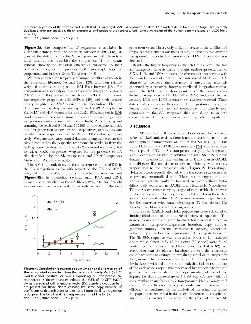
Finally, we correlated the expression level of the reporter gene
with the copy number of the transposon. The positional effect
variegation primarily observed with retroviral and lentiviral
vectors [30] could lead to the silencing of the therapeutic gene
delivered by the vector. We asked weather the SB integrations
would be affected by this phenomenon. We correlated the
expression of Venus protein, measured by Mean Fluorescence
Intensity (M.F.I.), with copy number of either the SA and T2
transposon, as determined by Southern blot or q-PCR analyses of
62 GABEB clones. For comparison, we analysed the M.F.I of a
GFP reporter gene, driven by the human Keratin 14 promoter, in
70 HaCaT clones isolated upon LV transduction. A linear
correlation curve was traced to retrieve the R2 coefficient of
determination. Transposon samples show an R2 = 0.759 with a
statistically defined correlation between two variables (PN = 0.6).
LV samples display an R2 = 0.001 with a null defined correlation
(Figure 4). Independent analysis of transposed clones obtained in
different cells (HaCaT and GABEB) and carrying a reporter gene
driven by PGK or CAGGS promoter showed comparable results
indicating common directly correlation between MFI and copy
number (data not shown). We conclude that SB integrants tend to
express their cargo faithfully, and multi-copy integrants express in
a copy-number dependent manner, consistent with earlier
observations [31].
Integration pattern analysisIn the last few years, several papers described the integration
profile of the SB, piggyBac (PB), and Tol2 transposons [20–23,32–
36]. Here we report the integration profile and preference of the
sandwich compared with the first-generation SB transposon [20]
in human epithelial cells. To generate a library of SA integration
events, we transfected 20 million GABEB cells with SA
transposon- and SB100X-carrying plasmids. The 20% of Venus-
positive cells were sorted three days after transfection to enrich the
population expressing the reporter gene. A 90%-pure sorted
population was kept in culture for 3 weeks to dilute the un-
integrated SA vector reaching a stable 78% Venus+ bulk
population. We used LM-PCR and pyrosequencing to generate
6,084 non-redundant SA-linked genomic sequences in human
immortalized GABEB keratinocytes. The Blast alignment re-
trieved 2,019 unambiguously mapped integration sites. As a
control, 10,000 random unique sequences were generated in silico
balancing the biases introduced by the LM-PCR (amplicon lenght
and MseI proximity) and the availability of the TA dinucleotides in
the genome. In the analysis we also annotated a large dataset
(59,169 hits) generated in HeLa cells transposed with the first-
generation Tneo transposon and selected for 2 weeks with
neomycin [20]. The integration sites and control sites were
annotated as transcriptional start site (TSS)-proximal when
mapping in the 62.5 kb window around a TSS, intragenic when
mapping within a transcription unit, and intergenic in all other
cases. Among SA integrations, 58.6% were in an intergenic
position, 38.9% were within the transcribed portion of at least 1
gene, and 2.5% was within a 5 kb window encompassing the TSS
Figure 2. Transposition efficiency. HeLa (A) and GABEB (B) cellswere co-transfected with the T2 and SA transposons- and transposase-carrying plasmids. The transposition rate, on the Y axis, is derived by theratio between the percentage of Venus+ cells at about 20 and 2 dayspost transfection. Data are representative of three independentexperiments (mean 6 SEM; n = 3).doi:10.1371/journal.pone.0112712.g002
Sleeping Beauty Transposition in Human Cells
PLOS ONE | www.plosone.org 5 November 2014 | Volume 9 | Issue 11 | e112712

Figure 3. Molecular characterization of the Sleeping Beauty-mediated integration events in GABEB cell clones. (A) Southern blotanalysis of genomic DNA from GABEB cell clones digested with NheI (SA clones) or AflII (T2 clones), single cutter in the transposon cassette, andhybridized to a Venus probe. A single band higher than 4.2 kb (SA clones) and 3.4 kb (T2 clones) indicates integration of one copy of the transposoninto the genome. Multiple Venus-specific bands correspond to repeated integration events. (B) Southern Blot analysis of genomic DNA from 8 (SA)and 9 (T2) clones digested with NcoI (SA clones) or MfeI and NdeI (T2 clones). The expected Venus-specific band corresponding to 6 kb for SA and8.9 kb for T2 transposon indicates the correct integration of the transposons into the genome. C, mock-transfected cells; red bars, Venus-specificprobe. Clone showing rearrangement of the transposon cassette is highlighted by black asterisk. (C) Bi-directional mapping of the junctions betweentransposon and genomic DNA. The table summarizes 27 integrations belonging to 10 single clones. For each integrant, the underlined sequence
Sleeping Beauty Transposition in Human Cells
PLOS ONE | www.plosone.org 6 November 2014 | Volume 9 | Issue 11 | e112712
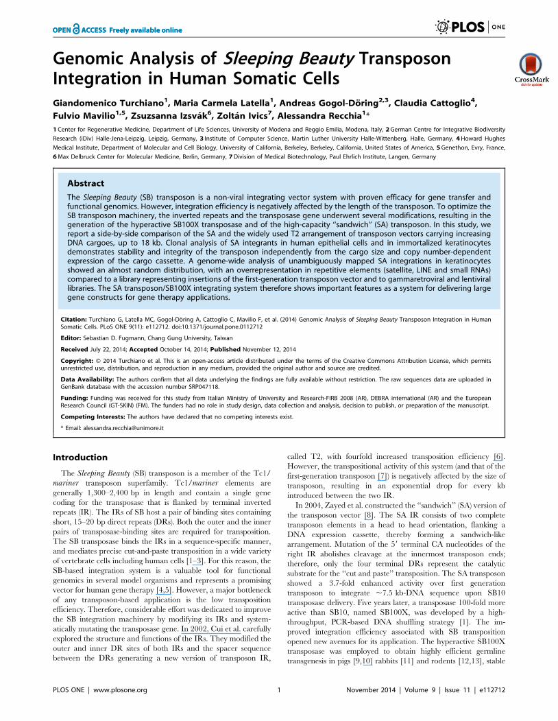
(Figure 5A; the complete list of sequences is available in
GenBank database with the accession number SRP047118). In
general, the distribution of the SB integrants in both datasets is
fairly random and resembles the composition of the human
genome showing no statistical differences compared to their
relative controls, i.e. all p-values (both two-sample tests for
proportions and Fisher’s Exact Tests) were .1022.
We then analysed the frequency of human repetitive elements in
the transposon libraries, SA and Tneo [20], and their relative
weighted controls availing of the RM Blast browser [28]. For
comparison we also analysed two viral-derived integration datasets
(MLV and HIV) generated in human CD34+ multipotent
hematopoietic progenitor cells (HPCs) [29] and their control
library weighted for MseI restriction site distribution. The raw
data generated by deep sequencing of the LM-PCR (applied to
SA, MLV and HIV treated cells) and LAM-PCR (applied in [20])
products were filtered and trimmed in order to rescue the genuine
integration events (see materials and methods). After filtering and
trimming we retrieved 6,084 and 165,887 unique sequences in SA
and first-generation vector libraries, respectively, and 37,873 and
31,204 unique sequences from MLV and HIV datasets, respec-
tively. We generated large control datasets taking into account the
bias introduced by the respective technique. In particular from the
hg19 genome database we retrieved 45,235 control reads weighted
for MseI, 65,570 sequences weighted for the presence of TA
dinucleotide hit by the SB transposons, and 209,913 sequences
MseI- and TA-double weighted.
The RM Blast analysis revealed an overrepresentation of REs in
the SA integrations (34%) with respect to the TA and MseI-
weighted control (14%) and to all the other datasets analysed
(Figure 5B). In particular, Satellite, small RNA and LINE
elements were enriched in the SA library (24-, 7.6- and 3.5-fold
increase over the background, respectively) whereas in the first-
generation vector library only a slight increase in the satellite and
simple repeats elements was measurable (3.5- and 2.6-fold over the
background, respectively); comparable LINE frequency was
detected.
Besides the higher frequency in the satellite elements, the two
SB transposon datasets share a slight under-representation of
SINE, LTR and DNA transposable elements in comparison with
their random control libraries. We introduced MLV and HIV
libraries to compare the frequency of integration into RE
generated by a retroviral integrase-mediated integration mecha-
nism. The RM Blast analysis pointed out that viral vectors
disfavour integration in RE (14–16% vs 24%), and, in particular,
satellite, LTR and LINE elements are underrepresented. These
data clearly confirm a difference in the integration site selection
between viral vectors and SB transposons and identify new
signatures in the SA integrome that should be taken into
consideration when using them as tools for genetic manipulation.
Discussion
The SB transposon IRs were mutated to improve their capacity
to be mobilized and, to date, there is not a direct comparison that
define genetic characteristics of the T2 and SA IRs [8]. In this
study, HeLa cells and GABEB keratinocytes [24] were transfected
with a panel of T2 or SA transposons carrying size-increasing
Venus expression cassette in combination with SB100X plasmid
(Figure 1). Transfection rate was higher in HeLa than in GABEB
cells (Figure S1) and the transposition efficiency was inversely
proportional to the transposon size (Figure 2). Interestingly,
HeLa cells were severely affected by the transposon size compared
to primary immortalized cells. These results suggest that the
transposase activity could be favoured by some cellular factor
differentially expressed in GABEB and HeLa cells. Nonetheless,
T2 and SA constructs carrying cargos of comparable size showed
similar transposition efficiency in both cell lines. From these data
we can conclude that the T2 IR construct is interchangeable with
the SA construct with some advantages: T2 has shorter IRs
thereby it could accept a larger cargo cassette.
Transposed GABEB and HeLa populations were subjected to
limiting dilution to obtain a single cell derived expansion. The
derived clones were employed to characterize several molecular
parameters: transposon-independent insertion, copy number,
genomic stability, faithful transposition activity, correlation
between copy number and expression of the integrated cassette.
The SB100X sequence was retrieved in 6 out of 211 analysed
clones while almost 14% of the clones (30 clones) were found
positive for the transposon backbone sequences (Table S2). We
hypothesize that the plasmid backbone carrying the transposon
could have some advantages to remain episomal or to integrate in
the genome. The transposon excision step from the plasmid leaves
the backbone with a double strand break that induce recruitment
of the endogenous repair machinery and integration into the cell
genome. We also analysed the copy number of the clones.
Figure 3A shows an average of 1.3 SA copies/clone while T2
copy number spans from 1 to 7 transposons with an average of 3
copies. This difference mostly depends on the transfection
efficiency as confirmed by the analysis of the other transposed
cell populations generated in this study. Therefore, it is possible to
fine tune this parameter by adjusting the ratios of the two SB
represents a portion of the transposon IRs, left (CAGTT) and right (AACTG) separated by dots; TA dinucleotide (in bold) is the target site correctlyduplicated after transposition. Hit chromosomes and positions are reported. UnK, unknown region of the human genome based on UCSC hg19assembly.doi:10.1371/journal.pone.0112712.g003
Figure 4. Correlation between copy number and expression ofthe integrated cassette. Mean fluorescence intensity (M.F.I.) of 62GABEB clones positive for Venus expressing SB transposons arerepresented as circles; triangles indicate the M.F.I. of 70 GFP+ HaCaTclones transduced with a lentiviral vector (LV). Standard deviation barsare present for those clones carrying the same copy number. R2
coefficients of determination were extracted from the linear regressionplot, green line for SA and T2 transposons and red line for LV.doi:10.1371/journal.pone.0112712.g004
Sleeping Beauty Transposition in Human Cells
PLOS ONE | www.plosone.org 7 November 2014 | Volume 9 | Issue 11 | e112712

components used for transfection or, as previously reported,
bypass the transfection procedure through the viral delivery of
transposase and transposon by adenoviral vector [37], integration
defective lentiviral vector [38,39], retroviral particle [40] and
adeno-associated vectors [41].
We were able to associate copy number of the transposon with
the expression level of the Venus fluorescence gene. Mean
Fluorescence Intensity does follow a direct proportion with the
copies harboured (Figure 4). In contrast, expression of the
reporter gene in lentiviral-mediated integrants does not correlate
with copy number and is more subjected to the activity of
surrounding genomic sequences [42,43].
Next, the integrated transposons in these clones were also
analysed for their integrity via Southern Blot. Retroviral and
lentiviral vectors can rearrange during the reverse transcription
step resulting in partially-deleted integrated proviruses, a frequent
occurrence in transgene hosting repetitive sequences [44,45]. The
SB mediated integration, by contrast, does not require reverse
transcription and thus is expected to preserve the integrity of the
transgene. Ninety-eight percent of the integrants, resulting from
T2 and SA transposition, have a correct size (Figure 3 B).
The sandwich transposon has a doubled IR/DR structure at
both ends with 8 transposase binding sites in total. In principle,
every transposase unit, bound to one DR site, could interact with
the others to create different chiasm geometries (also described in
[6]); some of these conformations could modify the integration
activity resulting in chromosomal aberrations. To investigate the
fidelity of the transposition process 10 GABEB clones were
mapped bi-directionally by LM-PCR and transposon-genome
junction was amplified by site-specific PCR. Twenty-five integra-
tions, out of 27 (92.6%), were validated for a canonical
transposition event with the TA target site duplication signature
at both ends (Figure 3C). Two integrations mapped by LM-PCR
were not confirmed in the opposite transposon end suggesting
Figure 5. Integration pattern analysis. (A) Integration sites were annotated as ‘‘TSS-proximal’’ when occurring within a distance of 62.5 kb fromthe gene’s TSS, as ‘‘Intragenic’’ when occurring in a gene body and as ‘‘Intergenic’’ in all other cases. Black bars represent exons of a schematic gene,arrowhead indicates the direction of transcription. Distribution of SA, Tneo and random integration sites in the genome is plotted accordingly todefined annotations. (B) Distribution of Repetitive Elements in SB SA and Tneo libraries, in MLV and HIV libraries. Relative weighted random librarieswere reported: TA and MseI-weighted for SA, TA-weighted for Tneo and MseI-weighted for MLV and HIV libraries. **p#1023, *p#1022.doi:10.1371/journal.pone.0112712.g005
Sleeping Beauty Transposition in Human Cells
PLOS ONE | www.plosone.org 8 November 2014 | Volume 9 | Issue 11 | e112712

rearrangements probably caused by the repair mechanism
occurred in the transposition break.
LM-PCR was also employed to derive a high-definition map of
SA/SB100X integration sites in the genome of a transposed
GABEB bulk population. This analysis is commonly applied to
integrating vectors (i.e. retroviral and lentiviral vectors) because it
allows to evaluate genotoxicity [46,47] and to understand
molecular mechanism driving the integration towards specific
regions of the genome [29,48–50]. The technique returned 2,019
SA unambiguously mappable integration sites randomly distrib-
uted throughout the human genome, in accordance with
previously published data on first-generation transposon [20,23]
(Figure 5A). For gene therapy purposes, the SB system results in a
safer integration profile compared to other integrating vector such
as Tol2, PB transposon and retroviral vectors [20–23,32,33],
which favor TSS-proximal regions or gene body sequences.
Although the integration site distribution in relation to genes
was found close to random, the RM Blast analysis shows a
significant bias distribution of SA integrations in repetitive
elements (RE), particularly in satellite, LINE and small RNA
genes. It could be that these genomic regions are favourable for
integration due to their base composition (TA-richness) or there
might be molecular mechanisms that actively recruit the
transposon/transposase complex at specific RE sites [51–55].
Curiously, the frequency of RE elements in the first-generation
transposon library and its weighted control were comparable.
Differently from the SA (obtained in 80% Venus expressing
immortalized keratinocytes without selective pressure), the first-
generation transposon library derives from transposed HeLa cells
[56] selected for two weeks by antibiotic resistance. This culture
condition could negatively select those integrations landing into
poorly expressed genomic loci or into heterochromatin regions
[35]. Nevertheless, the first-generation transposon integrations
were slightly increased into satellite regions and SINE, whereas
LTR and DNA elements were underrepresented compared to the
background.
These data identify some common features in SB datasets.
Conversely, MLV and HIV-derived viral vectors disfavour
integration in RE (satellite, LTR and LINE accordingly also to
[57]) suggesting an active role of viral integrase in the selection of
integration sites that could better support the expression,
replication and survival of the viral progeny. The genomic
features newly identified in the SA integrome raise an interesting
matter that needs to be deeply investigated for future application.
Supporting Information
Figure S1 Transposition trend. Expression of Venus
fluorescence protein was detected by cytofluorimetric analyses at
different time points in HeLa cells (A) and in GABEB cells (B). The
days post transfection (p.t.) are plotted on the X axis, while the
percentage of Venus+ cells are represented on the Y axis. The
maximum expression from a transfected reporter gene was
achieved 2 days p.t (black vertical dotted line). The plot shows
the samples co-transfected with SB100X and transposon plasmids:
T2 3.2 or the SA 5.7 (blue), T2 10 or SA 9.7 (green), T2 14 or SA
14 (purple), and the 18 kb transposons (orange). SA constructs
represented with dashed lines, T2 with continuous lines. In gold
are represented the negative controls transfected with T2 3.2 or
SA 5.7 alone, without the SB100X plasmid.
(EPS)
Table S1 List of primers used for plasmid episomialamplification, LM-PCR, and site-specific amplificationof the SA-genome junctions.
(DOC)
Table S2 Transposed clones were analysed to show thefollowing parameters: number of retrieved Venus+
clones for each bulk; percentage of Venus+ cells in bulkpopulations 48 hours p.t.; percentage of stable Venusexpressing cells in bulk populations; percentage ofclones positive for the Ampicillin or SB100X sequencecarried by transfected plasmids; mean copy numberretrieved by Southern blot analysis; recombinant eventsdetected in transposed clones by Southern blot analysis.
(DOC)
Acknowledgments
We thank Davide Pietrobon for compiling the scripts used for filtering and
trimming the raw sequences from SB and viral libraries.
Author Contributions
Conceived and designed the experiments: GT MCL. Performed the
experiments: GT MCL. Analyzed the data: GT MCL CC AGD FM Z.
Izsvak Z. Ivics AR. Wrote the paper: GT AR.
References
1. Mates L, Chuah M, Belay E, Jerchow B, Manoj N, et al. (2009) Molecularevolution of a novel hyperactive Sleeping Beauty transposase enables robust
stable gene transfer in vertebrates. Nature genetics 41: 753–761.
2. Ivics Z, Izsvak Z, Minter A, Hackett PB (1996) Identification of functional
domains and evolution of Tc1-like transposable elements. Proc Natl AcadSci U S A 93: 5008–5013.
3. Ivics Z, Hackett P, Plasterk R, Izsvak Z (1997) Molecular reconstruction ofSleeping Beauty, a Tc1-like transposon from fish, and its transposition in human
cells. Cell 91: 501–510.
4. Hackett PB, Largaespada DA, Cooper LJ (2010) A transposon and transposasesystem for human application. Mol Ther 18: 674–683.
5. Ivics Z, Kaufman C, Zayed H, Miskey C, Walisko O, et al. (2004) The SleepingBeauty transposable element: evolution, regulation and genetic applications.
Current issues in molecular biology 6: 43–55.
6. Cui Z, Geurts A, Liu G, Kaufman C, Hackett P (2002) Structure-function
analysis of the inverted terminal repeats of the sleeping beauty transposon.Journal of molecular biology 318: 1221–1235.
7. Izsvak Z, Ivics Z, Plasterk R (2000) Sleeping Beauty, a wide host-rangetransposon vector for genetic transformation in vertebrates. Journal of molecular
biology 302: 93–102.
8. Zayed H, Izsvak Z, Walisko O, Ivics Z (2004) Development of hyperactive
sleeping beauty transposon vectors by mutational analysis. Mol Ther 9: 292–
304.
9. Ivics Z, Garrels W, Mates L, Yau TY, Bashir S, et al. (2014) Germline
transgenesis in pigs by cytoplasmic microinjection of Sleeping Beauty
transposons. Nat Protoc 9: 810–827.
10. Garrels W, Holler S, Taylor U, Herrmann D, Niemann H, et al. (2014)
Assessment of fetal cell chimerism in transgenic pig lines generated by sleeping
beauty transposition. PLoS One 9: e96673.
11. Ivics Z, Hiripi L, Hoffmann OI, Mates L, Yau TY, et al. (2014) Germline
transgenesis in rabbits by pronuclear microinjection of Sleeping Beauty
transposons. Nat Protoc 9: 794–809.
12. Katter K, Geurts AM, Hoffmann O, Mates L, Landa V, et al. (2013)
Transposon-mediated transgenesis, transgenic rescue, and tissue-specific gene
expression in rodents and rabbits. FASEB J 27: 930–941.
13. Ivics Z, Mates L, Yau TY, Landa V, Zidek V, et al. (2014) Germline transgenesis
in rodents by pronuclear microinjection of Sleeping Beauty transposons. Nat
Protoc 9: 773–793.
14. Jin Z, Maiti S, Huls H, Singh H, Olivares S, et al. (2011) The hyperactive
Sleeping Beauty transposase SB100X improves the genetic modification of T
cells to express a chimeric antigen receptor. Gene Ther 18: 849–856.
15. Liu L, Sanz S, Heggestad AD, Antharam V, Notterpek L, et al. (2004)
Endothelial targeting of the Sleeping Beauty transposon within lung. Mol Ther
10: 97–105.
Sleeping Beauty Transposition in Human Cells
PLOS ONE | www.plosone.org 9 November 2014 | Volume 9 | Issue 11 | e112712

16. Belur LR, Frandsen JL, Dupuy AJ, Ingbar DH, Largaespada DA, et al. (2003)
Gene insertion and long-term expression in lung mediated by the SleepingBeauty transposon system. Mol Ther 8: 501–507.
17. Zhu J, Kren B, Park C, Bilgim R, Wong P, et al. (2007) Erythroid-specific
expression of beta-globin by the sleeping beauty transposon for Sickle celldisease. Biochemistry 46: 6844–6858.
18. Wilber A, Linehan JL, Tian X, Woll PS, Morris JK, et al. (2007) Efficient andstable transgene expression in human embryonic stem cells using transposon-
mediated gene transfer. Stem Cells 25: 2919–2927.
19. Grabundzija I, Wang J, Sebe A, Erdei Z, Kajdi R, et al. (2013) Sleeping Beautytransposon-based system for cellular reprogramming and targeted gene insertion
in induced pluripotent stem cells. Nucleic Acids Res 41: 1829–1847.20. Ammar I, Gogol-Doring A, Miskey C, Chen W, Cathomen T, et al. (2012)
Retargeting transposon insertions by the adeno-associated virus Rep protein.Nucleic acids research 40: 6693–6712.
21. Huang X, Guo H, Tammana S, Jung Y-C, Mellgren E, et al. (2010) Gene
transfer efficiency and genome-wide integration profiling of Sleeping Beauty,Tol2, and piggyBac transposons in human primary T cells. Mol Ther 18: 1803–
1813.22. Huang X, Wilber A, Bao L, Tuong D, Tolar J, et al. (2006) Stable gene transfer
and expression in human primary T cells by the Sleeping Beauty transposon
system. Blood 107: 483–491.23. Voigt K, Gogol-Doring A, Miskey C, Chen W, Cathomen T, et al. (2012)
Retargeting sleeping beauty transposon insertions by engineered zinc fingerDNA-binding domains. Mol Ther 20: 1852–1862.
24. Borradori L, Chavanas S, Schaapveld R, Gagnoux-Palacios L, Calafat J, et al.(1998) Role of the bullous pemphigoid antigen 180 (BP180) in the assembly of
hemidesmosomes and cell adhesion–reexpression of BP180 in generalized
atrophic benign epidermolysis bullosa keratinocytes. Experimental cell research239: 463–476.
25. McCormack W, Seiler M, Bertin T, Ubhayakar K, Palmer D, et al. (2006)Helper-dependent adenoviral gene therapy mediates long-term correction of the
clotting defect in the canine hemophilia A model. Journal of thrombosis and
haemostasis 4: 1218–1225.26. Sambrook J, Russell DW (2001) Molecular cloning: a laboratory manual. CSHL
press 1.27. Schmidt M, Hoffmann G, Wissler M, Lemke N, Mussig A, et al. (2001)
Detection and direct genomic sequencing of multiple rare unknown flankingDNA in highly complex samples. Hum Gene Ther 12: 743–749.
28. Smit AFA, Hubley R, Green P (1996-2010) RepeatMasker Open-3.0. Available:
http://repeatmasker.org. Accessed 2014 Oct 23.29. Cattoglio C, Facchini G, Sartori D, Antonelli A, Miccio A, et al. (2007) Hot
spots of retroviral integration in human CD34+ hematopoietic cells. Blood 110:1770–1778.
30. Cavazza A, Cocchiarella F, Bartholomae C, Schmidt M, Pincelli C, et al. (2013)
Self-inactivating MLV vectors have a reduced genotoxic profile in humanepidermal keratinocytes. Gene Ther 20: 949–957.
31. Garrels W, Mates L, Holler S, Dalda A, Taylor U, et al. (2011) Germlinetransgenic pigs by Sleeping Beauty transposition in porcine zygotes and targeted
integration in the pig genome. PLoS One 6: e23573.32. Hackett P, Largaespada D, Switzer K, Cooper L (2013) Evaluating risks of
insertional mutagenesis by DNA transposons in gene therapy. Translational
research.33. Yant S, Wu X, Huang Y, Garrison B, Burgess S, et al. (2005) High-resolution
genome-wide mapping of transposon integration in mammals. Molecular andcellular biology 25: 2085–2094.
34. Zhang W, Muck-Hausl M, Wang J, Sun C, Gebbing M, et al. (2013) Integration
profile and safety of an adenovirus hybrid-vector utilizing hyperactive sleepingbeauty transposase for somatic integration. PLoS One 8: e75344.
35. de Jong J, Akhtar W, Badhai J, Rust AG, Rad R, et al. (2014) Chromatinlandscapes of retroviral and transposon integration profiles. PLoS Genet 10:
e1004250.
36. Wang Y, Wang J, Devaraj A, Singh M, Jimenez Orgaz A, et al. (2014) Suicidalautointegration of sleeping beauty and piggyBac transposons in eukaryotic cells.
PLoS Genet 10: e1004103.
37. Yant S, Ehrhardt A, Mikkelsen J, Meuse L, Pham T, et al. (2002) Transposition
from a gutless adeno-transposon vector stabilizes transgene expression in vivo.
Nature biotechnology 20: 999–1005.
38. Moldt B, Miskey C, Staunstrup N, Gogol-Doring A, Bak R, et al. (2011)
Comparative genomic integration profiling of Sleeping Beauty transposons
mobilized with high efficacy from integrase-defective lentiviral vectors in
primary human cells. Mol Ther 19: 1499–1510.
39. Field AC, Vink C, Gabriel R, Al-Subki R, Schmidt M, et al. (2013) Comparison
of lentiviral and sleeping beauty mediated alphabeta T cell receptor gene
transfer. PLoS One 8: e68201.
40. Galla M, Schambach A, Falk C, Maetzig T, Kuehle J, et al. (2011) Avoiding
cytotoxicity of transposases by dose-controlled mRNA delivery. Nucleic acids
research 39: 7147–7160.
41. Zhang W, Solanki M, Muther N, Ebel M, Wang J, et al. (2013) Hybrid adeno-
associated viral vectors utilizing transposase-mediated somatic integration for
stable transgene expression in human cells. PloS one 8.
42. Moiani A, Paleari Y, Sartori D, Mezzadra R, Miccio A, et al. (2012) Lentiviral
vector integration in the human genome induces alternative splicing and
generates aberrant transcripts. The Journal of clinical investigation 122: 1653–
1666.
43. Cesana D, Sgualdino J, Rudilosso L, Merella S, Naldini L, et al. (2012) Whole
transcriptome characterization of aberrant splicing events induced by lentiviral
vector integrations. The Journal of clinical investigation 122: 1667–1676.
44. Titeux M, Pendaries V, Zanta-Boussif MA, Decha A, Pironon N, et al. (2010)
SIN retroviral vectors expressing COL7A1 under human promoters for ex vivo
gene therapy of recessive dystrophic epidermolysis bullosa. Mol Ther 18: 1509–
1518.
45. Holkers M, Maggio I, Liu J, Janssen JM, Miselli F, et al. (2013) Differential
integrity of TALE nuclease genes following adenoviral and lentiviral vector gene
transfer into human cells. Nucleic Acids Res 41: e63.
46. Aiuti A, Cassani B, Andolfi G, Mirolo M, Biasco L, et al. (2007) Multilineage
hematopoietic reconstitution without clonal selection in ADA-SCID patients
treated with stem cell gene therapy. The Journal of clinical investigation 117:
2233–2240.
47. Biffi A, Bartolomae C, Cesana D, Cartier N, Aubourg P, et al. (2011) Lentiviral
vector common integration sites in preclinical models and a clinical trial reflect a
benign integration bias and not oncogenic selection. Blood 117: 5332–5339.
48. Bushman F, Lewinski M, Ciuffi A, Barr S, Leipzig J, et al. (2005) Genome-wide
analysis of retroviral DNA integration. Nature reviews Microbiology 3: 848–858.
49. Bushman F (2007) Retroviral integration and human gene therapy. The Journal
of clinical investigation 117: 2083–2086.
50. Montini E, Cesana D, Schmidt M, Sanvito F, Bartholomae C, et al. (2009) The
genotoxic potential of retroviral vectors is strongly modulated by vector design
and integration site selection in a mouse model of HSC gene therapy. The
Journal of clinical investigation 119: 964–975.
51. Liu G, Geurts AM, Yae K, Srinivasan AR, Fahrenkrug SC, et al. (2005) Target-
site preferences of Sleeping Beauty transposons. J Mol Biol 346: 161–173.
52. Vigdal T, Kaufman C, Izsvak Z, Voytas D, Ivics Z (2002) Common physical
properties of DNA affecting target site selection of sleeping beauty and other
Tc1/mariner transposable elements. Journal of molecular biology 323: 441–452.
53. Olson W, Zhurkin V (2011) Working the kinks out of nucleosomal DNA.
Current opinion in structural biology 21: 348–357.
54. Foltz D, Jansen L, Black B, Bailey A, Yates J, et al. (2006) The human CENP-A
centromeric nucleosome-associated complex. Nature cell biology 8: 458–469.
55. Masumoto H, Nakano M, Ohzeki J-I (2004) The role of CENP-B and alpha-
satellite DNA: de novo assembly and epigenetic maintenance of human
centromeres. Chromosome research 12: 543–556.
56. De Luca M, Pellegrini G, Mavilio F (2009) Gene therapy of inherited skin
adhesion disorders: a critical overview. The British journal of dermatology 161:
19–24.
57. Carteau S, Hoffmann C, Bushman F (1998) Chromosome structure and human
immunodeficiency virus type 1 cDNA integration: centromeric alphoid repeats
are a disfavored target. J Virol 72: 4005–4014.
Sleeping Beauty Transposition in Human Cells
PLOS ONE | www.plosone.org 10 November 2014 | Volume 9 | Issue 11 | e112712