Genome sequence of Ensifer meliloti strain WSM1022; a highly effective microsymbiont of the model...
10
Standards in Genomic Sciences (2013) 9: 315-32 4 DOI:10.4056/sigs.4608286 The Genomic Standards Consortium Genome sequence of Ensifer meliloti strain WSM1022; a highly effective microsymbiont of the model legume Medicago truncatula A17 Jason Terpolilli 1 , Yvette Hill 1 , Rui Tian 1 , John Howieson 1 , Lambert Bräu 2 , Lynne Goodwin 3 , James Han 4 , Konstantinos Liolios 4 , Marcel Huntemann 4 , Amrita Pati 5 , Tanja Woyke 4 , Konstantinos Mavromatis 5 , Victor Markowitz 5 , Natalia Ivanova 3 , Nikos Kyrpides 3 , Wayne Reeve 1* . 1 Centre for Rhizobium Studies, Murdoch University, Western Australia, Australia 2 School of Life and Environmental Sciences, Deakin University, Victoria, Australia 3 Los Alamos National Laboratory, Bioscience Division, Los Alamos, New Mexico, USA 4 DOE Joint Genome Institute, Walnut Creek, California, USA 5 Biological Data Management and Technology Center, Lawrence Berkeley National Labora- tory, Berkeley, California, USA *Correspondence: Wayne Reeve ( [email protected]) Keywords: root-nodule bacteria, nitrogen fixation, rhizobia, Alphaproteobacteria Ensifer meliloti WSM1022 is an aerobic, motile, Gram-negative, non-spore-forming rod that can exist as a soil saprophyte or as a legume microsymbiont of Medicago. WSM1022 was isolated in 1987 from a nodule recovered from the roots of the annual Medicago orbicularis growing on the Cyclades Island of Naxos in Greece. WSM1022 is highly effective at fixing ni- trogen with M. truncatula and other annual species such as M. tornata and M. littoralis and is also highly effective with the perennial M. sativa (alfalfa or lucerne). In common with other characterized E. meliloti strains, WSM1022 will nodulate but fixes poorly with M. polymorpha and M. sphaerocarpos and does not nodulate M. murex. Here we describe the features of E. meliloti WSM1022, together with genome sequence information and its annota- tion. The 6,649,661 bp high-quality-draft genome is arranged into 121 scaffolds of 125 contigs containing 6,323 protein-coding genes and 75 RNA-only encoding genes, and is one of 100 rhizobial genomes sequenced as part of the DOE Joint Genome Institute 2010 Ge- nomic Encyclopedia for Bacteria and Archaea-Root Nodule Bacteria (GEBA-RNB) project. Introduction An available source of nitrogen (N) is essential to life on Earth. Although the atmosphere consists of approximately 80% N, the overwhelming propor- tion of this is present in the form of dinitrogen (N2) which is biologically inaccessible to the vast majority of higher organisms. Only a subset of mi- crobes has the necessary molecular machinery to make atmospheric N2 bioavailable by enzymatical- ly reducing N2 to NH3. The fact that plant growth is most commonly limited by the availability of N may have provided the selective pressure for a wide range of plant genera, most of which are leg- umes, to evolve a symbiotic relationship with the- se N2-fixing microbes. These microsymbionts, col- lectively termed root nodule bacteria, receive a carbon source from the plant and in return supply the host with biologically fixed N. When these symbiotic interactions are optimally harnessed in agriculture, all the N-requirements of the host can be met, without the need to apply industrially syn- thesized N-based fertilizers, thereby increasing both the economic and environmental sustainabil- ity of the farming system [1]. Forage and fodder legumes play an integral role in sustainable farming practice, providing feed for stock while also enriching soil with bioavailable N. Worldwide, there are approximately 110 million ha of forage and fodder legumes under production [2], of which members of the Medicago genus comprise a considerable component. Two bacteri- al species, Ensifer meliloti and E. medicae are known to nodulate and fix N2 with Medicago spp.
-
Upload
independent -
Category
Documents
-
view
0 -
download
0
Transcript of Genome sequence of Ensifer meliloti strain WSM1022; a highly effective microsymbiont of the model...

Standards in Genomic Sciences (2013) 9:315-324 DOI:10.4056/sigs.4608286
The Genomic Standards Consortium
Genome sequence of Ensifer meliloti strain WSM1022; a highly effective microsymbiont of the model legume Medicago truncatula A17 Jason Terpolilli1, Yvette Hill1, Rui Tian1, John Howieson1, Lambert Bräu2, Lynne Goodwin3,
James Han4, Konstantinos Liolios4, Marcel Huntemann4, Amrita Pati5, Tanja Woyke4, Konstantinos Mavromatis5, Victor Markowitz5, Natalia Ivanova3, Nikos Kyrpides3, Wayne
Reeve1*.
1 Centre for Rhizobium Studies, Murdoch University, Western Australia, Australia 2 School of Life and Environmental Sciences, Deakin University, Victoria, Australia 3 Los Alamos National Laboratory, Bioscience Division, Los Alamos, New Mexico, USA 4 DOE Joint Genome Institute, Walnut Creek, California, USA 5 Biolog ical Data Management and Technology Center, Lawrence Berkeley National Labora-
tory, Berkeley, California, USA
*Correspondence: Wayne Reeve ([email protected])
Keywords: root-nodule bacteria, nitrogen fixation, rhizobia, Alphaproteobacteria
Ensifer meliloti WSM1022 is an aerobic, motile, Gram-negative, non-spore-forming rod that can exist as a soil saprophyte or as a legume microsymbiont of Medicago. WSM1022 was isolated in 1987 from a nodule recovered from the roots of the annual Medicago orb icularis g rowing on the Cyclades Island of Naxos in Greece. WSM1022 is highly effective at fixing ni-trogen with M. truncatula and other annual species such as M. tornata and M. littoralis and is also highly effective with the perennial M. sativa (alfalfa or lucerne). In common with other characterized E. meliloti strains, WSM1022 will nodulate but fixes poorly with M. polymorpha and M. sphaerocarpos and does not nodulate M. murex. Here we describe the features of E. meliloti WSM1022, together with genome sequence information and its annota-tion. The 6,649,661 bp high-quality-draft genome is arranged into 121 scaffolds of 125 contigs containing 6,323 protein-coding genes and 75 RNA-only encoding genes, and is one of 100 rhizobial genomes sequenced as part of the DOE Joint Genome Institute 2010 Ge-nomic Encyclopedia for Bacteria and Archaea-Root Nodule Bacteria (GEBA-RNB) project.
Introduction An available source of nitrogen (N) is essential to life on Earth. Although the atmosphere consists of approximately 80% N, the overwhelming propor-tion of this is present in the form of dinitrogen (N2) which is biologically inaccessible to the vast majority of higher organisms. Only a subset of mi-crobes has the necessary molecular machinery to make atmospheric N2 bioavailable by enzymatical-ly reducing N2 to NH3. The fact that plant growth is most commonly limited by the availability of N may have provided the selective pressure for a wide range of plant genera, most of which are leg-umes, to evolve a symbiotic relationship with the-se N2-fixing microbes. These microsymbionts, col-lectively termed root nodule bacteria, receive a carbon source from the plant and in return supply
the host with biologically fixed N. When these symbiotic interactions are optimally harnessed in agriculture, all the N-requirements of the host can be met, without the need to apply industrially syn-thesized N-based fertilizers, thereby increasing both the economic and environmental sustainabil-ity of the farming system [1]. Forage and fodder legumes play an integral role in sustainable farming practice, providing feed for stock while also enriching soil with bioavailable N. Worldwide, there are approximately 110 million ha of forage and fodder legumes under production [2], of which members of the Medicago genus comprise a considerable component. Two bacteri-al species, Ensifer meliloti and E. medicae are known to nodulate and fix N2 with Medicago spp.
Ensifer meliloti strain WSM1022
316 Standards in Genomic Sciences
[3], although they differ in their symbiotic proper-ties on some Medicago hosts. Specifically, while E. medicae can nodulate and fix N2 with M. murex, M. arabica and M. polymorpha, E. meliloti does not nodulate M. murex, does not fix with M. polymorpha and fixes N2 very poorly with M. ara-bica [4-6]. E. meliloti strain WSM1022 was isolated in 1987 from a nodule collected from the annual M. orbicu-laris growing on the Cyclades Island of Naxos in Greece. E. meliloti WSM1022 is a highly effective microsymbiont of Medicago, forming efficient N2-fixing associations with the annual species M. littoralis and M. tornata [7]. In common with E. medicae WSM419 [8], WSM1022 also fixes ap-proximately twice as much N2 as E. meliloti 1021 on the model legume M. truncatula A17 [7]. How-ever, unlike E. medicae WSM419, E. meliloti WSM1022 is also highly effective with the peren-nial M. sativa (alfalfa or lucerne) [7]. Therefore, E. meliloti WSM1022 is a broadly effective microsymbiont of Medicago spp. and as such rep-resents a unique tool for the molecular analysis of effective N2 fixation with fully sequenced macro-and microsymbionts. Here we present a summary classification and a set of general features for E. meliloti strain WSM1022 together with a descrip-tion of its genome sequence and annotation.
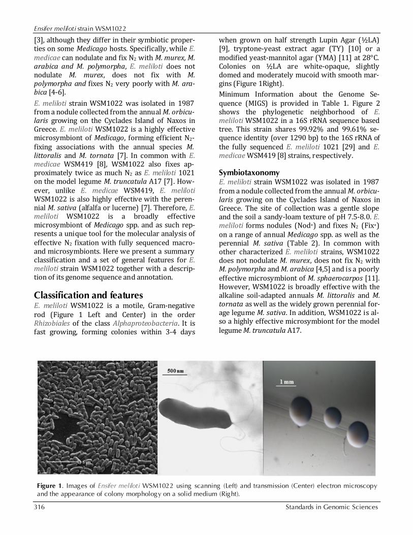
Classification and features E. meliloti WSM1022 is a motile, Gram-negative rod (Figure 1 Left and Center) in the order Rhizobiales of the class Alphaproteobacteria. It is fast growing, forming colonies within 3-4 days
when grown on half strength Lupin Agar (½LA) [9], tryptone-yeast extract agar (TY) [10] or a modified yeast-mannitol agar (YMA) [11] at 28°C. Colonies on ½LA are white-opaque, slightly domed and moderately mucoid with smooth mar-gins (Figure 1Right). Minimum Information about the Genome Se-quence (MIGS) is provided in Table 1. Figure 2 shows the phylogenetic neighborhood of E. meliloti WSM1022 in a 16S rRNA sequence based tree. This strain shares 99.92% and 99.61% se-quence identity (over 1290 bp) to the 16S rRNA of the fully sequenced E. meliloti 1021 [29] and E. medicae WSM419 [8] strains, respectively.
Symbiotaxonomy E. meliloti strain WSM1022 was isolated in 1987 from a nodule collected from the annual M. orbicu-laris growing on the Cyclades Island of Naxos in Greece. The site of collection was a gentle slope and the soil a sandy-loam texture of pH 7.5-8.0. E. meliloti forms nodules (Nod+) and fixes N2 (Fix+) on a range of annual Medicago spp. as well as the perennial M. sativa (Table 2). In common with other characterized E. meliloti strains, WSM1022 does not nodulate M. murex, does not fix N2 with M. polymorpha and M. arabica [4,5] and is a poorly effective microsymbiont of M. sphaerocarpos [11]. However, WSM1022 is broadly effective with the alkaline soil-adapted annuals M. littoralis and M. tornata as well as the widely grown perennial for-age legume M. sativa. In addition, WSM1022 is al-so a highly effective microsymbiont for the model legume M. truncatula A17.
Figure 1. Images of Ensifer meliloti WSM1022 using scanning (Left) and transmission (Center) electron microscopy and the appearance of colony morphology on a solid medium (Right).

Terpolilli et al.
http://standardsingenomics.org 317
Table 1. Classification and general features of Ensifer meliloti WSM1022 according to the MIGS recommendations [12]
MIGS ID Property Term Evidence code
Current classification
Domain Bacteria TAS [13]
Phylum Proteobacteria TAS [14]
Class Alphaproteobacteria TAS [15,16]
Order Rhizob iales TAS [16,17]
Family Rhizob iaceae TAS [18,19]
Genus Ensifer TAS [20-22]
Species Ensifer meliloti TAS [21]
Gram stain Negative IDA
Cell shape Rod IDA
Motility Motile IDA
Sporulation Non-sporulating NAS
Temperature range Mesophile NAS
Optimum temperature 28°C NAS
Salinity Non-halophile NAS
MIGS-22 Oxygen requirement Aerobic TAS [7]
Carbon source Varied NAS
Energy source Chemoorganotroph NAS
MIGS-6 Habitat Soil, root nodule, on host TAS [7]
MIGS-15 Biotic relationship Free living , symbiotic TAS [7]
MIGS-14 Pathogenicity Non-pathogenic NAS
Biosafety level 1 TAS [23]
Isolation Root nodule TAS [11]
MIGS-4 Geographic location Naxos, Greece TAS [11]
MIGS-5 Soil collection date 28 April 1987 IDA
MIGS-4.1 Longitude 37.107772 IDA
MIGS-4.2 Latitude 25.387841
MIGS-4.3 Depth 0-10cm
MIGS-4.4 Altitude Not recorded
Evidence codes – IDA: Inferred from Direct Assay; TAS: Traceable Author Statement (i.e., a direct report exists in the literature); NAS: Non-traceable Author Statement (i.e., not di-rectly observed for the living , isolated sample, but based on a generally accepted property for the species, or anecdotal evidence). These evidence codes are from the Gene Ontolo-gy project [24].

Ensifer meliloti strain WSM1022
318 Standards in Genomic Sciences
Figure 2. Phylogenetic tree showing the relationship of Ensifer meliloti WSM1022 (shown in bold print) to other Ensifer spp. in the order Rhizob iales based on aligned sequences of the 16S rRNA gene (1,290 bp internal region). All sites were informative and there were no gap-containing sites. Phylogenetic analyses were performed using MEGA, version 5 [25]. The tree was built using the Maximum-Likelihood method with the General Time Reversible model [26]. Bootstrap analysis [27] with 500 replicates was performed to assess the support of the clusters. Type strains are indicated with a superscript T. Brackets after the strain name contain a DNA database accession number and/or a GOLD ID (beg inning with the prefix G) for a sequencing project registered in GOLD [28]. Published genomes are indicated with an asterisk.

Terpolilli et al.
http://standardsingenomics.org 319
Table 2. Nodulation and N2 fixation properties of E. meliloti WSM1022 on selected Medicago spp. Data compiled from [7,11]†
Species Name Cultivar or Accession
Growth Habit Nodulation N2 fixation Comment
M. truncatula A17 Annual Nod+ Fix+ Highly effective
M. truncatula Jemalong Annual Nod+ Fix+ Highly effective
M. truncatula Caliph Annual Nod+ Fix+ Highly effective
M. littoralis Harbinger Annual Nod+ Fix+ Highly effective
M. tornata Tornafield Annual Nod+ Fix+ Highly effective
M. sphaerocarpos Orion Annual Nod+ Fix+ Poorly effective
M. arabica SA36043 Annual Nod+ Fix- No fixation
M. polymorpha Santiago Annual Nod+ Fix- No fixation
M. murex Zodiac Annual Nod- Fix- No nodulation
M. sativa Sceptre Perennial Nod+ Fix+ Highly effective
†Note that ‘+’ and ‘-’ denote presence or absence, respectively, of nodulation (Nod) or N2 fixation (Fix).
Genome sequencing and annotation Genome project history This organism was selected for sequencing on the basis of its environmental and agricultural rele-vance to issues in global carbon cycling, alterna-tive energy production, and biogeochemical im-portance, and is part of the Community Sequenc-ing Program at the U.S. Department of Energy, Joint Genome Institute (JGI) for projects of rele-vance to agency missions. The genome project is deposited in the Genomes OnLine Database [28] and an improved-high-quality-draft genome se-quence in IMG. Sequencing, finishing and annota-
tion were performed by the JGI. A summary of the project information is shown in Table 3.
Growth conditions and DNA isolation E. meliloti WSM1022 was cultured to mid loga-rithmic phase in 60 ml of TY rich medium [30] on a gyratory shaker at 28°C. DNA was isolated from the cells using a CTAB (Cetyl trimethyl ammonium bromide) bacterial genomic DNA isolation method [31].

Ensifer meliloti strain WSM1022
320 Standards in Genomic Sciences
Table 3. Genome sequencing project information for E. meliloti WSM1022. MIGS ID Property Term
MIGS-31 Finishing quality Improved high-quality draft
MIGS-28 Libraries used 1× Illumina library
MIGS-29 Sequencing platforms Illumina HiSeq 2000
MIGS-31.2 Sequencing coverage Illumina: 275×
MIGS-30 Assemblers Velvet version 1.1.04; Allpaths-LG version r42328
MIGS-32 Gene calling methods Prodigal 1.4, GenePRIMP
GOLD ID Gi08916
NCBI project ID 78233
Database: IMG 2510065057
Project relevance Symbiotic N2 fixation, agriculture
Genome sequencing and assembly The genome of Ensifer meliloti WSM1022 was se-quenced at the Joint Genome Institute (JGI) using Illumina technology [32]. An Illumina standard shotgun library was constructed and sequenced using the Illumina HiSeq 2000 platform which generated 12,082,430 reads totaling 1812.4 Mbp. All general aspects of library construction and se-quencing performed at the JGI can be found at the JGI website [31]. All raw Illumina sequence data was passed through DUK, a filtering program de-veloped at JGI, which removes known Illumina sequencing and library preparation artifacts (Mingkun, L., Copeland, A. and Han, J., un-published). The following steps were then per-formed for assembly: (1) filtered Illumina reads were assembled using Velvet [33] (version 1.1.04), (2) 1–3 kb simulated paired end reads were created from Velvet contigs using wgsim (https://github.com/lh3/wgsim), (3) Illumina reads were assembled with simulated read pairs using Allpaths–LG [34] (version r42328). Parame-ters for assembly steps were: 1) Velvet (velveth: 63 –shortPaired and velvetg: –veryclean yes –exportFiltered yes –mincontiglgth 500 –scaffolding no–covcutoff 10) 2) wgsim (–e 0 –1 100 –2 100 –r 0 –R 0 –X 0) 3) Allpaths–LG (PrepareAllpathsInputs:PHRED64=1 PLOIDY=1 FRAGCOVERAGE=125 JUMPCOVERAGE=25 LONGJUMPCOV=50, RunAllpath-sLG: THREADS=8 RUN=stdshredpairs TARGETS=standard VAPIWARNONLY=True OVERWRITE=True). The final draft assembly contained 125 contigs in 121 scaffolds. The total size of the genome is 6.6 Mb and the final assembly is based on 1,812.4 Mbp of
Illumina data, which provides an average 275× coverage of the genome.
Genome annotation Genes were identified using Prodigal [35] as part of the DOE-JGI annotation pipeline [36]. The pre-dicted CDSs were translated and used to search the National Center for Biotechnology Information (NCBI) nonredundant database, UniProt, TIGRFam, Pfam, PRIAM, KEGG, COG, and InterPro databases. The tRNAScanSE tool [37] was used to find tRNA genes, whereas ribosomal RNA genes were found by searches against models of the ri-bosomal RNA genes built from SILVA [38]. Other non–coding RNAs such as the RNA components of the protein secretion complex and the RNase P were identified by searching the genome for the corresponding Rfam profiles using INFERNAL (http://infernal.janelia.org). Additional gene pre-diction analysis and manual functional annotation was performed within the Integrated Microbial Genomes (IMG-ER) platform [39].
Genome properties The genome is 6,649,661 nucleotides with 62.16% GC content (Table 4) and comprised of 121 scaf-folds (Figure 3) of 125 contigs. From a total of 6,398 genes, 6,323 were protein encoding and 75 RNA only encoding genes. The majority of genes (80.78%) were assigned a putative function whilst the remaining genes were annotated as hypothet-ical. The distribution of genes into COGs functional categories is presented in Table 5.

Terpolilli et al.
http://standardsingenomics.org 321
Table 4. Genome Statistics for Ensifer meliloti WSM1022 Attribute Value % of Total Genome size (bp) 6,649,661 100.00 DNA coding reg ion (bp) 5,733,017 86.22 DNA G+C content (bp) 4,133,661 62.16 Number of scaffolds 121 Number of contigs 125 Total gene 6,398 100.00 RNA genes 75 1.17 rRNA operons 1 0.02 Protein-coding genes 6,323 98.83 Genes with function prediction 5,168 80.78 Genes assigned to COGs 5,147 80.45 Genes assigned Pfam domains 5,331 83.32 Genes with signal peptides 563 8.80 Genes with transmembrane helices 1,437 22.93 CRISPR repeats 0
Figure 3. Graphical map of the genome of Ensifer meliloti WSM1022 showing the seven largest scaffolds. From bottom to the top of each scaffold: Genes on forward strand (color by COG categories as denoted by the IMG platform), Genes on reverse strand (color by COG categories), RNA genes (tRNAs green, sRNAs red, other RNAs black), GC content, GC skew.

Ensifer meliloti strain WSM1022
322 Standards in Genomic Sciences
Table 5. Number of protein coding genes of Ensifer meliloti WSM1022 associated with the general COG functional categories.
Code Value % age COG Category
J 194 3.38 Translation, ribosomal structure and biogenesis
A 0 0.00 RNA processing and modification
K 497 8.65 Transcription
L 196 3.41 Replication, recombination and repair
B 1 0.02 Chromatin structure and dynamics
D 38 0.66 Cell cycle control, mitosis and meiosis
Y 0 0.00 Nuclear structure
V 61 1.06 Defence mechanisms
T 235 4.09 Signal transduction mechanisms
M 301 5.24 Cell wall/membrane biogenesis
N 71 1.24 Cell motility
Z 0 0.00 Cytoskeleton
W 1 0.02 Extracellular structures
U 113 1.97 Intracellular trafficking and secretion
O 177 3.08 Posttranslational modification, protein turnover, chaperones
C 357 6.21 Energy production conversion
G 606 10.54 Carbohydrate transport and metabolism
E 623 10.84 Amino acid transport metabolism
F 109 1.90 Nucleotide transport and metabolism
H 200 3.48 Coenzyme transport and metabolism
I 207 3.60 Lipid transport and metabolism
P 312 5.43 Inorganic ion transport and metabolism
Q 158 2.75 Secondary metabolite biosynthesis, transport and catabolism
R 708 12.32 General function prediction only
S 583 10.14 Function unknown
- 1,251 19.55 Not in COGS
Total 5,748 - -
Acknowledgements This work was performed under the auspices of the US Department of Energy’s Office of Science, Biological and Environmental Research Program, and by the Universi-ty of California, Lawrence Berkeley National Laborato-ry under contract No. DE-AC02-05CH11231, Lawrence Livermore National Laboratory under Contract No. DE-AC52-07NA27344, and Los Alamos National Laborato-ry under contract No. DE-AC02-06NA25396. We grate-
fully acknowledge the funding received from the Mur-doch University Strategic Research Fund through the Crop and Plant Research Institute (CaPRI) and the Cen-tre for Rhizobium Studies (CRS) at Murdoch Universi-ty. We also acknowledge ECR funding for J. Terpolilli awarded by the School of Veterinary and Life Sciences at Murdoch University.

Terpolilli et al.
http://standardsingenomics.org 323
References 1. Howieson JG, O’Hara GW, Carr SJ. Changing roles
for legumes in Mediterranean agriculture: develop-ments from an Australian perspective. Field Crops Res 2000; 65:107-122. http://dx.doi.org/10.1016/S0378-4290(99)00081-7
2. Herridge DF, Peoples MB, Boddey RM. Global in-puts of biological nitrogen fixation in agricultural systems. Plant Soil 2008; 311:1-18. http://dx.doi.org/10.1007/s11104-008-9668-3
3. Graham P. Ecology of the root-nodule bacteria of legumes. In: Dilworth MJ, James EK, Sprent JI, New-ton WE, editors. Nitrogen-Fixing Leguminous Sym-bioses. Dodrecht: The Netherlands: Springer; 2008. p 23-43.
4. Garau G, Reeve WG, Brau L, Yates RJ, James D, Tiwari R, O'Hara GW, Howieson JG. The symbiotic requirements of different Medicago spp. suggest the evolution of Sinorhizobium meliloti and S. medicae with hosts differentially adapted to soil pH. Plant Soil 2005; 276:263-277. http://dx.doi.org/10.1007/s11104-005-0374-0
5. Rome S, Cleyet-Marel JC, Materon LA, Normand P, Brunel B. Rapid identification of Medicago nodulating strains by using two oligonucleotide probes complementary to 16S rDNA sequences. Can J Microbiol 1997; 43:854-861. PubMed http://dx.doi.org/10.1139/m97-124
6. Brunel B, Rome S, Ziani R, Cleyet-Marel JC. Com-parison of nucleotide diversity and symbiotic prop-erties of Rhizobium meliloti populations from annual Medicago species. FEMS Microbiol Ecol 1996; 19:71-82. http://dx.doi.org/10.1111/j.1574-6941.1996.tb00200.x
7. Terpolilli JJ, O'Hara GW, Tiwari RP, Dilworth MJ, Howieson JG. The model legume Medicago truncatula A17 is poorly matched for N2 fixation with the sequenced microsymbiont Sinorhizobium meliloti 1021. New Phytol 2008; 179:62-66. Pub-Med http://dx.doi.org/10.1111/j.1469-8137.2008.02464.x
8. Reeve W, Chain P, O'Hara G, Ardley J, Nandesena K, Brau L, Tiwari R, Malfatti S, Kiss H, Lapidus A, et al. Complete genome sequence of the Medicago microsymbiont Ensifer (Sinorhizobium) medicae strain WSM419. Stand Genomic Sci 2010; 2:77-86. PubMed http://dx.doi.org/10.4056/sigs.43526
9. Howieson JG, Ewing MA, D'antuono MF. Selection for acid tolerance in Rhizobium meliloti. Plant Soil 1988; 105:179-188. http://dx.doi.org/10.1007/BF02376781
10. Beringer JE. R factor transfer in Rhizobium leguminosarum. J Gen Microbiol 1974; 84:188-198. PubMed http://dx.doi.org/10.1099/00221287-84-1-188
11. Terpolilli JJ. Why are the symbioses between some genotypes of Sinorhizobium and Medicago subop-timal for N2 fixation? Perth: Murdoch University; 2009. 223 p.
12. Field D, Garrity G, Gray T, Morrison N, Selengut J, Sterk P, Tatusova T, Thomson N, Allen M, Angiuoli SV, et al. Towards a richer description of our com-plete collection of genomes and metagenomes "Minimum Information about a Genome Sequence " (MIGS) specification. Nat Biotechnol 2008; 26:541-547. PubMed http://dx.doi.org/10.1038/nbt1360
13. Woese CR, Kandler O, Wheelis ML. Towards a nat-ural system of organisms: proposal for the domains Archaea, Bacteria, and Eucarya. Proc Natl Acad Sci USA 1990; 87:4576-4579. PubMed http://dx.doi.org/10.1073/pnas.87.12.4576
14. Garrity GM, Bell JA, Lilburn T. Phylum XIV. Proteobacteria phyl. nov. In: Garrity GM, Brenner DJ, Krieg NR, Staley JT (eds), Bergey's Manual of Systematic Bacteriology, Second Edition, Volume 2, Part B, Springer, New York, 2005, p. 1.
15. Garrity GM, Bell JA, Lilburn T. Class I. Alphaproteobacteria class. In: Garrity GM, Brenner DJ, Kreig NR, Staley JT, editors. Bergey's Manual of Systematic Bacteriology. Second ed: New York: Springer - Verlag; 2005, p. 1.
16. Validation List No. 107. List of new names and new combinations previously effectively, but not validly, published. Int J Syst Evol Microbiol 2006; 56:1-6. PubMed http://dx.doi.org/10.1099/ijs.0.64188-0
17. Kuykendall LD. Order VI. Rhizobiales ord. nov. In: Garrity GM, Brenner DJ, Krieg NR, Staley JT (eds), Bergey's Manual of Systematic Bacteriology, Second Edition, Volume 2, Part C, Springer, New York, 2005, p. 324.
18. Skerman VBD, McGowan V, Sneath PHA. Ap-proved Lists of Bacterial Names. Int J Syst Bacteriol 1980; 30:225-420. http://dx.doi.org/10.1099/00207713-30-1-225
19. Conn HJ. Taxonomic relationships of certain non-sporeforming rods in soil. J Bacteriol 1938; 36:320-321.
20. Casida LE. Ensifer adhaerens gen. nov., sp. nov.: a bacterial predator of bacteria in soil. Int J Syst Bacteriol 1982; 32:339-345. http://dx.doi.org/10.1099/00207713-32-3-339

Ensifer meliloti strain WSM1022
324 Standards in Genomic Sciences
21. Young JM. The genus name Ensifer Casida 1982 takes priority over Sinorhizobium Chen et al. 1988, and Sinorhizobium morelense Wang et al. 2002 is a later synonym of Ensifer adhaerens Casida 1982. Is the combination Sinorhizobium adhaerens (Casida 1982) Willems et al. 2003 legitimate? Request for an Opinion. Int J Syst Evol Microbiol 2003; 53:2107-2110. PubMed http://dx.doi.org/10.1099/ijs.0.02665-0
22. Judicial Commission of the International Committee on Systematics of Prokaryotes. The genus name Sinorhizobium Chen et al. 1988 is a later synonym of Ensifer Casida 1982 and is not conserved over the latter genus name, and the species name 'Sinorhizobium adhaerens' is not validly published. Opinion 84. Int J Syst Evol Microbiol 2008; 58:1973. PubMed http://dx.doi.org/10.1099/ijs.0.2008/005991-0
23. Gubler M, Hennecke H, Fix A. B and C genes are essential for symbiotic and free-living, microaerobic nitrogen fixation. FEBS Lett 1986; 200:186-192. http://dx.doi.org/10.1016/0014-5793(86)80536-1
24. Ashburner M, Ball CA, Blake JA, Botstein D, Butler H, Cherry JM, Davis AP, Dolinski K, Dwight SS, Eppig JT, et al. Gene ontology: tool for the unifica-tion of biology. The Gene Ontology Consortium. Nat Genet 2000; 25:25-29. PubMed http://dx.doi.org/10.1038/75556
25. Tamura K, Peterson D, Peterson N, Stecher G, Nei M, Kumar S. MEGA5: Molecular Evolutionary Ge-netics Analysis using Maximum Likelihood, Evolu-tionary Distance, and Maximum Parsimony Meth-ods. Mol Biol Evol 2011; 28:2731-2739. PubMed http://dx.doi.org/10.1093/molbev/msr121
26. Nei M, Kumar S. Molecular Evolution and Phylogenetics. New York: Oxford University Press; 2000.
27. Felsenstein J. Confidence limits on phylogenies: an approach using the bootstrap. Evolution 1985; 39:783-791. http://dx.doi.org /10.2307/2408678
28. Liolios K, Mavromatis K, Tavernarakis N, Kyrpides NC. The Genomes On Line Database (GOLD) in 2007: status of genomic and metagenomic projects and their associated metadata. Nucleic Acids Res 2008; 36:D475-D479. PubMed http://dx.doi.org/10.1093/nar/gkm884
29. Galibert F, Finan TM, Long SR, Puhler A, Abola P, Ampe F, Barloy-Hubler F, Barnett MJ, Becker A, Boistard P, et al. The composite genome of the leg-ume symbiont Sinorhizobium meliloti. Science
2001; 293:668-672. PubMed http://dx.doi.org/10.1126/science.1060966
30. Reeve WG, Tiwari RP, Worsley PS, Dilworth MJ, Glenn AR, Howieson JG. Constructs for insertional mutagenesis, transcriptional signal localization and gene regulation studies in root nodule and other bacteria. Microbiology 1999; 145:1307-1316. PubMed http://dx.doi.org/10.1099/13500872-145-6-1307
31. DOE Joint Genome Institute user home. http://my.jgi.doe.gov/general/index.html
32. Bennett S. Solexa Ltd. Pharmacogenomics 2004; 5:433-438. PubMed http://dx.doi.org/10.1517/14622416.5.4.433
33. Zerbino DR. Using the Velvet de novo assembler for short-read sequencing technologies. Current Proto-cols in Bioinformatics 2010;Chapter 11:Unit 11 5.
34. Gnerre S, MacCallum I, Przybylski D, Ribeiro FJ, Burton JN, Walker BJ, Sharpe T, Hall G, Shea TP, Sykes S, et al. High-quality draft assemblies of mammalian genomes from massively parallel se-quence data. Proc Natl Acad Sci USA 2011; 108:1513-1518. PubMed http://dx.doi.org/10.1073/pnas.1017351108
35. Hyatt D, Chen GL, Locascio PF, Land ML, Larimer FW, Hauser LJ. Prodigal: prokaryotic gene recogni-tion and translation initiation site identification. BMC Bioinformatics 2010; 11:119. PubMed http://dx.doi.org/10.1186/1471-2105-11-119
36. Mavromatis K, Ivanova NN, Chen IM, Szeto E, Mar-kowitz VM, Kyrpides NC. The DOE-JGI Standard operating procedure for the annotations of microbial genomes. Stand Genomic Sci 2009; 1:63-67. Pub-Med http://dx.doi.org/10.4056/sigs.632
37. Lowe TM, Eddy SR. tRNAscan-SE: a program for improved detection of transfer RNA genes in ge-nomic sequence. Nucleic Acids Res 1997; 25:955-964. PubMed
38. Pruesse E, Quast C, Knittel K. Fuchs BdM, Ludwig W, Peplies J, Glöckner FO. SILVA: a comprehensive online resource for quality checked and aligned ri-bosomal RNA sequence data compatible with ARB. Nucleic Acids Res 2007; 35:7188-7196. PubMed http://dx.doi.org/10.1093/nar/gkm864
39. Markowitz VM, Mavromatis K, Ivanova NN, Chen IM, Chu K, Kyrpides NC. IMG ER: a system for mi-crobial genome annotation expert review and curation. Bioinformatics 2009; 25:2271-2278. PubMed http://dx.doi.org/10.1093/bioinformatics/btp393