The Transcription Factor MtSERF1 of the ERF Subfamily Identified by Transcriptional Profiling Is...
15
The Transcription Factor MtSERF1 of the ERF Subfamily Identified by Transcriptional Profiling Is Required for Somatic Embryogenesis Induced by Auxin Plus Cytokinin in Medicago truncatula 1[W][OA] Feky R. Mantiri 2 , Sergey Kurdyukov 2 , Dasharath P. Lohar, Natalya Sharopova, Nasir A. Saeed, Xin-Ding Wang, Kathryn A. VandenBosch, and Ray J. Rose* Australian Research Council Centre of Excellence for Integrative Legume Research, School of Environmental and Life Sciences, University of Newcastle, Callaghan, New South Wales 2308, Australia (F.R.M., S.K., N.A.S., X.-D.W., R.J.R.); and Department of Plant Biology, University of Minnesota, St. Paul, Minnesota 55108 (D.P.L., N.S., K.A.V.) Transcriptional profiling of embryogenic callus produced from Medicago truncatula mesophyll protoplasts indicated up- regulation of ethylene biosynthesis and ethylene response genes. Using inhibitors of ethylene biosynthesis and perception, it was shown that ethylene was necessary for somatic embryogenesis (SE) in this model legume. We chose several genes involved in ethylene biosynthesis and response for subsequent molecular analyses. One of these genes is a gene encoding a transcription factor that belongs to the AP2/ERF superfamily and ERF subfamily of transcription factors. We demonstrate that this gene, designated M. truncatula SOMATIC EMBRYO RELATED FACTOR1 (MtSERF1), is induced by ethylene and is expressed in embryogenic calli. MtSERF1 is strongly expressed in the globular somatic embryo and there is high expression in a small group of cells in the developing shoot meristem of the heart-stage embryo. RNA interference knockdown of this gene causes strong inhibition of SE. We also provide evidence that MtSERF1 is expressed in zygotic embryos. MtSERF1 appears to be essential for SE and may enable a connection between stress and development. There have been numerous studies concerning the hormonal induction of somatic embryogenesis (SE) in a wide range of species. In almost all cases, auxin has a critical role and cytokinins are frequently involved (Fehe ´r et al., 2003; Rose, 2004). Stress is also a factor that has been increasingly recognized as having an important role in the induction of SE (Touraev et al., 1997; Fehe ´r et al., 2003; Nolan et al., 2006). Hormones and stress collectively induce dedifferentiation of dif- ferentiated cells and the initiation of an embryogenic program (Fehe ´r et al., 2003; Ikeda-Iwai et al., 2003; Rose and Nolan, 2006). The molecular mechanisms involved in the induc- tion of SE from cultured tissue are not well understood. There has, however, been progress in identifying the involvement of the SOMATIC EMBRYO RECEPTOR KINASE (SERK) and a number of transcription factors. Arabidopsis (Arabidopsis thaliana) transformed with the AtSERK1 gene under the control of the cauliflower mosaic virus 35S promoter showed a marked increase in SE compared to wild-type cultures (Hecht et al., 2001). Ectopic expression of the transcription factors LEAFY COTYLEDON1 (LEC1; Lotan et al., 1998), LEC2 (Stone et al., 2001), BABY BOOM (Boutilier et al., 2002), and WUSCHEL (Zuo et al., 2002) in Arabidopsis cause spontaneous formation of somatic embryos on intact plants or explants. AGL15 is another transcription fac- tor that promotes SE in Arabidopsis (Harding et al., 2003). In addition, many other genes are specifically expressed in SE (Imin et al., 2005). In Medicago truncatula, high rates of somatic embryo formation can be induced in the Jemalong genotype 2HA (Rose et al., 1999) by application of the hormones auxin and cytokinin (Nolan et al., 2003). The 2HA ge- notype is a super embryogenic mutant that is 500-fold more embryogenic than wild-type Jemalong (Nolan et al., 1989; Rose et al., 1999; Rose and Nolan, 2006). M. truncatula is a model legume (Cook, 1999) with the sequencing of the gene-rich euchromatin nearing com- pletion (Young and Shoemaker, 2006). Mutant resources (Tadege et al., 2005), numerous ESTs, microarray chips, 1 This work was supported in part by the Australian Research Council Centre of Excellence (grant no. CEO348212) to the Univer- sity of Newcastle Node of the Centre of Excellence for Integrative Legume Research. Support for microarray analysis was provided by the National Science Foundation Plant Genome project (grant no. 0110206) and the University of Minnesota. 2 These authors contributed equally to the article. * Corresponding author; e-mail [email protected]. The author responsible for distribution of materials integral to the findings presented in this article in accordance with the policy described in the Instructions for Authors (www.plantphysiol.org) is: Ray J. Rose ([email protected]). [W] The online version of this article contains Web-only data. [OA] Open Access articles can be viewed online without a sub- scription. www.plantphysiol.org/cgi/doi/10.1104/pp.107.110379 1622 Plant Physiology, April 2008, Vol. 146, pp. 1622–1636, www.plantphysiol.org Ó 2008 American Society of Plant Biologists
-
Upload
xn--nibge-zha -
Category
Documents
-
view
2 -
download
0
Transcript of The Transcription Factor MtSERF1 of the ERF Subfamily Identified by Transcriptional Profiling Is...
The Transcription Factor MtSERF1 of the ERF SubfamilyIdentified by Transcriptional Profiling Is Required forSomatic Embryogenesis Induced by Auxin Plus
Cytokinin in Medicago truncatula1[W][OA]
Feky R. Mantiri2, Sergey Kurdyukov2, Dasharath P. Lohar, Natalya Sharopova, Nasir A. Saeed,Xin-Ding Wang, Kathryn A. VandenBosch, and Ray J. Rose*
Australian Research Council Centre of Excellence for Integrative Legume Research, School of Environmentaland Life Sciences, University of Newcastle, Callaghan, New South Wales 2308, Australia (F.R.M., S.K., N.A.S.,X.-D.W., R.J.R.); and Department of Plant Biology, University of Minnesota, St. Paul, Minnesota 55108(D.P.L., N.S., K.A.V.)
Transcriptional profiling of embryogenic callus produced from Medicago truncatula mesophyll protoplasts indicated up-regulation of ethylene biosynthesis and ethylene response genes. Using inhibitors of ethylene biosynthesis and perception, itwas shown that ethylene was necessary for somatic embryogenesis (SE) in this model legume. We chose several genes involvedin ethylene biosynthesis and response for subsequent molecular analyses. One of these genes is a gene encoding a transcriptionfactor that belongs to the AP2/ERF superfamily and ERF subfamily of transcription factors. We demonstrate that this gene,designated M. truncatula SOMATIC EMBRYO RELATED FACTOR1 (MtSERF1), is induced by ethylene and is expressed inembryogenic calli. MtSERF1 is strongly expressed in the globular somatic embryo and there is high expression in a small groupof cells in the developing shoot meristem of the heart-stage embryo. RNA interference knockdown of this gene causes stronginhibition of SE. We also provide evidence that MtSERF1 is expressed in zygotic embryos. MtSERF1 appears to be essential forSE and may enable a connection between stress and development.
There have been numerous studies concerning thehormonal induction of somatic embryogenesis (SE) ina wide range of species. In almost all cases, auxin has acritical role and cytokinins are frequently involved(Feher et al., 2003; Rose, 2004). Stress is also a factorthat has been increasingly recognized as having animportant role in the induction of SE (Touraev et al.,1997; Feher et al., 2003; Nolan et al., 2006). Hormonesand stress collectively induce dedifferentiation of dif-ferentiated cells and the initiation of an embryogenicprogram (Feher et al., 2003; Ikeda-Iwai et al., 2003;Rose and Nolan, 2006).
The molecular mechanisms involved in the induc-tion of SE from cultured tissue are not well understood.There has, however, been progress in identifying theinvolvement of the SOMATIC EMBRYO RECEPTORKINASE (SERK) and a number of transcription factors.Arabidopsis (Arabidopsis thaliana) transformed withthe AtSERK1 gene under the control of the cauliflowermosaic virus 35S promoter showed a marked increasein SE compared to wild-type cultures (Hecht et al.,2001). Ectopic expression of the transcription factorsLEAFY COTYLEDON1 (LEC1; Lotan et al., 1998), LEC2(Stone et al., 2001), BABY BOOM (Boutilier et al., 2002),and WUSCHEL (Zuo et al., 2002) in Arabidopsis causespontaneous formation of somatic embryos on intactplants or explants. AGL15 is another transcription fac-tor that promotes SE in Arabidopsis (Harding et al.,2003). In addition, many other genes are specificallyexpressed in SE (Imin et al., 2005).
In Medicago truncatula, high rates of somatic embryoformation can be induced in the Jemalong genotype2HA (Rose et al., 1999) by application of the hormonesauxin and cytokinin (Nolan et al., 2003). The 2HA ge-notype is a super embryogenic mutant that is 500-foldmore embryogenic than wild-type Jemalong (Nolanet al., 1989; Rose et al., 1999; Rose and Nolan, 2006). M.truncatula is a model legume (Cook, 1999) with thesequencing of the gene-rich euchromatin nearing com-pletion (Young and Shoemaker, 2006). Mutant resources(Tadege et al., 2005), numerous ESTs, microarray chips,
1 This work was supported in part by the Australian ResearchCouncil Centre of Excellence (grant no. CEO348212) to the Univer-sity of Newcastle Node of the Centre of Excellence for IntegrativeLegume Research. Support for microarray analysis was provided bythe National Science Foundation Plant Genome project (grant no.0110206) and the University of Minnesota.
2 These authors contributed equally to the article.* Corresponding author; e-mail [email protected] author responsible for distribution of materials integral to the
findings presented in this article in accordance with the policydescribed in the Instructions for Authors (www.plantphysiol.org) is:Ray J. Rose ([email protected]).
[W] The online version of this article contains Web-only data.[OA] Open Access articles can be viewed online without a sub-
scription.www.plantphysiol.org/cgi/doi/10.1104/pp.107.110379
1622 Plant Physiology, April 2008, Vol. 146, pp. 1622–1636, www.plantphysiol.org � 2008 American Society of Plant Biologists

proteomic tools, and physical and genetic maps areavailable for M. truncatula (VandenBosch and Stacey,2003). The 2HA genotype coupled with the genomicand molecular genetics tools makes M. truncatula anattractive system to investigate the molecular geneticsof SE (Nolan et al., 2003; Imin et al., 2005; Rose andNolan, 2006).
In addition to the application of hormones to induceSE, there is the stress component, induced by theexcision and culture of the explant, to consider (Nolanet al., 2006). In M. truncatula, there are many stress-related proteins associated with SE (Imin et al., 2004).A number of these proteins are differentially expressedbetween 2HA and Jemalong (Imin et al., 2005). Syn-thesis of the growth regulator ethylene can be rapidlyevoked in response to a variety of biotic and abioticstresses, including wounding (Kende and Zeevaart,1997; Wang et al., 2002). Here, microarray studies onthe induction of SE in M. truncatula identified genespredicted to encode ethylene biosynthesis and ethyl-ene response proteins that are differentially expressedin SE. More detailed analysis of the role of ethylene inSE showed that a transcription factor of the AP2/ERFsuperfamily and ERF gene subfamily, designatedSOMATIC EMBRYO RELATED FACTOR1 (MtSERF1),which is dependent on ethylene biosynthesis and per-ception for its expression, is required for SE in M.truncatula. MtSERF1 may enable a connection betweenstress and development.
RESULTS
Microarray Analysis
The use of mesophyll protoplasts was valuable forthe microarray analysis because cultures are derivedfrom one cell type and should identify critical geneexpression changes more clearly than leaf explants.Leaf explants in addition to mesophyll cells containcells of the vasculature, stomates, and epidermis. Trendsin gene expression from 40- to 80-d-old 2HA cultureswere profiled using a 16K oligonucleotide array andCy3 and Cy5 fluorescent labels. At 40 d, the culturesare at the cell proliferation stage, at 60 d globularembryos are forming, and at 80 d heart- and later-stageembryos are forming (Fig. 1; Supplemental Fig. S1). Wemade direct comparisons between 40- and 60-d-oldcultures, 60- and 80-d-old cultures, and 40- and 80-d-old cultures. The determination of up- and down-regulated genes was determined statistically using the
strategies described in ‘‘Materials and Methods.’’ Thestatistical test is very important because the develop-ing embryos are diluted among the proliferating cellsand the fold change may be relatively small. Further,whereas there is a degree of synchrony in the produc-tion of embryos from protoplasts, embryo develop-ment is not perfectly synchronized. At 80 d of culture,embryo development in many cases has reached theheart stage, but synchronicity starts to be lost. Vasculartissue has also started to form in the callus at 80 d. Wehave grouped genes into functional classes to assist inthe interpretation. These are the first transcriptionalprofiling data obtained from differentiating singleprotoplasts using large-scale microarrays.
In Figure 2, we show the distribution of the numberof genes associated with different functional classesthat are up- or down-regulated for 60 d compared to 40and 80 d. By including all genes that show statisticallysignificant changes in expression, transcriptionalchanges occurring in only small numbers of cells willbe included (see Supplemental Table S1). Our maininterest is the time point where the cell culture (Sup-plemental Fig. S1) switches to SE formation (60 d) fromproliferation (40 d). Statistically significant changes inexpression were found for more than 1,500 genes at60 d compared to 40 d: 883 and 823 genes were up- ordown-regulated, respectively. Comparison of 80 and60 d of culture revealed about 2,000 genes differen-tially expressed from which 889 were up-regulatedand 1,089 down-regulated.
Development-Related Genes
Figure 2 shows the number of genes whose expres-sion was up- or down-regulated within 27 functionalgroups. There is down-regulation of cell proliferationand protein synthesis genes (histones, DNA replica-tion factors, ribosomal and a number of other transla-tion associated proteins) as cells switch into SE. Twocyclin-dependent kinases, cdc2Ms1 and cdcMsF,which are actively expressed during the G2-to-Mphase in alfalfa (Medicago sativa) cells (Magyar et al.,1997), are down-regulated at 60 d. These data areconsistent with that of Thibaud-Nissen et al. (2003) insoybean (Glycine max), where the most rapid cell divi-sion occurs in early callus formation. Our data alsodemonstrate changes in expression of a number of cellwall-modifying enzymes as well as cell wall proteins. It isknown that cells undergoing SE as well as zygotic em-bryogenesis show changes in cell wall polysaccharides
Figure 1. Main stages of embryogenic callus de-velopment starting from single protoplasts. A to D,Microcalli (A); proliferating stage, 40 d of culture(B); appearance of embryos, globular stage, 60 dof culture (C); callus with embryos at heart andlater stages of development, 80 d of culture (D).Arrows indicate embryos. Bars 5 5 mm.
Transcription Factor MtSERF1 and Somatic Embryogenesis
Plant Physiol. Vol. 146, 2008 1623

and proteoglycans (Majewska-Sawka and Nothnagel,2000). There is up-regulation of embryo-specific genesas the somatic embryos within the callus develop. There
is also increased expression of chloroplast- and photo-synthesis-related genes, reflecting plastid changes asso-ciated with the development of the embryogenic callus
Figure 2. Distribution of number of genes of different functional classes that are up- or down-regulated, for 60 d versus 40 d and80 d versus 60 d of culture from single protoplasts. Adapted from Supplemental Table S1, which contains the significantly up- anddown-regulated genes.
Mantiri et al.
1624 Plant Physiol. Vol. 146, 2008

Table I. Genes up-regulated $2 times at 60 d of protoplast culture versus 40 d of culture
Fold Change IDs Similarity to Known Proteins
Chalcone and flavonoid metabolism
2.8–3.5 TC100398, TC100400, TC100398 Chalcone reductase
2.0–2.3 TC106536, TC106544 Chalcone synthase
3.4 TC107720 UDP-glycose:flavonoid glycosyltransferase
3.2–3.4 TC94281, TC96312 Isoflavone reductase
2.1 TC100787 Isoflavone 2#-hydroxylase
Transcription factors
2.5–3.6 TC107549, TC94651, TC1015293 MYB
2.3–2.8 TC97324, TC101761 WRKY
2.1 TC100528, TC96130 No apical meristem (NAM)
Nodulins
2.5–3.2 TC111031, AL383966 NOD-like membrane protein
2.5–3.6 TC100836, TC94419, TC107353 Nodule specific
2.2 TC103767 ENOD18
2.2 TC95616 N21-like protein
Cytokinin response
2.2 TC94601 Ser/Thr protein kinase
Auxin response
2.6 TC95234 Auxin-induced protein homolog F8F16.140 (Arabidopsis)
2.6 TC94351 Zea mays IN2-2 protein
Ethylene biosynthesis
2.8–6.9 TC106654, TC106655 ACO
2.3 TC95406 ACS
Ethylene response
2.8–3.3 TC43436, TC105017 Ethylene-induced esterase/lipase (Citrus sinensis)
1.94 TC102138 MtSERF1
ABA response
2.2–3.9 TC95327, TC106638 ABA and environmental stress-inducible protein
GA response
5 TC100404 LTCOR11,
3.8 TC94215 GAST-like gene product (Fragaria 3 ananassa)
2.2 TC95411 Snakin-1 (Solanum tuberosum)
GA metabolism
2.9 TC103730 GA 2-oxidase (Pisum sativum)
Jasmonic acid biosynthesis
2.2 TC107322 Allene-oxide cyclase
Stress and defense
2.3 TC106640 Ferritin
2.1–4.7 TC106639, TC106641, TC100143 Cold- and drought-regulated protein
2.2 TC108259 Dehydration stress-induced protein (Brassica napus)
2.8 TC101709 Putative esterase
2.1–7 TC101688, TC95383, TC94626, TC94626 Pathogenesis-related protein
2.4 TC94759, TC100686 Putative disease resistance protein
2.2–5 TC108315, TC107261, TC106851,
TC95164, TC100746
Peroxidase
4.7 TC100966 Environmental stress-induced protein
2 TC102781 Temperature stress-induced lipocalin, partial (87%)
2.2 TC97485 b-Glucan-elicitor receptor (soybean)
Signal transduction
2.7 TC100498 Protein kinase MMK4, cold- and drought-induced (alfalfa), complete
2.2 TC109312 Putative LRR receptor-like kinase
2–2.5 TC102218, TC94601, TC94008 Ser/Thr-specific protein kinase
2.5 TC103069 Similarity to calmodulin, partial (46%)
Ca21 related
2.3 TC108816 Ca21/H1-exchanging protein
Chloroplasts/photosynthesis
2–2.7 TC106570, TC93920, TC94106 Rubisco small subunit
2–2.2 TC100390, TC100390, TC106432 Chlorophyll a/b-binding protein
Cytochrome P450
2.1–2.3 TC100502, TC100504, BE941365 Cytochrome P450
Lipid transport
2.1–4.5 TC94445, TC95002, TC94143, TC94138, TC93922 Nonspecific lipid-transfer protein precursor (LTP)
Protein integrity
3.5 TC106781 Kunitz proteinase inhibitor
Redox-related
2 TC104047 Thioredoxin 3
Cell wall structure and biosynthesis
2.3–3.1 TC106576, TC106582 Extensin-like protein
2.5–2.7 TC94063, TC94968 Cyanogenic b-glucosidase
2.7 TC100486 Xyloglucan endotransglycosylase, brassinosteroid-regulated protein BRU1 (soybean)
2.1–2.4 TC101143, TC94366 Pectinesterase-like protein
2.2 TC100597 Haloacid dehalogenase-like hydrolase, putative ripening-related protein (Vitis vinifera)
2.1 TC100580 Pro-rich cell wall protein
2–2.5 TC94670, TC93935 Glucan endo-1,3-b-D-glucosidase
Transcription Factor MtSERF1 and Somatic Embryogenesis
Plant Physiol. Vol. 146, 2008 1625
in low light. The developmental changes are consistentwith the morphological development of the embryo-genic callus and support the reliability of the arrays.
Stress-Related Genes
There is up-regulation of genes involved in biosyn-thesis of flavonoids, redox, P450, and other stress-related genes that could be related to general stressthat is a part of cell culture and an important compo-nent in the induction of SE (Nishiwaki et al., 2000;Ikeda-Iwai et al., 2003; Belmonte and Yeung, 2004;Stasolla et al., 2004). Most of the enzymes from theisoflavonoid biosynthetic pathway are up-regulatedat 60 d (chalcone reductase and chalcone synthase,isoflavone 2#-hydroxylase, isoflavone reductase) andtheir product can be involved in defense or nodulationprocesses.
Hormone- and Regulatory-Related Genes
We categorized a number of functional groups thatwere likely to have regulatory roles and provide auseful overview of the potential contributors to theregulatory networks involved in somatic embryo in-duction and development. Transcriptional regulators,signal transduction and hormone biosynthesis, andhormone response genes are represented. Auxin andcytokinin are the hormones supplied so it might beexpected that there would be changes in gene expres-sion for many genes directly related to these hor-mones. This was the case for the auxin response genes,but less so for the cytokinin response genes. What wasof particular interest was the up-regulation of ethylenebiosynthesis genes at both time points and ethyleneresponse genes in the SE transition period.
To obtain a view of the major transcriptional changesinvolved in the induction of SE, we focused on aselection of genes from Supplemental Table S1 showinga 2-fold or greater change (e.g. see Hass et al., 2004) for60 d compared to 40 d. The data in Figure 2 are derivedfrom all genes that are statistically up- or down-regu-lated and are found in Supplemental Table S1. These
data reinforce the view that stress and hormone re-sponses are well represented. There are genes respon-sive to ethylene, abscisic acid (ABA), and GA, which arehormones not present in the culture medium, in addi-tion to the auxin and cytokinin response genes.
We were interested in the contribution of stress re-sponses to successful SE. Therefore, we focused on theethylene biosynthesis genes and an ethylene responsetranscription factor, the APETALA2/ETHYLENE RE-SPONSE ELEMENT BINDING PROTEIN (AP2/EREBP)
Figure 4. Relative expression of ACS (TC95406; A), ACO (TC106655;B), and MtSERF1 (TC102138; C) during the development of embryo-genic cultures from leaf explants of 2HA or nonembryogenic Jemalongusing qRT-PCR. The 2HA cultures were also treated with the ethylenebiosynthesis inhibitor AVG at 10 mM or the ethylene perception inhibi-tor Ag1 at 10 mM. All data for each time point are derived from the samecDNA. The fold change is normalized to the starting leaf tissue. SEM
indicated.
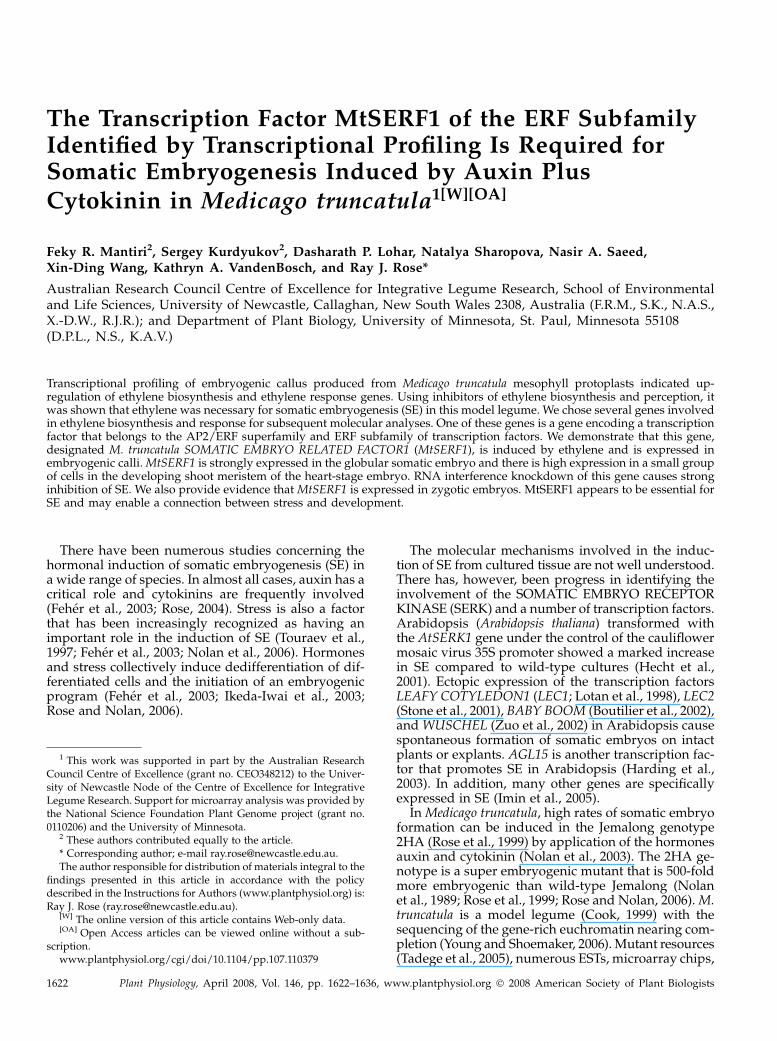
Figure 3. MtSERF1 (TC102138) expression in embryogenic 2HA andnonembryogenic Jemalong after 40 and 60 d of protoplast culture usingqRT-PCR. SEM indicated.
Mantiri et al.
1626 Plant Physiol. Vol. 146, 2008

homolog TC102138. The AP2/EREBP homolog was ofmore interest than other ethylene response genesbecause of its pattern of expression in quantitativereverse transcription (qRT)-PCR studies (detailed be-low); it showed a near 2-fold (1.94, included in Table I)increase and was a transcription factor. In a separateprotoplast experiment, the increase in expression inAP2/EREBP occurred in the highly embryogenic 2HAat 60 d, but not in the near nonembryogenic Jemalong(Fig. 3). We designated the ethylene-responsive AP2/EREBP homolog MtSERF1.
Gene Expression Analysis Using qRT-PCR
Measurements of gene expression using qRT-PCRwere carried out for both the ethylene biosynthesisgenes and the ethylene response gene on leaf explantsto see whether these genes were similarly up-regulatedas they were using mesophyll protoplasts. Leaf ex-plants are experimentally simpler than using isolated
single protoplasts and are commonly used to pro-duce embryogenic callus for legume transformationexperiments (Wang et al., 1996; Chabaud et al., 2003).Experiments can be turned over more quickly usingleaf explants because they produce embryos about40 d earlier than protoplasts These experiments werecarried out with both the highly embryogenic 2HAand the near nonembryogenic wild-type Jemalong.
The ethylene biosynthesis genes are expressed quiteearly and expression continues throughout the cultureperiod in the embryogenic 2HA. The expression pat-tern of the ethylene biosynthesis genes and MtSERF1,the ethylene response gene, is shown in Figure 4. Thepeak of expression in 2HA for ACC SYNTHASE (ACS)transcription is day 1 of culture and day 1 to 2 in thecase of ACC OXIDASE (ACO) transcription. When theexpression of MtSERF1 was measured, it first showedan increase in expression between day 7 and day 14and peaked at day 21 when embryos are starting toform in a partially synchronous fashion, the transition
Figure 5. Core and responsive element motifs in a 1,758-bp region upstream from the transcription start site of MtSERF1. Inaddition to the TATA and CAAT boxes, in silico analysis indicated that the promoter region contained a number of potentialregulatory elements indicated in the figure.
Transcription Factor MtSERF1 and Somatic Embryogenesis
Plant Physiol. Vol. 146, 2008 1627

period between day 40 and 60 in the protoplast ex-periments. Expression then declines, but continues asmore embryos are formed; the amount of expressionafter day 21 varies according to the amount of embryo-genesis. In all four biological repeats, the same in-ductive pattern was evident. Gene expression was alsomeasured with the nonembryogenic line Jemalong,which showed high expression of ACS, but littleexpression of ACO and MtSERF1. The data overallindicate that inhibitors of ethylene perception (Ag1)and biosynthesis (aminoethoxyvinylglycine [AVG])inhibit the expression of all three genes in embryo-genic tissue. In the case of ACO expression, the peak ofexpression is delayed and clearly reduced in the AVGtreatment.
As MtSERF1 is a member of the AP2/ERF family oftranscription factors, the promoter region was ex-amined for an ethylene response element (ERE). A1,758-bp region upstream from the transcription startsite was isolated, cloned, and sequenced. In additionto the TATA and CAAT boxes, in silico analysis indi-
cated that the promoter region contained a numberof potential regulatory elements (Fig. 5). Two EREelements were present, as well as two WUSCHEL-binding sites, four ARABIDOPSIS RESPONSE REGU-LATOR1 (ARR1) elements that are associated withcytokinin signaling, an AUXIN RESPONSE FACTOR(ARF) element, and a TOBACCO EIN3-LIKE (TEIL)element.
Somatic Embryo Induction
To further investigate the role of ethylene in SE,experiments were carried out with leaf explants usingstimulators of ethylene biosynthesis and inhibitors ofethylene biosynthesis and perception. These data areshown in Figure 6 and results clearly indicate a markedinfluence of ethylene on SE. The addition of the eth-ylene precursor 1-aminocyclopropane-1-carboxylicacid (ACC), the substrate for ACO (Pierik et al.,2006), caused a marked increase of SE at 10 mM. Sim-ilarly, methylglyoxal bis(guanylhydrazone) (MGBG),which increases the availability of the ethylene pre-cursor S-adenosyl-Met (SAM) by inhibiting polyaminesynthesis, which utilizes the same precursor, stimu-lates ethylene synthesis (Lee et al., 1997). MGBG at 100mM stimulated SE to the same extent as 10 mM ACC.Conversely, the ethylene biosynthesis inhibitor AVG,which inhibits the conversion of SAM to ACC (Yu andYang, 1979), and Ag1, an inhibitor of ethylene per-ception (Bleecker, 1999), strongly inhibited SE at 1 and10 mM, respectively, and there were no embryos at 10and 100 mM, respectively. Representative photographsof some treatments shown in Figure 7 also illustratethe influence of ethylene on callus development fromleaf explants. Stimulators of ethylene biosynthesis,using MGBG as the example, have small increases incallus development and inhibitors of ethylene action,such as Ag1, cause small decreases, but still allowcallus development to occur. This is clearly shown at100 mM Ag1, where there is complete inhibition ofembryo formation but callusing of the leaf explant hasstill occurred.
Given that embryogenic cultures are a mixture ofembryos and callus cells, to establish a stronger con-nection with ethylene biosynthesis and embryo for-
Figure 6. The effects of ethylene biosynthesis stimulators (ACC andMGBG) and inhibitors of biosynthesis and perception (AVG and Ag1)on embryo number in 2HA. To modify the levels of ethylene produc-tion, the following compounds were added to the medium: (1) theethylene precursor ACC, which stimulates ethylene biosynthesis; (2) thestimulator of ethylene biosynthesis MGBG; (3) the inhibitor of ethyleneperception silver nitrate (AgNO3); and (4) the inhibitor of ethylenebiosynthesis AVG. Three different concentrations (i.e. 1, 10, and 100mM) were employed for ACC, MGBG, and AgNO3, whereas AVG wasemployed at 0.1, 1, and 10 mM. Embryo numbers were counted after 11weeks of culture. SEM indicated.
Figure 7. The effect of the ethylene biosynthesisstimulator (MGBG) and inhibitor of perception(Ag1) on the development of embryogenic callusfrom 2HA leaf explants. Embryos have developed inthe dark for 11 weeks. Note large numbers of em-bryos at 100 mM MGBG and callus developmentwithout embryos in the case of 100 mM Ag1. Arrowspoint to embryos. Bars 5 8 mm. In parallel experi-ments with Jemalong, only two embryos wereproduced across all treatments. This confirms thatwild-type Jemalong rarely produces embryos.
Mantiri et al.
1628 Plant Physiol. Vol. 146, 2008

mation, we directly compared nonembryogenic calluswith embryogenic callus, somatic embryos, and ovules.The ACS and ACO genes are consistently expressed athigher levels in embryogenic tissue, somatic embryos,and ovules with globular-stage embryos compared tononembryogenic callus (Fig. 8).
Localization of MtSERF1 Expression and Requirementfor SE
To localize MtSERF1 expression, we carried out insitu hybridization. The MtSERF1-specific probe was a376-bp fragment from the 3# region. As shown in Fig-ure 9, MtSERF1 is strongly expressed in embryos. Inthe globular embryo, MtSERF1 is expressed through-out the embryo. This is very clearly shown in thicksections of fresh tissue where the callus cells show lit-tle, if any, hybridization signal. Later in the heart-stageembryo, using thinner sections of paraffin-embeddedtissue, we were able to show that hybridization pre-dominates in a small group of cells in the developingshoot meristem.
Given the MtSERF1 expression in somatic embryos,we investigated whether similar expression was pres-ent in zygotic embryos. Ovules at different times after
pollination were collected and MTSERF1 expressionmeasured using qRT-PCR. Expression increased dur-ing ovule development and then declined (Supple-mental Fig. S2), similar to the pattern in embryogeniccallus (Fig. 4C). The peak of ovule expression corre-sponded to the globular stage and had clearly declinedat the torpedo stage (7 d after pollination [dap]). In situhybridization studies showed expression was presentin the embryo, but not in the ovule wall (F.R. Mantiri,S. Kurdyukov, X.-D. Wang, and R.J. Rose, unpublisheddata).
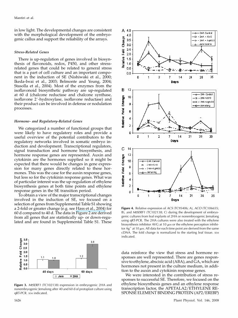
To examine whether MtSERF1 expression is requiredfor SE, we used an RNA interference (RNAi) approach.As shown in Figure 10, transgenic MtSERF1 knock-down calli produced no somatic embryos when com-pared to their empty vector counterparts. For the emptyvector control, 90% of 28 transformed calli producedembryos (with an average of 13.6 embryos/callus). Toconfirm the effects of RNA knockdown, we performedqRT-PCR on the calli. Results showed that the level ofthe transcripts in knockout calli was only 15% of that ofempty vector calli. We also obtained transformedplants using an inducible vector containing RNAi andproduced calli in the presence and absence of dexa-methasone. The induction of RNAi by dexamethasonereduced the number of calli-producing embryos by90%. The empty vector control showed no change in thepresence or absence of dexamethasone.
Sequence and Phylogenetic Analyses of the TranscriptionFactor MtSERF1
MtSERF1 is a protein of molecular mass 23 kD andcontains 201 amino acids. The amino acid sequenceof MtSERF1 contains a single AP2/ERF domain asshown by position-specific iterated and pattern hit-initiated BLAST. As indicated by Nakano et al. (2006),this domain is characteristic of the AP2/ERF super-family and the ERF subfamily contains a single do-main. An alignment of this domain with other proteins
Figure 8. Relative expression of ACS (TC95406) and ACO (TC106655)in nonembryogenic callus (Jem) compared to embryogenic callus (2HA)obtained from leaf explants, somatic embryos (S embryos), and ovuleswith globular-stage embryos 4 dap. The SE data are shown only for theembryogenic 2HA, and zygotic embryogenesis (ZE) is shown onlyfor Jem.
Figure 9. In situ hybridization of MtSERF1 RNA probe. The globular-stage embryo shows expression over the whole embryo (left), whereasin the heart-stage embryo (right), expression is localized to a smallgroup of cells (arrow) in the developing shoot meristem just below theapical notch (arrow). S, Suspensor-like structure; C, callus cells. Bars 5
40 mm.
Transcription Factor MtSERF1 and Somatic Embryogenesis
Plant Physiol. Vol. 146, 2008 1629
containing a single AP2/ERF domain shows highsimilarity (Fig. 11A).
To further investigate this uncharacterized ethylene-induced transcription factor, phylogenetic analyses ofAP2/ERF domain sequences of all 65 transcriptionfactors of Arabidopsis identified as members of the ERFsubfamily (Sakuma et al., 2002; Nakano et al., 2006) andother well-characterized ERFs from other species wereconducted. Phylogenetic analyses using the neighbor-joining method (Saitou and Nei, 1987) on ClustalX 1.8software showed that MtSERF1 belongs to Group IX ofthe classification of Nakano et al. (2006) or Group B-3according to the classification of Sakuma et al. (2002), asshown in an unrooted cladogram (Fig. 11B). To findorthologs of MtSERF1 in other species, phylogeneticanalyses of the entire amino acid sequences of ERFproteins included in Group IX were performed. Asshown in Figure 11C, we found that the MtSERF1 isclustered together with two uncharacterized genes ofArabidopsis, with At5g61590 being the closest ortho-log. This gene shares 41% identity with MtSERF1 (datanot shown). Both genes also share similar motifs outsidethe AP2/ERF domain when analyzed using the motifdiscovery software MEME (http://meme.nbcr.net).These findings suggest that MtSERF1 is distinct fromall AP2/ERF domain-containing transcription factorsof known function.
DISCUSSION
Transcriptional profiling of the development ofsomatic embryos from single isolated mesophyll pro-toplasts of the highly embryogenic M. truncatula geno-type 2HA revealed changes in the expression of manytranscripts. These data showed increased transcriptionof ethylene biosynthesis genes and ethylene responsegenes, which were of interest because of their commoninvolvement in stress and development responses.Subsequent experiments on ethylene response genesidentified an ethylene-responsive transcription factor,MtSERF1, which was essential for SE.
Some of the major changes in the microarray datarelate to stress, reflected in a range of genes connectedto abiotic, biotic, and oxidative stresses. This may havebeen predicted given that protoplast isolation (Pasternaket al., 2002) and tissue excision (Nolan et al., 2006)associated with the induction of SE is a very stressful,wound-related procedure. Transcriptional profiling inresponse to mechanical wounding has been carriedout in Arabidopsis (Cheong et al., 2002; Delessert et al.,2004) and a diverse group of genes previously relatedmore specifically to wounding, pathogen attack, abi-otic stress, and plant hormones are up-regulated. Inthis study, M. truncatula flavonoid biosynthesis geneswere also up-regulated and have also been related tostress protection (Winkel-Shirley, 2002). In M. truncatulaembryogenic cultures, there are many stress-relatedproteins associated with SE (Imin et al., 2004, 2005) asthere are in alfalfa (Domoki et al., 2006). In soybean, SEis induced by 2,4-dichlorophenoxyacetic acid in cotyle-dons and is associated with up-regulation of oxidativestress and defense genes (Thibaud-Nissen et al., 2003).Studies by Che et al. (2006) involving microarray anal-ysis of shoot, root, and callus development in Arabi-dopsis tissue culture also noted an increased expressionof specific stress-related genes.
Among the most highly induced genes in our studywas an ethylene biosynthesis gene (Table I). Up-regu-lation of transcripts of ethylene biosynthesis genes hasalso been seen in wounding (Cheong et al., 2002;Delessert et al., 2004) and SE in soybean cotyledons(Thibaud-Nissen et al., 2003). ACC synthase was up-regulated on an auxin-rich callus induction medium(Che et al., 2006) in Arabidopsis. We also noted an up-regulation of ethylene response genes and this con-tributed to ethylene becoming a focus of our studies. Inaddition to the suite of up-regulated genes related toethylene, it was of interest to note that it might beexpected that, because auxin and cytokinin were pres-ent in the medium, auxin and cytokinin responsegenes would be the only prominently featured hor-mone-related genes. However, this was not the case.Genes related to ABA, GA, and brassinosteroids werealso featured. We have recently discussed the pos-sible roles of these hormones in SE (Rose and Nolan,2006).
Whereas there is value in focusing on mesophyllprotoplasts as a uniform source of starting cells, ex-
Figure 10. The effect of MtSERF1 knockdown using RNAi on embryodevelopment. Empty vector control (left) and MtSERF1 knockdown(right). Bars 5 5 mm. The histogram indicates the reduction in MtSERF1expression due to RNAi.
Mantiri et al.
1630 Plant Physiol. Vol. 146, 2008

Figure 11. A, Alignment of the AP2/ERF domain of M. truncatula MtSERF1 with AP2/ERF domains from other plant species.Sequences were aligned with ClustalW implemented in ClustalX (1.8) using default parameters. Black and gray shading of the
Transcription Factor MtSERF1 and Somatic Embryogenesis
Plant Physiol. Vol. 146, 2008 1631
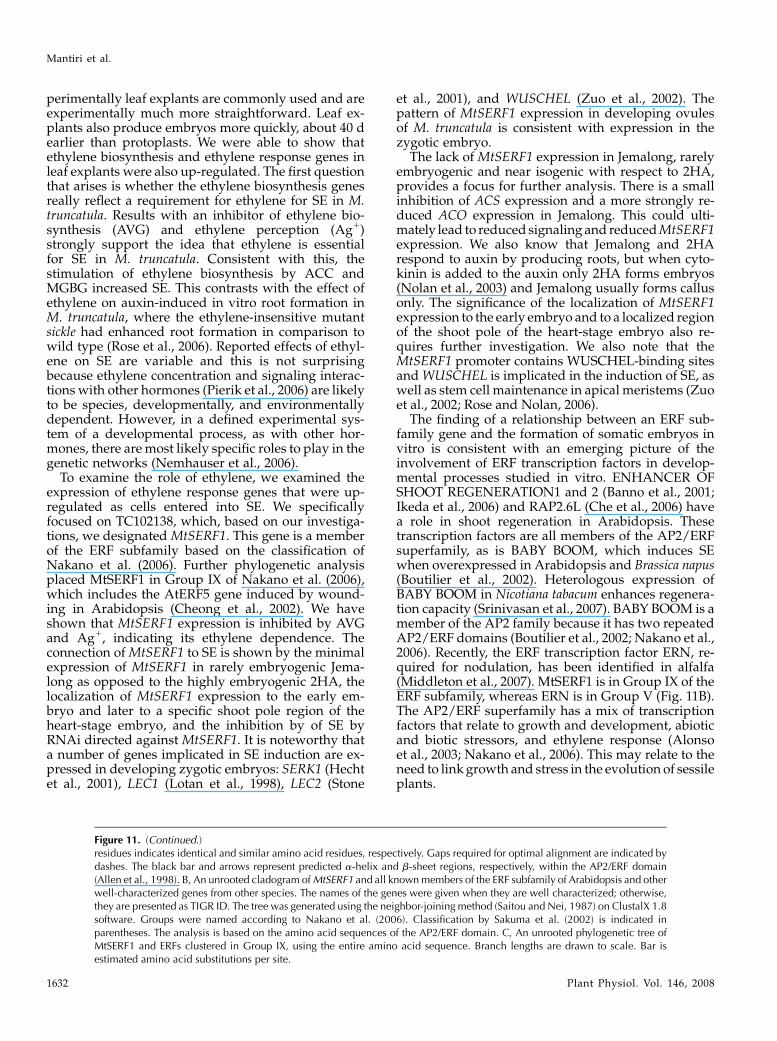
perimentally leaf explants are commonly used and areexperimentally much more straightforward. Leaf ex-plants also produce embryos more quickly, about 40 dearlier than protoplasts. We were able to show thatethylene biosynthesis and ethylene response genes inleaf explants were also up-regulated. The first questionthat arises is whether the ethylene biosynthesis genesreally reflect a requirement for ethylene for SE in M.truncatula. Results with an inhibitor of ethylene bio-synthesis (AVG) and ethylene perception (Ag1)strongly support the idea that ethylene is essentialfor SE in M. truncatula. Consistent with this, thestimulation of ethylene biosynthesis by ACC andMGBG increased SE. This contrasts with the effect ofethylene on auxin-induced in vitro root formation inM. truncatula, where the ethylene-insensitive mutantsickle had enhanced root formation in comparison towild type (Rose et al., 2006). Reported effects of ethyl-ene on SE are variable and this is not surprisingbecause ethylene concentration and signaling interac-tions with other hormones (Pierik et al., 2006) are likelyto be species, developmentally, and environmentallydependent. However, in a defined experimental sys-tem of a developmental process, as with other hor-mones, there are most likely specific roles to play in thegenetic networks (Nemhauser et al., 2006).
To examine the role of ethylene, we examined theexpression of ethylene response genes that were up-regulated as cells entered into SE. We specificallyfocused on TC102138, which, based on our investiga-tions, we designated MtSERF1. This gene is a memberof the ERF subfamily based on the classification ofNakano et al. (2006). Further phylogenetic analysisplaced MtSERF1 in Group IX of Nakano et al. (2006),which includes the AtERF5 gene induced by wound-ing in Arabidopsis (Cheong et al., 2002). We haveshown that MtSERF1 expression is inhibited by AVGand Ag1, indicating its ethylene dependence. Theconnection of MtSERF1 to SE is shown by the minimalexpression of MtSERF1 in rarely embryogenic Jema-long as opposed to the highly embryogenic 2HA, thelocalization of MtSERF1 expression to the early em-bryo and later to a specific shoot pole region of theheart-stage embryo, and the inhibition by of SE byRNAi directed against MtSERF1. It is noteworthy thata number of genes implicated in SE induction are ex-pressed in developing zygotic embryos: SERK1 (Hechtet al., 2001), LEC1 (Lotan et al., 1998), LEC2 (Stone
et al., 2001), and WUSCHEL (Zuo et al., 2002). Thepattern of MtSERF1 expression in developing ovulesof M. truncatula is consistent with expression in thezygotic embryo.
The lack of MtSERF1 expression in Jemalong, rarelyembryogenic and near isogenic with respect to 2HA,provides a focus for further analysis. There is a smallinhibition of ACS expression and a more strongly re-duced ACO expression in Jemalong. This could ulti-mately lead to reduced signaling and reduced MtSERF1expression. We also know that Jemalong and 2HArespond to auxin by producing roots, but when cyto-kinin is added to the auxin only 2HA forms embryos(Nolan et al., 2003) and Jemalong usually forms callusonly. The significance of the localization of MtSERF1expression to the early embryo and to a localized regionof the shoot pole of the heart-stage embryo also re-quires further investigation. We also note that theMtSERF1 promoter contains WUSCHEL-binding sitesand WUSCHEL is implicated in the induction of SE, aswell as stem cell maintenance in apical meristems (Zuoet al., 2002; Rose and Nolan, 2006).
The finding of a relationship between an ERF sub-family gene and the formation of somatic embryos invitro is consistent with an emerging picture of theinvolvement of ERF transcription factors in develop-mental processes studied in vitro. ENHANCER OFSHOOT REGENERATION1 and 2 (Banno et al., 2001;Ikeda et al., 2006) and RAP2.6L (Che et al., 2006) havea role in shoot regeneration in Arabidopsis. Thesetranscription factors are all members of the AP2/ERFsuperfamily, as is BABY BOOM, which induces SEwhen overexpressed in Arabidopsis and Brassica napus(Boutilier et al., 2002). Heterologous expression ofBABY BOOM in Nicotiana tabacum enhances regenera-tion capacity (Srinivasan et al., 2007). BABY BOOM is amember of the AP2 family because it has two repeatedAP2/ERF domains (Boutilier et al., 2002; Nakano et al.,2006). Recently, the ERF transcription factor ERN, re-quired for nodulation, has been identified in alfalfa(Middleton et al., 2007). MtSERF1 is in Group IX of theERF subfamily, whereas ERN is in Group V (Fig. 11B).The AP2/ERF superfamily has a mix of transcriptionfactors that relate to growth and development, abioticand biotic stressors, and ethylene response (Alonsoet al., 2003; Nakano et al., 2006). This may relate to theneed to link growth and stress in the evolution of sessileplants.
Figure 11. (Continued.)residues indicates identical and similar amino acid residues, respectively. Gaps required for optimal alignment are indicated bydashes. The black bar and arrows represent predicted a-helix and b-sheet regions, respectively, within the AP2/ERF domain(Allen et al., 1998). B, An unrooted cladogram of MtSERF1 and all known members of the ERF subfamily of Arabidopsis and otherwell-characterized genes from other species. The names of the genes were given when they are well characterized; otherwise,they are presented as TIGR ID. The tree was generated using the neighbor-joining method (Saitou and Nei, 1987) on ClustalX 1.8software. Groups were named according to Nakano et al. (2006). Classification by Sakuma et al. (2002) is indicated inparentheses. The analysis is based on the amino acid sequences of the AP2/ERF domain. C, An unrooted phylogenetic tree ofMtSERF1 and ERFs clustered in Group IX, using the entire amino acid sequence. Branch lengths are drawn to scale. Bar isestimated amino acid substitutions per site.
Mantiri et al.
1632 Plant Physiol. Vol. 146, 2008
MATERIALS AND METHODS
Protoplast Isolation and Culture
Protoplasts were isolated from leaves of the highly embryogenic 2HA
genotype of Medicago truncatula ‘Jemalong’. A wild-type Jemalong plant
frequently produces no embryos. The highest embryo-producing plant we
have ever recorded was one embryo per six explants (Nolan et al., 1989). The
2HA genotype was derived from a rare regenerated plant obtained by a single
cycle of tissue culture from wild-type Jemalong. This regenerated Jemalong
showed enhanced SE, and the seed progeny segregated into types with and
without the capacity to produce somatic embryos. Seed from the regenerated
Jemalong was used to continue to select for high embryogenicity over four
seed generations (Rose et al., 1999). The 2HA genotype can be considered to be
a near-isogenic, highly embryogenic mutant of Jemalong. Plants were grown
in controlled-environment rooms at low light intensity, as described by Tian
and Rose (1999). The flotation procedure utilized for protoplast isolation
yields almost exclusively mesophyll protoplasts. Isolated protoplasts were
grown in 1% low-melting-point agarose droplets and then transferred to agar
plates on P4 10:4 (10 mM naphthylacetic acid [NAA] and 4 mM benzylamino-
purine [BAP]) medium for culture as described by Rose and Nolan (1995). For
microarray analysis, calli derived from these isolated single protoplasts were
taken at the following stages of development (Fig. 1; Supplemental Fig. S1):
the cell proliferation stage (40 d of culture), the early globular stage (60 d of
culture), and the heart and later stages (80 d of culture).
Cultured Leaf Explants
Cultured M. truncatula leaf explants were obtained from glasshouse-grown
2HA or wild-type Jemalong. Seeds of wild-type Jemalong were originally
obtained from the National Medicago Collection, South Australian Research
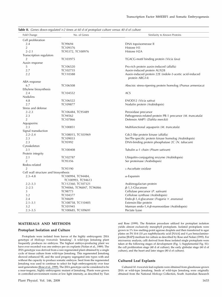
Table II. Genes down-regulated $2 times at 60 d of protoplast culture versus 40 d of culture
Fold Change No. of Genes Similarity to Known Proteins
Cell proliferation2.4 TC99694 DNA topoisomerase II2 TC109376 Histone H32–2.1 TC95372, TC100976 Histone H2A
Transcription regulators2 TC103975 TGACG-motif binding protein (Vicia fava)
Auxin response5 TC106520 Pro-rich protein auxin-induced (alfalfa)2.7 TC102735 Auxin-induced protein AUX282.2 TC110388 Auxin-induced protein 22E (indole-3-acetic acid-induced
protein ARG14)ABA response
6.7 TC106508 Abscisic stress-ripening protein homolog (Prunus armeniaca)Ethylene biosynthesis
2.4 TC104552 ACSNodulins
4.8 TC106522 ENOD12 (Vicia sativa)2.7 TC109877 Nodulin protein (Arabidopsis)
Stress and defense2–2.3 TC106484, TC95489 Peroxidase precursor2.3 TC94562 Pathogenesis-related protein PR-1 precursor (M. truncatula)2 TC107866 Defensin AMP1 (Dahlia merckii)
Aquaporins2.3 TC100851 Multifunctional aquaporin (M. truncatula)
Signal transduction2.2–2.4 TC108015, TC103969 Cdc2-like protein kinase (alfalfa)2.3 TC109033 Ser/Thr-specific protein kinase homolog (Arabidopsis)2.3 TC95992 DNA-binding protein phosphatase 2C (N. tabacum)
Cytoskeleton2.1 TC100408 Tubulin a-1 chain (Pisum sativum)
Protein integrity2.1 TC102787 Ubiquitin-conjugating enzyme (Arabidopsis)2 TC95356 Ser proteinase (Arabidopsis)
Redox-related2 TC95190 L-Ascorbate oxidase
Cell wall structure and biosynthesis2.3–4.8 TC100994, TC94484,
TC100993, TC94613a-Expansin
2.2–3.3 TC112360, TC107321 Arabinogalactan protein2–2.5 TC94066, TC96847, TC96066 b-1,3-Glucanase3 TC98773 Cellulase precursor (P. sativum)3.2 TC104377 Cellulose synthase (Arabidopsis)2.4 TC94609 Endo-b-1,4-glucanase (Fragaria 3 ananassa)2.1–3.1 TC108758, TC110405 Extensin-like protein3.2 TC101945 Mannan endo-1,4-b-mannosidase (Arabidopsis)2.3–3.5 TC100685, TC109691 Pectate lyase
Transcription Factor MtSERF1 and Somatic Embryogenesis
Plant Physiol. Vol. 146, 2008 1633

and Development Institute, Adelaide. The standard leaf culture procedure
was as described by Nolan et al. (2003). Explants were cultured on P4 10:4 for
3 weeks before transfer to P4 10:4:1 (10 mM NAA, 4 mM BAP, and 1 mM ABA).
16K Oligo Microarray Slides
The Medicago 16K microarray was utilized and has a probe set mapped to
Medicago Gene Index Release 8.0 (http//www.tigr.org/docs/tigr/scripts/
medicago/ARRAYS/array.TC mapping). The 70-mer oligos were synthesized
by Qiagen-Operon and the slides printed at the University of Arizona in the
laboratory of Dr. David Galbraith. After printing, the slides were baked for 80
min at 80�C. The oligonucleotide array elements were immobilized by UV
cross-linking at 300 mJ, then washed twice with gentle rocking for 2 min each
wash, in 23 SSC 1 0.2% SDS. The slides were then immersed in boiling hot
water for 2 min, blotted briefly, and transferred to ice-cold ethanol for 2 to
5 min. Slides were then dried by centrifugation at 1,500 rpm for 2 to 5 min
and finally stored in a light-proof box under cool dry conditions.
RNA Preparation, cDNA Synthesis, and Hybridization
of Microarrays
Calli grown from individual protoplasts in an isolation that produced
thousands of embryogenic microcalli, consistent with high protoplast quality,
were collected at 40, 60, and 80 d after initiation of culture. The calli were
frozen in liquid nitrogen and stored at 280�C until RNA was isolated. RNA
was isolated as described by Lohar et al. (2006) and stored at 280�C. Total
RNA (22 mg/sample) was pooled from three biological replicates giving 66 mg
of RNA and 33 mg of RNA was aliquoted into two separate tubes. The
Eppendorf tubes containing 33 mg of RNA were thawed on ice, spun dry in a
speed vac, and immediately returned to 280�C. The RNA was shipped from
Newcastle, Australia, to St. Paul, MN, on dry ice and transferred to 280�C
until required. This maintained the quality of the RNA. The 33 mg of RNA
were resuspended in 8 mL of nuclease-free double-distilled water and used for
cDNA synthesis with a reverse transcriptase primer for labeling with either
Cy3 or Cy5 dyes using a 3DNA Array50 kit (Genisphere) as previously
described (Lohar et al., 2006).
Experiments were conducted using a regular dye-swap design as de-
scribed earlier by Lohar et al. (2006). Microarrays for 60 versus 40 d, 80 versus
60 d, and 80 versus 40 d comparisons were hybridized with cDNAs from the
two different time points labeled with different dyes. Each hybridization was
repeated a total of six times to sample the technical variability, with three
repeats of each dye combination to control for dye effects (Lohar et al., 2006).
Microarray Analysis
Methods for array analysis were as described for a 6K microarray (Lohar
et al., 2006). Briefly, microarray slides were scanned using an Axon two-laser
scanner and image analysis was performed using GenePix (Axon) software.
Background-subtracted mean intensities for both tissues were log transformed
and normalized before further analysis. Normalization of microarray data
was performed using a statistical module developed as a part of Lab
Information System, which includes several scripts and modules written in
PERL and R languages.
Normalization steps included (1) within-slide normalization using local
linear regression (LOWESS function; Yang et al., 2000); and (2) between-slide
normalization using four-way ANOVA with replications for multislide dye-
swap experiments (Kerr et al., 2000). More detailed description of preprocess-
ing steps, such as log2 transformation of background-subtracted Cy5 and Cy3
intensities, are described by Lohar et al. (2006). Identification of differentially
expressed genes was done using SAM software (Stanford), which allows
flexible monitoring of the false discovery rate (Tusher et al., 2001). We applied
a false discovery rate of ,0.1% and the highest q value was ,0.06%.
All genes of statistical significance with predicted or known function or
that showed significant homology to characterized genes (annotated in the
The Institute for Genomic Research database at http://compbio.dfci.harvard.
edu/tgi) have been manually divided into 27 classes. Genes that did not fit
readily into one of these classes have been classified as ‘‘other genes with
defined function’’ and ‘‘genes with unknown function.’’ Supplemental Table
S1 lists all the genes incorporated into these classes. To obtain a subset of genes
that passed a statistical significance test we have also imposed a fixed ratio
threshold of 2.
Real-Time PCR
Total RNA was isolated from calli at different time points and from intact
leaves (as a calibrator) using the RNAqueous-4PCR kit (Ambion) according to
the manufacturer’s instructions. cDNA synthesis was performed using the
SuperScript II first-strand synthesis system for RT-PCR (Invitrogen) starting
with 2 mg of total RNAwith oligo(dT)15 primers. Real-time PCR was performed
using the SYBR GreenER qPCR SuperMix Universal kit (Invitrogen) and
analyzed in the DNA Engine Opticon 2 continuous fluorescence detection
system (Bio-Rad; formerly MJ Research). Primers 5#-TCATACGCCATCAT-
CTCTTAGGT-3# (forward) and 5#-AGGGGTTGTTTCCTTTGAAGAT-3# (re-
verse) were designed to quantify the MtSERF1 expression levels, which were
normalized to those of glyceraldehyde-3-P dehydrogenase (GAPDH), primers
5#-TGGTCATCAAACCCTCAACA-3# (forward) and 5#-CCTCGTTCTTTCC-
GCTATCA-3# (reverse), in each sample. To quantify the expression levels
of ACS, the primers were 5#-CCCACACAAATTCGCTTCTT-3# (forward) and
5#-TCACCATGTCCATCACCAGT-3# (reverse), whereas for ACO the primers
were 5#-GGGATTCTTTGAGCTGGTGA-3# (forward) and 5#-GACGAACA-
TGGAAGGTGCTT-3# (reverse). PCR cycling conditions included a 94�C
heating step for 1 min at the beginning of every run. The tubes were then
cycled at 94�C for 30 s, annealed at 60�C for 60 s, and extended at 72�C for 60 s. A
melting curve was generated at the end of every run to ensure product
uniformity. PCR reactions were performed in triplicate in at least two biological
repeats. Transcript abundance was estimated using a modification of the
comparative threshold cycle (Ct) method and was calculated as E2DDCt, where
DDCt 5 (Cttarget 2 CtGAPDH)Time x 2 (Cttarget 2 CtGAPDH)Calibrator and E is the
estimated amplification efficiency, which was calculated employing the linear
regression method on the log(fluorescence) per cycle number data for each
amplicon using the LinRegPCR software (Ramakers et al., 2003).
In Situ Hybridization
To generate the RNA probes, a 376-bp fragment specific to MtSERF1 was
first amplified by PCR with the primers 5#-CTGTGAAATTGATGCTGCAAA-3#(forward) and 5#-TGACATAATTGTTGAGCTCACTCC-3# (reverse). Then,
the promoter sequences of T7 and SP6 RNA polymerase were introduced to
this fragment by a two-step PCR. The first primers used were 5#-GAG-
GCCGCGTCTGTGAAATTGATGCTGCAAA-3# (forward) and 5#-ACCCGG-
GGCTTGACATAATTGTTGAGCTCACTCC-3# (reverse). The second set of
primers used was 5#-TTATGTAATACGACTCACTATAGGGAGGCCGCGT-3#(forward) and 5#-CCAATTTAGGTGACACTATAGAAGTACCCGGGGCT-3#(reverse). This PCR product was subsequently used as a template for in vitro
transcription employing T7 and SP6 RNA polymerase to synthesize digoxi-
genin (DIG)-labeled sense and antisense single-stranded RNA probes, re-
spectively, using a DIG RNA labeling kit (catalog no. 11 093 274 910; Roche
Diagnostics GmbH). Two different cytological procedures were used; paraffin
embedding and fresh tissue sectioned with a vibratome. For the paraffin
procedure, 4- to 5-week-old 2HA calli from leaf explants were fixed in 4%
formaldehyde in 0.025 M phosphate buffer at pH 7.2, dehydrated through an
ethanol and ethanol:histolene (Fronine Lab Supplies) series, embedded in
paraffin, sectioned (8 mM), and hybridized with the DIG-labeled sense and
antisense probes according to the manufacturer’s instructions. For the fresh
tissue procedure, the 2HA embryogenic tissue from leaf explant tissue was
embedded in agar and 40-mm sections cut with a vibratome. In both cases,
hybridization was detected using a fluorescent antibody enhancer set for DIG
detection (catalog no. 176756; Boehringer) and was visualized as a red/purple
color after the NBT/BCIP color reaction (Roche Diagnostics). In all cases, no
signal over background was observed using control sense-strand probes.
Construction of Constitutive and Inducible
RNAi Plasmids
For MtSERF1 RNAi construction, specific sequences in the 3#-end of
MtSERF1 mRNA were selected for construction of RNAi fragments. A cDNA
fragment of MtSERF1 was amplified by PCR with the primers 5#-CTGTG-
AAATTGATGCTGCAAA-3# (forward) and 5#-TGACATAATTGTTGAGCT-
CACTCC-3# (reverse). The MtSERF1-specific PCR products were cloned into
the vector pCR8/GW/TOPO (Invitrogen). After linearization of the plasmids,
the Gateway LR recombination reaction (Invitrogen) was conducted accord-
ing to the manufacturer’s protocol to incorporate the MtSERF1-specific
fragment into the binary T-DNA destination vector pH7GW1WG2(II) (Karimi
Mantiri et al.
1634 Plant Physiol. Vol. 146, 2008

et al., 2002) and pOpOff2(hyg) (Wielopolska et al., 2005) for constitutive and
inducible RNAi constructs, respectively. The resulting constructs were intro-
duced into Agrobacterium tumefaciens strain AGL1 by electroporation.
Transformation of M. truncatula
Transformation of M. truncatula 2HA leaf explants was carried out as
described by Wang et al. (1996) with some modifications. In brief, leaf pieces
were prepared and sterilized according to the method described by Nolan
et al. (2003) and dipped into bacterial solution, followed by cocultivation for 2
to 5 d. After cocultivation, the explants were decontaminated by dipping in a
solution containing 750 mg/L augmentin (5 parts amoxicillin/L part clav-
ulanic acid; Beecham Laboratories) before plating onto solid medium as
described previously in the section on cultured leaf explants. Transformed
calli were screened for hygromycin resistance by including hygromycin at
15 mg/L in the medium. Augmentin (500 mg/L) was also added in the
medium to eliminate the Agrobacterium. The explants were subcultured on
fresh medium every 4 weeks. RNAi constructs were induced by 2.5 mM
dexamethasone.
Sequence Analysis and Construction ofPhylogenetic Trees
Multiple alignment analyses were performed with ClustalW using a
ClustalX 1.8 software package. Phylogenetic trees were constructed using
the neighbor-joining method (Saitou and Nei, 1987) included in the ClustalX
1.8 software. Phylogenetic trees were drawn using TreeView (Win32) 1.6.0
software (Page, 1996).
Promoter Sequence Isolation and in Silico Analysis
Isolation of the MtSERF1 promoter was carried out according to the
GenomeWalker kit (CLONTECH) with minor modifications. In brief, for
the first round of amplification, a biotinylated gene-specific primer and the
adaptor primer AP1 were used. Immobilization of the PCR product to
streptavidin-coated particles and washing steps were conducted according
to the Dynal kilobase BINDER kit (Invitrogen). A one-tenth part of these beads
was used for nested PCR as described in the Genome Walker kit and the
fragment obtained sequenced. The proximal region of the promoter was
analyzed using eukaryotic transcription start site prediction software NNPP,
version 2.2 (Reese, 2000, 2001; www.fruitfly.org/seq_tools/promoter.html).
Search for core and responsive element motifs was performed in silico
by means of the Web Signal Scan Program (Prestridge, 1991; Higo et al.,
1999; http://www.dna.affrc.go.jp/Sigscan/signal.html) and MatInspector
(Cartharius et al., 2005; http://www.genomatix.de/products/MatInspector).
Supplemental Data
The following materials are available in the online version of this article.
Supplemental Figure S1. Histological examination of 40- and 60-d callus
used in microarray experiments.
Supplemental Figure S2. The bar graph represents the level of MtSERF1
expression normalized to the GAPDH gene.
Supplemental Table S1. All statistically up-regulated and down-regu-
lated genes at 60 versus 40 d, 80 versus 60 d, and 80 versus 40 d of
protoplast culture.
ACKNOWLEDGMENTS
We wish to thank Yoko Nitanai for assistance with gridding of the
microarray signals, Dr. Kim Nolan for assistance with tissue culture, and
Dr. Kim Nolan and Dr. Michael Sheahan for helpful discussion.
Received October 4, 2007; accepted January 16, 2008; published January 30, 2008.
LITERATURE CITED
Allen MD, Yamasaki K, Ohme-Takagi M, Tateno M, Suzuki M (1998) A
novel mode of DNA recognition by a beta-sheet revealed by the solution
structure of the GCC-box binding domain in complex with DNA. EMBO
J 17: 5484–5496
Alonso JM, Stepanova AN, Leisse TJ, Kim CJ, Chen H, Shinn P, Stevenson
DK, Zimmerman J, Barajas P, Cheuk R, et al (2003) Genome-wide inser-
tional mutagenesis of Arabidopsis thaliana. Science 301: 653–657
Banno H, Ikeda Y, Niu QW, Chua NH (2001) Overexpression of Arabidopsis
ESR1 induces initiation of shoot regeneration. Plant Cell 13: 2609–2618
Belmonte MF, Yeung EC (2004) The effects of reduced and oxidized
glutathione on white spruce somatic embryogenesis. In Vitro Cell Dev
Biol Plant 40: 61–66
Bleecker AB (1999) Ethylene perception and signalling: an evolutionary
perspective. Trends Plant Sci 4: 269–274
Boutilier K, Offringa R, Sharma VK, Kieft H, Ouellet T, Zhang LM,
Hattori J, Liu CM, van Lammeren AAM, Miki BLA, et al (2002) Ectopic
expression of BABY BOOM triggers a conversion from vegetative to
embryonic growth. Plant Cell 14: 1737–1749
Cartharius K, Frech K, Grote K, Klocke B, Haltmeier M, Klingenhoff A,
Frisch M, Bayerlein M, Werner T (2005) MatInspector and beyond:
promoter analysis based on transcription factor binding sites. Bioinfor-
matics 21: 2933–2942
Chabaud M, de Carvalho-Niebel F, Barker DG (2003) Efficient transfor-
mation of Medicago truncatula cv. Jemalong using the hypervirulent
Agrobacterium tumefaciens strain AGL1. Plant Cell Rep 22: 46–51
Che P, Lall S, Nettleton D, Howell SH (2006) Gene expression programs
during shoot, root, and callus development in Arabidopsis tissue
culture. Plant Physiol 141: 620–637
Cheong YH, Chang HS, Gupta R, Wang X, Zhu T, Luan S (2002) Tran-
scriptional profiling reveals novel interactions between wounding,
pathogen, abiotic stress, and hormonal responses in Arabidopsis. Plant
Physiol 129: 661–677
Cook DR (1999) Medicago truncatula—a model in the making! Curr Opin
Plant Biol 2: 301–304
Delessert C, Wilson IW, Van der Straeten D, Dennis ES, Dolferus R (2004)
Spatial and temporal analysis of the local response to wounding in
Arabidopsis leaves. Plant Mol Biol 55: 165–181
Domoki M, Gyorgyey J, Biro J, Pasternak TP, Zvara A, Bottka S, Puskas
LG, Dudits D, Feher A (2006) Identification and characterization
of genes associated with the induction of embryogenic competence
in leaf-protoplast-derived alfalfa cells. Biochim Biophys Acta 1759:
543–551
Feher A, Pasternak TP, Dudits D (2003) Transition of somatic plant cells to
an embryogenic state. Plant Cell Tissue Organ Cult 74: 201–228
Harding EW, Tang WN, Nichols KW, Fernandez DE, Perry SE (2003)
Expression and maintenance of embryogenic potential is enhanced
through constitutive expression of AGAMOUS-Like 15. Plant Physiol
133: 653–663
Hass C, Lohrmann J, Albrecht V, Sweere U, Hummel F, Yoo SD, Hwang I,
Zhu T, Schafer E, Kudla J, et al (2004) The response regulator 2 mediates
ethylene signalling and hormone signal integration in Arabidopsis.
EMBO J 23: 3290–3302
Hecht V, Vielle-Calzada JP, Hartog MV, Schmidt EDL, Boutilier K,
Grossniklaus U, de Vries SC (2001) The Arabidopsis SOMATIC EM-
BRYOGENESIS RECEPTOR KINASE 1 gene is expressed in developing
ovules and embryos and enhances embryogenic competence in culture.
Plant Physiol 127: 803–816
Higo K, Ugawa Y, Iwamoto M, Korenaga T (1999) Plant cis-acting regu-
latory DNA elements (PLACE) database: 1999. Nucleic Acids Res 27:
297–300
Ikeda Y, Banno H, Niu QW, Howell SH, Chua NH (2006) The ENHANCER
OF SHOOT REGENERATION 2 gene in Arabidopsis regulates CUP-
SHAPED COTYLEDON 1 at the transcriptional level and controls
cotyledon development. Plant Cell Physiol 47: 1443–1456
Ikeda-Iwai M, Umehara M, Satoh S, Kamada H (2003) Stress-induced
somatic embryogenesis in vegetative tissues of Arabidopsis thaliana.
Plant J 34: 107–114
Imin N, De Jong F, Mathesius U, van Noorden G, Saeed NA, Wang XD,
Rose RJ, Rolfe BG (2004) Proteome reference maps of Medicago
truncatula embryogenic cell cultures generated from single protoplasts.
Proteomics 4: 1883–1896
Imin N, Nizamidin M, Daniher D, Nolan KE, Rose RJ, Rolfe BG (2005)
Proteomic analysis of somatic embryogenesis in Medicago truncatula.
Explant cultures grown under 6-benzylaminopurine and 1-naphthale-
neacetic acid treatments. Plant Physiol 137: 1250–1260
Transcription Factor MtSERF1 and Somatic Embryogenesis
Plant Physiol. Vol. 146, 2008 1635
Karimi M, Inze D, Depicker A (2002) GATEWAY vectors for Agrobacterium-
mediated plant transformation. Trends Plant Sci 7: 193–195
Kende H, Zeevaart JAD (1997) The five ‘‘classical’’ plant hormones. Plant
Cell 9: 1197–1210
Kerr MK, Martin M, Churchill GA (2000) Analysis of variance for gene
expression microarray data. J Comput Biol 7: 819–837
Lee MM, Lee SH, Park KY (1997) Effects of spermine on ethylene biosyn-
thesis in cut carnation (Dianthus caryophyllus L.) flowers during senes-
cence. J Plant Physiol 151: 68–73
Lohar DP, Sharopova N, Endre G, Penuela S, Samac D, Town C, Silverstein
KAT, VandenBosch KA (2006) Transcript analysis of early nodulation events
in Medicago truncatula. Plant Physiol 140: 221–234
Lotan T, Ohto M, Yee KM, West MAL, Lo R, Kwong RW, Yamagishi
K, Fischer RL, Goldberg RB, Harada JJ (1998) Arabidopsis LEAFY
COTYLEDON1 is sufficient to induce embryo development in vegeta-
tive cells. Cell 93: 1195–1205
Magyar Z, Meszaros T, Miskolczi P, Deak M, Feher A, Brown S,
Kondorosi E, Athanasiadis A, Pongor S, Bilgin M, et al (1997) Cell
cycle phase specificity of putative cyclin-dependent kinase variants in
synchronized alfalfa cells. Plant Cell 9: 223–235
Majewska-Sawka A, Nothnagel EA (2000) The multiple roles of arabino-
galactan proteins in plant development. Plant Physiol 122: 3–9
Middleton PH, Jakab J, Penmetsa RV, Starker CG, Doll J, Kalo P, Prabhu
R, Marsh JF, Mitra RM, Kereszt A, et al (2007) An ERF transcription
factor in Medicago truncatula that is essential for nod factor signal
transduction. Plant Cell 19: 1221–1234
Nakano T, Suzuki K, Fujimura T, Shinshi H (2006) Genome-wide analy-
sis of the ERF gene family in Arabidopsis and rice. Plant Physiol 140:
411–432
Nemhauser JL, Hong F, Chory J (2006) Different plant hormones regulate
similar processes through largely nonoverlapping transcriptional re-
sponses. Cell 126: 467–475
Nishiwaki M, Fujino K, Koda Y, Masuda K, Kikuta Y (2000) Somatic
embryogenesis induced by the simple application of abscisic acid to
carrot (Daucus carota L.) seedlings in culture. Planta 211: 756–759
Nolan KE, Irwanto RR, Rose RJ (2003) Auxin up-regulates MtSERK1
expression in both Medicago truncatula root-forming and embryogenic
cultures. Plant Physiol 133: 218–230
Nolan KE, Rose RJ, Gorst JE (1989) Regeneration of Medicago truncatula
from tissue culture: increased somatic embryogenesis from regenerated
plants. Plant Cell Rep 8: 278–281
Nolan KE, Saeed NA, Rose RJ (2006) The stress kinase gene MtSK1 in
Medicago truncatula with particular reference to somatic embryogenesis.
Plant Cell Rep 25: 711–722
Page RDM (1996) TreeView: an application to display phylogenetic trees on
personal computers. Bioinformatics 12: 357–358
Pasternak TP, Prinsen E, Ayaydin F, Miskolczi P, Potters G, Asard H, Van
Onckelen HA, Dudits D, Feher A (2002) The role of auxin, pH, and
stress in the activation of embryogenic cell division in leaf protoplast-
derived cells of alfalfa. Plant Physiol 129: 1807–1819
Pierik R, Tholen D, Poorter H, Visser EJW, Voesenek L (2006) The Janus
face of ethylene: growth inhibition and stimulation. Trends Plant Sci 11:
176–183
Prestridge DS (1991) SIGNAL SCAN: a computer program that scans DNA
sequences for eukaryotic transcriptional elements. CABIOS 7: 203–206
Ramakers C, Ruijter JM, Lekanne Deprez RH, Moorman AFM (2003)
Assumption-free analysis of quantitative real-time polymerase chain
reaction (PCR) data. Neurosci Lett 339: 62–66
Reese MG (2000) Computational prediction of gene structure and regula-
tion in the genome of Drosophila melanogaster. PhD thesis. University of
California, Berkeley, CA/University of Hohenheim, Stuttgart, Germany
Reese MG (2001) Application of a time-delay neural network to promoter
annotation in the Drosophila melanogaster genome. Comput Chem 26: 51–56
Rose RJ (2004) Somatic embryogenesis in plants. In RM Goodman, ed,
Encyclopedia of Plant and Crop Science. Marcel Dekker, New York, pp
1165–1168
Rose RJ, Nolan KE (1995) Regeneration of Medicago truncatula from
protoplasts isolated from kanamycin-sensitive and kanamycin-resistant
plants. Plant Cell Rep 14: 349–354
Rose RJ, Nolan KE (2006) Genetic regulation of somatic embryogenesis
with particular reference to Arabidopsis thaliana and Medicago truncatula.
In Vitro Cell Dev Biol Plant 42: 473–481
Rose RJ, Nolan KE, Bicego L (1999) The development of the highly
regenerable seed line Jemalong 2HA for transformation of Medicago
truncatula—implications for regenerability via somatic embryogenesis.
J Plant Physiol 155: 788–791
Rose RJ, Wang X-D, Nolan KE, Rolfe BG (2006) Root meristems in Medicago
truncatula tissue culture arise from vascular-derived procambial-like cells in a
process regulated by ethylene. J Exp Bot 57: 2227–2235
Saitou N, Nei M (1987) The neighbour-joining method: a new method for
reconstructing phylogenetic trees. Mol Biol Evol 4: 406–425
Sakuma Y, Liu Q, Dubouzet JG, Abe H, Shinozaki K, Yamaguchi-
Shinozaki K (2002) DNA-binding specificity of the ERF/AP2 domain
of Arabidopsis DREBS, transcription factors involved in dehydration-
and cold-inducible gene expression. Biochem Biophys Res Commun
290: 998–1009
Srinivasan C, Liu ZR, Heidmann I, Supena EDJ, Fukuoka H, Joosen R,
Lambalk J, Angenent G, Scorza R, Custers JBM, et al (2007) Heterol-
ogous expression of the BABY BOOM AP2/ERF transcription factor
enhances the regeneration capacity of tobacco (Nicotiana tabacum L.).
Planta 225: 341–351
Stasolla C, Belmonte MF, van Zyl L, Craig DL, Liu WB, Yeung EC,
Sederoff RR (2004) The effect of reduced glutathione on morphology
and gene expression of white spruce (Picea glauca) somatic embryos.
J Exp Bot 55: 695–709
Stone SL, Kwong LW, Yee KM, Pelletier J, Lepiniec L, Fischer RL,
Goldberg RB, Harada JJ (2001) LEAFY COTYLEDON 2 encodes a B3
domain transcription factor that induces embryo development. Proc
Natl Acad Sci USA 98: 11806–11811
Tadege M, Ratet P, Mysore KS (2005) Insertional mutagenesis: a Swiss
army knife for functional genomics of Medicago truncatula. Trends Plant
Sci 10: 229–235
Thibaud-Nissen FO, Shealy RT, Khanna A, Vodkin LO (2003) Clustering
of microarray data reveals transcript patterns associated with somatic
embryogenesis in soybean. Plant Physiol 132: 118–136
Tian D, Rose RJ (1999) Asymmetric somatic hybridisation between the
annual legumes Medicago truncatula and Medicago scutellata. Plant Cell
Rep 18: 989–996
Touraev A, Vicente O, Heberle-Bors E (1997) Initiation of microspore
embryogenesis by stress. Trends Plant Sci 2: 297–302
Tusher VG, Tibshirani R, Chu G (2001) Significance analysis of micro-
arrays applied to the ionizing radiation response. Proc Natl Acad Sci
USA 98: 5116–5121
VandenBosch KA, Stacey G (2003) Summaries of legume genomics projects
from around the globe. Community resources for crops and models. Plant
Physiol 131: 840–865
Wang JH, Rose RJ, Donaldson BI (1996) Agrobacterium-mediated transfor-
mation and expression of foreign genes in Medicago truncatula. Aust J
Plant Physiol 23: 265–270
Wang KL, Li H, Ecker JR (2002) Ethylene biosynthesis and signaling
networks. Plant Cell 14: S131–S151
Wielopolska A, Townley H, Moore I, Waterhouse P, Helliwell C (2005) A
high-throughput inducible RNAi vector for plants. Plant Biotechnol J 3:
583–590
Winkel-Shirley B (2002) Biosynthesis of flavonoids and effects of stress.
Curr Opin Plant Biol 5: 218–223
Yang YH, Buckley MJ, Dudoit S, Speed TP (2000) Comparison of Methods
for Image Analysis on cDNA Microarray Data. Technical Report 2000.
Statistics Department, University of California, Berkeley, CA
Young ND, Shoemaker RC (2006) Genome studies and molecular genetics.
Part 1: Model legumes. Exploring the structure, function and evolution
of legume genomes. Curr Opin Plant Biol 9: 95–98
Yu YB, Yang SF (1979) Auxin-induced ethylene production and its inhi-
bition by aminoethoxyvinylglycine and cobalt ion. Plant Physiol 64:
1074–1077
Zuo JR, Niu QW, Frugis G, Chua NH (2002) The WUSCHEL gene pro-
motes vegetative-to-embryonic transition in Arabidopsis. Plant J 30:
349–359
Mantiri et al.
1636 Plant Physiol. Vol. 146, 2008