Genetically Encoded Short Peptide Tags for Orthogonal Protein Labeling by Sfp and AcpS...
10
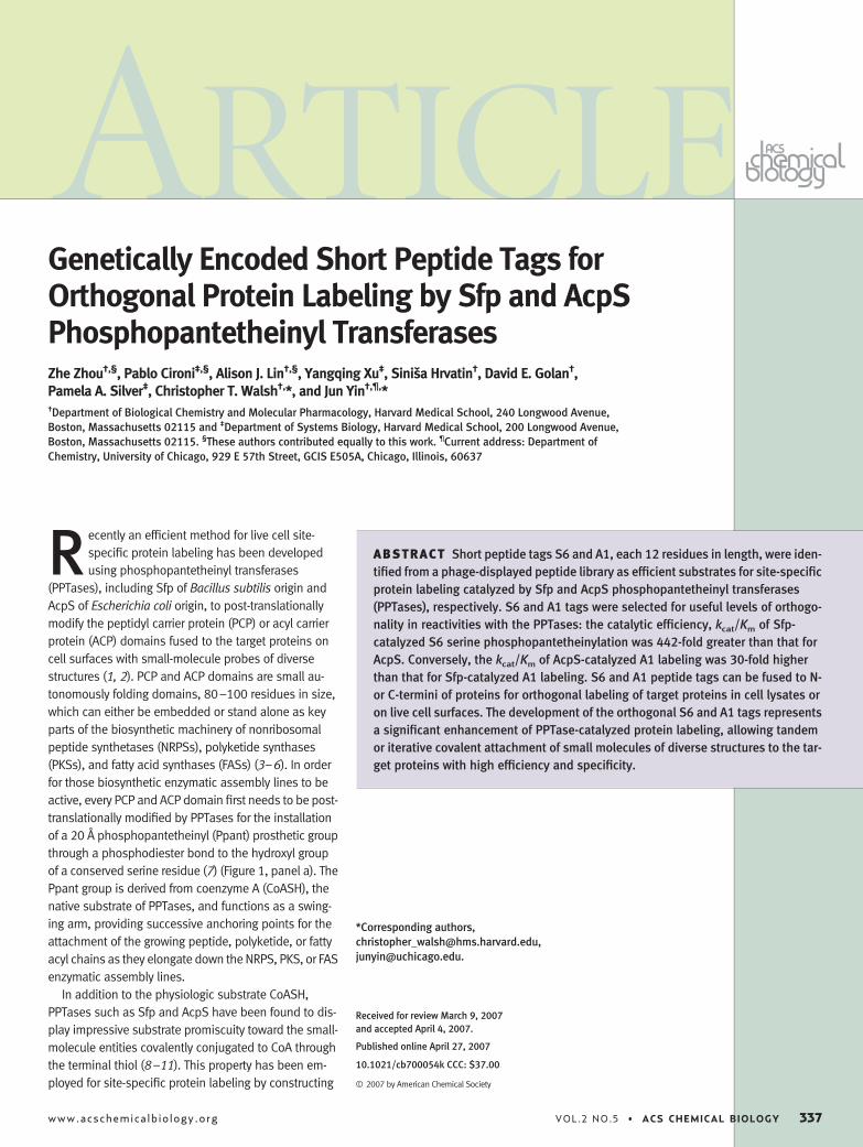
Genetically Encoded Short Peptide Tags for Orthogonal Protein Labeling by Sfp and AcpS Phosphopantetheinyl Transferases Zhe Zhou †,§ , Pablo Cironi ‡,§ , Alison J. Lin †,§ , Yangqing Xu ‡ , Siniša Hrvatin † , David E. Golan † , Pamela A. Silver ‡ , Christopher T. Walsh †, *, and Jun Yin †,¶, * † Department of Biological Chemistry and Molecular Pharmacology, Harvard Medical School, 240 Longwood Avenue, Boston, Massachusetts 02115 and ‡ Department of Systems Biology, Harvard Medical School, 200 Longwood Avenue, Boston, Massachusetts 02115. § These authors contributed equally to this work. ¶ Current address: Department of Chemistry, University of Chicago, 929 E 57th Street, GCIS E505A, Chicago, Illinois, 60637 R ecently an efficient method for live cell site- specific protein labeling has been developed using phosphopantetheinyl transferases (PPTases), including Sfp of Bacillus subtilis origin and AcpS of Escherichia coli origin, to post-translationally modify the peptidyl carrier protein (PCP) or acyl carrier protein (ACP) domains fused to the target proteins on cell surfaces with small-molecule probes of diverse structures ( 1, 2). PCP and ACP domains are small au- tonomously folding domains, 80 –100 residues in size, which can either be embedded or stand alone as key parts of the biosynthetic machinery of nonribosomal peptide synthetases (NRPSs), polyketide synthases (PKSs), and fatty acid synthases (FASs) ( 3– 6). In order for those biosynthetic enzymatic assembly lines to be active, every PCP and ACP domain first needs to be post- translationally modified by PPTases for the installation of a 20 Å phosphopantetheinyl (Ppant) prosthetic group through a phosphodiester bond to the hydroxyl group of a conserved serine residue ( 7) (Figure 1, panel a). The Ppant group is derived from coenzyme A (CoASH), the native substrate of PPTases, and functions as a swing- ing arm, providing successive anchoring points for the attachment of the growing peptide, polyketide, or fatty acyl chains as they elongate down the NRPS, PKS, or FAS enzymatic assembly lines. In addition to the physiologic substrate CoASH, PPTases such as Sfp and AcpS have been found to dis- play impressive substrate promiscuity toward the small- molecule entities covalently conjugated to CoA through the terminal thiol ( 8 –11). This property has been em- ployed for site-specific protein labeling by constructing *Corresponding authors, [email protected], [email protected]. Received for review March 9, 2007 and accepted April 4, 2007. Published online April 27, 2007 10.1021/cb700054k CCC: $37.00 © 2007 by American Chemical Society ABSTRACT Short peptide tags S6 and A1, each 12 residues in length, were iden- tified from a phage-displayed peptide library as efficient substrates for site-specific protein labeling catalyzed by Sfp and AcpS phosphopantetheinyl transferases (PPTases), respectively. S6 and A1 tags were selected for useful levels of orthogo- nality in reactivities with the PPTases: the catalytic efficiency, k cat /K m of Sfp- catalyzed S6 serine phosphopantetheinylation was 442-fold greater than that for AcpS. Conversely, the k cat /K m of AcpS-catalyzed A1 labeling was 30-fold higher than that for Sfp-catalyzed A1 labeling. S6 and A1 peptide tags can be fused to N- or C-termini of proteins for orthogonal labeling of target proteins in cell lysates or on live cell surfaces. The development of the orthogonal S6 and A1 tags represents a significant enhancement of PPTase-catalyzed protein labeling, allowing tandem or iterative covalent attachment of small molecules of diverse structures to the tar- get proteins with high efficiency and specificity. A RTICLE www.acschemicalbiology.org VOL.2 NO.5 • ACS CHEMICAL BIOLOGY 337
-
Upload
independent -
Category
Documents
-
view
0 -
download
0
Transcript of Genetically Encoded Short Peptide Tags for Orthogonal Protein Labeling by Sfp and AcpS...
Genetically Encoded Short Peptide Tags forOrthogonal Protein Labeling by Sfp and AcpSPhosphopantetheinyl TransferasesZhe Zhou†,§, Pablo Cironi‡,§, Alison J. Lin†,§, Yangqing Xu‡, Siniša Hrvatin†, David E. Golan†,Pamela A. Silver‡, Christopher T. Walsh†,*, and Jun Yin†,¶,*†Department of Biological Chemistry and Molecular Pharmacology, Harvard Medical School, 240 Longwood Avenue,Boston, Massachusetts 02115 and ‡Department of Systems Biology, Harvard Medical School, 200 Longwood Avenue,Boston, Massachusetts 02115. §These authors contributed equally to this work. ¶Current address: Department ofChemistry, University of Chicago, 929 E 57th Street, GCIS E505A, Chicago, Illinois, 60637
R ecently an efficient method for live cell site-specific protein labeling has been developedusing phosphopantetheinyl transferases
(PPTases), including Sfp of Bacillus subtilis origin andAcpS of Escherichia coli origin, to post-translationallymodify the peptidyl carrier protein (PCP) or acyl carrierprotein (ACP) domains fused to the target proteins oncell surfaces with small-molecule probes of diversestructures (1, 2). PCP and ACP domains are small au-tonomously folding domains, 80–100 residues in size,which can either be embedded or stand alone as keyparts of the biosynthetic machinery of nonribosomalpeptide synthetases (NRPSs), polyketide synthases(PKSs), and fatty acid synthases (FASs) (3–6). In orderfor those biosynthetic enzymatic assembly lines to beactive, every PCP and ACP domain first needs to be post-translationally modified by PPTases for the installationof a 20 Å phosphopantetheinyl (Ppant) prosthetic groupthrough a phosphodiester bond to the hydroxyl groupof a conserved serine residue (7) (Figure 1, panel a). ThePpant group is derived from coenzyme A (CoASH), thenative substrate of PPTases, and functions as a swing-ing arm, providing successive anchoring points for theattachment of the growing peptide, polyketide, or fattyacyl chains as they elongate down the NRPS, PKS, or FASenzymatic assembly lines.
In addition to the physiologic substrate CoASH,PPTases such as Sfp and AcpS have been found to dis-play impressive substrate promiscuity toward the small-molecule entities covalently conjugated to CoA throughthe terminal thiol (8–11). This property has been em-ployed for site-specific protein labeling by constructing
*Corresponding authors,[email protected],[email protected].
Received for review March 9, 2007and accepted April 4, 2007.
Published online April 27, 2007
10.1021/cb700054k CCC: $37.00
© 2007 by American Chemical Society
ABSTRACT Short peptide tags S6 and A1, each 12 residues in length, were iden-tified from a phage-displayed peptide library as efficient substrates for site-specificprotein labeling catalyzed by Sfp and AcpS phosphopantetheinyl transferases(PPTases), respectively. S6 and A1 tags were selected for useful levels of orthogo-nality in reactivities with the PPTases: the catalytic efficiency, kcat/Km of Sfp-catalyzed S6 serine phosphopantetheinylation was 442-fold greater than that forAcpS. Conversely, the kcat/Km of AcpS-catalyzed A1 labeling was 30-fold higherthan that for Sfp-catalyzed A1 labeling. S6 and A1 peptide tags can be fused to N-or C-termini of proteins for orthogonal labeling of target proteins in cell lysates oron live cell surfaces. The development of the orthogonal S6 and A1 tags representsa significant enhancement of PPTase-catalyzed protein labeling, allowing tandemor iterative covalent attachment of small molecules of diverse structures to the tar-get proteins with high efficiency and specificity.
ARTICLE
www.acschemicalbiology.org VOL.2 NO.5 • ACS CHEMICAL BIOLOGY 337
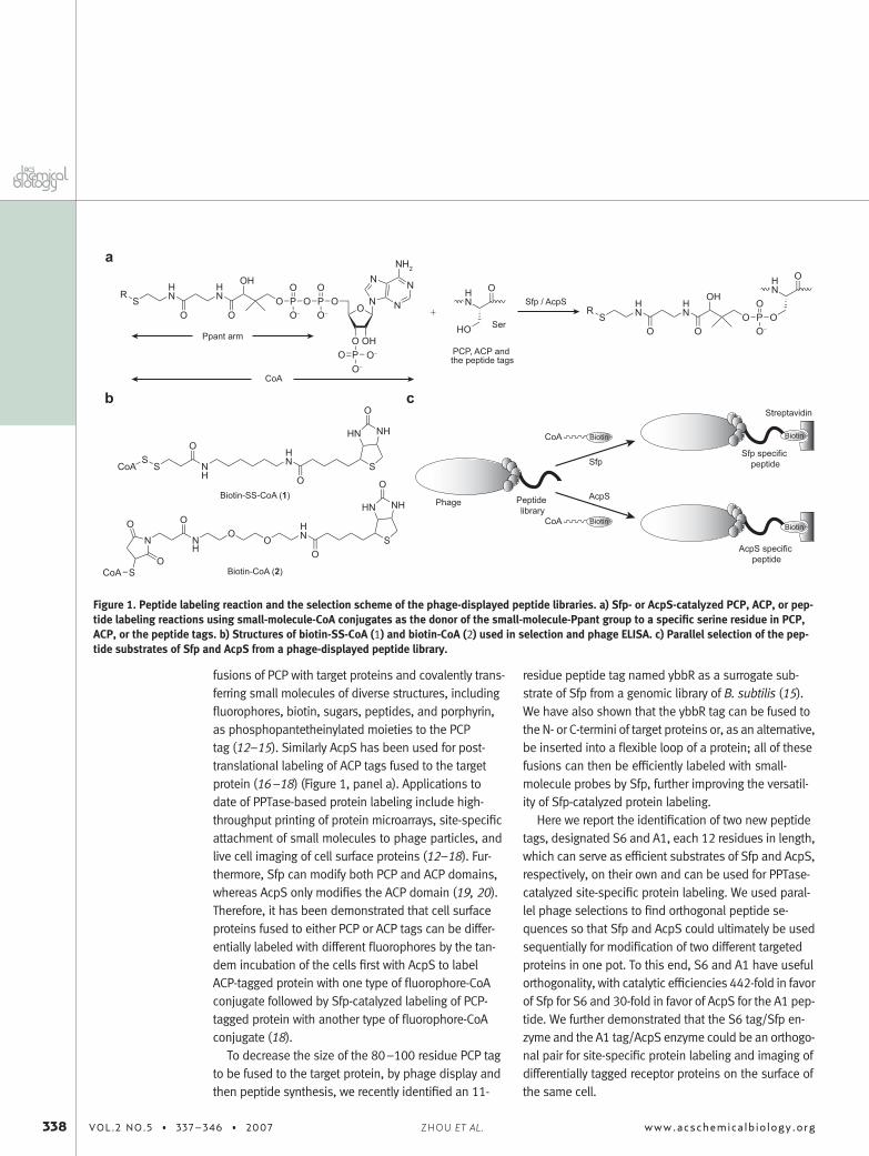
fusions of PCP with target proteins and covalently trans-ferring small molecules of diverse structures, includingfluorophores, biotin, sugars, peptides, and porphyrin,as phosphopantetheinylated moieties to the PCPtag (12–15). Similarly AcpS has been used for post-translational labeling of ACP tags fused to the targetprotein (16–18) (Figure 1, panel a). Applications todate of PPTase-based protein labeling include high-throughput printing of protein microarrays, site-specificattachment of small molecules to phage particles, andlive cell imaging of cell surface proteins (12–18). Fur-thermore, Sfp can modify both PCP and ACP domains,whereas AcpS only modifies the ACP domain (19, 20).Therefore, it has been demonstrated that cell surfaceproteins fused to either PCP or ACP tags can be differ-entially labeled with different fluorophores by the tan-dem incubation of the cells first with AcpS to labelACP-tagged protein with one type of fluorophore-CoAconjugate followed by Sfp-catalyzed labeling of PCP-tagged protein with another type of fluorophore-CoAconjugate (18).
To decrease the size of the 80–100 residue PCP tagto be fused to the target protein, by phage display andthen peptide synthesis, we recently identified an 11-
residue peptide tag named ybbR as a surrogate sub-strate of Sfp from a genomic library of B. subtilis (15).We have also shown that the ybbR tag can be fused tothe N- or C-termini of target proteins or, as an alternative,be inserted into a flexible loop of a protein; all of thesefusions can then be efficiently labeled with small-molecule probes by Sfp, further improving the versatil-ity of Sfp-catalyzed protein labeling.
Here we report the identification of two new peptidetags, designated S6 and A1, each 12 residues in length,which can serve as efficient substrates of Sfp and AcpS,respectively, on their own and can be used for PPTase-catalyzed site-specific protein labeling. We used paral-lel phage selections to find orthogonal peptide se-quences so that Sfp and AcpS could ultimately be usedsequentially for modification of two different targetedproteins in one pot. To this end, S6 and A1 have usefulorthogonality, with catalytic efficiencies 442-fold in favorof Sfp for S6 and 30-fold in favor of AcpS for the A1 pep-tide. We further demonstrated that the S6 tag/Sfp en-zyme and the A1 tag/AcpS enzyme could be an orthogo-nal pair for site-specific protein labeling and imaging ofdifferentially tagged receptor proteins on the surface ofthe same cell.
Streptavidin
Phage
CoA
CoA
AcpS
Sfp
Peptide library
AcpS specific peptide
Sfp specific peptide
Sfp / AcpS
Ppant arm
+
CoA
PCP, ACP and the peptide tags
Biotin-SS-CoA (1)
Biotin-CoA (2)
a
b c
Biotin
Biotin
Biotin
Biotin
RS
HN O
O O
OH
P O P OO O
O− O− O
O OHPO
N
N
N
N
NH2
O−
O−
O
HO SerR
S OO O
OH
P OO
O−
O
CoAS
S NH
O
OS
NHHN
O
HN
HN
HN H
NHN
HN
N NH
OO
O
O
SCoA
O
OS
NH
O
HN
HN
Figure 1. Peptide labeling reaction and the selection scheme of the phage-displayed peptide libraries. a) Sfp- or AcpS-catalyzed PCP, ACP, or pep-tide labeling reactions using small-molecule-CoA conjugates as the donor of the small-molecule-Ppant group to a specific serine residue in PCP,ACP, or the peptide tags. b) Structures of biotin-SS-CoA (1) and biotin-CoA (2) used in selection and phage ELISA. c) Parallel selection of the pep-tide substrates of Sfp and AcpS from a phage-displayed peptide library.
338 VOL.2 NO.5 • 337–346 • 2007 www.acschemicalbiology.orgZHOU ET AL.
RESULTS AND DISCUSSIONConstruction of the Phage-Displayed Peptide Library
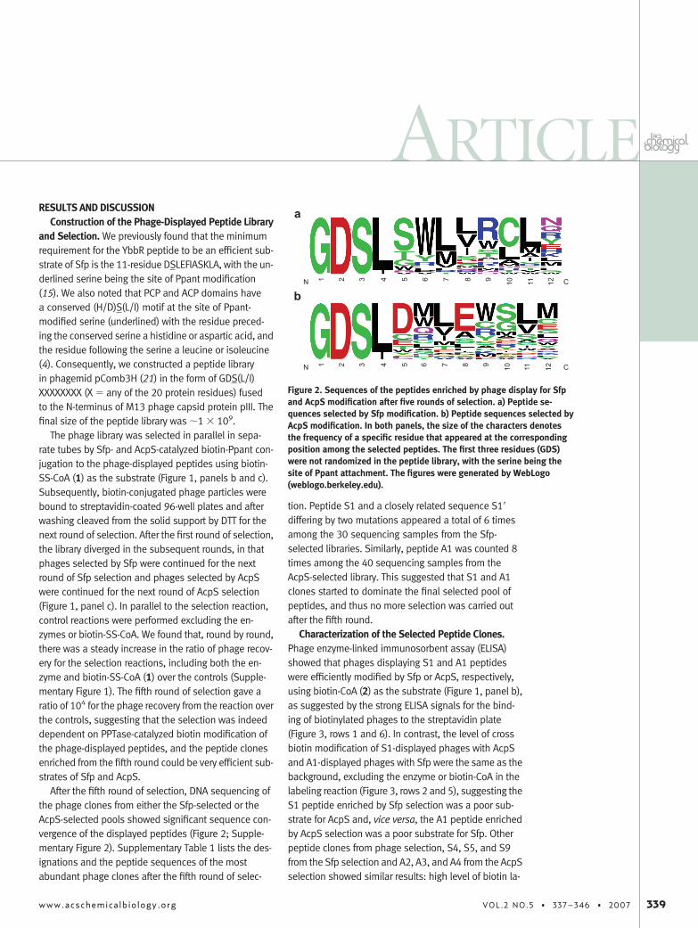
and Selection. We previously found that the minimumrequirement for the YbbR peptide to be an efficient sub-strate of Sfp is the 11-residue DSLEFIASKLA, with the un-derlined serine being the site of Ppant modification(15). We also noted that PCP and ACP domains havea conserved (H/D)S(L/I) motif at the site of Ppant-modified serine (underlined) with the residue preced-ing the conserved serine a histidine or aspartic acid, andthe residue following the serine a leucine or isoleucine(4). Consequently, we constructed a peptide libraryin phagemid pComb3H (21) in the form of GDS(L/I)XXXXXXXX (X � any of the 20 protein residues) fusedto the N-terminus of M13 phage capsid protein pIII. Thefinal size of the peptide library was �1 � 109.
The phage library was selected in parallel in sepa-rate tubes by Sfp- and AcpS-catalyzed biotin-Ppant con-jugation to the phage-displayed peptides using biotin-SS-CoA (1) as the substrate (Figure 1, panels b and c).Subsequently, biotin-conjugated phage particles werebound to streptavidin-coated 96-well plates and afterwashing cleaved from the solid support by DTT for thenext round of selection. After the first round of selection,the library diverged in the subsequent rounds, in thatphages selected by Sfp were continued for the nextround of Sfp selection and phages selected by AcpSwere continued for the next round of AcpS selection(Figure 1, panel c). In parallel to the selection reaction,control reactions were performed excluding the en-zymes or biotin-SS-CoA. We found that, round by round,there was a steady increase in the ratio of phage recov-ery for the selection reactions, including both the en-zyme and biotin-SS-CoA (1) over the controls (Supple-mentary Figure 1). The fifth round of selection gave aratio of 104 for the phage recovery from the reaction overthe controls, suggesting that the selection was indeeddependent on PPTase-catalyzed biotin modification ofthe phage-displayed peptides, and the peptide clonesenriched from the fifth round could be very efficient sub-strates of Sfp and AcpS.
After the fifth round of selection, DNA sequencing ofthe phage clones from either the Sfp-selected or theAcpS-selected pools showed significant sequence con-vergence of the displayed peptides (Figure 2; Supple-mentary Figure 2). Supplementary Table 1 lists the des-ignations and the peptide sequences of the mostabundant phage clones after the fifth round of selec-
tion. Peptide S1 and a closely related sequence S1=differing by two mutations appeared a total of 6 timesamong the 30 sequencing samples from the Sfp-selected libraries. Similarly, peptide A1 was counted 8times among the 40 sequencing samples from theAcpS-selected library. This suggested that S1 and A1clones started to dominate the final selected pool ofpeptides, and thus no more selection was carried outafter the fifth round.
Characterization of the Selected Peptide Clones.Phage enzyme-linked immunosorbent assay (ELISA)showed that phages displaying S1 and A1 peptideswere efficiently modified by Sfp or AcpS, respectively,using biotin-CoA (2) as the substrate (Figure 1, panel b),as suggested by the strong ELISA signals for the bind-ing of biotinylated phages to the streptavidin plate(Figure 3, rows 1 and 6). In contrast, the level of crossbiotin modification of S1-displayed phages with AcpSand A1-displayed phages with Sfp were the same as thebackground, excluding the enzyme or biotin-CoA in thelabeling reaction (Figure 3, rows 2 and 5), suggesting theS1 peptide enriched by Sfp selection was a poor sub-strate for AcpS and, vice versa, the A1 peptide enrichedby AcpS selection was a poor substrate for Sfp. Otherpeptide clones from phage selection, S4, S5, and S9from the Sfp selection and A2, A3, and A4 from the AcpSselection showed similar results: high level of biotin la-
a
bN C1 2 3 4 5 6 7 8 9 10 11 12
N C1 2 3 4 5 6 7 8 9 10 11 12
Figure 2. Sequences of the peptides enriched by phage display for Sfpand AcpS modification after five rounds of selection. a) Peptide se-quences selected by Sfp modification. b) Peptide sequences selected byAcpS modification. In both panels, the size of the characters denotesthe frequency of a specific residue that appeared at the correspondingposition among the selected peptides. The first three residues (GDS)were not randomized in the peptide library, with the serine being thesite of Ppant attachment. The figures were generated by WebLogo(weblogo.berkeley.edu).
ARTICLE
www.acschemicalbiology.org VOL.2 NO.5 • 337–346 • 2007 339
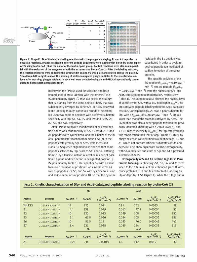
beling with the PPTase used for selection and back-ground level of cross-labeling with the other PPTase(Supplementary Figure 3). Thus our selection strategy,that is, starting from the same peptide library that wassubsequently diverged by either Sfp- or AcpS-catalyzedbiotin labeling through continued rounds of selection,led us to two pools of peptides with preferred substratespecificity with Sfp (S1, S4, S5, and S9) and AcpS (A1,A2, A3, and A4), respectively.
After PPTase-catalyzed modification of selected pep-tide clones was confirmed by ELISA, 12-residue S1 andA1 peptides were synthesized, and the kinetics of the bi-otin Ppant transfer reaction from biotin-CoA (2) to thepeptides catalyzed by Sfp or AcpS were measured(Table 1). Sequence alignment also showed that somepeptides selected by Sfp, such as S1= and S4, differingfrom S1 by a leucine instead of a valine residue at posi-tion 8 (Ppant-modified serine is designated position 3)(Supplementary Table 1). Thus peptide S2 with a valineto leucine mutation at position 8 was synthesized, aswell as peptides S3, S6, and S7 with cysteine to leucineand serine mutations at position 10, so that the cysteine
residue in the S1 peptide wassubstituted in order to avoid un-desired peptide tag mediated di-sulfide formation of the targetproteins.
The specific activities of theS6 peptide (kcat/Km � 0.19 �M�1
min�1) and A1 peptide (kcat/Km
� 0.015 �M�1 min�1) were the highest for Sfp- andAcpS-catalyzed peptide modification, respectively(Table 1). The S6 peptide also showed the highest levelof specificity for Sfp, with a 442-fold higher kcat/Km forSfp-catalyzed peptide labeling than the AcpS-catalyzedreaction. Correspondingly, A1 was a poor substrate forSfp, with a kcat/Km of 0.00049 �M�1 min�1, 30-foldlower than that of the reaction catalyzed by AcpS. TheS6 peptide was also a better peptide tag than the previ-ously identified YbbR tag with a 2-fold lower Km and�10� higher specificity (kcat/Km) for Sfp-catalyzed pep-tide modification than that of AcpS (Table 1). Thus, byphage selection we identified two peptide tags, S6 andA1, which not only are efficient substrates of Sfp andAcpS but also show significant catalytic orthogonality,with S6 a preferred substrate of Sfp and A1 a preferredsubstrate of AcpS.
Orthogonality of S and A1 Peptide Tags for in VitroProtein Labeling. Peptide tags S1, S2, S6, and A1 werefused to the N-terminus of the enhanced green fluores-cence protein (EGFP) and tested for biotin labeling bySfp or AcpS by ELISA (Figure 4). While the S tags and A
anti-M13 IgG HRP
Biotin
Streptavidin
Phage
pIII A/S peptide
+++
++++++
+++++
++++
S1phage
12345678
A1phage
Sfp50 nM
AcpS50 nM
Biotin-CoA100 nM
Figure 3. Phage ELISA of the biotin labeling reactions with the phages displaying S1 and A1 peptides. Inseparate reactions, phages displaying different peptide sequences were labeled with biotin by either Sfp orAcpS using biotin-CoA (2) as the donor of the biotin-Ppant group. Control reactions were also run in paral-lel with the exclusion of the enzymes or both the enzymes and biotin-CoA (2). After the labeling reaction,the reaction mixtures were added to the streptavidin-coated 96-well plate and diluted across the plate by5-fold from left to right to allow the binding of biotin-conjugated phage particles to the streptavidin sur-face. After washing, phages retained in each well were detected using an anti-M13 phage antibody conju-gated to horseradish peroxidase (HRP).
TABLE 1. Kinetic characterization of Sfp- and AcpS-catalyzed peptide labeling reaction by biotin-CoA (2)
Sfp AcpS
Peptide Sequence kcat (min�1) Km (�M)kcat/Km
(�M�1 min�1)kcat (min�1) Km (�M)
kcat/Km
(�M�1 min�1)kcat/Km (Sfp)/kcat/Km (AcpS)
YbbR13 DSLEFIASKLA 11 123 0.091 0.81 242 0.0033 28S1 GDSLSWLVRCLN 4.1 139 0.029 0.042 77.2 0.00054 53S2 GDSLSWLLRCLN 10 120 0.083 0.059 108 0.00055 150S3 GDSLSWLVRLLN 3.1 61.8 0.050 0.034 105 0.00032 156S6 GDSLSWLLRLLN 10 51.5 0.19 0.033 76.0 0.00043 442S7 GDSLSWLLRSLN 8.4 221 0.038 0.085 254 0.00033 115
Sfp AcpS
Peptide Sequence kcat (min�1) Km (�M)kcat/Km
(�M�1 min�1) kcat (min�1) Km (�M)kcat/Km
(�M�1 min�1)kcat/Km (AcpS)/
kcat/Km (Sfp)
A1 GDSLDMLEWSLM 0.26 534 0.00049 1.8 117 0.015 30
340 VOL.2 NO.5 • 337–346 • 2007 www.acschemicalbiology.orgZHOU ET AL.
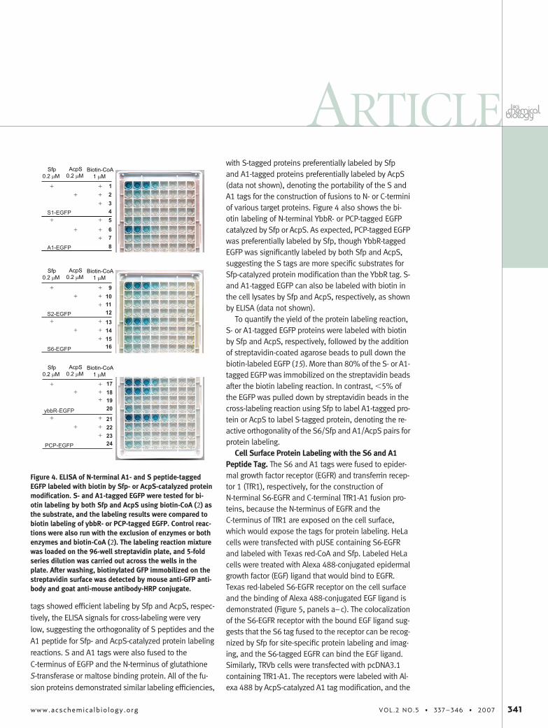
tags showed efficient labeling by Sfp and AcpS, respec-tively, the ELISA signals for cross-labeling were verylow, suggesting the orthogonality of S peptides and theA1 peptide for Sfp- and AcpS-catalyzed protein labelingreactions. S and A1 tags were also fused to theC-terminus of EGFP and the N-terminus of glutathioneS-transferase or maltose binding protein. All of the fu-sion proteins demonstrated similar labeling efficiencies,
with S-tagged proteins preferentially labeled by Sfpand A1-tagged proteins preferentially labeled by AcpS(data not shown), denoting the portability of the S andA1 tags for the construction of fusions to N- or C-terminiof various target proteins. Figure 4 also shows the bi-otin labeling of N-terminal YbbR- or PCP-tagged EGFPcatalyzed by Sfp or AcpS. As expected, PCP-tagged EGFPwas preferentially labeled by Sfp, though YbbR-taggedEGFP was significantly labeled by both Sfp and AcpS,suggesting the S tags are more specific substrates forSfp-catalyzed protein modification than the YbbR tag. S-and A1-tagged EGFP can also be labeled with biotin inthe cell lysates by Sfp and AcpS, respectively, as shownby ELISA (data not shown).
To quantify the yield of the protein labeling reaction,S- or A1-tagged EGFP proteins were labeled with biotinby Sfp and AcpS, respectively, followed by the additionof streptavidin-coated agarose beads to pull down thebiotin-labeled EGFP (15). More than 80% of the S- or A1-tagged EGFP was immobilized on the streptavidin beadsafter the biotin labeling reaction. In contrast, �5% ofthe EGFP was pulled down by streptavidin beads in thecross-labeling reaction using Sfp to label A1-tagged pro-tein or AcpS to label S-tagged protein, denoting the re-active orthogonality of the S6/Sfp and A1/AcpS pairs forprotein labeling.
Cell Surface Protein Labeling with the S6 and A1Peptide Tag. The S6 and A1 tags were fused to epider-mal growth factor receptor (EGFR) and transferrin recep-tor 1 (TfR1), respectively, for the construction ofN-terminal S6-EGFR and C-terminal TfR1-A1 fusion pro-teins, because the N-terminus of EGFR and theC-terminus of TfR1 are exposed on the cell surface,which would expose the tags for protein labeling. HeLacells were transfected with pUSE containing S6-EGFRand labeled with Texas red-CoA and Sfp. Labeled HeLacells were treated with Alexa 488-conjugated epidermalgrowth factor (EGF) ligand that would bind to EGFR.Texas red-labeled S6-EGFR receptor on the cell surfaceand the binding of Alexa 488-conjugated EGF ligand isdemonstrated (Figure 5, panels a–c). The colocalizationof the S6-EGFR receptor with the bound EGF ligand sug-gests that the S6 tag fused to the receptor can be recog-nized by Sfp for site-specific protein labeling and imag-ing, and the S6-tagged EGFR can bind the EGF ligand.Similarly, TRVb cells were transfected with pcDNA3.1containing TfR1-A1. The receptors were labeled with Al-exa 488 by AcpS-catalyzed A1 tag modification, and the
Sfp0.2 µM
12345678
910111213141516
17181920
21222324
S1-EGFP
A1-EGFP
S2-EGFP
S6-EGFP
ybbR-EGFP
PCP-EGFP
AcpS0.2 µM
Biotin-CoA1 µM
++
+
++
++
+
++
Sfp0.2 µM
AcpS0.2 µM
Biotin-CoA1 µM
++
+
++
++
+
++
Sfp0.2 µM
AcpS0.2 µM
Biotin-CoA1 µM
++
+
++
++
+
++
Figure 4. ELISA of N-terminal A1- and S peptide-taggedEGFP labeled with biotin by Sfp- or AcpS-catalyzed proteinmodification. S- and A1-tagged EGFP were tested for bi-otin labeling by both Sfp and AcpS using biotin-CoA (2) asthe substrate, and the labeling results were compared tobiotin labeling of ybbR- or PCP-tagged EGFP. Control reac-tions were also run with the exclusion of enzymes or bothenzymes and biotin-CoA (2). The labeling reaction mixturewas loaded on the 96-well streptavidin plate, and 5-foldseries dilution was carried out across the wells in theplate. After washing, biotinylated GFP immobilized on thestreptavidin surface was detected by mouse anti-GFP anti-body and goat anti-mouse antibody-HRP conjugate.
ARTICLE
www.acschemicalbiology.org VOL.2 NO.5 • 337–346 • 2007 341
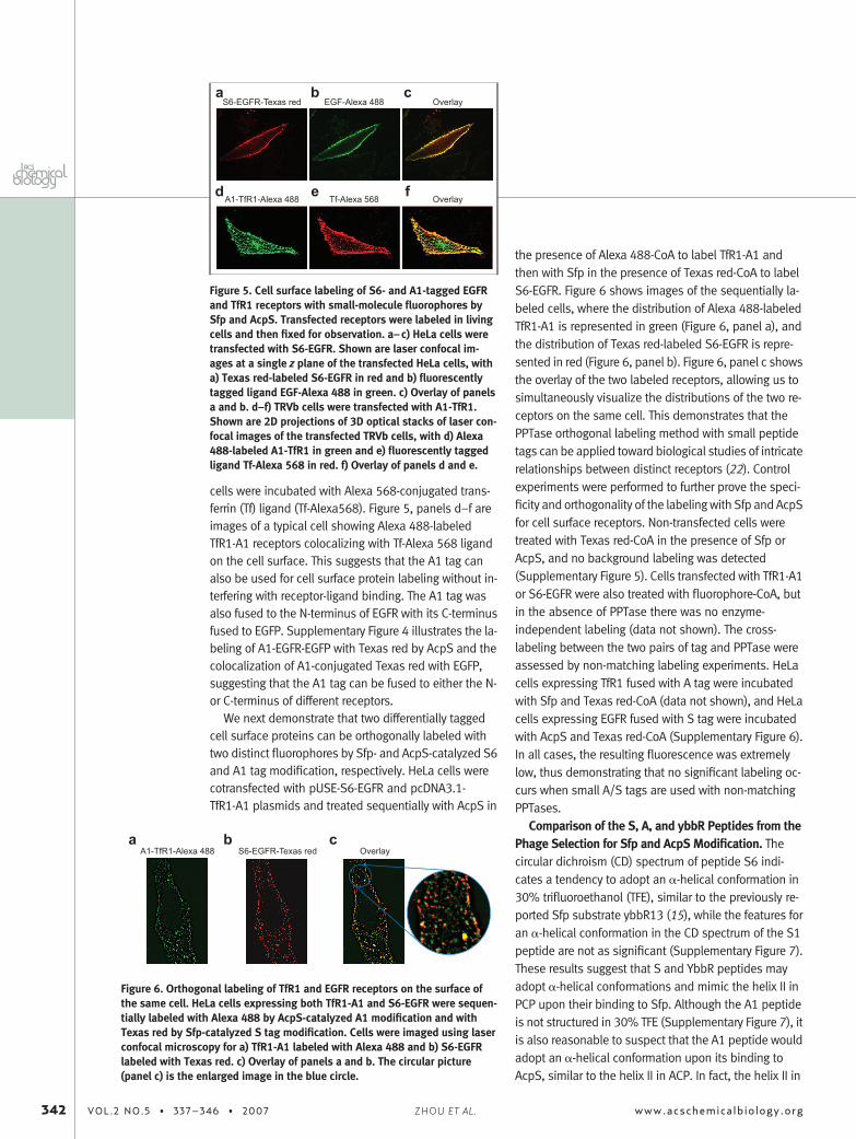
cells were incubated with Alexa 568-conjugated trans-ferrin (Tf) ligand (Tf-Alexa568). Figure 5, panels d–f areimages of a typical cell showing Alexa 488-labeledTfR1-A1 receptors colocalizing with Tf-Alexa 568 ligandon the cell surface. This suggests that the A1 tag canalso be used for cell surface protein labeling without in-terfering with receptor-ligand binding. The A1 tag wasalso fused to the N-terminus of EGFR with its C-terminusfused to EGFP. Supplementary Figure 4 illustrates the la-beling of A1-EGFR-EGFP with Texas red by AcpS and thecolocalization of A1-conjugated Texas red with EGFP,suggesting that the A1 tag can be fused to either the N-or C-terminus of different receptors.
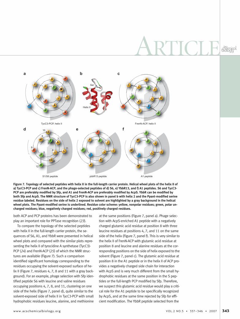
We next demonstrate that two differentially taggedcell surface proteins can be orthogonally labeled withtwo distinct fluorophores by Sfp- and AcpS-catalyzed S6and A1 tag modification, respectively. HeLa cells werecotransfected with pUSE-S6-EGFR and pcDNA3.1-TfR1-A1 plasmids and treated sequentially with AcpS in
the presence of Alexa 488-CoA to label TfR1-A1 andthen with Sfp in the presence of Texas red-CoA to labelS6-EGFR. Figure 6 shows images of the sequentially la-beled cells, where the distribution of Alexa 488-labeledTfR1-A1 is represented in green (Figure 6, panel a), andthe distribution of Texas red-labeled S6-EGFR is repre-sented in red (Figure 6, panel b). Figure 6, panel c showsthe overlay of the two labeled receptors, allowing us tosimultaneously visualize the distributions of the two re-ceptors on the same cell. This demonstrates that thePPTase orthogonal labeling method with small peptidetags can be applied toward biological studies of intricaterelationships between distinct receptors (22). Controlexperiments were performed to further prove the speci-ficity and orthogonality of the labeling with Sfp and AcpSfor cell surface receptors. Non-transfected cells weretreated with Texas red-CoA in the presence of Sfp orAcpS, and no background labeling was detected(Supplementary Figure 5). Cells transfected with TfR1-A1or S6-EGFR were also treated with fluorophore-CoA, butin the absence of PPTase there was no enzyme-independent labeling (data not shown). The cross-labeling between the two pairs of tag and PPTase wereassessed by non-matching labeling experiments. HeLacells expressing TfR1 fused with A tag were incubatedwith Sfp and Texas red-CoA (data not shown), and HeLacells expressing EGFR fused with S tag were incubatedwith AcpS and Texas red-CoA (Supplementary Figure 6).In all cases, the resulting fluorescence was extremelylow, thus demonstrating that no significant labeling oc-curs when small A/S tags are used with non-matchingPPTases.
Comparison of the S, A, and ybbR Peptides from thePhage Selection for Sfp and AcpS Modification. Thecircular dichroism (CD) spectrum of peptide S6 indi-cates a tendency to adopt an �-helical conformation in30% trifluoroethanol (TFE), similar to the previously re-ported Sfp substrate ybbR13 (15), while the features foran �-helical conformation in the CD spectrum of the S1peptide are not as significant (Supplementary Figure 7).These results suggest that S and YbbR peptides mayadopt �-helical conformations and mimic the helix II inPCP upon their binding to Sfp. Although the A1 peptideis not structured in 30% TFE (Supplementary Figure 7), itis also reasonable to suspect that the A1 peptide wouldadopt an �-helical conformation upon its binding toAcpS, similar to the helix II in ACP. In fact, the helix II in
Overlay
Overlay
S6-EGFR-Texas red EGF-Alexa 488
A1-TfR1-Alexa 488 Tf-Alexa 568
ba c
d e f
Figure 5. Cell surface labeling of S6- and A1-tagged EGFRand TfR1 receptors with small-molecule fluorophores bySfp and AcpS. Transfected receptors were labeled in livingcells and then fixed for observation. a–c) HeLa cells weretransfected with S6-EGFR. Shown are laser confocal im-ages at a single z plane of the transfected HeLa cells, witha) Texas red-labeled S6-EGFR in red and b) fluorescentlytagged ligand EGF-Alexa 488 in green. c) Overlay of panelsa and b. d–f) TRVb cells were transfected with A1-TfR1.Shown are 2D projections of 3D optical stacks of laser con-focal images of the transfected TRVb cells, with d) Alexa488-labeled A1-TfR1 in green and e) fluorescently taggedligand Tf-Alexa 568 in red. f) Overlay of panels d and e.
A1-TfR1-Alexa 488 S6-EGFR-Texas red Overlaya b c
Figure 6. Orthogonal labeling of TfR1 and EGFR receptors on the surface ofthe same cell. HeLa cells expressing both TfR1-A1 and S6-EGFR were sequen-tially labeled with Alexa 488 by AcpS-catalyzed A1 modification and withTexas red by Sfp-catalyzed S tag modification. Cells were imaged using laserconfocal microscopy for a) TfR1-A1 labeled with Alexa 488 and b) S6-EGFRlabeled with Texas red. c) Overlay of panels a and b. The circular picture(panel c) is the enlarged image in the blue circle.
342 VOL.2 NO.5 • 337–346 • 2007 www.acschemicalbiology.orgZHOU ET AL.
both ACP and PCP proteins has been demonstrated toplay an important role for PPTase recognition (23).
To compare the topology of the selected peptideswith helix II in the full-length carrier protein, the se-quences of S6, A1, and YbbR were presented in helicalwheel plots and compared with the similar plots repre-senting the helix II of tyrocidine A synthetase (TycC3)-PCP (24) and FrenN-ACP (25) of which the NMR struc-tures are available (Figure 7). Such a comparisonidentified significant homology corresponding to theresidues occupying the solvent-exposed surface of he-lix II (Figure 7, residues 4, 7, 8 and 11 with a gray back-ground). For an example, phage selection with Sfp iden-tified peptide S6 with leucine and valine residuesoccupying positions 4, 7, 8, and 11, clustering on oneside of the helix (Figure 7, panel d), quite similar to thesolvent-exposed side of helix II in TycC3-PCP with smallhydrophobic residues leucine, alanine, and methionine
at the same positions (Figure 7, panel a). Phage selec-tion with AcpS-enriched A1 peptide with a negativelycharged glutamic acid residue at position 8 with threeleucine residues at positions 4, 7, and 11 on the sameside of the helix (Figure 7, panel f). This is very similar tothe helix II of FrenN-ACP with glutamic acid residue atposition 8 and leucine and alanine residues at the cor-responding positions on the side of helix exposed to thesolvent (Figure 7, panel c). The glutamic acid residue atposition 8 in the A1 peptide or in the helix II of ACP pro-vides a negatively charged side chain for interactionwith AcpS and is very much different from the small hy-drophobic residues at the same position in the S pep-tides or the full-length PCP modified by Sfp. Therefore,we suspect this glutamic acid residue would play a criti-cal role for the A1 peptide to be specifically recognizedby AcpS, and at the same time rejected by Sfp for effi-cient modification. The YbbR peptide selected from the
LS
WL
V/L
C/L
L
S
N
R
S
LE
FI
A
S
K
L
A
S
S
LD
ML
E
W L
M
ybbR13 peptideS1/S6 peptide A1 peptide
3
45
67
8
9
10
11
12
LK
AM
A
V
A
A
V
H
R
E
A
S
LA
LL
E
T A
V
L
Q
Q
R
S
Q
TycC3-PCP, helix II FrenN-ACP, helix II
Ser
Helix II
a b c
d e f
Figure 7. Topology of selected peptides with helix II in the full-length carrier protein. Helical wheel plots of the helix II ofa) TycC3-PCP and c) FrenN-ACP, and the phage-selected peptides of d) S6, e) YbbR13, and f) A1 peptides. S6 and TycC3-PCP are preferably modified by Sfp, and A1 and FrenN-ACP are preferably modified by AcpS. YbbR can be modified byboth Sfp and AcpS. The NMR structure of TycC3-PCP is also shown in panel b with helix 2 and the Ppant-modified serineresidue labeled. Residues on the side of helix 2 exposed to solvent are highlighted by a gray background in the helicalwheel plots. The Ppant-modified serine is underlined. Residue color scheme: yellow, nonpolar residues; green, polar un-charged residues; blue, negatively charged residues; red, positively charged residues.
ARTICLE
www.acschemicalbiology.org VOL.2 NO.5 • 337–346 • 2007 343
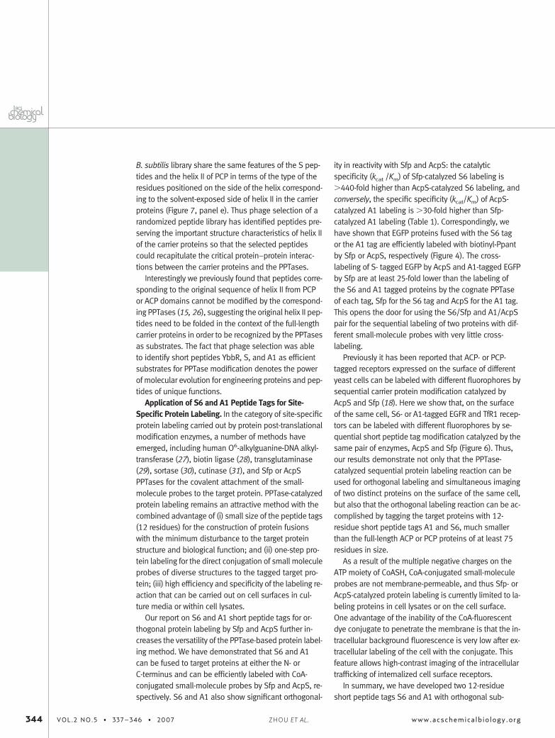
B. subtilis library share the same features of the S pep-tides and the helix II of PCP in terms of the type of theresidues positioned on the side of the helix correspond-ing to the solvent-exposed side of helix II in the carrierproteins (Figure 7, panel e). Thus phage selection of arandomized peptide library has identified peptides pre-serving the important structure characteristics of helix IIof the carrier proteins so that the selected peptidescould recapitulate the critical protein–protein interac-tions between the carrier proteins and the PPTases.
Interestingly we previously found that peptides corre-sponding to the original sequence of helix II from PCPor ACP domains cannot be modified by the correspond-ing PPTases (15, 26), suggesting the original helix II pep-tides need to be folded in the context of the full-lengthcarrier proteins in order to be recognized by the PPTasesas substrates. The fact that phage selection was ableto identify short peptides YbbR, S, and A1 as efficientsubstrates for PPTase modification denotes the powerof molecular evolution for engineering proteins and pep-tides of unique functions.
Application of S6 and A1 Peptide Tags for Site-Specific Protein Labeling. In the category of site-specificprotein labeling carried out by protein post-translationalmodification enzymes, a number of methods haveemerged, including human O6-alkylguanine-DNA alkyl-transferase (27), biotin ligase (28), transglutaminase(29), sortase (30), cutinase (31), and Sfp or AcpSPPTases for the covalent attachment of the small-molecule probes to the target protein. PPTase-catalyzedprotein labeling remains an attractive method with thecombined advantage of (i) small size of the peptide tags(12 residues) for the construction of protein fusionswith the minimum disturbance to the target proteinstructure and biological function; and (ii) one-step pro-tein labeling for the direct conjugation of small moleculeprobes of diverse structures to the tagged target pro-tein; (iii) high efficiency and specificity of the labeling re-action that can be carried out on cell surfaces in cul-ture media or within cell lysates.
Our report on S6 and A1 short peptide tags for or-thogonal protein labeling by Sfp and AcpS further in-creases the versatility of the PPTase-based protein label-ing method. We have demonstrated that S6 and A1can be fused to target proteins at either the N- orC-terminus and can be efficiently labeled with CoA-conjugated small-molecule probes by Sfp and AcpS, re-spectively. S6 and A1 also show significant orthogonal-
ity in reactivity with Sfp and AcpS: the catalyticspecificity (kcat /Km) of Sfp-catalyzed S6 labeling is�440-fold higher than AcpS-catalyzed S6 labeling, andconversely, the specific specificity (kcat/Km) of AcpS-catalyzed A1 labeling is �30-fold higher than Sfp-catalyzed A1 labeling (Table 1). Correspondingly, wehave shown that EGFP proteins fused with the S6 tagor the A1 tag are efficiently labeled with biotinyl-Ppantby Sfp or AcpS, respectively (Figure 4). The cross-labeling of S- tagged EGFP by AcpS and A1-tagged EGFPby Sfp are at least 25-fold lower than the labeling ofthe S6 and A1 tagged proteins by the cognate PPTaseof each tag, Sfp for the S6 tag and AcpS for the A1 tag.This opens the door for using the S6/Sfp and A1/AcpSpair for the sequential labeling of two proteins with dif-ferent small-molecule probes with very little cross-labeling.
Previously it has been reported that ACP- or PCP-tagged receptors expressed on the surface of differentyeast cells can be labeled with different fluorophores bysequential carrier protein modification catalyzed byAcpS and Sfp (18). Here we show that, on the surfaceof the same cell, S6- or A1-tagged EGFR and TfR1 recep-tors can be labeled with different fluorophores by se-quential short peptide tag modification catalyzed by thesame pair of enzymes, AcpS and Sfp (Figure 6). Thus,our results demonstrate not only that the PPTase-catalyzed sequential protein labeling reaction can beused for orthogonal labeling and simultaneous imagingof two distinct proteins on the surface of the same cell,but also that the orthogonal labeling reaction can be ac-complished by tagging the target proteins with 12-residue short peptide tags A1 and S6, much smallerthan the full-length ACP or PCP proteins of at least 75residues in size.
As a result of the multiple negative charges on theATP moiety of CoASH, CoA-conjugated small-moleculeprobes are not membrane-permeable, and thus Sfp- orAcpS-catalyzed protein labeling is currently limited to la-beling proteins in cell lysates or on the cell surface.One advantage of the inability of the CoA-fluorescentdye conjugate to penetrate the membrane is that the in-tracellular background fluorescence is very low after ex-tracellular labeling of the cell with the conjugate. Thisfeature allows high-contrast imaging of the intracellulartrafficking of internalized cell surface receptors.
In summary, we have developed two 12-residueshort peptide tags S6 and A1 with orthogonal sub-
344 VOL.2 NO.5 • 337–346 • 2007 www.acschemicalbiology.orgZHOU ET AL.
strate specificities for the site-specific protein post-translational labeling reaction catalyzed by PPTases Sfpand AcpS. The small size of the S6 and A1 tags com-pared to the full-length PCP and ACP domains (80–100residues), the versatility of those tags for fusion to targetproteins at N- or C-termini, the structural diversities of
the small-molecule probes for Sfp- and AcpS-catalyzedpeptide modification, and the high efficiency and speci-ficity of Sfp and AcpS for the S6 and A1 tags provide apowerful protein labeling method that would allow spe-cific orthogonal labeling of different target proteins oncell surfaces or in cell lysates.
METHODSCell Surface Labeling of TfR1-A1. TRVb cells (a kind gift of Timo-
thy E. McGraw, Weill Medical College, Cornell University) ex-pressing TfR1-A1 (transfection protocols available in Support-ing Information) were incubated with 1.98 �M AcpS, 1 �M CoA-Alexa Fluor 488, 10 mM MgCl2, 50 mM HEPES, pH 7.5 in 200 �Lof serum-free media for 30 min at 37 °C under 5% CO2. Cellswere washed five times with PBS, incubated with 10 �g mL�1
Alexa Fluor 568-conjugated Tf, and then washed five times withPBS. Finally, cells were fixed at 4 °C for 10 min using a 3.7%formaldehyde solution in PBS and mounted with mounting me-dium AEC (Immunotech) for optical microscopy studies. We haveconducted experiments to optimize the labeling time for singlecolor labeling and have found that 15–40 min of incubationwith the solution containing the enzyme and the fluorophoreswas the most favorable for imaging experiments (data notshown).
Cell Surface Labeling of S6-EGFR. HeLa cells expressing S6-EGFR (transfection protocols available in Supporting Informa-tion) were incubated with 1.98 �M Sfp, 1 �M CoA-Texas red,10 mM MgCl2, 50 mM HEPES, pH 7.5 in 200 �L of serum-free(not required) media for 20 min at 37 °C under 5% CO2. Cellswere washed five times with PBS, incubated with 5 �g mL�1
Alexa Fluor 488-conjugated EGF (Molecular Probes Inc.) for5 min, and then washed five times with PBS. Finally, cellswere fixed and mounted for optical microscopy studies.
Cell Surface Labeling of A1-TfR1 and S6-EGFR Expressed in HeLaCells. TfR1-A1 was labeled first by incubating the cells with1.98 �M AcpS, 1 �M CoA-Alexa Fluor 488, 10 mM MgCl2, 50 mMHEPES, pH 7.5 in 200 �L of serum-free media (not required) for30 min at 37 °C under 5% CO2. Labeled cells were washed fivetimes with PBS and then incubated with 1.98 �M Sfp, 1 �M CoA-Texas red, 10 mM MgCl2, 50 mM HEPES, pH 7.5 in 200 �L ofserum-free media for 20 min at 37 °C under 5% CO2. Cells werewashed five times with PBS then fixed and mounted with mount-ing medium for optical microscopy studies.
Images were acquired with a confocal microscopy using aNikon TE2000U inverted microscope in conjunction with aPerkinElmer Ultraview spinning disk confocal system or in aNikon Eclipse TE2000-E inverted epifluorescence microscopeequipped with a Hamamatsu Orca ER Cooled-CCD camera. Im-ages were acquired using a 40�, 60�, or 100� differential in-terference contrast oil immersion objective lens and analyzedusing MetaMorph software from Universal Imaging, Inc.
Acknowledgment: This work was supported by grantsHL32854 and HL70819 to D.E.G. and GM20011 to C.T.W. fromthe National Institutes of Health. P.C. was supported by aFulbright-M.E.C. (Spain) postdoctoral fellowship. J.Y. was sup-ported by a startup find from the University of Chicago. Confo-cal microscopy was performed in the Nikon Imaging Center atHarvard Medical School.
Supporting Information Available: This material is availablefree of charge via the Internet.
REFERENCES1. Johnsson, N., George, N., and Johnsson, K. (2005) Protein chemis-
try on the surface of living cells, ChemBioChem 6, 47–52.2. Yin, J., Lin, A. J., Golan, D. E., and Walsh, C. T. (2006) Site-specific
protein labeling by Sfp phosphopantetheinyl transferase, Nat. Pro-toc. 1, 280–285.
3. Cane, D. E., Walsh, C. T., and Khosla, C. (1998) Harnessing the bio-synthetic code: combinations, permutations, and mutations, Sci-ence 282, 63–68.
4. Marahiel, M. A., Stachelhaus, T., and Mootz, H. D. (1997) Modularpeptide synthetases involved in nonribosomal peptide synthesis,Chem. Rev. 97, 2651–2674.
5. Staunton, J., and Weissman, K. J. (2001) Polyketide biosynthesis: amillennium review, Nat. Prod. Rep. 18, 380–416.
6. Wakil, S. J., Stoops, J. K., and Joshi, V. C. (1983) Fatty acid synthe-sis and its regulation, Annu. Rev. Biochem. 52, 537–579.
7. Walsh, C. T., Gehring, A. M., Weinreb, P. H., Quadri, L. E., and Flu-gel, R. S. (1997) Post-translational modification of polyketide andnonribosomal peptide synthases, Curr. Opin. Chem. Biol. 1,309–315.
8. Belshaw, P. J., Walsh, C. T., and Stachelhaus, T. (1999) Aminoacyl-CoAs as probes of condensation domain selectivity in nonriboso-mal peptide synthesis, Science 284, 486–489.
9. Gehring, A. M., Lambalot, R. H., Vogel, K. W., Drueckhammer, D. G.,and Walsh, C. T. (1997) Ability of Streptomyces spp. acyl carrier pro-teins and coenzyme A analogs to serve as substrates in vitro forE. coli holo-ACP synthase, Chem. Biol. 4, 17–24.
10. La Clair, J. J., Foley, T. L., Schegg, T. R., Regan, C. M., and Burkart,M. D. (2004) Manipulation of carrier proteins in antibiotic biosynthe-sis, Chem. Biol. 11, 195–201.
11. Sieber, S. A., Walsh, C. T., and Marahiel, M. A. (2003) Loadingpeptidyl-coenzyme A onto peptidyl carrier proteins: a novel ap-proach in characterizing macrocyclization by thioesterase domains,J. Am. Chem. Soc. 125, 10862–10866.
12. Yin, J., Liu, F., Li, X., and Walsh, C. T. (2004) Labeling proteins withsmall molecules by site-specific posttranlational modification, J.Am. Chem. Soc. 126, 7754–7755.
13. Yin, J., Lin, A. J., Buckett, P. D., Wessling-Resnick, M., Golan, D. E.,and Walsh, C. T. (2005) Single-cell FRET imaging of transferrin recep-tor trafficking dynamics by Sfp catalyzed site specific protein label-ing, Chem. Biol. 12, 999–1006.
14. Yin, J., Liu, F., Schinke, M., Daly, C., and Walsh, C. T. (2004) Phage-mid encoded small molecules for high throughput screening ofchemical libraries, J. Am. Chem. Soc. 126, 13570–13571.
15. Yin, J., Straight, P. D., McLaughlin, S. M., Zhou, Z., Lin, A. J., Golan,D. E., Kelleher, N. L., Kolter, R., and Walsh, C. T. (2005) Geneticallyencoded short peptide tag for versatile protein labeling by Sfp phos-phopantetheinyl transferase, Proc. Natl. Acad. Sci. U.S.A. 102,15815–15820.
ARTICLE
www.acschemicalbiology.org VOL.2 NO.5 • 337–346 • 2007 345
16. George, N., Pick, H., Vogel, H., Johnsson, N., and Johnsson, K. (2004)Specific labeling of cell surface proteins with chemically diversecompounds, J. Am. Chem. Soc. 126, 8896–8897.
17. Meyer, B. H., Segura, J. M., Martinez, K. L., Hovius, R., George, N.,Johnsson, K., and Vogel, H. (2006) FRET imaging reveals that func-tional neurokinin-1 receptors are monomeric and reside in mem-brane microdomains of live cells, Proc. Natl. Acad. Sci. U.S.A. 103,2138–2143.
18. Vivero-Pol, L., George, N., Krumm, H., Johnsson, K., and Johnsson,N. (2005) Multicolor imaging of cell surface proteins, J. Am. Chem.Soc. 127, 12770–12771.
19. Flugel, R. S., Hwangbo, Y., Lambalot, R. H., Cronan, J. E., Jr., andWalsh, C. T. (2000) Holo-(acyl carrier protein) synthase and phos-phopantetheinyl transfer in Escherichia coli, J. Biol. Chem. 275,959–968.
20. Lambalot, R. H., Gehring, A. M., Flugel, R. S., Zuber, P., LaCelle, M.,Marahiel, M. A., Reid, R., Khosla, C., and Walsh, C. T. (1996) A newenzyme superfamily—the phosphopantetheinyl transferases,Chem. Biol. 3, 923–936.
21. Barbas, C. F., 3rd, Kang, A. S., Lerner, R. A., and Benkovic, S. J.(1991) Assembly of combinatorial antibody libraries on phage sur-faces: the gene III site, Proc. Natl. Acad. Sci. U.S.A. 88, 7978–7982.
22. Johannessen, L. E., Pedersen, N. M., Pedersen, K. W., Madshus, I. H.,and Stang, E. (2006) Activation of the epidermal growth factor (EGF)receptor induces formation of EGF receptor- and Grb2-containingclathrin-coated pits, Mol. Cell. Biol. 26, 389–401.
23. Mofid, M. R., Finking, R., and Marahiel, M. A. (2002) Recognition ofhybrid peptidyl carrier proteins/acyl carrier proteins in nonriboso-mal peptide synthetase modules by the 4=-phosphopantetheinyltransferases AcpS and Sfp, J. Biol. Chem. 277, 17023–17031.
24. Weber, T., Baumgartner, R., Renner, C., Marahiel, M. A., and Holak,T. A. (2000) Solution structure of PCP, a prototype for the peptidylcarrier domains of modular peptide synthetases, Structure 8,407–418.
25. Li, Q., Khosla, C., Puglisi, J. D., and Liu, C. W. (2003) Solution struc-ture and backbone dynamics of the holo form of the frenolicin acylcarrier protein, Biochemistry 42, 4648–4657.
26. Quadri, L. E., Weinreb, P. H., Lei, M., Nakano, M. M., Zuber, P., andWalsh, C. T. (1998) Characterization of Sfp, a Bacillus subtilis phos-phopantetheinyl transferase for peptidyl carrier protein domains inpeptide synthetases, Biochemistry 37, 1585–1595.
27. Keppler, A., Gendreizig, S., Gronemeyer, T., Pick, H., Vogel, H., andJohnsson, K. (2003) A general method for the covalent labeling of fu-sion proteins with small molecules in vivo, Nat. Biotechnol. 21,86–89.
28. Chen, I., Howarth, M., Lin, W., and Ting, A. Y. (2005) Site-specific la-beling of cell surface proteins with biophysical probes using biotinligase, Nat. Methods 2, 99–104.
29. Lin, C. W., and Ting, A. Y. (2006) Transglutaminase-catalyzed site-specific conjugation of small-molecule probes to proteins in vitroand on the surface of living cells, J. Am. Chem. Soc. 128,4542–4543.
30. Mao, H., Hart, S. A., Schink, A., and Pollok, B. A. (2004) Sortase-mediated protein ligation: a new method for protein engineering,J. Am. Chem. Soc. 126, 2670–2671.
31. Hodneland, C. D., Lee, Y. S., Min, D. H., and Mrksich, M. (2002) Se-lective immobilization of proteins to self-assembled monolayerspresenting active site-directed capture ligands, Proc. Natl. Acad. Sci.U.S.A. 99, 5048–5052.
32. Vivero-Pol, L., George, N., Krumm, H., Johnsson, K., and Johnsson,N. (2005) Multicolor imaging of cell surface proteins, J. Am. Chem.Soc. 127, 12770–12771.
346 VOL.2 NO.5 • 337–346 • 2007 www.acschemicalbiology.orgZHOU ET AL.