Basal Clades and Molecular Systematics of Heteromyid Rodents
Upload
independentCategory
view
2download
0
This article appeared in a journal published by Elsevier. The attachedcopy is furnished to the author for internal non-commercial researchand education use, including for instruction at the authors institution
and sharing with colleagues.
Other uses, including reproduction and distribution, or selling orlicensing copies, or posting to personal, institutional or third party
websites are prohibited.
In most cases authors are permitted to post their version of thearticle (e.g. in Word or Tex form) to their personal website orinstitutional repository. Authors requiring further information
regarding Elsevier’s archiving and manuscript policies areencouraged to visit:
http://www.elsevier.com/copyright
Author's personal copy
Virus Research 163 (2012) 486– 494
Contents lists available at SciVerse ScienceDirect
Virus Research
journa l h o me pag e: www.elsev ier .com/ locate /v i rusres
Genetic diversity of hantaviruses in Mexico: Identification of three novelhantaviruses from Neotominae rodents
Hiroaki Kariwaa,∗, Haruka Yoshidaa, Cornelio Sánchez-Hernándezb,María de Lourdes Romero-Almarazb, José Alberto Almazán-Catalánb, Celso Ramosc, Daisuke Miyashitaa,Takahiro Setoa, Ayako Takanoa, Masashi Totania, Ryo Murataa, Ngonda Saasaa, Mariko Ishizukaa,Takahiro Sanadaa, Kentaro Yoshii a, Kumiko Yoshimatsud, Jiro Arikawaa, Ikuo Takashimaa
a Graduate School of Veterinary Medicine, Hokkaido University, Kita-18, Nishi-9, Kita-Ku, Sapporo 060-0818, Hokkaido, Japanb Instituto de Biología, Universidad Nacional Autónoma de México, Apdo. Postal 70-153, 04510 México, D.F., Mexicoc Instituto Nacional de Salud Pública, Ave. Universidad 655, Colonia Santa Ahuacatitlán, 62508 Cuernavaca, Morelos, Mexicod Graduate School of Medicine, Hokkaido University, Kita-15, Nishi-7, Kita-Ku, Sapporo 060-8638, Hokkaido, Japan
a r t i c l e i n f o
Article history:Received 8 October 2011Received in revised form13 November 2011Accepted 15 November 2011Available online 25 November 2011
Keywords:HantavirusRodentGenetic diversityPhylogenyMexico
a b s t r a c t
A variety of hantaviruses are harbored by rodents in North and South America, some of which cancause hantavirus pulmonary syndrome. To obtain greater evolutionary insight into hantaviruses in theAmericas, a total of 211 rodents were captured in the Mexican states of Guerrero and Morelos in 2006.Anti-hantavirus antibodies were detected in 27 of 211 serum samples (12.8%) by ELISA. The distribution ofseropositive rodents was: 17 Peromyscus beatae, 1 Megadontomys thomasi, 1 Neotoma picta, 6 Reithrodon-tomys sumichrasti, and 2 Reithrodontomys megalotis. The hantavirus small (S), medium (M), and large (L)genome segments from P. beatae, R. sumichrasti, and R. megalotis were amplified and the sequences cov-ering the open reading frames were determined. The hantaviruses from P. beatae, R. sumichrasti, and R.megalotis were provisionally designated Montano (MTN), Carrizal (CAR), and Huitzilac (HUI), respectively.The M segment amino acid identities among the Mexican hantaviruses were 80.8–93.0%. When these Msegments were compared to those of known hantaviruses, MTN virus was most closely related to Lime-stone Canyon (LSC) virus (88.9% amino acid identity), while the CAR and HUI viruses were most closelyrelated to El Moro Canyon (ELMC) virus (90–91% identity). Phylogenetic analysis revealed that the MTN,CAR, and HUI viruses occupy a monophyletic clade with the LSC, ELMC, and Rio Segundo viruses, whichare harbored by Peromyscus boylii, R. megalotis, and Reithrodontomys mexicanus, respectively. The dataobtained in this study provide important information for understanding the evolution of hantaviruses inthe Americas.
© 2011 Elsevier B.V. All rights reserved.
1. Introduction
Hantaviruses, which comprise the genus Hantavirus in thefamily Bunyaviridae, contain a three-segment negative-sense RNAgenome. Small (S), medium (M), and large (L) RNA segments encodethe nucleocapsid protein (N), glycoproteins (Gn and Gc), and anRNA-dependent RNA polymerase, respectively (Schmaljohn et al.,1986). Hantaviruses are carried by a variety of rodents and sori-comorph species in nature and persistently infect their naturalhosts (Jonsson et al., 2010; Song et al., 2007). Some rodent-bornehantaviruses cause hantavirus infections in humans, includinghemorrhagic fever with renal syndrome (HFRS) and hantavirus
∗ Corresponding author. Tel.: +81 11 706 5212; fax: +81 11 706 5213.E-mail address: [email protected] (H. Kariwa).
pulmonary syndrome (HPS) (Peters et al., 1999; Schmaljohn andHjelle, 1997). HFRS, which is characterized by a high fever, hemor-rhage, and renal dysfunction with a fatality rate <10%, is distributedthroughout Europe, Russia, China, and Korea. HPS, which is charac-terized by a high fever, pulmonary dysfunction, and cardiac shockand has a ∼40% fatality rate (Peters and Khan, 2002), is distributedin North and South Americas. Humans acquire the infection pri-marily by the inhalation of virus-containing rodent excreta or bythe bite of infected animals (Lee and Johnson, 1982). An impor-tant characteristic of hantavirus is the close relationship betweenthe virus and its rodent host species, and co-evolution betweenthem may have occurred on a geological timescale (Plyusnin andMorzunov, 2001).
Since the identification of HPS in the United States of Amer-ica, patients have been reported from various countries in bothNorth and South America; more than 30 hantaviruses have been
0168-1702/$ – see front matter © 2011 Elsevier B.V. All rights reserved.doi:10.1016/j.virusres.2011.11.013
Author's personal copy
H. Kariwa et al. / Virus Research 163 (2012) 486– 494 487
identified on the American continents (Jonsson et al., 2010; Millset al., 2010), some of which are associated with HPS. In NorthAmerica, the Sin Nombre (SN), New York (NY), Monongahela, BlackCreek Canal (BCC), Bayou (BAY) viruses cause HPS and are car-ried by Peromyscus maniculatus, Peromyscus leucopus, Peromyscusleucopus nubiterrae, Sigmodon hispidus, Oryzomys palustis, and S.hispidus, respectively (Morzunov et al., 1995; Nichol et al., 1993;Ravkov et al., 1995; Rawlings et al., 1996; Song et al., 1994;Torrez-Martinez and Hjelle, 1995). In South America, the Andes(AND), Bermejo, Lechiguanas, Maciel, Oran (ORN), Laguna Negra,Araraquara, and Juquitiba viruses are associated with HPS andare carried by Oligoryzomys longicaudatus, Oligoryzomys chacoen-sis, Oligoryzomys flavescens, Bolomys obscurus, O. longicaudatus,Calomys laucha, Bolomys lasiurus, and Oligoryzomys nigripes, respec-tively (Bohlman et al., 2002; Calderón et al., 1999; Chu et al., 2006;Johnson et al., 1997; Padula et al., 2002; Suzuki et al., 2004). Otherhantaviruses that are not associated with human disease have beenidentified in various rodents in South America (Jonsson et al., 2010;Mills et al., 2010) such as Rio Mamore virus which was first identi-fied indigenous hantavirus in this geographical region (Bharadwajet al., 1997)
In Central America, Choclo virus, carried by Oligoryzomysfulvescens, has been identified as a cause of HPS in Panama(Vincent et al., 2000). Other viruses, including Rio Segundo (RIOS),Catacamas (CAT), and Calabazo, have been identified in Reithrodon-tomys mexicanus in Costa Rica, Oryzomys couesi in Honduras, andZygodontomys brevicauda in Panama, respectively; however, theirpathogenicity in humans is unknown (Hjelle et al., 1995; Milazzoet al., 2006; Vincent et al., 2000). Although there have been noreported cases of HPS, serological studies in rodents have indi-cated that several hantaviruses exist in Mexico (Castro-Arellanoet al., 2009; Hjelle et al., 1995; Mantooth et al., 2001; Suzán et al.,2001). The first hantavirus genome detected in Mexico was thatof El Moro Canyon (ELMC) virus from Reithrodontomys megalotis in1995 (Hjelle et al., 1995). Another hantavirus, Playa de Oro (ORO)virus, identified recently in O. couesi in Western Mexico is mostrelated genetically with the CAT, BAY, and BCC viruses (Chu et al.,2008). Because of the strong diversity among rodent species inMexico and the specific nature of the virus–rodent relationship,more unrecognized hantaviruses likely exist. The objective of thisstudy was to extend our knowledge of hantavirus ecology in Mexicoand to increase our understanding of hantavirus evolution in theAmericas.
Here we report the identification of three distinct hantaviruses,whose provisional names are Montano (MTN) virus in Peromyscusbeatae, Carrizal (CAR) virus in Reithrodontomys sumichrasti, andHuitzilac (HUI) virus in R. megalotis in the southern Mexican statesof Guerrero and Morelos. The nucleotide sequences of the openreading frames of the S, M, and L genome segments of the threeMexican hantaviruses were determined and a genetic analysis wasperformed. Our findings provide insight into the evolution of han-taviruses on the American continents.
2. Materials and methods
2.1. Rodent survey
In May of 2006, wild rodents were captured using live trapsplaced amid vegetation in a tropical moist forest 2400–2600 mabove sea level (asl) in Leonardo Bravo and in a dry forest around1000–1400 m asl in Eduardo Neri, near Zumpango del Río, in thestate of Guerrero. Rodents were also trapped at the AgriculturalExperiment Station in Zacatepec (600–1000 m asl), and in a pineforest with grasses above 2850 m asl in the municipality of Huitzi-lac near Tres Marías, in the state of Morelos. A total of 211 rodents
were captured at these survey points. Blood samples were col-lected from live animals via cardiac puncture under anesthesia,followed by the collection of lungs, kidneys, spleen, and liver. Thecollected organs were stored at −80 ◦C until use. Filter paper wasused to collect blood samples from dead animals. The serum andclots were separated by centrifugation at 12,000 × g for 5 min. Thesera were heat-inactivated at 56 ◦C for 30 min and stored at −40 ◦Cuntil required.
2.2. ELISA
The construct for expression of recombinant N of SNV (SNV-rN) was kindly supplied by Dr. C.J. Peters (University of TexasMedical Branch, Galveston, TX, USA). SNV-rN was expressed inEscherichia coli, purified by Ni-column chromatography, and storedat −80 ◦C until required. SNV-rN was added to 96-well plates(50 �l/well) and kept at 4 ◦C overnight. The plates were washedsix times with phosphate-buffered saline containing 0.05% Tween20 (PBST), blocked with 3% bovine serum albumin (Nakalai, Kyoto,Japan) in PBST, and incubated at 37 ◦C for 1 h. After washing,1:100 dilutions of wild rodent sera were applied to the plates(50 �l/well) and incubated at 37 ◦C for 1 h. The plates were washedand incubated with 50 �l/well of peroxidase (PO)-conjugated anti-P. leucopus IgG (H + L) antibodies (KPL, Inc., Gaithersburg, MD,USA) or PO-conjugated protein G (Zymed, San Francisco, CA,USA; 50 �l/well) at 37 ◦C for 1 h. After washing, 200 �l of O-phenylenediamine tablets (Sigma–Aldrich, St. Louis, MO, USA) withhydrogen peroxide was added to each well, and the plate wasleft at room temperature for 30 min. Optical density values werethen measured at 450 nm using a spectrophotometer (Labsystem,Helsinki, Finland).
2.3. Western blotting
SNV-rN was diluted to 1 �g/ml in sample buffer [1% sodiumdodecyl sulfate (SDS), 1% �-mercaptoethanol, 0.05% bromophenolblue, and 10% glycerol] and boiled for 1 h. SNV-rN was subjected toSDS–PAGE at 40 mA for 2 h, and then transferred to a polyvinyli-dene difluoride (PVDF) membrane in transfer buffer (0.1 M Tris,0.192 M glycine, and 20% methanol) at 1 mA/cm2 for 90 min usinga semi-dry system. The membrane was cut to about 5 mm in width,immersed in BlockAce (Dai Nippon Pharmaceutical, Osaka, Japan)at 4 ◦C overnight, and washed four times with PBST. Rodent serawere diluted 1:400 in PBS and each was applied to the PVDF mem-brane. The membrane was then washed with PBST and immersed ina 1:5000 dilution of PO-conjugated anti-Peromyscus IgG (KPL, Inc.)at room temperature for 1 h. After washing, the membrane wassoaked in ECL Detection Reagent (GE Healthcare, Little Chalfont,Buckinghamshire, UK) at room temperature for 1 min. Chemi-cal luminescence was then detected using a CCD camera system(Aishin, Kariya, Japan).
2.4. Reverse transcription-polymerase chain reaction (RT-PCR)
The lungs of the captured wild rodents were lyophilized inISOGEN RNA extraction reagent (Nippon Gene, Tokyo, Japan) byshaking with zirconium beads at 3/s for 3 min, using a Qiagen M300mixer (Retsch, Haan, Germany). After being kept at room tem-perature for 5 min, the lyophilized samples were stored at −80 ◦Cuntil they were used for RNA extraction. Total RNA was extractedaccording to the manufacturer’s instructions. RNA (2 �g) was firstreverse-transcribed using 200 U of Superscript II RNase H-reversetranscriptase (Invitrogen Corp., San Diego, CA, USA) and 5 ng of ran-dom primers (Invitrogen Corp.) according to the manufacturer’sinstructions. Partial S genome segments were amplified using thecDNA, a forward primer, SN-S143Fw (5′-tgg acc cvg atg ayg tya aca
Author's personal copy
488 H. Kariwa et al. / Virus Research 163 (2012) 486– 494
a-3′), a reverse primer, SN-S716Rv (5′-aan ccw ats ach ccc atg ac-3′),and Platinum Taq DNA Polymerase High Fidelity (Invitrogen Corp.).The thermal conditions for PCR were 94 ◦C for 2 min, followed by35 cycles of 94 ◦C for 30 s, 55 ◦C for 30 s, and 68 ◦C for 1 min.
2.5. Sequencing of the hantavirus genome segments
The amplified DNA fragments were purified using the Wizard SVGel and PCR Clean-Up System (Promega, Madison, WI, USA) thensequenced using a Big Dye Terminator v1.1 Cycle Sequencing Kit(Applied Biosystems, Foster City, CA, USA) according to the manu-facturer’s instructions. In some of the samples from which partialhantavirus S genome segments were amplified, the nucleotidesequences of all genome segments (S, M, and L) covering open read-ing frames were determined by direct sequencing of the amplifiedfragments.
2.6. Genetic analysis
The hantavirus nucleotide sequences used in this study werelisted in supplemental Table S1 and were compared using Genetyxversion 8.0.0 (Genetyx Corp., Tokyo, Japan) to calculate nucleotideand amino acid identities. A software package for genetic analy-sis, MEGA 4 (http://www.megasoftware.net/mega.html) was usedto generate multiple alignments and phylogenetic trees by theneighbor-joining method. The reliability of the dendrogram wasevaluated using 1000 bootstrap replicates.
3. Results
3.1. Investigation of hantavirus infection in Mexican rodents
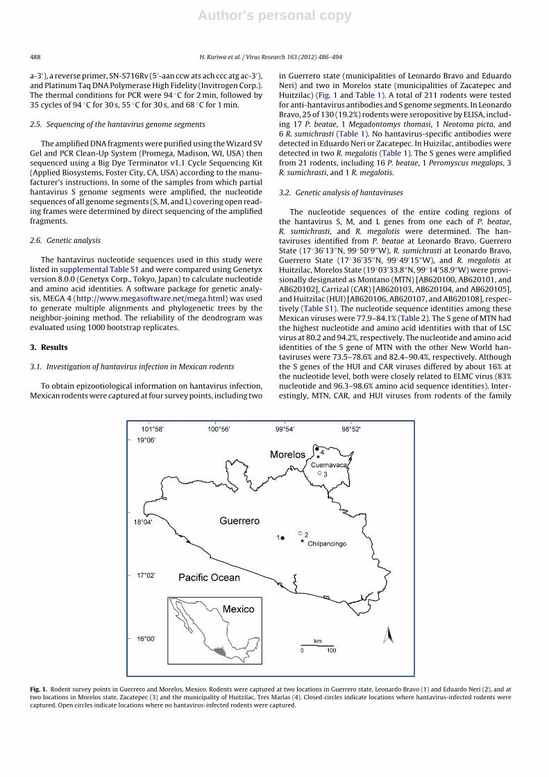
To obtain epizootiological information on hantavirus infection,Mexican rodents were captured at four survey points, including two
in Guerrero state (municipalities of Leonardo Bravo and EduardoNeri) and two in Morelos state (municipalities of Zacatepec andHuitzilac) (Fig. 1 and Table 1). A total of 211 rodents were testedfor anti-hantavirus antibodies and S genome segments. In LeonardoBravo, 25 of 130 (19.2%) rodents were seropositive by ELISA, includ-ing 17 P. beatae, 1 Megadontomys thomasi, 1 Neotoma picta, and6 R. sumichrasti (Table 1). No hantavirus-specific antibodies weredetected in Eduardo Neri or Zacatepec. In Huizilac, antibodies weredetected in two R. megalotis (Table 1). The S genes were amplifiedfrom 21 rodents, including 16 P. beatae, 1 Peromyscus megalops, 3R. sumichrasti, and 1 R. megalotis.
3.2. Genetic analysis of hantaviruses
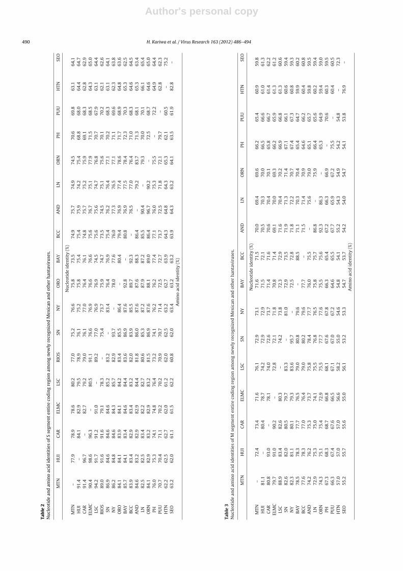
The nucleotide sequences of the entire coding regions ofthe hantavirus S, M, and L genes from one each of P. beatae,R. sumichrasti, and R. megalotis were determined. The han-taviruses identified from P. beatae at Leonardo Bravo, GuerreroState (17◦36′13′′N, 99◦50′9′′W), R. sumichrasti at Leonardo Bravo,Guerrero State (17◦36′35′′N, 99◦49′15′′W), and R. megalotis atHuitzilac, Morelos State (19◦03′33.8′′N, 99◦14′58.9′′W) were provi-sionally designated as Montano (MTN) [AB620100, AB620101, andAB620102], Carrizal (CAR) [AB620103, AB620104, and AB620105],and Huitzilac (HUI) [AB620106, AB620107, and AB620108], respec-tively (Table S1). The nucleotide sequence identities among theseMexican viruses were 77.9–84.1% (Table 2). The S gene of MTN hadthe highest nucleotide and amino acid identities with that of LSCvirus at 80.2 and 94.2%, respectively. The nucleotide and amino acididentities of the S gene of MTN with the other New World han-taviruses were 73.5–78.6% and 82.4–90.4%, respectively. Althoughthe S genes of the HUI and CAR viruses differed by about 16% atthe nucleotide level, both were closely related to ELMC virus (83%nucleotide and 96.3–98.6% amino acid sequence identities). Inter-estingly, MTN, CAR, and HUI viruses from rodents of the family
Fig. 1. Rodent survey points in Guerrero and Morelos, Mexico. Rodents were captured at two locations in Guerrero state, Leonardo Bravo (1) and Eduardo Neri (2), and attwo locations in Morelos state, Zacatepec (3) and the municipality of Huitzilac, Tres Marías (4). Closed circles indicate locations where hantavirus-infected rodents werecaptured. Open circles indicate locations where no hantavirus-infected rodents were captured.
Author's personal copy
H. Kariwa et al. / Virus Research 163 (2012) 486– 494 489
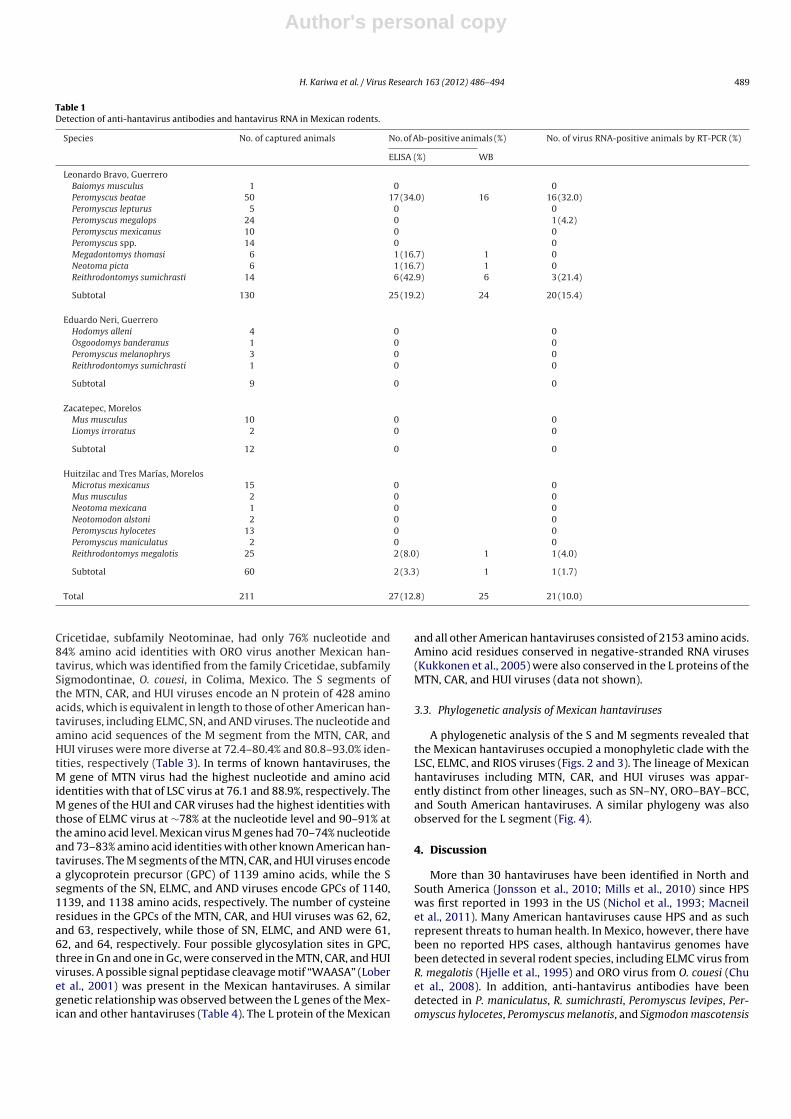
Table 1Detection of anti-hantavirus antibodies and hantavirus RNA in Mexican rodents.
Species No. of captured animals No. of Ab-positive animals (%) No. of virus RNA-positive animals by RT-PCR (%)
ELISA (%) WB
Leonardo Bravo, GuerreroBaiomys musculus 1 0 0Peromyscus beatae 50 17 (34.0) 16 16 (32.0)Peromyscus lepturus 5 0 0Peromyscus megalops 24 0 1 (4.2)Peromyscus mexicanus 10 0 0Peromyscus spp. 14 0 0Megadontomys thomasi 6 1 (16.7) 1 0Neotoma picta 6 1 (16.7) 1 0Reithrodontomys sumichrasti 14 6 (42.9) 6 3 (21.4)
Subtotal 130 25 (19.2) 24 20 (15.4)
Eduardo Neri, GuerreroHodomys alleni 4 0 0Osgoodomys banderanus 1 0 0Peromyscus melanophrys 3 0 0Reithrodontomys sumichrasti 1 0 0
Subtotal 9 0 0
Zacatepec, MorelosMus musculus 10 0 0Liomys irroratus 2 0 0
Subtotal 12 0 0
Huitzilac and Tres Marías, MorelosMicrotus mexicanus 15 0 0Mus musculus 2 0 0Neotoma mexicana 1 0 0Neotomodon alstoni 2 0 0Peromyscus hylocetes 13 0 0Peromyscus maniculatus 2 0 0Reithrodontomys megalotis 25 2 (8.0) 1 1 (4.0)
Subtotal 60 2 (3.3) 1 1 (1.7)
Total 211 27 (12.8) 25 21 (10.0)
Cricetidae, subfamily Neotominae, had only 76% nucleotide and84% amino acid identities with ORO virus another Mexican han-tavirus, which was identified from the family Cricetidae, subfamilySigmodontinae, O. couesi, in Colima, Mexico. The S segments ofthe MTN, CAR, and HUI viruses encode an N protein of 428 aminoacids, which is equivalent in length to those of other American han-taviruses, including ELMC, SN, and AND viruses. The nucleotide andamino acid sequences of the M segment from the MTN, CAR, andHUI viruses were more diverse at 72.4–80.4% and 80.8–93.0% iden-tities, respectively (Table 3). In terms of known hantaviruses, theM gene of MTN virus had the highest nucleotide and amino acididentities with that of LSC virus at 76.1 and 88.9%, respectively. TheM genes of the HUI and CAR viruses had the highest identities withthose of ELMC virus at ∼78% at the nucleotide level and 90–91% atthe amino acid level. Mexican virus M genes had 70–74% nucleotideand 73–83% amino acid identities with other known American han-taviruses. The M segments of the MTN, CAR, and HUI viruses encodea glycoprotein precursor (GPC) of 1139 amino acids, while the Ssegments of the SN, ELMC, and AND viruses encode GPCs of 1140,1139, and 1138 amino acids, respectively. The number of cysteineresidues in the GPCs of the MTN, CAR, and HUI viruses was 62, 62,and 63, respectively, while those of SN, ELMC, and AND were 61,62, and 64, respectively. Four possible glycosylation sites in GPC,three in Gn and one in Gc, were conserved in the MTN, CAR, and HUIviruses. A possible signal peptidase cleavage motif “WAASA” (Loberet al., 2001) was present in the Mexican hantaviruses. A similargenetic relationship was observed between the L genes of the Mex-ican and other hantaviruses (Table 4). The L protein of the Mexican
and all other American hantaviruses consisted of 2153 amino acids.Amino acid residues conserved in negative-stranded RNA viruses(Kukkonen et al., 2005) were also conserved in the L proteins of theMTN, CAR, and HUI viruses (data not shown).
3.3. Phylogenetic analysis of Mexican hantaviruses
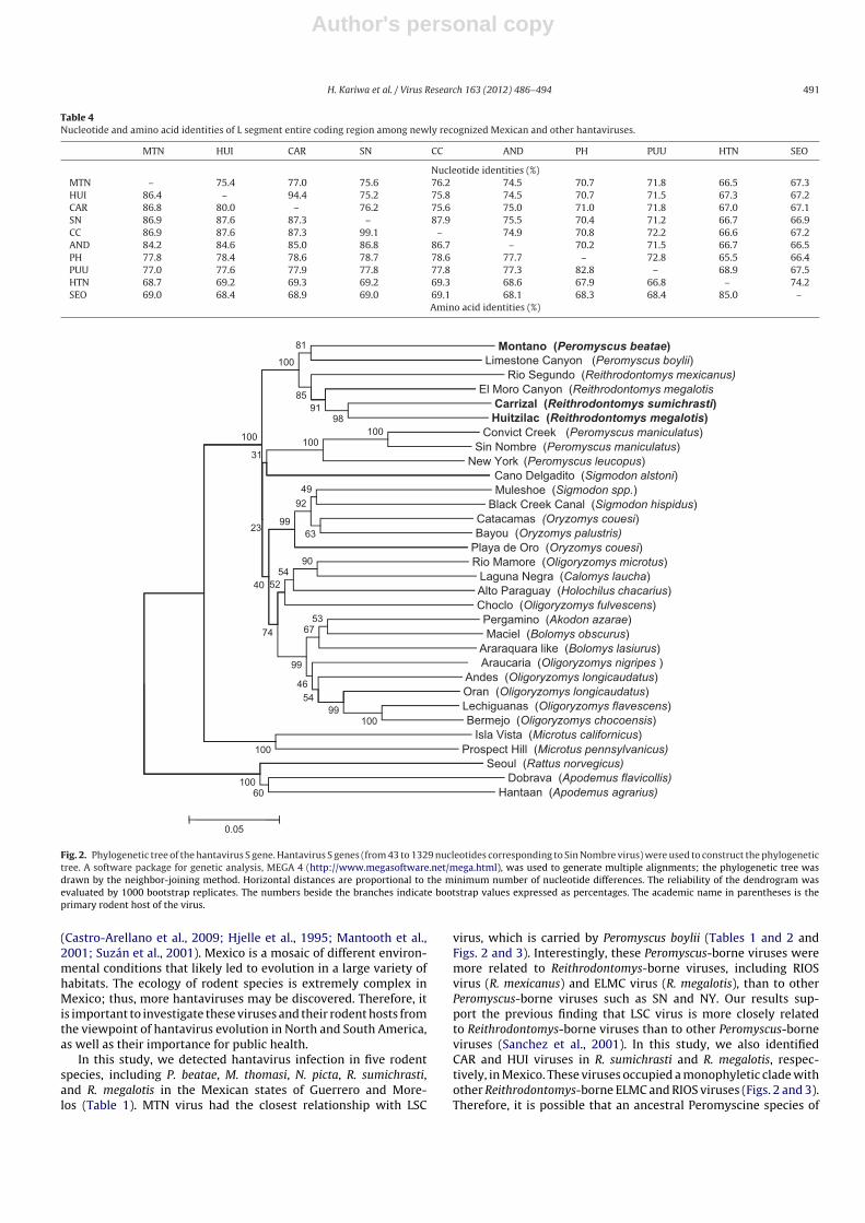
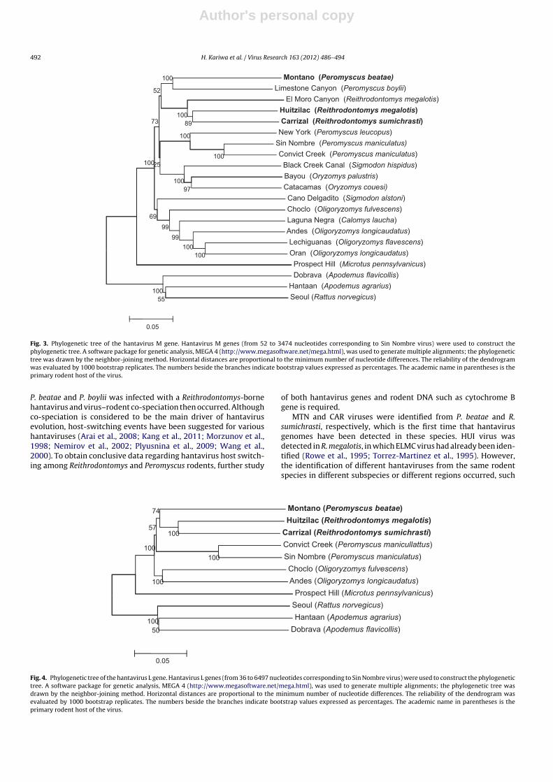
A phylogenetic analysis of the S and M segments revealed thatthe Mexican hantaviruses occupied a monophyletic clade with theLSC, ELMC, and RIOS viruses (Figs. 2 and 3). The lineage of Mexicanhantaviruses including MTN, CAR, and HUI viruses was appar-ently distinct from other lineages, such as SN–NY, ORO–BAY–BCC,and South American hantaviruses. A similar phylogeny was alsoobserved for the L segment (Fig. 4).
4. Discussion
More than 30 hantaviruses have been identified in North andSouth America (Jonsson et al., 2010; Mills et al., 2010) since HPSwas first reported in 1993 in the US (Nichol et al., 1993; Macneilet al., 2011). Many American hantaviruses cause HPS and as suchrepresent threats to human health. In Mexico, however, there havebeen no reported HPS cases, although hantavirus genomes havebeen detected in several rodent species, including ELMC virus fromR. megalotis (Hjelle et al., 1995) and ORO virus from O. couesi (Chuet al., 2008). In addition, anti-hantavirus antibodies have beendetected in P. maniculatus, R. sumichrasti, Peromyscus levipes, Per-omyscus hylocetes, Peromyscus melanotis, and Sigmodon mascotensis
Author's personal copy
490 H. Kariwa et al. / Virus Research 163 (2012) 486– 494Ta
ble
2N
ucl
eoti
de
and
amin
o
acid
iden
titi
es
of
S
segm
ent
enti
re
cod
ing
regi
on
amon
g
new
ly
reco
gniz
ed
Mex
ican
and
oth
er
han
tavi
ruse
s.
MTN
HU
I
CA
R
ELM
C
LSC
RIO
S
SN
NY
OR
O
BA
Y
BC
C
AN
D
LN
OR
N
PH
PUU
HTN
SEO
Nu
cleo
tid
e
iden
tity
(%)
MTN
–
77.9
78.9
78.6
80.2
77.0
75.2
76.6
75.8
73.5
74.9
75.7
74.9
74.5
70.6
69.8
63.1
64.1
HU
I
91.4
–
84.1
82.9
79.5
78.9
76.1
75.2
75.8
75.4
75.4
75.9
74.2
75.4
68.8
68.0
64.4
64.7
CA
R
91.4
96.7
–
82.7
79.2
79.0
76.1
77.0
76.4
76.1
74.8
75.7
75.2
75.9
69.1
68.3
62.8
62.9
ELM
C90
.4
98.6
96.3
–
80.5
91.1
76.6
76.9
76.6
76.6
75.6
76.7
75.1
77.9
71.5
68.5
64.3
65.0
LSC
94.2
91.7
91.2
91.0
–
89.2
77.0
76.9
76.9
74.5
75.6
75.6
74.7
76.8
70.7
67.9
63.1
64.4
RIO
S89
.0
91.6
91.6
79.1
78.3
–
75.4
73.7
75.9
74.7
73.5
74.5
75.1
75.6
70.1
70.2
62.1
62.6
SN
86.9
84.6
84.6
84.6
85.2
83.2
–
83.4
76.4
76.9
75.4
76.2
76.4
77.1
70.2
68.3
63.1
64.1
NY
86.2
84.8
84.6
84.3
85.7
82.9
93.7
–
78.0
77.6
76.0
77.3
76.5
77.1
71.1
69.6
62.3
63.8
OR
O
84.1
84.1
83.9
84.1
84.2
83.4
85.5
86.4
–
80.4
79.4
76.9
77.4
78.6
71.7
68.9
64.8
63.6
BA
Y
85.7
84.1
83.4
84.6
84.4
83.6
86.9
87.6
92.8
–
80.8
76.9
77.5
78.4
72.3
70.3
65.3
62.5
BC
C83
.9
83.4
82.9
83.4
83.2
82.0
83.9
85.5
89.7
92.3
–
76.5
77.0
76.4
71.0
68.3
64.6
64.5
AN
D
84.6
83.2
82.9
82.9
84.4
81.8
86.0
87.6
87.6
88.3
86.4
–
79.2
83.7
71.3
68.1
65.3
63.4
LN82
.5
82.5
83.4
82.2
82.7
80.6
85.3
87.2
87.9
87.2
85.5
90.4
–
79.3
70.0
70.1
66.1
65.4
OR
N
84.1
82.9
83.2
82.9
83.2
81.5
86.9
87.6
88.1
89.0
86.4
96.7
90.2
–
72.5
68.7
64.6
65.0
PH
76.0
75.3
74.1
74.8
76.4
73.2
74.1
76.2
76.2
77.4
77.1
76.0
75.3
75.5
–
72.2
64.9
64.4
PUU
70.7
70.4
71.1
70.2
70.2
70.9
70.7
71.4
72.5
72.7
73.7
72.5
71.8
72.1
79.7
–
62.8
62.3
HTN
62.2
62.5
62.7
62.0
61.2
62.0
62.5
63.2
62.7
63.9
64.3
64.8
64.3
65.3
62.1
60.5
–
75.2
SEO
63.2
62.0
61.1
61.5
62.2
60.8
62.0
63.4
63.2
63.2
63.9
64.3
63.2
64.1
63.5
61.9
82.8
–A
min
o
acid
iden
tity
(%)
Tab
le
3N
ucl
eoti
de
and
amin
o
acid
iden
titi
es
of
M
segm
ent
enti
re
cod
ing
regi
on
amon
g
new
ly
reco
gniz
ed
Mex
ican
and
oth
er
han
tavi
ruse
s.
MTN
HU
I
CA
R
ELM
C
LSC
SN
NY
BA
Y
BC
C
AN
D
LN
OR
N
PH
PUU
HTN
SEO
Nu
cleo
tid
e
iden
tity
(%)
MTN
–
72.4
73.4
71.6
76.1
72.9
73.1
71.6
71.5
70.0
69.4
69.6
66.2
65.4
60.9
59.8
HU
I
81.1
–
80.4
78.7
74.2
72.9
72.9
71.5
72.1
70.5
70.3
70.0
66.5
66.6
61.0
61.3
CA
R
80.8
93.0
–
78.1
74.0
72.6
73.7
71.4
71.6
70.6
70.4
70.1
65.8
66.7
61.4
62.2
ELM
C
79.7
91.0
90.2
–
72.8
72.1
71.8
70.8
71.4
69.1
70.0
69.3
66.2
65.9
61.3
61.2
LSC
88.9
83.4
82.6
80.3
–
74.2
73.8
72.3
72.9
71.6
70.4
70.2
66.9
66.8
61.3
60.6
SN
82.6
82.0
80.5
79.7
83.3
–
81.0
72.9
73.5
71.9
71.3
71.4
67.1
66.1
60.6
59.4
NY
82.3
81.1
80.1
79.3
83.6
95.7
–
72.5
72.8
71.8
72.2
70.7
67.4
67.3
60.8
59.3
BA
Y
78.5
78.3
77.7
76.5
79.0
80.8
79.6
–
88.5
71.1
70.3
70.4
65.4
64.7
59.9
60.2
BC
C
77.6
78.3
77.0
76.4
79.0
80.2
79.6
77.7
–
71.5
71.4
70.9
64.6
66.2
60.4
60.8
AN
D
74.2
76.2
75.3
73.7
75.8
78.4
77.7
76.0
75.5
–
75.6
79.0
65.1
65.7
59.8
59.5
LN
72.9
75.3
75.0
74.1
75.5
76.8
76.5
75.8
75.7
86.8
–
75.9
66.4
65.6
60.2
59.4
OR
N
74.3
75.1
75.4
72.9
75.5
77.7
77.6
75.5
75.6
92.3
86.3
–
65.3
64.9
59.4
59.0
PH
67.3
68.3
68.7
66.8
68.1
67.6
67.8
66.3
65.4
67.2
66.3
66.9
–
70.6
60.3
59.5
PUU
66.3
67.4
67.6
66.5
67.1
67.0
67.2
64.6
65.5
67.7
65.9
67.2
75.5
–
60.4
60.5
HTN
57.0
57.0
57.0
56.6
58.2
55.0
54.6
54.8
54.1
55.2
54.3
54.9
54.2
54.8
–
72.3
SEO
55.2
55.7
55.6
55.0
56.1
53.2
53.3
54.7
53.7
54.2
54.0
54.7
54.1
53.8
76.9
–A
min
o
acid
iden
tity
(%)
Author's personal copy
H. Kariwa et al. / Virus Research 163 (2012) 486– 494 491
Table 4Nucleotide and amino acid identities of L segment entire coding region among newly recognized Mexican and other hantaviruses.
MTN HUI CAR SN CC AND PH PUU HTN SEO
Nucleotide identities (%)MTN – 75.4 77.0 75.6 76.2 74.5 70.7 71.8 66.5 67.3HUI 86.4 – 94.4 75.2 75.8 74.5 70.7 71.5 67.3 67.2CAR 86.8 80.0 – 76.2 75.6 75.0 71.0 71.8 67.0 67.1SN 86.9 87.6 87.3 – 87.9 75.5 70.4 71.2 66.7 66.9CC 86.9 87.6 87.3 99.1 – 74.9 70.8 72.2 66.6 67.2AND 84.2 84.6 85.0 86.8 86.7 – 70.2 71.5 66.7 66.5PH 77.8 78.4 78.6 78.7 78.6 77.7 – 72.8 65.5 66.4PUU 77.0 77.6 77.9 77.8 77.8 77.3 82.8 – 68.9 67.5HTN 68.7 69.2 69.3 69.2 69.3 68.6 67.9 66.8 – 74.2SEO 69.0 68.4 68.9 69.0 69.1 68.1 68.3 68.4 85.0 –
Amino acid identities (%)
(PeromyscusMontano beatae)(PeromyscusLimestone Canyon boylii)
(ReithrodontomysRio Segundo mexicanus)
81
100
El Moro Canyon (Reithrodontomys megalotis(ReithrodontomysCarrizal sumichrasti)(ReithrodontomysHuitzilac megalotis)
(PeromyscusConvict Creek maniculatus)(PeromyscusSin Nombre maniculatus)
(PeromyscusNew York leucopus)
10098
100
9185
31
100
(SigmodonCano Delgadito alstoni)(SigmodonMuleshoe spp.)
(SigmodonBlack Creek Canal hispidus)Catacamas couesi)(Oryzomys
(OryzomysBayou palustris)63
4992
9923
(OryzomysPlaya de Oro couesi)(OligoryzomysRio Mamore microtus)
(CalomysLaguna Negra laucha)(HolochilusAlto Paraguay chacarius)
(OligoryzomysChoclo fulvescens)
9054
5240
Pergamino (Akodon az arae) (BolomysMaciel obscurus)
(BolomysAraraquara like lasiurus)(Oligoryzomys nigripes )Araucaria
(OligoryzomysAndes longicaudatus)
5367
46
99
74
Oran (Oligoryzomys longicaudatus)(OligoryzomysLechiguanas flavescens)
(OligoryzomysBermejo chocoensis)(MicrotusIsla Vista californicus)(MicrotusProspect Hill pennsylvanicus)100
10099
54
Seoul (Rattus norvegicus)(ApodemusDobrava flavicollis)
(ApodemusHantaan agrarius)60100
0.05
Fig. 2. Phylogenetic tree of the hantavirus S gene. Hantavirus S genes (from 43 to 1329 nucleotides corresponding to Sin Nombre virus) were used to construct the phylogenetictree. A software package for genetic analysis, MEGA 4 (http://www.megasoftware.net/mega.html), was used to generate multiple alignments; the phylogenetic tree wasdrawn by the neighbor-joining method. Horizontal distances are proportional to the minimum number of nucleotide differences. The reliability of the dendrogram wasevaluated by 1000 bootstrap replicates. The numbers beside the branches indicate bootstrap values expressed as percentages. The academic name in parentheses is theprimary rodent host of the virus.
(Castro-Arellano et al., 2009; Hjelle et al., 1995; Mantooth et al.,2001; Suzán et al., 2001). Mexico is a mosaic of different environ-mental conditions that likely led to evolution in a large variety ofhabitats. The ecology of rodent species is extremely complex inMexico; thus, more hantaviruses may be discovered. Therefore, itis important to investigate these viruses and their rodent hosts fromthe viewpoint of hantavirus evolution in North and South America,as well as their importance for public health.
In this study, we detected hantavirus infection in five rodentspecies, including P. beatae, M. thomasi, N. picta, R. sumichrasti,and R. megalotis in the Mexican states of Guerrero and More-los (Table 1). MTN virus had the closest relationship with LSC
virus, which is carried by Peromyscus boylii (Tables 1 and 2 andFigs. 2 and 3). Interestingly, these Peromyscus-borne viruses weremore related to Reithrodontomys-borne viruses, including RIOSvirus (R. mexicanus) and ELMC virus (R. megalotis), than to otherPeromyscus-borne viruses such as SN and NY. Our results sup-port the previous finding that LSC virus is more closely relatedto Reithrodontomys-borne viruses than to other Peromyscus-borneviruses (Sanchez et al., 2001). In this study, we also identifiedCAR and HUI viruses in R. sumichrasti and R. megalotis, respec-tively, in Mexico. These viruses occupied a monophyletic clade withother Reithrodontomys-borne ELMC and RIOS viruses (Figs. 2 and 3).Therefore, it is possible that an ancestral Peromyscine species of
Author's personal copy
492 H. Kariwa et al. / Virus Research 163 (2012) 486– 494
(PeromyscusMontano beatae)(PeromyscusLimestone Canyon boylii)(ReithrodontomysEl Moro Canyon megalotis)
100
52
(ReithrodontomysHuitzilac megalotis)(ReithrodontomysCarrizal sumichrasti)(PeromyscusNew York leucopus)100
89100
73
Sin Nombre (Peromyscus maniculatus)(PeromyscusConvict Creek maniculatus)
(SigmodonBlack Creek Canal hispidus)(OryzomysBayou palustris)
10010025
(OryzomysCatacamas couesi)(SigmodonCano Delgadito alstoni)
(OligoryzomysChoclo fulvescens)
97100
69 Laguna Negra (Calomys laucha)(OligoryzomysAndes longicaudatus)
(OligoryzomysLechiguanas flavescens)(OligoryzomysOran longicaudatus)100
10099
99
(MicrotusProspect Hill pennsylvanicus)(ApodemusDobrava flavicollis)
(ApodemusHantaan agrarius)100
Seoul (Rattus norvegicus)55
0.05
Fig. 3. Phylogenetic tree of the hantavirus M gene. Hantavirus M genes (from 52 to 3474 nucleotides corresponding to Sin Nombre virus) were used to construct thephylogenetic tree. A software package for genetic analysis, MEGA 4 (http://www.megasoftware.net/mega.html), was used to generate multiple alignments; the phylogenetictree was drawn by the neighbor-joining method. Horizontal distances are proportional to the minimum number of nucleotide differences. The reliability of the dendrogramwas evaluated by 1000 bootstrap replicates. The numbers beside the branches indicate bootstrap values expressed as percentages. The academic name in parentheses is theprimary rodent host of the virus.
P. beatae and P. boylii was infected with a Reithrodontomys-bornehantavirus and virus–rodent co-speciation then occurred. Althoughco-speciation is considered to be the main driver of hantavirusevolution, host-switching events have been suggested for varioushantaviruses (Arai et al., 2008; Kang et al., 2011; Morzunov et al.,1998; Nemirov et al., 2002; Plyusnina et al., 2009; Wang et al.,2000). To obtain conclusive data regarding hantavirus host switch-ing among Reithrodontomys and Peromyscus rodents, further study
of both hantavirus genes and rodent DNA such as cytochrome Bgene is required.
MTN and CAR viruses were identified from P. beatae and R.sumichrasti, respectively, which is the first time that hantavirusgenomes have been detected in these species. HUI virus wasdetected in R. megalotis, in which ELMC virus had already been iden-tified (Rowe et al., 1995; Torrez-Martinez et al., 1995). However,the identification of different hantaviruses from the same rodentspecies in different subspecies or different regions occurred, such
Montano (Peromysc us beat ae)(ReithrodontomysHuitzilac megalotis )
(ReithrodontomysCarrizal sumichrasti )100
74
57
Con vict Cre ek (Peromyscus manicul lat tus)(PeromyscusSin Nombre maniculatus )
(OligoryzomysChoclo fulvescens)100
100
(OligoryzomysAndes longicaudatus )(MicrotusProspect Hill pennsylvanicus)
(RattusSeoul norvegicus )
100
(ApodemusHantaan agrarius )(ApodemusDobrava flavicollis)50
100
0.05
Fig. 4. Phylogenetic tree of the hantavirus L gene. Hantavirus L genes (from 36 to 6497 nucleotides corresponding to Sin Nombre virus) were used to construct the phylogenetictree. A software package for genetic analysis, MEGA 4 (http://www.megasoftware.net/mega.html), was used to generate multiple alignments; the phylogenetic tree wasdrawn by the neighbor-joining method. Horizontal distances are proportional to the minimum number of nucleotide differences. The reliability of the dendrogram wasevaluated by 1000 bootstrap replicates. The numbers beside the branches indicate bootstrap values expressed as percentages. The academic name in parentheses is theprimary rodent host of the virus.
Author's personal copy
H. Kariwa et al. / Virus Research 163 (2012) 486– 494 493
as ORO and CAT viruses in O. couesi, SN and Monongahela viruses inP. maniculatus, AND and ORN viruses in O. longicaudatus, and Han-taan and Saaremaa viruses in Apodemus agrarius (Chu et al., 2008;Lee et al., 1978; Levis et al., 1998; López et al., 1996; Milazzo et al.,2006; Nemirov et al., 1999; Nichol et al., 1993; Song et al., 1996).Therefore, it is possible that R. megalotis may be the host speciesof HUI virus. However, to obtain more conclusive data on hostspecies of Mexican hantaviruses further epizootiological surveysare required.
The MTN, CAR, and HUI viruses are distant from ORO virus,which has been identified in O. couesi in Mexico. ORO virusoccupies a monophyletic clade with hantaviruses associated withNorth American Sigmodontinae rodents, including BAY and BCC.In contrast, the MTN, CAR, and HUI viruses are associated withNeotominae rodents. The genetic distance between the three Mex-ican viruses and ORO may reflect the long history of hantavirusevolution in two separate rodent subfamilies, Neotominae and Sig-modontinae.
It is important to know whether hantavirus infection exists inMexican people, though no HPS cases have been reported so far. Itis evident that five hantaviruses, ELMC, ORO, MTN, CAR, and HUI,are circulating in Mexican rodents. Furthermore, P. maniculatus, P.leucopus, and S. hispidus are the hosts of the pathogenic SN, NY, andBCC viruses, respectively, in the US and these species are also widelydistributed in Mexico (Mills et al., 2010). Therefore, large-scalenationwide epidemiological and epizootiological surveys should beconducted to verify whether human infections occur in Mexico.
In conclusion, we identified three hantaviruses in Mexico: theprovisional names are MTN, CAR, and HUI viruses from P. beatae, R.sumichrasti, and R. megalotis, respectively. These viruses are morerelated to hantaviruses associated with Neotominae rodents, suchas LSC, RIOS, and ELMC, than those associated with Sigmodontinaerodents, such as ORO virus, which have previously been identifiedin O. couesi in Mexico. The information obtained in this study isimportant for understanding the evolution of hantaviruses in theAmericas.
Acknowledgments
We thank Dr. C.J. Peters for providing us with the construct forexpression of the nucleocapsid protein of SN virus. We also thankto Y.Q. Jiménez-Salmerón, E. Guerrero-Ibarra, A. Taboada-Salgado,L. Sánchez-Vázquez, A.L. Ortiz V., and M.A. Lozano G. who workedwith us in the field in Mexico for assistance with rodent trappingand supported our survey. This work was supported financiallyby Grants-in-Aid for Scientific Research (16405034 and 17255009)from the Japanese Ministry of Education, Culture, Sports, Science,and Technology, and by a Health and Labor Sciences ResearchGrant on Emerging and Re-Emerging Infectious Diseases from theJapanese Ministry of Health, Labor, and Welfare. This work wasalso supported by the global COE Program for Zoonosis Control(Hokkaido University). A permit for this study was provided by theInstituto Nacional de Ecología, Dirección General de Vida SilvestreFAUT.0103 to C. Sánchez-Hernández.
Appendix A. Supplementary data
Supplementary data associated with this article can be found, inthe online version, at doi:10.1016/j.virusres.2011.11.013.
References
Arai, S., Ohdachi, S.D., Asakawa, M., Kang, H.J., Mocz, G., Arikawa, J., Okabe, N., Yanag-ihara, R., 2008. Molecular phylogeny of a newfound hantavirus in the Japaneseshrew mole (Urotrichus talpoides). Proc. Natl. Acad. Sci. U.S.A. 105, 16296–16301.
Bharadwaj, M., Botten, J., Torrez-Martinez, N., Hjelle, B., 1997. Rio Mamore virus:genetic characterization of a newly recognized hantavirus of the pygmy rice rat,Oligoryzomys microtis, from Bolivia. Am. J. Trop. Med. Hyg. 57, 368–374.
Bohlman, M.C., Morzunov, S.P., Meissner, J., Taylor, M.B., Ishibashi, K., Rowe, J.,Levis, S., Enria, D., St Jeor, S.C., 2002. Analysis of hantavirus genetic diversityin Argentina: S segment-derived phylogeny. J. Virol. 76, 3765–3773.
Calderón, G., Pini, N., Bolpe, J., Levis, S., Mills, J., Segura, E., Guthmann, N., Cantoni,G., Becker, J., Fonollat, A., Ripoll, C., Bortman, M., Benedetti, R., Enria, D., 1999.Hantavirus reservoir hosts associated with peridomestic habitats in Argentina.Emerg. Infect. Dis. 5, 792–797.
Castro-Arellano, I., Suzán, G., León, R.F., Jiménez, R.M., Lacher Jr., T.E., 2009. Surveyfor antibody to hantaviruses in Tamaulipas, México. J. Wildl. Dis. 45, 207–212.
Chu, Y.K., Milligan, B., Owen, R.D., Goodin, D.G., Jonsson, C.B., 2006. Phylogeneticand geographical relationships of hantavirus strains in eastern and westernParaguay. Am. J. Trop. Med. Hyg. 75, 1127–1134.
Chu, Y.K., Owen, R.D., Sánchez-Hernández, C., Romero-Almaraz, M.L., Jonsson, C.B.,2008. Genetic characterization and phylogeny of a hantavirus from WesternMexico. Virus Res. 131, 180–188.
Hjelle, B., Anderson, B., Torrez-Martinez, N., Song, W., Gannon, W.L., Yates, T.L., 1995.Prevalence and geographic genetic variation of hantaviruses of New World har-vest mice (Reithrodontomys): identification of a divergent genotype from a CostaRican Reithrodontomys mexicanus. Virology 207, 452–459.
Johnson, A.M., Bowen, M.D., Ksiazek, T.G., Williams, R.J., Bryan, R.T., Mills, J.N.,Peters, C.J., Nichol, S.T., 1997. Laguna Negra virus associated with HPS in westernParaguay and Bolivia. Virology 238, 115–127.
Jonsson, C.B., Figueiredo, L.T., Vapalahti, O., 2010. A global perspective on hantavirusecology, epidemiology, and disease. Clin. Microbiol. Rev. 23, 412–441.
Kang, H.J., Bennett, S.N., Hope, A.G., Cook, J.A., Yanagihara, R., 2011. Shared ances-try between a newfound mole-borne hantavirus and hantaviruses harbored bycricetid rodents. J. Virol. 85, 7496–7503.
Kukkonen, S.K., Vaheri, A., Plyusnin, A., 2005. L protein, the RNA-dependent RNApolymerase of hantaviruses. Arch. Virol. 150, 533–556.
Lee, H.W., Johnson, K.M., 1982. Laboratory-acquired infections with Hantaan virus,the etiologic agent of Korean hemorrhagic fever. J. Infect. Dis. 146, 645–651.
Lee, H.W., Lee, P.W., Johnson, K.M., 1978. Isolation of the etiologic agent of Koreanhemorrhagic fever. J. Infect. Dis. 137, 298–308.
Levis, S., Morzunov, S.P., Rowe, J.E., Enria, D., Pini, N., Calderon, G., Sabattini, M., StJeor, S.C., 1998. Genetic diversity and epidemiology of hantaviruses in Argentina.J. Infect. Dis. 177, 529–538.
Lober, C., Anheier, B., Lindow, S., Klenk, H.D., Feldmann, H., 2001. The Hantaan virusglycoprotein precursor is cleaved at the conserved pentapeptide WAASA. Virol-ogy 289, 224–229.
López, N., Padula, P., Rossi, C., Lázaro, M.E., Franze-Fernández, M.T., 1996. Geneticidentification of a new hantavirus causing severe pulmonary syndrome inArgentina. Virology 220, 223–226.
Macneil, A., Ksiazek, T.G., Rollin, P.E., 2011. Hantavirus pulmonary syndrome, UnitedStates, 1993–2009. Emerg. Infect. Dis. 17, 1195–1201.
Mantooth, S.J., Milazzo, M.L., Bradley, R.D., Hice, C.L., Ceballos, G., Tesh, R.B., Fulhorst,C.F., 2001. Geographical distribution of rodent-associated hantaviruses in Texas.J. Vector Ecol. 26, 7–14.
Milazzo, M.L., Cajimat, M.N., Hanson, J.D., Bradley, R.D., Quintana, M., Sherman, C.,Velásquez, R.T., Fulhorst, C.F., 2006. Catacamas virus, a hantaviral species nat-urally associated with Oryzomys couesi (Coues’ oryzomys) in Honduras. Am. J.Trop. Med. Hyg. 75, 1003–1010.
Mills, J.N., Amman, B.R., Glass, G.E., 2010. Ecology of hantaviruses and their hosts inNorth America. Vector Borne Zoonotic Dis. 10, 563–574.
Morzunov, S.P., Feldmann, H., Spiropoulou, C.F., Semenova, V.A., Rollin, P.E., Ksi-azek, T.G., Peters, C.J., Nichol, S.T., 1995. A newly recognized virus associatedwith a fatal case of hantavirus pulmonary syndrome in Louisiana. J. Virol. 69,1980–1983.
Morzunov, S.P., Rowe, J.E., Ksiazek, T.G., Peters, C.J., St Jeor, S.C., Nichol, S.T., 1998.Genetic analysis of the diversity and origin of hantaviruses in Peromyscus leuco-pus mice in North America. J. Virol. 72, 57–64.
Nemirov, K., Vapalahti, O., Lundkvist, A., Vasilenko, V., Golovljova, I., Plyusnina, A.,Niemimaa, J., Laakkonen, J., Henttonen, H., Vaheri, A., Plyusnin, A., 1999. Isolationand characterization of Dobrava hantavirus carried by the striped field mouse(Apodemus agrarius) in Estonia. J. Gen. Virol. 80, 371–379.
Nemirov, K., Henttonen, H., Vaheri, A., Plyusnin, A., 2002. Phylogenetic evidence forhost switching in the evolution of hantaviruses carried by Apodemus mice. VirusRes. 90, 207–215.
Nichol, S.T., Spiropoulou, C.F., Morzunov, S., Rollin, P.E., Ksiazek, T.G., Feldmann,H., Sanchez, A., Childs, J., Zaki, S., Peters, C.J., 1993. Genetic identification of ahantavirus associated with an outbreak of acute respiratory illness. Science 262,914–917.
Padula, P.J., Sanchez, A.J., Edelstein, A., Nichol, S.T., 2002. Complete nucleotidesequence of the M RNA segment of Andes virus and analysis of the variabil-ity of the termini of the virus S, M and L RNA segments. J. Gen. Virol. 83,2117–2122.
Peters, C.J., Khan, A.S., 2002. Hantavirus pulmonary syndrome: the new Americanhemorrhagic fever. Clin. Infect. Dis. 34, 1224–1231.
Peters, C.J., Simpson, G.L., Levy, H., 1999. Spectrum of hantavirus infection: hemor-rhagic fever with renal syndrome and hantavirus pulmonary syndrome. Annu.Rev. Med. 50, 531–545.
Plyusnin, A., Morzunov, S.P., 2001. Virus evolution and genetic diversity ofhantaviruses and their rodent hosts. Curr. Top. Microbiol. Immunol. 256,47–75.
Author's personal copy
494 H. Kariwa et al. / Virus Research 163 (2012) 486– 494
Plyusnina, A., Ibrahim, I.N., Plyusnin, A., 2009. A newly recognized hantavirus in theAsian house rat (Rattus tanezumi) in Indonesia. J. Gen. Virol. 90, 205–209.
Ravkov, E.V., Rollin, P.E., Ksiazek, T.G., Peters, C.J., Nichol, S.T., 1995. Genetic andserologic analysis of Black Creek Canal virus and its association with humandisease and Sigmodon hispidus infection. Virology 210, 482–489.
Rawlings, J.A., Torrez-Martinez, N., Neill, S.U., Moore, G.M., Hicks, B.N., Pichuantes,S., Nguyen, A., Bharadwaj, M., Hjelle, B., 1996. Cocirculation of multiple han-taviruses in Texas, with characterization of the small (S) genome of a previouslyundescribed virus of cotton rats (Sigmodon hispidus). Am. J. Trop. Med. Hyg. 55,672–679.
Rowe, J.E., St Jeor, S.C., Riolo, J., Otteson, E.W., Monroe, M.C., Henderson, W.W.,Ksiazek, T.G., Rollin, P.E., Nichol, S.T., 1995. Coexistence of several novel han-taviruses in rodents indigenous to North America. Virology 213, 122–130.
Sanchez, A.J., Abbott, K.D., Nichol, S.T., 2001. Genetic identification and characteriza-tion of limestone canyon virus, a unique Peromyscus-borne hantavirus. Virology286, 345–353.
Schmaljohn, C., Hjelle, B., 1997. Hantaviruses: a global disease problem. Emerg.Infect. Dis. 3, 95–104.
Schmaljohn, C.S., Jennings, G.B., Hay, J., Dalrymple, J.M., 1986. Coding strategy of theS genome segment of Hantaan virus. Virology 155, 633–643.
Song, J.W., Baek, L.J., Gajdusek, D.C., Yanagihara, R., Gavrilovskaya, I., Luft, B.J.,Mackow, E.R., Hjelle, B., 1994. Isolation of pathogenic hantavirus from white-footed mouse (Peromyscus leucopus). Lancet 344, 1637.
Song, J.W., Baek, L.J., Nagle, J.W., Schlitter, D., Yanagihara, R., 1996. Genetic and phy-logenetic analyses of hantaviral sequences amplified from archival tissues of
deer mice (Peromyscus maniculatus nubiterrae) captured in the eastern UnitedStates. Arch. Virol. 141, 959–967.
Song, J.W., Baek, L.J., Schmaljohn, C.S., Yanagihara, R., 2007. Thottapalayam virus, aprototype shrewborne hantavirus. Emerg. Infect. Dis. 13, 980–985.
Suzán, G., Ceballos, G., Mills, J., Ksiazek, T.G., Yates, T., 2001. Serologic evidenceof hantavirus infection in sigmodontine rodents in Mexico. J. Wildl. Dis. 37,391–393.
Suzuki, A., Bisordi, I., Levis, S., Garcia, J., Pereira, L.E., Souza, R.P., Sugahara, T.K.N.,Pini, N., Enria, D., Souza, L.T.M., 2004. Identifying rodent hantavirus reservoirs,Brazil. Emerg. Infect. Dis. 10, 2127–2134.
Torrez-Martinez, N., Hjelle, B., 1995. Enzootic of Bayou hantavirus in rice rats (Ory-zomys palustris) in 1983. Lancet 346, 780–781.
Torrez-Martinez, N., Song, W., Hjelle, B., 1995. Nucleotide sequence analysis of theM genomic segment of El Moro Canyon hantavirus: antigenic distinction fromfour corners hantavirus. Virology 211, 336–338.
Vincent, M.J., Quiroz, E., Gracia, F., Sanchez, A.J., Ksiazek, T.G., Kitsutani, P.T., Ruedas,L.A., Tinnin, D.S., Caceres, L., Garcia, A., Rollin, P.E., Mills, J.N., Peters, C.J., Nichol,S.T., 2000. Hantavirus pulmonary syndrome in Panama: identification of novelhantaviruses and their likely reservoirs. Virology 277, 14–19.
Wang, H., Yoshimatsu, K., Ebihara, H., Ogino, M., Araki, K., Kariwa, H., Wang, Z., Luo,Z., Li, D., Hang, C., Arikawa, J., 2000. Genetic diversity of hantaviruses isolatedin china and characterization of novel hantaviruses isolated from Niviventerconfucianus and Rattus rattus. Virology 278, 332–345.
Copyright © 2022 FDOKUMEN