Genetic characterization of K13965, a strain of Oak Vale virus from Western Australia
19
Genetic characterization of K13965, a strain of Oak Vale virus from Western Australia Phenix-Lan Quan a , David T. Williams b , Cheryl A. Johansen c , Komal Jain a , Alexandra Petrosov a , Sinead M. Diviney b , Alla Tashmukhamedova a , Stephen K. Hutchison d , Robert B. Tesh e , John S. Mackenzie b,f , Thomas Briese a,g,* , and W. Ian Lipkin a,g,h a Center for Infection and Immunity, Columbia University, New York, NY, USA b School of Biomedical Sciences, Curtin University, Perth, WA, Australia c Discipline of Microbiology and Immunology, University of Western Australia, Nedlands, WA, Australia d 454 Roche Life Sciences, Branford, CT, USA e Department of Pathology, University of Texas Medical Branch, Galveston, TX, USA f Burnet Institute, Melbourne, Australia g Department of Epidemiology, Mailman School of Public Health, Columbia University, New York, NY, USA h Departments of Pathology and Neurology, College of Physicians and Surgeons, Columbia University, New York, NY, USA Abstract K13965, an uncharacterized virus, was isolated in 1993 from Anopheles annulipes mosquitoes collected in the Kimberley region of northern Western Australia. Here, we report its genomic sequence, identify it as a rhabdovirus, and characterize its phylogenetic relationships. The genome comprises a P′ (C) and SH protein similar to the recently characterized Tupaia and Durham viruses, and shows overlap between G and L genes. Comparison of K13965 genome sequence to other rhabdoviruses identified K13965 as a strain of the unclassified Australian Oak Vale rhabdovirus, whose complete genome sequence we also determined. Phylogenetic analysis of N and L sequences indicated genetic relationship to a recently proposed Sandjima virus clade, although the Oak Vale virus sequences form a branch separate from the African members of that group. Keywords rhabdovirus; Oak Vale virus; Anopheles annulipes; Culex edwardsi; virus phylogenetics; Australia; molecular characterization © 2011 Elsevier B.V. All rights reserved. * Corresponding author: Center for Infection and Immunity, Mailman School of Public Health, Columbia University, 722 West 168 th Street, New York, NY 10032, USA; Phone: 212-342-9031; Fax: 212-342-9044; [email protected]. Publisher's Disclaimer: This is a PDF file of an unedited manuscript that has been accepted for publication. As a service to our customers we are providing this early version of the manuscript. The manuscript will undergo copyediting, typesetting, and review of the resulting proof before it is published in its final citable form. Please note that during the production process errors may be discovered which could affect the content, and all legal disclaimers that apply to the journal pertain. NIH Public Access Author Manuscript Virus Res. Author manuscript; available in PMC 2012 September 1. Published in final edited form as: Virus Res. 2011 September ; 160(1-2): 206–213. doi:10.1016/j.virusres.2011.06.021. NIH-PA Author Manuscript NIH-PA Author Manuscript NIH-PA Author Manuscript
-
Upload
independent -
Category
Documents
-
view
3 -
download
0
Transcript of Genetic characterization of K13965, a strain of Oak Vale virus from Western Australia

Genetic characterization of K13965, a strain of Oak Vale virusfrom Western Australia
Phenix-Lan Quana, David T. Williamsb, Cheryl A. Johansenc, Komal Jaina, AlexandraPetrosova, Sinead M. Divineyb, Alla Tashmukhamedovaa, Stephen K. Hutchisond, Robert B.Teshe, John S. Mackenzieb,f, Thomas Briesea,g,*, and W. Ian Lipkina,g,h
aCenter for Infection and Immunity, Columbia University, New York, NY, USAbSchool of Biomedical Sciences, Curtin University, Perth, WA, AustraliacDiscipline of Microbiology and Immunology, University of Western Australia, Nedlands, WA,Australiad454 Roche Life Sciences, Branford, CT, USAeDepartment of Pathology, University of Texas Medical Branch, Galveston, TX, USAfBurnet Institute, Melbourne, AustraliagDepartment of Epidemiology, Mailman School of Public Health, Columbia University, New York,NY, USAhDepartments of Pathology and Neurology, College of Physicians and Surgeons, ColumbiaUniversity, New York, NY, USA
AbstractK13965, an uncharacterized virus, was isolated in 1993 from Anopheles annulipes mosquitoescollected in the Kimberley region of northern Western Australia. Here, we report its genomicsequence, identify it as a rhabdovirus, and characterize its phylogenetic relationships. The genomecomprises a P′ (C) and SH protein similar to the recently characterized Tupaia and Durhamviruses, and shows overlap between G and L genes. Comparison of K13965 genome sequence toother rhabdoviruses identified K13965 as a strain of the unclassified Australian Oak Valerhabdovirus, whose complete genome sequence we also determined. Phylogenetic analysis of Nand L sequences indicated genetic relationship to a recently proposed Sandjima virus clade,although the Oak Vale virus sequences form a branch separate from the African members of thatgroup.
Keywordsrhabdovirus; Oak Vale virus; Anopheles annulipes; Culex edwardsi; virus phylogenetics;Australia; molecular characterization
© 2011 Elsevier B.V. All rights reserved.*Corresponding author: Center for Infection and Immunity, Mailman School of Public Health, Columbia University, 722 West 168thStreet, New York, NY 10032, USA; Phone: 212-342-9031; Fax: 212-342-9044; [email protected]'s Disclaimer: This is a PDF file of an unedited manuscript that has been accepted for publication. As a service to ourcustomers we are providing this early version of the manuscript. The manuscript will undergo copyediting, typesetting, and review ofthe resulting proof before it is published in its final citable form. Please note that during the production process errors may bediscovered which could affect the content, and all legal disclaimers that apply to the journal pertain.
NIH Public AccessAuthor ManuscriptVirus Res. Author manuscript; available in PMC 2012 September 1.
Published in final edited form as:Virus Res. 2011 September ; 160(1-2): 206–213. doi:10.1016/j.virusres.2011.06.021.
NIH
-PA Author Manuscript
NIH
-PA Author Manuscript
NIH
-PA Author Manuscript

1. IntroductionViruses with nonsegmented, single-stranded negative sense RNA genomes are subsumed inthe order Mononegavirales that currently includes the four families Bornaviridae,Filoviridae, Paramyxoviridae and Rhabdoviridae. The family Rhabdoviridae comprisesviruses of fish in the genus Novirhabdovirus and arthropod-borne viruses that infect plantsin the genera Nucleorhabdovirus and Cytorhabdovirus. The remaining generaEphemerovirus, Lyssavirus and Vesiculovirus include both arthropod-borne and non vector-borne pathogens of mammals, birds, reptiles and fish that include significant threats toagriculture and public health, such as vesicular stomatitis Indiana virus (VSIV) and rabiesvirus (RABV) (Kuzmin et al., 2009). In addition, more than 100 viruses are tentativelyassigned to this family based on their characteristic bullet-shaped virion morphology, or viaserologic cross-reactivity with one or more of the classified members of the family (Tordo etal., 2005).
Rhabdoviruses have genomes that range in approximate size from 11,000 to 16,000nucleotides (nt). The genome organization of many classic rhabdoviruses, as represented byRABV, has been considered to be one of the simplest in the viral kingdom with only 5genes: the nucleoprotein (N), phosphoprotein (P), matrix protein (M), glycoprotein (G) andpolymerase (L) in the order 3′-N-P-M-G-L-5′ (Kuzmin et al., 2009). However, recentmolecular analyses of novel rhabdoviruses indicated considerable genetic variability andmore complex genomes, with up to 10 additional open reading frames (ORF) located withinor interposed between the five core genes, many still without known function. In addition tooverlapping ORFs characterized within the P gene of members of the Vesiculovirus genus(C and C′; (Kretzschmar et al., 1996; Peluso et al., 1996; Spiropoulou and Nichol, 1993),and in unassigned rhabdoviruses from vertebrates and dipterans (Allison et al., 2010; Gubalaet al., 2010; Nishizawa et al., 1997; Quan et al., 2010; Schutze et al., 1999; Springfeld et al.,2005; Tao et al., 2008; Zhu et al., 2011), ORFs that overlap within N, M or G genes havealso been identified (Gubala et al., 2008; Jayakar and Whitt, 2002; Tao et al., 2008). One tofour ORFs located between the P and M genes are reported for plant-infecting rhabdoviruses(Dietzgen et al., 2006; Huang et al., 2003; Reed et al., 2005; Revill et al., 2005; Scholthof etal., 1994; Tanno et al., 2000), the unassigned Drosophila sigma viruses (Longdon et al.,2010; Teninges et al., 1993), and the Culicoides-transmitted Ngaingan (NGAV) andWongabel viruses (WOGV) (Gubala et al., 2010; Gubala et al., 2008). In addition, an ORFlocated between the M and G genes has been identified in the unassigned NGAV, Durham(DURV), and Tupaia viruses (TUPV) (Allison et al., 2010; Gubala et al., 2010; Springfeld etal., 2005). All members of the genus Novirhadovirus, the nucleorhabdovirus rice yellowstunt virus (RYSV) and the unassigned Coastal Plains virus (CPV, Genbank accessionGQ294473) have a unique ORF located between G and L genes, whereas some members ofthe genus Ephemerovirus contain ORFs potentially resulting from gene duplication at thatjunction (Huang et al., 2003; McWilliam et al., 1997; Morzunov et al., 1995; Wang et al.,1994). Additional genes located between the N and P genes have not yet been reported.
Arboviruses of medical importance have been monitored in northern Western Australia(WA) since the 1970s through serology and annual surveys of mosquitoes during the latewet season in major towns and communities across the Kimberley region (Broom et al.,2003; Liehne et al., 1981). During the course of those surveillance campaigns, a novel virus,designated K13965, was isolated. After K13965 failed to react with a panel of monoclonalantibodies raised to alphaviruses and flaviviruses common in WA (Broom et al., 1998), anddid not yield amplification products using primers for the detection of common arboviruses,unbiased high-throughput pyrosequencing (UHTS) was employed for geneticcharacterization. Here we report the complete genome sequence of K13965, and its
Quan et al. Page 2
Virus Res. Author manuscript; available in PMC 2012 September 1.
NIH
-PA Author Manuscript
NIH
-PA Author Manuscript
NIH
-PA Author Manuscript

identification as a rhabdovirus that represents a strain of Oak Vale virus (OVRV), for whichwe also report full genomic sequence.
2. Materials and Methods2.1 Virus collection and culture
K13965 was isolated from a pool of female Anopheles annulipes s.l. mosquitoes collected inMay 1993 from a trapping site located approximately 3 km north of Kununurra in theKimberley region of WA (Fig. 1). Mosquitoes were collected in an annual survey ofmosquitoes and arboviruses across northern WA by the University of Western AustraliaArbovirus Surveillance and Research Laboratory (Broom et al., 2002; Johansen et al.,2009a). Mosquitoes were processed for virus isolation by inoculation of C6/36 cells,followed by serial passage of cell culture supernatant in PSEK and Vero cells (Johansen etal., 2000; Lindsay et al., 1993). Infected cultures were monitored by microscopicexamination for cytopathic effect. Culture supernatants were harvested, serially passaged,and used for subsequent studies. Virus identification using infected cell monolayers wasattempted by enzyme immunoassay using a panel of monoclonal antibodies to Australianarboviruses (Broom et al., 1998), and by reverse transcription-polymerase chain reaction(RT-PCR) assays on nucleic acid extracts using a panel of primer sets targetingalphaviruses, flaviviruses, orbiviruses and bunyaviruses (primer sequences available uponrequest).
OVRV strain CSIRO-1342 was obtained from the World Reference Center for EmergingViruses and Arboviruses collection at the University of Texas Medical Branch. The isolationof OVRV strain CSIRO-1342 was reported from Peachester, Queensland, in 1981/82 fromtruck-trapped Culex sp. mosquitoes, and in 1984 from Culex edwardsi mosquito poolscollected during an ephemeral fever outbreak (Calisher et al., 1989; Cybinski and Muller,1990; Muller and Standfast, 1986) (Fig. 1).
2.2 Unbiased High-Throughput Sequencing (UHTS)Culture supernatant from infected PSEK cells was clarified from cell debris bycentrifugation and total RNA was extracted from the clarified supernatant for UHTS (Trizol-LS, Invitrogen, Carlsbad, CA, USA). RNA (0.5 μg) was DNase I-digested (DNA-free;Ambion, Austin, TX, USA) and reverse transcribed using Superscript II (Invitrogen) withrandom octamer primers linked to an arbitrary, defined 17-mer primer sequence. The cDNAwas RNase H treated prior to random amplification by PCR using AmpliTaq (AppliedBiosystems, Foster City, CA, USA) and a primer mix including the octamer-linked 17-mersequence primer and the defined 17-mer sequence primer in a 1:9 ratio (Quan et al., 2007).Products >70 bp were purified (MinElute, Qiagen, Hilden, Germany) and ligated to linkersfor sequencing on a GSL FLX Sequencer (454 Life Sciences, Branford, CT, USA)(Margulies et al., 2005). After trimming primer sequences and eliminating highly repetitiveelements, sequences were clustered and assembled into contiguous fragments (contigs) forcomparison by the Basic Local Alignment Search Tool (blast; (Altschul et al., 1990) to theGenbank database at nucleotide (nt) and deduced amino acid (aa) level, applying blastn andfastx algorithms.
2.3 Specific RT-PCR and quantitative real time RT-PCRMultiple primer sets were designed based on sequences obtained through UHTS. The draftgenome sequence was validated by sequencing overlapping PCR products that covered theentire genome. Genomic termini were characterized by RACE (Invitrogen). RNA wastranscribed with Superscript II (Invitrogen) using random hexamer priming. PCR primerswere applied at 0.2 μM concentration with 1 μL cDNA and Platinum Taq DNA polymerase
Quan et al. Page 3
Virus Res. Author manuscript; available in PMC 2012 September 1.
NIH
-PA Author Manuscript
NIH
-PA Author Manuscript
NIH
-PA Author Manuscript

(Invitrogen). Products were purified (QIAquick PCR purification kit; Qiagen) and directlydideoxy-sequenced on both strands (Genewiz, South Plainfield, NJ, USA).
Primers and probe for quantitative real-rime PCR were selected within the L gene usingGeneious software (Biomatters, Auckland, New Zealand): K13965-fwd 5′-TGGAGGAGACCATGACCAGCACA, K13965-rev 5′-GCTCAGACAGTTGGCTATGTTAGGAAG, K13965-probe 5′-FAM-AGCAAGAGGTATGCAAGAATGTGCA-TAMRA. A calibration standard was generatedby cloning a 102 nt genomic fragment (pGEM-T Easy; Promega, Madison, WI, USA).Assays were performed in duplicate using a StepOnePlus Real-time PCR mix (AppliedBiosystems), and a standard cycling profile with 45 cycles in a volume of 25 μL, containingrandom hexamer-primed cDNA, 300 nM primer (each) and 200 nM probe.
2.4 Phylogenetic analysisPhylogenetic trees were constructed based on aa sequences of L (970 aa, N=49, orconserved block III, 156 aa, n=77) and N (325 aa, n=60). Sequences were aligned by theMUSCLE 3.6 software algorithm (Edgar, 2004) and manually adjusted by using MEGA 4.0software (Tamura et al., 2007) after terminal regions with low alignment confidence wereclipped. Phylogenetic trees were generated by applying the Bayesian method of theMrBayes software package (v 3.1.2) (Huelsenbeck and Ronquist, 2001), using a Whelan andGoldman (WAG) evolutionary model of aa replacement (Whelan and Goldman, 2001) witha gamma distribution of rate variation among sites. Chains were run for 10 milliongenerations and sampled every 100th generation, applying a 25% burn-in rate at which pointall parameters had converged.
2.5 Protein analysisSimilarity to protein families and prediction of functional protein domains were obtainedthrough sequence analyses with PFAM (http://pfam.sanger.ac.uk/) and PROSITE(http://ca.expasy.org/prosite). Predictions of physico-chemical properties of deducedproteins of K13965 (molecular weight (MW), isoelectric point (pI), grand average ofhydropathicity (GRAVY)) were generated by using the Protparam tool(http://expasy.ch/tools/). Phobius software (http://www.ebi.ac.uk/Tools/phobius/) was usedto predict signal peptides and protein topology. Predictions of N-glycosylation andphosphorylation sites were derived with the respective algorithms of the Center forBiological Sequence Analysis (http://www.cbs.dtu.dk/services/). Amino acid sequenceidentity and similarity were calculated by applying the Needleman algorithm with anEBLOSUM30 substitution matrix (gap open/extension penalties of 10/2 for aa alignments;EMBOSS (Rice et al., 2000)) and a Perl script to compile the results for all comparisons.
3. Results3.1 K13965 genome organization
The complete K13965 genomic sequence was determined using infected cell culturesupernatant. UHTS yielded approximately 112,800 sequence reads with a mean length of245 nt. After primer trimming and assembly, 6 contigs ranging in size from 96 to 5,362 nt(14,363 sequence reads, mean length 353 nt) showed homology to rhabdovirus sequences inthe NCBI non-redundant database (http://www.ncbi.nlm.nih.gov/Genbank). The assembledsequence covered full-length genome sequence, except for leader and trailer regions.Genomic termini were subsequently characterized by RACE. The generated draft sequencewas confirmed by overlapping PCR across the whole genome (GenBank accessionJF705877).
Quan et al. Page 4
Virus Res. Author manuscript; available in PMC 2012 September 1.
NIH
-PA Author Manuscript
NIH
-PA Author Manuscript
NIH
-PA Author Manuscript

The negative-sense genome sequence of K13965 comprises 11,220 nt; the antigenomeencodes at least eight ORFs (Fig. 2A). In addition to the five core rhabdoviral genes,K13965 includes an additional ORF (SH) located between the M and G genes, and twoputative ORFs (P′, P″) in overlapping reading frames within the P gene to result in a genomeorganization 3′-Leader-N-P-(P′, P″)-M-SH-G-L-Trailer-5′. Putative transcription initiation(3′-CUGU) and termination (3′-GUACU7) signals are recognized flanking the identifiedORFs, resulting in six transcription units (Fig. 2A, B). Gene junctions show the usualseparation of the upstream termination signal by one to two nt from the downstreaminitiation signal, except at the G-L junction. The intergenic regions consist of a dinucleotide(GA) at the N-P and SH-G junctions, whereas a single nucleotide (G or A) is present at theP-M and M-SH junctions (Fig. 2B). In contrast, the termination/polyadenylation signal of Gis located 2 nt downstream of the L initiation sequence, resulting in overlap of the G and Lgenes without overlap of their ORFs.
A common feature of rhabdovirus genomes is partial complementary of their 3′- and 5′-termini (Whelan et al., 2004). The 3′-leader and the 5′-trailer sequences of K13965 comprise39 and 51 nt, respectively; with eleven of the fifteen terminal nt being complementary (Fig.2C).
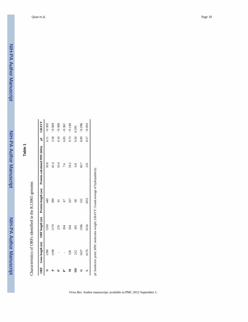
3.2 Open reading framesFeatures of the ORFs identified in the K13965 antigenome are summarized in Table 1.ORF-1 is compatible with nucleoproteins (N) of other rhabdoviruses by location, andcontains a highly conserved aa motif S289PYS that is proposed to be involved in RNA-binding in vertebrate rhabdoviruses (Crysler et al., 1990; Tordo et al., 1986). Alignmentwith other N sequences also indicates conserved aa identified as characteristic ofrhabdoviruses that infect vertebrates (Kuzmin et al., 2006). ORF-II contains severalpotential phosphorylation sites, including 2 tyrosine, 24 serine, and 7 threoninephosphorylation sites as anticipated for a phosphoprotein (P), the cofactor of the active viraltranscriptase/replicase complex. In addition, two non-overlapping ORFs in the +1 frame ofthe P gene may encode small acidic proteins designated P′ and P″ (Table 1, Figure 2). P′ islocated close to the 5′-end of the P ORF, similar to C proteins identified in VSIV andvesicular stomatitis New Jersey virus (VSNJV) (Peluso et al., 1996; Spiropoulou andNichol, 1993). Putative start codons for P′ and P″ are 65 nt and 758 nt downstream of the Pstart codon. Both putative proteins show no significant similarity to known proteins in thePFAM or PROSITE databases. ORF-III commonly codes for a matrix protein (M). The Manalog of K13965 includes potential serine (6 sites) and threonine (1 site) phosphorylationsites, and no sequence similarity to other known proteins is detected. K13965 ORF-IVincludes 2 hydrophobic aa clusters located at the N terminus and the center of the sequence,resulting in a grand average of hydropathicity of +0.595, analogous to the small hydrophobic(SH) proteins recently identified for TUPV and DURV (Allison et al., 2010; Springfeld etal., 2005). Phobius analysis indicated a signal peptide (aa 1-23) but no transmembraneanchor, suggesting that a soluble protein may be generated. Despite the limited primarysequence conservation amongst glycoproteins (G) of rhabdoviruses, general structuralfeatures including glycosylation sites, and cysteine residues are commonly conserved (Coll,1995; Walker and Kongsuwan, 1999). Accordingly, ORF-V of K13965 shows features oftype I glycoproteins, including an N-terminal signal peptide (aa 1-17), an ectodomain (aa18-501), a transmembrane domain (aa 502-521) followed by a short cytoplasmic tail (aa522-531), two potential N-glycosylation sites (N378KTL and N405GTT), and fourteencysteine residues including 7 of the 12 cysteines conserved among animal rhabdoviruses(CI40, CIII81, CV119, CVI158, CVII163, CXI309, CXII342; (Walker and Kongsuwan, 1999).The last and largest ORF-VI is predicted to translate into a 2,051 aa (231 kDa) L-polymerase. The L-polymerase of K13965 contains the conserved residues present in L-
Quan et al. Page 5
Virus Res. Author manuscript; available in PMC 2012 September 1.
NIH
-PA Author Manuscript
NIH
-PA Author Manuscript
NIH
-PA Author Manuscript

polymerase of negative strand RNA viruses (Poch et al., 1990). In addition, all four highlyconserved motifs A through D of block III (Poch et al., 1990; Poch et al., 1989) areconserved in L of K13965 (motif A (A577NHMDYSKWNNHQR); motif B(A647CWRGQAGGLEGLRQKGWTITSLLMI); motif C (T685LAQGDNQIV); motif D(Y758RGNLCNPKSKRY)).
3.3 Sequence comparison of K13965 with OVRV CSIRO-1342 and other rhabdovirusesIn an independent approach we also determined the complete genomic sequence of anotherunassigned Australian rhabdovirus, OVRV strain CSIRO-1342. As with K13965, culturesupernatant of CSIRO-1342 was analyzed by UHTS, sequence gaps filled with specific PCRand RACE, and the draft sequence finally confirmed through re-sequencing by overlappingPCR (GenBank accession JF705876). The total RNA preparations from CSIRO-1342 andK13965 that were used for UHTS were subsequently evaluated by a real-time RT-PCRassay developed based on the consensus genome sequences. The quantitative OVRV assayindicated virus loads of 1.5x 10⁵ copies/ng total RNA for CSIRO-1342, and 3.5 x 10⁵
copies/ng for K13965.
Like K13965, the negative-sense genome of OVRV comprises 11,220 nt with a comparablegenome organization. Furthermore, identical putative transcription initiation, terminationand intergenic regions were identified for CSIRO-1342, except for a change at the Mtermination sequence (3′-GCACU7). As in K13965 the G and L genes overlap. The putativeP′ ORFs of K13965 and CSIRO-1342 show high nt sequence conservation of 98.6%, and areidentical at the aa level. However, the putative P″ start and stop codons are positioneddifferently in the same +1 frame, generating proteins of 67 aa for K13965, or 48 aa forCSIRO-1342. Comparison of OVRV CSIRO-1342 and K13965 genomes indicated 93 % ntidentity. The P (92 %) and SH (93 %) ORFs show the least, and N (100 %) the highest aasequence identity (Suppl. Table 1). Comparison of individual K13965 ORFs to homologousproteins of other representative rhabdoviruses for which complete sequence is availableindicated that K13965 shares at most 36 % aa identity for L with TUPV and DURV; P andM show less then 17 % identity with the analyzed sequences, and identities for N rangedbetween 15 and 30 % (Suppl. Table 2).
3.4 Phylogenetic analysesPhylogenetic trees constructed from partial L aa sequences of K13965, CSIRO-1342 andrepresentative rhabdoviruses suggest that K13965 and CSIRO-1342 are closely related to,but distinct from, members of the recently proposed Sandjimba group (Fig. 3A) (Dacheux etal., 2010). Analysis of N aa sequences similarly grouped K13965 and CSIRO-1342 with theavailable Sandjimba group viruses KOLV and SJAV (Fig. 3B), but also showed arelationship to TUPV and DURV. Only the latter relationship could be confirmed by usingnearly full-length L sequence due to a lack of sequence information for the other viruses(Fig. 3C).
4. DiscussionK13965, an uncharacterized Australian virus isolate, was genetically identified as a strain ofOVRV by comparison of its genome to the full length genomic sequence that we obtainedfor the Australian OVRV CSIRO-1342, which matched the previously available partialsequence for CSIRO-1342 (GenBank Accession GQ294474). The K13965 genome, inaddition to 5 core proteins typical for rhabdoviruses, encodes a small hydrophobic (SH)protein, and putative P′ (C) and P″ proteins that may be expressed from alternative ORFscontained within the P gene. Like sigma viruses in Drosophila spp. (Longdon et al., 2010;
Quan et al. Page 6
Virus Res. Author manuscript; available in PMC 2012 September 1.
NIH
-PA Author Manuscript
NIH
-PA Author Manuscript
NIH
-PA Author Manuscript

Teninges et al., 1993), K13965, isolated from mosquitoes has overlap between the G and Lgene without ORF overlap.
Within the order Mononegavirales, SH and C proteins were initially identified inparamyxoviruses (Bellini et al., 1985; Curran and Kolakofsky, 2008; Elango et al., 1989).Paramyxovirus C proteins have been shown to inhibit interferon signaling (Nagai and Kato,2004), and SH proteins to inhibit TNF alpha production (Li et al., 2011) and apoptosis(Wilson et al., 2006). Protein products coded in analogous genome locations toparamyxovirus C proteins have been identified in the rhabdoviruses VSIV and VSNJV(Kretzschmar et al., 1996; Peluso et al., 1996; Spiropoulou and Nichol, 1993), and productscomparable to SH proteins were identified in TUPV (Springfeld et al., 2005) and predictedfor DURV (Allison et al., 2010). The functions of these proteins in rhabdoviruses areunknown although it has been hypothesized that C proteins may play a role in viralpathogenesis and enhance transcriptional activity (Peluso et al., 1996).
The putative P′ (C) of K13965 is larger than those reported for members of the genusVesiculovirus, but smaller than P′ (C) reported for TUPV or DURV (Suppl. Table 3). The P′(C) of K13965 is predicted to have an acidic pI whereas other rhabdoviral P′ (C) proteins,with the exception of that of DURV, have a basic pI >9. P′ (C) of K13965 shows norecognized sequence homology to other reported P′ (C), except for that of OVRVCSIRO-1342. In addition, a small P″ ORF located downstream of P′ was identified inK13965, resembling the P gene organization of NGAV, which is reported to also code fortwo putative non-overlapping small ORFs (Gubala et al., 2010). Although a P″ ORF ispresent also in CSIRO-1342, it is not conserved with respect to K13965; different start andstop codons predict a smaller protein for CSIRO-1342. This lack of conservation betweenthe two isolates indicates a lower selective pressure on these sequences compared to theother coding sequences.
Both OVRV K13965 and CSIRO-1342 encode a putative hydrophobic SH protein of smallersize than those of TUPV and DURV (Suppl. Table 4) that has no sequence homology withthese SHs. TUPV and DURV SHs are predicted to adopt a type I, or type II, transmembranetopology, respectively (Allison et al., 2010; Springfeld et al., 2005). In contrast, SH ofOVRV is not predicted to be a transmembrane protein. Interestingly, in NGAV an ORF (U4;(Gubala et al., 2010) is present in the same genomic position as SH proteins that also likelygenerates a soluble but not hydrophobic protein (NC_013955; Phobius). No aa conservationwas found in the N-terminal half of the ORF or the entire aa sequence. We also found noconservation of the leucine residues with respect to TUPV and DURV in the SH of K13965or CSIRO-1342. Together with TUPV and DURV (isolated from mammal and bird,respectively), the mosquito-borne OVRV represents the third example of a rhabdovirusencoding a P′ (C) and SH protein.
The geographic region from where K13965-infected Anopheles annulipes s.l. mosquitoeswere collected in 1993 is an enzootic focus for arboviruses, as the presence of the Ord Riverirrigation area enables mosquito species to breed continuously (Broom et al., 2002; Broomet al., 2003; Liehne et al., 1976; Mackenzie and Broom, 1999). The high level of rainfallduring the 1992/1993 wet season was associated with an increase in arbovirus activity inthat area (Broom et al., 1997). An isolate of the recently identified Stretch Lagoon orbiviruswas also recovered from mosquitoes collected from the Ord River area in 1993 (D.T.W. andC.A.J., unpublished), suggesting that environmental conditions were suitable for heightenedarbovirus activity at that time. OVRV CSIRO-1342 was isolated in the early 1980s fromCulex edwardsi and Culex spp. mosquitoes from southeast Queensland (Calisher et al.,1989; Cybinski and Muller, 1990; Muller and Standfast, 1986). In addition, OVRV was alsoisolated in 1984 from Aedes vigilax mosquitoes collected in Darwin (Weir et al., 1997).
Quan et al. Page 7
Virus Res. Author manuscript; available in PMC 2012 September 1.
NIH
-PA Author Manuscript
NIH
-PA Author Manuscript
NIH
-PA Author Manuscript

Taken together, OVRV strains have been isolated from disparate regions of tropicalAustralia, suggesting that this virus has been circulating widely for more than a decade.
An. annulipes s.l. is a species complex distributed across Australia, whilst Ae. vigilax has acoastal distribution and Cx. edwardsi is restricted to Queensland and the northern coast ofNew South Wales (Lee et al., 1989; Lee et al., 1987; Lee et al., 1984). Laboratory-basedvector competence studies will be required to confirm whether mosquitoes of these speciesare vectors for OVRV. Blood meal studies suggest that An. annulipes s.l. and Ae. vigilaxfeed on a variety of vertebrate hosts, including birds, marsupials and cattle (Jansen et al.,2009; Johansen et al., 2009b; Lee et al., 1987; Lee et al., 1984), whereas Cx. edwardsiappear to prefer humans and birds (Lee et al., 1989). No serosurveys of antibodies toK13965 in humans or animals in the Ord River area have been conducted, and no antibodiesto CSIRO-1342 were found in cattle sera at Peachester (Cybinski and Muller, 1990). OVRVlikely cycles between mosquitoes and vertebrate host(s), possibly birds and/or pigs, assuggested by a reported detection of antibodies to OVRV in feral pigs (Bourhy et al., 2008).
Phylogenetic analysis of the block III L aa sequence indicates that OVRV is closely relatedto members of a recently proposed Sandjimba group (Dacheux et al., 2010) within thetentative Dimarhabdovirus supergroup (Bourhy et al., 2005) of the Rhabdoviridae family.Other members of this group were isolated from birds or mosquitoes from the CentralAfrican Republic. However, the Australian viruses K13965 and CSIRO-1342 form a branchseparate from the African viruses, likely reflecting independent evolutionary pathwaysconsistent with their geographic separation. In this regard it is of interest that the Sandjimbagroup forms a sister clade with the proposed Almpiwar group (Fig. 3), whose four membershave been isolated from southern Queensland (Charleville virus strain Ch9824 fromCharleville), from northern Queensland (Almpiwar virus and Charleville virus strainCh9847 from Mitchell River), and from the Northern Territory (Humpty Doo virus fromBeatrice Hill), similar geographic areas as Peachester, Darwin and the Kimberley regionwhere OVRV and K13965 have been found (Fig. 1) (Bourhy et al., 2008). Thus, OVRV andAlmpiwar group viruses have shared or overlapping geographic distributions. Analysis ofthe N aa sequence showed a similar relationship of OVRV to two viruses from the Sandjimagroup for which N sequence is available, and also indicated a relationship to TUPV andDURV that was corroborated by the long L sequence analysis; however, further analyses arehampered by the lack of long L and N sequences for other viruses, especially for Almpiwargroup viruses. Additional genomic sequence information will be necessary to providefurther insights into the evolutionary history and relationships of members of these proposedgroups.
Arbovirus surveillance in northern Australia has yielded insights into the incidence,geographic range and ecology of major human flavi- and alphaviral pathogens throughcontinued annual studies over the last 5 decades (Broom et al., 1998; Johansen et al., 2009a;Johansen et al., 2009b; Liehne et al., 1976). Our identification of Oak Vale rhabdovirus inuncharacterized isolates identified through those surveillance efforts further defines viralflora in that area and enhances our knowledge of the diversity and complexity of rhabdoviralgenomes and their phylogenetic relationships.
Supplementary MaterialRefer to Web version on PubMed Central for supplementary material.
AcknowledgmentsThe authors are grateful to Michael D.A. Lindsay (Arbovirus Surveillance and Research Laboratory, the Universityof Western Australia, Nedlands, Western Australia), Annette K. Broom (Arbovirus Surveillance and Research
Quan et al. Page 8
Virus Res. Author manuscript; available in PMC 2012 September 1.
NIH
-PA Author Manuscript
NIH
-PA Author Manuscript
NIH
-PA Author Manuscript
Laboratory, the University of Western Australia, Nedlands, Western Australia), and Tony E. Wright (MosquitoBorne Disease Control Branch, Western Australian Department of Health, Claremont, Western Australia) formosquito collection, and technical staff in the Arbovirus Surveillance and Research Laboratory for virus isolation.We also thank Craig Street for bioinformatic analyses. This work was supported by National Institutes of Healthaward AI57158 (Northeast Biodefense Center-Lipkin), contract HHSN27220100004OI/HHSN27200004/DO4, theUnited States Agency for International Development’s (USAID) Emerging Pandemic Threats (EPT) Program,PREDICT project, under terms of Cooperative Agreement Number GHN-A-OO-09-00010-00, the United StatesDepartment of Defense and The Western Australian Department of Health by funding the Arbovirus Surveillanceand Research Program.
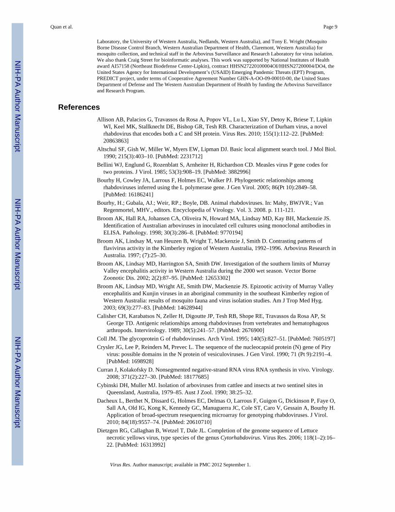
ReferencesAllison AB, Palacios G, Travassos da Rosa A, Popov VL, Lu L, Xiao SY, Detoy K, Briese T, Lipkin
WI, Keel MK, Stallknecht DE, Bishop GR, Tesh RB. Characterization of Durham virus, a novelrhabdovirus that encodes both a C and SH protein. Virus Res. 2010; 155(1):112–22. [PubMed:20863863]
Altschul SF, Gish W, Miller W, Myers EW, Lipman DJ. Basic local alignment search tool. J Mol Biol.1990; 215(3):403–10. [PubMed: 2231712]
Bellini WJ, Englund G, Rozenblatt S, Arnheiter H, Richardson CD. Measles virus P gene codes fortwo proteins. J Virol. 1985; 53(3):908–19. [PubMed: 3882996]
Bourhy H, Cowley JA, Larrous F, Holmes EC, Walker PJ. Phylogenetic relationships amongrhabdoviruses inferred using the L polymerase gene. J Gen Virol. 2005; 86(Pt 10):2849–58.[PubMed: 16186241]
Bourhy, H.; Gubala, AJ.; Weir, RP.; Boyle, DB. Animal rhabdoviruses. In: Mahy, BWJVR.; VanRegenmortel, MHV., editors. Encyclopedia of Virology. Vol. 3. 2008. p. 111-121.
Broom AK, Hall RA, Johansen CA, Oliveira N, Howard MA, Lindsay MD, Kay BH, Mackenzie JS.Identification of Australian arboviruses in inoculated cell cultures using monoclonal antibodies inELISA. Pathology. 1998; 30(3):286–8. [PubMed: 9770194]
Broom AK, Lindsay M, van Heuzen B, Wright T, Mackenzie J, Smith D. Contrasting patterns offlavivirus activity in the Kimberley region of Western Australia, 1992–1996. Arbovirus Research inAustralia. 1997; (7):25–30.
Broom AK, Lindsay MD, Harrington SA, Smith DW. Investigation of the southern limits of MurrayValley encephalitis activity in Western Australia during the 2000 wet season. Vector BorneZoonotic Dis. 2002; 2(2):87–95. [PubMed: 12653302]
Broom AK, Lindsay MD, Wright AE, Smith DW, Mackenzie JS. Epizootic activity of Murray Valleyencephalitis and Kunjin viruses in an aboriginal community in the southeast Kimberley region ofWestern Australia: results of mosquito fauna and virus isolation studies. Am J Trop Med Hyg.2003; 69(3):277–83. [PubMed: 14628944]
Calisher CH, Karabatsos N, Zeller H, Digoutte JP, Tesh RB, Shope RE, Travassos da Rosa AP, StGeorge TD. Antigenic relationships among rhabdoviruses from vertebrates and hematophagousarthropods. Intervirology. 1989; 30(5):241–57. [PubMed: 2676900]
Coll JM. The glycoprotein G of rhabdoviruses. Arch Virol. 1995; 140(5):827–51. [PubMed: 7605197]Crysler JG, Lee P, Reinders M, Prevec L. The sequence of the nucleocapsid protein (N) gene of Piry
virus: possible domains in the N protein of vesiculoviruses. J Gen Virol. 1990; 71 (Pt 9):2191–4.[PubMed: 1698928]
Curran J, Kolakofsky D. Nonsegmented negative-strand RNA virus RNA synthesis in vivo. Virology.2008; 371(2):227–30. [PubMed: 18177685]
Cybinski DH, Muller MJ. Isolation of arboviruses from cattlee and insects at two sentinel sites inQueensland, Australia, 1979–85. Aust J Zool. 1990; 38:25–32.
Dacheux L, Berthet N, Dissard G, Holmes EC, Delmas O, Larrous F, Guigon G, Dickinson P, Faye O,Sall AA, Old IG, Kong K, Kennedy GC, Manuguerra JC, Cole ST, Caro V, Gessain A, Bourhy H.Application of broad-spectrum resequencing microarray for genotyping rhabdoviruses. J Virol.2010; 84(18):9557–74. [PubMed: 20610710]
Dietzgen RG, Callaghan B, Wetzel T, Dale JL. Completion of the genome sequence of Lettucenecrotic yellows virus, type species of the genus Cytorhabdovirus. Virus Res. 2006; 118(1–2):16–22. [PubMed: 16313992]
Quan et al. Page 9
Virus Res. Author manuscript; available in PMC 2012 September 1.
NIH
-PA Author Manuscript
NIH
-PA Author Manuscript
NIH
-PA Author Manuscript
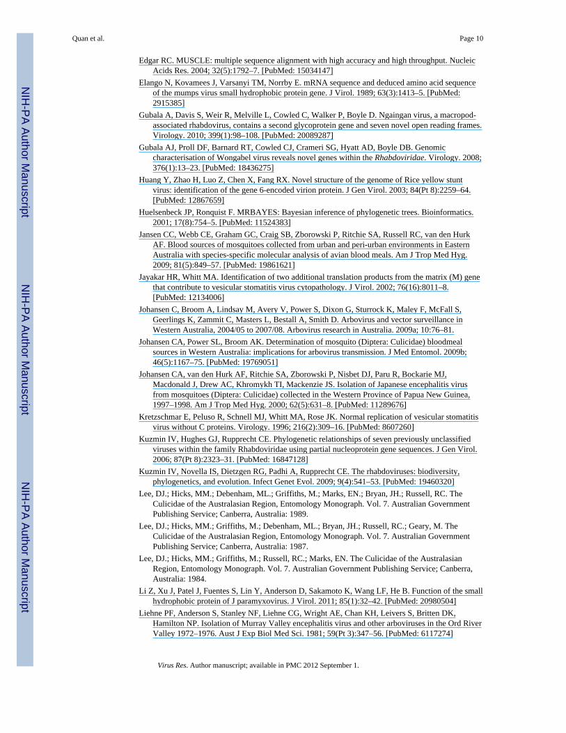
Edgar RC. MUSCLE: multiple sequence alignment with high accuracy and high throughput. NucleicAcids Res. 2004; 32(5):1792–7. [PubMed: 15034147]
Elango N, Kovamees J, Varsanyi TM, Norrby E. mRNA sequence and deduced amino acid sequenceof the mumps virus small hydrophobic protein gene. J Virol. 1989; 63(3):1413–5. [PubMed:2915385]
Gubala A, Davis S, Weir R, Melville L, Cowled C, Walker P, Boyle D. Ngaingan virus, a macropod-associated rhabdovirus, contains a second glycoprotein gene and seven novel open reading frames.Virology. 2010; 399(1):98–108. [PubMed: 20089287]
Gubala AJ, Proll DF, Barnard RT, Cowled CJ, Crameri SG, Hyatt AD, Boyle DB. Genomiccharacterisation of Wongabel virus reveals novel genes within the Rhabdoviridae. Virology. 2008;376(1):13–23. [PubMed: 18436275]
Huang Y, Zhao H, Luo Z, Chen X, Fang RX. Novel structure of the genome of Rice yellow stuntvirus: identification of the gene 6-encoded virion protein. J Gen Virol. 2003; 84(Pt 8):2259–64.[PubMed: 12867659]
Huelsenbeck JP, Ronquist F. MRBAYES: Bayesian inference of phylogenetic trees. Bioinformatics.2001; 17(8):754–5. [PubMed: 11524383]
Jansen CC, Webb CE, Graham GC, Craig SB, Zborowski P, Ritchie SA, Russell RC, van den HurkAF. Blood sources of mosquitoes collected from urban and peri-urban environments in EasternAustralia with species-specific molecular analysis of avian blood meals. Am J Trop Med Hyg.2009; 81(5):849–57. [PubMed: 19861621]
Jayakar HR, Whitt MA. Identification of two additional translation products from the matrix (M) genethat contribute to vesicular stomatitis virus cytopathology. J Virol. 2002; 76(16):8011–8.[PubMed: 12134006]
Johansen C, Broom A, Lindsay M, Avery V, Power S, Dixon G, Sturrock K, Maley F, McFall S,Geerlings K, Zammit C, Masters L, Bestall A, Smith D. Arbovirus and vector surveillance inWestern Australia, 2004/05 to 2007/08. Arbovirus research in Australia. 2009a; 10:76–81.
Johansen CA, Power SL, Broom AK. Determination of mosquito (Diptera: Culicidae) bloodmealsources in Western Australia: implications for arbovirus transmission. J Med Entomol. 2009b;46(5):1167–75. [PubMed: 19769051]
Johansen CA, van den Hurk AF, Ritchie SA, Zborowski P, Nisbet DJ, Paru R, Bockarie MJ,Macdonald J, Drew AC, Khromykh TI, Mackenzie JS. Isolation of Japanese encephalitis virusfrom mosquitoes (Diptera: Culicidae) collected in the Western Province of Papua New Guinea,1997–1998. Am J Trop Med Hyg. 2000; 62(5):631–8. [PubMed: 11289676]
Kretzschmar E, Peluso R, Schnell MJ, Whitt MA, Rose JK. Normal replication of vesicular stomatitisvirus without C proteins. Virology. 1996; 216(2):309–16. [PubMed: 8607260]
Kuzmin IV, Hughes GJ, Rupprecht CE. Phylogenetic relationships of seven previously unclassifiedviruses within the family Rhabdoviridae using partial nucleoprotein gene sequences. J Gen Virol.2006; 87(Pt 8):2323–31. [PubMed: 16847128]
Kuzmin IV, Novella IS, Dietzgen RG, Padhi A, Rupprecht CE. The rhabdoviruses: biodiversity,phylogenetics, and evolution. Infect Genet Evol. 2009; 9(4):541–53. [PubMed: 19460320]
Lee, DJ.; Hicks, MM.; Debenham, ML.; Griffiths, M.; Marks, EN.; Bryan, JH.; Russell, RC. TheCulicidae of the Australasian Region, Entomology Monograph. Vol. 7. Australian GovernmentPublishing Service; Canberra, Australia: 1989.
Lee, DJ.; Hicks, MM.; Griffiths, M.; Debenham, ML.; Bryan, JH.; Russell, RC.; Geary, M. TheCulicidae of the Australasian Region, Entomology Monograph. Vol. 7. Australian GovernmentPublishing Service; Canberra, Australia: 1987.
Lee, DJ.; Hicks, MM.; Griffiths, M.; Russell, RC.; Marks, EN. The Culicidae of the AustralasianRegion, Entomology Monograph. Vol. 7. Australian Government Publishing Service; Canberra,Australia: 1984.
Li Z, Xu J, Patel J, Fuentes S, Lin Y, Anderson D, Sakamoto K, Wang LF, He B. Function of the smallhydrophobic protein of J paramyxovirus. J Virol. 2011; 85(1):32–42. [PubMed: 20980504]
Liehne PF, Anderson S, Stanley NF, Liehne CG, Wright AE, Chan KH, Leivers S, Britten DK,Hamilton NP. Isolation of Murray Valley encephalitis virus and other arboviruses in the Ord RiverValley 1972–1976. Aust J Exp Biol Med Sci. 1981; 59(Pt 3):347–56. [PubMed: 6117274]
Quan et al. Page 10
Virus Res. Author manuscript; available in PMC 2012 September 1.
NIH
-PA Author Manuscript
NIH
-PA Author Manuscript
NIH
-PA Author Manuscript

Liehne PF, Stanley NF, Alpers MP, Liehne CG. Ord River arboviruses--the study site and mosquitoes.Aust J Exp Biol Med Sci. 1976; 54(5):487–97. [PubMed: 14612]
Lindsay MD, Broom AK, Wright AE, Johansen CA, Mackenzie JS. Ross River virus isolations frommosquitoes in arid regions of Western Australia: implication of vertical transmission as a means ofpersistence of the virus. Am J Trop Med Hyg. 1993; 49(6):686–96. [PubMed: 8279636]
Longdon B, Obbard DJ, Jiggins FM. Sigma viruses from three species of Drosophila form a major newclade in the rhabdovirus phylogeny. Proc Biol Sci. 2010; 277(1678):35–44. [PubMed: 19812076]
Mackenzie, JS.; Broom, AK. Ord river irrigation area: the effect of dam construction and irrigation onthe incidence of Murray Valley encephalitis virus. In: Kay, BH., editor. Water Resources - Health,Environment and Development. Spon; London: 1999. p. 108-122.
Margulies M, Egholm M, Altman WE, Attiya S, Bader JS, Bemben LA, Berka J, Braverman MS,Chen YJ, Chen Z, Dewell SB, Du L, Fierro JM, Gomes XV, Godwin BC, He W, Helgesen S, HoCH, Irzyk GP, Jando SC, Alenquer ML, Jarvie TP, Jirage KB, Kim JB, Knight JR, Lanza JR,Leamon JH, Lefkowitz SM, Lei M, Li J, Lohman KL, Lu H, Makhijani VB, McDade KE,McKenna MP, Myers EW, Nickerson E, Nobile JR, Plant R, Puc BP, Ronan MT, Roth GT, SarkisGJ, Simons JF, Simpson JW, Srinivasan M, Tartaro KR, Tomasz A, Vogt KA, Volkmer GA,Wang SH, Wang Y, Weiner MP, Yu P, Begley RF, Rothberg JM. Genome sequencing inmicrofabricated high-density picolitre reactors. Nature. 2005; 437(7057):376–80. [PubMed:16056220]
McWilliam SM, Kongsuwan K, Cowley JA, Byrne KA, Walker PJ. Genome organization andtranscription strategy in the complex GNS-L intergenic region of bovine ephemeral feverrhabdovirus. J Gen Virol. 1997; 78 (6):1309–17. [PubMed: 9191923]
Morzunov SP, Winton JR, Nichol ST. The complete genome structure and phylogenetic relationship ofinfectious hematopoietic necrosis virus. Virus Res. 1995; 38(2–3):175–92. [PubMed: 8578857]
Muller, MJ.; Standfast, HA. Vectors of ephemeral fever group viruses. In: St George, TD.; Kay, BH.;Blok, J., editors. Arbovirus research in Australia: Proceedings of the Fourth Symposium. CSIRO/QMIR; Brisbane: 1986. p. 295-300.p. 295-298.
Nagai Y, Kato A. Accessory genes of the paramyxoviridae, a large family of nonsegmented negative-strand RNA viruses, as a focus of active investigation by reverse genetics. Curr Top MicrobiolImmunol. 2004; 283:197–248. [PubMed: 15298171]
Nishizawa T, Kurath G, Winton JR. Sequence analysis and expression of the M1 and M2 matrixprotein genes of hirame rhabdovirus (HIRRV). Diseases of Aquatic Organisms. 1997; 31:9–17.
Peluso RW, Richardson JC, Talon J, Lock M. Identification of a set of proteins (C′ and C) encoded bythe bicistronic P gene of the Indiana serotype of vesicular stomatitis virus and analysis of theireffect on transcription by the viral RNA polymerase. Virology. 1996; 218(2):335–42. [PubMed:8610460]
Poch O, Blumberg BM, Bougueleret L, Tordo N. Sequence comparison of five polymerases (Lproteins) of unsegmented negative-strand RNA viruses: theoretical assignment of functionaldomains. J Gen Virol. 1990; 71 (Pt 5):1153–62. [PubMed: 2161049]
Poch O, Sauvaget I, Delarue M, Tordo N. Identification of four conserved motifs among the RNA-dependent polymerase encoding elements. Embo J. 1989; 8(12):3867–74. [PubMed: 2555175]
Quan PL, Junglen S, Tashmukhamedova A, Conlan S, Hutchison SK, Kurth A, Ellerbrok H, EgholmM, Briese T, Leendertz FH, Lipkin WI. Moussa virus: a new member of the Rhabdoviridae familyisolated from Culex decens mosquitoes in Cote d’Ivoire. Virus Res. 2010; 147(1):17–24.[PubMed: 19804801]
Quan PL, Palacios G, Jabado OJ, Conlan S, Hirschberg DL, Pozo F, Jack PJ, Cisterna D, Renwick N,Hui J, Drysdale A, Amos-Ritchie R, Baumeister E, Savy V, Lager KM, Richt JA, Boyle DB,Garcia-Sastre A, Casas I, Perez-Brena P, Briese T, Lipkin WI. Detection of respiratory viruses andsubtype identification of influenza A viruses by GreeneChipResp oligonucleotide microarray. JClin Microbiol. 2007; 45(8):2359–64. [PubMed: 17553978]
Reed SE, Tsai CW, Willie KJ, Redinbaugh MG, Hogenhout SA. Shotgun sequencing of the negative-sense RNA genome of the rhabdovirus Maize mosaic virus. J Virol Methods. 2005; 129(1):91–6.[PubMed: 16005085]
Quan et al. Page 11
Virus Res. Author manuscript; available in PMC 2012 September 1.
NIH
-PA Author Manuscript
NIH
-PA Author Manuscript
NIH
-PA Author Manuscript

Revill P, Trinh X, Dale J, Harding R. Taro vein chlorosis virus: characterization and variability of anew nucleorhabdovirus. J Gen Virol. 2005; 86(Pt 2):491–9. [PubMed: 15659770]
Rice P, Longden I, Bleasby A. EMBOSS: the European Molecular Biology Open Software Suite.Trends Genet. 2000; 16(6):276–7. [PubMed: 10827456]
Scholthof KB, Hillman BI, Modrell B, Heaton LA, Jackson AO. Characterization and detection of sc4:a sixth gene encoded by sonchus yellow net virus. Virology. 1994; 204(1):279–88. [PubMed:8091658]
Schutze H, Mundt E, Mettenleiter TC. Complete genomic sequence of viral hemorrhagic septicemiavirus, a fish rhabdovirus. Virus Genes. 1999; 19(1):59–65. [PubMed: 10499451]
Spiropoulou CF, Nichol ST. A small highly basic protein is encoded in overlapping frame within the Pgene of vesicular stomatitis virus. J Virol. 1993; 67(6):3103–10. [PubMed: 8388490]
Springfeld C, Darai G, Cattaneo R. Characterization of the Tupaia rhabdovirus genome reveals a longopen reading frame overlapping with P and a novel gene encoding a small hydrophobic protein. JVirol. 2005; 79(11):6781–90. [PubMed: 15890917]
Tamura K, Dudley J, Nei M, Kumar S. MEGA4: Molecular Evolutionary Genetics Analysis (MEGA)software version 4.0. Mol Biol Evol. 2007; 24(8):1596–9. [PubMed: 17488738]
Tanno F, Nakatsu A, Toriyama S, Kojima M. Complete nucleotide sequence of Northern cereal mosaicvirus and its genome organization. Arch Virol. 2000; 145(7):1373–84. [PubMed: 10963343]
Tao JJ, Zhou GZ, Gui JF, Zhang QY. Genomic sequence of mandarin fish rhabdovirus with an unusualsmall non-transcriptional ORF. Virus Res. 2008; 132(1–2):86–96. [PubMed: 18068257]
Teninges D, Bras F, Dezelee S. Genome organization of the sigma rhabdovirus: six genes and a geneoverlap. Virology. 1993; 193(2):1018–23. [PubMed: 8384742]
Tordo N, Benmansour A, Calisher C, Dietzgen RG, Fang RX, Jackson AO, Kurath G, Nadin-Davis S,Tesh RB, Walker PJ. Virus Taxonomy. Eighth Report of the International Committee onTaxonomy of Viruses. 2005:623–644.
Tordo N, Poch O, Ermine A, Keith G, Rougeon F. Walking along the rabies genome: is the large G-Lintergenic region a remnant gene? Proc Natl Acad Sci U S A. 1986; 83(11):3914–8. [PubMed:3459163]
Walker PJ, Kongsuwan K. Deduced structural model for animal rhabdovirus glycoproteins. J GenVirol. 1999; 80 (Pt 5):1211–20. [PubMed: 10355768]
Wang Y, McWilliam SM, Cowley JA, Walker PJ. Complex genome organization in the GNS-Lintergenic region of Adelaide River rhabdovirus. Virology. 1994; 203(1):63–72. [PubMed:8030285]
Weir RP, Hyatt AD, CHC, Whelan PI. New records of arboviruses isolated from mosquitoes in theNorthern Territory, 1982–1992. Arbovirus research in Australia. 1997; 7:311–321.
Whelan S, Goldman N. A general empirical model of protein evolution derived from multiple proteinfamilies using a maximum-likelihood approach. Mol Biol Evol. 2001; 18(5):691–9. [PubMed:11319253]
Whelan SP, Barr JN, Wertz GW. Transcription and replication of nonsegmented negative-strand RNAviruses. Curr Top Microbiol Immunol. 2004; 283:61–119. [PubMed: 15298168]
Wilson RL, Fuentes SM, Wang P, Taddeo EC, Klatt A, Henderson AJ, He B. Function of smallhydrophobic proteins of paramyxovirus. J Virol. 2006; 80(4):1700–9. [PubMed: 16439527]
Zhu RL, Lei XY, Ke F, Yuan XP, Zhang QY. Genome of turbot rhabdovirus exhibits unusual non-coding regions and an additional ORF that could be expressed in fish cell. Virus Res. 2011;155(2):495–505. [PubMed: 21185339]
Quan et al. Page 12
Virus Res. Author manuscript; available in PMC 2012 September 1.
NIH
-PA Author Manuscript
NIH
-PA Author Manuscript
NIH
-PA Author Manuscript

HighlightsFull genome sequence of Australian viruses K13965 and Oak Vale was determined byunbiased pyrosequencing. Genetic analysis identified K13965 as a rhabdovirus and as astrain of Oak Vale virus. Genome organization is unusual for P′(C)/P″ combined with aSH gene, and overlap of G and L genes. Phylogenetic analyses indicate relationships tothe African Sandjimba clade and Australian Almpiwar clade.
Quan et al. Page 13
Virus Res. Author manuscript; available in PMC 2012 September 1.
NIH
-PA Author Manuscript
NIH
-PA Author Manuscript
NIH
-PA Author Manuscript

Figure 1.Map of Australia by states and territories showing the geographic locations where OVRVK13965 and CSIRO-1342 were isolated. K13965 was isolated from Anopheles annulipes s.l.collected 3 km north of Kununurra. OVRV CSIRO-1342 was isolated from Culex edwardsicollected at Peachester. Another isolation of OVRV has been reported from Aedes vigilaxcollected at Darwin. ACT, Australian Capital Territory; NSW, New South Wales; NT,Northern Territory; QLD, Queensland; SA, South Australia; TAS, Tasmania; VIC, Victoria,WA, Western Australia.
Quan et al. Page 14
Virus Res. Author manuscript; available in PMC 2012 September 1.
NIH
-PA Author Manuscript
NIH
-PA Author Manuscript
NIH
-PA Author Manuscript

Figure 2.K13965 genome organization. (A) Schematic organization of the K13965 genome. The barrepresents the 11,120 nucleotide (nt) antigenome. The eight open arrows indicate theposition of the open reading frames (ORFs) for putative N, P, P′, P″, M, SH, G, and Lproteins. (B) Transcription initiation, intergenic and transcription termination/polyadenylation sequences. The start and stop codon for each gene is in bold, the intergenicregion is underlined. (C) Complementarity between the 3′ leader and the 5′ trailer sequences.Bases conserved among rhabdoviruses known to infect vertebrates are shown in bold. Startand stop codons are underlined.
Quan et al. Page 15
Virus Res. Author manuscript; available in PMC 2012 September 1.
NIH
-PA Author Manuscript
NIH
-PA Author Manuscript
NIH
-PA Author Manuscript

Figure 3.Phylogenetic relationships of K13965 and CSIRO-1342 with representative rhabdovirusesderived from partial L (156 aa; A), N (325 aa; B) and full L amino acid sequence (970 aa;C). The geographic origin (*Africa, # Australia) is indicated for K13965, CSIRO-1342 andmembers of the Sandjimba and Almpiwar groups. K13965 is indicated by a circledarrowhead. Analysis was performed using a Bayesian method applying a WAG model ofamino acid replacement with a gamma distribution of rate variation among sites for 10million generations (with a 25% burn-in). Support for each node is provided by BPP(Bayesian posterior probability) (>0.8). Branch lengths are drawn to scale and the trees weremid-point rooted. Amino acid sequences used were: ABLV, Australian bat lyssavirus(L:NP_478343; N:AAD01267); ALMV, Almpiwar virus (L:AY854645); ARAV, Aravanvirus (L:ABV03822; N:Q6X1D8); ARV, Adelaide River virus (L:AF234534;N:AAC54627); BBOV, Bimbo virus (L:GU816016); BEFV, bovine ephemeral fever virus(L:NP_065409; N:NP_065398); BGNV, Bangoran virus (L:GU816010); BRMV, Berrimahvirus (L:AAZ43265); BTKV, Boteke virus (L:GU816014); BYSMV, barley yellow striatemosaic virus (L:ACT21686); CFRV, China fish rhabdovirus (L:AAX86686); CHPV,Chandipura virus (L:P13179; N:P11211); CHVV9824, Charleville virus (L:AY854644);CHVV9847, Charleville virus (L:AY854672); COCV, Cocal virus (L:ACB47438;N:ACB47434); CPV, coastal plains virus (L:ADG86364; N:ADG86356); DAffSV,Drosophila affinis sigma virus 10 (L:GQ410980); DobsSV, Drosophila obscura sigma virus10A (N:GQ410979); DURV, Durham virus (L:ADB88761; N:ADB88758); DUVV,Duvenhage virus (L:ABZ81216; N:Q66453); EBLV1, european bat lyssavirus 1(L:ABZ81181; N:AAX62875); EBLV2, european bat lyssavirus 2 (L:ABZ81191;N:YP_001285393); FLAV, Flanders virus (L:AAN73288; N:AAN73283); FUKV, Fukuokavirus (L:AAZ43279); GARV, Garba virus (L:GU816018); HDOOV, Humpty doo virus(L:AAZ43271); HIRRV, Hirame rhabdovirus (L:NP_919035; N:NP_919030); IHNV,infectious hematopoietic necrosis virus (L:CAA52076; N:NP042676); IRKV, Irkut virus(L:ABV03823; N:Q5VKP6); ISFV, Isfahan virus (L:Q5K2K3; N:Q5K2K7); KAMV,Kamese virus (L:GU816011); KCV, Kern Canyon virus (N:ABE69215); KEUV, Keuralibavirus (L:GU816021); KHUV, Khujand virus (L:ABV03824; N:Q6X1D4); KIMV,Kimberley virus (L:AAZ43266); KOLV, Kolongo virus (L:GU816020; N:ABE69214);KOTV, Kotonkan virus (L:AAZ43267; N:ABE69213); LBV, Lagos bat virus(L:ABZ81171; N:ABF56214); LDV, Le Dantec virus (L:AAZ43278); LYMoV, lettuceyellow mottle virus (L:YP_002308376; N:YP_00208371); LNYV, lettuce necrotic yellowsvirus (L:YP_425092; N:CAG34083); MEBV, mount Elgon bat virus (N:ABE69217);
Quan et al. Page 16
Virus Res. Author manuscript; available in PMC 2012 September 1.
NIH
-PA Author Manuscript
NIH
-PA Author Manuscript
NIH
-PA Author Manuscript
MFSV, maize fine streak virus (L:YP_052849; N:YP_052843); MMV, maize mosaic virus(L:YP_052855; N:YP_052850); MOKV, Mokola virus (L:ABZ81211; N:YP_142350);MOSV, Mossuril virus (L:GU816012); MOUV, Moussa virus (L:ACZ81402;N:ACZ81403); NASV, Nasoule virus (L:GU816017); NCMV, northern cereal mosaic virus(L:NP_597914; N:NP_057954); NGAV, Ngaingan virus (L:YP_003518294;N:YP_003518280); NKOV, Nkolbisson virus (L:GU816022); OBOV, Obodhiang virus(N:ABE69212); OFV, orchid fleck virus (L:YP_001294929; N:BAH97109); OITAV, Oitarhabdovirus (N:BAD13431); OUAV, Ouango virus (L:GU816015); PCRV, Parry Creekvirus (L:AAZ43275); PERV, Perinet virus (L:AAZ43280); PFRV, Pike fry rhabdovirus(L:ACP28002; N:ACP27998); PIRYV, Piry virus (N:P26037); PORV, Porton virus(L:GU816013); PYDV, potato yellow dwarf virus (N:ABW35154); RABV, Rabies virus(L:Q66T60; N:ACN15666); RBUV, Rochambeau virus (N:ABE69218); RYSV, rice yellowstunt virus (L:NP_620502; N:NP_620496); SCRV, Siniperca chuatsi rhabdovirus(L:YP_802942; N:YP_802937); SCV, strawberry crinkle virus (L:AAP03645); SFRV,Starry flounder rhabdovirus (L:AY450644); SHRV, snakehead rhabdovirus (L:NP_050585;N:NP_050580); SIGMAV, Drosophila melanogaster sigma virus (N:ACV67011);SIGMAVHAP23, Drosophila melanogaster sigma virus HAP23 (L:GQ375258;N:GQ375258); SIGMAVHAP30, Drosophila melanogaster sigma virus AP30(L:YP_003126913; N:YP_003126908); SJAV, Sandjimba virus (L:GU816019;N:ABE69216); STRV, sea trout rhabdovirus (N:AAL35756); SVCV, spring viraemia ofcarp virus (L:Q91DR9; N:ABW24033); SYNV, Sonchus yellow net virus (L:NP_042286;N:NP_042281); TaVCV, Taro vein chlorosis virus (L:YP_224083; N:YP_224078); TIBV,Tibrogargan virus (L:ADG86355; N:ADG86347); TUPV, Tupaia virus (L:YP_238534;N:YP_238528); VHSV, viral hemorrhagic septicemia virus (L:CAB40833; N:P24378);VSAV, vesicular stomatitis Alagoas virus (L:ACB47443; N:ACB47439); VSIV, vesicularstomatitis Indiana virus (L:NP_041716; N:P11212); VSNJV, vesicular stomatitis NewJersey virus (L:P16379; N:P04881); WCBV, West Caucasian bat virus (L:ABV03821;N:Q5VKP2); WOGV, Wongabel virus (L:AAZ43276; N:YP_002333271).
Quan et al. Page 17
Virus Res. Author manuscript; available in PMC 2012 September 1.
NIH
-PA Author Manuscript
NIH
-PA Author Manuscript
NIH
-PA Author Manuscript
NIH
-PA Author Manuscript
NIH
-PA Author Manuscript
NIH
-PA Author Manuscript
Quan et al. Page 18
Tabl
e 1
Cha
ract
eris
tics o
f OR
Fs id
entif
ied
in th
e K
1396
5 ge
nom
e.
OR
FG
ene
leng
th (n
t)O
RF
leng
th (n
t)Pr
otei
n le
ngth
(aa)
Prot
ein
calc
ulat
ed M
W (k
Da)
pIG
RA
VY
N13
9013
5044
950
.86.
11−0.302
P11
9811
7038
941
.45.
58−0.569
P′-
276
9110
.46.
10−0.300
P″-
204
677.
66.
83−0.367
M52
850
416
719
.29.
73−0.140
SH21
218
360
6.8
6.50
0.59
5
G16
3715
9653
160
.78.
89−0.296
L61
7661
5620
5123
28.
57−0.203
pI: I
soel
ectri
c po
int;
MW
: mol
ecul
ar w
eigh
t; G
RA
VY
: Gra
nd a
vera
ge o
f hyd
ropa
thic
ity.
Virus Res. Author manuscript; available in PMC 2012 September 1.

NIH
-PA Author Manuscript
NIH
-PA Author Manuscript
NIH
-PA Author Manuscript
Quan et al. Page 19
Tabl
e 2
Gen
ome
orga
niza
tion
of re
pres
enta
tive
rhab
dovi
ruse
s.
Vir
usG
enus
Gen
ome
stru
ctur
eH
ost/v
ecto
rL
engt
h (n
t)G
enba
nk A
cces
sion
no.
K13
956
UC
N-P
(P′,
P″)-
M-S
H-G
-Lm
osqu
itoes
1112
0JF
7058
77
RA
BV
Lyss
avir
usN
-P-M
-G-L
bat
1192
3EU
2931
16
VSI
VVe
sicu
lovi
rus
N-P
(C)-
M-G
-Lbo
vine
1115
5A
F473
865
SIG
MA
VU
AN
-P-X
-M-G
-LD
roso
phila
mel
anog
aste
r12
625
GQ
3752
58
SCR
VU
CN
-P(C
)-M
(Ms)
-G-L
fish
1154
5N
C_0
0851
4
IHN
VN
ovir
habd
ovir
usN
-P-M
-G-N
V-L
fish
1113
1N
C_0
0165
2
TUPV
Tent
ativ
e Ve
sicu
lovi
rus
N-P
(C)-
M-S
H-G
-Ltre
e sh
rew
1144
0N
C_0
0702
0
RY
SVN
ucle
orha
bdov
irus
N-P
-3-M
-G-6
-Lpl
ant
1404
2N
C_0
0374
6
MFS
VN
ucle
orha
bdov
irus
N-P
-3-4
-M-G
-Lpl
ant
1378
2N
C_0
0597
4
TIB
VU
AN
-P-M
-U1-
U2-
G(G′)-
U3-
Lca
ttle,
dip
tera
n13
298
GQ
2944
72
CPV
UC
N-P
(P′)-
M-U
1-U
2-G
-U3-
Lliv
esto
ck13
203
GQ
2944
73
WO
GV
UC
N(U
4)-P
-U1-
U2-
U3-
M-G
(U5)
-Lbi
ting
mid
ges
1319
6N
C_0
1163
9
NC
MV
Cyt
orha
bdov
irus
N-P
-3-4
-5-6
-M-G
-Lpl
ant
1322
2N
C_0
0225
1
AR
VEp
hem
erov
irus
N-P
-M-G
-GN
S-(α
1/α2
)-β-
Lbo
vine
NA
U05
987
U10
363
L092
06
BEF
VEp
hem
erov
irus
N-P
(P′)-
M-G
-GN
S-(α
1/α2
/α3)
-(β/γ)
-Lbo
vine
1490
0N
C_0
0252
6
NG
AV
UA
N-P
(P′1
, P′2
)-(U
1/U
2)-U
3-M
-U4-
G-G
ns-(
U5/
U6)
-U7-
Lbi
ting
mid
ges
1576
4N
C_0
1395
5
UA
: una
ssig
ned
rhab
dovi
rus;
UC
: unc
lass
ified
viru
s (no
t fou
nd in
the
VII
Ith re
port
of th
e In
tern
atio
nal C
omm
ittee
on
taxo
nom
y vi
ruse
s). T
he 5
typi
cal c
ore
gene
s N, P
, M, G
and
L p
rese
nt in
all
rhab
dovi
ruse
s are
indi
cate
d in
bol
d. O
RFs
tran
slat
ed fr
om p
olyc
istro
nic
RN
A a
re in
dica
ted
in p
aren
thes
is.
Virus Res. Author manuscript; available in PMC 2012 September 1.