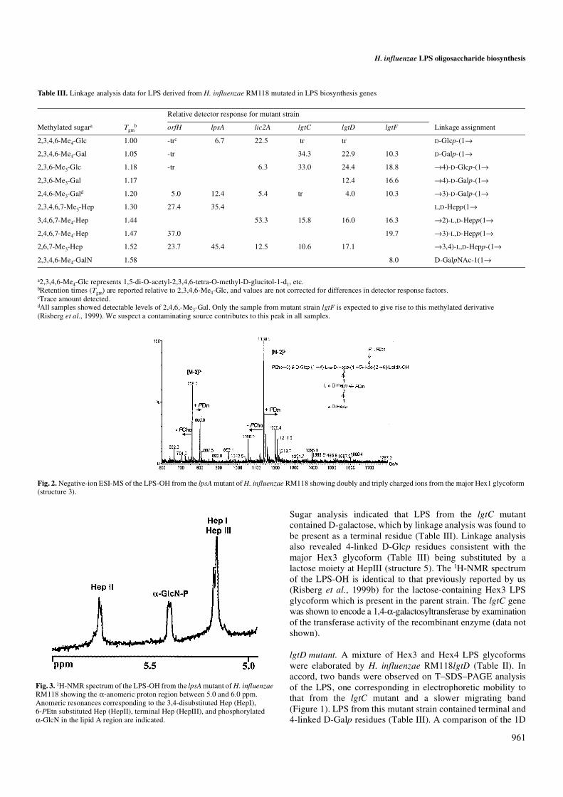
Genetic basis for expression of the major globotetraose-containing lipopolysaccharide from H....
12
Glycobiology vol. 11 no. 11 pp. 957–967, 2001 © 2001 Oxford University Press 957 Genetic basis for expression of the major globotetraose-containing lipopolysaccharide from H. influenzae strain Rd (RM118) Derek W. Hood 1,2 , Andrew D. Cox 3 , Warren W. Wakarchuk 3 , Melissa Schur 3 , Elke K.H. Schweda 4 , Shannon L. Walsh 2 , Mary E. Deadman 2 , Adele Martin 3 , E. Richard Moxon 2 , and James C. Richards 3 2 Molecular Infectious Diseases Group, University of Oxford Department of Paediatrics, Weatherall Institute of Molecular Medicine, John Radcliffe Hospital, Headington, Oxford, OX3 9DS, UK; 3 Institute for Biological Sciences, National Research Council of Canada, Ottawa, Ontario, K1A 0R6 Canada; and 4 Clinical Research Centre, Karolinska Institutet and University College of South Stockholm, NOVUM, S-141 86 Huddinge, Sweden. Received on April 19, 2001; revised on July 9, 2001; accepted on July 11, 2001 A genetic basis for the biosynthetic assembly of the globotetraose containing lipopolysaccharide (LPS) of Haemo- philus influenzae strain RM118 (Rd) was determined by structural analysis of LPS derived from mutant strains. We have previously shown that the parent strain RM118 elaborates a population of LPS molecules made up of a series of related glycoforms differing in the degree of oligosaccharide chain extension from the distal heptose residue of a conserved phosphorylated inner-core element, L-α-D-Hepp-(1→2)-L-α- D-Hepp-(1→3)-[β-D-Glcp-(1→4)-]-L-α-D-Hepp-(1→5)-α-Kdo. The fully extended LPS glycoform expresses the globotetraose structure, β-D-GalpNAc-(1→3)-α-D-Galp- (1→4)-β-D-Galp-(1→4)-β-D-Glcp. A fingerprinting strategy was employed to establish the structure of LPS from strains mutated in putative glycosyltransferase genes compared to the parent strain. This involved glycose and linkage analysis on intact LPS samples and analysis of O-deacylated LPS samples by electrospray ionization mass spectrometry and 1D 1 H-nuclear magnetic resonance spectroscopy. Four genes, lpsA, lic2A, lgtC, and lgtD, were required for sequential addition of the glycoses to the terminal inner-core heptose to give the globotetraose structure. lgtC and lgtD were shown to encode glycosyltransferases by enzymatic assays with synthetic acceptor molecules. This is the first genetic blueprint determined for H. influenzae LPS oligosaccharide biosynthesis, identifying genes involved in the addition of each glycose residue. Key words: globotetraose/Haemophilus influenzae/ lipopolysaccharide Introduction Haemophilus influenzae is a bacterium that routinely colonizes the upper respiratory tract of humans. It is also the cause of both upper and lower respiratory tract infections that result from contiguous spread. Occasionally, H. influenzae can cause systemic and life-threatening bacteraemic diseases, such as septicaemia and meningitis. Lipopolysaccharide (LPS) of H. influenzae functions as a virulence determinant (Preston et al., 1996a). LPS results in major cytotoxic injury to host cells, is a target for host immune responses, and can influence each stage of the pathogenesis of H. influenzae infection (Moxon and Maskell, 1992). Changes in LPS expression can alter the viru- lence of this pathogen (Kimura and Hansen, 1986; Cope et al., 1990; Weiser et al., 1990b; Hood et al., 1996a). A feature of H. influenzae LPS is that surface-exposed epitopes of the oligosac- charide are subject to high frequency on-off switching of expression (phase variation) (Weiser et al., 1990a; High et al., 1993; Jarosik and Hansen, 1994). This heterogeneity may be an advantage to the bacteria, allowing them to better confront different host compartments and microenvironments and to survive the host immune response (Weiser and Pan, 1998). Determination of structure is crucial to understanding the biology of H. influenzae LPS and its role in bacterial virulence. H. influenzae LPS comprises a variable oligosaccharide moiety and a membrane-anchoring lipid A component (Zamze and Moxon, 1987). LPS from a number of different strains have been shown to contain a common L-glycero-D-manno- heptose-containing inner-core trisaccharide unit attached to the lipid A moiety via a phosphorylated 2-keto-3-deoxyoctu- losonic acid (Kdo) residue (Phillips et al., 1992, 1993, 1996; Gibson et al., 1993; Schweda et al., 1993, 1995; Masoud et al., 1997; Risberg et al., 1997, 1999a,b; Rahman et al., 1999). Each of the Hep residues can provide a point for the addition of Hex residues, which in turn can lead to oligosaccharide chain extensions. The degree of substitution and chain extension from the triheptose unit varies within and between strains (Masoud et al., 1997; Risberg et al., 1999b). In addition, phos- phate-containing substituents that include free phosphate groups (P), phosphoethanolamine (PEtn), pyrophospho- ethanolamine (PPEtn), and phosphocholine (PCho) also contribute to the structural variability of these molecules. Recently we reported the structure of a globotetraose (β-D- GalpNAc-(1→3)-α-D-Galp-(1→4)-β-D-Galp-(1→4)-β-D-Glcp) containing LPS from H. influenzae strain RM118 (Risberg et al., 1999b), the strain (Rd) for which the complete genome sequence has been determined (Fleischmann et al., 1995). For strain RM118, three major populations of LPS glycoforms were identified, all containing a PCho→6)-β-D-Glcp group off the Hep attached to the Kdo unit, but differing in the length of the oligosaccharide chains off the third Hep of the inner-core element. LPS glycoforms expressing a fully assembled globotetraose side chain and sequentially truncated glycoforms containing globoside (α-D-Galp-(1→4)-β-D-Galp-(1→4)-β-D-Glcp) and 1 To whom correspondence should be addressed
-
Upload
independent -
Category
Documents
-
view
2 -
download
0
Transcript of Genetic basis for expression of the major globotetraose-containing lipopolysaccharide from H....
Glycobiology vol. 11 no. 11 pp. 957–967, 2001
© 2001 Oxford University Press 957
Genetic basis for expression of the major globotetraose-containing lipopolysaccharide from H. influenzae strain Rd (RM118)
Derek W. Hood1,2, Andrew D. Cox3, Warren W. Wakarchuk3, Melissa Schur3, Elke K.H. Schweda4, Shannon L. Walsh2, Mary E. Deadman2, Adele Martin3, E. Richard Moxon2, and James C. Richards3
2Molecular Infectious Diseases Group, University of Oxford Department of Paediatrics, Weatherall Institute of Molecular Medicine, John Radcliffe Hospital, Headington, Oxford, OX3 9DS, UK; 3Institute for Biological Sciences, National Research Council of Canada, Ottawa, Ontario, K1A 0R6 Canada; and 4Clinical Research Centre, Karolinska Institutet and University College of South Stockholm, NOVUM, S-141 86 Huddinge, Sweden.
Received on April 19, 2001; revised on July 9, 2001; accepted on July 11, 2001
A genetic basis for the biosynthetic assembly of theglobotetraose containing lipopolysaccharide (LPS) of Haemo-philus influenzae strain RM118 (Rd) was determined bystructural analysis of LPS derived from mutant strains. Wehave previously shown that the parent strain RM118 elaboratesa population of LPS molecules made up of a series of relatedglycoforms differing in the degree of oligosaccharide chainextension from the distal heptose residue of a conservedphosphorylated inner-core element, L-α-D-Hepp-(1→2)-L-α-D-Hepp-(1→3)-[β-D-Glcp-(1→4)-]-L-α-D-Hepp-(1→5)-α-Kdo.The fully extended LPS glycoform expresses theglobotetraose structure, β-D-GalpNAc-(1→3)-α-D-Galp-(1→4)-β-D-Galp-(1→4)-β-D-Glcp. A fingerprinting strategywas employed to establish the structure of LPS from strainsmutated in putative glycosyltransferase genes compared tothe parent strain. This involved glycose and linkage analysison intact LPS samples and analysis of O-deacylated LPSsamples by electrospray ionization mass spectrometry and1D 1H-nuclear magnetic resonance spectroscopy. Fourgenes, lpsA, lic2A, lgtC, and lgtD, were required for sequentialaddition of the glycoses to the terminal inner-core heptoseto give the globotetraose structure. lgtC and lgtD wereshown to encode glycosyltransferases by enzymatic assayswith synthetic acceptor molecules. This is the first geneticblueprint determined for H. influenzae LPS oligosaccharidebiosynthesis, identifying genes involved in the addition ofeach glycose residue.
Key words: globotetraose/Haemophilus influenzae/lipopolysaccharide
Introduction
Haemophilus influenzae is a bacterium that routinely colonizesthe upper respiratory tract of humans. It is also the cause of
both upper and lower respiratory tract infections that resultfrom contiguous spread. Occasionally, H. influenzae can causesystemic and life-threatening bacteraemic diseases, such assepticaemia and meningitis. Lipopolysaccharide (LPS) ofH. influenzae functions as a virulence determinant (Preston et al.,1996a). LPS results in major cytotoxic injury to host cells, is atarget for host immune responses, and can influence each stageof the pathogenesis of H. influenzae infection (Moxon andMaskell, 1992). Changes in LPS expression can alter the viru-lence of this pathogen (Kimura and Hansen, 1986; Cope et al.,1990; Weiser et al., 1990b; Hood et al., 1996a). A feature of H.influenzae LPS is that surface-exposed epitopes of the oligosac-charide are subject to high frequency on-off switching ofexpression (phase variation) (Weiser et al., 1990a; High et al.,1993; Jarosik and Hansen, 1994). This heterogeneity may bean advantage to the bacteria, allowing them to better confrontdifferent host compartments and microenvironments and tosurvive the host immune response (Weiser and Pan, 1998).
Determination of structure is crucial to understanding thebiology of H. influenzae LPS and its role in bacterial virulence.H. influenzae LPS comprises a variable oligosaccharidemoiety and a membrane-anchoring lipid A component (Zamzeand Moxon, 1987). LPS from a number of different strainshave been shown to contain a common L-glycero-D-manno-heptose-containing inner-core trisaccharide unit attached to thelipid A moiety via a phosphorylated 2-keto-3-deoxyoctu-losonic acid (Kdo) residue (Phillips et al., 1992, 1993, 1996;Gibson et al., 1993; Schweda et al., 1993, 1995; Masoud et al.,1997; Risberg et al., 1997, 1999a,b; Rahman et al., 1999).Each of the Hep residues can provide a point for the addition ofHex residues, which in turn can lead to oligosaccharide chainextensions. The degree of substitution and chain extensionfrom the triheptose unit varies within and between strains(Masoud et al., 1997; Risberg et al., 1999b). In addition, phos-phate-containing substituents that include free phosphategroups (P), phosphoethanolamine (PEtn), pyrophospho-ethanolamine (PPEtn), and phosphocholine (PCho) alsocontribute to the structural variability of these molecules.Recently we reported the structure of a globotetraose (β-D-GalpNAc-(1→3)-α-D-Galp-(1→4)-β-D-Galp-(1→4)-β-D-Glcp)containing LPS from H. influenzae strain RM118 (Risberg etal., 1999b), the strain (Rd) for which the complete genomesequence has been determined (Fleischmann et al., 1995). Forstrain RM118, three major populations of LPS glycoformswere identified, all containing a PCho→6)-β-D-Glcp group offthe Hep attached to the Kdo unit, but differing in the length of theoligosaccharide chains off the third Hep of the inner-core element.LPS glycoforms expressing a fully assembled globotetraose sidechain and sequentially truncated glycoforms containinggloboside (α-D-Galp-(1→4)-β-D-Galp-(1→4)-β-D-Glcp) and1To whom correspondence should be addressed
D.W. Hood et al.
958
lactose (β-D-Galp-(1→4)-β-D-Glcp) were characterized(Risberg et al., 1999b).
The availability of the complete genome sequence ofH. influenzae strain Rd facilitated a comprehensive study of LPSbiosynthetic loci in the type b strains RM153 and RM7004.Many predicted gene functions were correlated with particularsteps in the synthesis of the LPS in strain RM153 (Hood et al.,1996a). The LPS from strain RM118 has a significantlydifferent structure to that of RM153, the pattern and degree ofsubstitution of oligosaccharide chain extensions being entirelydifferent. Thus it is not possible to assign the genetic basis forbiosynthetic functions for RM118 LPS from the informationcurrently available. In particular, the genetic basis for expressionof the globotetraose structure and Hex addition to the first Hephas not previously been reported.
In this study we employ a structural fingerprinting strategyto determine and compare the structures of LPS obtained froma series of defined mutants in LPS biosynthetic genes inH. influenzae strain RM118. We identify the glycosyltrans-ferases involved in the assembly of the globotetraose sidechain and in the biosynthesis of the inner-core region of theLPS molecule. The transferase functions of gene productsinvolved in sequential addition of α-1,4-linked Galp (LgtC)and β-1,3-linked GalpNAc (LgtD) to give the globoside andglobotetraose structures, respectively, were unambiguouslydetermined by enzymatic assays with synthetic acceptors.
Results
Construction and screening of mutant strains
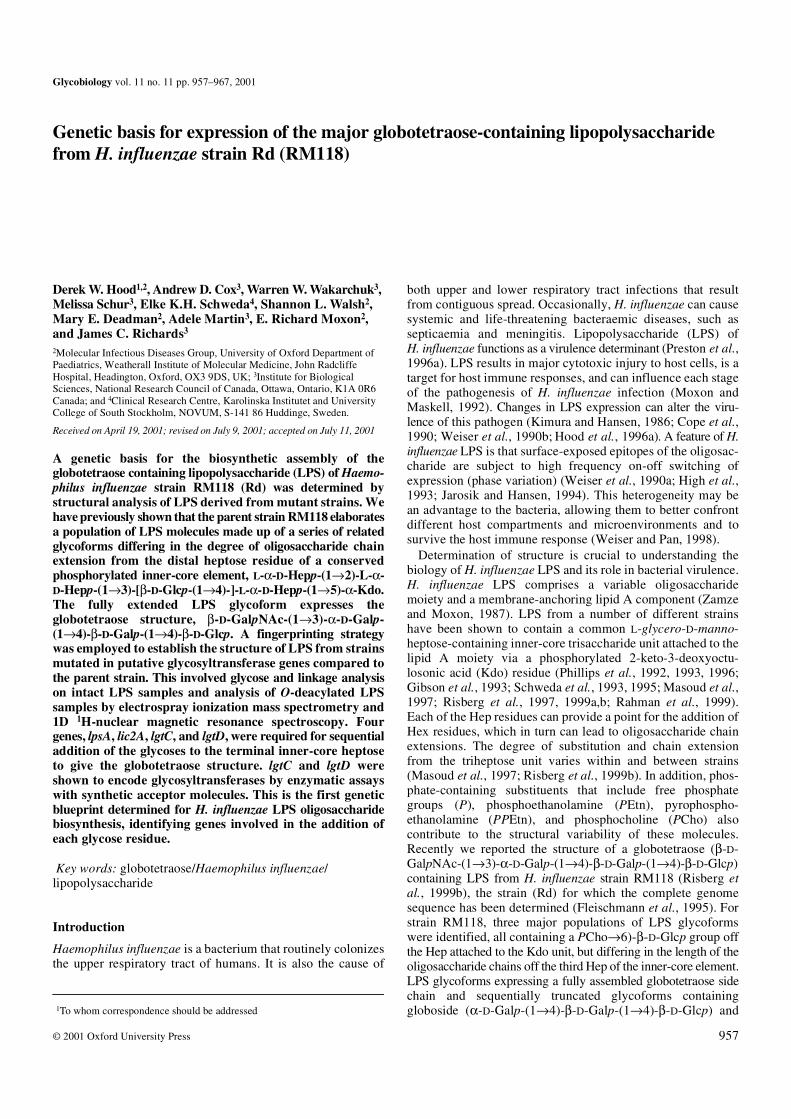
A set of mutants was made (Table I) to investigate in detail thegenetic basis of the biosynthesis of the oligosaccharide portionof LPS from H. influenzae strain RM118. The DNA constructsused to mutate the majority of these genes had been previouslyreported (Hood et al., 1996a). lic1 and lic2A are phase-variableLPS biosynthetic loci described previously (Weiser et al.,1989; High et al., 1993). In the course of this study there wasno obvious candidate gene responsible for adding the Glc tothe first Hep. Searching the Rd genome sequence databasewith the LgtF sequence from Neisseria gave a match (31%identify over 247 amino acids) to reading frame HI0653. lgtFwas amplified by polymerase chain reaction (PCR) fromchromosomal DNA of strain RM118, cloned then inactivatedand used to transform H. influenzae. Genes responsible for thesynthesis of Kdo (kdsA, kdsB) and the Kdo transferase (kdtA)have been identified from the genome sequence (Fleischmannet al., 1995). Various attempts to construct strains mutated inthe kdtA gene failed, similar to findings with type b strains(Hood et al., 1996a). This is assumed to be due to nonviabilityof this mutant. LPS isolated from RM118 and the isogenicmutant strains was analysed by tricine–sodium dodecylsulfate–polyacrylamde gel electrophoresis (T–SDS–PAGE)(data not shown). Strains mutated in genes most likelyencoding glycosyltransferases for RM118 oligosaccharidesynthesis and that showed an altered pattern of LPS bandswhen compared to wild type (Figure 1), were selected fordetailed structural analysis of their LPS as described below. Amutant in which the lic1 locus is inactivated was also investi-gated.
Structural characterization of LPS
Analysis of LPS from strain RM118 by T–SDS–PAGE (Figure 1)showed a heterogeneous pattern of bands corresponding inelectrophoretic mobility to populations of low-molecular-massLPS composed of lipid A and oligosaccharide componentsdiffering in the number of attached sugar residues. We havepreviously shown that strain RM118 grown under similar
Table I. LPS-related genes investigated in strain RM118 in this study
HI numbers are the ORF designations given by the Institute for Genomic Research for the H. influenzae genome sequence database. The genes given in boldface type are those selected for detailed comparative analysis of the expressed LPS glycoforms.
Gene HI number Reference
kdtA 0652 Hood et al., 1996a
lgtC 0259 Hood et al., 1996a
lgtD 1578 Hood et al., 1996a
lpsA 0765 Hood et al., 1996a
orfZ 0260.1 Hood et al., 1996a
opsX 0261 Hood et al., 1996a
orfH 0523 Hood et al., 1996a
rfaF 1105 Hood et al., 1996a
lic2A 0550 High et al., 1993
lic1 1537–1540 Weiser et al., 1990a
lgtF 0653 This study
cld 0866 Hood et al., 1996a
galU 0812 Hood et al., 1996a
kfiC 0868 Hood et al., 1996a
lsg1 0867 Hood et al., 1996a
orfM 0260 Hood et al., 1996a
orfE 0869 Hood et al., 1996a
orfO 0870 Hood et al., 1996a
orfY 0871 Hood et al., 1996a
pgmB 0740 Hood et al., 1996a
pgmC 1337 Hood et al., 1996a
rfe 1716 Hood et al., 1996a
rfbP 0872 Hood et al., 1996a
rfbB 0873 Hood et al., 1996a
Fig. 1. The electrophoretic gel-migration patterns after T–SDS–PAGE of LPS purified from RM118 wild type and strains mutated in putative glycosyltransferase genes. RM118 corresponds to the wild-type strain, and the isogenic mutants are listed by the relevant LPS gene.

H. influenzae LPS oligosaccharide biosynthesis
959
conditions expresses populations of LPS containing three tofive glycose residues attached to a common inner-core element(Risberg et al., 1999b). LPS from strains with mutations inlic1, lgtF, and lgtD showed similar complex banding patterns,and those from strains with mutations in lgtC, lic2A, lpsA,orfH, rfaF, and opsX gave less complex patterns comprisingbands having consecutively faster mobilities consistent withsuccessive sugar deletions. Identification of the nature of sugardeletions in the LPS samples from mutant strains, grown inliquid culture, was achieved by comparative structuralanalysis. Structural fingerprinting was done using electrosprayionization mass spectrometry (ESI-MS) and 1D 1H–nuclearmagnetic resonance (NMR) analysis of O-deacylated LPS(LPS-OH) samples. In addition, glycose and linkage analyseswere carried out on intact LPS samples. Such analyses establishedthe key structural features of the altered LPS glycoforms. TheESI-MS data obtained in the negative ion mode is presented inTable II. The LPS-OH samples from the mutant strains gavedata consistent with the presence of oligosaccharides linkedvia Kdo-4-phosphate to a common O-deacylated lipid Amoiety (lipid A-OH) differing in the number of Hep, Hex, andphosphate-containing substituents (Phillips et al., 1992, 1996;Schweda et al., 1993, 1995; Masoud et al., 1997; Risberg et al.,1997, 1999b). H. influenzae lipid A-OH is known (Masoud et al.,1997; Helander et al., 1988) to be composed of bisphos-phorylated β-1,6-linked glucosamine disaccharide substitutedby 3-hydroxytetradecanoamide groups at C-2 and C-2′.
opsX mutant. Inactivation of opsX gave rise to deep-roughLPS, which was devoid of Hep or hexose residues, containingonly a phosphorylated Kdo attached to lipid A (Table II). Wehave previously shown that mutation in the opsX gene ofH. influenzae type b strain RM153 results in truncation of theLPS between HepI and Kdo (Hood et al., 1996a). Tandemmass spectrometry (MS-MS) analysis of the LPS-OH sampleby low-energy collisional activation of the doubly chargedmolecular ions (m/z 625.5 and 616.5) afforded in both cases amajor fragment ion at m/z 952 (lipid A-OH) arising fromcleavage of the Kdo-β-D-glucosamine bond (data not shown).The mass of this fragment ion is consistent with that expectedfor H. influenzae lipid A-OH (Helander et al., 1988). It seemslikely that the anhydro-Kdo-P derivative (corresponding to m/z616.5) is due to the Kdo molecule in this mutant only, notbeing substituted by the HepI residue at the 5-position. It isevident that RM118 and RM153 opsX mutants express LPSsimilar to that from the previously characterized Rdisn (I69)strain (Helander et al., 1988; Preston et al., 1996b). The I69 LPSphenotype arises from a mutation in the heptose biosynthetic genegmhA (Brook and Valvano, 1996), rendering the mutant strainincapable of adding heptose to LPS.
rfaF mutant. 1H-NMR analysis of LPS-OH from RM118rfaFshowed, in addition to the expected 1H resonance from the α-linkedglucosamine residue of lipid A, an anomeric proton resonance(∼5.19 ppm) in the low-field region from a single heptose unit.Sugar analysis confirmed the Hep residue to be L-glycero-D-manno heptose. Correspondingly, the ESI-MS spectrum wasdominated by a single abundant doubly charged ion at m/z721.6 consistent with the structure Hep1-Kdo-lipid A-OH(Table II).
orfH mutant. Strain RM118orfH gave a mixture of LPS glyco-forms, each containing two Hep residues, as evidenced fromthe ESI-MS data (Table II). In addition to the major populationof glycoforms containing an additional Hep residue, that is,Hep2•PEtn0-2•Kdo-lipid A-OH, compared to RM118rfaF LPS,were species containing a Hex-PCho unit. Sugar analysisindicated the presence of D-glucose and the PCho methylprotons gave an intense signal in the 1H-NMR at 3.24 ppm..LPS from this strain reacted with TEPC-15, a PCho specificmonoclonal antibody (MAb) (Weiser et al., 1997). Linkageanalysis revealed the presence of terminal Hep, 3-substitutedHep and 3,4,-disubsituted Hep residues (Table III). This data isconsistent with RM118orfH expressing the two major LPSglycoform structures 1 and 2 (see Scheme 1, PEtn showspartial substitution). The occurrence of two bands for the LPSof RM118orfH when analyszed by T–SDS–PAGE (Figure 1)is consistent with this conclusion.
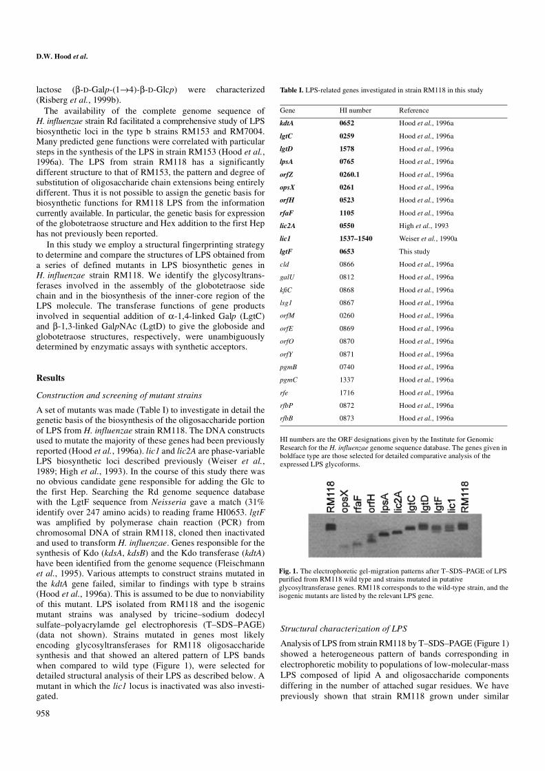
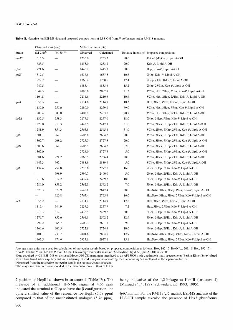
lpsA mutant. ESI-MS analysis of LPS-OH from RM118lpsAindicated it to contain glycoforms having an additional Hepresidue when compared to RM118orfH (Figure 2, Table II),the PCho containing Hex1 glycoform being the major LPSspecies. Linkage analysis was consistent with sequentialaddition of Hep to the terminal Hep in structure 2 (Table III).Correspondingly, the 1H-NMR spectrum of this LPS-OHshowed the characteristic pattern in the low-field region(5.0–6.0 ppm) for the LPS tri-Hep inner-core element (HepII,5.76 ppm; HepI/HepIII, 5.16/5.15 ppm) of H. influenzae(Figure 3) (Risberg et al., 1999b). This data is consistent withthe RM118lpsA derived LPS having the structure 3 (Figure 2,Table IV).
lic2A mutant. ESI-MS analysis of the LPS-OH samples fromstrain RM118lic2A revealed the presence of Hex2 glycoformsas the major LPS species (Table II). Analysis of theRM118lic2A LPS indicated the presence of D-glucose as theonly neutral hexose, linkage analysis indicating it to be aterminal residue (Table III). A significant proportion of 2-linkedHep residues was also revealed by linkage analysis. It is note-worthy, that 2-subsituted Hep residues were not detected in theLPS sample from the lpsA mutant due to substitution of thatresidue by PEtn groups (cf. structure 3), which are not readilycleaved under the hydrolysis conditions employed in thelinkage analysis procedure. In accord with these findings, itcan be concluded that LPS from the lic2A mutant differs fromthat of the lpsA mutant in that it carries a glucose residue at the
Scheme 1. Structures 1 and 2 of the LPS glycoform.
D.W. Hood et al.
960
2-position of HepIII as shown in structure 4 (Table IV). Thepresence of an additional 1H-NMR signal at 4.65 ppmindicated the terminal D-Glcp to have the β-configuration, theupfield shifted value of the resonance for HepII (5.58 ppm)compared to that of the unsubstituted analogue (5.76 ppm),
being indicative of the 1,2-linkage to HepIII (structure 4)(Masoud et al., 1997; Schweda et al., 1993, 1995).
lgtC mutant. For the RM118lgtC mutant, ESI-MS analysis of theLPS-OH sample revealed the presence of Hex3 glycoforms.
Table II. Negative ion ESI-MS data and proposed compositions of LPS-OH from H. influenzae strain RM118 mutants.
Average mass units were used for calculation of molecular weight based on proposed composition as follows: Hex, 162.15; HexNAc, 203.19; Hep, 192.17; Kdo-P, 300.16; PEtn, 123.05; PCho, 165.05. The average molecular mass of O-deacylated lipid A (lipid A-OH) is 953.03.aData acquired by CE-ESI- MS on a crystal Model 310 CE instrument interfaced to an API 3000 triple quadrupole mass spectrometer (Perkin-Elmer/Sciex) fitted with a bare fused silica capillary column and using 30 mM morpholine-acetate (pH 9.0) containing 5% methanol as the separation buffer.bMeasured from the respective molecular ions in the reconstructed spectrum.cThe major ion observed corresponded to the molecular ion –18 (loss of H2O).
Observed ions (m/z) Molecular mass (Da)
Strain (M-2H)2– (M-3H)3– Observed Calculated Relative intensityb Proposed composition
opsXa 616.5 — 1235.0 1235.2 80.0 Kdo-P (-H2O)c, Lipid A-OH
625.5 — 1253.0 1253.2 20.0 Kdo-P, Lipid A-OH
rfaF 721.6 — 1445.2 1445.3 100.0 Hep, Kdo-P, Lipid A-OH
orfH 817.5 1637.5 1637.5 10.6 2Hep, Kdo-P, Lipid A-OH
879.2 — 1760.4 1760.6 42.4 2Hep, PEtn, Kdo-P, Lipid A-OH
940.5 — 1883.4 1883.6 15.2 2Hep, 2PEtn, Kdo-P, Lipid A-OH
1042.3 — 2086.6 2087.8 21.2 PCho, Hex, 2Hep, PEtn, Kdo-P, Lipid A-OH
1104.8 — 2211.6 2210.8 10.6 PCho, Hex, 2Hep, 2PEtn, Kdo-P, Lipid A-OH
lpsA 1056.3 — 2114.6 2114.9 10.3 Hex, 3Hep, PEtn, Kdo-P, Lipid A-OH
1139.0 759.0 2280.0 2279.9 69.0 PCho, Hex, 3Hep, PEtn, Kdo-P, Lipid A-OH
1200.4 800.0 2402.9 2403.0 20.7 PCho, Hex, 3Hep, 2PEtn, Kdo-P, Lipid A-OH
lic2A 1137.5 758.3 2277.5 2277.0 18.0 2Hex, 3Hep, PEtn, Kdo-P, Lipid A-OH
1220.0 813.3 2442.5 2442.1 51.0 PCho, 2Hex, 3Hep, PEtn, Kdo-P, Lipid A-O H
1281.9 854.3 2565.8 2565.1 31.0 PCho, 2Hex, 3Hep, 2PEtn, Kdo-P, Lipid A-OH
lgtC 1301.1 867.1 2603.8 2604.2 80.0 PCho, 3Hex, 3Hep, PEtn, Kdo-P, Lipid A-OH
1362.7 908.2 2727.5 2727.3 20.0 PCho, 3Hex, 3Hep, 2PEtn, Kdo-P, Lipid A-OH
lgtD 1300.6 867.1 2603.9 2604.2 62.0 PCho, 3Hex, 3Hep, PEtn, Kdo-P, Lipid A-OH
1362.0 2726.0 2727.3 5.0 PCho, 3Hex, 3Hep, 2PEtn, Kdo-P, Lipid A-OH
1381.6 921.2 2765.5 2766.4 28.0 PCho, 4Hex, 3Hep, PEtn, Kdo-P, Lipid A-OH
1443.3 962.1 2888.9 2889.4 5.0 PCho, 4Hex, 3Hep, 2PEtn, Kdo-P, LipidA-OH
lgtF 1137.4 757.8 2276.6 2277.0 16.0 2Hex, 3Hep, PEtn, Kdo-P, Lipid A-OH
— 798.9 2399.7 2400.0 5.0 2Hex, 3Hep, 2PEtn, Kdo-P, Lipid A-OH
1218.6 812.2 2439.4 2439.2 18.0 3Hex, 3Hep, PEtn, Kdo-P, Lipid A-OH
1280.0 853.2 2562.3 2562.2 7.0 3Hex, 3Hep, 2PEtn, Kdo-P, Lipid A-OH
1320.3 879.9 2642.8 2642.4 38.0 HexNAc, 3Hex, 3Hep, PEtn, Kdo-P, Lipid A-OH
— 920.8 2765.4 2765.4 16.0 HexNAc, 3Hex, 3Hep, 2PEtn, Kdo-P, Lipid A-OH
lic1 1056.2 — 2114.4 2114.9 12.8 Hex, 3Hep, PEtn, Kdo-P, Lipid A-OH
1117.4 744.9 2237.3 2237.9 7.2 Hex, 3Hep, 2PEtn, Kdo-P, Lipid A-OH
1218.3 812.1 2438.9 2439.2 20.0 3Hex, 3Hep, PEtn, Kdo-P, Lipid A-OH
1279.7 852.6 2561.1 2562.2 12.9 3Hex, 3Hep, 2PEtn, Kdo-P, Lipid A-OH
1299.2 865.7 2600.3 2601.3 9.0 4Hex, 3Hep, PEtn, Kdo-P, Lipid A-OH
1360.6 906.5 2722.9 2724.4 10.0 4Hex, 3Hep, 2PEtn, Kdo-P, Lipid A-OH
1401.1 933.7 2804.6 2804.5 12.9 HexNAc, 4Hex, 3Hep, PEtn, Kdo-P, Lipid A-OH
1462.5 974.6 2927.1 2927.6 15.1 HexNAc, 4Hex, 3Hep, 2PEtn, Kdo-P, Lipid A-OH
H. influenzae LPS oligosaccharide biosynthesis
961
Sugar analysis indicated that LPS from the lgtC mutantcontained D-galactose, which by linkage analysis was found tobe present as a terminal residue (Table III). Linkage analysisalso revealed 4-linked D-Glcp residues consistent with themajor Hex3 glycoform (Table III) being substituted by alactose moiety at HepIII (structure 5). The 1H-NMR spectrumof the LPS-OH is identical to that previously reported by us(Risberg et al., 1999b) for the lactose-containing Hex3 LPSglycoform which is present in the parent strain. The lgtC genewas shown to encode a 1,4-α-galactosyltransferase by examinationof the transferase activity of the recombinant enzyme (data notshown).
lgtD mutant. A mixture of Hex3 and Hex4 LPS glycoformswere elaborated by H. influenzae RM118lgtD (Table II). Inaccord, two bands were observed on T–SDS–PAGE analysisof the LPS, one corresponding in electrophoretic mobility tothat from the lgtC mutant and a slower migrating band(Figure 1). LPS from this mutant strain contained terminal and4-linked D-Galp residues (Table III). A comparison of the 1D
Table III. Linkage analysis data for LPS derived from H. influenzae RM118 mutated in LPS biosynthesis genes
a2,3,4,6-Me4-Glc represents 1,5-di-O-acetyl-2,3,4,6-tetra-O-methyl-D-glucitol-1-d1, etc.bRetention times (Tgm) are reported relative to 2,3,4,6-Me4-Glc, and values are not corrected for differences in detector response factors.cTrace amount detected.dAll samples showed detectable levels of 2,4,6,-Me3-Gal. Only the sample from mutant strain lgtF is expected to give rise to this methylated derivative (Risberg et al., 1999). We suspect a contaminating source contributes to this peak in all samples.
Relative detector response for mutant strain
Methylated sugara Tgmb orfH lpsA lic2A lgtC lgtD lgtF Linkage assignment
2,3,4,6-Me4-Glc 1.00 -trc 6.7 22.5 tr tr D-Glcp-(1→
2,3,4,6-Me4-Gal 1.05 -tr 34.3 22.9 10.3 D-Galp-(1→
2,3,6-Me3-Glc 1.18 -tr 6.3 33.0 24.4 18.8 →4)-D-Glcp-(1→
2,3,6-Me3-Gal 1.17 12.4 16.6 →4)-D-Galp-(1→
2,4,6-Me3-Gald 1.20 5.0 12.4 5.4 tr 4.0 10.3 →3)-D-Galp-(1→
2,3,4,6,7-Me5-Hep 1.30 27.4 35.4 L,D-Hepp(1→
3,4,6,7-Me4-Hep 1.44 53.3 15.8 16.0 16.3 →2)-L,D-Hepp(1→
2,4,6,7-Me4-Hep 1.47 37.0 19.7 →3)-L,D-Hepp(1→
2,6,7-Me3-Hep 1.52 23.7 45.4 12.5 10.6 17.1 →3,4)-L,D-Hepp-(1→
2,3,4,6-Me4-GalN 1.58 8.0 D-GalpNAc-1(1→
Fig. 2. Negative-ion ESI-MS of the LPS-OH from the lpsA mutant of H. influenzae RM118 showing doubly and triply charged ions from the major Hex1 glycoform (structure 3).
Fig. 3. 1H-NMR spectrum of the LPS-OH from the lpsA mutant of H. influenzae RM118 showing the α-anomeric proton region between 5.0 and 6.0 ppm. Anomeric resonances corresponding to the 3,4-disubstituted Hep (HepI), 6-PEtn substituted Hep (HepII), terminal Hep (HepIII), and phosphorylated α-GlcN in the lipid A region are indicated.
D.W. Hood et al.
962
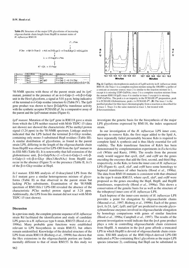
1H-NMR spectra with those of the parent strain and its lgtCmutant, pointed to the presence of an α-D-Galp-(1→4)-β-D-Galpunit in the Hex4 glycoform, a signal at 5.01 p.p.m. being indicativeof the terminal α-D-Galp residue (structure 6) (Table IV). The lgtDgene product was shown to have β-GalpNAc transferase activitywith the synthetic acceptor FCHASE-pk, by a comparative assay ofthe parent and the lgtD mutant strains (Figure 4).
lgtF mutant. Mutation of the lgtF gene in RM118 gave a strainfrom which the LPS neither reacted with MAb TEPC-15 (datanot shown) nor showed the characteristic PCho methyl protonsignal (3.24 ppm) in the 1H-NMR spectrum. Linkage analysisindicated that the LPS lacked the terminal β-D-Glcp residue,containing only mono-3-substituted HepI residues (Table III).A similar distribution of glycoforms, as found in the parentstrain LPS, differing in the length of the oligosaccharide chainfrom HepIII was observed for LPS-OH from the lgtF mutant inits ESI-MS (Table II). It is noteworthy that full extension of theglobotetraose unit, β-D-GalpNAc-(1→3)-α-D-Galp-(1→4)-β-D-Galp-(1→4)-β-D-Glcp (Hex3.HexNAc) from HepIII canoccur in the absence (Figure 5) or the presence (Table II, lic1)of the β-D-Glcp residue at HepI.
lic1 mutant. ESI-MS analysis of O-deacylated LPS from thelic1 mutant gave a similar heterogeneous mixture of glyco-forms (Table II) as that observed in the parent strain butlacking PCho substituents. Examination of the 1H-NMRspectrum of RM118lic1 LPS-OH revealed the absence of thecharacteristic PCho methyl proton signal at 3.24 ppm.Additionally, the LPS from this mutant did not react with MAbTEPC-15 (not shown).
Discussion
In a previous study, the complete genome sequence of H. influenzaestrain Rd facilitated the identification and study of candidateLPS genes in a H. influenzae type b strain, RM153 (Hood et al.,1996a). Many probable gene functions were establishedrelevant to LPS biosynthesis in strain RM153, but othersremain unidentified. Knowledge of the detailed structure of theLPS from strain RM118 (Risberg et al., 1999b) has shown thatthe Hex extensions in the oligosaccharide portion are funda-mentally different to that of strain RM153. In this study we
investigate the genetic basis for the biosynthesis of the majorLPS glycoforms expressed by RM118, the index sequencedstrain.
In our investigation of the H. influenzae LPS inner core,attempts to remove Kdo, the first sugar added to the lipid A,have repeatedly failed presumably because Kdo is required tocomplete lipid A synthesis and is thus likely essential for cellviability. The Kdo transferase function of KdtA has beendemonstrated by complementation experiments in Escherichiacoli (White and Raetz, 1998). The results from the presentstudy would suggest that opsX, rfaF, and orfH are the genesencoding the enzymes that add the first, second, and third Hep,respectively, to the Kdo, to form the inner core of H. influenzaeLPS (Figure 6). opsX, rfaF, and orfH have some homology toheptosyl transferases of other bacteria (Hood et al., 1996a).The data from RM118 mutants is consistent with that obtainedin the type b strain RM153, where opsX, rfaF, and orfH wereproposed as the genes encoding the HepI, HepII, and HepIIItransferases, respectively (Hood et al., 1996a). This shows aconservation of the genetic basis for as well as the structure ofthe triheptosyl inner core of H. influenzae LPS.
Each heptose of the inner-core moiety of H. influenzae LPSprovides a point for elongation by oligosaccharide chains(Masoud et al., 1997; Risberg et al., 1999b). Each of the geneslpsA, lic2A, lgtC, lgtD, and lgtF are predicted to encode glyco-syltransferase enzymes involved in oligosaccharide elongationby homology comparisons with genes of similar function(Hood et al., 1996a; Campbell et al., 1997). The results of thepresent investigation would indicate that the lpsA gene productplays a role in controlling oligosaccharide chain extensionfrom HepIII. A mutation in the lpsA gene affords a truncatedLPS in which HepIII is devoid of oligosaccharide chain exten-sions. ESI-MS analysis of the RM118lpsA-derived LPS-OHindicated a PCho-containing Hex1 glycoform as the major LPSspecies (structure 2), confirming that HepI can be substituted in
Table IV. Structure of the major LPS glycoforms of increasing oligosaccharide chain length from HepIII in mutant stains of H. influenzae RM118
Fig. 4. Capillary electrophoresis analysis of LgtD activity in H. influenzae strain RM118. (A) Trace 1 is a complete reaction mixture using the 100,000 × g pellet of a sonicate as enzyme source; trace 2 is similar to the reaction mixture in 1, except it is missing UDP-GalNAc; trace 3 is a complete reaction mixture from the mutant RM118:lgtD; trace 4 is similar to trace 3 except it is missing UDP-GalNAc. The peak a is an impurity in the FCHASE-PK preparation, peak b is FCHASE-Globotetraose, peak c is FCHASE-PK. (B) The trace 1 is the purified product for thin-layer chromatography from a reaction as described for A trace 1. Trace 2 is the same material as trace 1, but treated with β-hexosaminidase.

H. influenzae LPS oligosaccharide biosynthesis
963
the absence of Hex extension from HepIII. The lic2A, lgtC, andlgtD mutants, which contain a functional lpsA gene, arecapable of adding a β-D-Glcp residue in a 1,2-linkage toinitiate chain extension from HepIII (Table IV). LpsA hashomology to the group of galactosyltransferases typified byLic2A and LgtB of Haemophilus and Neisseria, respectively.In strain RM153, it was also found that LPS from a lpsAmutant lacked any extension from the third Hep (Hood et al.,1996a). However, in strain RM153, lpsA apparently plays aslightly different role being responsible for the addition of Galas the sole extension from the third Hep (Hood et al., 1996a).Thus, LpsA is likely the transferase for the addition of the firstglycose to HepIII in H. influenzae LPS biosynthesis.
The RM118lic2A mutant showed a PCho-containing Hex2glycoform as a major LPS species (structure 4) and RM118lgtC,which contains a functional lic2A gene, elaborates LPScontaining a lactose side chain at HepIII (structure 5). This isconsistent with the involvement of the lic2A gene to add the
β-D-Galp unit in a 1,4 linkage to the terminal β-D-Glcp residueattached to HepIII. Lic2A homologues in type b strains havebeen shown to be involved in expression of the digalactoside-containing Pk epitope (α-D-Galp-[1→4]-β-D-Galp-[1→4]-β-D-Glcp). Homology comparisons with other databank sequencessupport the function of Lic2A as a β-galactosyltransferase;importantly, it has significant homology to the Neisseria LgtBand LgtE proteins, both of which are galactosyltransferases(Wakarchuk et al., 1996). Structural analysis of LPS from anRM118 strain mutated in lgtC confirmed the loss of α-D-Galp,supporting the α-galactosyltransferase function for this gene.Correspondingly the lgtD mutant and the parent strain, whichcontain a functional lgtC gene, are capable of adding a α-D-Galpin a 1,4 linkage to the terminal β-D-Galp of the lactose epitope(structure 6). The function of LgtC was confirmed by demon-strating α-galactosyltransferase activity with the recombinantprotein and a synthetic FCHASE-Lac acceptor. In N. meningitidis,LgtC is also an α-galactosyltransferase (Gotschlich, 1994;Wakarchuk et al., 1998). It follows that the lgtC gene encodesthe specific α-galactosyltransferase for the synthesis of theα-D-Galp-(1→4)-β-D-Galp of the RM118 Hex4 LPS glycoform(Figure 6). The parent strain RM118 that contains a functionallgtD gene is capable (unlike the mutant) of elaborating thecomplete globotetraose unit, which is indicative of its role inadding the terminal β-D-GalpNAc. The H. influenzae lgtDgene is a homologue of two related Neisseria genes, lgtA andlgtD, which add GlcpNAc and GalpNAc, respectively, toN. gonorrhoeae LPS (Gotschlich, 1994). Enzyme assays withextracts of RM118 and the RM118lgtD mutant confirmed theβ-D-GalpNAc transferase activity. The lgtD gene was foundnot to be present in the type b strains RM153 and RM7004(called lgtA in Hood et al., 1996a). Correspondingly, the LPSelaborated by strain RM153 does not contain a GalpNAcmoiety (Masoud et al., 1997).
It is noteworthy that, though the activity of glycosyltrans-ferases adding the distal residues of the globotetraose (lgtD)and globoside (lgtC) oligosaccharide side chains could beassayed with the appropriate synthetic acceptor, similar experi-ments to assay the activity of the transferases involved insynthesis of the lactose moiety (Lic2A and LpsA) wereunsuccessful. It is likely that the latter two enzymes have morestringent specificities that require the acceptor sugar to be
Fig. 5. Negative ion ESI-MS of the triply charged molecular ion region of the LPS-OH from the lgtF mutant of H. influenzae RM118. Peaks arising from the Hex 2 (β-D-Galp-(1→4)-β-D-Glcp), Hex 3 (α-D-Galp-(1→4)-β-D-Galp-(1→4)-β-D-Glcp), and Hex3•HexNAc (β-D-GalpNAc-(1→3)-α-D-Galp-(1→4)-β-D-Galp-(1→4)-β-D-Glcp) are indicated.
Fig. 6. A schematic representation of the structure of LPS from H. influenzae strain RM118 based on the results of the analysis of Risberg et al., (1999b). The proposed site of action in LPS biosynthesis of loci characterized in this study are shown, linked by arrows to the relevant saccharide linkage. The phase variable loci are lic1, lic2A, and lgtC. Represented in the LPS structure: KDO, 2-keto-3-deoxyoctulosonic acid; Hep, L-glycero-D-manno-heptose; Glc, D-glucose; Gal, D-galactose; GalNAc, N-acetylgalactosamine; PEtn, phosphoethanolamine; P, phosphate; PCho, phosphocholine. For the heptose residues, listed top to bottom are heptose I, heptose II, then heptose III.
D.W. Hood et al.
964
linked to the inner-core Hep residues. Characterization of theinitial set of genes and mutant strains available for study ofRM118 LPS biosynthesis gave no obvious candidate respon-sible for addition of the β-D-Glcp unit to HepI. An lgtFhomologue was identified in strain RM118 by searching thestrain Rd genome sequence for matches to genes required forthe addition of Hex sugars to Hep residues in the LPS of otherorganisms. These search sequences included the rfaK and lgtFgenes of Neisseria (Kahler et al., 1996). Analysis of the LPSfrom strain RM118lgtF supported a role for LgtF in chainextension from HepI. The ESI-MS showed molecular ionscorresponding to a mixture of glycoforms having chainextensions, including lactose and globotetraose, from HepIII ofa triheptosyl inner-core unit that lacks PCho→6)-β-D-Glcp atHepI (Figure 3). Thus, the processes of chain extension fromboth HepI and HepIII appear to be largely independent in theLPS of strain RM118.
The heterogeneity observed in H. influenzae LPS structuremay be due in part to intrinsic variation in the biosynthesis ofsuch a complex structure, but the majority of variationobserved is presumed to be due to specific LPS biosyntheticgenes capable of variable expression (phase variation). Thisstudy has allowed us for the first time to confirm the genesinvolved in the synthesis of an important phase-variableepitope of H. influenzae LPS, the digalactoside. In strainRM118, Lic2A adds the proximal β-D-Galp and LgtC theterminal α-D-Galp to the digalactoside (-D-Galp-(1→4)-β-D-Galp) as part of the extension from HepIII, whereas the sameepitope is expressed as the terminal extension from a diglucosideon the second Hep in the type b strain RM153 (Masoud et al.,1997). Both lic2A and lgtC are phase-variable genes (High et al.,1993; Hood et al., 1996b), making the expression of theepitope highly variable within and between organisms. Thedigalactoside epitope is expressed in the LPS of many relatedbacteria, including Neisseria (Virji et al., 1990). The epitope ispotentially immunodominant, and its presence offers thepotential for molecular mimicry of host structures and caninfluence the survival of Haemophilus within experimentalsystems (Hood et al., 1996b; Weiser and Pan, 1998).
In addition to the order and stereochemistry of the sugarresidues, the location, type and frequency of substituents suchas P, PEtn, and PCho can have a profound affect on LPSstructure and biological function. The lic1 locus is essential forthe phase-variable addition of PCho to the H. influenzae LPSmolecule (Weiser et al., 1997; Lysenko et al., 2000). DNAsequence polymorphisms in lic1 direct the different acceptorspecificity observed for PCho incorporation and influence theresistance of H. influenzae to innate humoral immunity(Weiser and Pan, 1998; Lysenko et al., 2000). The geneencoding a Kdo kinase, kdkA, responsible for phosphorylationof Kdo, has been identified (White et al., 1999). This gene haspreviously been investigated by us as orfZ and when mutatedwas shown to alter bacterial survival in an infant rat model ofinfection (Hood et al., 1996a). The only remaining substituentsin the core oligosaccharide, whose genetic control remainsunknown, therefore, are the PEtn residues that are attached tothe 6-position of the HepII residue stoichiometrically andsometimes to the phosphate group on Kdo.
In summary, the genetic blueprint for synthesis of the majorglobotetraose-containing oligosaccharide of RM118 LPS hasbeen elucidated. The type b strain RM153 and strain RM118
have a gene pool for LPS biosynthesis that is generally thesame for the two strains but with some key differences relatedto their respective LPS structure and biology.
Materials and methods
Bacterial strains and culture conditions
The H. influenzae Rd strain was originally obtained fromAlexander and Leidy by Herriot. It was given to H. O. Smith,who named the strain KW-20 and used it in the genomesequencing project (Fleischmann et al., 1995). This same strainobtained from the Smith laboratory has been used by us(RM118). The genotypes of mutants derived from this strainare listed in Table I. H. influenzae strains were grown at 37°Cin brain heart infusion broth supplemented with hemin (10 µg/ml),nicotinamide adenine dinucleotide (2 µg/ml), and kanamycin(10 µg/ml) when appropriate.
E. coli strain DH5α was used to propagate cloned PCRproducts and gene constructs and was grown at 37°C in Luria-Bertani broth supplemented with ampicillin (100 µg/ml) orkanamycin (50 µg/ml) as required (Sambrook et al., 1989).
Identification of LPS-related genes from the H. influenzae genome sequence
Putative LPS biosynthetic genes had been previously identifiedby an in silico search of the H. influenzae genome sequencewith heterologous sequences of LPS biosynthetic genes from awide range of organisms obtained from publicly availabledatabases (Hood et al., 1996a). The RM118lgtF locus(HI0653) was identified by searching the Institute for GenomicResearch H. influenzae strain Rd sequence database(www.tigr.org/tdb/CMR/ghi/htmls/SplashPage.html) for matcheswith the LgtF protein sequence from N. meningitidis(GenBank accession no. U58765).
Recombinant DNA methodology, cloning, and mutation
Restriction endonucleases and DNA-modifying enzymes wereobtained from Boehringer Mannheim and used according tothe manufacturer’s instructions. Plasmid DNA preparation,Southern blotting, and hybridization analysis were performedas described by Sambrook et al. (1989). Chromosomal DNAwas prepared from Haemophilus by the method describedelsewhere (High et al., 1993).
Apart from lgtF, putative H. influenzae LPS biosyntheticgenes were cloned and mutated as previously reported (Hoodet al., 1996a). For the lgtF locus, oligonucleotide primers, lgtFa(5′-TGGTGGTGGGCAAGACGC-3′) and lgtFb (5′- AGCCTG-AATTCGACAGCC-3′), amplified a 1461-bp DNA fragmentincluding HI0653 by PCR. PCR conditions were for 1-minperiods of denaturation (94°C), annealing (50°C), and poly-merization (72°C) for 30 cycles. One microliter of PCRproduct was ligated with 50 ng of plasmid pT7Blue (Novagen)and transformed into E. coli strain DH5α. Recombinantplasmids were confirmed by restriction endonuclease digestionand sequencing from plasmid-specific primers (Hood et al.,1996a). The lgtF gene was inactivated by inserting a kana-mycin resistance cassette (released by digestion with EcoR1from pUC4Kan, Pharmacia) into a MunI restriction site 257 bpinside the 5′ end of HI0653 to give plasmid pDQ1.

H. influenzae LPS oligosaccharide biosynthesis
965
Construction of mutant strains
Two to three micrograms of linearized plasmid, containingmutated LPS biosynthetic genes, was used to transformH. influenzae strain RM118 by the MIV procedure (Herriottet al., 1970) and transformants were selected on kanamycin.To construct strain RM118lic1, RM118 was transformed with5 µg of sheared chromosomal DNA isolated from the corre-sponding RM153 mutant. Strain RM118lic2A was constructedby transformation of RM118 with 1 µg of a PCR productincluding inactivated lic2A and the adjacent gene ksgAamplified from strain RM153lic2A. PCR used the primers L2A(5′-CTCCATATTACATAAT-3′) and L2D (5′-AAACACT-TAGGCCATACG-3′) under conditions as described above.All transformants were recultured on appropriate brain heartinfusion/antibiotic plates, then were confirmed as mutants byPCR amplification and/or Southern blotting/hybridization ofdigested chromosomal DNA.
Analysis of LPS by immunoblotting and electrophoresis
LPS isolated from wild type and mutants of H. influenzaestrain RM118 was analyzed using LPS-specific monoclonalantibodies and by T–SDS–PAGE) as described previously(Hood et al., 1996a).
Structural fingerprinting of LPS
Cells from 10-L batch cultures (10 lots of 1 L) were harvestedafter overnight growth, LPS was extracted by the hot phenol-water method (Westphal and Jann, 1965), followed by ethanolprecipitation as described by Thibault and Richards (2000).LPS was purified and O-deacylated as previously described(Holst et al., 1993). Sugars were identified by gas-liquidchromatography mass spectrometry (GLC-MS) as their alditolacetates as previously described (Masoud et al., 1997). Linkageanalysis was accomplished following acetylation of the oligo-saccharides with acetic anhydride (0.5 ml) and 4-dimethyl-aminopyridine (0.5 mg) at room temperature for 24 h.Peracetylated material was then treated with methyl iodide indimethylsulfoxide in the presence of lithium methylsulfinyl-methanide to afford the methylated oligosaccharides, whichwere recovered using a SepPak C18 cartridge and subjected tosugar analysis (Blakeney and Stone, 1985). The relativeproportions of the various alditol acetates and partially methyl-ated alditol acetates obtained in sugar and methylationanalyses were measured from the detector response of theGLC-MS and are uncorrected. GLC-MS was carried out with aDelsi Di200 chromatograph equipped with a NERMAG R10–10Hquadrupole mass spectrometer or with a Varian Iontrap systemusing a DB-5 fused silica capillary column (25 m × 0.25 mm ×0.25 µm) and a temperature gradient of 160°C (1 min) →250°C at 3°C/min.
ESI-MS was performed on a VG Quattro Mass Spectrometer(Micromass, Manchester, UK) in the negative-ion mode.Samples were dissolved in water and then mixed in a 1:1 ratiowith 50% aqueous acetonitrile containing 1% acetic acid.Sample solutions were injected via a syringe pump into arunning solvent of H2O:CH3CN (1:1) at a flow rate of 5 µl/min.1D 1H-NMR spectra were recorded at 500 MHz for solutionsin deuterium oxide at 22°C, after several lyophyllizations withD2O, on a Bruker AMX 500 spectrometer. To enhance spectralresolution, perdeutero-EDTA (2 mM) and perdeutero-SDS
(10 mg/ml) were added to the D2O solutions (Risberg et al.,1997). Chemical shifts are referenced to the methyl protonresonance (δ; 2.225 ppm) of internal acetone.
Analysis of enzymatic activity from LgtC and LgtD
The enzyme encoded by lgtD was assayed with the syntheticacceptor FCHASE-Pk using capillary electrophoresis fordetection of the product (Wakarchuk et al., 1996). FCHASE-Pk
was synthesized from FCHASE-Lac using the N. meningitidisLgtC enzyme as previously described (Gotschlich, 1994;Wakarchuk et al., 1998). The reaction conditions were 0.5 mMacceptor, 1 mM UDP-GalNAc, 50 mM HEPES–NaOH(pH 7.0), 10 mM MgC12, 10 mM MnC12. Extracts fromRM118 and RM118:lgtD were made by sonicating the cells,and then collecting the membrane fraction by centrifugation at100,000 × g for 30 min. The product was isolated by thin-layerchromatography as previously described (Wakarchuk et al.,1996). Since the proportion of acceptor converted to productwas small, some of the starting material was also isolated. Therecovered mixture was divided into two parts and then treatedwith β-hexosaminidase under conditions recommended by theenzyme supplier (NEB).
Activity for LgtC was below the limits of detection inextracts of RM118, so the lgtC gene was cloned into anexpression vector and activity was assayed in E. coli. The genewas amplified by PCR (as described above) using primerslgtCa (5′GGGGGGCATATGGGACGGACTGTCAGTCAGA-CAATG) and lgtCb (5′GGGGGGGTCGACTCATTAAT-TATCTTTTATTCTCTTTCTTAATC). The gene was theninserted into plasmid pCWori plus at the NdeI and SalI sitessimilar to what was described for lgtC from N. meningitidis(Gotschlich, 1994; Wakarchuk et al., 1998). Crude sonicatedextracts of the recombinant clone were assayed with 1 mMFCHASE-Lac, 1 mM UDP-Gal, 10 mM MnC12, 5 mM dithio-threitol, 50 mM HEPES, pH 7.5. The enzyme was shown to beunstable in E. coli and was assayed within a few hours of whenthe extracts were made. The product of the enzyme reactionwas analyzed by specific glycosidase digestion, massspectrometry, and cochromatography with authentic FCHASE-Pk.
Acknowledgments
We thank Don Krajcarski for assistance in ESI-MS analysis,Jianjun Li for CE-ESI-MS analysis, and Johan Sagemark andAnna-Karin Karlsson for methylation analysis. D.H., S.W.,and M.D. were supported by a program grant from the MedicalResearch Council, UK.
Abbreviations
ESI-MS, electrospray ionization mass spectrometry; GLC-MS,gas-liquid chromatography mass spectrometry; Kdo, 2-keto-3-deoxyoctulosonic acid; LPS, lipopolysaccharide; LPS-OH,O-deacylated lipopolysaccharide; MAb, monoclonal antibody;MS-MS, tandem mass spectrometry; NMR, nuclear magneticresonance; P, free phosphate group; PCR, polymerase chainreaction PEtn, phosphoethanolamine; PPEtn, pyrophospho-ethanolamine; PCho, phosphocholine; T–SDS–PAGE, tricine–sodium dodecyl sulfate–polyacrylamide gel electrophoresis.
D.W. Hood et al.
966
References
Blakeney, A.B. and Stone, B.A. (1985) Methylation of carbohydrates withlithium methylsulphinyl carbanion. Carbohydr. Res., 140, 319–324.
Brook, J.S. and Valvano, M.A. (1996) Molecular cloning of the Haemophilusinfluenzae gmhA (lpcA) gene encoding a phosphoheptose isomerase requiredfor lipooligosaccharide biosynthesis. J. Bacteriol., 178, 3339–3341.
Campbell, J.A., Davies, G.J., Bulone, V., and Henrissat, B. (1997) A classificationof nucleotide-diphospho-sugar glycosyltransferases based on amino acidsequence similarities. Biochem. J., 326, 929–942.
Cope, L.D., Yogev, R., Mertsola, J., Argyle, J.C., McCracken, G.H., andHansen, E.J. (1990) Effect of mutation in lipopolysaccharide biosyntheticgenes on virulence of Haemophilus influenzae type b. Infect. Immun., 58,2343–2351.
Fleischmann, R.D., Adams, M.D., White, O., Clayton, R.A., Kirkness, E.F.,Kerlavage, A.R., Butt, C.J., Tomb, J-F., Dougherty, B.A., Merrick, J.M.,and others (1995) The genome of Haemophilus influenzae Rd. Science,269, 496–512.
Gibson, B.W., Melaugh, W., Phillips, N.J., Apicella, M.A., Campagnari, A.A.,and Griffiss, J.M. (1993) Investigation of the structural heterogeneity oflipooligosaccharides from pathogenic Haemophilus and Neisseria speciesand of R-type lipopolysaccharides from Salmonella typhimurium byelectrospray mass spectrometry. J. Bacteriol., 175, 2702–2712.
Gotschlich, E.C. (1994) Genetic locus for the biosynthesis of the variableportion of Neisseria gonorrhoeae lipooligosaccharide. J. Exp. Med., 180,2181–2190.
Helander, I.M., Lindner, B., Brade, H., Altmann, K., Lindberg, K.K.,Rietschel, E.T., and Zahringer, U. (1988) Chemical structure of the lipo-polysaccharide of Haemophilus influenzae strain I-69 Rd–/b+: Description of anovel deep-rough chemotype. Eur. J. Biochem., 177, 483–492.
Herriott, R.M., Meyer, E.M., and Vogt, M.J. (1970) Defined non-growthmedia for stage II development of competence in Haemophilus influenzae.J. Bacteriol., 101, 517–524.
High, N.J., Deadman, M.E., and Moxon, E.R. (1993) The role of the repetitiveDNA motif (5′-CAAT-3′) in the variable expression of the Haemophilusinfluenzae lipopolysaccharide epitope Galα(1-4)βGal. Mol. Microbiol., 9,1275–1282.
Holst, O., Broer, W., Thomas-Oates, J.E., Mamat, U., and Brade, H. (1993)Structural analysis of two oligosaccharide bisphosphates isolated from thelipopolysaccharide of a recombinant strain of Escherichia coli F515 (Rechemotype) expressing the genus-specific epitope of Chlamydia lipo-polysaccharide. Eur. J. Biochem., 214, 703–710.
Hood, D.W., Deadman, M.E., Allen, T., Masoud, H., Martin, A., Brisson, J.R.,Fleischmann, R., Venter, J.C., Richards, J.C., and Moxon, E.R. (1996a)Use of the complete genome sequence information of Haemophilusinfluenzae strain Rd to investigate lipopolysaccharide biosynthesis.Mol. Microbiol., 22, 951–965.
Hood, D.W., Deadman, M.E., Jennings, M.P., Bisceric, M., Fleischmann, R.D.,Venter, J.C., and Moxon, E.R. (1996b) DNA repeats identify novelvirulence genes in Haemophilus influenzae. Proc. Natl. Acad. Sci. USA,93, 11121–11125.
Jarosik, G.P. and Hansen, E.J. (1994) Identification of a new locus involved inthe expression of Haemophilus influenzae type b lipooligosaccharide.Infect. Immun., 62, 4861–4867.
Kahler, C.M., Carlson, R.W., Rahman, M.M., Martin, L.E., and Stephens, D.S.(1996) Two glycosyltransferase genes, lgtF and rfaK, constitute thelipooligosaccharide ice (Inner Core Extension) biosynthesis operon ofNeisseria meningitidis. J. Bacteriol., 178, 6677–6684.
Kimura, A. and Hansen, E.J. (1986) Antigenic and phenotypic variants ofHaemophilus influenzae type b lipopolysaccharide and their relationship invirulence. Infect. Immun., 51, 60–79.
Lysenko, E., Richards, J.C., Cox, A.D., Stewart, A., Martin, A., Kapoor, M.,and Weiser, J.N. (2000) The position of phosphorylcholine on the lipopoly-saccharide of Haemophilus influenzae affects binding and sensitivity toC-reactive protein-mediated killing. Mol. Microbiol., 35, 234–245.
Masoud, H., Moxon, E.R., Martin, A., Krajcarski, D., and Richards, J.C.(1997) Structure of the variable and conserved lipopolysaccharideoligosaccharide epitopes expressd by Haemophilus influenzae serotype bstrain Eagan. Biochem., 36, 2091–2103.
Moxon, E.R. and Maskell, D.J. (1992) “Haemophilus influenzae lipopoly-saccharide: the biochemistry and biology of a virulence factor.” InHormaeche, C.E., Penn, C.W., Smythe, C.J. (eds.), Molecular biology ofbacterial infection, current status and future perspectives. Society for
General Microbiology Symposium 49. Cambridge University Press,Cambridge, UK.
Phillips, N.J., Apicella, M.A., Griffiss, J.M., and Gibson, B.W. (1992)Structural characterization of the cell surface lipooligosaccharides from anon-typable strain of Haemophilus influenzae. Biochem., 31, 4515–4526.
Phillips, N.J., Apicella, M.A., Griffiss, J.M., and Gibson, B.W. (1993)Structural studies of the lipooligosaccharides from Haemophilus influenzaetype b strain A2. Biochem., 32, 2003–2012.
Phillips, N.J., McLaughlin, R., Miller, T.J., Apicella, M.A., and Gibson, B.W.(1996) Characterization of two transposon mutants of Haemophilusinfluenzae type b with altered lipooligosaccharide biosynthesis. Biochem.,35, 5937–5947.
Preston, A., Mandrell, R.E., Gibson, B.W., and Apicella, M.A. (1996a) Thelipopolysaccharides of pathogenic Gram-negative bacteria. Crit. Rev.Microbiol., 22, 139–180.
Preston, A., Maskell, D., Johnson, A., and Moxon, E.R. (1996b) Alteredlipopolysaccharide characteristic of the I69 phenotype in Haemophilusinfluenzae results from mutation in a novel gene, isn. J. Bacteriol., 178,396–402.
Rahman, M.M., Gu, X-X., Tsai, C-M., Kolli, V.S.K., and Carlson, R.W.(1999) The structural heterogeneity of the lipooligosaccharide (LOS)expressed by pathogenic non-typeable Haemophilus influenzae strainNTHi 9274. Glycobiology, 9, 1371–1380.
Risberg, A., Schweda, E.K.H., and Jansson, P.-E. (1997) Structural studies ofthe cell-envelope oligosaccharide from the lipopolysaccharide of Haemophilusinfluenzae strain RM.118-28. Eur. J. Biochem., 243, 701–707.
Risberg, A., Alvelius, G., and Schweda, E.K.H. (1999a) Structural analysis ofthe lipopolysaccharide oligosaccharide epitopes expressed by Haemophilusinfluenzae strain RM.118-26. Eur. J. Biochem., 265, 1067–1074.
Risberg, A., Masoud, H., Martin, A., Richards, J.C., Moxon, E.R., andSchweda, E.K.H. (1999b) Structural analysis of the lipopolysaccharideepitopes expressed by a capsular deficient strain of Haemophilus influenzaeRd. Eur. J. Biochem., 261, 171–180.
Sambrook, J., Fritsch, E. F., and Maniatis, T. (1989) Molecular cloning; alaboratory manual, 2d ed. Cold Spring Harbor Laboratory, Cold SpringHarbor, NY.
Schweda, E.K.H., Hegedus, O.E., Borrelli, S., Lindberg, A.A., Weiser, J.W.,Maskell, D.J., and Moxon, E.R. (1993) Structural studies of the saccharideportion of cell envelope lipopolysaccharide from Haemophilus influenzaestrain AH1-3 (lic3+). Carbohydr. Res., 246, 319–330.
Schweda, E.K.H., Jansson, P.-E., Moxon, E.R., and Lindberg, A.A. (1995)Structural studies of the saccharide part of the cell envelope lipooligo-saccharide from Haemophilus influenzae strain galEgalK. Carbohydr.Res., 272, 213–224.
Thibault, P. and Richards, J.C. (2000) Application of combined capillaryelectrophoresis electrospray mass spectrometry in the characterisation ofshort chain lipopolysaccharides: Haemophilus influenzae. In Holst, O.(ed.), Methods of molecular biology, vol. 45, Bacterial toxins: methods andprotocols. Humana Press, Totowa, NJ.
Virji, M., Weiser, J.N., Lindberg, A.A., and Moxon, E.R. (1990) Antigenicsimilarities in lipopolysaccharides of Haemophilus and Neisseria andexpression of a digalactoside structure also present on human cells.Microb. Pathogen., 9, 441–450.
Wakarchuk, W., Martin, A., Jennings, M.P., Moxon, E.R., and Richards, J.C.(1996) Functional relationships of the genetic locus encoding the glycosyltransferase enzymes involved in the expression of the lacto-N-neotetraoseterminal lipopolysaccharide structure in Neisseria meningitidis. J. Biol.Chem., 271, 19166–1917.
Wakarchuk, W.W., Cunningham, A.M., Watson, D.C., and Young, N.M. (1998)Role of paired basic residues in the expression of active recombinant galacto-syltransferases from the bacterial pathogen Neisseria meningitidis. Prot. Eng.,11, 295–302.
Weiser, J.N. and Pan, N. (1998) Adaptation of Haemophilus influenzae toacquired and innate humoral immunity based on phase variation of lipo-polysaccharide. Mol. Microbiol., 30, 767–775.
Weiser, J.N., Love, J.M., and Moxon, E.R. (1989) The molecular mechanismof phase variation of Haemophilus influenzae lipopolysaccharide. Cell, 59,657–665.
Weiser, J.N., Maskell, D.J., Butler, P.D., Lindberg, A.A., and Moxon, E.R.(1990a) Characterisation of repetitive sequences controlling phasevariation of Haemophilus influenzae lipopolysaccharide. J. Bacteriol.,172, 3304–3309.
H. influenzae LPS oligosaccharide biosynthesis
967
Weiser, J.N., Williams, A., and Moxon, E.R. (1990b) Phase-variable lipo-polysaccharide structures enhance the invasive capacity of Haemophilusinfluenzae. Infect. Immun., 58, 3455–3457.
Weiser, J.N., Shchepetov, M., and Chong, S.T.H. (1997) Decoration oflipopolysaccharide with phosphorylcholine: a phase variable characteristicof Haemophilus influenzae. Infect. Immun., 65, 943–950.
Westphal, O. and Jann, K. (1965) Bacterial lipopolysaccharides. Extractionwith phenol-water and further applications of the procedure. Meth. Carbohydr.Chem., 5, 83–91.
White, K.A. and Raetz, C.R.H. (1998) Complementation of a genomic disruptionof the E.coli bifunctional KDO transferase by the H. influenzae mono-functional KDO transferase. FASEB J., 12, L44.
White, K.A., Lin, S., Cotter, R.J., and Raetz, C.R.H. (1999) A Haemophilus influen-zae gene that encodes a membrane bound 3-deoxy-D-manno-octulosonic acid(Kdo) kinase: Possible involvement of KDO phosphorylation in bacterialvirulence. J. Biol. Chem., 274, 31391–31400.
Zamze, S.E. and Moxon, E.R. (1987) Composition of the lipopolysaccharidefrom different capsular serotype strains of Haemophilus influenzae. J. Gen.Microbiol., 133, 1443–1451.

D.W. Hood et al.
968