Remodeling of second-order neurons in the retina of rd/rd mutant mice
11
Remodeling of second-order neurons in the retina of rd/rd mutant mice Enrica Strettoi a, * , Vincenzo Pignatelli a , Chiara Rossi a , Vittorio Porciatti a,b , Benedetto Falsini a,c a Laboratorio di Neurofisiologia, Istituto di Neuroscienze del CNR, Area della Ricerca, Via G. Moruzzi 1, 56100 Pisa, Italy b Bascom Palmer Eye Institute, 900 N.W. 17th Street, 33136 Miami, USA c Clinica Oculistica, Universit a Cattolica del Sacro Cuore, Largo S. Vito 1, 00168 Roma, Italy Received 31 July 2002; received in revised form 5 November 2002 Abstract This is a brief review of data obtained by analyzing the morphology and the physiology of the retinas in rd/rd and normal, wt mice, aged 10–90 days. Second-order neurons of the rd/rd show abnormalities that start with the anomalous development of rod bipolar cells around P10 and culminate with the atrophy of dendrites in cone bipolar cells, mostly evident at P90. Horizontal cells remodel considerably. Cone-mediated ERGs, (recorded between 13 and 16 days of age) have reduced a-wave and b-wave amplitudes and longer b-wave latency and duration. B-wave abnormalities indicate specific postreceptoral dysfunction. Morphological and ERG changes in rd/rd retinas are consistent with substantial inner retinal remodeling associated to photoreceptor degeneration. Ó 2003 Elsevier Science Ltd. All rights reserved. Abbreviations: rd/rd mouse: retinal degeneration mouse, also known as rd1; wt: wild-type mouse; P: postnatal day; opl, onl, ipl, gcl: outer plexiform, outer nuclear, inner plexiform, ganglion cell layer of the retina; PKC: protein kinase C; ERG: electroretinogram Keywords: Bipolar cell; Dendritic atrophy; Horizontal cells; ERG; Retinitis pigmentosa 1. Introduction The Central Nervous System of mammals undergoes remodeling during physiological processes, such as de- velopment, plasticity and learning, as well as in response to traumatic injuries or diseases. In the case of patho- logical or experimental conditions causing the death of sensory receptors, secondary effects upon postsynaptic cells usually occur. Structural effects of de-afferentation range from the change in the distribution of various proteins, to the rewiring, atrophy and transneuronal de- generation of target neurons (see, for instance, Baeke- landt et al., 1994; Gilbert, 1998; Milam, Li, & Fariss, 1998; Rubel & Fritzsch, 2002; Sernagor, Eglen, & Wong, 2001). Retinal neurons of individuals affected by inherited photoreceptor degeneration are confronted with the death of a very large population of afferent nerve cells, namely rods and cones. The retina of a typical labora- tory mouse contains over 4,000,000 photoreceptors that converge upon approximately 600,000 bipolar cells (roughly 200,000 rod bipolar and 400,000 cone bipolar cells) and 17,000 horizontal cells (Jeon, Strettoi, & Masland, 1998; Strettoi & Pignatelli, 2000; Strettoi & Volpini, 2002). An obvious question to ask is how these cells react, when they are deprived of their major input neurons, because of one of the many mutations causing retinal degeneration in the mouse (reviewed in Chang et al., 2002). The identification of the effects of photoreceptor ab- normal development and/or degeneration upon other retinal cells can contribute to a deeper knowledge of the normal biology of the retina. In addition, understanding the impact of photoreceptor loss upon inner retinal structure and function, is a prerequisite for most of the approaches aimed at restoring vision in retinal degen- eration: cellular and retinal transplant (Lund et al., 2001), implant of prostheses (i.e. Humayun, 2001; Pea- chey & Chow, 1999; Zrenner, 2002), gene therapy (Bennett, 2000). * Corresponding author. Tel.: +39-50-315-3213; fax: +39-50-315- 3220. E-mail address: [email protected] (E. Strettoi). 0042-6989/03/$ - see front matter Ó 2003 Elsevier Science Ltd. All rights reserved. doi:10.1016/S0042-6989(02)00594-1 Vision Research 43 (2003) 867–877 www.elsevier.com/locate/visres
-
Upload
independent -
Category
Documents
-
view
0 -
download
0
Transcript of Remodeling of second-order neurons in the retina of rd/rd mutant mice

Remodeling of second-order neurons in the retina of rd/rdmutant mice
Enrica Strettoi a,*, Vincenzo Pignatelli a, Chiara Rossi a, Vittorio Porciatti a,b,Benedetto Falsini a,c
a Laboratorio di Neurofisiologia, Istituto di Neuroscienze del CNR, Area della Ricerca, Via G. Moruzzi 1, 56100 Pisa, Italyb Bascom Palmer Eye Institute, 900 N.W. 17th Street, 33136 Miami, USA
c Clinica Oculistica, Universit�aa Cattolica del Sacro Cuore, Largo S. Vito 1, 00168 Roma, Italy
Received 31 July 2002; received in revised form 5 November 2002
Abstract
This is a brief review of data obtained by analyzing the morphology and the physiology of the retinas in rd/rd and normal, wt
mice, aged 10–90 days. Second-order neurons of the rd/rd show abnormalities that start with the anomalous development of rod
bipolar cells around P10 and culminate with the atrophy of dendrites in cone bipolar cells, mostly evident at P90. Horizontal cells
remodel considerably. Cone-mediated ERGs, (recorded between 13 and 16 days of age) have reduced a-wave and b-wave amplitudes
and longer b-wave latency and duration. B-wave abnormalities indicate specific postreceptoral dysfunction. Morphological and
ERG changes in rd/rd retinas are consistent with substantial inner retinal remodeling associated to photoreceptor degeneration.
� 2003 Elsevier Science Ltd. All rights reserved.
Abbreviations: rd/rd mouse: retinal degeneration mouse, also known as rd1; wt: wild-type mouse; P: postnatal day; opl, onl, ipl, gcl: outer plexiform,
outer nuclear, inner plexiform, ganglion cell layer of the retina; PKC: protein kinase C; ERG: electroretinogram
Keywords: Bipolar cell; Dendritic atrophy; Horizontal cells; ERG; Retinitis pigmentosa
1. Introduction
The Central Nervous System of mammals undergoesremodeling during physiological processes, such as de-
velopment, plasticity and learning, as well as in response
to traumatic injuries or diseases. In the case of patho-
logical or experimental conditions causing the death of
sensory receptors, secondary effects upon postsynaptic
cells usually occur. Structural effects of de-afferentation
range from the change in the distribution of various
proteins, to the rewiring, atrophy and transneuronal de-generation of target neurons (see, for instance, Baeke-
landt et al., 1994; Gilbert, 1998; Milam, Li, & Fariss, 1998;
Rubel & Fritzsch, 2002; Sernagor, Eglen, &Wong, 2001).
Retinal neurons of individuals affected by inherited
photoreceptor degeneration are confronted with the
death of a very large population of afferent nerve cells,
namely rods and cones. The retina of a typical labora-
tory mouse contains over 4,000,000 photoreceptors that
converge upon approximately 600,000 bipolar cells(roughly 200,000 rod bipolar and 400,000 cone bipolar
cells) and 17,000 horizontal cells (Jeon, Strettoi, &
Masland, 1998; Strettoi & Pignatelli, 2000; Strettoi &
Volpini, 2002). An obvious question to ask is how these
cells react, when they are deprived of their major input
neurons, because of one of the many mutations causing
retinal degeneration in the mouse (reviewed in Chang
et al., 2002).The identification of the effects of photoreceptor ab-
normal development and/or degeneration upon other
retinal cells can contribute to a deeper knowledge of the
normal biology of the retina. In addition, understanding
the impact of photoreceptor loss upon inner retinal
structure and function, is a prerequisite for most of the
approaches aimed at restoring vision in retinal degen-
eration: cellular and retinal transplant (Lund et al.,2001), implant of prostheses (i.e. Humayun, 2001; Pea-
chey & Chow, 1999; Zrenner, 2002), gene therapy
(Bennett, 2000).
*Corresponding author. Tel.: +39-50-315-3213; fax: +39-50-315-
3220.
E-mail address: [email protected] (E. Strettoi).
0042-6989/03/$ - see front matter � 2003 Elsevier Science Ltd. All rights reserved.
doi:10.1016/S0042-6989(02)00594-1
Vision Research 43 (2003) 867–877
www.elsevier.com/locate/visres

Some of the restorative approaches cited above are
already in the phase of clinical trials; it is reasonable to
assume that the efficacy of others will soon reach the
levels necessary for experimentation on human patients
(Acland et al., 2001). The chances of such approaches to
lead to functional recovery of the retinal function could
be limited in those pathological conditions in which the
inner retinal layers reorganized severely because ofphotoreceptor death.
The number of published papers addressing the issue
of inner retina preservation in retinal degeneration is not
very high when compared to the publications dedicated
to study the genetics of diseases and the morpho-
logy and biochemistry of degenerating photoreceptors
(around 400 over a total of 13.000 citations in a Medline
search). In the past, this has lead to the quite generalconclusion that major modifications occur only at late
stages of photoreceptor degeneration, when thinning of
inner retinal layers becomes very evident in various
pathologies (see Milam et al., 1998, for changes associ-
ated to retinitis pigmentosa).
Particular attention has been paid to survival and
death of photoreceptors (see, for instance, Adler, Cur-
cio, Hicks, Price, & Wong, 1999) and, to some extent, ofother retinal cells. Survival is usually evaluated by
counting cells stained with assorted methods, including
basic dyes or DNA-binding molecules. These tech-
niques, however, are rarely suitable to reveal cell-type
specific abnormalities.
An increasing number of reports, based on more se-
lective analysis of retinal cells and networks, has recently
brought to light remarkable changes in the morphology(i.e. Fariss, Li, & Milam, 2000), synaptic arrangement
(Peng, Hao, Petters, & Wong, 2000) and histochemical
features (i.e. de Raad, Szczesny, Munz, & Rem�ee, 1996;Lund et al., 2001) of cells of the inner layers, in both
human and animal retinas with photoreceptor degener-
ation. Selective alterations of the ERG have been de-
scribed as well (Falsini et al., 1994; Hood & Birch,
1996). The nature and entity of inner retinal alterationsvary according to the kinetics of photoreceptor degen-
eration (i.e., fast or slow), the molecular defect, the
onset-time, the age of the individual etc. Hence, it seems
important to identify, for each specific type of disease
leading to photoreceptor degeneration, a temporal
window, during which the inner retina might still be
receptive to successful therapeutic intervention.
We addressed the issue of inner retinal remodeling bystarting an analytical study, based on morphological
techniques and ERG analysis, of the retina of the rd/rd
mutant mouse, perhaps the best-known animal model of
retinitis pigmentosa (RP). In rd/rd mice, a mutation of
the beta subunit of the rod-specific phosphodiesterase
leads to the rapid death of rod photoreceptors starting
from the second week of life (Bowes et al., 1990; Farber
& Lolley, 1974). Thus, the first phase of rod death
overlaps partially with retinal synaptogenesis (Blanks,
Adinolfi, & Lolley, 1974). At one month of age, rods are
virtually lost. Secondary death of cones follows the onset
of rod death and proceeds slowly over a period of several
months (Carter-Dawson, LaVail, & Sidman, 1978).
The present paper represents a brief review of the
work carried on by our laboratory on the retina of rd/rd
mice in the last three years, with the addition of therecently developed visualization of retinal cells, labeled
by gene-gun delivery of fluorescent dyes. Selective
staining and electrophysiological recordings show major
changes in the rd/rd retina, that go along with the loss of
photoreceptors.
Our analysis focuses on second order neurons, in
which changes are very evident, and is obviously in-
complete. Still, it can be extended to other cellular typesin future and provide a framework for the study of other
animal models of retinopathies.
2. Methods
Experimental procedures were done in agreement
with the ARVO Statement for the use of animals in
ophthalmology and vision research and the rules for
animal experimentation of the Italian Ministry of
Health that follow the European Community Council
Directive of 1986.Animals were C3Hpdebrd1 mice, homozygous for the
rd mutation (rd/rd) and C57Bl6J, wild type (wt) mice.
All animals were born and maintained under controlled
ambient illumination on a 12 h light/dark cycle with the
illumination level below 60 photopic lux.
2.1. Morphology
The numbers of animals used for morphological
analysis, as well as the methods used for immunocyto-
chemistry (ICCH), cell counting and electron micro-
scopy, have been given in detail in Strettoi and Pignatelli
(2000) and Strettoi, Porciatti, Falsini, Pignatelli, and
Rossi (2002). The latter reference provides the types and
the sources of the antibodies used for specific labeling of
retinal cells. ICCH was performed on animals rangingfrom 10 days of age (P10) to P90, both on retinal whole
mounts and on retinal sections. Secondary antibodies
were conjugated with Oregon Green 488, Alexa 568
(from Molecular Probes, Netherlands) or with Cy3
(Sigma). Fluorescent retinal preparations were exam-
ined with a Leica TCS-NT confocal microscope equip-
ped with a krypton–argon laser.
2.2. Gene-gun labeling
Six additional animals (three rd/rd and three wt, 3–5
months old) were used for gene-gun labeling of cells
868 E. Strettoi et al. / Vision Research 43 (2003) 867–877

with lipophilic dyes (DiI and DiO), according to the
protocol described in Gan, Grutzendler, Wong, Wong,
and Lichtman, 2000. A Bio-Rad helium gene-gun was
loaded with dye-bullets (kindly donated by Dr. R.
Wong) and set up at a pressure of 60–80 psI. Retinas
were dissected from quickly enucleated eyes of wt and rd
animals, 3–5-months old, and immersed in ice-cold,
oxygenated saline solution for 5 min. Each retina wasthen moved to the stage of an automatically advanced
tissue chopper (McIlwain; 2Biological, Italy) and cut in
radial slices, 150 lm thick. Retinal slices were collected
in saline, partially dried and exposed to one or two shots
of dyes from the gun. Upon successful labeling of cells,
retinal specimens were fixed for 30 min in 2% para-
formaldheyde, rinsed in buffer and counterstained with
DAPI for visualization of nuclei. During the choppingand the subsequent mounting, retinal slices assumed a
semirandom orientation, so that labeled cells became
visible in radial as well as in tangential view. Images
were collected both with a Zeiss Axiocam digital color
camera, attached to a Zeiss Axioscope fluorescence mi-
croscope, and with the confocal microscope.
2.3. Electrophysiology
ERGs were conventionally recorded in response to
light flashes of different intensity (0.2–20 cd/m2 s�1) both
under dark- and light-adaptation (12 cd/m2). The in-terval between repeated flashes was set to allow com-
plete recovery of the b-wave between flashes, except for
the experiments where the effect of repetition rate (0.1
and 20 Hz) was specifically evaluated. A- and b-waves
were quantified in their amplitude (baseline to negative
a-wave peak, a- to positive b-wave peak) and time-
to-peak (from stimulus onset to negative a-wave through,
from stimulus onset to positive b-wave peak). For b-
wave analysis, oscillatory potentials were removed by
digital filtering (Lyubarsky, Falsini, Pennesi, Valentini,& Pugh, 1999). Light-intensity dependence of the a- and
b-wave amplitudes was determined (Robson & Frish-
man, 1995, 1996; Rohrer, Korenbrot, LaVail, Reic-
hardt, & Xu, 1999). The numbers of animals used are
given in Strettoi et al. (2002).
3. Results
3.1. Morphological changes observed in the rd/rd retina
In adult rd/rd mice, nuclear staining of vertical retinal
sections (Fig. 1) fails to reveal major abnormalities in
inner layers. Similarly, a survey of semithin sections
obtained from well-preserved material prepared for
electron microscopy and stained with basic dyes, sug-
gests considerable preservation of inner retinal cells
(Fig. 2). Only the reduction or the absence of the pho-toreceptor layer is evident. Selective staining reveals
changes in the morphology of rod bipolar and hori-
zontal cells first, followed by similar modifications
in cone bipolar cells. Similar to the degeneration of
Fig. 1. Fluorescence nuclear staining of vertical sections of adult wt and rd/rd retinas. Age of animals and retinal eccentricities match. Only the
absence of photoreceptors is evident in the mutant. Bar is 20 lm.
E. Strettoi et al. / Vision Research 43 (2003) 867–877 869

photoreceptors, morphological abnormalities tend tofollow a center-to-periphery gradient.
3.2. Changes in rod bipolar cells
Rod bipolar cells stain with antibodies against the
alpha isoform of protein kinase C (PKC). ICCH reveals
that both dendritic arbors and axonal endings of rod
bipolar cells in the rd/rd retina fail to develop normally.
Between the second and third postnatal week, dendrites
appear evidently shorter and spatially disordered as
compared to their wt counterparts (Strettoi & Pignatelli,2000). While the latter grow regularly and assume the
normal, bushy morphology, obvious at one month of
age, dendrites of rd/rd rod bipolar cells remain atrophic,
disorganized and poorly oriented, as shown by confocal
analysis of both vertical sections (Fig. 3A and B) and
retinal whole mounts (Fig. 3C–E). There is never a uni-
form plane formed by rod bipolar dendrites, as it is pre-
sent in the opl of the normal retina. Staining with otherbipolar-specific antibodies (such as L7 and PKC-beta),
alone or in combination, confirms this observation.
Atrophy of dendrites goes along with decreased im-
munoreactivity and spatial misplacement of mGluR6,
the major glutamate receptor responsible for synaptic
transmission between photoreceptors and depolarizing
bipolar cells (rod bipolar and ‘‘on-center’’ cone bipolar
cells). Labeling for mGluR6 show clusters of the re-ceptor in the apical parts of bipolar cell bodies and also
in their axons, in the inner nuclear layer (Strettoi &
Pignatelli, 2000).
Axonal arborizations of rod bipolar cells are alsoanomalous in the rd retina, for single varicosities seem
to stop growing at P10, and most of them remain
smaller than the average size of their wt counterparts
(see Figs. 3A, B and 4). They also exhibit ultrastructural
features typical of immature endings, such as the pres-
ence of tubular profiles and abnormally small and round
synaptic ribbons (Strettoi et al., 2002).
At three months of age, the number of rod bipolarcells in the central retina has dropped by 30% (Strettoi &
Pignatelli, 2000). In the same period and in the following
months, single cell staining with the gene-gun confirms
the poor morphology of dendritic arbors in surviving
rod bipolar cells (Fig. 4A and B).
3.3. Changes in horizontal cells
Antibodies against calbindin D stain horizontal cells
entirely, while antibodies against neurofilaments label
their axonal complexes (Peichl & Gonzalez-Soriano,1994). ICCH with these two antibodies reveals sprouting
of processes from horizontal cells, mostly originating
from their axonal complexes (postsynaptic to rods) and
oriented radially toward the inner nuclear layer, in
which they run for short tracts. Sprouts become clearly
visible around the end of the second postnatal week; at
that time they are eight times more frequent than in their
wt counterpart, in which they are encountered occa-sionally.
From around P15 on, the main branches of axonal
complexes grow to be progressively thicker, at the same
Fig. 2. Vertical semithin sections from wt and rd/rd retinal tissue, obtained after glutaraldheyde fixation and plastic embedding. Conventional
toluidine blue staining. Examination with high power light microscopy and interference contrast optics shows good structural preservation of inner
retinal layers in the rd/rd at three months. Large differences between wt and mutant are not obvious. Bar is 20 lm.
870 E. Strettoi et al. / Vision Research 43 (2003) 867–877

time loosing thin processes (Fig. 5A and B). Covering of
the retinal surface becomes poor, as primary branches
appear more spaced. This series of events progresses
continuously, until, at three months of age, individualaxonal arbors of horizontal cells show striking remod-
eling: they have become wider, with thicker branches but
poor of thin processes (Fig. 5C).
Cell bodies of horizontal cells remodel following a
slower time scale. Up to four weeks of age, no major
changes are obvious. During the following month, cell
bodies become hypertrophic and spaced irregularly
across the retina, while their thin dendrites are lostprogressively. By three months, there is an 18% decrease
in the overall retinal number of horizontal cells (Strettoi
& Pignatelli, 2000); morphological changes in the central
retina are obvious (compare Fig. 5D and E).
3.4. Changes in cone bipolar cells
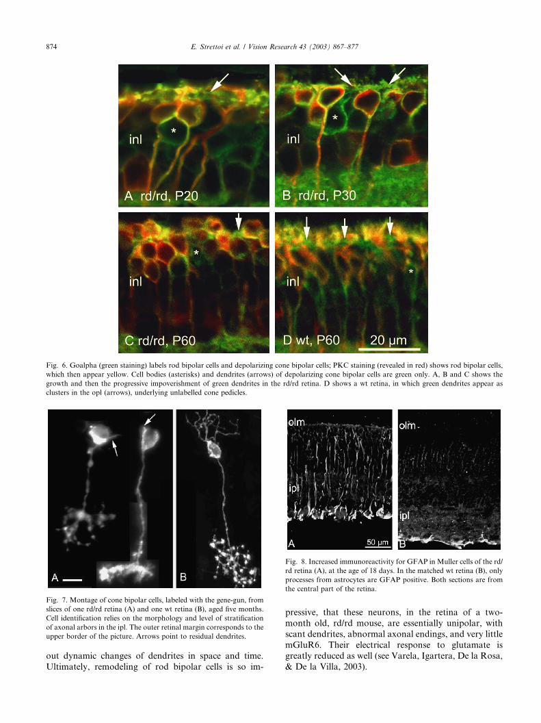
The analysis of cone bipolar cells in the mouse retina
is limited by technical reasons: cone bipolar dendrites
are hard to visualize in retinal sections stained with PKC
(labeling rod bipolar cells) and Goalpha (that stains cell
bodies, dendrites and axons of both rod bipolar and
depolarizing cone bipolar cells; Vardi, 1998). This diffi-
culty increases in a pathological preparation such as therd/rd retina. Other antibodies, that allow a more fa-
vorable study of dendrites of these cells (i.e., NK3 re-
ceptor antibodies: Casini, Brecha, Bosco, & Rickman,
2000; caldendrin antibodies: Haverkamp & Wassle,
2000), stain fractions of the varieties of cone bipolar
cells that are present in the retina of the mouse (see, for
instance, Tsukamoto, Morigiwa, Ueda, & Sterling,
2001). Combining several of the markers available forcone bipolar cells at this time, we start detecting major
morphological alterations in these cells between one and
two months of age. Cells stained with Goalpha do de-
velop dendrites that are still visible at P20 and at P30,
when dendrites of rod bipolar cells have already un-
dergone major changes (Fig. 6A and B). As is the case of
horizontal cells, in the following period, dendrites of
cone bipolar cells undergo a process of progressive re-traction, while their plexus in the opl becomes increas-
ingly discontinuous (compare Fig. 6C and D). Fig. 7A
Fig. 3. Changes in rod bipolar cells revealed by PKC staining. A and B shows retinal vertical sections obtained from animals of approximately 30
days. Arrows point to well developed dendrites of rod bipolar cells in A, and to cell bodies with no processes (single arrow) or few dendrites with
radial orientation in B (double arrows). C–E shows whole mounts of wt (C and D) and rd/rd (E) retinas. C corresponds to the focal plane of rod
bipolar dendrites and D to the focal plane of cell bodies. In the rd/rd (E), the equivalent of C is not detectable; rod bipolar cells appear carrying few,
disorganized processes. Bars are 10 lm.
E. Strettoi et al. / Vision Research 43 (2003) 867–877 871

shows individual cone bipolar cells, labeled in a five-
month old preparation. Short stumps have replaced
dendrites, clearly visible in single cone bipolar cells of a
normal retina (Fig. 7B).
3.5. Other cell types
3.5.1. Muller cells
As other authors have described in various forms of
retinopathies (de Raad et al., 1996; Rungger-Brandle,
Dosso, & Leuenberger, 2000; Sheedlo, Jaynes, Bolan, &
Turner, 1995), we observe a typical sign of glial reaction
in the form of increased immunoreactivity for glial
fibrillary acidic protein (GFAP) in Muller cells (Fig. 8).This is best visible from approximately P18/P20 to P30,
and attenuates thereafter (Strettoi et al., 2002). Major
morphological abnormalities in Muller cells (labeled by
glutamine synthetase) are not evident, except for the
expected shortening of their apical processes, associated
to the decrease in thickness of the photoreceptor layer.
3.5.2. Amacrine cells
We studied three well know amacrine cell types,namely cholinergic, dopaminergic, and type AII amac-
rines. Each of them can be labeled as a population by
specific antibodies, respectively against Choline acetyl-
transferase, Tyrosine hydroxylase and disabled-1 mole-
cules (Rice & Curran, 2000). The overall morphology of
these three types of cells is definitely more preserved
than the morphology of bipolar and horizontal cells, at
least up to the age of three months. The total retinal
number of cholinergic and dopaminergic cells does not
change during the same time interval (Strettoi et al.,
2002). More detailed studies on amacrine cells are inprogress in our laboratory.
3.6. Electrophysiological changes
Rod-mediated ERGs cannot be recorded from rd/rdmice of any age. A comparison between wt and rd ERGs
is possible for cone-mediated responses, which we ob-
tained reproducibly in a short time window comprised
between P13 and P16.
The major differences observed in the rd/rd ERG, at
all stimulation intensities, consist in an overall ampli-
tude reduction associated to an evident change in the
waveform. As shown in Fig. 9, the change in thewaveform is mainly due to specific alterations of the b-
wave, delayed and of abnormally long duration. At low
flash rates, the ERG of the rd/rd mouse, as compared to
the wild type, is dominated by the negative component
(a-wave). By increasing the flash rate, the sluggish pos-
itive ERG component (i.e. the b-wave) of the rd/rd
mouse becomes relatively more attenuated, resulting in a
progressive ‘‘smearing out’’ of the ERG waveform.Decreasing the stimulus intensity in the wild-type mouse
in order to mimic the subnormal a-wave amplitude of
the rd/rd mouse does not normalize the b-wave abnor-
malities. This indicates specific defects in the kinetics of
the b-wave.
4. Discussion
Even though our study is limited to some morpho-
logical and electrophysiological analysis of one specific
animal model, some conclusions on retinal remodeling
can be drawn. Indeed, the rd/rd postreceptoral retina
undergoes a major anatomical and functional rear-
rangement induced by the loss of photoreceptors. The
changes we describe follow a clear temporal trend: theyappear first in cells connected to rods, that degenerate
first, and than in those connected to cones. Interestingly,
horizontal cells, which connect to both, show the first
signs of remodeling at the side associated to rods. The
kinds of remodeling that we observe are a combination
of various cellular responses: sprouting (evident in
horizontal cell axonal endings), hypertrophy (again in
processes and bodies of horizontal cells) and atrophy, ofaxonal endings and dendrites. There is also cellular loss
(which we quantified, so far, only for rod bipolar and
horizontal cells). Similar responses (sprouting, cellular
Fig. 4. (A) and (B) Wt and rd/rd rod bipolar cells, individually labeled
by gene-gun delivery of lipophilic dyes on retinal slices, from retinas of
three and five months, respectively. Differences in dendritic number
and orientation (arrows), as well as in the sizes of axonal arbors are
very evident. Bar is 10 lm.
872 E. Strettoi et al. / Vision Research 43 (2003) 867–877

loss) have been described in RP (Fariss et al., 2000;
Santos et al., 1997). Sprouting from horizontal cells has
been described in other retinas with photoreceptor de-
generation (i.e. Park et al., 2001) and in experimental
retinal detachment (Lewis, Linberg, & Fisher, 1998).
Disorganization in the morphology and in the distri-
bution of horizontal cells has been described in theretina of RCS rats (Chu, Humphrey, & Constable,
1993).
Note that some of the changes we observed could be
very deceiving. For instance, losses of thin processes
from horizontal and bipolar cells, and the concomitant
hypertrophy of larger axonal branches in horizontal
cells, produce the effect of an apparently unchanged size
of the opl. At the same time, while immunocytochem-
istry clearly shows a great loss of dendrites in rod bi-
polar cells, gene-gun labeling reveals few, anomalous,
processes in some cells. These could be the dendritesestablishing ectopic connections with cones, described
by Peng et al. (2000) in young rd/rd mice; alternatively,
they could be newly formed dendrites with aberrant
orientation. Further analysis will be necessary to find
Fig. 5. Changes in horizontal cells. A and B shows neurofilament staining of vertical sections in wt (A) and rd/rd (B) retinas shows hypertrophy and
moderate sprouting (arrow) of axonal branches of horizontal cells in the mutants at P30. The plane of section in A is slightly oblique. C shows
individual axonal arbor of horizontal cell stained with the gene-gun and viewed as a retinal whole mount. The diameter of the axonal arborization is
three times as wide as a normal one. Arrow points to the axon. D and E shows Calbindin staining in whole mount preparations reveals cellular loss,
hypertrophy of bodies and absence of thin dendrites in horizontal cells from central retinal areas of rd/rd mice. Bars are 20 lm.
E. Strettoi et al. / Vision Research 43 (2003) 867–877 873
out dynamic changes of dendrites in space and time.
Ultimately, remodeling of rod bipolar cells is so im-
pressive, that these neurons, in the retina of a two-
month old, rd/rd mouse, are essentially unipolar, with
scant dendrites, abnormal axonal endings, and very little
mGluR6. Their electrical response to glutamate isgreatly reduced as well (see Varela, Igartera, De la Rosa,
& De la Villa, 2003).
Fig. 6. Goalpha (green staining) labels rod bipolar cells and depolarizing cone bipolar cells; PKC staining (revealed in red) shows rod bipolar cells,
which then appear yellow. Cell bodies (asterisks) and dendrites (arrows) of depolarizing cone bipolar cells are green only. A, B and C shows the
growth and then the progressive impoverishment of green dendrites in the rd/rd retina. D shows a wt retina, in which green dendrites appear as
clusters in the opl (arrows), underlying unlabelled cone pedicles.
Fig. 7. Montage of cone bipolar cells, labeled with the gene-gun, from
slices of one rd/rd retina (A) and one wt retina (B), aged five months.
Cell identification relies on the morphology and level of stratification
of axonal arbors in the ipl. The outer retinal margin corresponds to the
upper border of the picture. Arrows point to residual dendrites.
Fig. 8. Increased immunoreactivity for GFAP in Muller cells of the rd/
rd retina (A), at the age of 18 days. In the matched wt retina (B), only
processes from astrocytes are GFAP positive. Both sections are from
the central part of the retina.
874 E. Strettoi et al. / Vision Research 43 (2003) 867–877

Already in 1978, Blanks et al. described the lack of
connections between rods and rod bipolar dendrites
during postnatal development of the rd/rd retina, in
classical EM studies. We can confirm now that rod bi-
polar dendrites, for reasons we do not know yet, fail to
grow appropriately in the outer plexiform layer. Ab-
normal development of dendrites is linked to an arrest in
growth of rod bipolar axonal endings. An obvious hy-pothesis is that rod bipolar cells are missing a signal,
necessary to develop successfully and possibly associ-
ated to the synaptic terminal of healthy photoreceptors.
Of course, intervening glial cells and/or other factors
could also be very important.
Dendritic underdevelopment and atrophy in rod bi-
polar cells and loss of thin processes from axonal arbors
of horizontal cell are predictive of the late retraction ofdendrites from cone bipolar cells. Similarly, the latter
phenomenon could be predictive of the changes one can
expect in animal models of slow retinal degeneration.
Indeed, in the retina of crx-null mice (Furukawa, Mor-
row, Li, Davis, & Cepko, 1999), in which photorecep-
tors die over a slow time scale, we observe that dendrites
of rod bipolar cells are fully developed at first, and
subsequently retract; hence, at late stages of photore-
ceptor degeneration, they are similar to rod bipolar cells
of an earlier rd/rd retina (Pignatelli, Cepko, & Strettoi,
in preparation). Thus, progressive dendritic atrophy
could be a common avenue for retinal neurons deprived
by their input cells, similarly to what found in central
auditory nuclei after de-afferentation (Deitch & Rubel,1984). This might be of relevance in uncovering appro-
priate time windows for therapeutic strategies: inter-
ventions that rely upon the integrity of second order
neurons have to be designed early for fast-degenerating
forms of photoreceptor diseases, particularly in the in-
stance that death of photoreceptor and synaptogenesis
overlap. The reason for which restoration of vision in
the rd/rd mice is possible only upon early intervention(Bennett et al., 1996; Kwan, Wang, & Lund, 1999;
Radner et al., 2001) could be the extensive and early
remodeling observed in this mutant.
With the exposed technical limits described above, it
is somewhat encouraging that cone bipolar cells start to
show dendritic atrophy later than rod bipolar cells. They
are a very large population of second order neurons in
the retinal of mammals and therefore a potential plat-form for therapeutic intervention (i.e., based on photo-
receptor transplant).
However, cone-mediated ERG recordings performed
near P14 show abnormalities of the b-wave that appear
to depend upon specific deficits in the retinal cone-
pathway. At this early stage, morphological analysis is
unable to uncover changes clearly linked to the cone
pathway alone.A decreased sensitivity to glutamate in depolarizing
cone bipolar cells (which give a large contribution to the
b-wave in photopic conditions) could explain these ab-
normalities. This goes along with the observed decrease
in immunoreactivity for mGluR6.
An increased sensitivity to GABA in cone bipolar
cells of the rd/rd mouse (similar to that found in rod
bipolar cells by Varela et al., 2003) could also result in asuppressive effect of their depolarizing response and al-
tered kinetic, ultimately reflected in the b-wave of the
ERG. In the human GABA-associated retinal dysfunc-
tion (Krauss, Johnson, & Miller, 1998), ERG recordings
show reduced cone-mediate responses (b-waves and os-
cillatory potentials), with increased b-wave duration.
It is to note that changes in cone-mediated flicker
ERGs are reminiscent of the ERG changes found atleast in some RP patients. Massof, Johnson, Sunness,
Perry, and Finkelstein (1986) described progressive
‘‘smearing out’’ of the RP flicker ERG waveforms with
increasing temporal frequency. The differences in time-
to-peak between the positive and negative ERG com-
ponents were longer in RP patients than in the normal,
suggesting that the temporal anomalies in the flicker
ERG are due to changes in both amplitude and time
Fig. 9. Cone-mediated ganzfeld electroretinograms recorded at dif-
ferent flash frequencies between 0.5 and 20 s�1 from a wt (black
tracings) and an rd/rd mouse (red tracings) of 14 days of age (P14).
Flash intensity is 2 cd/m2 s�1. Background intensity 12 cd/m2. Note the
marked b-wave attenuation and delay in the rd/rd compared to the WT
mouse at all flash rates. The a-wave of the rd/rd animal is relatively
preserved. However, the delayed b-wave onset results in an increased
a-wave time-to-peak, and an overall ‘‘smearing out’’ of the rd/rd ERG
waveform.
E. Strettoi et al. / Vision Research 43 (2003) 867–877 875

constants of the ERG components. Hood and Birch
(1996) observed similar changes. Falsini et al. (1999)
evaluated the fundamental and second harmonic com-
ponents of flicker ERGs, as a function of temporal
frequency, in human patients with typical RP. They
reported that both components showed temporal fre-
quency-dependent abnormalities in both amplitude and
latency, with increasing response delays at higher tem-poral frequencies. Taken together, these data suggest
that the abnormalities found in rd mice and in human
RP patients share a similar mechanism, involving an
abnormality of the temporal response properties of
postreceptoral generators subtending the origin of ERG
b-wave.
Several techniques are now available to study retinal
cell types individually or as homogeneous populations:selective staining methods, mosaic analysis, electro-
physiological recordings from single units, single-cell
PCR, DNA-arrays, to mention some of them. It is likely
that in the next few years the associated efforts of dif-
ferent research groups to apply these techniques to ret-
inal pathology will contribute to gain a deeper view of
the whole concept of remodeling in retinal disease,
which should be considered as the rule more than theexception.
Acknowledgements
We like to thank the colleagues who donated anti-
bodies for this and previous studies (C. Cepko; T.
Curran; B. Howell; S. Nakanishi; D. Rice; R. Wong), aswell as all the colleagues of the Institute of Neuroscience
in Pisa for administrative, technical and scientific sup-
port. Funded by the Italian CNR, TeleThon Project
E833, NIH-R01EY12654.
References
Acland, G. M., Aguirre, G. D., Ray, J., Zhang, Q., Aleman, T. S.,
Cideciyan, A. V., Pearce-Kelling, S. E., Anand, V., Zeng, Y.,
Maguire, A. M., Jacobson, S. G., Hauswirth, W. W., & Bennett, J.
(2001). Gene therapy restores vision in a canine model of childhood
blindness. Nature Genetics, 28, 92–95.
Adler, R., Curcio, C., Hicks, D., Price, D., & Wong, F. (1999). Cell
death in age-related macular degeneration. Molecular Vision, 3, 5–
31.
Baekelandt, V., Arckens, L., Annaert, W., Eysel, U. T., Orban, G. A.,
& Vandesande, F. (1994). Alterations in GAP-43 and synapsin
immunoreactivity provide evidence for synaptic reorganization in
adult cat dorsal lateral geniculate nucleus following retinal lesions.
European Journal of Neuroscience, 6, 754–765.
Bennett, J. (2000). Gene therapy for retinitis pigmentosa. Current
Opinions in Molecular Therapy, 2, 420–425.
Bennett, J., Tanabe, T., Sun, D., Zeng, Y., Kjeldbye, H., Gouras, P., &
Maguire, A. M. (1996). Photoreceptor cell rescue in retinal
degeneration (rd) mice by in vivo gene therapy. Nature Medicine,
2, 649–654.
Blanks, J. C., Adinolfi, A. M., & Lolley, R. N. (1974). Photoreceptor
degeneration and synaptogenesis in retinal-degenerative (rd) mice.
The Journal of Comparative Neurology, 156, 95–106.
Bowes, C., Li, T., Danciger, M., Baxter, L. C., Applebury, M. L., &
Farber, D. B. (1990). Retinal degeneration in the rd mouse is
caused by a defect in the beta subunit of rod cGMP-phospho-
diesterase. Nature, 347, 677–680.
Carter-Dawson, L. D., LaVail, M. M., & Sidman, R. L. (1978).
Differential effect of the rd mutation on rods and cones in the
mouse retina. Investigative Ophthalmology and Visual Science, 17,
489–498.
Casini, G., Brecha, N. C., Bosco, L., & Rickman, D. W. (2000).
Developmental expression of neurokinin-1 and neurokinin-3
receptors in the rat retina. The Journal of Comparative Neurology,
42, 275–287.
Chang, B., Hawes, N. L., Hurd, R. E., Davisson, M. T., Nusinowitz,
S., & Heckenlively, J. R. (2002). Retinal degeneration mutants in
the mouse. Vision Research, 42, 517–525.
Chu, Y., Humphrey, M. F., & Constable, I. J. (1993). Horizontal cells
of the normal and dystrophic rat retina: a wholemount study using
immunolabelling for the 28-kDa calcium-binding protein. Exper-
imental Eye Research, 57, 141–148.
de Raad, S., Szczesny, P. J., Munz, K., & Rem�ee, C. E. (1996). Lightdamage in the rat retina: glial fibrillary acidic protein accumulates
in Muller cells in correlation with photoreceptor damage. Oph-
thalmic Research, 28, 99–107.
Deitch, J. S., & Rubel, E. W. (1984). Afferent influences on brain stem
auditory nuclei of the chicken: time course and specificity of
dendritic atrophy following deafferentation. The Journal of Com-
parative Neurology, 10, 66–79.
Falsini, B., Iarossi, G., Fadda, A., Porrello, G., Valentini, P., Piccardi,
M., & Scullica, L. (1999). The fundamental and second harmonic
of the photopic flicker electroretinogram: temporal frequency-
dependent abnormalities in retinitis pigmentosa. Clinical Neuro-
physiology, 110, 1554–1562.
Falsini, B., Iarossi, G., Porciatti, V., Merendino, E., Fadda, A.,
Cermola, S., & Buzzonetti, L. (1994). Postreceptoral contribution
to macular dysfunction in retinitis pigmentosa. Investigative
Ophtalmology and Visual Science, 35, 4282–4290.
Farber, D. B., & Lolley, R. N. (1974). Cyclic guanosine monophos-
phate: elevation in degenerating photoreceptor cells of the C3H
mouse retina. Science, 186, 449–451.
Fariss, R. N., Li, Z. Y., & Milam, A. H. (2000). Abnormalities in rod
photoreceptors, amacrine cells and horizontal cells in human
retinas with retinitis pigmentosa. American Journal of Ophthalmol-
ogy, 129, 215–223.
Furukawa, T., Morrow, E. M., Li, T., Davis, F. C., & Cepko, C. L.
(1999). Retinopathy and attenuated circadian entrainment in Crx-
deficient mice. Nature Genetics, 23, 466–470.
Gan, W. B., Grutzendler, J., Wong, W. T., Wong, R. O., & Lichtman,
J. (2000). Multicolor ‘‘DiOlistic’’ labeling of the nervous system
using lipophilic dye combinations. Neuron, 27, 219–225.
Gilbert, C. D. (1998). Adult cortical dynamics. Physiological Reviews,
78, 467–485.
Haverkamp, S., & Wassle, H. (2000). Immunocytochemical analysis
of the mouse retina. The Journal of Comparative Neurology, 424, 1–
23.
Hood, D. C., & Birch, D. G. (1996). Abnormalities of the retinal cone
system in retinitis pigmentosa. Vision Research, 36, 1699–1709.
Humayun, M. S. (2001). Intraocular retinal prosthesis. Transactions of
American Ophthalmological Society, 99, 271–300.
Jeon, C. J., Strettoi, E., & Masland, R. H. (1998). The major cell
populations of the mouse retina. Journal of Neuroscience, 18, 8936–
8946.
Krauss, G. L., Johnson, M. A., & Miller, N. R. (1998). Vigabatrin-
associated retinal cone system dysfunction: electroretinogram and
ophthalmologic findings. Neurology, 50, 614–618.
876 E. Strettoi et al. / Vision Research 43 (2003) 867–877
Administrator
Note

Kwan, A. S., Wang, S., & Lund, R. D. (1999). Photoreceptor layer
reconstruction in a rodent model of retinal degeneration. Exper-
imental Neurology, 159, 21–33.
Lewis, G. P., Linberg, K. A., & Fisher, S. K. (1998). Neurite
outgrowth from bipolar and horizontal cells after experimental
retinal detachment. Investigative Ophthalmology and Visual Science,
39, 424–434.
Lund, R. D., Kwan, A. S., Keegan, D. J., Sauve, Y., Coffey, P. J., &
Lawrence, J. M. (2001). Cell transplantation as a treatment for
retinal disease. Progress in Retinal and Eye Research, 20, 415–449.
Lyubarsky, A. L., Falsini, B., Pennesi, M. E., Valentini, P., & Pugh, E.
N., Jr. (1999). UV- and midwave-sensitive cone-driven retinal
responses of the mouse: a possible phenotype for coexpression of
cone photopigments. Journal of Neuroscience, 19, 442–455.
Massof, R. W., Johnson, M. A., Sunness, J. S., Perry, C., &
Finkelstein, D. (1986). Flicker electroretinogram in retinitis
pigmentosa. Documenta Ophthalmologica, 62, 231–245.
Milam, A. H., Li, Z. Y., & Fariss, R. N. (1998). Histopathology of the
human retina in retinitis pigmentosa. Progress in Retinal and Eye
Research, 17, 175–205.
Park, S. J., Kim, I. B., Choi, K. R., Moon, J. I., Oh, S. J., Chung, J.
W., & Chun, M. H. (2001). Reorganization of horizontal cell
processes in the developing FVB/N mouse retina. Cell and Tissue
Research, 306, 341–346.
Peachey, N. S., & Chow, A. Y. (1999). Subretinal implantation of
semiconductor-based photodiodes: progress and challenges. Jour-
nal of Rehabilitation Research and Development, 36, 371–376.
Peichl, L., & Gonzalez-Soriano, J. (1994). Morphological types of
horizontal cell in rodent retinae: a comparison of rat, mouse, gerbil,
and guinea pig (1994). Visual Neuroscience, 11, 501–517.
Peng, Y. W., Hao, Y., Petters, R. M., & Wong, F. (2000). Ectopic
synaptogenesis in the mammalian retina caused by rod photo-
receptor-specific mutations. Nature Neuroscience, 3, 1121–1127.
Radner, W., Sadda, S. R., Humayun, M. S., Suzuki, S., Melia, M.,
Weiland, J., & de Juan, E., Jr. (2001). Light-driven retinal ganglion
cell responses in blind rd mice after neural retinal transplantation.
Investigative Ophthalmology and Visual Science, 42, 1057–1065.
Rice, D. S., & Curran, T. (2000). Disabled-1 is expressed in type AII
amacrine cells in the mouse retina. The Journal of Comparative
Neurology, 424, 327–338.
Robson, J. G., & Frishman, L. J. (1995). Response linearity and
kinetics of the cat retina: the bipolar cell component of the dark-
adapted electroretinogram. Visual Neuroscience, 12, 837–850.
Robson, J. G., & Frishman, L. J. (1996). Photoreceptor and bipolar
cell contributions to the cat electroretinogram: a kinetic model for
the early part of the flash response. Journal of Optical Society of
America A, 13, 613–622.
Rohrer, B., Korenbrot, J. I., LaVail, M. M., Reichardt, L. F., & Xu,
B. (1999). Role of neurotrophin receptor TrkB in the maturation
of rod photoreceptors and establishment of synaptic transmis-
sion to the inner retina. Journal of Neuroscience, 19, 8919–
8930.
Rubel, E. W., & Fritzsch, B. (2002). Auditory system development:
primary auditory neurons and their targets. Annual Review of
Neuroscience, 25, 51–101.
Rungger-Brandle, E., Dosso, A. A., & Leuenberger, P. M. (2000).
Glial reactivity, an early feature of diabetic retinopathy. Investiga-
tive Ophthalmology and Visual Science, 41, 1971–1980.
Santos, A., Humayun, M. S., Juan, E. J., Greenburg, R. J., Marsh, M.
J., Klock, I. B., & Milam, A. H. (1997). Preservation of the inner
retina in retinitis pigmentosa. A morphometric analysis. Archives of
Ophtalmology, 115, 511–515.
Sernagor, E., Eglen, S. J., & Wong, R. O. (2001). Development of
retinal ganglion cell structure and function. Progress in Retinal and
Eye Research, 20, 139–174.
Sheedlo, H. J., Jaynes, D., Bolan, A. L., & Turner, J. E. (1995).
Mullerian glia in dystrophic rodent retinas: an immunocytochem-
ical analysis. Brain Research Developmental Brain Research, 85,
171–180.
Strettoi, E., & Pignatelli, V. (2000). Modifications of retinal neurons in
a mouse model of retinitis pigmentosa. In: Proceedings of the
National Academy of Sciences of the USA, Vol. 97, pp. 11020–
11025.
Strettoi, E., Porciatti, V., Falsini, B., Pignatelli, V., & Rossi, C. (2002).
Morphological and functional abnormalities in the inner retina of
the rd/rd mouse. Journal of Neuroscience, 22, 5492–5504.
Strettoi, E., & Volpini, M. (2002). Retinal organization in the bcl-
2-overexpressing transgenic mouse. The Journal of Comparative
Neurology, 446, 1–10.
Tsukamoto, Y., Morigiwa, K., Ueda, M., & Sterling, P. J. (2001).
Microcircuits for night vision in mouse retina. Journal of Neuro-
science, 21, 8616–8623.
Vardi, N. (1998). Alpha subunit of Go localizes in the dendritic tips of
ON bipolar cells. The Journal of Comparative Neurology, 395, 43–
52.
Varela, C., Igartera, I., De la Rosa, E. J., & De la Villa, P. (2003).
Functional modifications in rod bipolar cells in a mouse model of
retinitis pigmentosa. Vision Research.
Zrenner, E. (2002). Will retinal implants restore vision? Science, 295,
1022–1025.
E. Strettoi et al. / Vision Research 43 (2003) 867–877 877