Genetic and morphometric evidence for parallel evolution of the Globigerinella calida morphotype
17
Research paper Genetic and morphometric evidence for parallel evolution of the Globigerinella calida morphotype Agnes K.M. Weiner a, ⁎, Manuel F.G. Weinkauf a , Atsushi Kurasawa b , Kate F. Darling c,d , Michal Kucera a a MARUM Center for Marine Environmental Sciences, University of Bremen, Leobenerstrasse, 28359 Bremen, Germany b Research and Development Center for Global Change, Japanese Agency for Marine Earth Science and Technology, Natsushima-cho 2-15, Yokosuka 237-0061, Japan c School of GeoSciences, University of Edinburgh, Edinburgh EH9 3JW, UK d School of Geography and GeoSciences, University of St Andrews, Fife KY16 9AL, UK abstract article info Article history: Received 1 April 2014 Received in revised form 14 October 2014 Accepted 16 October 2014 Available online 25 October 2014 Keywords: planktonic foraminifera Globigerinella shell morphology shell porosity taxonomy parallel evolution Molecular genetic investigations of the highly abundant extant planktonic foraminifera plexus Globigerinella siphonifera/Globigerinella calida have recently shown this group to be the genetically most diverse one within planktonic foraminifera, separating it into 12 distinct genetic types belonging to three main genetic lineages. In- dependently, several morphological or physiological variants have been described within the group, but the cor- relation between the high genetic diversity and the phenotypic variability remains unclear. In this study, we combine genetic data with morphometric analyses of shell shape and porosity of genotyped individuals of the different genetic lineages. Our morphometric measurements suggest a differentiation of three morphotypes within the plexus, two of which possess the elongated chambers described as a typical trait of G. calida. These two morphotypes with elongated chambers are associated with two distinct genetic lineages. The G. calida mor- phology therefore appears to have evolved twice in parallel. Unexpectedly, we show that the two morphotypes with elongated chambers can be separated from each other by characters seen in the lateral view of their shells. This implies that the taxonomy of the extant members of the genus Globigerinella should be revised. A compar- ison with the original descriptions and type specimens of members of the genus shows that two genetic types of one major lineage correspond to G. calida. The second group with elongated chambers is associated with a recent- ly diverged genetic type and we propose to reinstate the name Globigerinella radians for this distinct form. The remaining nine of the 12 genetic types correspond to the G. siphonifera morphology, and in the absence of evi- dence for morphological differentiation, they form a paraphyletic morpho-taxon. Our results highlight the prev- alence of parallelism in the evolution of shell morphology in planktonic foraminifera even at the lowest level of relatedness represented by genetic types. © 2014 The Authors. Published by Elsevier B.V. This is an open access article under the CC BY license (http://creativecommons.org/licenses/by/4.0/). 1. Introduction Molecular genetic studies of extant planktonic foraminifera con- tinue to challenge our perception on the diversity within the group (e.g. Darling et al., 1999; de Vargas et al., 1999; de Vargas et al., 2002; Aurahs et al., 2009; Seears et al., 2012; Quillévéré et al., in press). The relatively low number of accepted morphospecies (e.g. Hemleben et al., 1989) is significantly exceeded by the number of their constit- uent genetic types (e.g. Darling and Wade, 2008). Since most of these genetic types cannot be differentiated morphologically, they are often referred to as “cryptic species” and their discovery usually has no impact on the taxonomy of the morphospecies. Exceptions hereto are Neogloboquadrina incompta, which could be separated from Neogloboquadrina pachyderma based on genetic data confirming the observation that the two species are characterized by different coiling directions (Darling et al., 2006) as well as Globigerinoides elongatus that was initially synonymized with Globigerinoides ruber, but recently shown to be genetically as well as morphologically distinct (Aurahs et al., 2011). Morphometric studies on Orbulina universa, Globoconella inflata and Globorotalia truncatulinoides revealed only slight morphological differences between the genetic types that were statistically significant, but did not allow sufficiently precise discrimina- tion of individuals to warrant a taxonomic revision (Morard et al., 2009; Morard et al., 2011; Quillévéré et al., in press). A study on the morpho- species complex Globigerinoides sacculifer surprisingly revealed that also the opposite scenario can exist: a worldwide screening of all morphotypes associated with this taxon showed that this morphospe- cies is genetically homogenous despite high morphological variability (André et al., 2013). In this case an over-interpretation of morphological characteristics had taken place, which led to the usage of multiple morphospecies concepts that do not appear justified in the light of the Marine Micropaleontology 114 (2015) 19–35 ⁎ Corresponding author. Tel.: +49 421 21865972; fax: +49 421 21865505. E-mail address: [email protected] (A.K.M. Weiner). http://dx.doi.org/10.1016/j.marmicro.2014.10.003 0377-8398/© 2014 The Authors. Published by Elsevier B.V. This is an open access article under the CC BY license (http://creativecommons.org/licenses/by/4.0/). Contents lists available at ScienceDirect Marine Micropaleontology journal homepage: www.elsevier.com/locate/marmicro
-
Upload
independent -
Category
Documents
-
view
1 -
download
0
Transcript of Genetic and morphometric evidence for parallel evolution of the Globigerinella calida morphotype

Marine Micropaleontology 114 (2015) 19–35
Contents lists available at ScienceDirect
Marine Micropaleontology
j ourna l homepage: www.e lsev ie r .com/ locate /marmicro
Research paper
Genetic and morphometric evidence for parallel evolution of theGlobigerinella calida morphotype
Agnes K.M. Weiner a,⁎, Manuel F.G. Weinkauf a, Atsushi Kurasawa b, Kate F. Darling c,d, Michal Kucera a
a MARUM Center for Marine Environmental Sciences, University of Bremen, Leobenerstrasse, 28359 Bremen, Germanyb Research and Development Center for Global Change, Japanese Agency for Marine Earth Science and Technology, Natsushima-cho 2-15, Yokosuka 237-0061, Japanc School of GeoSciences, University of Edinburgh, Edinburgh EH9 3JW, UKd School of Geography and GeoSciences, University of St Andrews, Fife KY16 9AL, UK
⁎ Corresponding author. Tel.: +49 421 21865972; fax:E-mail address: [email protected] (A.K.M. Weiner).
http://dx.doi.org/10.1016/j.marmicro.2014.10.0030377-8398/© 2014 The Authors. Published by Elsevier B.V
a b s t r a c t
a r t i c l e i n f oArticle history:Received 1 April 2014Received in revised form 14 October 2014Accepted 16 October 2014Available online 25 October 2014
Keywords:planktonic foraminiferaGlobigerinellashell morphologyshell porositytaxonomyparallel evolution
Molecular genetic investigations of the highly abundant extant planktonic foraminifera plexus Globigerinellasiphonifera/Globigerinella calida have recently shown this group to be the genetically most diverse one withinplanktonic foraminifera, separating it into 12 distinct genetic types belonging to three main genetic lineages. In-dependently, several morphological or physiological variants have been described within the group, but the cor-relation between the high genetic diversity and the phenotypic variability remains unclear. In this study, wecombine genetic data with morphometric analyses of shell shape and porosity of genotyped individuals of thedifferent genetic lineages. Our morphometric measurements suggest a differentiation of three morphotypeswithin the plexus, two of which possess the elongated chambers described as a typical trait of G. calida. Thesetwo morphotypes with elongated chambers are associated with two distinct genetic lineages. The G. calidamor-phology therefore appears to have evolved twice in parallel. Unexpectedly, we show that the two morphotypeswith elongated chambers can be separated from each other by characters seen in the lateral view of their shells.This implies that the taxonomy of the extant members of the genus Globigerinella should be revised. A compar-ison with the original descriptions and type specimens of members of the genus shows that two genetic types ofonemajor lineage correspond toG. calida. The second groupwith elongated chambers is associatedwith a recent-ly diverged genetic type and we propose to reinstate the name Globigerinella radians for this distinct form. Theremaining nine of the 12 genetic types correspond to the G. siphonifera morphology, and in the absence of evi-dence for morphological differentiation, they form a paraphyletic morpho-taxon. Our results highlight the prev-alence of parallelism in the evolution of shell morphology in planktonic foraminifera even at the lowest level ofrelatedness represented by genetic types.
© 2014 The Authors. Published by Elsevier B.V. This is an open access article under the CC BY license(http://creativecommons.org/licenses/by/4.0/).
1. Introduction
Molecular genetic studies of extant planktonic foraminifera con-tinue to challenge our perception on the diversity within the group(e.g. Darling et al., 1999; de Vargas et al., 1999; de Vargas et al., 2002;Aurahs et al., 2009; Seears et al., 2012; Quillévéré et al., in press). Therelatively low number of accepted morphospecies (e.g. Hemlebenet al., 1989) is significantly exceeded by the number of their constit-uent genetic types (e.g. Darling andWade, 2008). Since most of thesegenetic types cannot be differentiated morphologically, they areoften referred to as “cryptic species” and their discovery usuallyhas no impact on the taxonomy of the morphospecies. Exceptionshereto are Neogloboquadrina incompta, which could be separatedfrom Neogloboquadrina pachyderma based on genetic data confirming
+49 421 21865505.
. This is an open access article under
the observation that the two species are characterized by differentcoiling directions (Darling et al., 2006) as well as Globigerinoideselongatus that was initially synonymized with Globigerinoides ruber,but recently shown to be genetically as well as morphologically distinct(Aurahs et al., 2011). Morphometric studies on Orbulina universa,Globoconella inflata and Globorotalia truncatulinoides revealed onlyslight morphological differences between the genetic types that werestatistically significant, but did not allow sufficiently precise discrimina-tion of individuals towarrant a taxonomic revision (Morard et al., 2009;Morard et al., 2011; Quillévéré et al., in press). A study on the morpho-species complex Globigerinoides sacculifer surprisingly revealed thatalso the opposite scenario can exist: a worldwide screening of allmorphotypes associated with this taxon showed that this morphospe-cies is genetically homogenous despite high morphological variability(André et al., 2013). In this case an over-interpretation ofmorphologicalcharacteristics had taken place, which led to the usage of multiplemorphospecies concepts that do not appear justified in the light of the
the CC BY license (http://creativecommons.org/licenses/by/4.0/).

20 A.K.M. Weiner et al. / Marine Micropaleontology 114 (2015) 19–35
genetic evidence. These examples underline that the connectionbetween genetic andmorphologic variability in planktonic foraminiferais complex and the resolution of species delineation requires a detailedcombined genetic and morphometric analysis. Here we present suchcombined analysis for the genetically and morphologically diverseGlobigerinella siphonifera/Globigerinella calida plexus.
The genus Globigerinella was first described by Cushman (1927) toinclude individuals with near-planispirally coiled shells, globular toovate chambers and fine rounded spines (Kennett and Srinivasan,1983). Three extant morphospecies can be attributed to this highlydiverse and abundant genus. The most abundant morphospeciesis G. siphonifera, described as Globigerina siphonifera by d'Orbigny(1839), with spherical to ovate chambers and a rather tight coiling.Globigerina aequilateralis, described by Brady (1879), which has a verysimilar morphology, was later declared a junior synonym (Banner andBlow, 1960). The second most abundant morphospecies, G. calida, wasdescribed as Globigerina calida by Parker (1962). It was characterizedas having trochospirally coiled evolute shells with radially elongatedchambers, the final chamber separated from the previous ones andbeing perforated by large circular pores (Parker, 1962; Saito et al.,1981). Both species occur globally in the surface waters of the tropics,subtropics and the temperate regions (e.g. Bé, 1977; Huber et al.,1997). The third morphospecies is Globigerinella adamsi, which wasoriginally described as Hastigerina adamsi (Banner and Blow, 1959)and is characterized by its elongated digitate chambers with pointedtips. This morphospecies is exceedingly rare. It inhabits mesopelagicwaters of the Indopacific low latitude realm (Bé and Tolderlund, 1971)and was never collected for genetic analysis. All three morphospeciesshow a considerable level of intraspecific morphological variability.Parker (1962) was the first to describe a potential separation ofG. siphonifera into two or even more groups based on shell size andthe degree of deviation from the planispiral coiling. She assumedthough that these forms represent ecophenotypic plasticity and there-fore did not treat her morphotypes taxonomically. Interestingly, theexistence of two groups within G. siphonifera was later inferred on thebasis of biological differences, especially the possession of differentendosymbiotic Chrysophycophyte species (Faber et al., 1988, 1989).Later studies suggested a correlation between these groups and the
BeellaTyp
Type IIIa a
Ty
Type IIa1
G
Hastigerina pelagica
Hastigerina pelagica
Type IIa5
Type IIIc
76
100
76
9982
95
46
96
10098
100
100
97
94
100
100
5653
Fig. 1.Maximum likelihood phylogenetic tree of the spinose planktonic foraminifera. Phylogenstatus of Globigerinella and its affinity with the sister taxon Beella digitata and highlighting i(Katoh and Standley, 2013) alignment of SSU rDNA sequences andwere calculated using RAxMLmorphospecies has been collapsed, except for G. siphonifera/G. calida where only terminal bran
then known two genetic types (Huber et al., 1997), including a potentialdifferentiation between the two types based on shell porosity. In a sub-sequent study, Bijma et al. (1998) observed differences in cell physiolo-gy and different isotopic compositions of the shell for these two groups.However, none of these discoveries had an impact on the taxonomy ofthe genus.
Genetic studies conducted on the ribosomal small subunit RNA gene(SSU rDNA) of G. siphonifera subsequently demonstrated that the highdiversity in this morphospecies is not limited to the morphology, butis also represented at the genetic level (Huber et al., 1997; de Vargaset al., 2002; Darling andWade, 2008; Göker et al., 2010). Most recently,Weiner et al. (2014) showed that the high sequence diversity in thegroup could be assigned to three major genetic lineages, which furthersplit into 12 distinct genetic types (Fig. 1). Since no signs of hybridiza-tion are found between these genetic types, they may be consideredto represent biological species (compare e.g. de Vargas et al., 2001;Darling et al., 2007). In these genetic studies, the exact status ofG. calida remained unclear. The distinction of this morphospecies fromG. siphonifera is difficult and in many cases the two morphospecieswere lumped together for studies on fossils from the sediment (e.g.Siccha et al., 2009). The distinction is especially difficult among pre-adult individuals that are often encountered in the plankton. As a result,only a preliminary identification has been presented by genetic studiespublished to date, in which G. calida was suggested to represent one ofthe genetic types of theGlobigerinella plexus (Type IV of de Vargas et al.,2002, and G. calida in Darling and Wade, 2008).
In order to resolve the relationship between genetic andmorpholog-ic variability in the genus, we have taken advantage of the recently de-veloped methods for extraction of DNA from planktonic foraminiferathat leave the shells intact for morphometric analysis (Morard et al.,2009; Weiner et al., 2014). Using these methods in combination withthe imaging of genotyped specimens prior to DNA extraction, we haveamassed a dataset of morphological measurements from 181 individualspecimens identified by several researchers as G. siphonifera andG. calida, sampled within various regions of the world ocean. All of thespecimens were genetically analyzed and could be assigned to one ofthe delineated genetic types. We combined measurements of shellmorphology based on scanning electron microscopic as well as light
digitata
Globigerinella siphonifera G. calida/
e Ia and b
nd b
pe IIb
Globigerinoides conglobatus G. elongatus/
Globigerinoides ruber
Globigerinoides sacculifer
Orbulina universa
lobigerina falconensis
Globigerina bulloides
Turborotalita quinqueloba
Type IIa2, 3 and 4
and 6
0.2 substitutions/site
etic relationships among the spinose planktonic foraminifera showing the monophyleticts high genetic diversity. The tree as well as the bootstrap values are based on a MAFFT(Stamatakis, 2006) via the CIPRES gateway (Miller et al., 2010). Sequence diversitywithinches are collapsed.

21A.K.M. Weiner et al. / Marine Micropaleontology 114 (2015) 19–35
microscopic images with measurements of porosity and pore size. As aresult, we were able to resolve the identity of G. calida and revise thetaxonomic concept of the G. siphonifera/G. calida plexus.
2. Material and methods
2.1. Sampling, imaging and genetic analysis
In this study, images of 181 Globigerinella siphonifera andGlobigerinella calida individuals were analyzed for comparisons of shellmorphology with genetic identity. All of the individuals includedyielded DNA sequences that could be used to assign them to one ofthe 12 lineages described by Weiner et al. (2014). The specimenswere collected by stratified plankton tows during 13 expeditions be-tween 2006 and 2013 (Fig. 2, Table S1). The foraminifera were separat-ed from the rest of the plankton, taxonomically identified usingstereomicroscopes and in most cases digitally photographed directlyon board. Living specimens still containing cytoplasm were preparedfor DNA extraction. Methods for genetic analysis and the sequencedata of most individuals were presented inWeiner et al. (2014). Specif-ically for this study we genetically characterized 44 additional speci-mens from a transit through the South Pacific on board RV SONNE(SO226-3, Kucera and Cruise Participants, 2013). These new samplesrepresent topotypic material for the species concept of G. calida as de-veloped by Parker (1962). They were obtained by stratified tows usinga multiple closing net with a mesh size of 100 μm. Foraminifera wereisolated from the plankton residues, cleaned, dried and frozen on
Fig. 2. Geographic locations of sampled individuals. a) Sampling locations of all individuals uselightmicroscopic images, only SEM images or bothwere available, depending on the samplingmpling location. Gray shading indicates the relative abundance of Globigerinella siphonifera as it isdata in the MARGO database by Ocean Data View (Schlitzer, 2011). Diagonal lines indicate areaindicate different genetic types, following the classification by Weiner et al. (2014). Numbers wthe same genetic type. Gray shading indicates the relative abundance ofGlobigerinella calida as itdata in the MARGO database by Ocean Data View. Diagonal lines indicate areas where no data
cardboard slides until further processing in the lab. The guanidinemeth-od, which allows preservation of the shell, was used for DNA extraction(e.g. Morard et al., 2009). Light microscopic images showing the stan-dard taxonomic umbilical view were taken in the lab prior to DNA ex-traction and all specimens have been assigned to either G. siphoniferaor G. calida by the collector, following the current taxonomy as present-ed e.g. in Hemleben et al. (1989). Polymerase chain reaction (PCR) wasused to amplify a ~600 bp large fragment of the 3′end of the small sub-unit ribosomal RNA gene (SSU rDNA) using the GoTaq® G2 Hot Startpolymerase (Promega) and two different primer pairs as indicated inTable S1. PCR products were purified using the QIAquick® PCR Purifica-tion Kit (Qiagen) and afterwards sequenced directly by an external ser-vice provider (Agowa, Berlin). Sequence chromatograms were checkedmanually for ambiguous reads and correctedwhere possible. Sequencesof all 44 individuals were submitted to GenBank (http://www.ncbi.nlm.nih.gov/; Accession nos.: KJ202213–KJ202256). Shells that could be re-covered afterDNA extractionwere imaged by scanning electronmicros-copy (SEM) from spiral/umbilical and lateral view and highermagnification close-ups of chamber wall surface were taken. In total,37 individuals from the South Pacific yielded images that could beused for morphometric analysis.
2.2. ML tree inference and bootstrapping
In order to represent the phylogenetic position of G. siphonifera/G. calida in relation to the rest of the spinose planktonic foraminifera, se-quences of 11morphospecieswere included in an automated alignment
d in the morpho-genetic comparison in this study. Different symbols indicate where onlyethod applied. Numbers within the symbols denote number of individuals from one sam-found in planktonic foraminiferal assemblages from surface sediments interpolated froms where no data are available. b) The genetic identity of the analyzed individuals. Symbolsithin the symbols denote number of individuals from one sampling location belonging tois found inplanktonic foraminiferal assemblages fromsurface sediments interpolated fromare available.

22 A.K.M. Weiner et al. / Marine Micropaleontology 114 (2015) 19–35
using the online version of MAFFT v. 7 (File S1, Katoh and Standley,2013) as it is available on the CIPRES gateway (Miller et al., 2010),under default settings. This alignment was then used without furthermanipulation or filtering for tree inference under the maximum likeli-hood (ML) criterion with RAxML-HPC2 v. 7.6.3 (Stamatakis, 2006) viathe CIPRES Gateway. Branch support was established with the fast im-plementation (Stamatakis et al., 2008, option -x) of nonparametricbootstrapping (BS; Felsenstein, 1985). The number of necessary repli-cates was determined by automatic bootstopping with the majority-rule tree based criterion (option -#autoMRE). The per-site rateapproximation model (Stamatakis, 2006) was used for the fast BSphase followed by a slow final model optimization under the generaltime reversible model allowing for between-site variation modeled viaa gamma distribution (GTR + Г; option -m GTRCAT). Run parameterswere set to infer in one run the best-known ML tree and perform afull BS analysis (option –f a).
2.3. Measurements of shell morphology and porosity
SEM images suitable for morphometric analysis were obtained froma total of 63 specimens of the G. siphonifera/G. calida plexus in lateraland umbilical/spiral view to quantify the main morphological fea-tures of the shell which have been used to differentiate morphospe-cies in the plexus. The traits have been quantified as distances andlandmark positions (Fig. 3) extracted from the images in R v. 3.0.1 (RDevelopment Core Team, 2011). In lateral view thesemeasurements in-clude the height htotal of the specimen, the elongation of the last cham-ber (El), the deviation of the last whorl from the planispiral plane(expressed as angle α), and the extent to which the first chamber ofthe last whorl covers the aperture (PS). In umbilical/spiral view valuescomprise the elongation of the last chamber (EL), the mean elongation
Fig. 3.Morphometricmeasurements conducted on images ofGlobigerinella siphonifera/G. calida isize and porosity derived from SEM images, including equations for the calculation of the deriextracted from the images. Black and green lines show distances used for the calculation ofmorplines are auxiliary lines for the visualization of calculated angles. Distances Hi and Bi, and anglesumbilical viewwere also extracted from lightmicroscopic images. (For interpretation of the refe
of all chambers in the last whorl (E) and the number of chambersin the last whorl expressed as mean angle γ between successive cham-ber axes. To avoid the effect of unusual terminal morphologies, inKummerform-specimens, the penultimate chamber was treated as thelast chamber. Damaged specimens with fewer than three consecutivechambers in the last whorl preserved were excluded from the analysis(15 individuals). The data acquisition and parameter calculation wasreplicated, and the values used in the following represent the mean ofthe two replications to minimize subjectivity during data extraction.
To evaluate the degree of morphological separation obtained on thebasis of exactly positioned clean specimens on SEM images, for practicalapplication in the field, we subsequently tested the approach on 128light-microscopic images of imperfectly oriented specimens in umbili-cal/spiral view. In these images we extracted 13 landmark points each(Fig. 3) to calculate the elongations EL and E on the basis of the lastthree chambers, as well as the mean angle γ.
Porosity measurements were obtained using SEM images with amagnification of 4000 of the surfaces of the last chamber of 66 speci-mens. The images were treated for contrast enhancement and wherenecessary, pores were manually blackened to enable automatic mea-surements. The maximum Feret diameter (d) and centroid coordinatesof each pore were then extracted from black and white threshold im-ages in FIJI v. 1.47q (Schindelin et al., 2012). These values were thenused to calculate porosity as a fraction of the pore area relative to theshell surface area (Fig. 3). This approach yields reliable results as longas pores can be expected not to be significantly oval in first approxima-tion. The maximum pore diameter, in contrast to the directly measuredpore area, is invariant to the orientation of the pore, so that the curva-ture of the shell does not influence the results by distorting the poresin areas which are not perfectly perpendicular to the plane of view. Inthis study, we decided not to break the shells to measure the pores
ndividuals. Schematic representation of themeasurements of shell characteristics and poreved, size-invariant parameters. Blue points represent landmarks, whose coordinates werehological parameters, whichwere calculated on the basis of the landmarks. Yellowdashedγi are only shown exemplarily on the last two chambers in umbilical view. Points 1–13 inrences to color in thisfigure legend, the reader is referred to theweb version of this article.)

020
4060
Genotype
Cla
ssifi
ed a
s G
. cal
ida
(%)
I II III Ia Ib IIa IIb IIIa IIIb IIIc
80 228 74 23 57 191 37 2 38 34
Lineage
Fig. 4. Percentages of individuals classified as Globigerinella calida. Percentages of individ-uals in each genetic lineage/genetic type (following the classification by Weiner et al.(2014)) of Globigerinella that were classified upon collection as G. calida. The datasetincludes all 382 individuals that were genetically analyzed, independent of the existenceof morphometric measurements. Vertical bars represent 95% binomial confidence inter-vals after Agresti and Coull (1998). Total number of trials n is given at the bottom of thegraph. Most individuals classified upon collection as G. calida belong to either lineage Ior III.
23A.K.M. Weiner et al. / Marine Micropaleontology 114 (2015) 19–35
from the inside, like Huber et al. (1997). As a result, our values are likelyto overestimate pore size by a small amount, especially in large andthick shells.
In order to determine shell porosity, we calculated the distance ofeach pore to every other pore based on the obtained centroid coordi-nates in R v. 3.0.1 and then identified the nearest neighbor to eachpore. The mean distance l of all nearest-neighbor-pair-distances ofthe specimen was then assumed to be a good approximation of themean pore distances in that specimen. Assuming a regular pore dis-tribution with one pore at each corner of a square with edge length l,we could then approximate the mean porosity P of the specimen(P = (π / 4 × d2) / l2). Even if the real pore distribution deviates fromthis expectation, the fact that we treat all specimens alike, leads tomutually comparable results. In 40 specimens we have taken two SEMimages from the same individual, which we could use to test the repro-ducibility of our results using a paired t-test.
2.4. Statistical analysis of morphometric measurements
All statistical analyses were performed in R v. 3.0.1. We used princi-pal component analysis (PCA, Hotelling, 1933) to evaluate the continu-ity of themorphospace in the G. siphonifera/G. calida plexus on the basisof the morphological parameters (excluding porosity) obtained fromthe SEM images, without a priori assumption on their attribution togenetic types. During that step we excluded the parameter htotal fromthe analysis, because shell height of specimens from the plankton is afunction of their age and does not represent the final size at whichreproductionwould occur. Next, we explored towhat degree specimensof distinct genetic lineages can be distinguished from the rest of theplexus by performing linear discriminant analyses/canonical variateanalyses (LDA/CVA, Fisher, 1936) in the R-package MASS v. 7.3-26(Venables and Ripley, 2002). We then repeated the same steps on thedata obtained from light microscopic images.
The porosity data were tested for the influence of genetic type andsampling location and their interaction term on porosity and pore sizeof specimens. To that end the non-parametric Scheirer–Ray–Hare test(Scheirer et al., 1976) was applied. For all significant factors, pairwisecomparisons were performed using a Mann–Whitney U test (Mannand Whitney, 1947), during which the p-values were corrected afterBenjamini and Hochberg (1995). To test for a relationship betweenpore size/porosity and shell size (approximated via shell height, ht),we performed a Kendall–Theil robust line fitting (Kendall, 1938; Theil,1950; Sen, 1968) implemented in R, using the equations from Helseland Hirsch (2002) and Conover (1980). For specimens with two SEMimages of the same individual, we used the onewhich provided a largerdataset (i.e. more pore measurements) for the analysis.
3. Results
Of the 382 genetically analyzed Globigerinella specimens (Weineret al. (2014) and new data from South Pacific combined), 62 werelabeled upon collection as G. calida. In this respect, the subset used formorphometric analysis is representative, containing 42 specimens outof 181 in total originally labeled as G. calida. As a first step, we inves-tigated, whether the usage of the species name G. calida, as determinedby traditional taxonomy during the initial collection, correlated withany of the genetic types. In fact, the comparison of the taxonomic labelsand genetic identification reveals that the frequency of usage ofG. calidavaries significantly among the genetic lineages and genetic types(Fig. 4). Although there is no single genetic type which is associatedexclusively with specimens labeled as G. calida, this name has beenused more frequently for specimens in lineages I and III (Fig. 4).
Next, we ventured to resolve the correlation of genetic andmorpho-logical variability in the G. siphonifera/G. calida plexus. To this end, wefirst explored morphological differences among all analyzed specimensand determined how these relate to the genetic types found within this
group. The high number of SEM and light-microscopic images allowed amorphometric analysis of representatives of almost every genetic typefrom various parts of the world ocean (Figs. 2, 5, Table S2). Most genetictypes had sufficiently well preserved shells following DNA extraction toobtain representative SEM images, apart from Types IIa2, IIa6, IIb andIIIa. However, it was possible to include Types IIa2 and IIb in the mor-phometric analyses using their light-microscopic images, but those ofIIa6 and IIIa proved too poor to be useful.
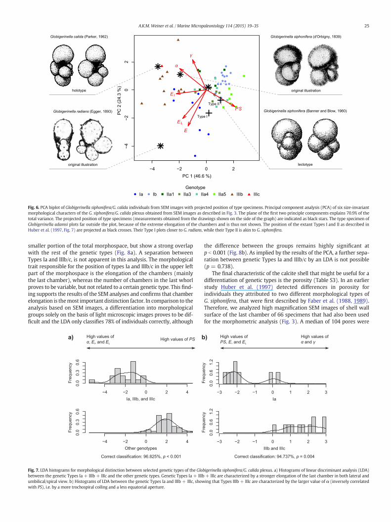
A PCA of the morphometric measurements carried out on SEMimages (Fig. 3, Table S2) revealed a significant size-independent varia-tion in morphology of the individuals belonging to the G. siphonifera/G. calida plexus (Fig. 6). The mapping of the genetic identity onto themorphospace reveals that three of the analyzed genetic types are asso-ciatedwith amorphology that is distinct from the rest of the plexus. Thegenetic Types Ia and IIIb/c appear to be separated from the rest of thegenetic types chiefly by higher chamber elongation (El, EL, Fig. 6). Thisseparation is supported by the LDA,which confirms a statistically signif-icant difference in the multivariate means between the groups(p b 0.001) and reveals that based on the same set of morphologicalmeasurements, 97% of the specimens can be correctly classified by thediscriminant function (Fig. 7a). Furthermore, these three types can notonly be separated from the rest, they also showmorphologic differenceswhen being compared with each other. Specimens of Type Ia are char-acterized by the highest values for chamber elongation in spiral/umbil-ical view (E and EL), while members of lineage III are marked by highestvalues for angle α, which describes the deviation of growth from theplanispiral plane (Fig. 6). This differentiation is also supported by theLDA (p = 0.004), and allows a correct classification of 95% of the spec-imens (Fig. 7b).
A CVA with the remainder of genetic types (Ib, IIa1, IIa3–5) showslow correct classification rates (73%) and a general distribution of allgenetic types over the whole morphospace, indicating that no distinctmorphotypes can be separated within this group (Fig. S1).
Having established the existence of three groups of genetic typesthat are morphologically distinct from each other, we attempted to

Fig. 5. Images of representative specimens of the genetic types of Globigerinella sp. SEM images and light microscopic images of representative individuals belonging to the differentgenetic types within the G. siphonifera/G. calida plexus. No SEM images are available for specimens representing Types IIa2, IIa6, IIb and IIIa. The exact sampling location of each specimenis shown in Table S1. The light microscopic images are taken immediately after sampling of living specimens.
24 A.K.M. Weiner et al. / Marine Micropaleontology 114 (2015) 19–35
determine whether or not these groups correspond to any of theexisting morphological species concepts. To this end, we extracted im-ages of type specimens from the literature, including original illustra-tions and designated types. This included the original illustrationsof Globigerina radians by Egger (1893), Globigerina siphonifera byd'Orbigny (1839), its lectotype by Banner and Blow (1960) and the ho-lotype of Globigerina calida by Parker (1962). The same morphologicalparameters have been extracted from these images as from the geno-typed individuals and based on these data the specimens wereprojected onto the plane of the first two principal component axes(Fig. 6). This analysis reveals that the holotype of G. calida shows thehighest similarity inmorphologywith the genetic Types IIIb/c. The orig-inal illustration of G. radians shows a specimen with highly elongated
chambers and a small value of α as is characteristic for individuals ofthe genetic Type Ia. The rest of the genetic types clusters aroundthe lectotype specimen of G. siphonifera.
In order to determine to what degree the morphological separationis possible without the time-consuming SEM imaging, we subsequentlyanalyzed light microscopic images of 128 genotyped individuals(Table S2). Since it is not possible to take images of the lateral viewwithout fixing the specimens, only pictures from the umbilical/spiralside were available. Consequently, the number of morphological vari-ableswas limited and characters like the angle α, that proved importantfor the separation into morphological groups, could not be measured.The PCA analysis of the measurements on the light microscopic imagesrevealed that specimens belonging to Types Ia, IIIb and IIIc occupy a
Ia Ib IIa1 IIa3 IIa4 IIa5 IIIb IIIcGenotype
Globigerinella calida (Parker, 1962)
holotype
Globigerinella radians (Egger, 1893)
original illustration
Globigerinella siphonifera (d'Orbigny, 1839)
original illustration
Globigerinella siphonifera (Banner and Blow, 1960)
lectotype−4 −2 0 2
−4−2
02
PC 1 (46.6 %)
PC
2 (2
4.3
%)
α
El
PS
E
EL
γ
Type II
Type I
Fig. 6. PCA biplot of Globigerinella siphonifera/G. calida individuals from SEM images with projected position of type specimens. Principal component analysis (PCA) of six size-invariantmorphological characters of the G. siphonifera/G. calida plexus obtained from SEM images as described in Fig. 3. The plane of the first two principle components explains 70.9% of thetotal variance. The projected position of type specimens (measurements obtained from the drawings shown on the side of the graph) are indicated as black stars. The type specimen ofGlobigerinella adamsi plots far outside the plot, because of the extreme elongation of the chambers and is thus not shown. The position of the extant Types I and II as described inHuber et al. (1997, Fig. 7) are projected as black crosses. Their Type I plots closer to G. radians, while their Type II is akin to G. siphonifera.
25A.K.M. Weiner et al. / Marine Micropaleontology 114 (2015) 19–35
smaller portion of the total morphospace, but show a strong overlapwith the rest of the genetic types (Fig. 8a). A separation betweenTypes Ia and IIIb/c, is not apparent in this analysis. The morphologicaltrait responsible for the position of types Ia and IIIb/c in the upper leftpart of the morphospace is the elongation of the chambers (mainlythe last chamber), whereas the number of chambers in the last whorlproves to be variable, but not related to a certain genetic type. This find-ing supports the results of the SEM analyses and confirms that chamberelongation is themost important distinction factor. In comparison to theanalysis based on SEM images, a differentiation into morphologicalgroups solely on the basis of light microscopic images proves to be dif-ficult and the LDA only classifies 78% of individuals correctly, although
a) High values ofα, El, and EL
High values of PS
−4 −2 0 2 4
0.0
0.3
0.6
−4 −2 0 2 4
0.0
0.3
0.6
Ia, IIIb, and IIIc
Other genotypes
Freq
uenc
yFr
eque
ncy
Correct classification: 96.825%, p < 0.001
b
Fig. 7. LDA histograms for morphological distinction between selected genetic types of the Globetween the genetic Types Ia + IIIb + IIIc and the other genetic types. Genetic Types Ia + IIIbumbilical/spiral view. b) Histograms of LDA between the genetic Types Ia and IIIb + IIIc, showwith PS), i.e. by a more trochospiral coiling and a less equatorial aperture.
the difference between the groups remains highly significant atp b 0.001 (Fig. 8b). As implied by the results of the PCA, a further sepa-ration between genetic Types Ia and IIIb/c by an LDA is not possible(p = 0.738).
The final characteristic of the calcite shell that might be useful for adifferentiation of genetic types is the porosity (Table S3). In an earlierstudy Huber et al. (1997) detected differences in porosity forindividuals they attributed to two different morphological types ofG. siphonifera, that were first described by Faber et al. (1988, 1989).Therefore, we analyzed high magnification SEM images of shell wallsurface of the last chamber of 66 specimens that had also been usedfor the morphometric analysis (Fig. 3). A median of 104 pores were
−3 −2 −1 0 1 2 3
0.0
0.6
1.2
−3 −2 −1 0 1 2 3
0.0
0.6
1.2
)
Ia
IIIb and IIIc
Freq
uenc
yFr
eque
ncy
High values ofα and γ
High values ofPS, E, and EL
Correct classification: 94.737%, p = 0.004
bigerinella siphonifera/G. calida plexus. a) Histograms of linear discriminant analysis (LDA)+ IIIc are characterized by a stronger elongation of the last chamber in both lateral anding that Types IIIb + IIIc are characterized by the larger value of α (inversely correlated

b)
−2 0 2 4
−2−1
01
2
PC 1
PC
2
GenoypeIaIbIIa1IIa2IIa3IIa4IIa5IIbIIIbIIIc
GroupIaIIIOtherEL
E
γ High values of EL
Freq
uenc
yFr
eque
ncy
−2 −1 0 1 2 3
0.0
0.3
0.6
Ia, IIIb, and IIIc
−2 −1 0 1 2 3
0.0
0.3
0.6
Other genotypes
Correct classification: 78.125%, p < 0.001
Low values of ELa)
Fig. 8. PCA biplot and LDA histograms of morphometric data obtained from light microscopic images of Globigerinella siphonifera/G. calida individuals. a) Principal component analysis(PCA) of three size-invariant characters (see Fig. 3) extracted from light microscopic images in umbilical/spiral view. Types Ia and III are situated in one sector of the plot and are mainlydistinguished from the other genetic types by a stronger elongation of the last chamber, albeit showing large overlap inmorphospacewith the other types. b) Linear discriminant analysis(LDA) histograms for the distinction of genetic Types Ia + IIIb + IIIc from the other types on the basis of the three characters extracted from light microscope images. The correct classi-fication rate in the LDA is larger than 78%, and the Hotelling's T2 value indicates a significant morphological difference between the groups. Nevertheless, the large overlap between thehistograms confirms the PCA results indicating that distinctions between the genetic types on the basis of umbilical/spiral lightmicroscopic images alone is less reliable than that based onexactly positioned SEM images including the lateral view.
26 A.K.M. Weiner et al. / Marine Micropaleontology 114 (2015) 19–35
measured per individual. Comparing the mean pore diameter and themean porosity with the size of the individuals we see a trend towardsincreasing pore parameters with larger shell sizes, when regarding thewhole plexus as a single group (p b 0.001, Fig. 9). When the differentgenetic types are regarded as separate entities, however, this trendis only significant in Types IIa4 (p(pore size) = p(porosity) = 0.028), andIIa5 (p(porosity) = 0.005, Table S3). In the majority of size classes wedetect the whole range of pore size and porosity values. The use of amathematical approach to calculate the pore parameters of specimensof the G. siphonifera/G. calida plexus on the basis of measurementsthat are widely independent of the viewing angle makes our resultsreliable, even though we could in rare cases only measure 10 pores/specimen. This is supported by the high degree of replicability of mea-surements on the same specimen (n = 40, paired t-test, p(pore size) =0.789, p(porosity) = 0.912).
200 300 400 500 600 700
23
45
67
Height of specimen (µm)
Mea
n po
re d
iam
eter
(µm
)
IaIbIIa1IIa3IIa4IIa5IIIbIIIc
Fig. 9. Relationship between pore parameters and shell size in individuals of the Globigerinellain the G. siphonifera/G. calida plexus. Though there is likely a relationship between shell size(p(pore size) = p(porosity) = 0.028), and IIa5 (p(porosity) = 0.005), Kendall–Theil robust line fittipore sizes and porosities is realized and the observed variation in these parameters is not mer
Testing for possible influences on the pore parameters using aScheirer–Ray–Hare test, we detected a significant influence of thegenetic background of the individuals as well as of the region in whichthey were sampled, but not of the interaction term of the two factors(Table 1).We observe large pore diameters and high porosity in individ-uals belonging to Types Ia and Ib and small pores and lowporosity valuesin the morphologically similar Types IIIb and IIIc (Fig. 10, Table S3). Thegenetic Type cluster IIa is marked by a high variability in pore sizes andporosities within genetic types, with three Types (IIa1, IIa3, and IIa4)showing lower values than Type IIa5. Comparing the different samplinglocalities, we detect smaller pore sizes and porosity values in the Pacificand off Japan compared to the Indian Ocean and the Mediterranean Sea.This finding is consistent for all genetic types, which implies that theyexhibit the same direction of reaction of the pore parameters to theenvironmental conditions at a certain sampling locality.
200 300 400 500 600 700
0.1
0.2
0.3
0.4
Height of specimen (µm)
Mea
n po
rosi
ty (f
ract
ion)
IaIbIIa1IIa3IIa4IIa5IIIbIIIc
siphonifera/G. calida plexus. The relationship between pore size and porosity to shell sizeand pore parameters (due to the small sample sizes this is only significant in Types IIa4ng), the graph shows that in the majority of the size range the whole observed range ofely reflecting shell size.

Table 1Results of a Scheirer–Ray–Hare test for the influence of genetic type, sampling region, andtheir interaction on the pore size and porosity of specimens of the Globigerinellasiphonifera/G. calida plexus. For a full cross-wise comparison of genetic types and samplingsites (Mann–Whitney U test with adjusted p-values) see Table S3.
Factor p-Value (pore size) p-Value (porosity)
Genetic type b0.001 b0.001Region 0.008 0.008Genetic type ∗ Region 0.100 0.053
27A.K.M. Weiner et al. / Marine Micropaleontology 114 (2015) 19–35
4. Discussion
4.1. Representativeness of sampling
To date, Globigerinella appears to be the most genetically diversegenus within the planktonic foraminifera (Figs. 1, 5; de Vargas et al.,2002; Darling andWade, 2008). However, the amount of genetic diver-sity is not endless and a Jackknifing analysis presented by Weiner et al.(2014) indicated that the 12 genetic types recorded at that time werelikely a comprehensive representation of the genetic diversity withinthe lineage. In this study, DNA sequences were obtained from an
Fig. 10. Boxplots for the analysis of influence of genetic type and sampling location on the porpoints showing the variability of pore size and porosity within the G. siphonifera/G. calida plexsignificant influence on both parameters (Table 1). In the porosity plots the porosity values deII are indicated by dotted lines.
additional 44 individuals from three stations in the southern Pacific, aregion that was not sampled before. Yet, all of these sequences couldbe assigned to one of the genetic types of Weiner et al. (2014). Thisfact supports the claim byWeiner et al. (2014) that the number of sam-pled genetic types is close to saturation both with respect to the addi-tion of more individuals as well as to sampling of new regions. This isimportant, because it allows us to assume that the image dataset we an-alyze is representative of the full diversity within the plexus.
4.2. Genetic identity of G. calida specimens
Due to the morphological similarity between G. calida andG. siphonifera, the genetic distinction between the two morphospe-cies remained uncertain. However, an analysis of the original attribu-tions given to each sampled individual included in this study indicatedthat the G. calidamorphology, mainly characterized by more elongatedchambers (Parker, 1962), is found in several of the delineated genetictypes (Types Ia, IIIb/c, Figs. 4 and 5). This analysis also revealed thatthe specimens have been labeled as G. calida conservatively, i.e., themajority of the specimens belonging to the genetic types associatedwith the G. calida morphology were labeled as G. siphonifera (Table S1).
e parameters in Globigerinella siphonifera/G. calida individuals. Boxplots and original dataus by genetic type and sampling location. Both genetic type and sampling location have atermined by Huber et al. (1997) as being typical for the last chamber of their Types I and

28 A.K.M. Weiner et al. / Marine Micropaleontology 114 (2015) 19–35
This is interesting considering that the SEM-based morphometric analy-sis revealed a strong separation of specimens with the general G. calidamorphology (Fig. 7a). Conversely, the analysis based on lightmicroscopeimages (Fig. 8) showed a higher degree of overlap between the twogroups, indicating that the distinction between the two morphospeciesis less obvious when it is difficult to orient the shells freely, such aswhen taking images of living specimens from the plankton.
The analysis of the genetic identity of specimens labeled as G. calidain the field (Figs. 4) and the morphometric analysis (Fig. 7a) both indi-cate that the general morphology of G. calida occurs independently intwo unrelated lineages. Moreover, specimens belonging to these line-ages can clearly be separated from each other morphologically usingSEM images (Fig. 7b). Since this separation is based on a characterthat is only visible in the lateral view, a validation on light microscopicimages was not possible. Nevertheless, specimens in the field can beobserved in lateral views and the character is thus likely to be usefulin field studies as well.
The association of two distinct “G. calida” morphologies with twogenetically distinct lineages underlines the existence of a taxonomicconfusion. Before any attempt to resolve this confusion, it has to beestablished that the morphological differences do not representecophenotypic variants. This possibility can be easily discarded onthe basis of our sampling. Specimens belonging to all three morpho-logically recognizable groups co-occur at the same stations anddepth intervals (Fig. 2, Table S1). If the characters associated withthe broad “G. calida”morphologywere due to ecophenotypic variabilitythen there should have been no distinction between specimens of theG. siphonifera and G. calida morphology from the same plankton haul.
4.3. Congruence of morphotypes with existing species concepts
To clarify the relation of the three morphotypes to the original mor-phological concepts we projected the morphometric values of the typespecimens and illustrations onto the PCA plot, revealing a surprisinglyhigh congruence with the three morphologic groups (Fig. 6). TheG. siphonifera morphology appears to be most akin to the largestgroup of genetic types, especially when the lectotype by Banner andBlow (1960) is considered. The lectotype specimen has been selectedby these authors out of the original material of d'Orbigny (1839). It rep-resents a typical specimen of what has been in their opinion commonlyassociated with the name G. siphonifera, and a better congruence of thelectotype than the original drawing by d'Orbigny (1839) with our sam-ples is thus not surprising. Since the exact specimen illustrated byd'Orbigny (1839) as G. siphonifera cannot be clearly identified withinhis collection, the lectotype by Banner and Blow (1960)must be consid-ered as the type of this species. Consequently, we conclude that most ofour genetic types correspond to the current morphospecies concept ofG. siphonifera and the lectotype of the species selected by Banner andBlow (1960) indeed represents a fair representation of the morphologyof this taxon.
The separation of the two “G. calida”morphologies is possible on thebasis of characters best seen in the lateral view. Individuals of thegenetic Types IIIb and c are characterized by a higher deviation fromplanspirality than individuals belonging to Type Ia and are thereforecloser to the original description of the G. calida morphology, whichwas described as having an umbilical aperture (Parker, 1962). This issupported by the fact that the G. calida holotype is projected veryclose to the IIIb/c group in the PCA biplot. In order to further test ourassumption that the genetic lineage III corresponds to G. calida, weused molecular clock estimates to compare the ages of the lineages de-rived from molecular data to those observed for morphospecies in thefossil record (Fig. 11; Weiner et al., 2014). The first appearance ofG. calida in the fossil record lies between 3 and 4 Ma according to theCHRONOS database (http://chronos.org), G. praecalida first occurred atabout 9Ma. These ages are consistentwith theG. praecalidamorphologyrepresenting ancestral populations of lineage III, their first appearance
marking the divergence between lineage III and lineage II. In this sce-nario, interestingly, the appearance of the G. calida morphologyaround 3 Ma corresponds to the oldest divergence among the genetictypes of lineage III. Importantly, the fossil record is not compatiblewith the divergence age of lineages Ia and Ib, which is too young. If lin-eage Ia represented G. calida, then that morphology would have to beassociated with lineage I until the divergence between genetic TypesIa and Ib, when Type Ib would have lost its chamber elongation. Weconsider this scenario less likely, because it would imply that the mor-phology of genetic Type Ib would have to revert back to the ancestralmorphology (the ancestral Miocene form of Globigerinella, G. obesa,does not possess radially elongated chambers). These observationsfurther support the assumption that extant representatives of the ge-netic lineage III best represent the morphospecies concept of G. calidaas it has been applied in the fossil record.
Since the genetic Type Ia can also be separatedmorphologically fromall other types we investigated whether it is related to some alreadyknown morphologic concept. Globigerinella adamsi was described as asister to G. siphonifera and G. calida (Banner and Blow, 1959) andcould be a potential candidate. However, G. adamsi is described as hav-ing even more elongated chambers (Banner and Blow, 1959; Parker,1962) and so far it was exclusively found in sediments from the Pacificor Indian Ocean (Bé and Tolderlund, 1971; Hemleben et al., 1989). Sincewe find our Type Ia also in the Caribbean Sea, G. adamsi is unlikely tocorrespond to it.
Searching the literature we discovered with Globigerina radians(Egger, 1893) an illustration of a specimen possessing a morphologythat closely resembles that of G. calida but appears more planispiral.Adding the morphometric parameters of the type illustration to thePCA we found that the illustration corresponds to our specimens ofType Ia, characterized by highly elongated chambers and a small valueof α. While the type specimen of G. radians arguably plots on themarginof the morphospace occupied by type Ia, this offset may be the result ofthe poor image quality, which hampered the extraction of the necessarymorphological parameters and may have resulted in a slight overesti-mation of the true chamber elongation in that species. We furthernote that the original description of G. radians by Egger (1893) appearsindistinguishable from the description of G. calida by Parker (1962), butthe distinctly planispiral specimen illustrated by Egger (1893) differsfrom the holotype of G. calida (Fig. 6).We therefore propose to reinstateG. radians as a name for specimens of genetic Type Ia (Fig. 12).
To compare our morphotypes with the two types that were first de-scribed by Faber et al. (1988) andmorphometrically analyzed by Huberet al. (1997), we projected the morphological traits of one specimen ofType I as well as Type II, figured in Huber et al. (1997, Fig. 7), into thePCA morphospace of our analysis (Fig. 6). Thereby we could show thattheir Type II resembles our G. siphonifera, whereas their Type I is closerrelated to our G. radians.
4.4. The paraphyletic status of G. siphonifera
A taxonomic revision of the G. siphonifera/G. calida plexus is con-founded by the fact that themorphology of several genetic types cannotbe evaluated (Table 2). Thus, genetic Type IIIa did not yield images of ahigh enough quality to be included in themorphometric analyses. It cantherefore not be ruled out that this genetic type is associated with theG. siphonifera morphology rather than G. calida like the rest of lineageIII. Further difficulty arises from the fact that themajority of the genetictypes appearmorphologically similar. This ismost troublesome for TypeIb, which cannot be included into the morphospecies concept ofG. radians, because it resembles the G. siphonifera morphology other-wise found in specimens of lineage II. Consequently, our taxonomicrevision based on shell morphology (Fig. 6) leads to a paraphyly inG. siphonifera, where the taxon includes specimens of genetic Type Iband lineage II, which are unrelated, but cannot be separatedmorpholog-ically from each other (see Fig. S1). It is entirely possible that traits other

Beella digitata
2.0 substitutions/site
10.86(11.08)
Type IIa1
Type IIa2+3
Type IIa4
Type IIa5+6
Type IIb
Type IIIc
Type IIIa+b
Type Ia+b
9.10
1.48
7.49
5.25
0.71
3.20
1.65
0.51
1.67
2.82
1.71
1.30
GlobigerinellasiphoniferaG.calida
/
3.73
12.5 10.0 7.5 5.0 2.5 0Age in Ma
0
10
20
30
40
50
Freq
uenc
y
G. praecalidaG. calida
Fig. 11.Molecular clock estimates for Globigerinella siphonifera/G. calida and their sister species Beella digitata. Molecular clock for G. siphonifera/G. calida and B. digitata based on a MAFFTalignment with time estimate ranges from the uncorrelated lognormal relaxedmolecular clock, modified afterWeiner et al. (2014). For details on the procedures see this previous study.Numbers at nodes indicate divergence ages with 95% confidence intervals. Number in brackets indicates fixed age for the split of Globigerinella and B. digitata. The histogram shows theoccurrence of G. calida (blue) and G. praecalida (red) as it is recorded in the CHRONOS database (http://chronos.org). (For interpretation of the references to color in this figure legend,the reader is referred to the web version of this article.)
29A.K.M. Weiner et al. / Marine Micropaleontology 114 (2015) 19–35
than those based on shell morphology will allow a separation of Type Ibfrom G. siphonifera and we note that biological or physiological differ-ences (including the possession of symbionts), were shown before todiverge between two different types of G. siphonifera (Faber et al.,1988, 1989; Huber et al., 1997).
In this context, a feature of the shell that was reported to differbetween the two different types originally described by Faber et al.(1988) is the shell porosity (Huber et al., 1997). Differences in poresize were used to differentiate between the two types for which also arelationship to genetic divergence was suggested. In the present studywe use porosity measures as a further set of characters to differentiategenetic types on a morphological basis and to assess the correlationwith the Types I and II as described by Faber et al. (1988). Becausepores appear to facilitate gas exchange between the cytoplasm andthe environment (Hemleben et al., 1989), shell porosity is primarilycontrolled by body size (Brummer et al., 1987). This is because cyto-plasm volume increases with the cube of chamber diameter, but porearea only with the square of chamber diameter. This relationshipexplains the observed relationship between porosity and shell size inour data (Fig. 9). However, within the range of shell sizes representedin our dataset, we only detected a minor influence of shell size on pore
parameters, explaining a maximum of 23% of the total variation (Fig. 9,Table S3). Therefore, we conclude that among the studied specimens po-rosity is not predominantly controlled by the individual ontogeny, andthe observed differences require another explanation. Taking other pa-rameters into account, both the genetic type and the sampling locationhave a significant influence on the pore parameters (Table 1). Especially,there is a strong genetic influencewith five of the eight analyzed genetictypes consistently showing low porosity values (Fig. 10).
The largest pores and higher porosity is observed in specimens oflineage I. This is consistent with the results by Huber et al. (1997), sug-gesting that pore parameters could beused to differentiate specimens ofgenetic lineage I from specimens of lineage II. The values ofmean poros-ity reported for the two genetic types in Huber et al. (1997) are slightlylower than those observed among the analyzed specimens (Fig. 10).This offset likely reflects the fact that we measured the pores from theoutside instead of breaking the shell to measure from the inside. Weobserve that large pores and high porosity also marks specimens ofgenetic Type IIa5 (Fig. 10). This means that the propensity for buildingdisproportionately large and more concentrated pores evolved at leasttwice in Globigerinella and cannot be universally used to differentiatebetween specimens of lineages I and II. On the other hand, the

Fig. 12. SEM images of the threemorphotypes. SEM images of the spiral, umbilical and lateral viewand close-up viewof the pores of two individuals of eachmorphotypewith their revisedtaxonomy. Scale bars at pictureswith thewhole shell are 60 μm, close-upshave a scale bar of 20 μm.Globigerinella radians specimens originate from theMozambiqueChannel, stationsMC-4 (1) and GLOW5 (2–4), Globigerinella siphonifera specimens from theMozambique Channel, stationMC-11 (5) and the Arabian Sea, station 945 (6–8) and Globigerinella calida specimensfrom the Mozambique Channel, stations MC-12 (9, 11, 12) and MC-4 (10).
30 A.K.M. Weiner et al. / Marine Micropaleontology 114 (2015) 19–35
observation that there is no statistically significant interaction betweengenetic type and sampling region suggests that the observed differencesin pore parameters are an inherent property of the genetic types and areat least partly genetically fixed. The existence of aweaker but significantrelationship between pore parameters and locality implies a secondaryecophenotypic effect. This effect is consistent between genetic types(has the same sign), meaning that the pore parameters will remainoffset at the same locality and may be used as a rough indicator todistinguish between the genetic Types I + IIa5 and rest of II + III,even though the threshold value will differ among localities.
4.5. Ecological differentiation
Inmany cases, genetic typeswere shown to exhibit amore restrictedbiogeographical distribution than the morphospecies they belong to(e.g. Aurahs et al., 2009; Weiner et al., 2012). De Vargas et al. (2002)reported a non-random distribution associated with the productivityin the water column for four different genetic lineages of G. siphonifera,corresponding to our lineages I, IIa, IIb and III. Therefore, we also testedfor a potential correlation between the distribution of the revised mor-phospecies and water mass characteristics. However, the fact that in

Table 2The correspondence between genetic diversity and morphological variability within theGlobigerinella siphonifera/G. calida plexus, including classification following classicaltaxonomy (e.g., Parker, 1962) and the revised taxonomy, based on the morphometricmeasurements from this study.Questionmarks stand for genetic typeswhosemorphologycould not be confirmedbyquantitative analysis, because no suitable imageswere available.
Genetic type Revised taxonomy Classical taxonomy
Ia G. radians G. calida or G. siphoniferaIb G. siphonifera G. siphoniferaIIa1 G. siphonifera G. siphoniferaIIa2 G. siphonifera G. siphoniferaIIa3 G. siphonifera G. siphoniferaIIa4 G. siphonifera G. siphoniferaIIa5 G. siphonifera G. siphoniferaIIa6 ? G. siphoniferaIIb G. siphonifera G. siphoniferaIIIa ? G. calidaIIIb G. calida G. calidaIIIc G. calida G. calida
31A.K.M. Weiner et al. / Marine Micropaleontology 114 (2015) 19–35
many regions all threemorphospecies co-occur indicates that there is nodifference in their biogeographic distribution and their co-occurrence inthe same depth intervals excludes a potential vertical separation patternin the water column. A comparison with data from surface sedimentsamples as reported in the MARGO database (Fig. 2, Barrows andJuggins, 2005; Kucera et al., 2005) also shows that G. siphonifera andG. calida share a common range of occurrence.
To test specifically for a possible affinity to different environmentalsettings between the two species with elongated chambers, we plottedthe localities of all genotyped specimens (Weiner et al., 2014 and thisstudy) of these morphospecies against the annual average temperatureand productivity at those localities. By comparing the occurrence ofG. calida and G. radians in the north Atlantic, Mediterranean Sea andCaribbean Sea with extracted sea surface temperature and chloro-phyll a data we observe the exact same temperature tolerance ofboth G. calida and G. radians (Fig. 13). Both morphotypes show twoabundance peaks, one at a higher temperature and one in colderwaters.When comparing those results with the distribution of “G. calida” inthe sediment according to theMARGO database it appears that this pat-tern resonates with the occurrence of two abundance maxima in the
15 20 25 30
0.00
0.05
0.10
0.15
Annual SST (°C)
G. c
alid
a ab
unda
nce
(%)
genotyped spec.
sedimentary spec.
Fig. 13. Distribution of Globigerinella calida and G. radians along sea surface temperature and(Barrows and Juggins, 2005; Kucera et al., 2005) along sea surface temperature (global SST aAtlas, Locarnini et al., 2013) and productivity (10-year averaged annual chlorophyll a concentraing Caribbean) and the Mediterranean Sea. Corresponding SST and chlorophyll a values for thG. radians from this study are added at the top of the graphs (filled symbols: genetic type and mto show the same preferences for primary productivity as well as water temperature.
morphospecies. Therefore, the “G. calida” assemblages seem to be amixture of G. calida and G. radians.
4.6. Parallel evolution of morphological traits
By comparing the morphology of the individuals to their geneticbackground we were able to support our first impression that morpho-logical divergence only maps partly onto the genetic diversity. We findelongated chambers in individuals of lineages I and III, leaving only lin-eage II to completely represent the typical G. siphonifera morphology.Thus, unexpectedly, we are confronted with the fact that a similarchamber morphology evolved twice in Globigerinella and can be foundin individuals belonging to different genetic lineages. The same seemsto apply to the evolution of larger pores and higher porosity. This char-acter has likely evolved early (late Miocene) in the evolutionary historyof lineage I, but it alsomust have evolved in parallel in genetic Type IIa5,most likely in the Quaternary (Fig. 11). We suggest that the evolution ofelongated chambers in twodifferent genetic lineages is the result of par-allel evolution, as it was shown before to have been the case for digitatechamber shapes in various morphospecies of planktonic foraminifera(Coxall et al., 2007) as well as for certain keel structures that appearedindependently in closely related lineages (Norris, 1991a). The pervasiveappearance of parallel evolution in foraminifera shell morphologymight either be the result of developmental constraints that allow foronly a certain number of possible shapes or of directional evolutionarytrends shaped by environmental forces (compare Norris, 1991b). Elon-gated chamberswere argued to likely represent an adaptation to feedingin the low productivity subsurface regions of the water column (Coxallet al., 2007). However, G. calida aswell asG. radians both occur in surfacewaters, suggesting that the formation of elongated chambers not neces-sarily indicates a deep dwelling habitat. Surprisingly, in the genusGlobigerinella we show that parallel evolution operates on the lowesttaxonomic level, and that it involves not only chamber shape but alsothe properties of the shell wall (pore parameters).
5. Conclusions
Themorphometric analysis of shell shape and porosity of genotypedindividuals of the Globigerinella siphonifera/Globigerinella calida plexusprovides evidence for the morphological differentiation of several SSU
0.05 0.10 0.20 0.50 1.00 2.00 5.00
0.00
0.05
0.10
0.15
Annual mean chlorophyll a (mg/m3)
G. c
alid
a ab
unda
nce
(%)
genotyped spec.
sedimentary spec.
G. radiansG. calida
primary productivity. Distribution of the morphotype generally referred to as G. calidas annual mean and mean of the upper 20 m of the water column from the World Oceantion from Ocean ColorWeb, Feldman andMcClain, 2013) in the Northern Atlantic (includ-e sampling sites of genotyped and morphologically analyzed individuals of G. calida andorphotype known, open symbols: only genetic type known). Both morphospecies appear

32 A.K.M. Weiner et al. / Marine Micropaleontology 114 (2015) 19–35
rDNA genetic types. A detailedmorpho-genetic comparison allows us touse this information to revise the taxonomy of the genus. Our analysesshow that the genetic Types Ia, IIIb and IIIc can be separated from therest of the altogether 12 genetic types due to their radially elongatedchambers and in case of Types IIIb/c also because of the high deviationfrom planspirality. Although a separation into three morphologicgroups proved to be difficult using light microscopic pictures, the differ-entiation conducted on SEM images is highly significant. We also dis-covered a difference in the porosity and pore size values between thedifferent genetic lineages. However, our data show that the pore pa-rameters are influenced not only by the genetic background of theindividual but also by environmental factors and that like chambershape this character also underwent parallel evolution. A comparisonof the three morphologic groups with the original descriptions formembers of the Globigerinella genus reveals that most of our genetictypes correspond to the morphology of G. siphonifera. The genetic line-age III could be shown to most resemble the true G. calida morphology(Parker, 1962), which is also supported by molecular clock estimates,dating the diversification in this lineage to the same age as the appear-ance of G. calida in the fossil record. For the third morphologic groupfound within the plexus, we propose the name Globigerinella radians,which was attributed to this morphology by Egger (1893) but virtuallyignored since. We are aware of the fact that a revision of the taxonomyin Globigerinella creates a paraphyletic group with genetic types of twodifferent lineages manifesting the G. siphonifera morphology, but ourdata do not show sufficient evidence for a separation of genetic TypeIb from the rest of the G. siphonifera group. The fact that we observeelongated chambers as well as high porosity in different genetic typesshows that in the genus Globigerinella parallel evolution is highly prev-alent acting on the lowest taxonomic level.
Supplementary data to this article can be found online at http://dx.doi.org/10.1016/j.marmicro.2014.10.003.
Acknowledgments
We thank the captain, crew and the scientists of cruises on RV Posei-don (P349 and 411), Meteor (M69/1, 71/2, 71/3, 74/1a,b and 78/1),Maria S. Merian (MSM 15/5), Sonne (SO226/3), Pelagia (64PE303 and65PE304) and Tansei-Maru (KT07-14) for their help in taking planktonsamples. We also thank the colleagues from the IUI Marine Station inEilat, Israel for their help in sampling. Technical assistance of MargretBayer and Petra Witte in taking SEM images and Michael Siccha inextracting oceanic chlorophyll data is gratefully acknowledged. WethankAlexander Altenbach andWinfriedWerner from the LMUMunichand the “bayerische Staatssammlung für Paläontologie und Geologie”for their help in searching for type material. Thanks to Helen Coxalland one anonymous reviewer for very helpful comments on themanuscript. The work was financially supported by the DeutscheForschungsgemeinschaft (grant KU 2259/19) and the Natural Environ-ment Research Council of the UK (grants NER/J/S/2000/00860 andNE/D009707/1).
Appendix 1. Systematic appendix
1.1. Genus Globigerinella Cushman, 1927
The genus Globigerinella as described by Cushman (1927) includesmorphospecies of planktonic foraminifera with nearly planispiraltests in the adult stage, globular to ovate chambers, umbilical toequatorial aperture and fine rounded spines (Kennett andSrinivasan, 1983). Three extant morphospecies have been assigned tothis genus (e.g. Hemleben et al., 1989): Globigerinella siphonifera(d'Orbigny, 1839),Globigerinella calida (Parker, 1962) andGlobigerinellaadamsi (Banner and Blow, 1959). In the present study based on geneticandmorphometric data,we further include among the extant species ofthe genus Globigerinella radians (Egger, 1893).
1.2. Globigerinella radians (Egger, 1893)
Figs. 12, 1–4.Globigerina radians Egger, 1893, p. 170, plate XIII (Figs. 22–24).Non Globigerina radians — Rhumbler, 1909, p. 148, plate XXIX
(Figs. 2–4) — Parker, 1958, p. 278, plate 5 (Fig. 10) — Drooger andKaasschieter, 1958, p. 84, plate 4 (Fig. 24) plate 5 (Fig. 6).
1.2.1. Type specimenNone designated; the specimen figured by Egger (1893) on plate
XIII, Figs. 22–24 is based on material of the Gazelle expedition from1874 to 1876, which was stored at the “bayerische Staatssammlungfür Paläontologie und Geologie”, but destroyed during second worldwar; the original type material is thus considered to be lost.
1.2.2. Type localityThe morphospecies was originally described from sediments from
the southern Indian and Pacific Ocean collected during the Gazelle expe-dition from 1874 to 1876, localities cited in Egger (1893) are west.Australia St. 87 (20°49 S, 113°46 E, depth 915 m), St. 90 (18°52 S,116°18 E, depth 357 m); Fiji St. 130 (14°52 S, 175° 32W, depth 1655 m).
1.2.3. Original description“Die Schale erreicht 0.35 Millimeter Höhe bei 0.27 Breite, findet
sich selten in grösseren, häufig in kleineren Dimensionen. Siekennzeichnet sich durch einen eigenthümlich losen Aufbau, welcherin der letzten Windung vier bis fünf kaum zusammenhängende, inder Regel mit ihrer längeren Achse senkrecht zum Mittel der Schalegerichtete Kammern hat. Das Wachstum dieser Kammern nimmtsehr rasch zu, die letzte Kammer ist viel grösser als die vorhergehende,und so zurück. Die Anfangswindung ist nur dürftig entwickelt. DieSeiten sind flach, die Nabelfläche ist wohl vertieft, aber in der Mittevöllig offen. Die Oberfläche ist rauh, stachelig. Von Globig. digitataunterscheidet die Formder hier gerundeten, dort zugespitzt verlängertenKammern, von Globig. aequilateralis die seitliche Aufrollung, diestrahlig abstrebende Kammerstellung. Glob. quadrilobata d'Orb. hatgleichmässigere Kammergrösse und stets nur vier Kammern.” (Egger,1893, p. 170).
1.2.4. Translated original descriptionThe shell is up to 0.35 mm high and 0.27 mm wide, but few shells
reach that sizewhilemany remain smaller. The very lobate shell is com-posed of 4–5 loosely arranged chambers in the last whorl, with a dis-tinct radial chamber elongation and a high relative size increase fromchamber to chamber. The initial spiral is diffuse, the spinose shell is lat-erally flattened and evolute. The species is distinguished fromG. digitataby rounded instead of pointed tips of the elongated chambers, fromG. aequilateralis by the higher trochospirality and the radially elongatedchambers, and G. quadrilobata has a more constant chamber size andonly four chambers.(Egger, 1983, p. 170).
1.2.5. Emended descriptionThe species concept forGlobigerinella radians is based on the descrip-
tion by Egger (1893), but it is extended in the present study as follows:Individuals of G. radians possess nearly planispirally coiled highly evo-lute shells with typically five chambers in the last whorl. Chambers inthe last whorl are radially elongated (mean values: El ≈ 0.9, EL ≈ 1.1,E≈ 1.0) with rounded tips. Aperture is equatorial, forming a high sym-metrical arch. The surface of the shell is covered by round spines. Themorphospecies differs from Globigerinella calida by a more planispiralcoiling (mean values: α≈ 7.9°, PS≈ 0.9) as well as by possessing largerpores and higher shell porosity. Unlike Globigerinella adamsi, G. radiansnever shows pointed chamber tips and the degree of chamber elonga-tion is not as extreme as in that morphospecies.

33A.K.M. Weiner et al. / Marine Micropaleontology 114 (2015) 19–35
1.2.6. Mean shell height in lateral view473–771 μm (mean= 633 μm, n = 8).
1.2.7. Observed occurrences in this study (genotyped individuals)Caribbean Sea, Mozambique Channel, southwestern Pacific Ocean.
1.2.8. RemarksThe original description of G. radians by Egger (1893) refers to
planispirally coiled shells with a loose chamber arrangement and asignificant size increase from one chamber to the next as well as aspinose surface. Subsequently, Rhumbler (1909) used the nameGlobigerina radians for a non-spinose foraminifera, although he isreferring to Egger's work (1893). Rhumbler's concept was adoptedby (Parker, 1958) until it was renamed as Globigerina atlantisae byCifelli and Smith (1970), and later synonymized with Tenuitella iota(Hemleben et al., 1989). Drooger and Kaasschieter (1958) used thenameG. radians for specimens from Caribbean surface sediments corre-sponding to a morphology that we consider to be G. calida.
1.3. Globigerinella calida (Parker, 1962)
Fig. 12, 5–8.Globigerina subcretacea — Drooger and Kaasschieter, 1958, p. 84,
plate 4 (Fig. 23) plate 5 (Fig. 5).Globigerina radians — Drooger and Kaasschieter, 1958, p. 84, plate 4
(Fig. 24) plate 5 (Fig. 6).Globigerina sp. — Bradshaw, 1959, p. 38, plate 6 (Figs. 19, 26–28).Globigerina calida Parker, 1962, p. 221, plate 1 (Figs. 9–13, 15).Globigerinella calida— Saito et al., 1976, p. 282, plate 1 (Fig. 2) plate 6
(Fig. 2) plate 8 (Fig. 1) — Saito et al., 1981, p. 32, plate 4 (Fig. 2a–d) —Kennett and Srinivasan, 1983, p. 240, plate 60 (Figs. 7–9) — Hemlebenet al., 1989, p. 18, Fig. 2.3 e, f.
1.3.1. Type specimenHolotype USNM no. 638685 (Parker, 1962).
1.3.2. Type localityThe morphospecies is originally described from surface sediments
from the central southern Pacific Ocean (14°44 S, 112°06 W, depth3120 m), Downwind BG 130 (0–4 cm.)
1.3.3. Original description“Test trochoid, with a low spire and rounded, highly lobulated
periphery: chambers fairly rapidly enlarging, early ones spherical,later ones somewhat elongated radially, 5 in the initial whorl, 4–5 inthe final one, up to 15 in the adult test; sutures distinct, depressed, radi-al on both sides; wall calcareous, radial, perforate, hispid, with long finespines in living forms; aperture umbilical, semicircular, becomingumbilical–extraumbilical and highly arched in the peripheral plane insome adults, with a narrow lip.” (Parker, 1962, pp. 221–222).
1.3.4. Emended descriptionParker (1962) describes a typical representative of the species
G. calida, towhich the use in this study is in total agreement. Individualsof G. calida possess slightly trochospirally coiled evolute shells with typ-ically five chambers in the last whorl. The last whorl is marked by radi-ally elongated chambers (mean values: El ≈ 0.9, EL ≈ 1.0, E≈ 1.0) withrounded tips. The aperture is in an interiomarginal position and cannotbe seen from the spiral view. The surface of the shell is marked by roundspines. The general appearance is similar to Globigerinella radians, how-ever, the chambers are less elongated in umbilical/spiral view, themor-phospecies has smaller pores and less shell porosity and shows a cleardeviation from planispirality (mean values: α ≈ 22.1°, PS≈ 0.5).
1.3.5. Mean shell height in lateral view226–675 μm (mean= 417 μm, n = 11).
1.3.6. Observed occurrences in this study (genotyped individuals)Caribbean Sea, Eastern Mediterranean Sea, Red Sea, Arabian Sea,
Mozambique Channel, middle-western Pacific Ocean.
1.3.7. RemarksThe original description of G. calida by Parker (1962) refers to
trochospiral shells with rapidly enlarging chambers and the lastchambers being elongated radially. Her specimens have ~5 chambersin the final whorl and the apertures are approaching an interiomarginalposition. She differentiates G. calida from G. siphonifera by having lessinvolute chambers and less spines. Parker (1962) synonymizes her de-scription with Globigerina sp. described by Bradshaw (1959), howevershe neither refers to the Globigerina radians described in Drooger andKaasschieter (1958) nor to the Globigerina subcretacea from the sameauthors, which both describe the samemorphology asG. calida. Parker'sdescription of themorphospecies still remains valid until today, howeverin Saito et al. (1976) the morphospecies was assigned to the genusGlobigerinella.
1.4. Globigerinella siphonifera (d'Orbigny, 1839)
Fig. 12, 9–12.Globigerina siphonifera d'Orbigny, 1839, p. 83, plate 4 (Figs. 15–18).Cassidulina globulosa — Egger, 1857, p. 296, plate 11 (Fig. 4).Globigerina aequilateralis— Brady, 1879, p. 285— Brady, 1884, p. 605,
plate 80 (Figs. 18–21) — Egger, 1893, p. 172, plate XIII (Figs. 5–8).Globigerinella aequilateralis — Cushman, 1927, p. 87 — Bradshaw,
1959, p. 38, plate 7 (Figs. 1, 2) — Cifelli and Smith, 1970, p. 35, plate 4(Figs. 2–4) — Walker and Vilks, 1973, p. 196, plate 1 (Figs. 6, 7) —
Saito et al., 1976, p. 281, plate 3 (Figs. 1, 2) plate 6 (Fig. 7) plate 8(Figs. 3, 8) — Saito et al., 1981, p. 26, plate 2 (Fig. 2a–d) — Kennett andSrinivasan, 1983, p. 238, plate 59 (Fig. 1) plate 60 (Figs. 4–6).
Hastigerina aequilateralis — Bolli et al., 1957, p. 29, plate 3 (Fig. 4).Hastigerina siphonifera — Banner and Blow, 1960, p. 22, Figs. 2, 3.Globigerinella siphonifera— Parker, 1962, p. 228, plate 2 (Figs. 22–28)
— Hemleben et al., 1989, p. 18, Fig. 2.3 i, k.
1.4.1. Type specimenLectotype: Alcide d'Orbigny collection at the Muséum Nationale de
l'Histoire Naturelle, Paris, designated by Banner and Blow (1960).
1.4.2. Type localityThe morphospecies is originally described from recent beach sand
on Cuba (Banner and Blow, 1960).
1.4.3. Original description“Globigerina. Testa creberrima, tubulifera, alba; spira plana, loculis
tribus sphaericis; apertura elongata. Dimensions: Diamètre 1/3 demillim. Coquille globuleuse, couverte partout d'un grand nombre depetits tubes percés à leur extrémité, qui la rendent comme hérissée.Spire non saillante, composée d'un tour seulement ou de cinq loges àl'âge adulte. Loges sphériques, ou un peu ovales, très distinctes, aunombre de trois au dernier tour. Ouverture en croissant sur le retouret la dernière loge. Couleur, blanc uniforme. Comme les Globigerinabulloides, vivant dans l'Adriatique et aux Canaries, cette espèce a quatreloges seulement au dernier tour de spire, caractère que nous trouvonssans exception chez tous les individus; mais elle s'en distinguefacilement par les pointes tubuleuses don't elle est couverte: cesmêmes pointes se retrouvent, il est vrai, chez la Globigérine hérissée;mais celle-ci a une toute autre forme, et le dernier tour y est composéde cinq loges.” (d'Orbigny, 1839, p. 83).
1.4.4. Translated original descriptionShells are thick, tubuliferous, white, planispiral, which places them
in the spherical tribus; elongated aperture. Dimension: 1/3 of mm indiameter. Globular shell, covered by a high number of tubes pierced at

34 A.K.M. Weiner et al. / Marine Micropaleontology 114 (2015) 19–35
their extremity, making it look spiky. The spire is not prominent, com-posed of one whorl only or five chambers to the adult age. Chambersare spherical, slightly oval, very distinct, at the number of three in thelast round. The aperture is at the overgrowth of the last chamber andthe preceding whorl. Color, white uniform. As Globigerina bulloides,occurring in the Adriatic Sea and the Canaries, this species has alwaysfour chambers in the last whorl, feature thatwe found in all individuals;she is distinguished easily by the tubular spines with which it is cov-ered: the same spines are found on all the spiky Globigerins. But thisone has another shape, and the last round is composed of 5 chambers(d'Orbigny, 1839, p. 83).
1.4.5. Emended descriptionIn this study the species concept is used according to Banner and
Blow (1960), who describe with their lectotype a typical representativeof the species Globigerinella siphonifera. Individuals of G. siphoniferapossess in adult stages nearly planispirally coiled involute shells withtypically five chambers in the last whorl. Chambers in the last whorlare globular or ovoid (mean values: El≈ 0.7, EL≈ 0.9, E≈ 0.9). Apertureis equatorial, forming a high symmetrical arch. The surface of the shell iscovered with round spines. The morphospecies differs from G. radiansand G. calida by less elongated chambers especially in lateral view. Incontrast to G. calida, G. siphonifera does not show a strong deviation ofthe growth axis from the planispiral plane (mean values: α ≈ 8.0°,PS≈ 1.0).
1.4.6. Mean shell height in lateral view200–744 μm (mean= 476 μm, n = 44).
1.4.7. Observed occurrences in this study (genotyped individuals)Caribbean Sea, middle-eastern Atlantic Ocean, Mediterranean Sea,
Red Sea, Arabian Sea, Mozambique Channel, western Pacific Ocean.
1.4.8. RemarksThe original description of Globigerinella siphonifera by d'Orbigny
(1839) refers to planispirally coiled shells with globular chambers andmany spines. His specimens have ~5 chambers in the last whorl andthe aperture is elongate. The morphospecies was later renamed inGlobigerina aequilateralis by Brady (1879), however, this name wasdeclared a junior synonym by Banner and Blow (1960). These authorsthough attributed the morphospecies to the genus Hastigerina, whichwas changed again by Parker (1962), who referred to the morphospe-cies as Globigerinella siphonifera, the name which is still valid today.
References
Agresti, A., Coull, B.A., 1998. Approximate is better than “exact” for interval estimation ofbinomial proportions. Am. Stat. 52, 119–126.
André, A., Weiner, A., Quillévéré, F., Aurahs, R., Morard, R., Douady, C.J., de Garidel-Thoron,T., Escarguel, G., de Vargas, C., Kucera, M., 2013. The cryptic and the apparentreversed: Lack of genetic differentiation within the morphologically diverse plexusof the planktonic foraminifer Globigerinoides sacculifer. Paleobiology 39, 21–39.
Aurahs, R., Grimm, G.W., Hemleben, V., Hemleben, C., Kucera, M., 2009. Geographical dis-tribution of cryptic genetic types in the planktonic foraminifer Globigerinoides ruber.Mol. Ecol. 18, 1692–1706.
Aurahs, R., Treis, Y., Darling, K., Kucera, M., 2011. A revised taxonomic and phylogeneticconcept for the planktonic foraminifer species Globigerinoides ruber based on molec-ular and morphometric evidence. Mar. Micropaleontol. 79, 1–14.
Banner, F.T., Blow, W.H., 1959. The classification and statigraphical distribution of theGlobigerinaceae. Palaeontology 2, 1–27.
Banner, F.T., Blow, W.H., 1960. The taxonomy, morphology and affinities of the generaincluded in the subfamily Hastigerininae. Micropaleontology 6, 19–31.
Barrows, T.T., Juggins, S., 2005. Sea-surface temperatures around the Australian marginand Indian Ocean during the Last Glacial Maximum. Quat. Sci. Rev. 24, 1017–1047.
Bé, A.W.H., 1977. An ecological, zoogeographic and taxonomic review of recent planctonicForaminifera. In: Ramsay, A.T.S. (Ed.), Oceanic Micropalaeontology. Academic Press,London, New York, San Francisco, pp. 1–100.
Bé, A.W.H., Tolderlund, D.S., 1971. Distribution and ecology of living planktonic foraminif-era in surface waters of the Atlantic and Indian Oceans. In: Funnell, B., Riedel, W.(Eds.), The Micropalaeontology of Oceans. Cambridge University Press, Cambridge,pp. 105–149.
Benjamini, Y., Hochberg, Y., 1995. Controlling the false discovery rate: a practical andpowerful approach to multiple testing. J. R. Stat. Soc. Ser. B Methodol. 57, 289–300.
Bijma, J., Hemleben, C., Huber, B.T., Erlenkeuser, H., Kroon, D., 1998. Experimental deter-mination of the ontogenetic stable isotope variability in two morphotypes ofGlobigerinella siphonifera (d'Orbigny). Mar. Micropaleontol. 35, 141–160.
Bolli, H.M., Loeblich, A.R.J., Tappan, H., 1957. Planktonic foraminiferal familiesHantkeninidae, Orbulinidae, Globorotaliidae and Globotruncanidae. Bull. US NatlMus. 215, 3–50.
Bradshaw, J.S., 1959. Ecology of living planktonic foraminifera in the north and equatorialPacific Ocean. Contrib. Cushman Lab. Foram. Res. 10, 25–46.
Brady, H.B., 1879. Notes on some of the reticularian Rhizopoda of the “Challenger” expe-dition. 2. Additions to the knowledge of porcellaneous and hyaline types. Q. J.Microsc. Sci. New Ser. 261–299.
Brady, H.B., 1884. Report on the foraminifera dredged by H.M.S. Challenger during theyears 1873-1876. H.M.S. Challenger Reports 9, 1–814.
Brummer, G.-J.A., Hemleben, C., Spindler, M., 1987. Ontogeny of extant spinose planktonicforaminifera (Globigerinidae): a concept exemplified by Globigerinoides sacculifer(Brady) and G. ruber (d'Orbigny). Mar. Micropaleontol. 12, 357–381.
Cifelli, R., Smith, R.K., 1970. Distribution of planktonic foraminifera in the vicinity of theNorth Atlantic current. Smithson. Contrib. Paleobiol. 4, 1–52.
Conover,W.J., 1980. Practical Nonparametric Statistics. JohnWiley & Sons, Ltd., New York.Coxall, H.K., Wilson, P.A., Pearson, P.N., Sexton, P.F., 2007. Iterative evolution of digitate
planktonic foraminifera. Paleobiology 33, 495–516.Cushman, J.A., 1927. Recent Foraminifera off the west coast of America. Bull. Scripps Inst.
Oceanogr. Tech. Ser. 1, 119–188.d'Orbigny, A., 1839. Foraminifèrs. In: de la Sagra, R. (Ed.), Histoire Physique et Naturelle
de L'ile de Cuba. Bertrand A, Paris, p. 82.Darling, K.F., Wade, C.M., 2008. The genetic diversity of planktic foraminifera and the
global distribution of ribosomal RNA genotypes. Mar. Micropaleontol. 67, 216–238.Darling, K., Wade, C.M., Kroon, D., Leigh Brown, A.J., Bijma, J., 1999. The diversity and dis-
tribution of modern planktic foraminiferal small subunit RNA genotypes and theirpotential as tracers of present and past ocean circulations. Paleoceanography 14,3–12.
Darling, K.F., Kucera, M., Kroon, D., Wade, C.M., 2006. A resolution for the coiling directionparadox in Neogloboquadrina pachyderma. Paleoceanography 21, PA2011.
Darling, K.F., Kucera, M., Wade, C.M., 2007. Global molecular phylogeography reveals per-sistent Arctic circumpolar isolation in a marine planktonic protist. Proc. Natl. Acad.Sci. U. S. A. 104, 5002–5007.
de Vargas, C., Norris, R., Zaninetti, L., Gibb, S.W., Pawlowski, J., 1999. Molecular evidence ofcryptic speciation in planktonic foraminifers and their relation to oceanic provinces.Proc. Natl. Acad. Sci. U. S. A. 96, 2864–2868.
de Vargas, C., Renaud, S., Hilbrecht, H., Pawlowski, J., 2001. Pleistocene adaptive radiationin Globorotalia truncatulinoides: genetic, morphologic, and environmental evidence.Paleobiology 27, 104–125.
deVargas, C., Bonzon,M., Rees, N.W., Pawlowski, J., Zaninetti, L., 2002. Amolecular approachto biodiversity and biogeography in the planktonic foraminifer Globigerinellasiphonifera (d'Orbigny). Mar. Micropaleontol. 45, 101–116.
Development Core Team, R., 2011. R: A Language and Environment for Statistical Com-puting. R Foundation for Statistical Computing, Vienna.
Drooger, C.W., Kaasschieter, J.P.H., 1958. Foraminifera of the Orinoco–Trinidad–Pariashelf. Verhandelingen der Koninklijke Nederlandse Akademie van Wetenschappen,Afd. Natuurkunde 22, 1–107.
Egger, J.G., 1857. Die foraminiferen der Miocän-Schichten bei Ortenburg in Nieder-Bayern. Neues Jahrbuch der Mineralogie Geognostik Geologie und Petrefakten-Kunde, Stuttgart.
Egger, J.G., 1893. Foraminiferen aus Meeresgrundproben, gelothet von 1874 bis 1876.Verlag der königlichen Akademie, Munich.
Faber, W.W., Anderson, O.R., Lindsey, J.L., Caron, D.A., 1988. Algal–foraminiferal symbiosisin the planktonic foraminifer Globigerinella aequilateralia; I. Occurrence and stabilityof two mutually exclusive chrysophyte endosymbionts and their ultrastructure. J.Foraminifer. Res. 18, 334–343.
Faber, W.W., Anderson, O.R., Caron, D.A., 1989. Algal–foraminiferal symbiosis in theplanktonic foraminifer Globigerinella aequilateralis; II. Effects of two symbiont specieson foraminiferal growth and longevity. J. Foraminifer. Res. 19, 185–193.
Feldman, G.C., McClain, C.R., 2013. Ocean Color Web, AquaMODIS Reprocessing 2012.0.NASA Goddard Space Flight Center.
Felsenstein, J., 1985. Confidence limits on phylogenies: an approach using the Bootstrap.Evolution 39, 783–791.
Fisher, R.A., 1936. The use of multiple measurements in taxonomic problems. Ann. Hum.Genet. 7, 179–188.
Göker, M., Grimm, G.W., Auch, A.F., Aurahs, R., Kucera, M., 2010. A clustering optimizationstrategy for molecular taxonomy applied to planktonic foraminifera SSU rDNA. Evol.Bioinforma. 6, 97–112.
Helsel, D.R., Hirsch, R.M., 2002. Statisticalmethods inwater resources. Hydrologic Analysisand Interpretation. U.S. Geological Survey, pp. 1–510.
Hemleben, C., Spindler, M., Anderson, O.R., 1989. Modern Planktonic Foraminifera.Springer, Heidelberg.
Hotelling, H., 1933. Analysis of a complex of statistical variables into principal compo-nents. J. Educ. Psychol. 24, 417–441.
Huber, B.T., Bijma, J., Darling, K.F., 1997. Cryptic speciation in the living planktonic fora-minifer Globigerinella siphonifera (d' Orbigny). Paleobiology 23, 33–62.
Katoh, K., Standley, D.M., 2013. MAFFT multiple sequence alignment software version 7:improvements in performance and usability. Mol. Biol. Evol. 30, 772–780.
Kendall, M.G., 1938. A new measurement of rank correlation. Biometrika 30, 81–93.Kennett, J.P., Srinivasan, M.S., 1983. Neogene Planktonic Foraminifera: A Phylogenetic
Atlas. Hutchinson Ross Publishing Co., Stroudsburg, PA.

35A.K.M. Weiner et al. / Marine Micropaleontology 114 (2015) 19–35
Kucera, M., Participants, Cruise, 2013. Cruise report of RV Sonne Cruise SO226-3 DipFIP—
the extent and structure of cryptic diversity in morphospecies of planktonic Forami-nifera of the Indopacific Warm Pool, Wellington–Kaohsiung, 04.03.2013–28.03.2013.Berichte, MARUM — Zentrum für Marine Umweltwissenschaften, FachbereichGeowissenschaften. Universität Bremen, Bremen.
Kucera, M., Weinelt, M., Kiefer, T., Pflaumann, U., Hayes, A., Weinelt, M., Chen, M.-T., Mix,A.C., Barrows, T.T., Cortijo, E., Duprat, J., Juggins, S., Waelbroeck, C., 2005. Reconstruc-tion of sea-surface temperatures from assemblages of planktonic foraminifera: multi-technique approach based on geographically constrained calibration data sets and itsapplication to glacial Atlantic and Pacific Oceans. Quat. Sci. Rev. 24, 951–998.
Locarnini, R.A., Mishonov, A.V., Antonov, J.I., Boyer, T.P., Garcia, H.E., Baranova, O.K.,Zweng, M.M., Paver, C.R., Reagan, J.R., Johnson, D.R., Hamilton, M., Seidov, D., 2013.World Ocean Atlas 2013. In: Levitus, S., Mishonov, A. (Eds.), Temperature Volume1. NOAA Atlas NESDIS.
Mann, H.B., Whitney, D.R., 1947. On a test of whether one of two random variables isstochastically larger than the other. Ann. Math. Stat. 18, 50–60.
Miller, M.A., Pfeiffer, W., Schwartz, T., 2010. Creating the CIPRES Science Gateway forinference of large phylogenetic trees. Proceedings of the Gateway ComputingEnvironments Workshop (GCE), New Orleans, LA, pp. 1–8.
Morard,R., Quillévéré, F., Escarguel, G., Ujiie, Y., deGaridel-Thoron, T.,Norris, R.D., deVargas,C., 2009. Morphological recognition of cryptic species in the planktonic foraminiferOrbulina universa. Mar. Micropaleontol. 71, 148–165.
Morard, R., Quillévéré, F., Douady, C.J., de Vargas, C., de Garidel-Thoron, T., Escarguel, G.,2011. Worldwide genotyping in the planktonic foraminifer Globoconella inflata:implications for life history and paleoceanography. PLoS ONE 6, e26665.
Norris, R.D., 1991a. Parallel evolution in the keel structure of planktonic foraminifera. J.Foraminifer. Res. 21, 319–331.
Norris, R.D., 1991b. Biased extinction and evolutionary trends. Paleobiology 17, 388–399.Parker, F.L., 1958. Eastern Mediterranean foraminifera. Sediment Cores from the Mediter-
ranean Sea and the Red SeaReports on the Swedish Deep Sea Expedition. Elander,Goteborg, pp. 217–283.
Parker, F.L., 1962. Planktonic foraminiferal species in pacific sediments. Micropaleontolo-gy 8, 219–254.
Quillévéré, F., Morard, R., Escarguel, G., Douady, C.J., Ujiié, Y., de Garidel-Thoron, T., de Vargas,C., 2013. Global scale same-specimen morpho-genetic analysis of Truncorotaliatruncatulinoides: A perspective on themorphological species concept in planktonic fora-minifera. Palaeogeography, Palaeoclimatology, Palaeoecology 391 Part A: 2-12.
Rhumbler, L., 1909. Die Foraminiferen (Thalamophoren) der Plankton-Expedition. Verlagvon Lipsius und Tischer, Kiel and Leipzig.
Saito, T., Thompson, P.R., Breger, D., 1976. Skeletal ultramicrostructure of some elongate-chambered planktonic foraminifera and related species. In: Takayanagi, Y., Saito, T.(Eds.), Progress in Micropaleontology. Micropaleontology Press, American MuseumNatural History, New York.
Saito, T., Thompson, P.R., Breger, D., 1981. Systematic Index of Recent and PleistocenePlanktonic Foraminifera. University of Tokyo Press, Tokyo.
Scheirer, J., Ray, W.S., Hare, N., 1976. The analysis of ranked data derived from completelyrandomized factorial designs. Biometrics 32, 429–434.
Schindelin, J., Arganda-Carreras, I., Frise, E., Kayning, V., Longair, M., Pietzsch, T., Preibisch,S., Rueden, C., Saalfeld, S., Schmid, B., Tinevez, J.-Y., White, D.J., Hartenstein, V., Eliceiri,K., Tomancak, P., Cardona, A., 2012. Fiji: an open-source platform for biological-imageanalysis. Nat. Methods 9, 676–682.
Schlitzer, R., 2011. Ocean Data View. http://odv.awi.de.Seears, H., Darling, K., Wade, C., 2012. Ecological partitioning and diversity in tropical
planktonic foraminifera. BMC Evol. Biol. 12, 54.Sen, P.K., 1968. Estimates of the regression coefficient based on Kendall's Tau. J. Am. Stat.
Assoc. 63, 1379–1389.Siccha, M., Trommer, G., Schulz, H., Hemleben, C., Kucera, M., 2009. Factors controlling the
distribution of planktonic foraminifera in the Red Sea and implications for the devel-opment of transfer functions. Mar. Micropaleontol. 72, 146–156.
Stamatakis, A., 2006. Phylogenetic models of rate heterogeneity: a high performancecomputing perspective. Parallel and Distributed Processing Symposium. IPDPS,Rhodos, Greece.
Stamatakis, A., Hoover, P., Rougemont, J., 2008. A rapid bootstrap algorithm for the RAxMLweb servers. Syst. Biol. 57, 758–771.
Theil, H., 1950. A rank-invariant method of linear and polynomial regression analysis, III.Proceedings of the Koninklijke Nederlandse Akademie van Wetenschappen 53, pp.1397–1412.
Venables, W.N., Ripley, B.D., 2002. Modern Applied Statistics with S, 4th ed. Springer-Verlag.
Walker, D.A., Vilks, G., 1973. Spinal ultrastructure of the planktonic foraminifersHastigerina THOMSON and Globigerinella CUSHMAN. J. Foraminifer. Res. 3, 196–198.
Weiner, A., Aurahs, R., Kurasawa, A., Kitazato, H., Kucera, M., 2012. Vertical nichepartitioning between cryptic sibling species of a cosmopolitan marine planktonicprotist. Mol. Ecol. 21, 4063–4073.
Weiner, A.K.M., Weinkauf, M.F.G., Kurasawa, A., Darling, K.F., Kucera, M., Grimm, G.W.,2014. Phylogeography of the tropical planktonic foraminifera lineage Globigerinellareveals isolation inconsistent with passive dispersal by ocean currents. PLoS ONE 9,e92148.