Metabolic and Nutritional Profile of Obese Adolescents With Nonalcoholic Fatty Liver Disease
Upload
independentCategory
view
0download
0
Generalizability of the NASH CRN Histological Scoring Systemfor Nonalcoholic Fatty Liver Disease
Ravi Juluri, MD1, Raj Vuppalanchi, MD1, John Olson, MD2, Aynur Ünalp, MD4, Mark L. VanNatta, MHS4, Oscar W. Cummings, MD3, James Tonascia, PhD4, and Naga Chalasani, MD1
1 Department of Medicine; Indiana University School of Medicine, Indianapolis, Indiana 2WithamHealth Services, Lebanon, Indiana 3 Department of Pathology; Indiana University School ofMedicine, Indianapolis, Indiana 4 Center for Clinical Trials, The Johns Hopkins University,Bloomberg School of Public Health, Baltimore, Maryland
AbstractGoals and Background—The recently developed histological scoring system for NAFLD bythe NASH Clinical Research Network (NASH CRN) is becoming increasingly popular. However,its generalizability to a community setting has not been evaluated. We conducted a study tocompare a community general pathologist to an expert hepatopathologist in assessing NAFLDusing the NASH CRN scoring system.
Study—Forty eight consecutive patients with suspected NAFLD underwent liver biopsy.Histological features of interest such as steatosis, lobular inflammation, balloon degeneration,fibrosis, NAFLD Activity Score (NAS) and the presence of NASH were scored in a blindedfashion by the two pathologists on two separate occasions 3 months apart.
Results—The mean (± SD) length of the liver biopsy samples was 25 ± 5 mm. Inter-observeragreement (kappa) between two pathologists was 0.62(0.45-0.80) for steatosis, 0.44(0.23-0.65) forlobular inflammation, 0.25(0.11-0.38) for ballooning, 0.40 for NAS(0.28-0.52) and 0.35(0.19-0.52) for fibrosis. The two pathologists diagnosed “definite NASH” in a similar proportionof patients (56% vs. 57%), but their inter-observer agreement was only 0.46 (0.24-0.67) as theyboth diagnosed different levels of NASH (borderline vs. definite) in different subjects. Intra-observer agreement was generally comparable for steatosis, lobular inflammation, NAS anddiagnosis of NASH, but not for fibrosis.
Conclusions—Clinically important differences exist between community general pathologistand expert hepatopathologist in assessing NAFLD using the NASH CRN scoring system. Morestudies are needed to investigate its suitability for community-based clinical practice.
KeywordsSampling error; Liver biopsy; Non-alcoholic steatohepatitis
Author for Correspondence Raj Vuppalanchi, MD Indiana University School of Medicine Regenstrief Health Center, Room 41001050 Wishard Blvd. Indianapolis, IN 46202-2859 Tel (317) 278-1652 Fax (317) 278-1949 [email protected] disclosures: Authors have no relevant conflicts of interest to declare.Publisher's Disclaimer: This is a PDF file of an unedited manuscript that has been accepted for publication. As a service to ourcustomers we are providing this early version of the manuscript. The manuscript will undergo copyediting, typesetting, and review ofthe resulting proof before it is published in its final citable form. Please note that during the production process errors may bediscovered which could affect the content, and all legal disclaimers that apply to the journal pertain.
NIH Public AccessAuthor ManuscriptJ Clin Gastroenterol. Author manuscript; available in PMC 2012 January 1.
Published in final edited form as:J Clin Gastroenterol. 2011 January ; 45(1): 55–58. doi:10.1097/MCG.0b013e3181dd1348.
NIH
-PA Author Manuscript
NIH
-PA Author Manuscript
NIH
-PA Author Manuscript
IntroductionNon alcoholic fatty liver disease (NAFLD) is one of the most common forms of chronicliver disease affecting nearly a third of adults in the United States.1-3 It is the hepaticcomponent of metabolic syndrome and is associated with deposition of triglycerides in thehepatocytes.4 The clinical spectrum of NAFLD ranges between hepatic steatosis that isgenerally considered benign to non-alcoholic steatohepatitis (NASH) that can progress tocirrhosis, liver failure or hepatocellular cancer.5-7 While the diagnosis of NAFLD can bemade based on clinical and imaging criteria, steatohepatitis (NASH) is a histologicaldiagnosis and requires liver biopsy.
Since the original description by Ludwig et al., in 1980,8 many histological features thatcharacterize NASH and several scoring schema have been published.9-12 The recentlypublished histological scoring scheme of the NASH Clinical Research Network (NASHCRN) is increasing in popularity by both clinical and research communities.13 This scoringsystem was developed by the Pathology Subcommittee of the NASH CRN that comprised ofnine expert hepato-pathologists. In principle, this scoring system comprises of NAFLDActivity Score (NAS), fibrosis stage and identification of NASH by pattern recognition(gestalt).13 The NAS can range from 0 to 8 and is calculated by the sum of scores ofsteatosis (0-3), lobular inflammation (0-3) and hepatocyte ballooning (0-2). In patients withNAFLD, NAS score of ≥ 5 strongly correlated with a diagnosis of “definite NASH”whereas NAS ≤ 3 correlated with a diagnosis of “not NASH”.13 As the NASH CRNScoring System was developed and validated by a group of expert academichepatopathologists, it is unclear if it can be generalized to community-based generalpathologists. Furthermore, the relationship between different NAS and the diagnosis ofNASH by pattern recognition for a community pathologist is not known. Therefore, weconducted a study to examine if the NASH CRN Scoring System is generalizable tocommunity-based general pathologists. Our study objectives were (a) to assess theperformance of a community-based general pathologist in the interpretation of histologicalfeatures of NAFLD as compared to an expert hepatopathologist and (b) to assess therelationship between NAS and the diagnosis of “definite NASH” for community-basedgeneral pathologist as compared to expert hepato-pathologist.
Materials and MethodsThis study consisted of forty eight consecutive patients with suspected NAFLD who hadundergone percutaneous liver biopsy for clinical purposes at Indiana University Hospitalfrom July 2004 to June 2006. These patients were extensively evaluated by blood tests andimaging studies to exclude competing etiologies. None of the subjects had ≥ 7 drinks perweek of alcohol on average over the preceding 5 year period. All liver biopsies wereperformed by radiologists percutaneously under ultrasound guidance using an 18 gaugeautomated biopsy gun (Bard® Monopty®). Liver biopsy samples are sent to thehistopathology lab in a single formalin bottle for standard tissue fixation. The liver biopsyslides from each patient were stained with H&E and Masson trichrome stain under astandard protocol. This study has been reviewed and approved by the local institutionalreview board. These liver biopsies were used for another study that examined therelationship between biopsy length and histological yield and the results are publishedelsewhere.14
A community-based general pathologist (J.O) and an expert hepato-pathologist (O.C)independently examined all liver biopsy slides, 3 months apart on two separate occasions ina blinded fashion to score steatosis, lobular inflammation, hepatocellular ballooning, fibrosisand calculated NAS using the published NASH CRN criteria.13 In addition, using pattern
Juluri et al. Page 2
J Clin Gastroenterol. Author manuscript; available in PMC 2012 January 1.
NIH
-PA Author Manuscript
NIH
-PA Author Manuscript
NIH
-PA Author Manuscript
recognition, both pathologists assessed for steatohepatitis in each set of slides andcategorized them as “definite”, “borderline” or “not”. The community-based generalpathologist has been in practice for nearly two decades and serves as the lead pathologist forone of the local community hospitals and interprets a wide variety of human pathologicalspecimens. The expert hepato-pathologist is a member of the Pathology Subcommittee thatdeveloped the NASH CRN Scoring System.13 To mimic real-life practice, we offered notraining session for the community general pathologist but he familiarized himself with theNASH CRN scoring system as published in the paper. For remainder of the manuscript,community-based general pathologist will be referred to as community pathologist andexpert hepatopathologist as expert pathologist.
Data analysisSteatosis, lobular inflammation, hepatocyte balloon degeneration, fibrosis, NAS and thepresence of NASH by pattern recognition were systematically assessed according to thepublished NASH CRN Scoring System. These outcome measures were compared betweencommunity pathologist and expert pathologist. Coefficient of concordance (Kappa statistic)was utilized to assess the intra- and inter-observer agreement in the interpretation ofhistological features. A Kappa value of 0.2 – 0.39 was considered as “fair”, 0.4 - 0.59 as“moderate”, 0.6 – 0.79 as “substantial” and ≥ 0.8 as “perfect” agreement.15 P-values werenominal and were derived from ordered logistic regression with robust variance estimationto account for within patient correlation. The test of whether the relationship of diagnosis ofNASH on NAS varied by type of pathologist was assessed using ordered logistic regressionon NASH with an interaction term for NAS by pathologist. Statistical analyses used bothStata 9.0 (StataCorp, Stata Statistical Software: Release 9. College Station, TX: StataCorpLP, 2005) and SAS 8.0 (SAS Institute Inc., SAS/STAT User's Guide, Version 8, Cary NC:SAS Institute Inc., 1999).
ResultsLiver biopsy samples from 48 patients with NAFLD were included in this study. All patientswere Caucasian (50% women) and their mean age (S.D.) was 46.2 ± 9.7 years and meanBMI was 33.2 ± 6.2 kg/m2. The biochemical tests revealed normal total bilirubin, alkalinephosphatase, albumin and coagulation parameters but AST and ALT were elevated at values72 ± 52 U/l and 81± 47 IU/L, respectively. The mean ± S.D. length of liver biopsy samplewas 25 ± 5 mm.
Compared to expert pathologist, general pathologist's mean (s.e.) grade for steatosis, (1.77 ±0.13 vs.1.38 ± 0.13, p=0.0002), ballooning (1.38 ± 0.07 vs. 0.70 ± 0.11, p<0.0001), fibrosis(2.13 ± 0.14 vs. 1.46 ± 0.17, p<0.0001) and NAS (4.71 ± 0.21 vs. 3.57± 0.23, p<0.0001)were significantly higher. There was no difference between two pathologists’ in the gradefor lobular inflammation (Table 1). The inter-observer agreement between general andexpert pathologists was “substantial” [0.62 (0.45-0.80)] for steatosis, “moderate” [0.44(0.23-0.65)] for lobular inflammation, “fair” for both ballooning [0.25 (0.11-0.38)] andfibrosis [0.35 (0.19-0.52)] (Table 1). Compared to expert pathologist, general pathologistclassified a significantly lower proportion of liver biopsies as “no NASH” (6% vs. 17%,p=0.01) but a comparable number of biopsies as “borderline” (38% vs. 26%, p=0.2) and“definite NASH” (56% vs. 57%, p=0.9) (Table 1). In their first reading out of 47 availablepairs, the general and expert pathologists agreed there were 3, 6, and 20 cases of no,borderline, and definite NASH, respectively. Disagreement occurred in which 5, 6, and 7cases of no, borderline, and definite NASH, respectively, diagnosed by the expert werecalled as borderline, definite, and borderline NASH, respectively, by the general pathologist.Note, there were no cases where the general pathologist called no NASH and the expertpathologist called borderline or definite NASH; similarly, there were no cases where the
Juluri et al. Page 3
J Clin Gastroenterol. Author manuscript; available in PMC 2012 January 1.
NIH
-PA Author Manuscript
NIH
-PA Author Manuscript
NIH
-PA Author Manuscript
expert pathologist called no NASH and the general pathologist called definite NASH.”Although they diagnosed “definite” and “borderline” NASH in comparable proportion ofliver biopsies, but their inter-observer agreement was only moderate (kappa=0.46) becausethey diagnosed different levels of NASH (borderline vs. definite) in different liver biopsies.
The intra-observer agreements for general and expert pathologists are shown in Table 2. Ingeneral, their intra-observer agreements were comparable for steatosis, lobularinflammation, NAS and diagnosis of NASH but not for ballooning or fibrosis. For bothballooning and fibrosis, community pathologist had lower intra-observer agreement than thehepatopathologist (Table 2).
The relationship between NAS and the diagnosis of NASH are shown in Figures 1 and 2.For biopsies read by the general pathologist, biopsies with NAS = 2 (there were no cases ofNAS = 1), NAS = 3 or 4 and NAS ≥ 5 were primarily diagnosed as “not NASH”, borderlineNASH, and “definite NASH”, respectively (Figure 1). However, the relationship betweendiagnosis of NASH and NAS was more complex for the expert pathologist (Figure 2). OnlyNAS = 1 and NAS > 5 completely correlated with “not NASH” and “definite NASH”,respectively. However, there was no statistical evidence that the relationship between NASHdiagnosis and NAS varied by type of pathologist (p=0.53 by ordered logistic regression).When receiver operating curves were constructed for the outcome of definite NASH(regressed on NAS as the explanatory variable), the area under the curve was 0.89 (95% CI:0.80-0.98) for the expert pathologist and 0.99 (95%CI=0.97, 1.00) for the generalpathologist.
DiscussionSince its publication, the NASH CRN histological scoring system has become widelypopular among the clinical investigators for systematically assessing liver histology inpatients with NAFLD.16-19 Although not necessarily intended for use in clinical practice, ithas been our anecdotal experience that many community pathologists have started to applythe NASH CRN scoring system in their clinical practice. As the NASH CRN scoring systemwas developed by a group of academic pathologists with extensive experience in liverpathology, it is not known if it can be generalized to community-based general pathologists.This prompted us to conduct this study in which we systematically compared and foundimportant clinical differences between community-based general pathologist and experthepatopathologist in assessing liver histology in patients with NAFLD according to theNASH CRN scoring system.
Our study makes several important observations. First, compared to an expert pathologist,community-based general pathologist assigned higher scores for almost all histologicalvariables which translated into significantly higher NAS and also diagnosed more patientswith borderline/definite NASH. If we were to assume expert pathologist as the goldstandard, then our study general pathologist has misdiagnosed NASH in 10% of our studypatients. Second, although both pathologists diagnosed definite NASH in almost equalproportion of study patients, inter-observer agreement was only moderate because theydiagnosed definite NASH in different patients. This finding may not have clinicalimplications currently because there are no approved treatments for NASH to beadministered in community clinical practice. However, if there were ever to be a provenmedical treatment then this discrepancy may lead to unnecessary treatment for somepatients. Third, compared to expert pathologist, community general pathologist hadgenerally comparable intra-observer agreement for steatosis, inflammation, NAS and NASHdiagnosis but much lower consistency in assessing ballooning and fibrosis. Althoughconsistency in assessing NAS and diagnosing NASH is reassuring, significantly lower
Juluri et al. Page 4
J Clin Gastroenterol. Author manuscript; available in PMC 2012 January 1.
NIH
-PA Author Manuscript
NIH
-PA Author Manuscript
NIH
-PA Author Manuscript
consistency in staging fibrosis is somewhat surprising because it is generally thought to beless influenced by subjectivity.20
Although it has been stated NASH should be histologically diagnosed only by patternrecognition rather than based on a numerical score,13 the relationship we found betweenNAS and NASH diagnosis is quite compelling. We believe that more work needs to be doneto explore if NAS or its modification including fibrosis stage could be used to diagnoseNASH.
One limitation of our study is that we evaluated only one community-based generalpathologist and thus it is unknown if our findings are valid reflection of communitypathologists at large. We have considered having more than one community-based generalpathologist participate in our study, but we believed that repeating our study at a differentgeographic location by a different group of investigators might be more informative.
In conclusion, we found clinically important differences between community-based generalpathologist and expert hepatopathologist in assessing NAFLD histology according to theNASH CRN scoring system. As there is compelling relationship between NAS and thediagnosis of NASH, more work needs to be done to explore the utility of NAS or itsmodification with fibrosis in diagnosing NASH in clinical practice.
AcknowledgmentsSupported by in part by NIH grant K24 DK69290 (NC).
References1. Clark JM, Brancati FL, Diehl AM. The prevalence and etiology of elevated aminotransferase levels
in the United States. Am J Gastroenterol 2003;98:960–7. [PubMed: 12809815]2. Browning JD, Szczepaniak LS, Dobbins R, et al. Prevalence of hepatic steatosis in an urban
population in the United States: impact of ethnicity. Hepatology 2004;40:1387–95. [PubMed:15565570]
3. Liangpunsakul S, Chalasani N. Unexplained elevations in alanine aminotransferase in individualswith the metabolic syndrome: results from the third National Health and Nutrition Survey(NHANES III). Am J Med Sci 2005;329:111–6. [PubMed: 15767815]
4. Marchesini G, Bugianesi E, Forlani G, et al. Nonalcoholic fatty liver, steatohepatitis, and themetabolic syndrome. Hepatology 2003;37:917–23. [PubMed: 12668987]
5. Adams LA, Lymp JF, St Sauver J, et al. The natural history of nonalcoholic fatty liver disease: apopulation-based cohort study. Gastroenterology 2005;129:113–21. [PubMed: 16012941]
6. Fassio E, Alvarez E, Dominguez N, et al. Natural history of nonalcoholic steatohepatitis: alongitudinal study of repeat liver biopsies. Hepatology 2004;40:820–6. [PubMed: 15382171]
7. Harrison SA, Neuschwander-Tetri BA. Nonalcoholic fatty liver disease and nonalcoholicsteatohepatitis. Clin Liver Dis 2004;8:861–79. ix. [PubMed: 15464659]
8. Ludwig J, Viggiano TR, McGill DB, et al. Nonalcoholic steatohepatitis: Mayo Clinic experienceswith a hitherto unnamed disease. Mayo Clin Proc 1980;55:434–8. [PubMed: 7382552]
9. Brunt EM, Janney CG, Di Bisceglie AM, et al. Nonalcoholic steatohepatitis: a proposal for gradingand staging the histological lesions. Am J Gastroenterol 1999;94:2467–74. [PubMed: 10484010]
10. Brunt EM, Neuschwander-Tetri BA, Oliver D, et al. Nonalcoholic steatohepatitis: histologicfeatures and clinical correlations with 30 blinded biopsy specimens. Hum Pathol 2004;35:1070–82. [PubMed: 15343508]
11. Matteoni CA, Younossi ZM, Gramlich T, et al. Nonalcoholic fatty liver disease: a spectrum ofclinical and pathological severity. Gastroenterology 1999;116:1413–9. [PubMed: 10348825]
12. Younossi ZM, Gramlich T, Liu YC, et al. Nonalcoholic fatty liver disease: assessment ofvariability in pathologic interpretations. Mod Pathol 1998;11:560–5. [PubMed: 9647594]
Juluri et al. Page 5
J Clin Gastroenterol. Author manuscript; available in PMC 2012 January 1.
NIH
-PA Author Manuscript
NIH
-PA Author Manuscript
NIH
-PA Author Manuscript
13. Kleiner DE, Brunt EM, Van Natta M, et al. Design and validation of a histological scoring systemfor nonalcoholic fatty liver disease. Hepatology 2005;41:1313–21. [PubMed: 15915461]
14. Vuppalanchi R, Unalp A, Natta ML, et al. Effects of Liver Biopsy Sample Length and Number ofReadings on Histologic Yield for Nonalcoholic Fatty Liver Disease. Clin Gastroenterol Hepatol.2008
15. Ratziu V, Charlotte F, Heurtier A, et al. Sampling variability of liver biopsy in nonalcoholic fattyliver disease. Gastroenterology 2005;128:1898–906. [PubMed: 15940625]
16. Guruprasad PA, James AT, Philip VK, et al. Randomized, placebo controlled trial of pioglitazonein non-diabetic subjects with nonalcoholic steatohepatitis (NASH). Gastroenterology. 2008
17. Belfort R, Harrison SA, Brown K, et al. A placebo-controlled trial of pioglitazone in subjects withnonalcoholic steatohepatitis. N Engl J Med 2006;355:2297–307. [PubMed: 17135584]
18. Ratziu V, Giral P, Jacqueminet S, et al. Rosiglitazone for nonalcoholic steatohepatitis: one-yearresults of the randomized placebo-controlled Fatty Liver Improvement with Rosiglitazone Therapy(FLIRT) Trial. Gastroenterology 2008;135:100–10. [PubMed: 18503774]
19. Harrison SA, Fecht W, Brunt EM, et al. Orlistat for overweight subjects with nonalcoholicsteatohepatitis: A randomized, prospective trial. Hepatology 2009;49:80–6. [PubMed: 19053049]
20. Wilson S, Chalasani N. Noninvasive markers of advanced histology in nonalcoholic fatty liverdisease: are we there yet? Gastroenterology 2007;133:1377–8. discussion 1378-9. [PubMed:17919509]
Juluri et al. Page 6
J Clin Gastroenterol. Author manuscript; available in PMC 2012 January 1.
NIH
-PA Author Manuscript
NIH
-PA Author Manuscript
NIH
-PA Author Manuscript

Figure 1. Relationship between NAFLD Activity Score and the Diagnosis of NASH by PatternRecognition by the Community General PathologistFor each activity score represented on the X axis, the percentage of observations with aparticular histological diagnosis is shown on the Y axis. The total number of observationsfor each activity score is shown across the top of the graph.
Juluri et al. Page 7
J Clin Gastroenterol. Author manuscript; available in PMC 2012 January 1.
NIH
-PA Author Manuscript
NIH
-PA Author Manuscript
NIH
-PA Author Manuscript
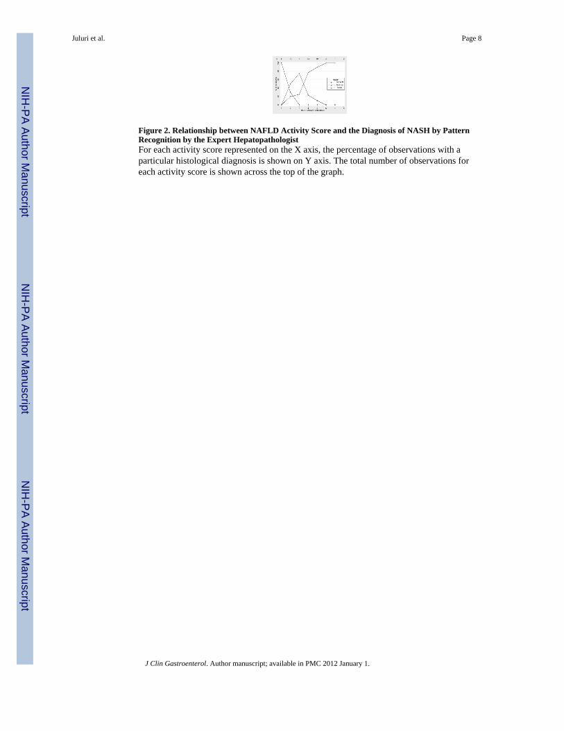
Figure 2. Relationship between NAFLD Activity Score and the Diagnosis of NASH by PatternRecognition by the Expert HepatopathologistFor each activity score represented on the X axis, the percentage of observations with aparticular histological diagnosis is shown on Y axis. The total number of observations foreach activity score is shown across the top of the graph.
Juluri et al. Page 8
J Clin Gastroenterol. Author manuscript; available in PMC 2012 January 1.
NIH
-PA Author Manuscript
NIH
-PA Author Manuscript
NIH
-PA Author Manuscript
NIH
-PA Author Manuscript
NIH
-PA Author Manuscript
NIH
-PA Author Manuscript
Juluri et al. Page 9
Table 1
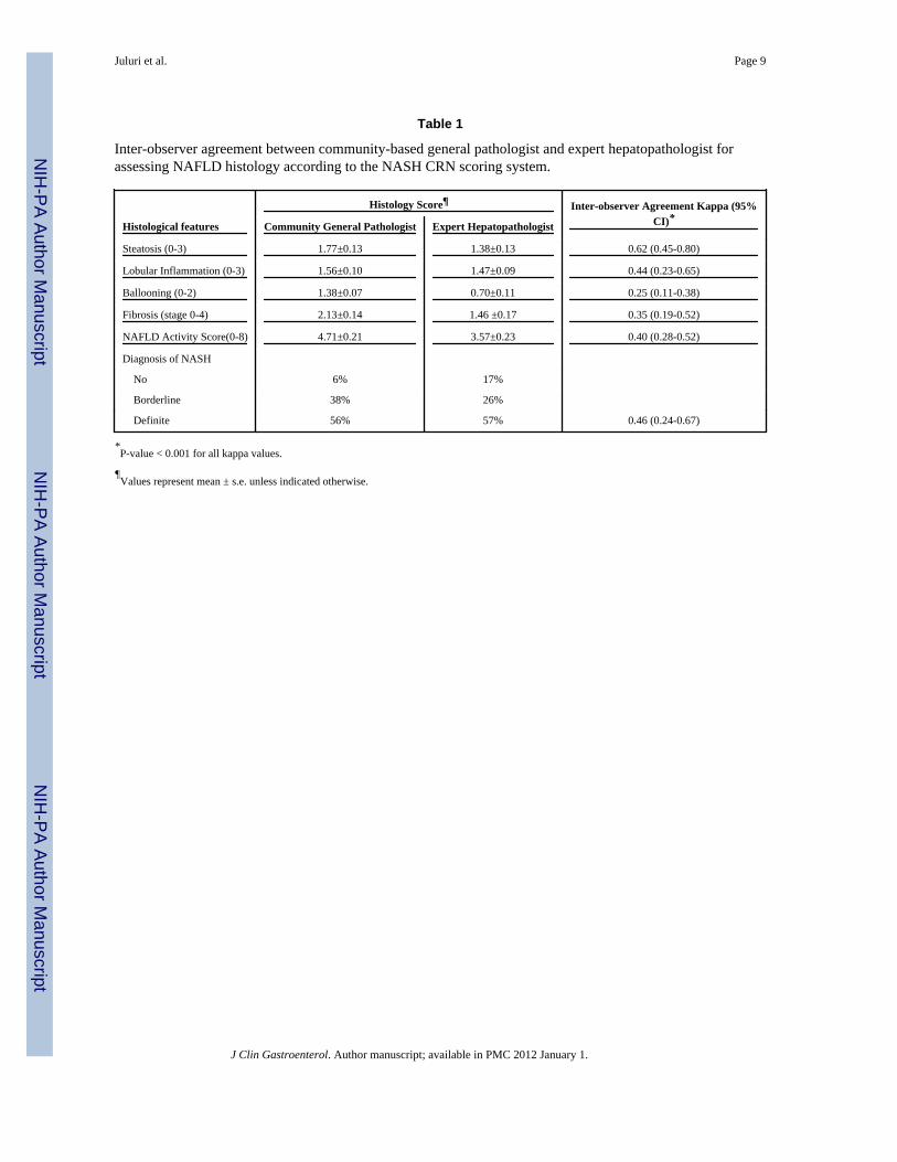
Inter-observer agreement between community-based general pathologist and expert hepatopathologist forassessing NAFLD histology according to the NASH CRN scoring system.
Histological features
Histology Score¶ Inter-observer Agreement Kappa (95%CI)*Community General Pathologist Expert Hepatopathologist
Steatosis (0-3) 1.77±0.13 1.38±0.13 0.62 (0.45-0.80)
Lobular Inflammation (0-3) 1.56±0.10 1.47±0.09 0.44 (0.23-0.65)
Ballooning (0-2) 1.38±0.07 0.70±0.11 0.25 (0.11-0.38)
Fibrosis (stage 0-4) 2.13±0.14 1.46 ±0.17 0.35 (0.19-0.52)
NAFLD Activity Score(0-8) 4.71±0.21 3.57±0.23 0.40 (0.28-0.52)
Diagnosis of NASH
No 6% 17%
Borderline 38% 26%
Definite 56% 57% 0.46 (0.24-0.67)
*P-value < 0.001 for all kappa values.
¶Values represent mean ± s.e. unless indicated otherwise.
J Clin Gastroenterol. Author manuscript; available in PMC 2012 January 1.
NIH
-PA Author Manuscript
NIH
-PA Author Manuscript
NIH
-PA Author Manuscript
Juluri et al. Page 10
Tabl
e 2
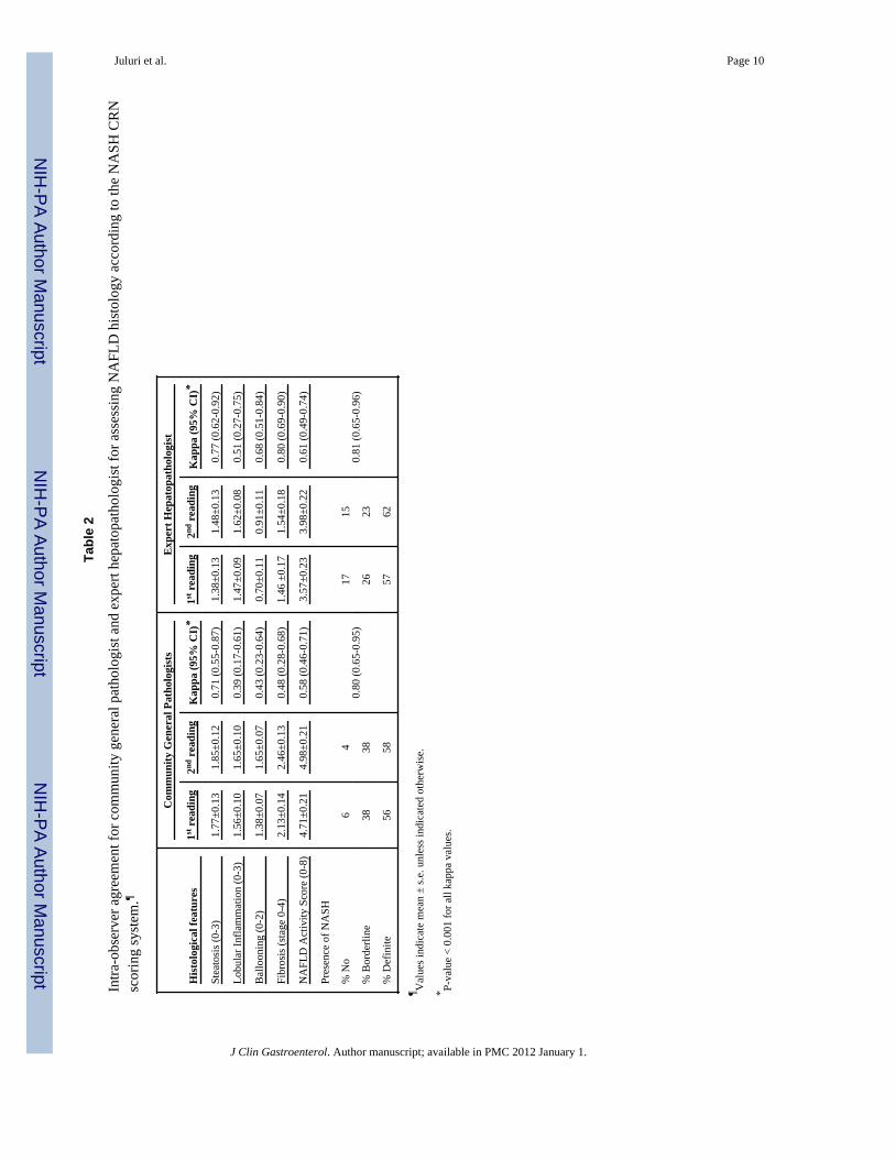
Intra
-obs
erve
r agr
eem
ent f
or c
omm
unity
gen
eral
pat
holo
gist
and
exp
ert h
epat
opat
holo
gist
for a
sses
sing
NA
FLD
his
tolo
gy a
ccor
ding
to th
e N
ASH
CR
Nsc
orin
g sy
stem
.
His
tolo
gica
l fea
ture
s
Com
mun
ity G
ener
al P
atho
logi
sts
Exp
ert H
epat
opat
holo
gist
1st r
eadi
ng2nd
rea
ding
Kap
pa (9
5% C
I)*
1st r
eadi
ng2nd
rea
ding
Kap
pa (9
5% C
I)*
Stea
tosi
s (0-
3)1.
77±0
.13
1.85
±0.1
20.
71 (0
.55-
0.87
)1.
38±0
.13
1.48
±0.1
30.
77 (0
.62-
0.92
)
Lobu
lar I
nfla
mm
atio
n (0
-3)
1.56
±0.1
01.
65±0
.10
0.39
(0.1
7-0.
61)
1.47
±0.0
91.
62±0
.08
0.51
(0.2
7-0.
75)
Bal
loon
ing
(0-2
)1.
38±0
.07
1.65
±0.0
70.
43 (0
.23-
0.64
)0.
70±0
.11
0.91
±0.1
10.
68 (0
.51-
0.84
)
Fibr
osis
(sta
ge 0
-4)
2.13
±0.1
42.
46±0
.13
0.48
(0.2
8-0.
68)
1.46
±0.
171.
54±0
.18
0.80
(0.6
9-0.
90)
NA
FLD
Act
ivity
Sco
re (0
-8)
4.71
±0.2
14.
98±0
.21
0.58
(0.4
6-0.
71)
3.57
±0.2
33.
98±0
.22
0.61
(0.4
9-0.
74)
Pres
ence
of N
ASH
0.80
(0.6
5-0.
95)
0.81
(0.6
5-0.
96)
% N
o6
417
15
% B
orde
rline
3838
2623
% D
efin
ite56
5857
62
¶ Val
ues i
ndic
ate
mea
n ±
s.e. u
nles
s ind
icat
ed o
ther
wis
e.
* P-va
lue
< 0.
001
for a
ll ka
ppa
valu
es.
J Clin Gastroenterol. Author manuscript; available in PMC 2012 January 1.
Copyright © 2022 FDOKUMEN