Cigarette smoking is not associated with specific histological features or severity of nonalcoholic...
11
CORRESPONDENCE Cigarette Smoking Is Not Associated with Specific Histological Features or Severity of Nonalcoholic Fatty Liver Disease To the Editor: We read with great interest the article by Azzalini et al., 1 who demonstrated that cigarette smoking worsens the severity of nonal- coholic fatty liver disease (NAFLD) in obese Zucker rats. In a related letter, Xu and coworkers 2 showed that cigarette smoking may act as a cofactor but not as an independent factor for NAFLD in humans. However, currently it is uncertain whether there is a significant association between smoking patterns and the severity of liver histology among patients with NAFLD. Clarification of this aspect may help to explain the underlying mechanisms and may be of clinical importance in planning preventive and therapeutic strat- egies. We have therefore assessed whether there is a significant asso- ciation between liver histology and smoking patterns among patients with biopsy-proven NAFLD. A total of 90 consecutive outpatients with NAFLD (43 males and 47 females, mean age, 47 6 8 years) were recruited from our clinics. All patients had chronically elevated liver enzymes and he- patic steatosis detected by ultrasonography. The NAFLD diagnosis was based on liver biopsy and exclusion of other known etiologic factors of chronic liver disease (alcohol abuse or intake 20 g/day, viral hepatitis, autoimmune hepatitis, and use of hepatotoxic drugs). An experienced pathologist blinded to clinical data scored the liver biopsies according to the National Institute of Diabetes and Digestive and Kidney Diseases NASH Clinical Research Net- work scoring system. 3 Pack-years of smoking were calculated as the product of the duration of smoking (in years) and the average number of cigarettes smoked per day. The protocol was approved by the local ethics committee, and all participants gave written informed consent. In multivariable-adjusted linear logistic regression models, each histological feature of NAFLD (i.e., steatosis grade, nec- roinflammatory grade, or fibrosis stage analyzed separately) was con- sidered as the dependent variable. Sex, age, body mass index, smok- ing, low-density lipoprotein cholesterol, homeostasis model assessment of insulin resistance (HOMA-IR) score, and metabolic syndrome (considered as a single clinical entity) were included as covariates. A total of 30 patients had ever smoked, 26 were past smokers, whereas 34 were current smokers. The distribution of nonsmokers, past smokers, and current smokers was not different in NAFLD patients classified according to liver histopathology (steatosis alone, borderline steatohepatitis, definite steatohepatitis). Notably, pack-years of smoking were not associated with degree of hepatic steatosis (P ¼ 0.67), necroinflammation (P ¼ 0.34), and fibrosis among patients with NAFLD (P ¼ 0.41). These results suggest that the severity of liver histopathology among patients with NAFLD is not associated with smoking patterns, after allowance for classical risk factors, insulin resistance, and the presence of the metabolic syndrome. This study has shown for the first time that the histological se- verity of NAFLD is not independently predicted by smoking pat- terns after adjustment for a broad spectrum of potential confound- ers, including the metabolic syndrome, a condition that is strongly correlated with NAFLD. Cigarette smoking is one of the major environmental factors suggested to play a crucial role in the devel- opment of several diseases. 4 Disorders such as atherosclerosis, lung cancer, or cardiovascular diseases are highly associated with tobacco consumption. However, cigarette smoking does not seem to influ- ence the histological features or the severity of NAFLD in a dose- dependent fashion. The biological mechanisms by which smoking could contribute to progressive NAFLD in humans are still poorly understood. Future follow-up studies are necessary to validate these findings and better estimate the risk of disease progression in rela- tion to smoking among patients with biopsy-proven NAFLD. YUSUF YILMAZ, M.D. OYA YONAL, M.D. RAMAZAN KURT , M.D. EROL AVSAR, M.D. Department of Gastroenterology Marmara University School of Medicine Altunizade, Istanbul, Turkey References 1. Azzalini L, Ferrer E, Ramalho LN, Moreno M, Domı ´nguez M, Colme- nero J, et al. Cigarette smoking exacerbates nonalcoholic fatty liver dis- ease in obese rats. HEPATOLOGY 2010; doi:10.1002/hep.23516. 2. Xu C, Yu C, Xu L, Miao M, Li Y. Is cigarette smoking an independent risk factor or a cofactor for nonalcoholic fatty liver disease? HEPATOLOGY 2010; doi:10.1002/hep.23644. 3. Kleiner DE, Brunt EM, Van Natta M, Behling C, Contos MJ, Cum- mings OW, et al. Design and validation of a histological scoring system for nonalcoholic fatty liver disease. HEPATOLOGY 2005;41:1313-1321. 4. Wipfli H, Samet JM. Global economic and health benefits of tobacco control: part 1. Clin Pharmacol Ther 2009;86:263-271. Copyright V C 2010 by the American Association for the Study of Liver Diseases. Published online in Wiley InterScience (www.interscience.wiley.com). DOI 10.1002/hep.23718 Potential conflict of interest: Nothing to report. Reply: We thank Dr. Yilmaz et al. for their interest in our recent study published in HEPATOLOGY . 1 As the authors described in their letter, our experimental study showing that cigarette smoking exacerbates nonalcoholic fatty liver disease (NAFLD) should be confirmed in human studies. In their letter, Yilmaz et al. show the results from a cross-sectional study including 90 patients with histologically-pro- ven NAFLD. The logistic regression analysis showed that cigarette smoking was not an independent factor associated with the severity of NAFLD, after adjusting for sex, age, BMI and other factors. The authors conclude that cigarette smoking per se does not wor- sen the severity of NAFLD. Although we acknowledge the effort of the authors to address this issue, we think that their observations are too preliminary to reach such strong conclusion. First, NAFLD is a highly heterogene- ous disease that involves many environmental and genetic factors and the series from Yilmaz et al. is clearly underpowered because it only includes 90 patients. Large epidemiological studies including a high number of well-characterized patients are clearly needed to elucidate the role of smoking on NAFLD. And second, the impact of total number of pack-years smoked on the development of severe forms of NAFLD according to Kleiner’s classification (i.e., definitive nonalcoholic steatohepatitis) should be specifically inves- tigated. In conclusion, carefully designed clinical studies including large number of patients are required to assess the role of smoking on the clinical course of NAFLD. LORENZO AZZALINI, M.D. JOSE ´ ALTAMIRANO, M.D. RAMO ´ N BATALLER, M.D., PH.D. Liver Unit, Hospital Clı´nic Institut d’Investigacions Biome`diques August Pi i Sunyer (IDIBAPS), Barcelona, Catalonia, Spain 391
-
Upload
independent -
Category
Documents
-
view
1 -
download
0
Transcript of Cigarette smoking is not associated with specific histological features or severity of nonalcoholic...

CORRESPONDENCE
Cigarette Smoking Is Not Associated with Specific Histological Features or Severity ofNonalcoholic Fatty Liver Disease
To the Editor:
We read with great interest the article by Azzalini et al.,1 whodemonstrated that cigarette smoking worsens the severity of nonal-coholic fatty liver disease (NAFLD) in obese Zucker rats. In arelated letter, Xu and coworkers2 showed that cigarette smokingmay act as a cofactor but not as an independent factor for NAFLDin humans. However, currently it is uncertain whether there is asignificant association between smoking patterns and the severity ofliver histology among patients with NAFLD. Clarification of thisaspect may help to explain the underlying mechanisms and may beof clinical importance in planning preventive and therapeutic strat-egies. We have therefore assessed whether there is a significant asso-ciation between liver histology and smoking patterns amongpatients with biopsy-proven NAFLD.
A total of 90 consecutive outpatients with NAFLD (43 malesand 47 females, mean age, 47 6 8 years) were recruited from ourclinics. All patients had chronically elevated liver enzymes and he-patic steatosis detected by ultrasonography. The NAFLD diagnosiswas based on liver biopsy and exclusion of other known etiologicfactors of chronic liver disease (alcohol abuse or intake �20 g/day,viral hepatitis, autoimmune hepatitis, and use of hepatotoxicdrugs). An experienced pathologist blinded to clinical data scoredthe liver biopsies according to the National Institute of Diabetesand Digestive and Kidney Diseases NASH Clinical Research Net-work scoring system.3 Pack-years of smoking were calculated as theproduct of the duration of smoking (in years) and the averagenumber of cigarettes smoked per day. The protocol was approvedby the local ethics committee, and all participants gave writteninformed consent. In multivariable-adjusted linear logistic regressionmodels, each histological feature of NAFLD (i.e., steatosis grade, nec-roinflammatory grade, or fibrosis stage analyzed separately) was con-sidered as the dependent variable. Sex, age, body mass index, smok-ing, low-density lipoprotein cholesterol, homeostasis model assessmentof insulin resistance (HOMA-IR) score, and metabolic syndrome(considered as a single clinical entity) were included as covariates.
A total of 30 patients had ever smoked, 26 were past smokers,whereas 34 were current smokers. The distribution of nonsmokers,past smokers, and current smokers was not different in NAFLDpatients classified according to liver histopathology (steatosis alone,borderline steatohepatitis, definite steatohepatitis). Notably, pack-yearsof smoking were not associated with degree of hepatic steatosis (P ¼0.67), necroinflammation (P ¼ 0.34), and fibrosis among patientswith NAFLD (P ¼ 0.41). These results suggest that the severity ofliver histopathology among patients with NAFLD is not associatedwith smoking patterns, after allowance for classical risk factors, insulinresistance, and the presence of the metabolic syndrome.
This study has shown for the first time that the histological se-verity of NAFLD is not independently predicted by smoking pat-terns after adjustment for a broad spectrum of potential confound-ers, including the metabolic syndrome, a condition that is stronglycorrelated with NAFLD. Cigarette smoking is one of the majorenvironmental factors suggested to play a crucial role in the devel-opment of several diseases.4 Disorders such as atherosclerosis, lungcancer, or cardiovascular diseases are highly associated with tobaccoconsumption. However, cigarette smoking does not seem to influ-ence the histological features or the severity of NAFLD in a dose-dependent fashion. The biological mechanisms by which smokingcould contribute to progressive NAFLD in humans are still poorlyunderstood. Future follow-up studies are necessary to validate thesefindings and better estimate the risk of disease progression in rela-tion to smoking among patients with biopsy-proven NAFLD.
YUSUF YILMAZ, M.D.OYA YONAL, M.D.RAMAZAN KURT, M.D.EROL AVSAR, M.D.Department of Gastroenterology
Marmara University School of MedicineAltunizade, Istanbul, Turkey
References1. Azzalini L, Ferrer E, Ramalho LN, Moreno M, Domınguez M, Colme-
nero J, et al. Cigarette smoking exacerbates nonalcoholic fatty liver dis-ease in obese rats. HEPATOLOGY 2010; doi:10.1002/hep.23516.
2. Xu C, Yu C, Xu L, Miao M, Li Y. Is cigarette smoking an independentrisk factor or a cofactor for nonalcoholic fatty liver disease? HEPATOLOGY
2010; doi:10.1002/hep.23644.3. Kleiner DE, Brunt EM, Van Natta M, Behling C, Contos MJ, Cum-
mings OW, et al. Design and validation of a histological scoring systemfor nonalcoholic fatty liver disease. HEPATOLOGY 2005;41:1313-1321.
4. Wipfli H, Samet JM. Global economic and health benefits of tobaccocontrol: part 1. Clin Pharmacol Ther 2009;86:263-271.
Copyright VC 2010 by the American Association for the Study of Liver Diseases.Published online in Wiley InterScience (www.interscience.wiley.com).DOI 10.1002/hep.23718Potential conflict of interest: Nothing to report.
Reply:
We thank Dr. Yilmaz et al. for their interest in our recent studypublished in HEPATOLOGY.1 As the authors described in their letter,our experimental study showing that cigarette smoking exacerbatesnonalcoholic fatty liver disease (NAFLD) should be confirmed inhuman studies. In their letter, Yilmaz et al. show the results from across-sectional study including 90 patients with histologically-pro-ven NAFLD. The logistic regression analysis showed that cigarettesmoking was not an independent factor associated with the severityof NAFLD, after adjusting for sex, age, BMI and other factors.The authors conclude that cigarette smoking per se does not wor-sen the severity of NAFLD.
Although we acknowledge the effort of the authors to addressthis issue, we think that their observations are too preliminary toreach such strong conclusion. First, NAFLD is a highly heterogene-ous disease that involves many environmental and genetic factorsand the series from Yilmaz et al. is clearly underpowered because itonly includes 90 patients. Large epidemiological studies includinga high number of well-characterized patients are clearly needed toelucidate the role of smoking on NAFLD. And second, the impactof total number of pack-years smoked on the development ofsevere forms of NAFLD according to Kleiner’s classification (i.e.,definitive nonalcoholic steatohepatitis) should be specifically inves-tigated. In conclusion, carefully designed clinical studies includinglarge number of patients are required to assess the role of smokingon the clinical course of NAFLD.
LORENZO AZZALINI, M.D.JOSE ALTAMIRANO, M.D.RAMON BATALLER, M.D., PH.D.Liver Unit, Hospital Clınic
Institut d’Investigacions Biomediques August Pi i Sunyer(IDIBAPS), Barcelona, Catalonia, Spain
391

Reference1. Azzalini L, Ferrer E, Ramalho LN, Moreno M, Domınguez M, Colme-
nero J, et al. Cigarette smoking exacerbates nonalcoholic fatty liver dis-ease in obese rats. HEPATOLOGY 2010;51:1567-1576.
Copyright VC 2010 by the American Association for the Study of Liver Diseases.Published online in Wiley InterScience (www.interscience.wiley.com).DOI 10.1002/hep.23749Potential conflict of interest: Nothing to report.
Ferritin and Liver Allocation? Impact on Mortality Not Only on the Waiting List But Also AfterOrthotopic Liver Transplantation Should Be Considered
To the Editor:
Recently, Walker and colleagues1 published a retrospective dual-center study of the impact of serum ferritin (SF) on the mortalityof candidates for orthotopic liver transplantation (OLT). Theyfound baseline SF levels greater than 200 lg/L to be an independ-ent predictor of waiting-list mortality, and they showed that theaddition of SF to Model for End-Stage Liver Disease (MELD) para-meters increased prognostic accuracy. Because additional factorsto improve MELD-based organ allocation would be generally de-sirable, the authors proposed the incorporation of SF into theMELD-based allocation system (as currently discussed for serumsodium2).
However, elevated SF not only reflects increased hepatic irondeposition but also indicates iron accumulation in extrahepaticsites. For example, cardiac iron deposition was found in transve-nous endomyocardial biopsy samples of 64% of patients with sub-stantial hepatic iron staining.3
Hence, impaired iron homeostasis not only may be predictiveof preoperative mortality but also may have a negative impact onthe outcome after OLT. In order to study this question, a numberof studies have compared the post-OLT survival of patients withnormal iron contents and patients exhibiting hepatic iron overloadin their explanted organs: Tung et al.4 reported significantly (P ¼0.0009) reduced 5-year post-OLT survival of only 40% in 37
patients with hepatic iron overload versus 62% in age-matchedcontrols. In 35 patients with hepatic iron overload (>40 lmol/g),the Queensland group5 found reduced 1- and 5-year unadjustedsurvival (P ¼ 0.27) after OLT of 74% and 63% versus 80% and72% in 178 patients with normal iron contents (<40 lmol/g). Ina multicenter study including 235 patients with hepatic iron over-load not associated with hereditary hemochromatosis, Kowdleyet al.6 observed reduced 5-year post-OLT survival of 63% versus72% in the overall population undergoing OLT (P ¼ 0.003).
Although these data suggest that elevated SF before OLT mayalso affect posttransplant outcomes, no study has been publishedso far concerning this issue.
Therefore, we looked at all adult patients who underwent trans-plantation at the Hannover Medical School Transplant Centerbetween January 1, 2004 and March 30, 2008. Patients with acuteliver failure, living donor OLT, and combined liver-heart or liver-lung transplantation and nine patients with hemochromatosis wereexcluded. Of the remaining 346 patients, pretransplant SF levelswere available for 92.2%. In a Kaplan-Meier analysis with a meanfollow-up of 1535 days, we found significantly (P ¼ 0.038, log-rank test) reduced survival (61.1% versus 71.9%) for patients withan SF level greater than or equal to 365 lg/L (Fig. 1).
To predict an optimal benefit from OLT, an ideal allocation pa-rameter predicting waiting-list mortality would be expected to havea low impact on posttransplant mortality. SF represents a parame-ter reflecting a variety of clinical problems that are capable of lim-iting outcomes and that are likely not all remedied by OLT.
In summary, the available data and our experience suggest thatwith the use of SF as an additional allocation parameter, mortalitymay potentially be shifted to some degree to the period after OLT.Further studies should therefore analyze the influence of SF notonly on waiting-list mortality but also on the posttransplantoutcome.
TOBIAS J. WEISMULLER, M.D.MICHAEL P. MANNS, M.D.CHRISTIAN P. STRASSBURG, M.D.Department of Gastroenterology, Hepatology, and Endocrinology
Hannover Medical School, Hannover, Germany
References1. Walker NM, Stuart KA, Ryan RJ, Desai S, Saab S, Nicol JA, et al.
Serum ferritin concentration predicts mortality in patients awaitingliver transplantation. HEPATOLOGY 2010;51:1683-1691.
2. Yun BC, Kim WR, Benson JT, Biggins SW, Therneau TM, KremersWK, et al. Impact of pretransplant hyponatremia on outcome followingliver transplantation. HEPATOLOGY 2009;49:1610-1615.
3. O’Glasser AY, Scott DL, Corless CL, Zaman A, Sasaki A, Gopal DV,et al. Hepatic and cardiac iron overload among patients with end-stageliver disease referred for liver transplantation. Clin Transplant; doi:10.1111/ j.1399–0012.2009.01136.x.
Fig. 1. Kaplan-Meier survival curves after OLT in patients with apretransplant SF level < 365 lg/L versus patients with an SF level� 365 lg/L. The curves are significantly different according to a log-rank test (P ¼ 0.038).
392 CORRESPONDENCE HEPATOLOGY, July 2010

4. Tung BY, Farrell FJ, McCashland TM, Gish RG, Bacon BR, KeeffeEB, et al. Long-term follow-up after liver transplantation in patientswith hepatic iron overload. Liver Transpl Surg 1999;5:369-374.
5. Stuart KA, Fletcher LM, Clouston AD, Lynch SV, Purdie DM, KerlinP, et al. Increased hepatic iron and cirrhosis: no evidence for an adverseeffect on patient outcome following liver transplantation. HEPATOLOGY
2000;32:1200-1207.6. Kowdley KV, Brandhagen DJ, Gish RG, Bass NM, Weinstein J, Schil-
sky ML, et al. Survival after liver transplantation in patients with he-patic iron overload: the national hemochromatosis transplant registry.Gastroenterology 2005;129:494-503.
Copyright VC 2010 by the American Association for the Study of Liver Diseases.Published online in Wiley InterScience (www.interscience.wiley.com).DOI 10.1002/hep.23732Potential conflict of interest: Nothing to report.
Reply:
We thank Weismuller et al. for their interesting commentsrelating to our recent article.1 Although we agree with their viewthat an ideal allocation parameter should have a low impact on thepost–liver transplant outcome, we assert that the severity of liverdisease and renal function are independent factors that influencethe outcome of orthotopic liver transplantation (OLT). However,these variables are constitutive elements of the Model for End-Stage Liver Disease score, which is currently widely applied todetermine organ allocation.2
Weismuller et al. presented novel data showing that patientswith a pre-OLT serum ferritin concentration greater than 365 lg/Lhad reduced survival in comparison with patients with a serum fer-ritin concentration below this threshold. Our previous studies haveshown that patients with cirrhosis-associated iron loading (CAFeL)are more likely to have an elevated serum ferritin concentrationand are also more likely to have a higher Child-Pugh score.3 Inour study, the Child-Pugh score and serum creatinine concentra-tion were associated with an adverse post-OLT outcome. Thus, theobservation that patients with an elevated serum ferritin concentra-tion have higher posttransplant mortality may simply reflect moreadvanced liver disease in those subjects. We look forward to a com-plete multivariate analysis of the Hannover data to confirm if thisis the case.
The observation that the serum ferritin concentration has im-portant prognostic significance with respect to pre-OLT (and possi-bly post-OLT) morbidity and mortality demands that the patho-physiological basis of this relationship be explored because apotential therapeutic target may be uncovered. The authors sug-gested that an elevated serum ferritin concentration may be associ-ated with iron deposition in extrahepatic sites (e.g., myocardium)that could compromise survival after OLT. Should the relationshipbe due to increased iron stores, then strategies to reduce body iron
need to be considered. In previous studies,3 we did not demon-strate a significant impact of CAFeL on the post-OLT outcomewhen all factors were considered. However, we recognize that thisissue remains controversial, and we acknowledge other reports ofadverse post-OLT outcomes associated with this condition and ex-perience with hereditary hemochromatosis by which affectedpatients are at increased risk of posttransplant mortality.4 An exactunderstanding of the clinical implications of CAFeL requires theproper identification of affected subjects, and this is difficult inpatients with cirrhosis because of the regional variation in the dis-tribution of iron in the same cirrhotic liver.5 Studies using mag-netic resonance imaging technology, which provides a global mea-surement of the hepatic iron concentration and thus overcomes theproblem of regional variation, may be particularly important indefining the exact clinical importance of this condition.
DARRELL H. G. CRAWFORD, M.D., FRACP1,2,3
LINDA M. FLETCHER, PH.D.1,2,3
KATHERINE A. STUART, PH.D., FRACP21Discipline of Medicine, University of Queensland
Brisbane, Queensland, Australia2Department of Gastroenterology and Hepatology
Princess Alexandra Hospital, BrisbaneQueensland, Australia
3Gallipoli Medical Research FoundationGreenslopes Hospital, Brisbane, Queensland, Australia
References1. Walker NM, Stuart KA, Ryan RJ, Desai S, Saab S, Nicol JA, et al.
Serum ferritin concentration predicts mortality in patients awaitingliver transplantation. HEPATOLOGY 2010;51:1683-1691.
2. Wiesner R, Edwards E, Freeman R, Harper A, Kim R, Kamath P, et al.Model of End-Stage Liver Disease (MELD) and allocation of donor liv-ers. Gastroenterology 2003;124:91-96.
3. Stuart KA, Fletcher LM, Clouston AD, Lynch SV, Purdie DM, KerlinP, et al. Increased hepatic iron and cirrhosis: no evidence for an adverseeffect on patient outcome following liver transplantation. HEPATOLOGY
2000;32:1200-1207.4. Crawford DHG, Fletcher LM, Hubscher S, Stuart KA, Gane E, Angus
PW, et al. Patient and graft survival after liver transplantation for he-reditary hemochromatosis: implications for pathogenesis. HEPATOLOGY
2004;39:1655-1662.5. Edmond MJ, Bronner MR, Carlson TH, Lin M, Labbe RF, Kowdley
KV. Quantitative study of the variability of hepatic iron concentration.Clin Chem 1999;45:340-346.
Copyright VC 2010 by the American Association for the Study of Liver Diseases.Published online in Wiley InterScience (www.interscience.wiley.com).DOI 10.1002/hep.23753Potential conflict of interest: Nothing to report.
Diagnostic Utility of Chromosome 17 and p16 Abnormalities in Fluorescence In SituHybridization Tests in Primary Sclerosing Cholangitis
To the Editor:
We read with great interest the article by Bangarulingam et al.regarding the long-term outcomes of positive fluorescence in situhybridization (FISH) in patients with primary sclerosing cholangi-tis (PSC).1 We applaud the authors for applying bile duct cytologyand FISH to a large cohort of patients with PSC to better charac-
terize the long-term outcomes. The authors used Vysis UroVysion,a commercially available kit that was approved by the U.S. Foodand Drug Administration in 2005 for use in the initial diagnosisof bladder cancer in patients with hematuria.2 This probe set hassince been applied to detect chromosomal abnormalities in variousbody sites including the detection of malignancies in biliary stric-tures.1,3-7 The UroVysion kit allows for the simultaneous testing of
HEPATOLOGY, Vol. 52, No. 1, 2010 CORRESPONDENCE 393

numeric aberrations, or aneusomy, of chromosome 3 (CEP3),
chromosome 7 (CEP7), and chromosome 17 (CEP17), as well as
band 9p21 (P16/CDKN2A) deletions.Unfortunately, the authors provide no information on the
results of CEP17 and p16 abnormalities in their cohort. We viewthe omission of the CEP17 and p16 results as a potential lost op-portunity. In histology specimens, p16 inactivation has been shownto be common in PSC-associated cholangiocarcinoma (CCA) with90% showing the loss of one allele which correlated with the lossof p16 expression in 57% of CCAs.8 Functional point mutationsin the p16 promoter likely contribute to the initiation and progres-sion of PSC-associated CCA.9 Using FISH, it was reported thatfour of six PSC-associated CCAs had CEP3, CEP7, and CEP17aneusomy.5 The two CCAs that did not have aneusomy had p16deletions.5 In addition, 64% of CCAs had CEP17 aneusomy, com-pared to 82% and 77% with aneusomy of CEP3 and CEP7,respectively.5 It appears that CEP17 aneusomy and p16 deletionsmay be more common in PSC-associated CCA than the authorsreport.
Since 2008, our liver program has adopted the use of FISH inaddition to cytology in the diagnosis of indeterminate stricturesand PSC-associated dominant strictures (n ¼ 56). In our initial se-ries, 12 tissue-proven CCAs were identified, of which 9 had non-diagnostic cytology.10 As reported previously, CEP3 and CEP7aneusomy were most commonly seen in CCA (7 of 12 CCAs).Among CCA cases with positive FISH and negative cytology, wefound that CEP17 aneusomy was present in 75% and p16 dele-tions were seen in 50%. Among the cases that had a p16 deletion(homozygous or heterozygous), nearly half of the cases (5 of 9)had no other chromosomal changes. Based on our experience andpreviously published data, we believe that the inclusion of CEP17and p16 status may have significant additional diagnosticimportance.
After reviewing their published data, we agree with the author’sconclusion that FISH is inadequate to be used as a CCA screeningmodality in unselected patients with PSC, but may have a role inpatients with a clinical or laboratory suspicion for PSC-associateddominant strictures. However, we question if their conclusionwould have changed with the inclusion of CEP17 aneusomy and/or p16 deletions. Because these results are currently available to theauthors for all patients, we ask that they reconsider the exclusionof this data from interpretation.
LANCE L. STEIN, M.D.1
TAMAS A. GONDA, M.D.2
PETER D. STEVENS2
ROBERT S. BROWN JR., M.D., M.P.H.11Department of Medicine, Center for Liver Disease and
Transplantation, and2Department of Medicine, Division of Gastroenterology, Columbia
University College of Physicians and Surgeons, New York, NY
References1. Bangarulingam SY, Bjornsson E, Enders F, Barr Fritcher EG, Gores G,
Halling KC, et al. Long-term outcomes of positive fluorescence in situhybridization tests in primary sclerosing cholangitis. HEPATOLOGY 2010;51:174-180.
2. Halling KC, King W, Sokolova IA, Meyer RG, Burkhardt HM, HallingAC, et al. A comparison of cytology and fluorescence in situ hybridiza-tion for the detection of urothelial carcinoma. J Urol 2000;164:1768-1775.
3. Moreno-Luna LE, Kipp B, Halling KC, Sebo TJ, Kremers WK, RobertsLR, et al. Advanced cytologic techniques for the detection of malignantpancreatobiliary strictures. Gastroenterology 2006;131:1064-1072.
4. Kipp BR, Stadheim LM, Halling SA, Pochron NL, Harmsen S, Nagor-ney DM, et al. A comparison of routine cytology and fluorescence in
situ hybridization for the detection of malignant bile duct strictures.Am J Gastroenterol 2004;99:1675-1681.
5. DeHaan RD, Kipp BR, Smyrk TC, Abraham SC, Roberts LR, HallingKC. An assessment of chromosomal alterations detected by fluorescencein situ hybridization and p16 expression in sporadic and primary scle-rosing cholangitis-associated cholangiocarcinomas. Hum Pathol 2007;38:491-499.
6. Barr Fritcher EG, Kipp BR, Slezak JM, Moreno-Luna LE, Gores GJ,Levy MJ, et al. Correlating routine cytology, quantitative nuclear mor-phometry by digital image analysis, and genetic alterations by fluores-cence in situ hybridization to assess the sensitivity of cytology fordetecting pancreatobiliary tract malignancy. Am J Clin Pathol 2007;128:272-279.
7. Levy MJ, Baron TH, Clayton AC, Enders FB, Gostout CJ, HallingKC, et al. Prospective evaluation of advanced molecular markers andimaging techniques in patients with indeterminate bile duct strictures.Am J Gastroenterol 2008;103:1263-1273.
8. Ahrendt SA, Eisenberger CF, Yip L, Rashid A, Chow JT, Pitt HA, et al.Chromosome 9p21 loss and p16 inactivation in primary sclerosing chol-angitis-associated cholangiocarcinoma. J Surg Res 1999;84:88-93.
9. Taniai M, Higuchi H, Burgart LJ, Gores GJ. p16INK4a promotermutations are frequent in primary sclerosing cholangitis (PSC) andPSC-associated cholangiocarcinoma. Gastroenterology 2002;123:1090-1098.
10. Glick M GT, Iqbal S, Nandula S, Kang JU, Murty V, Stevens P, et al.Fluorescent in situ hybridization in the diagnosis of indeterminate bili-ary strictures [Abstract]. Am J Gastroenterol 2009;104(Suppl. 3):177.
Copyright VC 2010 by the American Association for the Study of Liver Diseases.Published online in Wiley InterScience (www.interscience.wiley.com).DOI 10.1002/hep.23698Potential conflict of interest: Nothing to report.
Reply:
We would like to thank Dr. L. Stein and colleagues for thecomments on our article regarding long-term outcomes of positivefluorescence in situ hybridization (FISH) in patients with primarysclerosing cholangitis (PSC).1
With regard to the major concern about not providing informa-tion on results for CEP17 and P16, we were aware that the P16(at 9p21) and P53 (on 17p) genes are frequently inactivatedthrough gene deletion, point mutation, or promoter hypermethyl-ation in cholangiocarcinoma (CCA). The only one of these abnor-malities detectable with FISH is gene deletion. It is important tounderstand that the results we showed were for a clinically devel-oped and validated test and not a research study. The reason datawas not provided for P16 is that when the assay was developedand validated for clinical use, we found it difficult to use the 9p21(P16) probe for discerning cancer. One of the reasons was that the9p21 probe is yellow and bile autofluoresces yellow, so it was diffi-cult to see the 9p21 probe and to be comfortable with the numberof copies of 9p21 in a cell. We no longer use bile aspirates, so thatis not really an issue anymore (i.e., there is not a problem withyellow autofluorescence in bile duct brushings).
The more important reason we decided not to use 9p21 whenthis was clinically implemented in 2003 is that it would have madeanalyzing the specimens more complicated and laborious. Wewanted an assay that could be done in a reasonable amount oftime. The reason that it would create more work is that 9p21 isdirected to the P16 tumor suppressor gene and the change thatone would be looking for is deletion of the 9p21 probe (one orzero copies). It should be understood that a low but appreciablefraction of normal cells show what looks like 9p21 deletion. Forexample, an average of about 10% of normal cells might beexpected to show only a single copy of 9p21. This is not due to
394 CORRESPONDENCE HEPATOLOGY, July 2010

real deletion but overlap of the two probe signals such that itappears to be a single copy. Because of this, to feel comfortablecalling a case positive for 9p21 loss without generating false posi-tive calls would require having a cutoff of well over 10% of thecells showing hemizygous 9p21 loss. To capture this informationwould require formal enumeration of 50 or 100 cells and increasethe amount of time required to clinically interpret the assay. Asindicated before, we wanted a clinical assay that was not too labori-ous. However, we recognized that we might have some false nega-tive results because we might miss cases that showed only 9p21loss without any chromosomal gains. So, the bottom line for 9p21(P16) is that Stein et al. are correct that we probably failed todetect some cases of CCA due to not assessing for this. Onceagain, it is important to realize that this was a clinical study andnot a research study. We could always go back and try to capturethat data but it would not reflect the clinical test that was used.
With regard to CEP17, aneusomy can refer to gain or loss of aprobe. Stein et al. do not state which type of CEP17 aneusomythey were interested in knowing more about. If they are referringto loss of CEP17, then the reason we did not capture that infor-mation is for the same reasons listed above, i.e., that it wouldrequire a more laborious enumeration due to the low-level artifac-tual CEP17 loss that exists in normal cells. If they are referring togain of CEP17, then even though we did not specifically captureinformation on CEP17 gain, cells with CEP17 gain would havebeen categorized as trisomic (if only CEP17 was gained, which isvery rare in our patient population) or polysomic (if CEP17 wasgained along with gain of one or more of the other probes), andthus that information was captured within those diagnosticcategories. Incidentally, we know that cases with P53 loss (due topartial or complete loss of chromosome 17) tend to show
chromosomal instability (gains) which manifests as polysomy byFISH. For this additional reason, CEP17 (P53) loss is unlikelyto add any additional sensitivity beyond that obtained withpolysomy.
We are currently working to develop a FISH enumeration pro-cedure that will allow us to use 9p21 loss as a criterion for positiv-ity in the clinical assay that we use. This should allow us toincrease the sensitivity of the assay.
SANJAY Y. BANGARULINGAM, M.D.1
EINAR BJORNSSON, M.D.1
FELICITY ENDERS, PH.D.1
EMILY G. BARR FRITCHER2
GREGORY GORES, M.D.1
KEVIN C. HALLING, M.D., PH.D.2
KEITH D. LINDOR, M.D.11Division of Gastroenterology and Hepatology and the
2Division of Laboratory Medicine and PathologyMayo Clinic, Rochester, MN
Reference1. Bangarulingam SY, Bjornsson E, Enders F, Barr Fritcher EG, Gores G,
Halling KC, et al. Long-term outcomes of positive fluorescence in situhybridization tests in primary sclerosing cholangitis. HEPATOLOGY 2010;51:174-180.
Copyright VC 2010 by the American Association for the Study of Liver Diseases.Published online in Wiley InterScience (www.interscience.wiley.com).DOI 10.1002/hep.23758Potential conflict of interest: Nothing to report.
Methodological Issues in a Meta-Analysis
To the Editor:
In a recent article published in HEPATOLOGY, Awad et al.1 pres-ent a meta-analysis comparing peginterferon alfa-2a and peginter-feron alfa-2b for hepatitis C treatments. Using data from eight tri-als, the authors concluded that the proportion of patients achievingsustained virological response (SVR) with peginterferon alfa-2a wassignificantly higher than the proportion achieving SVR with pegin-terferon alfa-2b. Of the 3070 patients in the Individualized DosingEfficacy Versus Flat Dosing to Assess Optimal Pegylated InterferonTherapy (IDEAL) trial, 1016 were included in the meta-analysis,even though they received a dosage of 1.0 lg/kg/week, which islower than the approved starting dosage of peginterferon alfa-2b(1.5 lg/kg/week). This letter, however, focuses on concerns aboutmethodological issues in the meta-analysis.
The validity of the random effects model used in the article isdependent on a large number of individual studies, each with suffi-ciently large samples.2-4 In Awad et al.’s study,1 only eight trialswere used, five of which had fewer than 50 observations per arm.The largest trial5 alone represented nearly 70% of observations.Based on that trial, past research has shown no difference betweenpeginterferon alfa-2a and peginterferon alfa-2b with respect toSVR. This underlines the importance of sensitivity analyses forexamining the robustness of Awad et al.’s conclusions. Because theauthors showed that there was no evidence of heterogeneity (I2 ¼0) among the eight studies that they included, a fixed effects modelapproach is appropriate. I applied a fixed effects exact inferenceprocedure (proposed by Tian et al.6) as a sensitivity analysis to the
data shown in Fig. 2 of Awad et al.’s article. This method isunbiased even for small samples or when only a small number ofstudies are included in the meta-analysis. This robust methodyielded a 95% confidence interval for the risk ratio of 0.988-1.214, which included the null value of 1. A similar analysis lim-ited to the approved starting dose of 1.5 lg/kg/week (whichexcluded 1016 of the 3070 patients in the IDEAL trial) alsoshowed a lack of a statistically significant difference with an exact95% confidence interval of 0.967-1.214.
The discrepancy between the findings based on large samplemethods and those based on exact methods emphasizes the needfor thorough sensitivity analyses using a variety of appropriatestatistical methods whenever possible. Here, the use of an exactmethod, which is likely more appropriate because of the limited num-ber of studies, shows no statistically significant difference between thetwo drugs; this result directly contradicts the findings of Awad et al.1
Aside from the biases resulting from the reliance on large sam-ple properties when statistical analyses of small samples are beingperformed, current meta-analyses often reduce a complicated, mul-titrial meta-analysis to a single parameter from which absolute con-clusions are drawn. A number of recent articles, including ones byWang et al.7 and Cai et al.,8,9 present a method that, applied to theissue of peginterferon alfa-2a versus peginterferon alfa-2b, would pro-vide clinicians with clearer guidance about which product is mostlikely to be an appropriate treatment for any given patient.
PIERRE CREMIEUX, PH.D.Analysis Group, Inc., Boston, MA
HEPATOLOGY, Vol. 52, No. 1, 2010 CORRESPONDENCE 395

References1. Awad T, Thorlund K, Hauser G, Stimac D, Mabrouk M, Gluud C.
Peginterferon alpha-2a is associated with higher sustained virologicalresponse than peginterferon alfa-2b in chronic hepatitis C: systematicreview of randomized trials. HEPATOLOGY 2010;51:1176-1184.
2. Brockwell SE, Gordon IR. A comparison of statistical methods formeta-analysis. Stat Med 2001;20:825-840.
3. Bohning D, Malzahn U, Dietz E, Schlattmann P, Viwatwongkasem C,Biggeri A. Some general points in estimating heterogeneity variancewith the DerSimonian-Laird estimator. Biostatistics 2002;3:445-457.
4. Viechtbauer W. Confidence intervals for the amount of heterogeneityin meta-analysis. Stat Med 2007;26:37-52.
5. McHutchison JG, Lawitz EJ, Shiffman ML, Muir AJ, Galler GW,McCone J, et al. Peginterferon alfa-2b or alfa-2a with ribavirin fortreatment of hepatitis C infection. N Engl J Med 2009;361:580-593.
6. Tian L, Cai T, Pfeffer MA, Piankov N, Cremieux PY, Wei LJ. Exactand efficient inference procedure for meta-analysis and its applicationto the analysis of independent 2 � 2 tables with all available data butwithout artificial continuity correction. Biostatistics 2009;10:275-281.
7. Wang R, Tian L, Cai T, Wei LJ. Nonparametric inference procedurefor percentiles of the random effects distribution in meta analysis. AnnAppl Stat. 2010;4:520-532.
8. Cai T, Tian L, Wong PH, Wei LJ. Analysis of randomized comparativeclinical trial data for personalized treatment selections. Biostatistics. Inpress.
9. Cai T, Tian L, Uno H, Solomon SD, Wei LJ. Calibrating parametricsubject-specific risk estimation. Biometrika. In press.
Copyright VC 2010 by the American Association for the Study of Liver Diseases.Published online in Wiley InterScience (www.interscience.wiley.com).DOI 10.1002/hep.23762Potential conflict of interest: Nothing to report.
Reply:
We thank Dr. Cremieux for bringing this issue to our attention,and we agree that sensitivity analysis is an important and necessarycomponent of all systematic reviews. We are, however, concernedabout the validity of the method proposed by Tian et al.1 that Cre-mieux has chosen for his re–meta-analysis of sustained virologicalresponse. Currently, the method by Tian et al. has only beensparsely validated for meta-analysis scenarios (>30 trials with eventrates < 10%) that are far from representative of the sustained viro-logical response meta-analysis (8 trials with event rates rangingfrom 20% to 80%).2 Although the method by Tian et al. may intime be proven statistically superior, at the current stage, preferringthis method over the conventional meta-analysis methods is analo-gous to preferring a phase 1 drug over a Food and Drug Adminis-tration–approved drug in clinical practice.
Cremieux points out that the lack of evidence for heterogeneity(I2 ¼ 0%) makes the fixed-effects model appropriate. In otherwords, Cremieux suggests that the choice of model (fixed versusrandom) may be determined by the estimate of heterogeneity (I2).This is highly inappropriate meta-analytic conduct that theCochrane Collaboration abandoned some years ago.3 Findingsfrom empirical studies and simulation studies also testify to theinappropriateness of this conduct.4-6 However, since we do nothave evidence of heterogeneity, we cannot preclude the possibilitythat a fixed-effects model is appropriate. Using the conventionalMantel-Haenszel fixed-effects model, we obtained a pooled relativerisk of 1.10 (95% confidence interval ¼ 1.03-1.18). Excludinglow-dose peginterferon alfa-2b from the Individualized Dosing Ef-ficacy Versus Flat Dosing to Assess Optimal Pegylated InterferonTherapy (IDEAL) trial, we obtained a pooled relative risk of 1.10(95% confidence interval ¼ 1.03-1.19).
In his commentary, Cremieux interprets his findings as discrep-ant with the original findings of our systematic review. Althoughwe do agree that some caution should be exercised, the word dis-crepant seems too strong to describe the observed statistical differ-ence. In fact, this claim of discrepancy is based on an unfair com-parison. In our original analyses, we took precautions to interpretthe statistical inference according to the strength of the evidence.In this vein, we constructed adjusted thresholds for statistical sig-nificance by using an approach (trial sequential monitoring boun-daries) analogous to approaches used for interim analysis in clinicaltrials.7-10 Roughly speaking, we can state that this analysis trans-lates into an adjusted P value less than 5% but not less than 1%(the unadjusted meta-analyzed P value was 0.8%). In comparison,the confidence intervals proposed by Tian et al.1 would translateinto a P value of roughly 6%. Several authors have previouslywarned against relying on the conventional criterion for statisticalsignificance.7,8,11-14 In this vein, a P value of 1% to 5% and a Pvalue of 6% should be interpreted similarly.
We thank Cremieux for bringing up issues that may have beenhidden to the statistically inexperienced reader. However, even ifone were to take a leap of faith and believe that Cremieux’s analy-ses were based on a valid method, the conclusions of our analysisand his analysis still do not differ: there is statistical evidence,albeit moderate, that peginterferon alfa-2a is superior to peginter-feron alfa-2b in achieving sustained virological response.
KRISTIAN THORLUND, M.SC.TAHANY AWAD, M.SC.Cochrane Hepatobiliary Group
Copenhagen Trial UnitCenter for Clinical Intervention ResearchRigshospitalet, Copenhagen University HospitalCopenhagen, Denmark
References1. Tian L, Cai T, Pfeffer MA, Piankov N, Cremiex P. Exact and efficient
inference procedure for meta-analysis and its application to the analysisof independent 2x2 tables with all available data but without artificialcontinuity correction. Biostatistics 2009;10:275-281.
2. Awad T, Thorlund K, Hauser G, Stimac D, Mabrouk M, Gluud C.Peginterferon alpha-2a is associated with higher sustained virologicalresponse than peginterferon alfa-2b in chronic hepatitis C: systematicreview of randomized trials. HEPATOLOGY 2010;51:1176-1184.
3. Higgins JPT, Green S, editors. Cochrane Handbook for SystematicReviews of Interventions, Version 5.0.1 [updated September 2008].The Cochrane Collaboration, 2008. Available from www.cochrane-handbook.org.
4. Ioannidis JP, Patsopoulos NA, Evangelou E. Uncertainty in heterogene-ity estimates in meta-analyses. BMJ 2007;335:914-916.
5. Rucker G, Schwarzer G, Carpenter JR, Schumacher M. Undue relianceon I(2) in assessing heterogeneity may mislead. BMC Med Res Meth-odol 2008;8:79.
6. Sidik K, Jonkman JN. A comparison of heterogeneity variance estima-tors in combining results of studies. Stat Med 2007;26:1964-1981.
7. Pogue J, Yusuf S. Cumulating evidence from randomized trials: utiliz-ing sequential monitoring boundaries for cumulative meta-analysis.Control Clin Trials 1997;18:580-593.
8. Thorlund K, Devereaux PJ, Wetterslev J, Guyatt G, Ioannidis JP, Tha-bane L, et al. Can trial sequential monitoring boundaries reduce spuri-ous inferences from meta-analyses? Int J Epidemiol 2009;38:276-286.
9. Wetterslev J, Thorlund K, Brok J, Gluud C. Trial sequential analysismay establish when firm evidence is reached in cumulative meta-analy-sis. J Clin Epidemiol 2008;61:64-75.
10. Wetterslev J, Thorlund K, Brok J, Gluud C. Estimating required infor-mation size by quantifying diversity in a random-effects meta-analysis.BMC Med Res Methodol 2009;9:86.
396 CORRESPONDENCE HEPATOLOGY, July 2010

11. Berkey C, Mosteller F, Lau J. Uncertainty of the time of first signifi-cance in random-effects cumulative meta-analysis. Control Clin Trials1996;17:357-371.
12. Gehr B, Weiss C, Porzsolt F. The fading of reported effectiveness. Ameta-analysis of randomised controlled trials. BMC Med Res Methodol2006;6:25.
13. Sterne JA, Davey SG. Sifting the evidence—what’s wrong with signifi-cance tests? Br Med J 2001;322:226-231.
14. Trikalinos TA, Churchill R, Ferri M, Leucht S, Tuunainen A, WahlbeckK, et al. Effect sizes in cumulative meta-analyses of mental health random-ized trials evolved over time. J Clin Epidemiol 2004;57:1124-1130.
Copyright VC 2010 by the American Association for the Study of Liver Diseases.Published online in Wiley InterScience (www.interscience.wiley.com).DOI 10.1002/hep.23764Potential conflict of interest: Nothing to report.
Routine Hepatitis B Virus DNA Testing in Patients with Human Immunodeficiency Virus
To the Editor:
We read the article by Nunez with great interest.1 In the literature,three cases who were positive for human immunodeficiency virus(HIV) were described with hepatitis B reactivation after withdrawal ofhepatitis B virus (HBV)-active drug due to the virologic failure ofHIV. All three of the patients were positive for antibody to hepatitis Bcore antigen (anti-HBc).2,3 The HBV reactivations could be controlledby highly active antiretroviral therapy regimens including lamivudineand tenofovir in the first patient,2 tenofovir/emtricitabine in the sec-ond patient,3 and without any HBV-active drug in the third patient.2
The author’s concerns were mostly based on economics. How-ever, without knowing the HBV DNA presence, we should get somedifferent recommendations for clinicians, such as choosing an HBV-active drug in all anti-HBc–positive patients with HIV. Moreover,some authors also suggest hat the follow-up should be based onHBV DNA levels in only anti-HBc–positive patients with HIV.2
AKIF ALTINBAS, M.D.FUAT EKIZ, M.D.OSMAN YUKSEL, M.D., ASSOC. PROF
Department of GastroenterologyDıskapı Yıldırım Beyazıt Education and Research HospitalAnkara, Turkey
References1. Marina Nunez. Routine hepatitis B virus DNA testing in human im-
munodeficiency virus-infected patients with positive hepatitis B coreantibody but negative hepatitis B surface antigen is not justified by cur-rent evidence. HEPATOLOGY 2010; doi:10.1002/hep.23395.
2. Chamorro AJ, Casado JL, Bellido D, Moreno S. Reactivation of hepati-tis B in an HIV-infected patient with antibodies against hepatitis Bcore antigen as the only serological marker. Eur J Clin Microbiol InfectDis 2005;24:492-494.
3. Bloquel B, Jeulin H, Burty C, Letranchant L, Rabaud C, Venard V.Occult hepatitis B infection in patients infected with HIV: Report oftwo cases of hepatitis B reactivation and prevalence in a hospitalcohort. J Med Virol 2010;82:206-212.
Copyright VC 2010 by the American Association for the Study of Liver Diseases.Published online in Wiley InterScience (www.interscience.wiley.com).DOI 10.1002/hep.23553Potential conflict of interest: Nothing to report.
Think Twice If You Consider Entecavir Treatment in Cases with Lamivudine Refractoriness
To the Editor:
We read with interest the article by Shim et al.1 concerning theefficacy of entecavir (ETV) in patients with chronic hepatitis B re-sistant to both lamivudine (LAM) and adeofovir (ADV) or toLAM alone. In this study, they indicated that ETV given at 1 mg/day for 48 weeks resulted in lesser hepatitis B virus (HBV) DNAand alanine aminotransferase (ALT) reduction in the LAM/ADV-resistant group than compared with the LAM-resistant group.They also noted that HBV DNA loss was significantly higher inthe LAM-resistant group compared with the LAM/ADV-resistantgroup (34% versus 10%). However, they showed that virologicalbreakthrough was similar in both groups. They also underlinedthat virological response at 12 weeks determined the degree ofHBV DNA reduction over 48 weeks of therapy, regardless of previ-ous antiviral treatment.
In their study, Shim et al. underlined the importance of multi-drug resistance in cases with inappropriate use of antivirals in
HBV infection. The same authors suggested, in the introductionsection, that ‘‘in terms of salvage therapy for LAM-resistant orADV-resistant chronic hepatitis B infection, the American Associa-tion for the Study of Liver Diseases (AASLD) practice guidelinerecommended switching to ETV’’ monotherapy as an optimalstrategy. However, according to the current guidelines, includingAASLD 2009,2 European Association for the Study of the Liver2009,3 and Asian Pacific Association for the Study of the Liver2008,4 what the authors did seemed to be inappropriate to suggestto the readers. The guidelines mentioned above unanimously indi-cated that ETV can be recommended as a rescue therapy only forADV-resistant chronic HBV infection having Asp236-to-Thr236(N236T) and/or Ala181-to-Thr181/Val181 (A181T/V) substitu-tions. Contrary to what the authors wrote in the introduction sec-tion of their article, AASLD guidelines in 20092 on HBV infectionclearly indicate that ETV is not an optimal treatment for LAM-re-fractory HBV. It is clearly known that Leu180-to-Met180(L180M) þ Met204-to-Val204 (M204V) and L180M þ M204V
HEPATOLOGY, Vol. 52, No. 1, 2010 CORRESPONDENCE 397

þ Asn236-to-Thr236 (N236T) mutants behaved 6.25-fold resist-ant to ETV compared with wild-type HBV.5 We also know thatgenotypic resistance to ETV will develop at a rate of 43% at theend of 4 years.6 Expectedly, two patients in the series of Shimet al.1 developed virological breakthrough with Ser202-to-Gly202(S202G) ETV resistance substitutions at 36 weeks of treatment,and one patient developed biochemical breakthrough in the LAM-resistant group of patients. Unfortunately, the readers were notinformed in this article how the authors treated these two cases intheir series. Another relevant article in this field showed thatalthough HBV DNA suppression was achieved in a higher percent-age of patients, there was an emergence of nearly 8% resistance toETV monotherapy in cases with previous LAM resistance in year2.7 Thus, this strategy led to selection of multidrug-resistant HBVstrains with maximal viral resistance in the near future. Anotherconcern with this article is that the authors also underlined theimpact of 1 log HBV DNA reduction at 12 weeks on antiviral effi-cacy at 48 weeks. However, it would be more valuable if theycould provide us with the threshold level of HBV DNA reductionat 12 weeks to achieve HBV undetectability in their cases.
As a result, the authors in this article1 tested an approach whichis absolutely not valid and nor practical at present. Currently, webelieve that ETV monotherapy is not a good alternative as a rescuetherapy for cases with LAM and or LAM/ADV resistance, whereascontinued treatment resulted in virus suppression in a higher per-centage of patients in the series of Shim and colleagues. ETV isobviously not a drug with a high genetic barrier to resistance inthe setting of LAM refractoriness. In such situations, we have toadmit the effectiveness of other alternative drugs, includingtenofovir.
YUCEL USTUNDAG1
OMER TOPALAK2
1Zonguldak Karaelmas University School of Medicine,Department of Internal Medicine, Gastroenterology Clinics,Zonguldak, Turkey
2Dokuz Eylul University School of Medicine,Department of Internal Medicine, Gastroenterology Clinics,Izmir, Turkey
References1. Shim JH, Suh DJ, Kim KM, Lim YS, Lee HC, Chung YH, et al. Effi-
cacy of entecavir in patients with chronic hepatitis B resistant to both
lamivudine and adefovir or to lamivudine alone. HEPATOLOGY 2009;50:
1064-1071.2. Lok AS, MacMahon BJ. Chronic hepatitis B: Update 2009. Hepato-
logy 2009;50:661-666.3. European Association for the Study of the Liver.EASL clinical practice
guidelines: Management of chronic hepatitis B. J Hepatol 2009;50:
227-242.4. Leung N. Recent data on treatment of chronic hepatitis B with nucleos
(t)ide analogues. Hepatol Int 2008;2:163-178.5. Brunelle MN, Jacquard AC, Pichoud C, Durantel D, Carrouee-Duran-
tel S, Villeneuve JP, et al. Susceptibility to antivirals of a human HBV
strain with mutations conferring resistance to both lamivudine and ade-
fovir. HEPATOLOGY 2005;41:1391-1398.6. Colonno RJ, Rose RE, Pokornowski K. Four year assessment of entec-
vir resistance in nucleoside resistance in nucleoside naive and lamivudin
refractory patients. J Hepatol 2007;46:S294.7. Sherman M, Yurdaydin C, Simsek H, Silva M, Liaw YF, Rustgi VK,
et al. Benefits of Entecavir for Hepatitis B Liver Disease (BEHoLD)
Study Group. Entecavir therapy for lamivudine-refractory chronic hepa-
titis B: improved virologic, biochemical, and serology outcomes
through 96 weeks. HEPATOLOGY 2008;48:99-108.
Copyright VC 2010 by the American Association for the Study of Liver Diseases.Published online in Wiley InterScience (www.interscience.wiley.com).DOI 10.1002/hep.23555Potential conflict of interest: Nothing to report.
Hemochromatosis Protein HFE C282Y Conformational Considerations
To the Editor:
I read with interest the results of the recent study by Vecchiet al. on HFE C282Y mutation impairing protein traffic to theplasma membrane, which is associated with lower hepcidin expres-sion.1 Indeed, hepcidin suppression has been consistently linked toiron overload. Furthermore, the homozygous Cys-to-Tyr mutationat residue 282 (C282Y) of the hemochromatosis protein HFE (themost common form of iron overload) is recognized to induce theformation of aggregates that are retained in the endoplasmic reticu-lum (ER).2,3
The report by Vecchi et al. importantly suggests that the abnor-mal protein trafficking of the mutant HFE C282Y protein directlyresults in the suppression of hepcidin expression. Viewing HFEC282Y hereditary hemochromatosis in the context of aberrant pro-tein trafficking that leads to the suppression of hepcidin has impor-tant implications for iron overload regulation, thus highlightingthe conformational aspects of HFE C282Y protein in the onsetand variable pathogenesis of this conditions.4
A close relationship exists between abnormal protein traffickingand clinical consequences, evidence of which can be observed in arange of disorders.5 This emphasis of the study by Vecchi et al. isthat investigations of the misfolding protein (HFE C282Y) may
provide further intriguing possibilities for the understanding of thiscondition.
Thus, the recognition of HFE C282Y hereditary hemochroma-tosis aberrant protein trafficking as an important consideration forthis condition may reveal new and more-effective approaches to di-agnosis and treatment of iron overload.
MATTHEW W. LAWLESS
Centre for Liver Disease, Mater Misericordiae University Hospital,Dublin, IrelandE-mail: [email protected]
References1. Vecchi C, Montosi G, Pietrangelo A. Huh-7: a human ‘‘hemochroma-
totic’’ cell line. HEPATOLOGY 2010;51:654-659.2. de Almeida SF, Picarote G, Fleming JV, Carmo-Fonseca M, Azevedo
JE, de Sousa M. Chemical chaperones reduce endoplasmic reticulumstress and prevent mutant HFE aggregate formation. J Biol Chem2007;282:27905-27912.
3. Lawless MW, Mankan AK, White M, O’Dwyer MJ, Norris S. Expres-sion of hereditary hemochromatosis C282Y HFE protein in HEK293cells activates specific endoplasmic reticulum stress responses, BMCCell Biol 2007;8:30.
398 CORRESPONDENCE HEPATOLOGY, July 2010

4. Gray SG, Crowe J, Lawless MW. Hemochromatosis: as a conforma-tional disorder. Int J Biochem Cell Biol 2009;41:2094-2097.
5. Carrell RW, Lomas DA. Conformational disease. Lancet 1997;350:134-138.
Copyright VC 2010 by the American Association for the Study of Liver Diseases.Published online in Wiley InterScience (www.interscience.wiley.com).DOI 10.1002/hep.23557Potential conflict of interest: Nothing to report.
Natural Approach Against Lipotoxic Traffic in Nonalcoholic Fatty Liver Disease
To the Editor:
We read with great interest the editorial by Bass, recently pub-lished in HEPATOLOGY.1 The author, based on the results of a recentarticle in an earlier issue of HEPATOLOGY
2 and another reference ofinterest in the last 2 years,3 highlights benefits and chances that theanalysis of plasma lipid profile could provide. In these two articles,Puri et al. characterized circulating lipidome in normal subjectsand extrapolated the significance of the variations observed inpatients with nonalcoholic fatty liver disease (NAFLD). The lipido-mic profile of patients with simple steatosis was different from thatof lean normal controls, and, more interestingly, it also differedfrom that observed in subjects with nonalcoholic steatohepatitis(NASH).
All these findings suggest the possibility of drawing a lipid pro-file which typifies the patients suffering from various forms thatcharacterize NAFLD. However, Bass1 emphasizes the role of acomprehensive picture of the state of lipid metabolism in NAFLDnot only as the basis to expand our knowledge about the molecularpathogenesis of the disease, but also to identify novel diagnostic se-rum biomarkers and efficient therapeutic natural agents. We wouldlike to stress, in particular, the implications that these works havein therapeutic terms.
Current management of NAFLD includes diet regimen, aerobicexercise, and interventions toward the associated metabolic abnor-malities.4 Certain nutrients may also be of benefit; in fact, encour-aging results demonstrate that antioxidant supplementation may beconsidered as adjunctive therapy.5 Furthermore, in light of remarksmade by Bass, it also reinforces the idea that the restoration of nor-mal lipid profile could be one of the major targets of an effectiveand safe natural therapy for patients with NAFLD. In fact, thereare promising data from both animal models and human trials onthe use of N-3 long-chain fatty acids (long-chain polyunsaturatedfatty acids, or LCPUFAs), including eicosapentaenoic acid anddocosahexaenoic acid (DHA), as potential natural treatments forNAFLD.6 LCPUFAs are found naturally in fish oil, flaxseed, andsome nuts. Interestingly, in a recent clinical trial (registered athttp://clinicaltrials.gov/ with the NCT00885313 identifier), weinvestigated the effect of dietary supplementation with DHA (250mg/day) on plasma lipid traffic in children affected by NAFLD.Although the study is still ongoing, unpublished data from the first6 months of follow-up show that DHA supplementation increasesinsulin sensitivity, which is paralleled by a reduction in insulin re-sistance, and decreases fat liver content, thus restoring part of thenormal lipidomic profile.
In conclusion, we highlight the importance of adopting a safeand nontoxic new therapy that is able to reverse the metabolicdisturbances and the intense lipotoxic traffic in the hepatocytes ofpatients with NAFLD. The choice of a natural agent such asDHA could be a suitable answer to this need, even if, because thenatural history of disease as well as pathogenetic mechanisms areonly partly known, further studies are needed to evaluate thepotential of DHA to prevent the transition from simple steatosisto NASH.
VALERIO NOBILI1
GIORGIO BEDOGNI2
ANNA ALISI1
CARLO AGOSTONI3
1Liver Unit, Bambino Gesu Children’s Hospital andResearch Institute, Rome, Italy
2Clinical Epidemiology Unit, Liver Research CenterBasovizza Trieste, Italy
3Department of Maternal and Pediatric SciencesUniversity of Milan, Fondazione IRCCS Ospedale MaggiorePoliclinico, Milan, Italy
References1. Bass NM. Lipidomic dissection of nonalcoholic steatohepatitis: moving
beyond foie gras to fat traffic. HEPATOLOGY 2010;51:4-7.2. Puri P, Wiest MM, Cheung O, Mirshahi F, Sargeant C, Min HK,
et al. The plasma lipidomic signature of nonalcoholic steatohepatitis.
HEPATOLOGY 2009;50:1827-1838.3. Puri P, Baillie RA, Wiest MM, Mirshahi F, Choudhury J, Cheung O,
et al. A lipidomic analysis of nonalcoholic fatty liver disease. HEPATO-
LOGY 2007;46:1081-1090.4. Perlemuter G, Bigorgne A, Cassard-Doulcier AM, Naveau S. Nonalco-
holic fatty liver disease: from pathogenesis to patient care. Nat ClinPract Endocrinol Metab 2007;3:458-469.
5. Hickman I, Macdonald G. Is vitamin E beneficial in chronic liver dis-ease? HEPATOLOGY 2007;46:288-290.
6. Masterton GS, Plevris JN, Hayes PC. Review article: omega-3 fatty acids -a promising novel therapy for non-alcoholic fatty liver disease. AlimentPharmacol Ther 2009; doi: 10.1111/j.1365–2036.2009.04230.x.
Copyright VC 2010 by the American Association for the Study of Liver Diseases.Published online in Wiley InterScience (www.interscience.wiley.com).DOI 10.1002/hep.23560Potential conflict of interest: Nothing to report.
HEPATOLOGY, Vol. 52, No. 1, 2010 CORRESPONDENCE 399
Genetic Variations in Heme Oxygenase-1 and Chronic Hepatitis
To the Editor:
We read with interest the recent article by Lehmann et al. in HE-
PATOLOGY,1 showing that biliverdin decreased expression of hepatitis Cvirus (HCV) genes in cell lines expressing HCV replicons. Hemeoxygenase-1 (HMOX1), which catalyzes the rate-controlling step ofheme catabolism, with formation of equimolar amounts of biliverdin,carbon monoxide, and iron, is recognized to be a key cytoprotectiveand antioxidant enzyme.2,3 Although the HMOX1 gene is up-regu-lated by many stressful stimuli, including increased oxidative stress,2,3
its activity has been reported to be low in livers of subjects withchronic hepatitis C,4 even though this is a condition characterized byincreased hepatic oxidative stress.5 Genetic variations in the promoterregion of the HMOX1 genes, including the A/T polymorphism atposition �413 and the length of (GT)n repeats closer to the transcrip-tion starting point, have been reported to influence HMOX1 geneexpression. Specifically, variants associated with higher activities ofHMOX1 (the �413 A allele and shorter lengths of (GT)n repeats[n � 27]) correlate with less severe chronic inflammatory diseases,including chronic obstructive pulmonary disease, coronary artery dis-ease, diabetes mellitus, and arthritis.3 We hypothesized that carriage ofgenetic variants associated with higher HMOX1 gene expressionwould be associated with slower progression and/or better outcomesof advanced chronic hepatitis C.
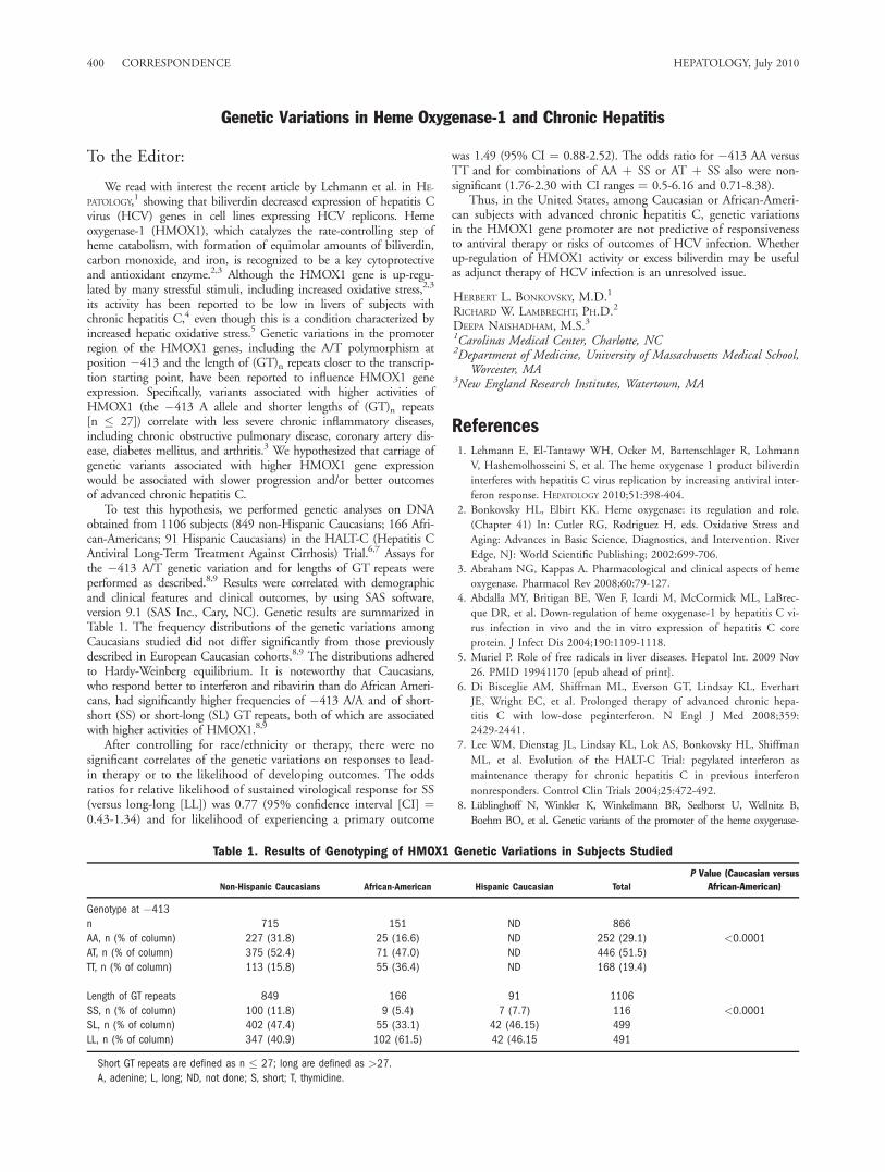
To test this hypothesis, we performed genetic analyses on DNAobtained from 1106 subjects (849 non-Hispanic Caucasians; 166 Afri-can-Americans; 91 Hispanic Caucasians) in the HALT-C (Hepatitis CAntiviral Long-Term Treatment Against Cirrhosis) Trial.6,7 Assays forthe �413 A/T genetic variation and for lengths of GT repeats wereperformed as described.8,9 Results were correlated with demographicand clinical features and clinical outcomes, by using SAS software,version 9.1 (SAS Inc., Cary, NC). Genetic results are summarized inTable 1. The frequency distributions of the genetic variations amongCaucasians studied did not differ significantly from those previouslydescribed in European Caucasian cohorts.8,9 The distributions adheredto Hardy-Weinberg equilibrium. It is noteworthy that Caucasians,who respond better to interferon and ribavirin than do African Ameri-cans, had significantly higher frequencies of �413 A/A and of short-short (SS) or short-long (SL) GT repeats, both of which are associatedwith higher activities of HMOX1.8,9
After controlling for race/ethnicity or therapy, there were nosignificant correlates of the genetic variations on responses to lead-in therapy or to the likelihood of developing outcomes. The oddsratios for relative likelihood of sustained virological response for SS(versus long-long [LL]) was 0.77 (95% confidence interval [CI] ¼0.43-1.34) and for likelihood of experiencing a primary outcome
was 1.49 (95% CI ¼ 0.88-2.52). The odds ratio for �413 AA versusTT and for combinations of AA þ SS or AT þ SS also were non-significant (1.76-2.30 with CI ranges ¼ 0.5-6.16 and 0.71-8.38).
Thus, in the United States, among Caucasian or African-Ameri-can subjects with advanced chronic hepatitis C, genetic variationsin the HMOX1 gene promoter are not predictive of responsivenessto antiviral therapy or risks of outcomes of HCV infection. Whetherup-regulation of HMOX1 activity or excess biliverdin may be usefulas adjunct therapy of HCV infection is an unresolved issue.
HERBERT L. BONKOVSKY, M.D.1
RICHARD W. LAMBRECHT, PH.D.2
DEEPA NAISHADHAM, M.S.31Carolinas Medical Center, Charlotte, NC2Department of Medicine, University of Massachusetts Medical School,
Worcester, MA3New England Research Institutes, Watertown, MA
References1. Lehmann E, El-Tantawy WH, Ocker M, Bartenschlager R, Lohmann
V, Hashemolhosseini S, et al. The heme oxygenase 1 product biliverdin
interferes with hepatitis C virus replication by increasing antiviral inter-
feron response. HEPATOLOGY 2010;51:398-404.2. Bonkovsky HL, Elbirt KK. Heme oxygenase: its regulation and role.
(Chapter 41) In: Cutler RG, Rodriguez H, eds. Oxidative Stress and
Aging: Advances in Basic Science, Diagnostics, and Intervention. River
Edge, NJ: World Scientific Publishing; 2002:699-706.3. Abraham NG, Kappas A. Pharmacological and clinical aspects of heme
oxygenase. Pharmacol Rev 2008;60:79-127.4. Abdalla MY, Britigan BE, Wen F, Icardi M, McCormick ML, LaBrec-
que DR, et al. Down-regulation of heme oxygenase-1 by hepatitis C vi-
rus infection in vivo and the in vitro expression of hepatitis C core
protein. J Infect Dis 2004;190:1109-1118.5. Muriel P. Role of free radicals in liver diseases. Hepatol Int. 2009 Nov
26. PMID 19941170 [epub ahead of print].6. Di Bisceglie AM, Shiffman ML, Everson GT, Lindsay KL, Everhart
JE, Wright EC, et al. Prolonged therapy of advanced chronic hepa-titis C with low-dose peginterferon. N Engl J Med 2008;359:2429-2441.
7. Lee WM, Dienstag JL, Lindsay KL, Lok AS, Bonkovsky HL, Shiffman
ML, et al. Evolution of the HALT-C Trial: pegylated interferon as
maintenance therapy for chronic hepatitis C in previous interferon
nonresponders. Control Clin Trials 2004;25:472-492.
8. Lublinghoff N, Winkler K, Winkelmann BR, Seelhorst U, Wellnitz B,
Boehm BO, et al. Genetic variants of the promoter of the heme oxygenase-
Table 1. Results of Genotyping of HMOX1 Genetic Variations in Subjects Studied
Non-Hispanic Caucasians African-American Hispanic Caucasian Total
P Value (Caucasian versus
African-American)
Genotype at �413
n 715 151 ND 866
AA, n (% of column) 227 (31.8) 25 (16.6) ND 252 (29.1) <0.0001
AT, n (% of column) 375 (52.4) 71 (47.0) ND 446 (51.5)
TT, n (% of column) 113 (15.8) 55 (36.4) ND 168 (19.4)
Length of GT repeats 849 166 91 1106
SS, n (% of column) 100 (11.8) 9 (5.4) 7 (7.7) 116 <0.0001
SL, n (% of column) 402 (47.4) 55 (33.1) 42 (46.15) 499
LL, n (% of column) 347 (40.9) 102 (61.5) 42 (46.15 491
Short GT repeats are defined as n � 27; long are defined as >27.
A, adenine; L, long; ND, not done; S, short; T, thymidine.
400 CORRESPONDENCE HEPATOLOGY, July 2010

1 gene and their influence on cardiovascular disease (the Ludwigshafen Risk
and Cardiovascular Health Study). BMC Med Genet 2009;10:36.9. Exner M, Minar E, Wagner O, Schillinger M. The role of heme oxy-
genase-1 promoter polymorphisms in human disease. Free Radic BiolMed 2004;37:1097-1104.
Copyright VC 2010 by the American Association for the Study of Liver Diseases.Published online in Wiley InterScience (www.interscience.wiley.com).DOI 10.1002/hep.23562Potential conflict of interest: Nothing to report.
Quantification of Genotype 4 Serum Samples: Impact of Hepatitis C Virus Genetic Variability
To the Editor:
We read with interest the recent correspondence of Germeret al.1 and Chevaliez et al.2 regarding the quantification of geno-type 4 hepatitis C virus (HCV) RNA by the COBAS AmpliPrep/COBAS TaqMan HCV Test (CAP/CTM) (Roche Molecular Sys-tems Inc., Branchburg, NJ). Several publications evaluating thequantification of genotype 4 serum samples have led to conflictingreports.3-9
We evaluated the correlation between viral load results in theserum samples of 75 pretreatment patients infected with genotype4 and the impact of nucleotide (nt) polymorphism at nt 145 andnt 165 on viral load quantification. HCV RNA measurementswere performed with the Versant HCV 3.0 Assay (branched DNA)(Siemens Healthcare Diagnostics Inc., Saint Denis, France); theCAP/CTM test; and the Abbott m2000sp extraction/m2000rtamplification system (ART) (Abbott Laboratories Inc.) and theCOBAS AmpliPrep/COBAS TaqMan HCV Test (Roche MolecularSystems).
HCV genotypes were identified using the TruGene HCV 50NCgenotyping kit (Siemens Healthcare Diagnostics Inc.). HCVsubtyping was performed in the NS5 B nonstructural region ofthe HCV genome with the Open Gene Thermo sequenase fluores-cent-labeled primer cycle sequencing kit (Siemens HealthcareDiagnostics Inc.).10
The mean viral loads for the 75 serum samples (HCV genotype4a, n ¼ 36; 4c, n ¼ 1; 4d, n ¼ 16; 4e, n ¼ 10; 4f, n ¼ 4; 4h,n ¼ 4; 4i, n ¼ 2; and 4l, n ¼ 2) were: 5.300 6 0.751, 5.334 60.941, and 5.419 6 0.820 with the branched DNA, CAP/CTM,and ART tests, respectively (all values are not significant). Ourresults showed similar quantification levels for HCV genotype 4subtype, no matter which assay was used. These data are in agree-ment with those reported by Germer et al. on a cohort of 100clinical genotype 4 samples.1
In our 75 patients, HCV 50 noncoding region sequencesrevealed no sequence containing a G-to-A substitution at nt 145,which was reported to be associated with failure of CAP/CTM byChevaliez et al.2; three sequences contained an A-to-T substitutionat nt 165, which was previously associated with under-quantifica-tion by CAP/CTM.2 Two of these three substitutions at nt 165yielded under-quantification of 0.5 log10 IU/mL and 0.988 log10IU/mL with CAP/CTM. These results are in accordance with thoseof Germer et al.1 and confirm that substitutions at nt 145 and 165are very rare. Thus, one would be unlikely to encounter failure ofCAP/CTM to detect HCV genotype 4 strains not only in U.S.samples but also in European samples.
In conclusion, our results show similar quantification levels forthe different HCV genotype 4 subtypes, irrespective of whicheverassay was used, and that a G-to-A substitution at nt 145 and anA-to-T substitution at nt 165 are very rare, confirming that onewould be unlikely to encounter failure of CAP/CTM to detectHCV genotype 4 strains found in the United States and in Europe.Nevertheless, it is important to consistently use the same HCVRNA assay throughout patient treatment follow-up.
PHILIPPE HALFON1
MICHELLE MARTINOT-PEIGNOUX2
HACENE KHIRI1
PATRICK MARCELLIN2
1Laboratoire Alphabio, Hopital Ambroise Pare Marseille, France2Institut National de la Sante et de la Recherche Medicale, U-773,
Centre de Recherche Biomedicale Bichat-Beaujon CRB3 and Serviced’Hepatologie, Hopital Beaujon, Universite Paris 7, Clichy, France
References1. Germer JJ, Bommersbach CE, Schmidt DM, Bendel JL, Yao JDC. Quantifi-
cation of genotype 4 hepatitis C virus RNA by the COBAS AmpliPrep/COBAS TaqMan hepatitis C virus test. HEPATOLOGY 2009;50:1679-1680.
2. Chevaliez S, Bouvier-Alias M, Castera L, Pawlotsky JM. The CobasAmpliPrep-Cobas TaqMan real-time polymerase chain reaction assayfails to detect hepatitis C virus RNA in highly viremic genotype 4 clin-ical samples. HEPATOLOGY 2009;49:1397-1398.
3. Sarrazin C, Dragan A, Gartner BC, Forman MS, Traver S, Zeuzem S,et al. Evaluation of an automated highly sensitive real-time PCR basedassay (COBAS AmpliPrep/COBAS TaqMan) for quantification ofHCV RNA. J Clin Virol 2008;43:162-168.
4. Pittaluaga F, Allice C, Abate ML, Ciancio F, Varetto S, et al. Clinical evaluationof the COBAS AmpliPrep/COBAS TaqMan for the HCV RNA quantificationin comparison with the branched-DNA assay. J Med Virol 2008;80:254-260.
5. Michelin BD, Muller Z, Stelzl E, Marth E, Kessler HH. Evaluation ofthe Abbott real time HCV assay for quantitative detection of hepatitisC virus RNA. J Clin Virol 2008;38:91-100.
6. Chevaliez S, Bouvier-Alias M, Brillet R, Pawlotsky JM. Overestimation andunderestimation of hepatitis C virus RNA levels in a widely used real-timepolymerase chain reaction-based method. HEPATOLOGY 2007;46:22-31.
7. Sarrazin C, Gartner BC, Sizmann D, Babiel R, Mihm U, Hofmann WP,et al. Comparison of conventional PCR with real-time PCR and branchedDNA-based assays for hepatitis C virus RNA quantification and clinicalsignificance for genotype 1 to 5. J Clin Microbiol 2006;44:729-737.
8. Tuaillon E, Mondain AM, Ottomani L, Roudiere L, Perney P, Picot MC, et al.Impact of hepatitis C virus genotype on quantification of HCV RNA in serumby COBAS AmpliPrep/COBAS Taqman HCV test, Abbott HCV RealTimeassay, and VERSANT HCV RNA assay. J Clin MicroBiol 2007;45:3077-3081.
9. Vermehren J, Kau A, Gartner B, Gobel R, Zeuzem S, Sarrazin C. Dif-ferences between two RealTime PCR-based hepatitis C assays(RealTimeHCV and Cobas AmpliPrep/Cobas TaqMan) and one signal amplifica-tion assay (Versant HCV RNA 3,0) for RNA detection and quantifica-tion. J Clin Microbiol 2008;46:3880-3891.
10. Halfon P, Trimoulet P, Bourliere M, Khiri H, De Ledinghen V, CouzigouP, et al. Hepatitis C virus genotyping based on 50 non coding sequenceanalysis (TrugeneTM). J Clin Microbiol 2001;39:1771-1773.
Copyright VC 2010 by the American Association for the Study of Liver Diseases.Published online in Wiley InterScience (www.interscience.wiley.com).DOI 10.1002/hep.23578Potential conflict of interest: Dr. Marcellin received grants, is on the speakers’
bureau of, and advises Roche, Schering-Plough, and Gilead. He also advises and is onthe speakers’ bureau of Bristol-Myers Squibb, Novartis, and Intermune. He advisesVertex, Pharmasset, Tibotec, Merck Sharp & Dohme, Boehringer, Biolex, andZymogenetics.
HEPATOLOGY, Vol. 52, No. 1, 2010 CORRESPONDENCE 401