Functional Role of Autophagy-Mediated Proteome Remodeling in Cell Survival Signaling and Innate...
15
Molecular Cell Article Functional Role of Autophagy-Mediated Proteome Remodeling in Cell Survival Signaling and Innate Immunity Robin Mathew, 1,2,6 Sinan Khor, 1,2,6 Sean R. Hackett, 3 Joshua D. Rabinowitz, 3 David H. Perlman, 4,5 and Eileen White 1,2, * 1 Rutgers Cancer Institute of New Jersey, 195 Little Albany Street, New Brunswick, NJ 08903 2 Department of Molecular Biology and Biochemistry, Rutgers University, Piscataway, NJ, 08854 3 Department of Chemistry, and the Lewis-Sigler Institute for Integrative Genomics, Princeton University, Washington Road, Princeton, NJ 08544 4 Department of Molecular Biology, the Lewis-Sigler Institute for Integrative Genomics, and the Princeton Proteomics and Mass Spectrometry Core, Princeton University, Washington Road, Princeton, NJ 08544 5 Present address: Department of Chemistry, Princeton University, Washington Road, Princeton, NJ 08544 6 Co-first author *Correspondence: [email protected] http://dx.doi.org/10.1016/j.molcel.2014.07.019 SUMMARY Ras-driven cancer cells upregulate basal autophagy that degrades and recycles intracellular proteins and organelles. Autophagy-mediated proteome de- gradation provides free amino acids to support metabolism and macromolecular synthesis, which confers a survival advantage in starvation and pro- motes tumorigenesis. While the degradation of iso- lated protein substrates by autophagy has been implicated in controlling cellular function, the extent and specificity by which autophagy remodels the cellular proteome and the underlying functional con- sequences were unknown. Here we compared the global proteome of autophagy-functional and -defi- cient Ras-driven cancer cells, finding that autophagy affects the majority of the proteome yet is highly selective. While levels of vesicle trafficking proteins important for autophagy are preserved during star- vation-induced autophagy, deleterious inflammatory response pathway components are eliminated even under basal conditions, preventing cytokine-induced paracrine cell death. This reveals the global, func- tional impact of autophagy-mediated proteome remodeling on cell survival and identifies critical autophagy substrates that mediate this process. INTRODUCTION Macroautophagy (hereafter referred to as autophagy) is a stress- induced, self-cannibalization mechanism that captures da- maged proteins and organelles in autophagosomes, which then fuse with lysosomes where their cargo is degraded. In normal cells, basal autophagy occurs at low levels to prevent chronic tissue damage and to maintain homeostasis (Mathew et al., 2007a, 2007b). Autophagy substrates are degraded and recycled to provide carbon and nitrogen sources for metabolism and biosynthesis of macromolecules and for production of cofactors for redox balance and energy (Mathew and White, 2011; Rabinowitz and White, 2010). The specificity of cargo de- livery is poorly understood but partly achieved by ubiquitin modi- fication on cargo that is recognized by specific domains in the cargo receptor proteins such as p62/SQSTM1 (p62) (Vadlamudi and Shin, 1998). The captured cargo is then sequestered by au- tophagosomes that assemble around them, through coordinate action of the products of essential autophagy genes such as Atg5 and Atg7, deletions of which abrogate autophagy (Mizush- ima, 2007). While short-lived intracellular proteins are degraded primarily by the ubiquitin proteasome system (UPS), autophagy remains the only known mechanism by which cells eliminate aggregated proteins and entire organelles. Recent evidence suggests that autophagy is critical for cancer cell metabolism, survival, and tumor maintenance (Guo et al., 2011, 2013a; Rao et al., 2014; Rosenfeldt et al., 2013; Strohecker et al., 2013). In contrast to normal cells, basal autophagy is dramatically upregulated in many cancers, such as those driven by activation of Ras oncogenes, where it is critical for survival and for tumorigenesis (Guo et al., 2011, 2013b; Lock et al., 2011; Yang et al., 2011). Metabolic stress due to a deficient microenvironment and deregulated cell growth may contribute to this autophagy addiction (Degenhardt et al., 2006), but the mechanism for this autophagy-mediated survival and growth of Ras-driven cancers is only beginning to emerge. Autophagy degrades the proteome, a major amino acid and energy source for metabolically stressed cancer cells, facilitating both survival and proliferation. Oncogenic mutations in Ras sup- press mitochondrial production of acetyl-coenzyme A and the utilization of fatty acids via b-oxidation, presumably elevating the demand for protein degradation by autophagy to support mitochondrial function through anaplerosis (Mathew and White, 2011; White, 2013). Consistent with this, autophagy deficiency in Ras-expressing tumors causes damaged proteins and defec- tive mitochondria to accumulate, leading to impairment of energy metabolism and reduced tumor growth. Although the exact mechanism by which autophagy supports metabolism and Molecular Cell 55, 1–15, September 18, 2014 ª2014 Elsevier Inc. 1 Please cite this article in press as: Mathew et al., Functional Role of Autophagy-Mediated Proteome Remodeling in Cell Survival Signaling and Innate Immunity, Molecular Cell (2014), http://dx.doi.org/10.1016/j.molcel.2014.07.019
-
Upload
independent -
Category
Documents
-
view
0 -
download
0
Transcript of Functional Role of Autophagy-Mediated Proteome Remodeling in Cell Survival Signaling and Innate...
Please cite this article in press as: Mathew et al., Functional Role of Autophagy-Mediated Proteome Remodeling in Cell Survival Signaling and InnateImmunity, Molecular Cell (2014), http://dx.doi.org/10.1016/j.molcel.2014.07.019
Molecular Cell
Article
Functional Role of Autophagy-MediatedProteome Remodeling in Cell SurvivalSignaling and Innate ImmunityRobin Mathew,1,2,6 Sinan Khor,1,2,6 Sean R. Hackett,3 Joshua D. Rabinowitz,3 David H. Perlman,4,5 and Eileen White1,2,*1Rutgers Cancer Institute of New Jersey, 195 Little Albany Street, New Brunswick, NJ 089032Department of Molecular Biology and Biochemistry, Rutgers University, Piscataway, NJ, 088543Department of Chemistry, and the Lewis-Sigler Institute for Integrative Genomics, Princeton University, Washington Road, Princeton,NJ 085444Department of Molecular Biology, the Lewis-Sigler Institute for Integrative Genomics, and the Princeton Proteomics andMass Spectrometry
Core, Princeton University, Washington Road, Princeton, NJ 085445Present address: Department of Chemistry, Princeton University, Washington Road, Princeton, NJ 085446Co-first author
*Correspondence: [email protected]
http://dx.doi.org/10.1016/j.molcel.2014.07.019
SUMMARY
Ras-driven cancer cells upregulate basal autophagythat degrades and recycles intracellular proteinsand organelles. Autophagy-mediated proteome de-gradation provides free amino acids to supportmetabolism and macromolecular synthesis, whichconfers a survival advantage in starvation and pro-motes tumorigenesis. While the degradation of iso-lated protein substrates by autophagy has beenimplicated in controlling cellular function, the extentand specificity by which autophagy remodels thecellular proteome and the underlying functional con-sequences were unknown. Here we compared theglobal proteome of autophagy-functional and -defi-cient Ras-driven cancer cells, finding that autophagyaffects the majority of the proteome yet is highlyselective. While levels of vesicle trafficking proteinsimportant for autophagy are preserved during star-vation-induced autophagy, deleterious inflammatoryresponse pathway components are eliminated evenunder basal conditions, preventing cytokine-inducedparacrine cell death. This reveals the global, func-tional impact of autophagy-mediated proteomeremodeling on cell survival and identifies criticalautophagy substrates that mediate this process.
INTRODUCTION
Macroautophagy (hereafter referred to as autophagy) is a stress-
induced, self-cannibalization mechanism that captures da-
maged proteins and organelles in autophagosomes, which
then fuse with lysosomes where their cargo is degraded. In
normal cells, basal autophagy occurs at low levels to prevent
chronic tissue damage and to maintain homeostasis (Mathew
et al., 2007a, 2007b). Autophagy substrates are degraded and
M
recycled to provide carbon and nitrogen sources for metabolism
and biosynthesis of macromolecules and for production of
cofactors for redox balance and energy (Mathew and White,
2011; Rabinowitz and White, 2010). The specificity of cargo de-
livery is poorly understood but partly achieved by ubiquitin modi-
fication on cargo that is recognized by specific domains in the
cargo receptor proteins such as p62/SQSTM1 (p62) (Vadlamudi
and Shin, 1998). The captured cargo is then sequestered by au-
tophagosomes that assemble around them, through coordinate
action of the products of essential autophagy genes such as
Atg5 and Atg7, deletions of which abrogate autophagy (Mizush-
ima, 2007). While short-lived intracellular proteins are degraded
primarily by the ubiquitin proteasome system (UPS), autophagy
remains the only known mechanism by which cells eliminate
aggregated proteins and entire organelles.
Recent evidence suggests that autophagy is critical for cancer
cell metabolism, survival, and tumor maintenance (Guo et al.,
2011, 2013a; Rao et al., 2014; Rosenfeldt et al., 2013; Strohecker
et al., 2013). In contrast to normal cells, basal autophagy is
dramatically upregulated in many cancers, such as those driven
by activation of Ras oncogenes, where it is critical for survival
and for tumorigenesis (Guo et al., 2011, 2013b; Lock et al.,
2011; Yang et al., 2011). Metabolic stress due to a deficient
microenvironment and deregulated cell growth may contribute
to this autophagy addiction (Degenhardt et al., 2006), but the
mechanism for this autophagy-mediated survival and growth
of Ras-driven cancers is only beginning to emerge.
Autophagy degrades the proteome, a major amino acid and
energy source for metabolically stressed cancer cells, facilitating
both survival and proliferation. Oncogenic mutations in Ras sup-
press mitochondrial production of acetyl-coenzyme A and the
utilization of fatty acids via b-oxidation, presumably elevating
the demand for protein degradation by autophagy to support
mitochondrial function through anaplerosis (Mathew and White,
2011; White, 2013). Consistent with this, autophagy deficiency
in Ras-expressing tumors causes damaged proteins and defec-
tivemitochondria to accumulate, leading to impairment of energy
metabolism and reduced tumor growth. Although the exact
mechanism by which autophagy supports metabolism and
olecular Cell 55, 1–15, September 18, 2014 ª2014 Elsevier Inc. 1

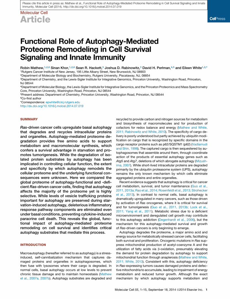
Figure 1. SILAC as a Means to Monitor Proteome Remodeling by Autophagy
(A) SILAC-based proteomics comparing autophagy-deficient and -competent cells. Autophagy-deficient cells were given medium containing 13C-Lysine and13C15N Arginine for at least eight doublings to fully label their proteome. Cells were then subjected to a starvation time course (0, 3, and 5 hr Hank’s balanced salt
solution [HBSS]), trypsin digested, and analyzed by ultraperformance liquid chromatography-mass spectrometry (UPLC-MS).
(B) Atg5- or Atg7-deficient cell lines do not have functional autophagy. Starved iBMK cells or TDCLs were lysed and immunoblotted for the indicated antibodies.
b-actin was used as a loading control.
(C) Autophagy-deficient cells have ER stress in starvation. Starved iBMK cells or TDCLs were lysed and immunoblotted for the indicated antibodies. b-actin was
used as a loading control.
(legend continued on next page)
Molecular Cell
Autophagy Defects Prime the Interferon Response
2 Molecular Cell 55, 1–15, September 18, 2014 ª2014 Elsevier Inc.
Please cite this article in press as: Mathew et al., Functional Role of Autophagy-Mediated Proteome Remodeling in Cell Survival Signaling and InnateImmunity, Molecular Cell (2014), http://dx.doi.org/10.1016/j.molcel.2014.07.019
Molecular Cell
Autophagy Defects Prime the Interferon Response
Please cite this article in press as: Mathew et al., Functional Role of Autophagy-Mediated Proteome Remodeling in Cell Survival Signaling and InnateImmunity, Molecular Cell (2014), http://dx.doi.org/10.1016/j.molcel.2014.07.019
stress survival is unknown, defects in autophagy cause dere-
gulation of amino acid levels and depletion of key substrates in
mitochondrial metabolism, compromising mitochondrial respira-
tion (Guo et al., 2011, 2013a; Rao et al., 2014; Rosenfeldt et al.,
2013; Yang et al., 2011). Cancer cells are increasingly reliant on
amino acids such as glutamine for mitochondrial function, for
nucleotide and lipid synthesis, and for redox homeostasis (De-
Berardinis et al., 2007; Son et al., 2013). Despite this important
role for autophagy in cancer metabolism and stress survival, a
comprehensive understanding of autophagy substrates and the
specific mechanisms by which autophagy supports cancer cell
survival are currently lacking. Additionally, although autophagy
is selective to certain cellular substrates in specific contexts,
whether or not autophagy targets specific pathways to alter
cell function is not understood (Mizushima and Komatsu, 2011).
Using isogenic cell models comparing autophagy-intact and
-deficient cells (Atg5+/+ and Atg5�/�) and the technique of Stable
Isotope Labeling by Amino acids in Cell culture (SILAC) for proteo-
mic analysis, we examined the role of autophagy in remodeling the
proteome in basal and starvation conditions. We discovered that
autophagy-mediated proteome remodeling affects the majority
of the proteome and is selective, preserving cellular function of
Ras-driven cancer cells. Our results show that autophagy selec-
tively eliminates proteins (autophagy protein substrates) involved
in pathways that are nonessential or toxic for survival under stress,
whilepreferentially sparing those involved inpathwaysessential for
themaintenanceof functional autophagy and stress survival (auto-
phagy-resistant proteins). Specifically, we found that proteins
involved in the innate immune response, such as the retinoic-
acid-inducible gene-I (RIG-I) pathway, accumulated in auto-
phagy-deficient cells. Conversely, the soluble N-ethylmaleimide-
sensitive factor (NSF) attachment receptor (SNARE)-mediated
vesicle trafficking, lysosome, and endocytosis pathways essential
for intracellular protein trafficking between endoplasmic reticulum
(ER), Golgi apparatus, endosomes, and lysosomes escaped auto-
phagy-mediated elimination. These findings suggest that auto-
phagy extensively remodels the proteome and that defects in
autophagy may prime nonimmune cells for constitutive innate im-
mune and inflammatory signaling with profound implications for
cell survival, partly explaining the requirement for autophagy for
sustaining aggressive cancer growth.
RESULTS
Global Impact of Autophagy on the Cellular ProteomeTo assess the impact of autophagy on the overall cellular prote-
ome, we employed SILAC (Ong et al., 2002) to compare relative
protein levels in isogenic wild-type (WT) (Atg5+/+) and auto-
phagy-deficient (Atg5�/�) HRasG12V-transformed immortalized
baby mouse kidney epithelial (iBMK) cells (Guo et al., 2011)
in normal growth media and following 3 and 5 hr of starvation
(Figure 1A). Ras expression levels in these iBMK cells are com-
(D and E) Starvation robustly induces autophagy in iBMK cells and TDCLs. iBMK c
lysed, and immunoblotted for the indicated antibodies. Quantitation of the LC3-II
(n = 2).
Data are representative of at least two independent experiments.
M
parable to those of human cancer cell lines with activating Ras
mutations and render them tumorigenic, although Atg5 defi-
ciency compromises tumor growth (Guo et al., 2011).
Western blot analysis revealed induction of autophagy in the
WT cells in starvation, and was completely blocked by Atg5
and Atg7 deletion as indicated by the absence of LC3-I-to-
LC3-II processing in the autophagy-deficient cell lines, which
instead accumulated the autophagy substrate p62 (Figure 1B).
Autophagy defects also caused accumulation of ER chaperones
GRp170 and GRp78 and protein disulphide isomerase (PDI) in
both iBMK cell lines (Figure 1C; top panel), consistent with
previous findings (Mathew et al., 2009). We then examined the
autophagy flux with the lysosomal inhibitor Bafilomycin A1,
which resulted in the accumulation of LC3-II in the WT cells,
but not in the autophagy-deficient cells (Figure 1D). In tumor-
derived cell lines (TDCLs) from the KRasG12D;p53�/� genetically
engineeredmousemodel (GEMM) for non-small-cell lung cancer
(NSCLC) (Guo et al., 2013a), starvation robustly induced auto-
phagy in the WT cells, while autophagy defects resulted in p62
accumulation and elevated expression of ER stress markers
(Figures 1B, 1C, and 1E). Thus, the Ras-transformed Atg5
iBMK cells used for the SILAC are representative of autophagy
functionality independent of tissue type and Ras subfamily.
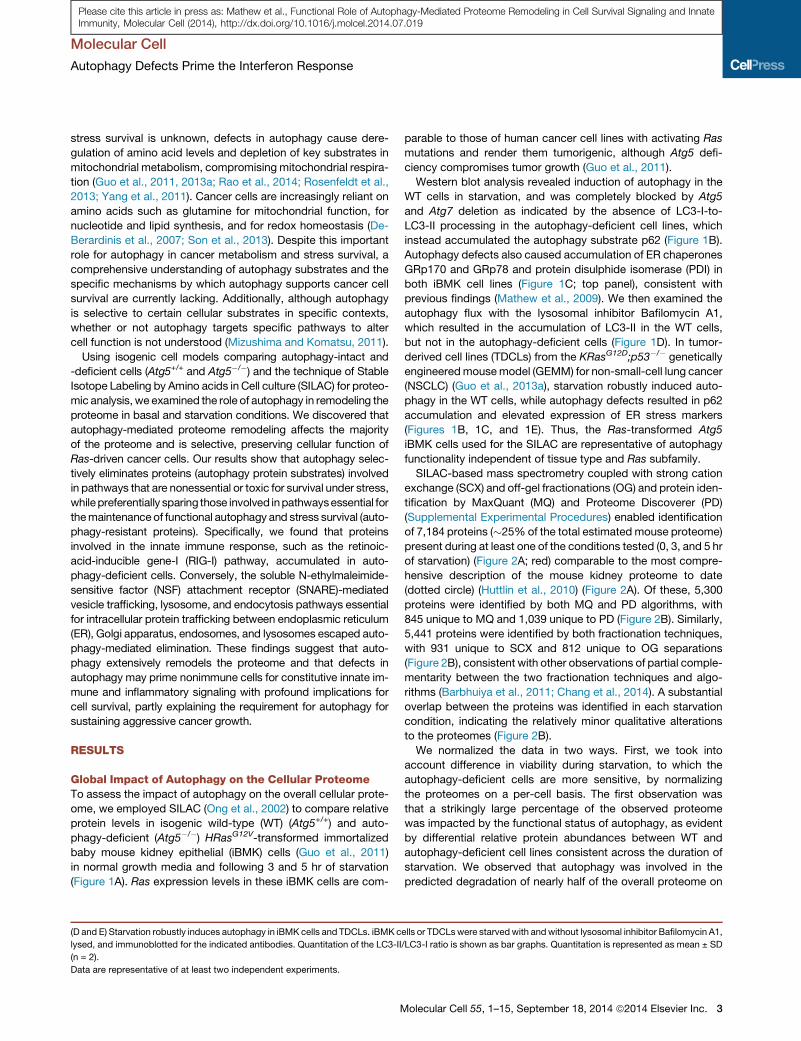
SILAC-based mass spectrometry coupled with strong cation
exchange (SCX) and off-gel fractionations (OG) and protein iden-
tification by MaxQuant (MQ) and Proteome Discoverer (PD)
(Supplemental Experimental Procedures) enabled identification
of 7,184 proteins (�25% of the total estimated mouse proteome)
present during at least one of the conditions tested (0, 3, and 5 hr
of starvation) (Figure 2A; red) comparable to the most compre-
hensive description of the mouse kidney proteome to date
(dotted circle) (Huttlin et al., 2010) (Figure 2A). Of these, 5,300
proteins were identified by both MQ and PD algorithms, with
845 unique to MQ and 1,039 unique to PD (Figure 2B). Similarly,
5,441 proteins were identified by both fractionation techniques,
with 931 unique to SCX and 812 unique to OG separations
(Figure 2B), consistent with other observations of partial comple-
mentarity between the two fractionation techniques and algo-
rithms (Barbhuiya et al., 2011; Chang et al., 2014). A substantial
overlap between the proteins was identified in each starvation
condition, indicating the relatively minor qualitative alterations
to the proteomes (Figure 2B).
We normalized the data in two ways. First, we took into
account difference in viability during starvation, to which the
autophagy-deficient cells are more sensitive, by normalizing
the proteomes on a per-cell basis. The first observation was
that a strikingly large percentage of the observed proteome
was impacted by the functional status of autophagy, as evident
by differential relative protein abundances between WT and
autophagy-deficient cell lines consistent across the duration of
starvation. We observed that autophagy was involved in the
predicted degradation of nearly half of the overall proteome on
ells or TDCLswere starvedwith andwithout lysosomal inhibitor Bafilomycin A1,
/LC3-I ratio is shown as bar graphs. Quantitation is represented as mean ± SD
olecular Cell 55, 1–15, September 18, 2014 ª2014 Elsevier Inc. 3

Figure 2. Effect of Autophagy Deficiency on the Cellular Proteome
(A) Venn diagrams showing statistically validated proteins by SILAC-MS, compared to the total proteins identified in our study (red), a previously publishedmouse
proteome (gray), and the total estimated mouse proteome (turquoise).
(legend continued on next page)
Molecular Cell
Autophagy Defects Prime the Interferon Response
4 Molecular Cell 55, 1–15, September 18, 2014 ª2014 Elsevier Inc.
Please cite this article in press as: Mathew et al., Functional Role of Autophagy-Mediated Proteome Remodeling in Cell Survival Signaling and InnateImmunity, Molecular Cell (2014), http://dx.doi.org/10.1016/j.molcel.2014.07.019

Molecular Cell
Autophagy Defects Prime the Interferon Response
Please cite this article in press as: Mathew et al., Functional Role of Autophagy-Mediated Proteome Remodeling in Cell Survival Signaling and InnateImmunity, Molecular Cell (2014), http://dx.doi.org/10.1016/j.molcel.2014.07.019
a per-cell basis within 3 hr of starvation, which increased to 70%
at 5 hr (Figure 2C). This suggests that autophagy is a significant
mechanism for turnover and remodeling of the cellular proteome.
Second, to examine the specificity by which autophagy im-
pacts the global proteome, proteins in each SILAC channel
were normalized to total protein for each cell type and examined
for relative protein abundance ratios (ratios of levels in auto-
phagy-deficient compared to WT; Atg5�/�/Atg5+/+: Heavy/Light[H/L]) in normal growth and starvation conditions. Each condition
contained groups of proteins with disproportionately higher or
lower levels in autophagy-deficient cells compared to the WT,
representing proteins that appear to be selectively eliminated
or spared by autophagy, respectively (Figure 2D). Out of the
7,184 total proteins identified, under normal conditions, 6.4%
(462 proteins) were elevated (>1.5-fold) in the autophagy-defi-
cient cells, while 6.7% (484 proteins) showed decreased levels
compared to the WT cells. Following 3 hr of starvation, 5.8%
(419 proteins) showed elevation and 7.2% (517 proteins) showed
a decrease in relative protein levels. Similarly, after 5 hr of starva-
tion, 4.9% (352 proteins) were elevated, while 7.2% (520 pro-
teins) showed reduced relative proteins levels (Figures 2B and
2D). Thus, autophagy has a dramatic impact on the overall pro-
teome, with its loss surprisingly producing both up- and downre-
gulation of specific proteins relative to the WT.
To examine if the changes in the proteome caused by auto-
phagy deficiency were specific to cellular activities, we looked
at the molecular functions enriched among proteins with
extreme changes in their relative abundance in each condition
using Gene Set Enrichment Analysis (GSEA) as described previ-
ously (Subramanian et al., 2005). Interestingly, the Rig-I like
receptor (RLR) signaling pathway, DNA replication, and one car-
bon pool by folate metabolism were highly enriched among pro-
teins elevated in autophagy-deficient cells at all the conditions
tested (Figure 2E). Alternatively, proteins involved in cellular pro-
cesses that include proteasomes and ribosomes were selec-
tively preserved with functional autophagy as indicated by low
H/L ratios.
We then examined how relative protein abundances changed
across time during starvation by subjecting the log geometric
means of the relative abundance values normalized to their basal
ratio (0 hr) to hierarchical clustering (Figure 2F). While the major-
ity of proteins had relative protein abundances (H/L ratio) consis-
tent across all conditions, there were clusters of proteins whose
relative levels decreased (green) or increased (red) during starva-
tion. Interestingly, pathways related to glycerophospholipid
metabolism, angiogenesis (VEGF signaling), SNARE vesicle
trafficking, etc. were enriched among proteins that showed a
(B) Venn diagrams showing proteins identified using MaxQuant (MQ), Proteome D
the distribution of protein identifications at three different time points (0, 3, and 5
(C) Bar graph showing the increase in the total number of proteins that showed r
compared to the WT cells at 0, 3, and 5 hr of starvation on a per-cell basis (norm
(D) S curve displaying relative protein abundances of all the proteins between auto
protein.
(E) Bar graphs showing NES for pathways enriched among extremes of chang
deficiency (only normalized p values < 0.05 are shown). Positive NES indicates
negative NES indicates enrichment for H/L ratios < 1 (relative increase in autoph
(F) Hierarchical clustered heatmap with the 0 hr normalized log2 H/L values sho
starvation using the top and bottom 10% most-pronounced changes.
M
progressive decrease in protein abundance in starvation (Fig-
ure 2F). In contrast, pathways related to lysosome, protein
degradation (ubiquitin mediated proteolysis), etc. were enriched
among proteins that showed an increase (Figure 2F), consistent
with the need to preserve autophagy machinery and protein ho-
meostasis during starvation.
To examine any inherent basal difference due to the auto-
phagy defect, and to identify a subset of proteins that showed
statistically significant changes in normal and starvation condi-
tions, we compared the overall data in Figure 2D by t statistics
calculated for each methodology and performed hierarchical
clustering of relative abundance measurements across the
starvation time course. We then computed p values based on
t statistics to determine whether the time zero intercept of
proteins log2 (H/L) relative abundances differed significantly
over time from time zero (Figures 3A and 3B). We identified a
subset of 3,181 proteins that were either elevated (yellow) or
depressed (blue) due to autophagy deficiency (Figure 3A),
with over 50% of the proteins showing autophagy-dependent
alteration.
Autophagy-Mediated Protein Remodeling Is SelectiveIn the subset of 3,181 proteins that had statistically significant
changes in normal and starvation conditions that were attribut-
able to autophagy defect, 1,638 behaved as major autophagy
substrates (high H/L ratio) and thus are potential markers for
autophagy-mediated protein degradation (Figure 3C). Surpris-
ingly, 1,543 proteins behaved as if they were resistant to de-
gradation (low H/L ratio); that is, their levels were retained in
autophagy-competent cells in starvation (Figure 3C).
Among the proteins that were enriched on both extremes, a
common theme of molecular functions suggested that auto-
phagy selectively targets protein substrates to enable cell sur-
vival. For example, DNA replication proteins, including PCNA,
POLA2, MCM3, MCM, and LIG1, were among the proteins
elevated in autophagy-deficient cells (Figure 3C and Table S1).
Importantly, levels of poly(ADP-ribose) polymerase (PARP), a
critical component of the single-stranded DNA repair, energy
sensing machinery, and apoptotic cell death, and STAT1, an
important mediator of interferon-mediated cell death, were
significantly elevated in the autophagy-deficient cells (Figure 3C
and Table S1), suggesting a prosurvival role for autophagy
through selective targeting of protein substrates.
Proteins Preserved during AutophagyConsistent with a stress survival role for autophagy, specific pro-
teins with purported roles in cellular functions essential for stress
iscoverer (PD), and different fractionation techniques (SCX and OG), as well as
hr).
elative (>2-fold) increase (red) or decrease (green) in the autophagy-defective
alized to cell number).
phagy-deficient versusWT cells (log2 H/L ratios) shown in (A) normalized to total
es in relative protein abundances (H/L ratios) in starvation due to autophagy
enrichment for H/L ratios > 1 (relative increase in autophagy-defective), and
agy-competent).
wing time-dependent changes in relative protein levels across time points in
olecular Cell 55, 1–15, September 18, 2014 ª2014 Elsevier Inc. 5

Figure 3. Pathway Enrichment in Autophagy-Deficient Cells Due to Defects in Protein Degradation(A) Volcano plot showing statistical significance of pre-starvation differences in protein abundances due to autophagy function. Proteins whose abundance is
significantly altered due to autophagy are shown: a total of 1,638 proteins (51.5%) were preferentially degraded in autophagy-competent cells (yellow), while
1,543 (46.7%) were selectively preserved (blue). Significance of PD-based quantification is shown.
(B) Scatter plot comparing statistical significance as determined by either MQ or PD. Proteins found to be elevated or depleted due to autophagy were those that
exceeded a false discovery rate of 0.05 when analyzed with both MQ and PD platforms.
(C) S plot showing relative protein abundance (H/L ratio) in 3,181 proteins found to be significantly altered due to autophagy deficiency. Some select examples of
proteins that showed extreme changes are highlighted.
(D) NES from GSEA analysis of the 3,181 proteins found to be significantly altered due to autophagy deficiency with normalized p value < 0.05 are shown.
(E) Enrichment plots showing relative enrichment for significantly enriched pathways in GSEA analysis in (D).
See also Table S1.
Molecular Cell
Autophagy Defects Prime the Interferon Response
6 Molecular Cell 55, 1–15, September 18, 2014 ª2014 Elsevier Inc.
Please cite this article in press as: Mathew et al., Functional Role of Autophagy-Mediated Proteome Remodeling in Cell Survival Signaling and InnateImmunity, Molecular Cell (2014), http://dx.doi.org/10.1016/j.molcel.2014.07.019

Molecular Cell
Autophagy Defects Prime the Interferon Response
Please cite this article in press as: Mathew et al., Functional Role of Autophagy-Mediated Proteome Remodeling in Cell Survival Signaling and InnateImmunity, Molecular Cell (2014), http://dx.doi.org/10.1016/j.molcel.2014.07.019
survival, such as poly(ADP-ribose) glycohydrolase (PARG, poly
[ADP-ribose] catabolism), adenylate kinase 4 (AK4, ATP/ADP
homeostasis), CTP synthase 2 (CTPS, glutaminolysis), and cit-
rate synthase (CS), were among those that showed relative
accumulation in the WT cells (low H/L ratio) (Figure 3C and Table
S1). Similarly, several of the proteasome components (PSMC2,
PSMC4, PSMC5, PSMD6, etc.) were among the proteins highly
retained by autophagy in starvation (Figure 3C and Table S1).
Importantly, critical components of vesicle trafficking and endo-
cytosis, such as synaptosomal-associated protein 29 (SNAP29),
were also selectively preserved, indicating that maintaining func-
tional autophagy is critical in remodeling the proteome. This sug-
gests that autophagy selectively retains protein components of
distinct pathways that are essential for cell survival.
Defects in Proteome Remodeling Alter CellularSignaling PathwaysAlterations in protein homeostasis of signaling pathway compo-
nents may impact the function of these pathways. Therefore, in
order to test the functional consequences of this preferential pro-
teome remodeling by autophagy on cellular stress signaling,
3,181 proteins (UniProt IDs) ranked by change in relative abun-
dance were subjected to GSEA that allowed statistical enrich-
ment of pathways and cellular functions on both extremes of
changes. Interestingly, proteins related to DNA replication and
innate immunity-related pathways, such as RLR signaling, Toll-
like receptor (TLR) signaling, and RNA-degradation, were among
the most significantly enriched in the autophagy-defective cells
(Figures 3D and 3E). In contrast, pathways such as the protea-
some, ribosome, and lysosome pathways were enriched in the
autophagy-competent cells (Figures 3D and 3E), supporting
the importance of these processes in cell survival. This consis-
tently indicated a high degree of selectivity during starvation.
To verify the major implicated pathways by means orthogonal
to SILAC-based proteomics, we explored the constituents of
these pathways by western blotting. Consistent with the SILAC
data as indicated by H/L ratios for major members of the
pathway (Figure 4A; red indicates high, green indicates low or
unchanging), proteins in the RIG-I pathway were upregulated
in the autophagy-deficient compared to the WT cells. Members
of the RIG-I pathway, including the double-stranded RNA virus
(dsRNA) sensing RNA helicases, melanoma differentiation-
associated gene 5 (MDA-5), and RIG-I, and the downstream ki-
nases, the phosphorylated form of TANK-binding kinase 1 (p-
TBK1) and the inhibitor of NF-kB kinase subunit epsilon
(IKKε), accumulated in autophagy-deficient cells, consistent
with SILAC observations (Figures 4A and 4B). Autophagy-defi-
cient TDCLs showed similar accumulation of these proteins,
although MDA-5 or RIG-I were undetectable, consistent with
the requirement for p53 for the expression of these proteins
(Munoz-Fontela et al., 2008). Note that the TDCLs are p53 defi-
cient, whereas the iBMKs express dominant-negative p53 (p53-
DD) and perhaps do not have a complete p53 block.
In order to validate that autophagy selectively retains specific
proteins essential to preserve functional autophagy and protein
homeostasis, we examined proteins in different subdivisions of
the endosome-lysosome pathway. Proteins involved in vesicle
formation at the cell surface (caveolin and clathrin) and early
M
endosome/vesicle fusion proteins, including DCC-interacting
protein 13-alpha (APPL1), early endosome antigen 1 (EEA1),
RAB4, RAB5, and SYNTAXIN 6, were generally retained in the
autophagy-WT cells, suggesting selective resistance to degra-
dation (Figure 4C). Interestingly, there were differences in the
mechanisms of vesicle internalization that were cell-type depen-
dent; higher levels of clathrin, but not caveolin, were detected in
WT iBMK cells, whereas the opposite trend was observed in the
TDCLs. Thus, although mechanisms may differ depending on
cell type, in general autophagy preserves functional vesicle
trafficking.
Autophagy Limits the Interferon Response bySuppressing IRF3 ActivationSelective accumulation of members of the RIG-I pathway in the
autophagy-defective cells suggested that autophagy may be
involved in suppressing innate immune and type I interferon
(IFN) responses. Therefore, we examined the activation of the
transcription factor interferon regulatory factor 3 (IRF3), which
is the target of p-TBK1 and IKKε. The dimerization of IRF3 is a
hallmark of its activation (Takahasi et al., 2003), andwe assessed
this by starving or treating cells with the synthetic dsRNA virus
analog poly I:C in complexed aswell as uncomplexed forms (Fig-
ure 5A). Uncomplexed poly I:C stimulates TLR3, and complexed
poly I:C is detected by MDA-5. Consistent with our hypothesis,
dimerized IRF3 was increased in autophagy-deficient cells,
especially in response to complexed poly I:C (Figure 5A). IRF3
activation and subsequent nuclear translocation results in the
transcription of genes involved in IFN response. Therefore, we
investigated if the activation of the innate immune response re-
sulted in the transcriptional activation of the IFN response by
gene expression profiling of these cells under similar conditions
of cellular stress (Figure 5B and Table S2). GSEA analysis iden-
tified the cytokine-cytokine receptor interactions, TLR, and JAK-
STAT signaling pathways as the most significantly enriched
pathways, consistent with our hypothesis (Figure 5C). Impor-
tantly, autophagy defects caused significant alterations in the
expression of several IRF3 and NF-kB target genes even under
basal conditions (Figure 5D). Interestingly, JAK-STAT mediates
the interferon response upon interferon alpha or beta (IFN-a or
IFN-b) stimulation, and autophagy-defective cells showed levels
of STAT1 by SILAC >10-fold higher (Table S1). This further sup-
ported our recent finding of an elevated inflammatory response
with tumor-specific Atg7 deletion in KRas-driven spontaneous
lung cancer (Guo et al., 2013a). Thus, autophagy functions to
suppress IRF3 activation and the interferon response.
Defects in protein homeostasis induced by autophagy defi-
ciency deregulate protein components of the innate immunity
pathway sufficient to trigger and elicit activation of IFN response.
To further confirm this hypothesis, we performed interleukin-6
(IL-6) and IFN-b luciferase reporter assays. Despite similar basal
levels, autophagy-deficient iBMKs showed increased IL-6 pro-
moter activity in response to different stimuli, including tumor ne-
crosis factor alpha (TNF-a), a known inducer of NF-kB signaling,
poly I:C, and starvation (Figure 5E). In contrast to IL-6, IFN-b
expression was increased in the autophagy-deficient cells by
poly I:C, but not by TNF-a or starvation. Uncomplexed poly I:C
significantly increased IL-6 and IFN-b expression, pointing to
olecular Cell 55, 1–15, September 18, 2014 ª2014 Elsevier Inc. 7

Figure 4. Validation of Proteins Designated as Autophagy Substrates and Proteins Designated as Selectively Preserved
(A) Pathway diagram indicating t-intercept estimates fromMaxQuant log2 H/L ratios for RIG-I pathway members, showing high (red) and low or unchanging ratio
(green). Blank circles indicate proteins not designated by SILAC.
(B) Autophagy-deficient cells accumulate RIG-I pathway proteins. iBMK cells and TDCLs were starved, lysed, and immunoblotted for the indicated antibodies.
b-actin was used as a loading control.
(C) Autophagy-competent cells retain vesicle trafficking proteins in starvation. iBMK cells and TDCLs were starved, lysed, and immunoblotted for the indicated
antibodies. b-actin was used as a loading control. Quantitation of the ratio of protein between autophagy-deficient and autophagy-competent cells at the
indicated time point normalized to b-actin is shown as well. Quantitation is represented as mean ± SD (n = 2).
Data are representative of at least two independent experiments.
Molecular Cell
Autophagy Defects Prime the Interferon Response
Please cite this article in press as: Mathew et al., Functional Role of Autophagy-Mediated Proteome Remodeling in Cell Survival Signaling and InnateImmunity, Molecular Cell (2014), http://dx.doi.org/10.1016/j.molcel.2014.07.019
simultaneous IFN and NF-kB activation. Recently, it was re-
ported that activation of RIG-I signaling activates the mitochon-
drial antiviral-signaling protein (MAVS) by aggregation, which in
turn activates IRF3 and NF-kB upon viral infection (Hou et al.,
2011). In order to test if the IRF3 activation due to autophagy
deficiency was mediated through MAVS, we expressed in-
creasing amounts of MAVS, which showed a dose-dependent
decrease in cell viability (Figures S1A and S1B). We then per-
formed luciferase assays with increasing levels of MAVS expres-
8 Molecular Cell 55, 1–15, September 18, 2014 ª2014 Elsevier Inc.
sion to see if we could further induce IRF3 signaling (Figure S1C).
In general, the reporter activity for IFN-b increased with ectopic
MAVS expression in a dose-dependent manner under basal and
poly I:C treatment, but not starvation. This suggests that the level
of MAVS protein, or potentially other RIG-I components, corre-
lates with levels of signaling through the pathway even under
basal conditions. To further confirm increased expression
of these cytokines, we performed ELISA for IL-6, IFN-a, and
IFN-b (Figures 5F and S1D), which showed dramatically

Figure 5. Autophagy Suppresses IRF3 Activation and Subsequent Transcription of IRF3-Target Genes
(A) Autophagy-deficient cells have increased IRF3 activation. iBMK cells were either starved or treated with poly I:C, lysed, and probed by native PAGE for
dimerized IRF3.
(B) Gene expression forHRasG12V-expressing Atg5+/+ (top) and Atg5�/� (bottom) iBMK cells in starvation. Functionally enriched genes in Atg5�/� iBMK cells (red)
included those involved in the inflammatory response as shown in (C) and (D) below.
(C) GSEA analysis of data in (B), showing enrichment of cytokine-cytokine receptor interaction and Toll-like receptor signaling pathways in Atg5�/� iBMK cells
under basal condition (0 hr).
(D) Table of inflammatory genes identified in (B) by gene expression as enriched in Atg5�/� iBMK cells (0 hr).
(E) Luciferase reporter assays in iBMK cells showing increased IL-6 and IFN-b activity in autophagy-deficient iBMK cells. Relative fold changes of Atg5�/� as
compared to Atg5+/+ iBMK cells are also shown.
(legend continued on next page)
Molecular Cell
Autophagy Defects Prime the Interferon Response
Molecular Cell 55, 1–15, September 18, 2014 ª2014 Elsevier Inc. 9
Please cite this article in press as: Mathew et al., Functional Role of Autophagy-Mediated Proteome Remodeling in Cell Survival Signaling and InnateImmunity, Molecular Cell (2014), http://dx.doi.org/10.1016/j.molcel.2014.07.019

Molecular Cell
Autophagy Defects Prime the Interferon Response
Please cite this article in press as: Mathew et al., Functional Role of Autophagy-Mediated Proteome Remodeling in Cell Survival Signaling and InnateImmunity, Molecular Cell (2014), http://dx.doi.org/10.1016/j.molcel.2014.07.019
increased levels in media from poly I:C-treated autophagy-defi-
cient cells compared to those from the WT.
Autophagy Defects Disrupt Proteome Composition andPrime Nonimmune Cells for Innate Immunity and theInterferon ResponseWe hypothesized that accumulation of RIG-I-related proteins
produced by autophagy defects allows nonimmune cells to be
primed to mount an innate immune response, leading to cell
death. We assessed the viability and clonogenicity of these cells
when exposed to starvation or poly I:C and found that auto-
phagy-deficient iBMK cells were more sensitive to all stressors,
most prominently complexed poly I:C (Figures 6A, 6B, and S2A).
To confirm that cytokines in the media caused the reduced
viability, we treated cells with poly I:C in the presence of neutral-
izing antibodies against IFN-a and IFN-b, which efficiently
rescued the cell death (Figure 6C). Next, we performed condi-
tioned media experiments to determine if interferon acts through
a paracrine mechanism. Autophagy-deficient iBMK cells had
reduced viability and clonogenicity when givenmedia from either
autophagy-competent or -deficient cells that had been treated
with poly I:C (Figures 6D and S2B), which was rescued by the
addition of neutralizing antibodies to the media (Figure 6E, red
boxes). IFN-a/b activate JAK-STAT signaling, which ultimately
increases transcription of interferon-stimulated genes, possibly
leading to apoptosis (Stawowczyk et al., 2011). To determine
the mechanism of cell death, we blocked apoptotic cell death
by treating cells with a pan-caspase inhibitor Z-VAD-FMK. Unex-
pectedly, cell deathwasmuchmore pronounced upon treatment
with Z-VAD-FMK (Figures 7A and S3A). It was previously shown
inmacrophages that treatment with poly I:C and Z-VAD-FMK ac-
tivates programmed necrosis (He et al., 2011). Consistently, we
found that treatment with poly I:C in the presence of Z-VAD-FMK
amplifies cell death. Addition of the receptor-interacting serine/
threonine-protein kinase 1 (RIP1) inhibitor necrostatin-1 (Nec-1)
effectively reduced this increased cell death, while Nec-1 alone
did not have any effect (Figures 7A and S3A). Levels of RIP1
confirmed that Nec-1 had available substrate (Figure 7B). We
then sought to determine the mechanism of this cell death in
response to poly I:C by assessing caspase-3 cleavage. Indeed,
we observed higher levels of cleaved caspase-3 in autophagy-
deficient cells when treated with complexed poly I:C, consistent
with diminished viability and clonogenic survival. Together, these
results suggest that autophagy-deficient cells are primed to
mount a robust interferon response upon stress stimulus, such
as RNA-helicase-mediated detection of cytoplasmic dsRNAs.
Activation of interferon signaling occurs through a paracrine
mechanism, which leads to cell death by apoptosis.
DISCUSSION
Autophagy promotes the stress survival, growth, and aggres-
siveness of Ras-driven lung cancers (Guo et al., 2013a) by recy-
(F) Autophagy-deficient cells have elevated secretion of inflammatory cytokines.
collected, and ELISA was performed for IL-6 and IFN-b.
Data in (E) and (F) are represented asmean ± SD (n = 2). n.s., not significant; *p < 0
and Table S2.
10 Molecular Cell 55, 1–15, September 18, 2014 ª2014 Elsevier Inc.
cling macromolecules to fuel mitochondrial respiration and
metabolism (Mathew and White, 2011; Rabinowitz and White,
2010; White, 2013), but the mechanism is still under inves-
tigation. In the absence of external nutrients, a selective auto-
phagy-mediated degradation of the cellular proteome may
facilitate this process, promoting survival. Recently, a model
for selective autophagosome formation was proposed wherein
autophagy receptors p62 and NBR1 directly engage ubiquitin-
like modifiers (UBLs) for selective autophagosome formation
around the cargo in a context-dependent manner (Rogov et al.,
2014). In addition, it was shown that autophagosomes are en-
riched in specific cargo receptors that mediate iron metabolism,
indicating the selectivity of autophagy substrates (Mancias et al.,
2014). However, a comprehensive understanding of the selec-
tivity for autophagy substrates, and if or how this impacts cellular
function to promote tumor growth, has been lacking.
The finding that autophagy deficiency altered amino acid and
mitochondrial metabolism, rendering lung cancer cells suscepti-
ble to stress (Guo et al., 2013a), raised several important ques-
tions. First, does autophagy-mediated protein degradation alter
cellular proteome composition? Second, if true, do these auto-
phagy-mediated proteome alterations modify cellular function?
While defects in autophagy have been shown to have indirect
effects on cells, such as compromising the UPS, the extent to
which autophagy deficiency affects proteins with long half-lives,
and its impact on cell survival signaling, is unknown (Korolchuk
et al., 2009). Herein, we report that in starvation, Atg5 deficiency
in HRasG12V-transformed cells significantly altered relative pro-
teins levels of the majority of cellular proteins compared to those
in WT cells, revealing the magnitude of the impact of autophagy
on protein homeostasis. The major downstream consequence of
disruption in protein homeostasis was 2-fold. First, when auto-
phagy was functional, there was selective elimination of proteins
detrimental for cell survival to stress, while those that support cell
survival were preserved. Second, defects in autophagy caused
accumulation of putative autophagy substrates, many of which
were members of signaling pathways detrimental to cell survival.
Moreover, their accumulation was sufficient to mediate cell
death, providing an explanation, at least in part, for the require-
ment of autophagy for tumor maintenance. Importantly, auto-
phagy-deficient cells accumulate levels of PARP familymembers
while depleting their catabolic counterpart, PARG (Figure 3C).
This alteration could occur in response to alterations in NAD+
regulation and changes in cellular energy homeostasis and likely
leads to increased ADP-ribosylation of PARP targets. Alteration
in PARP levels is implicated in NAD+ depletion, mitochondrial
dysfunction, inflammatory-response gene expression, senes-
cence, and susceptibility to cell death, all of which are pheno-
typeswe and others have observed in autophagy-defective cells.
Most striking was the observation that autophagy defects
result in accumulation of proteins, such as RIG-I (6.75-fold)
and MDA-5 (12.36-fold), that in turn primed cells for subsequent
activation of the innate immune response. Ligand activation of
Supernatants from iBMK cells in normal media or poly I:C-treated cells were
.05, **p < 0.01, and ***p < 0.001 by unpaired Student’s t test. See also Figure S1

Figure 6. Autophagy Defects Prime Cells for Innate Immune Response and Subsequent Cell Death
(A) Autophagy-deficient cells are more susceptible to metabolic stress or poly I:C treatment. iBMK cells were treated with HBSS, uncomplexed poly I:C, or
complexed poly I:C, and cell viability was examined.
(B) Cells treated as in (A) were allowed to recover in normal medium and assessed for clonogenic survival. Triplicate wells are shown in Figure S2A.
(C) Neutralizing antibodies rescue poly I:C-induced cell death. iBMK cells were stimulated with complexed or uncomplexed poly I:C in the presence or absence of
neutralizing antibodies to IFN-a and IFN-b, and cell viability was assessed.
(D) Autophagy-deficient cells have reduced viability with conditioned media. iBMK cells were stimulated with complexed or uncomplexed poly I:C and then given
fresh media to allow for interferon production. This media was then collected and overlayed onto untreated cells overnight, after which cell viability or clonogenic
survival was assessed.
(E) Neutralizing antibodies rescue cell death in conditioned media. iBMK cells were treated as in (D), and collected media was mixed with either PBS or
neutralizing antibodies to IFN-a and IFN-b and overlayed onto untreated cells overnight, upon which cells were assessed for clonogenic survival.
Data are represented as mean ± SD (n = 3). n.s., not significant; *p < 0.05, **p < 0.01, and ***p < 0.001 by unpaired Student’s t test. Data are representative of at
least two independent experiments. See also Figure S2.
Molecular Cell
Autophagy Defects Prime the Interferon Response
Molecular Cell 55, 1–15, September 18, 2014 ª2014 Elsevier Inc. 11
Please cite this article in press as: Mathew et al., Functional Role of Autophagy-Mediated Proteome Remodeling in Cell Survival Signaling and InnateImmunity, Molecular Cell (2014), http://dx.doi.org/10.1016/j.molcel.2014.07.019

(legend on next page)
Molecular Cell
Autophagy Defects Prime the Interferon Response
12 Molecular Cell 55, 1–15, September 18, 2014 ª2014 Elsevier Inc.
Please cite this article in press as: Mathew et al., Functional Role of Autophagy-Mediated Proteome Remodeling in Cell Survival Signaling and InnateImmunity, Molecular Cell (2014), http://dx.doi.org/10.1016/j.molcel.2014.07.019

Molecular Cell
Autophagy Defects Prime the Interferon Response
Please cite this article in press as: Mathew et al., Functional Role of Autophagy-Mediated Proteome Remodeling in Cell Survival Signaling and InnateImmunity, Molecular Cell (2014), http://dx.doi.org/10.1016/j.molcel.2014.07.019
RIG-I induces apoptosis inmelanoma cells (Besch et al., 2009). A
recent report showed that the autophagy-activating kinaseULK1
inhibits IRF3 and the interferon response through phosphoryla-
tion of stimulator of interferon genes (STING) (Konno et al.,
2013). In addition, conditional knockout of the essential auto-
phagy gene FIP200 elevates interferon response inmammary tu-
mors (Wei et al., 2011). Hence, derepression of innate immunity
with autophagy deficiency was not surprising, although whether
accumulation of pathway proteins in tumor cells was sufficient to
activate interferon response was unclear.
The highlight of our study is the finding that selective protein
accumulation due to autophagy defects alone is sufficient to
prime cells for the interferon response, which upon activation
by ligands leads to cell death by apoptosis via a paracrine mech-
anism. It was recently shown that in the absence of viral infec-
tion, RIG-I induction alone could trigger apoptosis-independent
cell death via AKT-mTOR inhibition in an AMLmodel, supporting
our findings (Li et al., 2014). This inhibition also activated auto-
phagy, possibly as a stress response, indicating the importance
of autophagy in this setting. Inherent metabolic stress and cell
death in autophagy-deficient cancer cells can lead to incom-
pletely digested nucleic acids that may mimic an antiviral-like in-
flammatory response, leading to reduced tumor burden. Indeed,
failure to degrademitochondrial DNA throughmitophagy causes
inflammation and heart failure inmice (Oka et al., 2012), and DNA
and toxic radicals from depolarized mitochondria, a conse-
quence of defective autophagy, activate inflammasomes (Zhou
et al., 2011), producing proinflammatory cytokines such as
IL-1b and IL-18. Alternatively, autophagy may be required to
degrade the protein components of inflammatory signaling as
a negative feedback mechanism that is absent in autophagy-
deficient cells, explaining the activated state of these pathways.
Regardless, our data suggest that accumulation of autophagy
substrates in autophagy-deficient cells may contribute to activa-
tion of the interferon response, leading to apoptosis, andwarrant
further investigation.
In stark contrast to autophagy substrates, proteins whose
levels were refractory to autophagy-mediated downregulation
were found to be essential for maintaining autophagy-related
processes, such as vesicle-mediated trafficking, endocytosis,
and lysosomal protein degradation, among others. While auto-
phagy machinery has been known to interact with vesicle
trafficking components (Behrends et al., 2010), their selective
retention in starvation-induced autophagy was not known. This
underscores the importance of selective autophagy as a mech-
anism to preserve the autophagy process itself and cell survival
in starvation. Strategies to inhibit these processes may provide
Figure 7. Activation of Interferon Response Triggers Cell Death by Ap
(A) Z-VAD-FMK accelerates cell death under poly I:C treatment by activating necr
Nec-1, followed by HBSS, uncomplexed poly I:C, or complexed poly I:C. Cells we
(B) Poly I:C induces cell death by apoptosis. iBMK cells were treated as in (A), l
loading control.
(C) A model for autophagy-mediated immune suppression. In normal cells, autop
limiting IRF3 activation and the interferon response. Autophagy deficiency cau
members, which is sufficient to prime the host cells for interferon response. Up
signaling, leading to cell death by apoptosis, which when blocked reverts to nec
Data are represented as mean ± SD (n = 3). n.s., not significant; *p < 0.05, **p < 0
least two independent experiments. See also Figure S3.
M
additional opportunities to augment cell death by autophagy in-
hibition for enhanced therapeutic efficacy.
In summary, our results indicate that autophagy plays a major
role in modulating the cellular proteome and that deregulation of
this process alters cell function. Autophagy-mediated suppres-
sion of the antitumor immune response may be a mechanism
by which autophagy supports tumor growth (Figure 7C). We
report that autophagy specifically targets mediators of inflam-
mation to suppress inflammatory pathways, unraveling an
important consequence of defective autophagy in activating
the innate immune and interferon responses. This selective auto-
phagic degradationmay potentially explain the increased inflam-
mation in mice with tumor-specific autophagy defects (Guo
et al., 2011, 2013a). Together, these observations suggest that
autophagy selectively targets substrates to maintain cell survival
and protein homeostasis, the deregulation of which has the
potential to alter signaling pathways critical for survival in Ras-
driven cancers. Autophagy substrates and autophagy-resistant
proteins identified here will serve as biomarkers for monitoring
autophagy function in clinical settings.
EXPERIMENTAL PROCEDURES
Tissue Culture, SILAC, Viability, and Clonogenic Assays
All cell lines were cultured as previously described (Guo et al., 2011, 2013a).
SILAC labeling was performed using heavy arginine (613C, 415N) and lysine
(613C) (Pierce, cat. # 1860972, 89990). Cell viability was assessed by Vi-
CELL cell viability analyzer (Beckman Coulter). For clonogenic assays, cells
were treated, allowed to recover for 2 days in normal medium, fixed, and
stained with Giemsa.
Luciferase Assays
Luciferase assays were performed as described previously (Mathew et al.,
2009). A detailed description can be found in the Supplemental Experimental
Procedures.
Mass Spectrometry
SILAC-labeled cells were subjected to a starvation time course and lysed.
Extracts were trypsin digested and separated by strong cation exchange
(SCX) or off-gel fractionation (OG). Peptide fractions were subjected to
reverse-phase nano-LC-MS and MS/MS performed on Nano Ultra 2D plus
UPLC system (Eksigent) coupled to an LTQ-Orbitrap hybrid MS (Thermo
Fisher Scientific). The raw MS data set acquired for this study can be down-
loaded at the following URL: https://chorusproject.org/anonymous/download/
experiment/28d353ecd5924727900e231d0a11ecc5.
SUPPLEMENTAL INFORMATION
Supplemental Information includes Supplemental Experimental Procedures,
three figures, and two tables and can be found with this article online at
http://dx.doi.org/10.1016/j.molcel.2014.07.019.
optosis
optosis. iBMK cells were pretreated with Z-VAD-FMK, Nec-1, or Z-VAD-FMK +
re allowed to recover in normal medium and assessed for clonogenic survival.
ysed, and immunoblotted for the indicated antibodies. b-actin was used as a
hagy functions to suppress accumulation of RIG-I pathway proteins, thereby
ses deregulation of protein homeostasis and accumulation of RIG-I pathway
on activation by appropriate stress stimulus, host cells activate paracrine IFN
roptosis.
.01, and ***p < 0.001 by unpaired Student’s t test. Data are representative of at
olecular Cell 55, 1–15, September 18, 2014 ª2014 Elsevier Inc. 13

Molecular Cell
Autophagy Defects Prime the Interferon Response
Please cite this article in press as: Mathew et al., Functional Role of Autophagy-Mediated Proteome Remodeling in Cell Survival Signaling and InnateImmunity, Molecular Cell (2014), http://dx.doi.org/10.1016/j.molcel.2014.07.019
AUTHOR CONTRIBUTIONS
R.M., S.K., D.H.P., and E.W. designed the experiments. R.M. and S.K. per-
formed the experiments and analyzed the data. S.R.H. performed the statisti-
cal analysis. R.M., S.K., D.H.P., and E.W. interpreted the data and prepared
the manuscript. J.D.R. provided advice.
ACKNOWLEDGMENTS
We thankmembers of theWhite and Rabinowitz laboratories for insightful sug-
gestions. We thank Drs. Z.J. Chen (UT Southwestern Medical Center, TX) for
IFN-b-luciferase and FLAG-MAVS plasmids, C. Gelinas (Rutgers University,
NJ) for pIL6kB-Luc plasmid, J.Y. Guo for generating TDCLs, and J.Y. Guo
and H.Y. Chen for generating iBMK cell lines. We also thank C. Hulderman
and J. Park for assistance with western blotting and cell culture. This work
was supported by grants from NIH (R37 CA53370 and R01 CA130893 to
E.W., RC1 CA147961 and R01 CA163591 to E.W. and J.D.R) and DOE (DE-
AC05-06OR23100 to S.R.H.).
Received: May 8, 2014
Revised: June 27, 2014
Accepted: July 24, 2014
Published: August 28, 2014
REFERENCES
Barbhuiya, M.A., Sahasrabuddhe, N.A., Pinto, S.M., Muthusamy, B., Singh,
T.D., Nanjappa, V., Keerthikumar, S., Delanghe, B., Harsha, H.C.,
Chaerkady, R., et al. (2011). Comprehensive proteomic analysis of human
bile. Proteomics 11, 4443–4453.
Behrends, C., Sowa, M.E., Gygi, S.P., and Harper, J.W. (2010). Network orga-
nization of the human autophagy system. Nature 466, 68–76.
Besch, R., Poeck, H., Hohenauer, T., Senft, D., Hacker, G., Berking, C.,
Hornung, V., Endres, S., Ruzicka, T., Rothenfusser, S., and Hartmann, G.
(2009). Proapoptotic signaling induced by RIG-I and MDA-5 results in type I
interferon-independent apoptosis in human melanoma cells. J. Clin. Invest.
119, 2399–2411.
Chang, C., Zhang, J., Han,M., Ma, J., Zhang,W.,Wu, S., Liu, K., Xie, H., He, F.,
and Zhu, Y. (2014). SILVER: an efficient tool for stable isotope labeling LC-MS
data quantitative analysis with quality control methods. Bioinformatics 30,
586–587.
DeBerardinis, R.J., Mancuso, A., Daikhin, E., Nissim, I., Yudkoff, M., Wehrli, S.,
and Thompson, C.B. (2007). Beyond aerobic glycolysis: transformed cells can
engage in glutamine metabolism that exceeds the requirement for protein and
nucleotide synthesis. Proc. Natl. Acad. Sci. USA 104, 19345–19350.
Degenhardt, K., Mathew, R., Beaudoin, B., Bray, K., Anderson, D., Chen, G.,
Mukherjee, C., Shi, Y., Gelinas, C., Fan, Y., et al. (2006). Autophagy promotes
tumor cell survival and restricts necrosis, inflammation, and tumorigenesis.
Cancer Cell 10, 51–64.
Guo, J.Y., Chen, H.Y., Mathew, R., Fan, J., Strohecker, A.M., Karsli-Uzunbas,
G., Kamphorst, J.J., Chen, G., Lemons, J.M., Karantza, V., et al. (2011).
Activated Ras requires autophagy to maintain oxidative metabolism and
tumorigenesis. Genes Dev. 25, 460–470.
Guo, J.Y., Karsli-Uzunbas, G., Mathew, R., Aisner, S.C., Kamphorst, J.J.,
Strohecker, A.M., Chen, G., Price, S., Lu, W., Teng, X., et al. (2013a).
Autophagy suppresses progression of K-ras-induced lung tumors to oncocy-
tomas and maintains lipid homeostasis. Genes Dev. 27, 1447–1461.
Guo, J.Y., Xia, B., and White, E. (2013b). Autophagy-mediated tumor promo-
tion. Cell 155, 1216–1219.
He, S., Liang, Y., Shao, F., and Wang, X. (2011). Toll-like receptors activate
programmed necrosis in macrophages through a receptor-interacting ki-
nase-3-mediated pathway. Proc. Natl. Acad. Sci. USA 108, 20054–20059.
Hou, F., Sun, L., Zheng, H., Skaug, B., Jiang, Q.X., and Chen, Z.J. (2011).
MAVS forms functional prion-like aggregates to activate and propagate anti-
viral innate immune response. Cell 146, 448–461.
14 Molecular Cell 55, 1–15, September 18, 2014 ª2014 Elsevier Inc.
Huttlin, E.L., Jedrychowski, M.P., Elias, J.E., Goswami, T., Rad, R., Beausoleil,
S.A., Villen, J., Haas, W., Sowa, M.E., and Gygi, S.P. (2010). A tissue-specific
atlas of mouse protein phosphorylation and expression. Cell 143, 1174–1189.
Konno, H., Konno, K., and Barber, G.N. (2013). Cyclic dinucleotides trigger
ULK1 (ATG1) phosphorylation of STING to prevent sustained innate immune
signaling. Cell 155, 688–698.
Korolchuk, V.I., Mansilla, A., Menzies, F.M., and Rubinsztein, D.C. (2009).
Autophagy inhibition compromises degradation of ubiquitin-proteasome
pathway substrates. Mol. Cell 33, 517–527.
Li, X.Y., Jiang, L.J., Chen, L., Ding, M.L., Guo, H.Z., Zhang, W., Zhang, H.X.,
Ma, X.D., Liu, X.Z., Xi, X.D., et al. (2014). RIG-I modulates Src-mediated AKT
activation to restrain leukemic stemness. Mol. Cell 53, 407–419.
Lock, R., Roy, S., Kenific, C.M., Su, J.S., Salas, E., Ronen, S.M., and Debnath,
J. (2011). Autophagy facilitates glycolysis during Ras-mediated oncogenic
transformation. Mol. Biol. Cell 22, 165–178.
Mancias, J.D., Wang, X., Gygi, S.P., Harper, J.W., and Kimmelman, A.C.
(2014). Quantitative proteomics identifies NCOA4 as the cargo receptor medi-
ating ferritinophagy. Nature 509, 105–109.
Mathew, R., and White, E. (2011). Autophagy, stress, and cancer metabolism:
what doesn’t kill you makes you stronger. Cold Spring Harb. Symp. Quant.
Biol. 76, 389–396.
Mathew, R., Karantza-Wadsworth, V., and White, E. (2007a). Role of auto-
phagy in cancer. Nat. Rev. Cancer 7, 961–967.
Mathew, R., Kongara, S., Beaudoin, B., Karp, C.M., Bray, K., Degenhardt, K.,
Chen, G., Jin, S., andWhite, E. (2007b). Autophagy suppresses tumor progres-
sion by limiting chromosomal instability. Genes Dev. 21, 1367–1381.
Mathew, R., Karp, C.M., Beaudoin, B., Vuong, N., Chen, G., Chen, H.Y., Bray,
K., Reddy, A., Bhanot, G., Gelinas, C., et al. (2009). Autophagy suppresses
tumorigenesis through elimination of p62. Cell 137, 1062–1075.
Mizushima, N. (2007). Autophagy: process and function. Genes Dev. 21, 2861–
2873.
Mizushima, N., and Komatsu, M. (2011). Autophagy: renovation of cells and
tissues. Cell 147, 728–741.
Munoz-Fontela, C., Macip, S., Martınez-Sobrido, L., Brown, L., Ashour, J.,
Garcıa-Sastre, A., Lee, S.W., and Aaronson, S.A. (2008). Transcriptional role
of p53 in interferon-mediated antiviral immunity. J. Exp. Med. 205, 1929–1938.
Oka, T., Hikoso, S., Yamaguchi, O., Taneike, M., Takeda, T., Tamai, T., Oyabu,
J., Murakawa, T., Nakayama, H., Nishida, K., et al. (2012). Mitochondrial DNA
that escapes from autophagy causes inflammation and heart failure. Nature
485, 251–255.
Ong, S.E., Blagoev, B., Kratchmarova, I., Kristensen, D.B., Steen, H., Pandey,
A., and Mann, M. (2002). Stable isotope labeling by amino acids in cell culture,
SILAC, as a simple and accurate approach to expression proteomics. Mol.
Cell. Proteomics 1, 376–386.
Rabinowitz, J.D., and White, E. (2010). Autophagy and metabolism. Science
330, 1344–1348.
Rao, S., Tortola, L., Perlot, T., Wirnsberger, G., Novatchkova, M., Nitsch, R.,
Sykacek, P., Frank, L., Schramek, D., Komnenovic, V., et al. (2014). A dual
role for autophagy in a murine model of lung cancer. Nat Commun 5, 3056.
Rogov, V., Dotsch, V., Johansen, T., and Kirkin, V. (2014). Interactions between
autophagy receptors and ubiquitin-like proteins form the molecular basis for
selective autophagy. Mol. Cell 53, 167–178.
Rosenfeldt, M.T., O’Prey, J., Morton, J.P., Nixon, C., MacKay, G., Mrowinska,
A., Au, A., Rai, T.S., Zheng, L., Ridgway, R., et al. (2013). p53 status determines
the role of autophagy in pancreatic tumour development. Nature 504,
296–300.
Son, J., Lyssiotis, C.A., Ying, H., Wang, X., Hua, S., Ligorio, M., Perera, R.M.,
Ferrone, C.R., Mullarky, E., Shyh-Chang, N., et al. (2013). Glutamine supports
pancreatic cancer growth through a KRAS-regulated metabolic pathway.
Nature 496, 101–105.
Stawowczyk, M., Van Scoy, S., Kumar, K.P., and Reich, N.C. (2011). The inter-
feron stimulated gene 54 promotes apoptosis. J. Biol. Chem. 286, 7257–7266.

Molecular Cell
Autophagy Defects Prime the Interferon Response
Please cite this article in press as: Mathew et al., Functional Role of Autophagy-Mediated Proteome Remodeling in Cell Survival Signaling and InnateImmunity, Molecular Cell (2014), http://dx.doi.org/10.1016/j.molcel.2014.07.019
Strohecker, A.M., Guo, J.Y., Karsli-Uzunbas, G., Price, S.M., Chen, G.J.,
Mathew, R., McMahon, M., and White, E. (2013). Autophagy sustains mito-
chondrial glutamine metabolism and growth of BrafV600E-driven lung tumors.
Cancer Discov 3, 1272–1285.
Subramanian, A., Tamayo, P., Mootha, V.K., Mukherjee, S., Ebert, B.L.,
Gillette, M.A., Paulovich, A., Pomeroy, S.L., Golub, T.R., Lander, E.S., and
Mesirov, J.P. (2005). Gene set enrichment analysis: a knowledge-based
approach for interpreting genome-wide expression profiles. Proc. Natl.
Acad. Sci. USA 102, 15545–15550.
Takahasi, K., Suzuki, N.N., Horiuchi, M., Mori, M., Suhara, W., Okabe, Y.,
Fukuhara, Y., Terasawa, H., Akira, S., Fujita, T., and Inagaki, F. (2003). X-ray
crystal structure of IRF-3 and its functional implications. Nat. Struct. Biol. 10,
922–927.
M
Vadlamudi, R.K., and Shin, J. (1998). Genomic structure and promoter analysis
of the p62 gene encoding a non-proteasomal multiubiquitin chain binding pro-
tein. FEBS Lett. 435, 138–142.
Wei, H., Wei, S., Gan, B., Peng, X., Zou, W., and Guan, J.L. (2011).
Suppression of autophagy by FIP200 deletion inhibits mammary tumorigen-
esis. Genes Dev. 25, 1510–1527.
White, E. (2013). Exploiting the bad eating habits of Ras-driven cancers. Genes
Dev. 27, 2065–2071.
Yang, S., Wang, X., Contino, G., Liesa, M., Sahin, E., Ying, H., Bause, A., Li, Y.,
Stommel, J.M., Dell’antonio, G., et al. (2011). Pancreatic cancers require auto-
phagy for tumor growth. Genes Dev. 25, 717–729.
Zhou, R., Yazdi, A.S., Menu, P., and Tschopp, J. (2011). A role formitochondria
in NLRP3 inflammasome activation. Nature 469, 221–225.
olecular Cell 55, 1–15, September 18, 2014 ª2014 Elsevier Inc. 15