Functional MRI of Verbal Self-monitoring in Schizophrenia: Performance and Illness-Specific Effects
16
Functional MRI of Verbal Self-monitoring in Schizophrenia: Performance and Illness-Specific Effects Veena Kumari 1,2,5 , Dominic Fannon 3 , Dominic H. ffytche 4 , Vinodkumar Raveendran 2 , Elena Antonova 2 , Preethi Premkumar 2 , Michael A. Cooke 2 , Ananatha P.P. Anilkumar 3 , Steven C.R. Williams 4 , Christopher Andrew 4 , Louise C. Johns 2,3 , Cynthia H.Y. Fu 3,4 , Philip K. McGuire 3 , and Elizabeth Kuipers 2,5 2 Department of Psychology; 3 Division of Psychological Medicine and Psychiatry; 4 Centre for Neuroimaging Sciences, Institute of Psychiatry, King’s College London, London, UK; 5 NIHR Biomedical Research Centre for Mental Health, South London and Maudsley NHS Trust, London, UK Previous small-sample studies have shown altered fronto- temporal activity in schizophrenia patients with auditory hallucinations and impaired monitoring of self-generated speech. We examined a large cohort of patients with schizo- phrenia (n 5 63) and a representative group of healthy con- trols (n 5 20) to disentangle performance, illness, and symptom-related effects in functional magnetic resonance imaging–detected brain abnormalities during monitoring of self- and externally generated speech in schizophrenia. Our results revealed activation of the thalamus (medial genicu- late nucleus, MGN) and frontotemporal regions with accu- rate monitoring across all participants. Less activation of the thalamus (MGN, pulvinar) and superior-middle tempo- ral and inferior frontal gyri occurred in poorly performing patients (1 standard deviation below controls’ mean; n 5 36), relative to the combined group of controls and well- performing patients. In patients, (1) greater deactivation of the ventral striatum and hypothalamus to own voice, com- bined with nonsignificant activation of the same regions to others’ voice, associated positively with negative symptoms (blunted affect, emotional withdrawal, poor rapport, passive social avoidance) regardless of performance and (2) exagger- ated activation of the right superior-middle temporal gyrus during undistorted, relative to distorted, feedback associated with both positive symptoms (hallucinations, persecution) and poor performance. A further thalamic abnormality char- acterized schizophrenia patients regardless of performance and symptoms. We conclude that hypoactivation of a neural network comprised of the thalamus and frontotemporal regions underlies impaired speech monitoring in schizophre- nia. Positive symptoms and poor monitoring share a common activation abnormality in the right superior temporal gyrus during processing of degraded speech. Altered striatal and hy- pothalamic modulation to own and others’ voice characterizes emotionally withdrawn and socially avoidant patients. Key words: psychosis/self/others/social avoidance/fMRI/ frontal/temporal/thalamus Introduction According to Kircher and David, 1 self-consciousness consists of (a) self-agency, the sense of authorship of one’s actions, (b) self-coherence, the sense of being a bounded physical whole, (c) self-affectivity, experienc- ing affect in relation to other experiences of self, and (d) self-history, the sense of one’s temporal continuity. Self- monitoring, the cognitive ability distinguishing the prod- ucts of self-generated actions or thoughts from those of other-generated actions or thoughts, contributes to the sense of self-agency. 2 Some of the core symptoms of schizophrenia are thought to stem from impaired self- monitoring. 3 Empirical investigations of patients with schizophrenia have shown deficient self-monitoring in the visual, 4–7 tactile, 8 and verbal domains. 9–13 The temporal and frontal lobes are implicated in successful self-monitoring. 13 A few published studies addressing the functional neuroanatomy of verbal self- monitoring in schizophrenia have shown reduced supe- rior-middle temporal lobe (TL) activity during verbal imagery in patients with auditory hallucinations (AH) 14–16 and reduced left but increased right TL activa- tion to external speech in association with hallucinatory behavior. 17 More recently, functional magnetic reso- nance imaging (fMRI) studies have revealed reduced con- nectivity between the left superior temporal gyrus (STG) and anterior cingulate (AC) during the appraisal of own speech in patients with AH (n = 10) compared with healthy controls (HC; n = 10) 18 and altered activation in the left 1 To whom correspondence should be addressed; Department of Psychology, P078, Institute of Psychiatry, King’s College London, De Crespigny Park, London SE5 8AF, UK; tel: 00-44-207-848- 0233, fax: 00-44-207-848-0860, e-mail: [email protected]. Schizophrenia Bulletin doi:10.1093/schbul/sbn148 Ó 2008 The Authors This is an Open Access article distributed under the terms of the Creative Commons Attribution Non-Commercial License (http://creativecommons.org/licenses/ by-nc/2.0/uk/) which permits unrestricted non-commercial use, distribution, and reproduction in any medium, provided the original work is properly cited. 1 Schizophrenia Bulletin Advance Access published November 7, 2008 by guest on July 1, 2015 http://schizophreniabulletin.oxfordjournals.org/ Downloaded from
Transcript of Functional MRI of Verbal Self-monitoring in Schizophrenia: Performance and Illness-Specific Effects

Functional MRI of Verbal Self-monitoring in Schizophrenia: Performance andIllness-Specific Effects
VeenaKumari1,2,5, Dominic Fannon3, DominicH. ffytche4,Vinodkumar Raveendran2, Elena Antonova2,Preethi Premkumar2, Michael A. Cooke2, AnanathaP.P. Anilkumar3, Steven C.R. Williams4,Christopher Andrew4, Louise C. Johns2,3, CynthiaH.Y. Fu3,4, Philip K. McGuire3, and Elizabeth Kuipers2,5
2Department of Psychology; 3Division of Psychological Medicineand Psychiatry; 4Centre for Neuroimaging Sciences, Institute ofPsychiatry, King’s College London, London, UK; 5NIHRBiomedical Research Centre for Mental Health, South Londonand Maudsley NHS Trust, London, UK
Previous small-sample studies have shown altered fronto-temporal activity in schizophrenia patients with auditoryhallucinations and impaired monitoring of self-generatedspeech.We examined a large cohort of patients with schizo-phrenia (n5 63) and a representative group of healthy con-trols (n 5 20) to disentangle performance, illness, andsymptom-related effects in functional magnetic resonanceimaging–detected brain abnormalities during monitoring ofself- and externally generated speech in schizophrenia. Ourresults revealed activation of the thalamus (medial genicu-late nucleus, MGN) and frontotemporal regions with accu-rate monitoring across all participants. Less activation ofthe thalamus (MGN, pulvinar) and superior-middle tempo-ral and inferior frontal gyri occurred in poorly performingpatients (1 standard deviation below controls’ mean; n 536), relative to the combined group of controls and well-performing patients. In patients, (1) greater deactivationof the ventral striatumand hypothalamus to own voice, com-bined with nonsignificant activation of the same regions toothers’ voice, associated positively with negative symptoms(blunted affect, emotionalwithdrawal, poor rapport, passivesocialavoidance)regardlessofperformanceand(2)exagger-ated activation of the right superior-middle temporal gyrusduringundistorted, relative todistorted, feedbackassociatedwith both positive symptoms (hallucinations, persecution)andpoorperformance.Afurther thalamicabnormalitychar-
acterized schizophrenia patients regardless of performanceand symptoms. We conclude that hypoactivation of a neuralnetwork comprised of the thalamus and frontotemporalregions underlies impaired speech monitoring in schizophre-nia. Positive symptoms and poormonitoring share a commonactivation abnormality in the right superior temporal gyrusduringprocessingofdegradedspeech.Alteredstriatal andhy-pothalamicmodulation toownandothers’ voicecharacterizesemotionally withdrawn and socially avoidant patients.
Key words: psychosis/self/others/social avoidance/fMRI/frontal/temporal/thalamus
Introduction
According to Kircher and David,1 self-consciousnessconsists of (a) self-agency, the sense of authorship ofone’s actions, (b) self-coherence, the sense of beinga bounded physical whole, (c) self-affectivity, experienc-ing affect in relation to other experiences of self, and (d)self-history, the sense of one’s temporal continuity. Self-monitoring, the cognitive ability distinguishing the prod-ucts of self-generated actions or thoughts from those ofother-generated actions or thoughts, contributes to thesense of self-agency.2 Some of the core symptoms ofschizophrenia are thought to stem from impaired self-monitoring.3 Empirical investigations of patients withschizophrenia have shown deficient self-monitoring inthe visual,4–7 tactile,8 and verbal domains.9–13
The temporal and frontal lobes are implicated insuccessful self-monitoring.13 A few published studiesaddressing the functional neuroanatomy of verbal self-monitoring in schizophrenia have shown reduced supe-rior-middle temporal lobe (TL) activity during verbalimagery in patients with auditory hallucinations(AH)14–16 and reduced left but increased right TL activa-tion to external speech in association with hallucinatorybehavior.17 More recently, functional magnetic reso-nance imaging (fMRI) studies have revealed reduced con-nectivity between the left superior temporal gyrus (STG)and anterior cingulate (AC) during the appraisal of ownspeech in patients with AH (n= 10) compared with healthycontrols (HC; n = 10)18 and altered activation in the left
1To whom correspondence should be addressed; Department ofPsychology, P078, Institute of Psychiatry, King’s College London,De Crespigny Park, London SE5 8AF, UK; tel: 00-44-207-848-0233, fax: 00-44-207-848-0860, e-mail: [email protected].
Schizophrenia Bulletindoi:10.1093/schbul/sbn148
� 2008 The AuthorsThis is an Open Access article distributed under the terms of the Creative Commons Attribution Non-Commercial License (http://creativecommons.org/licenses/by-nc/2.0/uk/) which permits unrestricted non-commercial use, distribution, and reproduction in any medium, provided the original work is properly cited.
1
Schizophrenia Bulletin Advance Access published November 7, 2008 by guest on July 1, 2015
http://schizophreniabulletin.oxfordjournals.org/D
ownloaded from

STG and AC during misattribution of self-generatedspeech in patients with AH (n = 11) compared with thosewithout AH (n= 10) and HC (n= 10).19 The patients show-ing activation abnormalities in these studies were also, onaverage, impaired in judging self-generated speech.
The present study aimed to disentangle performance,illness, and symptom-related effects in fMRI-detectedbrain abnormalities during monitoring of self- and exter-nally generated speech in schizophrenia using a largegroup of patients and a representative group of HCemploying an established fMRI-compatible task.20 Wehypothesized that (1) patients will show deficient perfor-mance compared with HC and (2) there will be decreasedfrontotemporal activation in patients performing in the‘‘deficient’’ range. We further examined illness pro-cess–related (present across the patient group regardlessof performance or symptoms) and symptoms-related (as-sociated with symptoms but unrelated to performance)abnormalities. Based on previous evidence,13 we antici-pated an association between STG dysfunction and pos-itive symptoms, especially hallucinations. Although thereis evidence for a positive association between negativesymptomsanderror-correctingability inaself-monitoringparadigm,21 no previous study has examined the relation-ship between negative symptoms and fMRI-detectedbrain activity during monitoring of self- and externallygenerated speech in schizophrenia. This part of our inves-tigation was exploratory.
Methods
Participants and Design
Seventy right-handed22 outpatients with schizophrenia,of whom 63 provided usable data. All patients were ina chronic illness phase and on stable doses of antipsy-chotics for at least 3 months. Twenty right-handed HCmatched, on average, on age and sex to the patient groupwere also studied (see table 1 for participants’ character-istics). All participants had intact hearing.
The study had local ethics committee approval. Allparticipants provided written informed consent.
Assessments
The diagnosis23 in patients was confirmed, and symptomswere rated using the Positive and Negative SyndromeScale (PANSS)24 by a trained psychiatrist. The absenceof a clinical diagnosis25 in HC was also confirmed.The current IQ26 was measured in all participants.
fMRI Paradigm and Procedure
All participants performed a self-monitoring task whileundergoing fMRI. Participants were presented with sin-gle words on a computer screen (visible for 750 ms, inter-stimulus internal 16.25 s), viewed via a prismatic mirrorfitted in the radiofrequency head coil, as they lay in the
scanner, and were instructed to read each word aloud.The participant’s speech was transformed through a soft-ware program and a DSP.FX digital effects processor(Power Technology, Brisbane, CA), amplified by a com-puter sound card, and relayed back through an acousticMRI sound system (Ward Ray-Premis, Hampton Court,UK) and pneumatic tubes within the ear protectors ata volume of 91 dB (SD 2). The volume of the feedbackwas sufficient to overcome the bone conduction of theparticipant’s own voice. The verbal feedback was (a) theirown voice (self-undistorted), (b) their own voice loweredin pitch by 4 semitones (self-distorted), (c) voice ofanother person matched on participant’s sex (other-undistorted), or (d) another person’s voice with the pitchlowered by 4 semitones (other-distorted). The level ofpitch distortion was determined based on findingsfrom previous neuroimaging studies of verbal self-mon-itoring.20,27 Participants registered their responses re-garding the origin of feedback by using the button boxwith the ‘‘self’’ button press for their voice, the ‘‘other’’button press for ‘‘other’’ voice, or the ‘‘unsure’’ button ifthey were unsure about the nature of the feedback.
Table 1. Demographic and Clinical Characteristics of StudyParticipants
Demographics
HealthyParticipants(n = 20, 70% Men)
Patients(n = 63,74.6% Men)
Mean (SD) Mean (SD)
Age (y)a 33.95 (10.37) 37.95 (9.63)
Education (y)b 15.70 (2.73) 13.79 (2.57)
Current IQc 122.15 (12.06) 103.98 (20.44)
Clinical Characteristics(Patients Only)
Range Mean (SD)
Age at illness onset (y) 10–50 24.71 (7.97)
Duration of illness 1–43 13.24 (9.51)
PANSS: positivesymptomsd
7–25 16.17 (4.81)
PANSS: negativesymptoms
7–27 17.40 (4.77)
PANSS: generalpsychopathology
18–56 30.51 (6.75)
Total PANSS score 37–108 65.78 (13.81)
Medication Forty-eight patients(75%) on atypicaland 10 (17.2%) ontypical antipsychotics.Remaining 5 patientson both atypical andtypical antipsychotics.
at (df = 81) = 1.59, P > .10.bt (df = 81) = 2.84, P = .01.ct (df = 80) = 2.99, P = .004 (IQ not assessed in one patient, nreduced to 62).dPANSS = Positive and Negative Syndrome Scale.24
V. Kumari et al.
2
by guest on July 1, 2015http://schizophreniabulletin.oxfordjournals.org/
Dow
nloaded from

Words ‘‘self,’’ ‘‘other,’’ and ‘‘unsure’’ were displayed onthe screen and were outlined in black after each partici-pant’s response. Accuracy of the responses was recordedonline. Participants’ occasional failures to press a buttonwere recorded as nonresponses. In total, 64 words werepresented during the experiment. Each condition oc-curred 16 times in a pseudorandom order. Participantswere familiarized with the experimental procedures priorto scanning.
Image Acquisition
Echoplanar MR brain images were acquired using a 1.5 TGE Signa system (General Electric, Milwaukee, WI). Aquadrature birdcage head coil was used for radio fre-quency transmission and reception. In each of 14 near-ax-ial noncontiguous planes (slice thickness = 7.0 mm,interslice gap = 1 mm) parallel to the intercommissural(ac-pc) plane, T2*-weighted MR images depicting bloodoxygenation level–dependent (BOLD) contrast were ac-quired over 1.1 seconds using a ‘‘clustered’’ acquisition(12–14) (echo time = 40 ms, 70� flip angle), which createda relative silent period of 2.15 seconds for each stimuluswithin a repetition time of 3.25 seconds and the interstim-ulus interval of 16.25 seconds, yielding 5 brain volumesfor each trial. A clustered acquisition sequence was usedto minimize artifacts associated with overt speech duringimage acquisition.20
Data Analysis
Demographic and Behavioral Measures. Patients andHC were compared on demographic characteristics usingindependent sample t tests. Group differences in perfor-mance (the percentage of correct, incorrect, or unsureresponses as well as non-responses) were examined bygroup (patients, HC) 3 source (self, other) 3 distortion(undistorted, distorted) analysis of variance (ANOVA)with group as a between-subjects factor and sourceand distortion as within-subjects factors, followedby post hoc analyses as appropriate. Sex as a between-subjects factor showed no main or interactive effects inperformance and was subsequently removed. Effect sizesfor group differences in performance are reported aspartial g2 (the proportion of variance associated witha factor).
The associations between performance variables differ-entiating patients from HC and PANSS scores (subscaleand total) were examined using Pearson correlations.
All analyses were carried out using SPSS 15. Alphalevel for testing significance of effects wasP = .05, 2 tailed,unless stated otherwise.
Functional Magnetic Resonance Imaging
Preprocessing. For each participant, the volume func-tional time series were motion corrected, transformed
into stereotactic space, spatially smoothed with a10-mm full-width at half-maximum Gaussian filter andband pass filtered using statistical parametric mappingsoftware (SPM2; http://www.fil.ion.ucl.ac.uk/spm2).
Creation of Patient Subgroups
The patient sample was divided, on the basis of their totalpercentage of correct answers (accuracy rates for the4 conditions were highly positively correlated), into2 groups: good and poor performers. The mean percent-age of total correct answers in HC minus 1 SD (mean =78.91, SD = 11.00) was used to define a deficit cutoff scoreof 67.91 (�67.91% = deficient performance).
Models and Inferences
We focused on brain activity during trials with correctanswers. There were too few errors for the self-undistorted condition in the majority of HC and patientsclassified as good performers to allow the analysis ofbrain responses during errors.
fMRI data were analyzed using a 2-stage random-effect procedure.28 The first stage identified subject-specificactivations. Although there were relatively fewer correcttrials per participant in the poor performance group, thisgroup had sufficient (and the largest) number of partic-ipants to allow ample power in further analysis steps. Wethen identified task-related activations (threshold P< .05corrected for multiple comparisons at cluster level) using1-sample t tests across all participants. The second stageof analysis involved separate ANOVAs within SPM foreach task condition and then at the levels of source (self-undistorted þ self-distorted vs other undistorted þ otherdistorted) and distortion (self-undistorted þ other-undis-torted vs self-distorted þ other-distorted) to identifyregions of activity (P = .05 corrected for multiple compar-isons at cluster level) differentiating (a) good (HC þpatients) from poor performers (patients only) and (b)patients (regardless of performance) from HC.
Next, subject-specific activation values were extractedfrom voxels showing the maximum group difference ineach cluster and explored for possible relationshipswith performance and symptom scores (within SPSS).The associations with performance and PANSS subscaleand total scores were examined using Pearson correla-tions and with individual PANSS item scores (if detectedat the total or subscale level) using Spearman rank-ordercorrelations.
Results
Participant Characteristics
HC and patients were comparable on age and sex distri-bution, but patients had significantly fewer years ofeducation and lower IQ (table 1).
3
fMRI of Verbal Self-monitoring in Schizophrenia
by guest on July 1, 2015http://schizophreniabulletin.oxfordjournals.org/
Dow
nloaded from

Performance
Years in education was not associated with performancewhen examined separately in HC and patients or acrossthe entire sample (P’s > .15). IQ showed modestlypositive correlations with percentage of correct answersduring the other-distorted condition (r20 = 0.530, P =.016) but not during the other 3 conditions (self-undistorted r = 0.360, self-distorted r = 0.129, other-undistorted r = 0.10; P’s > .10) in controls and duringthe self-undistorted (r62 = 0.474, P = .016) and other-distorted (r = 0.503, P = .001) but not (P’s > .10) duringthe self-undistorted (r = 0.205) or other-undistorted (r =0.197) conditions in patients.
HC showed more accurate performance than patients(F1,81 = 19.52, P = .001, g2 = 0.194). Both groups hadmore accurate performance during the undistortedthan the distorted conditions (F1,81 = 76.33; P < .001,g2 = 0.485). A group 3 source 3 distortion interactionwas present (F1,81 = 4.33, P = .04, g2 = 0.051), withHC performing significantly better than patients duringthe self-undistorted, self-distorted, and other-undistortedconditions but not during the other-distorted condition,possibly due to a much reduced accuracy in this conditionin HC (table 2).
Complementing the results for percentage of correctanswers, patients made more misattributions than HC(F1,81 = 14.26, P < .001, g2 = 0.150). This effect was sig-nificantly present only for the self-distorted and other-
undistorted conditions (table 2), although the group 3
source 3 distortion interaction fell short of formal signif-icance (F1,81 = 3.78, P = .06, g2 = 0.045). Both groupsmade more errors during the distorted than the undis-torted conditions (F1,81 = 46.21, P < .001, g2 = 0.363),especially during the other-distorted condition (source 3
distortion: F1,81 = 6.72, P = .01, g2 = 0.08).The patients and HC were not significantly different
for the percentage of unsure responses (F < 2.40 forgroup and group 3 source 3 distortion). Both groupsmade more unsure responses during the distorted, rela-tive to undistorted (F1,81 = 14.40, P < .001, g2 = 0.15),and during the other, relative to self, conditions(F1,81 = 9.60, P = .003, g2 = 0.11).
No significant effects involving the group weredetected for the percentage of nonresponses (F < 2.15for group and group 3 source 3 distortion).
Across the entire patient sample, no significant orconsistent relationships were found between PANSS to-tal or subscale scores and performance accuracy (r range:�0.20 to þ0.28) or error rate (r range: �0.22 to þ0.17).
Good and Poor Performance Patients
Twenty-seven patients met the criterion for good and36 for poor performance. Of 27 patients classified asgood performers, 4 patients were excluded due to anodd performance pattern: 3 patients did not make anycorrect responses during the self-distorted condition
Table 2. Verbal Monitoring Performance in Patients and Healthy Participants and the Results of the Analysis of Simple Main Effects forVariables Showing Significant Main or Interactive Effects Involving the Group Factor
ConditionHealthy Participants(n = 20) Mean (SD)
Patients (n = 63),Mean (SD)
P Value(Statistic)
Effect Size(Partial g2)
% CorrectSelf-undistorted 92.81 (6.17) 82.94 (19.64) .03 (F1,81 = 4.87) 0.057Self-distorted 75.31 (28.49) 50.19 (36.49) .006 (F1,81 = 7.91) 0.089Other-undistorted 85.31 (20.00) 61.01 (31.88) .002 (F1,81 = 10.28) 0.113Other-distorted 62.19 (25.44) 54.26 (35.35) ns (F1,81 = 0.86)Total 78.91 (11.00) 62.11 (15.79) <.001 (F1,81 = 19.51) 0.194
% IncorrectSelf-undistorted 1.87 (4.58) 3.37 (6.43) ns (F1,81 = 0.93)Self-distorted 16.25 (22.43) 33.93 (33.63) .03 (F1,81 = 4.82) 0.056Other-undistorted 10.62 (14.06) 27.18 (29.12) .017 (F1,81 = 5.98) 0.069Other-distorted 20.93 (22.15) 29.36 (32.49) ns (F1,81 = 1.17)Total 12.42 (7.27) 23.47 (12.41) <.001 (F1,81 = 14.24) 0.149
% UnsureSelf-undistorted 0.31 (1.40) 5.26 (12.91)Self-distorted 6.87 (15.16) 12.80 (17.00)Other-undistorted 3.75 (9.80) 9.23 (12.34)Other-distorted 15.00 (21.11) 13.79 (20.05)Total 6.48 (8.62) 10.26 (12.08)
% NonresponsesSelf-undistorted 5.00 (3.85) 8.43 (9.99)Self-distorted 1.56 (4.91) 3.07 (5.60)Other-undistorted 0.31 (1.39) 2.48 (7.06)Other-distorted 1.87 (3.57) 2.58 (7.15)Total 2.19 (2.45) 4.14 (5.24)
4
V. Kumari et al.
by guest on July 1, 2015http://schizophreniabulletin.oxfordjournals.org/
Dow
nloaded from

and 1 patient made no correct response during the other-distorted condition despite excellent performance duringthe remaining 3 conditions.
As expected, patients classified as good performers hadbetter performance than poorly performing patients (P’s< .05), but no clinical or demographic characteristics dif-ferentiated the 2 groups (P’s > .05) (table 3).
Functional Magnetic Resonance Imaging
Task-Related Activation Patterns Across All Participants.
Individual Conditions. A network of regions involving (bi-laterally) the thalamus (medial geniculate nucleus, MGN),superior-middle TL, inferior frontal gyrus (IFG), insula,and putamen were activated in all conditions, across allparticipants (table 4, figure 1). The lingual gyrus (mainlyright-sided) was activated during the self conditions.
The parahippocampal gyrus, posterior cingulate (PC),and medial frontal gyrus (MFG)/AC were deactivatedduring all conditions. The caudate nucleus was deacti-vated during the 2 self conditions and the angular gyrusduring the 2 other conditions.
Source and Distortion. The angular gyrus, extending tothe PC, showed differential activity in the other < self-contrast; this effect was due to stronger deactivation of
these areas during the other, compared with self, condi-tions regardless of distortion (table 5, figure 1). The cau-date activity also differentiated the self and otherconditions (regardless of distortion) (see further, diagno-sis and symptom effects in this area). A cluster located inthe right transverse temporal gyrus, extending to the rightIFG, showed greater activity during the undistorted,compared with distorted, feedback conditions (see fur-ther, diagnosis and performance effects). A small clusterin the AC showed greater activity during the distorted,relative to undistorted, conditions, but this effect failedto reach corrected significance.
Good Performers Vs Poor Performers
Good performers showed greater activity bilaterally inthe superior-middle TL during all conditions thanpoor performers (table 6, figure 2). They also showedgreater thalamic and putamen activity during all, exceptother-undistorted, conditions, and in the IFG and middleoccipital gyrus during the self-undistorted condition.
Poor performers showed more activity than good per-formers in the medial prefrontal and posterior temporalparietal cortices during all conditions (failed to reach cor-rected significance in the self-distorted condition, thusnot presented in table or figure). These differencesoccurred due to stronger deactivation of these areas(figure 1) in good, relative to poor, performers.
Poor and good performers were also differentiated byright IFG activity. Good, but not poor, performersshowed more activity in these regions during the self, com-pared with other, conditions (regardless of distortion).
Patients Vs Healthy Participants
Patients, regardless of performance, showed less activitythan HC in the thalamus (pulvinar) during the self-undistorted condition. They showed more activity bilat-erally in the STG during the self-undistorted conditionand in the left STG during the other-distorted condition.Subject-specific activation values in these regions did notcorrelate with age, age at illness onset, illness duration,PANSS scores, IQ, or performance in patients.
Patients showed more activity than HC in the ventralstriatum, hypothalamus, and part of the thalamus in theother > self-contrast (table 7, figure 3, source 3 group).This effect occurred due to greater deactivation of theseregions to own voice (note that the caudate showed deac-tivation during the self-conditions across the whole sampledue to this effect mainly in patients, figure 1), combinedwith nonsignificant activation of the same regions to some-one else’s voice (not shown) and associated positively withnegative symptoms. Within the patient group, greaterventral striatal-hypothamic activity during other > self-contrast (figure 3, source 3 group) correlated withhigher negative symptoms score (r = 0.293, P = .05). Atthe individual symptom level, blunted affect (q = 0.369,P = .01), emotional withdrawal (q = 0.332, P = .026),
Table 3. Performance and Demographic and ClinicalCharacteristics of Good and Poor Performance Patient Groups
Performance
Good PerformancePatients (n = 23,78.3% Men),Mean (SD)
Poor PerformancePatients (n = 36,69.4% Men),Mean (SD)
% CorrectSelf-undistorted 93.21 (5.93) 75.52 (22.97)Self-distorted 67.12 (29.99) 42.53 (35.91)Other-undistorted 79.35 (15.47) 45.49 (32.34)Other-distorted 64.67 (24.69) 45.65 (38.08)Total 76.09 (7.24) 52.32 (2.21)
Demographic andclinical characteristics
Age (y) 35.57 9.61 40.08 (9.47)Education (y) 14.21 (2.83) 13.67 (2.51)Current IQ 101.26 (21.06) 101.43 (20.84)Age at illness onset (y) 23.78 (8.08) 25.22 (8.22)Duration of illness 11.78 (8.92) 14.86 (9.95)PANSS: positive
symptoms17.30 (4.60) 15.64 (4.92)
PANSS: negativesymptoms
17.09 (4.76) 17.89 (4.97)
PANSS: generalpsychopathology
32.35 (6.81) 32.66 (6.82)
Total PANSS score 66.74 (14.33) 66.19 (13.85)Medication Twenty patients
on atypical, 2 ontypical, and 1 onboth atypicaland typicalantipsychotics.
Twenty-six patientson atypical, 7 ontypical, and 3 on
both atypicaland typicalantipsychotics.
Note: PANSS = Positive and Negative Syndrome Scale.24
5
fMRI of Verbal Self-monitoring in Schizophrenia
by guest on July 1, 2015http://schizophreniabulletin.oxfordjournals.org/
Dow
nloaded from

Table 4. Brain Areas Showing Significant Activation Increases and Decrease in Association With Individual Task Conditions (VoxelThreshold P = .005)
4a. Increases
Cluster Size(Voxels n) Brain Region
BrodmannArea (BA) Side MNI Coordinates
VoxelT Value
Cluster P (Correctedfor MultipleComparisons)
Self-undistorted x y z
20225 Superior temporal gyrus(extends bilaterally tothe inferior frontalgyrus, insula, putamen,and thalamus)
22 R 56 �24 4 14.33 <.00122 L �50 �36 16 14.2522 R 56 �34 16 11.21
2181 Lingual gyrus/cuneus 18 R 18 �66 6 6.32 <.00118 L �14 �76 12 5.8418 L �18 �78 22 4.85
Self-distorted
7212 Superior temporal gyrus(extends to the inferiorfrontal gyrus, insula,putamen, and thalamus)
22 R 58 �26 4 13.44 <.00122 R 58 �36 14 11.42
Postcentral gyrus 40 R 60 �22 20 10.59
9231 Heschl’s gyrus (extendsto the inferior frontalgyrus, insula, putamen,and thalamus)
41 L �54 �36 14 11.91 <.001
Superior temporal gyrus 22 L �58 �24 4 9.85Thalamus n/a R 10 �16 4 9.65
1305 Cuneus 19 L �20 �80 34 7.50 .00218 L �14 �76 12 5.06
2226 Middle occipital gyrus 19 R 30 �78 26 5.43 <.00119 R 16 �78 26 5.3619 R 44 �70 2 4.76
Other-undistorted
7258 Superior temporal gyrus(extends to the inferiorfrontal gyrus, insula,and putamen)
22 R 56 �26 0 12.56 <.00122 R 62 �36 8 10.7822 R 46 �30 14 9.43
7066 (extends anteriorly to theBroca’s area and posteriorlyto Wernicke’s area, insula,and putamen)
22 L �54 �38 14 11.53 <.00144 L �34 22 8 7.9122 L �58 2 �2 5.80
2296 Brain stem n/a L �2 �30 �8 7.34 <.001Thalamus n/a R 12 �16 8 6.68Thalamus n/a L �6 �20 4 5.45Other-distorted
9567 Superior temporal gyrus(extends anteriorly tothe Broca’s area andposteriorly to Wernicke’s area)
22 L �52 �32 10 10.71 <.001
Thalamus n/a R 10 �18 4 10.58Superior temporal gyrus 22 L �56 �12 12 10.48
5945 Superior temporal gyrus(extends anteriorly tothe inferior frontalgyrus and insula)
22 R 62 �16 4 10.51 <.00122 R 64 �36 14 9.05
Precentral gyrus 6 R 60 �4 12 8.08
6
V. Kumari et al.
by guest on July 1, 2015http://schizophreniabulletin.oxfordjournals.org/
Dow
nloaded from
poor rapport (q= 0.359,P= .015), and passive social avoid-ance (q = 0.335, P = .025) contributed to this relationship;poor abstract thinking, lack of spontaneity, and stereo-typed thinking items were uncorrelated (P > .15).
Patients also showed greater activity in the right TLextending to the right IFG and parietal regions duringconditions with undistorted, relative to distorted, feed-back (regardless of source); HC showed the opposite pat-tern although to a lesser degree. This activity change inpatients did not correlate with IQ (r = �0.134) but cor-related negatively with performance (percentage of totalcorrect r = �0.364, P = .014) and positively with positivesymptoms (r = 0.349, P = .019), specifically with halluci-nations (q = 0.437, P = .003), persecution (q = 0.320, P =.032), and disorganization (q = 0.320, P = .032).
Discussion
We studied a large number of patients and a representa-tive group of HC using fMRI and an established fMRI-
compatible task with the main objective of disentanglingperformance, illness, and symptom-related effects infMRI-detected brain abnormalities during self- and ex-ternally generated speech monitoring in schizophrenia.
Behavioral Findings: Impaired Performance in Patients
The data confirm our hypothesis of impaired perfor-mance in patients, relative to HC. This deficit, however,was not limited to misattribution of ‘‘self-generated’’voice (externalizing bias) because the patients alsomisattributed another person’s voice to themselves (inter-nalizing bias). While it is possible that a subgroup ofpatients show only externalizing bias, studies using similarparadigms to those used in the present study point toa more general monitoring deficit affecting judgmentsof both self-generated and another person’s voice inschizophrenia.11,29 A possible reason for this might bethat participants can perceive their own voice while read-ing the words across all conditions,30,31 meaning all
Table 4. Continued
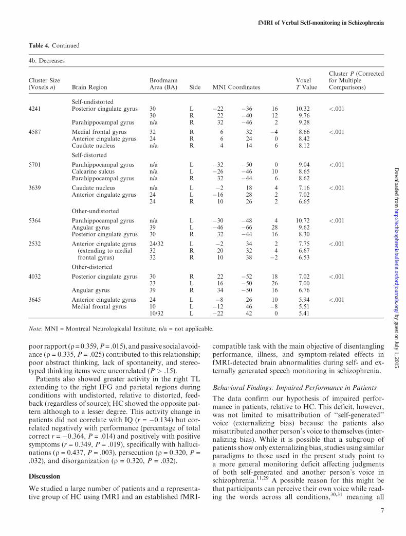
4b. Decreases
Cluster Size(Voxels n) Brain Region
BrodmannArea (BA) Side MNI Coordinates
VoxelT Value
Cluster P (Correctedfor MultipleComparisons)
Self-undistorted4241 Posterior cingulate gyrus 30 L �22 �36 16 10.32 <.001
30 R 22 �40 12 9.76Parahippocampal gyrus n/a R 32 �46 2 9.28
4587 Medial frontal gyrus 32 R 6 32 �4 8.66 <.001Anterior cingulate gyrus 24 R 6 24 0 8.42Caudate nucleus n/a R 4 14 6 8.12
Self-distorted
5701 Parahippocampal gyrus n/a L �32 �50 0 9.04 <.001Calcarine sulcus n/a L �26 �46 10 8.65Parahippocampal gyrus n/a R 32 �44 6 8.62
3639 Caudate nucleus n/a L �2 18 4 7.16 <.001Anterior cingulate gyrus 24 L �16 28 2 7.02
24 R 10 26 2 6.65
Other-undistorted
5364 Parahippocampal gyrus n/a L �30 �48 4 10.72 <.001Angular gyrus 39 L �46 �66 28 9.62Posterior cingulate gyrus 30 R 32 �44 16 8.30
2532 Anterior cingulate gyrus(extending to medialfrontal gyrus)
24/32 L �2 34 2 7.75 <.00132 R 20 32 �4 6.6732 R 10 38 �2 6.53
Other-distorted
4032 Posterior cingulate gyrus 30 R 22 �52 18 7.02 <.00123 L 16 �50 26 7.00
Angular gyrus 39 R 34 �50 16 6.76
3645 Anterior cingulate gyrus 24 L �8 26 10 5.94 <.001Medial frontal gyrus 10 L �12 46 �8 5.51
10/32 L �22 42 0 5.41
Note: MNI = Montreal Neurologicalal Institute; n/a = not applicable.
7
fMRI of Verbal Self-monitoring in Schizophrenia
by guest on July 1, 2015http://schizophreniabulletin.oxfordjournals.org/
Dow
nloaded from

Fig. 1. Task-Related Changes in Brain Activity Across All Participants (Maps Thresholded at P5 .005, uncorrected). The top row showsactivity increases, and the middle row shows activity decreases associated with individual task conditions in sagittal, axial, and coronal viewswith associated Montreal Neurologicalal Institute coordinates (x,y, z). The bottom row shows activity changes associated with source (self vsother regardless of the level of distortion) and distortion (distorted vs undistorted regardless of the source) factors. All displayed clusters,except the anterior cingulate cluster for distortion factor (distorted > undistorted), are significant (P < .05) after correction for multiplecomparisons. Left hemisphere is shown on the left of the coronal view.
Table 5. Brain Areas Showing Activation Changes for Source and Distortion Effects Across All Participants (Voxel Threshold P = .005)
Cluster Size(Voxels n) Brain Region
BrodmannArea (BA) Side
MNICoordinates
VoxelT Value
Cluster P (Corrected forMultiple Comparisons)
Self > other x y z2584 Angular gyrus (extends to
posterior cingulate)39 R 44 �42 22 4.86 <.00139 L �34 �48 20 4.75
Precuneus 31 R 16 �52 24 4.33Other > self
956 Caudate n/a L �2 10 8 4.41 .012n/a n/a 0 0 �8 4.04
Undistorted > distorted1874 Transverse temporal gyrus 41 R 38 �22 22 3.97 <.001
Inferior frontal gyurs/Insula 44 R 32 30 0 3.62Globus pallidus n/a R 20 �4 �2 3.57
None Distorted > undistorted
Note: MNI = Montreal Neurologicalal Institute; n/a = not applicable.
8
V. Kumari et al.
by guest on July 1, 2015http://schizophreniabulletin.oxfordjournals.org/
Dow
nloaded from

Table 6. Neural Activity Differentiating Groups of Good (Regardless of Diagnosis) and Poor Performers
Individual task conditions (voxel threshold P = .005)
Cluster Size(Voxels n) Brain Region
BrodmannArea Side MNI Coordinate
VoxelT Value
Cluster P(Corrected forMultipleComparisons)
DirectionofEffects
Poor performers > good performersSelf un-distorted x y z
11038 Superior temporal gyrus,inferior frontal gyrus(extends to putamenand thalamus)
22 R 54 -24 0 7.18 <.001 Relatively strongeractivity increases ingood performers
45/47 R 34 6 �2 5.7822 L �52 �36 10 5.76
836 Middle occipital gyrus 18 L -14 -82 14 4.89 0.013 As above18/19 L -10 -96 22 4.0619 L -24 -90 28 3.62
653 Middle occipital gyrus 18 R 16 �70 10 3.84 .032 As above19 R 24 �60 4 3.6118 R 16 �78 14 3.44
Self-distorted
4523 Middle-superior temporalgyrus (extends to thethalamus and the putamen)
21 R 56 -26 2 7.25 <0.001 As above22 R 46 -30 12 5.32
Precentral gyrus 6 R 56 -6 18 5.29
3650 Superior temporal gyrus(extends to the insulaand the inferior frontal gyrus)
22 L �44 �38 14 5.06 <.001 As above22/42 L �54 �26 18 4.6922 L �54 �34 16 4.62
Other-undistorted
1259 Superior temporal gyrus 22 L �52 �40 14 6.57 .007 As aboveInferior parietal cortex 40 L �52 �36 26 2.87
1593 Middle-superior temporal gyrus 21 R 56 �28 0 6.46 .002 As above42 R 62 �34 6 5.2722 R 52 �36 16 4.53
Other-distorted
5186 Precentral gyrus 6 L �58 �6 10 6.60 <.001 As aboveInsula (extends to the thalamus
and the inferior frontal gyrus)n/a L �38 14 �2 5.50
Superior temporal gyrus 42/22 L �58 �24 6 5.26
6129 Superior temporal gyrus(extends to the thalamus,putamen, and the inferiorfrontal gyrus)
22 R 60 �26 0 6.23 <.001 As above22 R 62 �12 0 6.1942 R 54 �22 6 5.79
Poor performers > good performersSelf-undistorted
1492 Medial prefrontal/cingulate gyrus
32 R 2 32 4 6.78 .001 Greater deactivationin good, relative topoor, performers
11 L -22 -28 -14 4.2432 L -14 40 6 4.08
None Self-distortedOther-undistorted
749 Medial prefrontal/cingulate gyrus
32 L �24 42 6 4.38 .045 As above32/24 L �4 28 6 3.64
831 Middle temporal gyrus(extends to PHG)
39 R 30 �52 16 4.28 .032 As above21 R 36 �52 8 4.02
Posterior cingulate 23 R 28 �52 24 3.98Other-distorted
1844 Posterior cingulate 29/30 L �12 �52 22 5.50 <.001 As aboveInferior parietal cortex 40 L �44 �66 28 4.87
9
fMRI of Verbal Self-monitoring in Schizophrenia
by guest on July 1, 2015http://schizophreniabulletin.oxfordjournals.org/
Dow
nloaded from

conditions involve monitoring of self-generated voice insome way. Furthermore, the performance on our task islikely to be affected not only by the ability to recognizeand discriminate between the ‘‘self’’ and ‘‘others’’2 butalso to include other influences such as prior experienceof having used the word stimuli facilitating recognitionin self-generated voice, working memory (WM, remember-ing the feedback while making a decision), and the ability toswitch between and tendency to consider various possibleoptions whilemakinga judgmentaboutoriginof the voices.
Neural Findings
Task-RelatedActivationPatternsAcrossAllParticipants.
Individual Task Conditions.
Increases. We found activation of a large neural networkcomprised of the thalamus (MGN), superior-middle TL,and IFG with successful monitoring of own or others’voice in both distorted and undistorted conditions. Therewas remarkable overlap across the 4 conditions.
An important finding is the robust thalamic, especiallyMGN, activation. Thalamic activity has been considerednecessary for conscious awareness of auditory signals.32
Specifically, the MGN is the main area of thalamic relayfor ascending auditory information, receiving auditoryinformation from the inferior colliculus via the brainstem and then relaying it to the primary auditory cor-tex.33 Previous studies may have failed to observeMGN activation with this or very similar tasks, mostlikely because they had relatively smaller samples ordid not consider relevant activation contrasts.
The activation of frontotemporal regions with success-ful performance is generally consistent with previous
studies.20,27 Despite the classical view that the left TLdominates speech perception, recent studies34–37 empha-size the roles of STG and the superior temporal sulcus inboth hemispheres in the auditory representation ofspeech.38 Furthermore, areas in the right anterior supe-rior temporal sulcus are known to show sensitivity to au-ditory distinctiveness39 required for correct attribution ofauditory feedback across all conditions. The TL activa-tions extended into the inferior parietal lobe, hypothe-sized to be an extension of the higher auditoryassociation cortex.33 The left IFG (Broca’s area) activa-tion was also expected given its role in speech and lan-guage,37,38 while the right IFG activation may relate tothe earlier noted WM requirement of our task.40
Decreases. The parahippocampal gyrus, PC (extendingto the angular gyrus for ‘‘other’’ conditions), and MFGwere deactivated during all conditions. These areas areinvolved in a ‘‘default’’ mode of conscious experience andoften found to be deactivated during goal-specific taskconditions.41
Source and Distortion. The comparison of the self withother conditions, regardless of presence or absence of dis-tortion, revealed differential activity in the PC and angu-lar gyrus. This effect was attributable to strongerdeactivation of these areas during the other, relative tothe self, conditions. The angular gyrus is involved inawareness of action authorship.42 The PC has been im-plicated in familiarity43 in addition to its known involve-ment in a ‘‘default’’ mode of conscious experience.41 Suchfunctions of the angular gyrus and PC may account forstronger deactivation of these regions during the other,relative to the self, conditions given the possible overlap
Table 6. Continued
Individual task conditions (voxel threshold P = .005)
Cluster Size(Voxels n) Brain Region
BrodmannArea Side MNI Coordinate
VoxelT Value
Cluster P(Corrected forMultipleComparisons)
DirectionofEffects
Poor performers > good performers987 Medial prefrontal/
cingulate gyrus24/32 R 20 32 6 3.61 .003 As above24 L �6 26 6 3.4924 L �18 32 4 3.48
Source and distortion effects(voxel threshold P = .05)
Self > other During self comparedwith other—moreactivity in goodperformers. Lessactivity in poorperformers.
5695 Inferior frontal gyrus 45 L 28 34 4 4.38 .015Hippocampus N/a R �28 �30 �8 3.17Parahippocampal gyrus 35 R �38 �34 �8 3.10
None Undistorted > distorted
Note: MNI = Montreal Neurologicalal Institute; n/a = not applicable.
10
V. Kumari et al.
by guest on July 1, 2015http://schizophreniabulletin.oxfordjournals.org/
Dow
nloaded from

between the self conditions that involved processing ofown (thus familiar) voice and the default baseline statethat itself may involve participants being aware of them-selves in the surroundings. Judging from the performancedata, the other conditions were also more difficult thatthe self-conditions and more difficult task conditionsgenerally produce greater deactivation. The caudatealso showed differential activity between the self andother conditions; this, however, was influenced by illnessthe and symptoms (discussed later).
A cluster located in the right transverse temporal gy-rus, extending to the IFG, showed greater activity duringthe undistorted, compared with the distorted, conditions.Although on the surface, this effect may seem inconsis-tent with the finding of Fu et al,20 it was influenced byillness and symptoms. Specifically, patients and HCshowed activity changes in opposite directions, andHC showed activity changes in the same direction (ie,greater activity during the distorted condition) as inthe study of Fu et al20 (see further for a discussion ofthe effect in patients). There was some (nonsignificant)AC activity during the distorted, relative to the undis-torted, feedback conditions. This is consistent with pre-
vious findings20 using a longer version of this task andperhaps related to greater effort or conflict in decisionmaking during the conditions of distorted feedback.44
Good Performers Vs Poor Performers. As expected,poor performers showed much reduced frontotemporalactivity than good performers. Activation deficit inpoor performers was most consistently localized in thesuperior-middle TL and appeared somewhat strongerand larger in extent on the right. This may be associatedwith low discrimination between self-generated vs an-other person’s voices in poorly performing patients giventhe sensitivity of right TL to auditory distinctiveness.39
Reduced left IFG activity in poor performers may sug-gest a language-related deficit.38 Reduced IFG activation(in either hemisphere) could also be the cause or effect ofreduced verbal WM.40 This in our task would mean lossof information after hearing the feedback, prior to beingable to make a response about its origin. Previous studiesexamining auditory verbal imagery have observed nor-mal frontal but reduced TL activity in patients withschizophrenia when they were required to imagine words
Fig. 2. Brain Activity Differentiating Good Performers (Healthy Participants and Well-Performing Patients) and Poor Performers. The topand the middle rows show group differences in activations and deactivations found in individual task condition comparisons (mapsthresholded atP5 .005 uncorrected) with associated Montreal Neurologicalal Institute coordinates (x, y, z). The bottom row shows group3source in sagittal and axial views effect (maps thresholded at P5 .05, uncorrected) with associated MNI coordinates (x, y, z). All displayedclusters are significant (P < .05) after correction for multiple comparisons. Left hemisphere is shown on the left of the axial view.
11
fMRI of Verbal Self-monitoring in Schizophrenia
by guest on July 1, 2015http://schizophreniabulletin.oxfordjournals.org/
Dow
nloaded from

spoken by others.14,15,45,46 The auditory verbal imageryparadigms, however, may not exert the same, time-constrained, load on the verbal WM system as the currentparadigm. Furthermore, the inferior frontal junction isactivated in paradigms that involve task switching andset shifting,47 and there may be a role for these functionsin successful performance on our task.
Poor performers also showed thalamic (MGN, lateralGN, pulvinar) hypoactivity during 3 conditions. Al-though the thalamus has traditionally been seen as a relaycenter, recent theoretical positions48–50 and empiricaldata51–53 point to the importance of these thalamic areas
in stimulus-driven attention and, more generally, in cor-tico-cortical processing achieved by flow of informationvia cortico-thalamo-cortical reentry routes. Poor atten-tion to and/or reduced cortical processing of speech stim-uli may contribute to poor performance.
The finding of relatively greater activity in the medialprefrontal and posterior temporal parietal cortices inpoor performers, relative to good performers, althoughnot specifically hypothesized, is an interesting one. Asnoted earlier, these areas were deactivated during activeconditions and the observed difference between the poorand good performers in the medial prefrontal and the
Table 7. Neural Activity Differentiating Groups of Healthy Participants and Patients With Schizophrenia Regardless of Performance
Individual task conditions (voxel threshold P = .005)
Cluster Size(Voxels n) Brain Region
BrodmannArea Side MNI Coordinates
VoxelT Value
Cluster P(Correctedfor MultipleComparisons)
Directionof Effects
Healthy participants > patientsNone Self-undistorted x y z .008 Less activity in patients
than healthy participants.942 Thalamus n/a R 8 �30 14 4.32n/a L �4 �30 14 4.16n/a L �14 �36 12 3.88
None Self-distortedOther-undistortedOther-distorted
Patients > healthy participantsSelf undistorted
997 Superior-middletemporal gyrus
22/42 L �48 �36 18 4.92 .006 Relatively stronger activityin patients.21 L �56 �28 0 3.74
939 Superior temporal gyrus 42 R 50 �32 20 4.74 .008 As above.42 R 58 �28 8 3.5822 R 64 �38 8 3.33
None Self distortedOther undistortedOther distorted
639 Superior-middletemporal gyrus
22 L �54 �32 10 3.95 .019 As above.22 L �56 �42 16 3.8621 L �50 -40 �2 3.51
Source and distortion effects (voxel threshold P = .05)
Other > self Deactivation during self andnon-significant activation duringother in patients, especially withhigh negative symptoms; oppositeeffect to some degree in healthyparticipants.
5695 Hippocampus n/a L �16 �38 0 3.75 .05Hypothalamus n/a R 4 -6 4 3.16Ventral striatum n/a L �6 10 6 3.11
Undistorted > distorted More activity during undistortedrelative to distorted in patients,especially with poor performanceand/or positive symptoms; oppositeeffect to some degree in healthyparticipants.
6525 Superior temporal gyrus 42 R 40 �24 18 3.64 .007Middle temporal gyrus 21 R 36 �22 �10 3.24Inferior frontal gyrus n/a R 42 30 12 3.15
Note: MNI = Montreal Neurologicalal Institute; n/a = not applicable.
12
V. Kumari et al.
by guest on July 1, 2015http://schizophreniabulletin.oxfordjournals.org/
Dow
nloaded from

posterior temporal parietal cortices arose because ofa lack of deactivation of these regions in the formergroup, perhaps reflecting the fact that this group acti-vated task-relevant areas to a markedly reduced degree.54
Finally, a significant source 3 performance group in-teraction indicated more activity in the right lateral fron-tal, extending to the left hippocampal, region during theself, compared with other, conditions in good but notpoor performers. Previous studies demonstrating coacti-vation of hippocampal and right inferior frontal regionsduring autobiographical retrieval, but not during seman-tic retrieval, have indicated the involvement of these areasin access of episodes from the personal past.55,56 Ourfinding thus may reflect successful recall of previous ex-perience of having spoken and listened to (in own voice)the word stimuli in good performers.
Patients Vs Healthy Participants: Illness and SymptomsInfluences. There was activation deficit in patients, un-related to their performance or symptoms, in the thala-mus (figure 3) during the self-undistorted condition. Thisabnormality seems located in the pulvinar region, ratherthan in the geniculate nucleus, suggesting perhaps thatthalamic first-order relay of auditory information maynot be affected by schizophrenic illness. Given the evi-dence for (a) strong thalamus/pulvinar involvement in
attention57,58 and (b) impaired attention as a schizophre-nia trait/risk marker,59,60 it may represent a trait-relatedeffect in schizophrenia. All patients included in this study,however, were on antipsychotic drugs. Altered activationpatterns found across the entire patient sample, there-fore, may have also included the neurotoxic (causing,or resulting in greater, abnormality) or neuroprotective(minimal abnormality) influences of long-term antipsy-chotic treatment.61,62 Future studies involving antipsy-chotic-naive patients with schizophrenia would help toclarify this issue.
Patients, relative to HC, also showed more activity inparts of the superior-middle TL bilaterally during theself-undistorted condition and in the same area butonly on the left during the other-distorted condition(figure 3). These 2 effects when combined contributedto the group 3 distortion interaction showing less activityin the right temporal (extending to the IFG) regionduring the distorted, compared with the undistorted, con-ditions in patients compared with HC. The degree of neg-ative change in the right TL activity from undistorted-to-distorted feedback associated with poor performance inpatients. This may suggest that the right TL dysfunction,specific to language and voice perception network, maybe particularly relevant to performance deficit in schizo-phrenia.63 The right TL dysfunction (less activity during
Fig. 3. Brain Activity Differentiating Patients From Healthy Participants. The top row shows group differences with individual taskconditions (maps thresholded atP5 .005 uncorrected), and the middle and bottom rows show group3 source and group3 level of distortioneffects, respectively, in sagittal and axial views (maps thresholded atP5 .05, uncorrected) with associated Montreal Neurologicalal Institutecoordinates (x,y, z). All displayed clusters are significant (P< .05) after correction for multiple comparisons. Left hemisphere is shown on theleft of the axial view.
13
fMRI of Verbal Self-monitoring in Schizophrenia
by guest on July 1, 2015http://schizophreniabulletin.oxfordjournals.org/
Dow
nloaded from

the distorted, compared with undistorted, conditions),supporting our hypothesis, was also associated with pos-itive symptoms, especially hallucinations.
We also observed, for the first time to our knowledge,in patients especially those with blunted affect, emotionalwithdrawal, poor rapport and passive social avoidancesymptoms, deactivation of the ventral striatum, midbrainand hypothalamus during processing of own voice and(nonsignificant) activation when processing someoneelse’s voice leading to the finding of significantly greateractivity during the other, relative to self, conditions(group 3 source, figure 3). The hypothalamus, thalamus,ventral striatum, and midbrain regions are activated byexpectation of unpleasant stimuli in healthy people,64
perhaps in association with autonomic arousal.65 The hy-pothalamus is particularly important within the stressaxis.66 It is possible that emotionally withdrawn and so-cially avoidant patients perceived the whole experimentalsetting (including baseline conditions) and someone else’svoice unpleasant (ie, stressful) and arousing unless theyheard their own voice. A recent study67 has shown greateramygdala activation to fearful faces in schizophreniapatients with (than without) flat affect and proposed lim-bic overstimulation with flat affect in schizophrenia. In-creased limbic activity to angry and contemptuous facestimuli has been seen in generalized social phobia.68 Dys-functions of the mesocortical and mesolimbic pathwaysare proposed as being the most relevant to socialanxiety.69 Striatal dysfunction has also been noted ingeneralized social phobia.70
Other Observations
Although the right TL dysfunction was associated withboth poor performance and positive symptoms, we didnot find significant associations between the PANSSsymptom dimensions and performance. This observationdeserves some discussion because self-monitoring hy-pothesis of schizophrenia was initially proposed to ex-plain some of the first-rank symptoms, which presumea loss of self-agency.3 Several studies using a range ofparadigms have supported the association between thesymptoms of delusions of control, thought insertion,and thought blocking and poor self-monitoring6–8,71–75
though there are others that did not find such associa-tions.21,76 Similarly, most,10–12,29,77 but not all,78 studiesusing verbal self-monitoring paradigms observed a rela-tionship between positive symptoms, mainly AH anddelusions, and impaired self-monitoring. An associationbetween self-monitoring deficit and positive symptomsmay more often be found in acutely psychotic samples.
Conclusions
Successful monitoring of own or someone else’s speechactivates a large neural network comprised of the tha-lamic (MGN), temporal, and IFG regions that have im-
portant roles in perception and processing of auditoryinformation and language. Reduced response, both interms of activations in this network and deactivationsin the associated ‘‘default’’ network involving primarilythe MFG and PC, underlies impaired monitoring ofself- or externally generated speech in schizophrenia.Within this population, emotionally withdrawn and so-cially avoidant patients show greater response modula-tion in autonomic arousal-linked neural systems withself-vs-others distinction. Positive symptoms, especiallyhallucinations and persecution, and poor monitoringshare a common activation abnormality in the rightSTG during processing of degraded speech.
Funding
Wellcome Trust, UK (067427/z/02/z). V.K. holds aWellcome Senior Research Fellowship in Basic Biomed-ical Science.
References
1. Kircher T, David AS. Self-consciousness: an integrative ap-proach from philosophy, psychopathology and the neuroscien-ces. In: Kircher T, David AS, eds. The Self in Neuroscienceand Psychiatry. Cambridge, UK: Cambridge University Press;2003:445–473.
2. Kircher TT, Leube DT. Self-consciousness, self-agency, andschizophrenia. Conscious Cognition. 2003;12:656–669.
3. Frith CD. The Cognitive Neuropsychology of Schizophrenia.Erlbaum, UK: Taylor and Francis; 1992.
4. Frith CD, Done DJ. Towards a neuropsychology of schizo-phrenia. Br J Psychiatry. 1988;153:437–443.
5. Malenka RC, Angel RW, Hampton B, Berger PA. Impairedcentral error-correcting behaviour in schizophrenia. ArchGen Psychiatry. 1982;39:101–107.
6. Mlakar J, Jensterle J, Frith CD. Central monitoring deficiencyand schizophrenic symptoms. Psychol Med. 1994;24:557–564.
7. Stirling JD, Hellewell JS, Quraishi N. Self-monitoring dys-function and the schizophrenic symptoms of alien control.Psychol Med. 1998;28:675–683.
8. Blakemore SJ, Smith J, Steel R, Johnstone CE, Frith CD.The perception of self-produced sensory stimuli in patientswith auditory hallucinations and passivity experiences:evidence for a breakdown in self-monitoring. Psychol Med.2000;30:1131–1139.
9. Cahill C, Silbersweig D, Frith CD. Psychotic experiences in-duced in deluded patients using distorted auditory feedback.Cogn Neuropsychiatry. 1996;1:201–211.
10. Johns LC, McGuire PK. Verbal self-monitoring and auditoryhallucinations in schizophrenia. Lancet. 1999;353:469–470.
11. Johns LC, Rossell S, Frith CD, et al. Verbal self-monitoringand auditory verbal hallucinations in patients with schizo-phrenia. Psychol Med. 2001;31:705–715.
12. Allen PP, Johns LC, Fu CH, Broome MR, Vythelingum GN,McGuire PK. Misattribution of external speech in patientswith hallucinations and delusions. Schizophr Res. 2004;69:277–287.
13. Raveendran V, Kumari V. Clinical, cognitive and neural cor-relates of self-monitoring deficits in schizophrenia: an update.Acta Neuropsychiatr. 2007;19:27–37.
14
V. Kumari et al.
by guest on July 1, 2015http://schizophreniabulletin.oxfordjournals.org/
Dow
nloaded from

14. McGuire PK, Silbersweig DA, Wright I, et al. Abnormalmonitoring of inner speech: a physiological basis for auditoryhallucinations. Lancet. 1995;346:596–600.
15. McGuire PK, Silbersweig DA, Wright I, Murray RM,Frackowiak RS, Frith CD. The neural correlates of innerspeech and auditory verbal imagery in schizophrenia: rela-tionship to auditory verbal hallucinations. Br J Psychiatry.1996;169:148–159.
16. Shergill SS, Bullmore E, Simmons A, Murray RM, McGuirePK. Functional anatomy of auditory verbal imagery inschizophrenic patients with auditory hallucinations. Am JPsychiatry. 2000;157:1691–1693.
17. Woodruff PW, Wright IC, Bullmore ET, et al. Auditory hal-lucinations and the temporal cortical response to speech inschizophrenia: a functional magnetic resonance imagingstudy. Am J Psychiatry. 1997;154:1676–1682.
18. Mechelli A, Allen PP, Amaro EJ, et al. Misattribution ofspeech and impaired connectivity in patients with auditoryverbal hallucinations. Hum Brain Mapp. 2007;28:1213–1222.
19. Allen PP, Amaro E, Fu CH, et al. Neural correlates of themisattribution of speech in schizophrenia. Br J Psychiatry.2007;190:162–169.
20. Fu CH, Vythelingum GN, Brammer MJ, et al. An fMRIstudy of verbal self-monitoring: neural correlates of auditoryverbal feedback. Cereb Cortex. 2006;16:969–977.
21. Goldberg TE, Gold JM, Coppola R, Weinberger DR. Unnat-ural practices, unspeakable actions: a study of delayed audi-tory feedback in schizophrenia. Am J Psychiatry. 1997;154:858–860.
22. Oldfield RC. The assessment and analysis of handedness: theEdinburgh inventory. Neuropsychologia. 1971;9:97–113.
23. First MB, Spitzer RL, Gibbon M, Williams JBW. StructuredClinical Interview for DSM-IV Axis I Disorders, Patient Edi-tion (SCID-P), Version 2. New York, NY: Biometrics Re-search, New York State Psychiatric Institute; 1995.
24. Kay SR, Fiszbein A, Opler LA. The positive and negativesyndrome scale (PANSS) for schizophrenia. Schizophr Bull.1987;13:261–276.
25. First MB, Spitzer RL, Gibbon M, Williams JBW. StructuredClinical Interview for DSM-IV-TR Axis I Disorders, ResearchVersion, Non-patient Edition. (SCID-I/NP). New York, NY:Biometrics Research, New York State Psychiatric Institute;2002.
26. Wechsler D. Wechsler Abbreviated Scale of Intelligence. SanAntonio, Tx: The Psychological Corporation; 1999.
27. McGuire PK, Silbersweig DA, Frith CD. Functionalneuroanatomy of verbal self-monitoring. Brain. 1996;119:907–917.
28. Friston KJ, Holmes AP, Worsley KJ. How many subjectsconstitute a study? Neuroimage. 1999;10:1–5.
29. Heinks-Maldonado TH, Mathalon DH, Houde JF, Gray M,Faustman WO, Ford JM. Relationship of imprecise corollarydischarge in schizophrenia to auditory hallucinations. ArchGen Psychiatry. 2007;64:286–296.
30. Price CJ, Wise RJ, Warburton EA, et al. Hearing and saying.The functional neuro-anatomy of auditory word processing.Brain. 1996;119(pt 3):919–931.
31. Price CJ. The anatomy of language: contributions from func-tional neuroimaging. J Anatomy. 2000;197(pt 3):335–359.
32. Silbersweig DA, Stern E. Towards a functional neuroanat-omy of conscious perception and its modulation by volition:implications of human auditory neuroimaging studies. PhilosTrans R Soc Lond B Biol Sci. 1998;353:1883–1888.
33. Henkel CK. The auditory system. In: Haines DE, ed. Funda-mentals Neuroscience for Basic and Clinical Applications. 3rded: Churchil Livingston Elsevier; 2006:334–349.
34. Belin P, Zatorre RJ, Lafaille P, Ahad P, Pike B. Voice-selective areas in human auditory cortex. Nature. 2000;403:309–312.
35. Binder JR, Frost JA, Hammeke TA, et al. Human temporallobe activation by speech and nonspeech sounds. CerebrCortex. 2000;10:512–528.
36. Binder JR, Liebenthal E, Possing ET, Medler DA, Ward BD.Neural correlates of sensory and decision processes inauditory object identification. Nat Neurosci. 2004;7:295–301.
37. Hickok G. Functional anatomy of speech perception andspeech production: psycholinguistic implications. J Psycholin-guistic Res. 2001;30:225–235.
38. Demonet JF, Thierry G, Cardebat D. Renewal of the neuro-physiology of language: functional neuroimaging. PhysiolRev. 2005;85:49–95.
39. Zatorre RJ, Bouffard M, Belin P. Sensitivity to auditory ob-ject features in human temporal neocortex. J Neurosci. 2004;24:3637–3642.
40. Cabeza R, Nyberg L. Imaging cognition II: an empirical re-view of 275 PET and fMRI studies. J Cognit Neurosci. 2001;12:1–47.
41. Gusnard DA, Raichle ME, Raichle ME. Searching for a base-line: functional imaging and the resting human brain. Nat RevNeurosci. 2001;2:685–694.
42. Farrer C, Frey SH, Van Horn JD, et al. The angular gyruscomputes action awareness representations. Cereb Cortex.2008;18:254–261.
43. Maddock RJ, Garrett AS, Buonocore MH. Remembering fa-miliar people: the posterior cingulate cortex and autobio-graphical memory retrieval. Neuroscience. 2001;104:667–676.
44. Botvinick MM. Conflict monitoring and decision making:reconciling two perspectives on anterior cingulate function.Cognit Affect Behav Neurosci. 2007;7:356–366.
45. Shergill SS, Bullmore E, Simmons A, Murray RM, McGuire PK.Mapping auditory hallucinations in schizophrenia using func-tional magnetic resonance imaging. Arch Gen Psychiatry.2000;59:468–489.
46. Shergill SS, Brammer MJ, Fukuda R, Williams SC, Murray RM,McGuire PK. Engagement of brain areas implicated in process-ing inner speech in people with auditory hallucinations. Br JPsychiatry. 2003;182:525–531.
47. Derrfuss J, Brass M, Neumann J, von Cramon DY. Involve-ment of the inferior frontal junction in cognitive control:meta-analyses of switching and Stroop studies. Hum BrainMapp. 2005;25:22–34.
48. Sherman SM, Guillery RW. The role of the thalamus in theflow of information to the cortex. Philos Trans R Soc LondB Biol Sci. 2002;357:1695–1708.
49. Guillery RW, Sherman SM. Thalamic relay functions andtheir role in corticocortical communication: generalizationsfrom the visual system. Neuron. 2002;33:163–175.
50. Sherman SM. The thalamus is more than just a relay. CurrOpin Neurobiol. 2007;17:417–422.
51. Bender DB, Youakim M. Effect of attentive fixation in ma-caque thalamus and cortex. J Neurophysiology. 2001;200185:219–234.
52. Michael GA, Buron V. The human pulvinar and stimulus-driven attentional control. Behav Neurosci. 2005;119:1353–1367.
15
fMRI of Verbal Self-monitoring in Schizophrenia
by guest on July 1, 2015http://schizophreniabulletin.oxfordjournals.org/
Dow
nloaded from

53. O’Connor DH, Fukui MM, Pinsk MA, Kastner S. Attentionmodulates responses in the human lateral geniculate nucleus.Nat Neurosci. 2002;5:1203–1209.
54. Fox MD, Snyder AZ, Vincent JL, Corbetta M, Van EssenDC, Raichle ME. The human brain is intrinsically organizedinto dynamic, anticorrelated functional networks. Proc NatlAcad Sci U S A. 2005;102:9673–9678.
55. Greenberg DL, Rice HJ, Cooper JJ, Cabeza R, Rubin DC,Labar KS. Co-activation of the amygdala, hippocampusand inferior frontal gyrus during autobiographical memoryretrieval. Neuropsychologia. 2005;43:659–674.
56. Daselaar SM, Rice HJ, Greenberg DL, Cabeza R, LaBar KS,Rubin DC. The spatiotemporal dynamics of autobiographicalmemory: neural correlates of recall, emotional intensity, andreliving. Cereb Cortex. 2008;18:217–229.
57. Adler CM, Sax KW, Holland SK, Schmithorst V, RosenbergL, Strakowski SM. Changes in neuronal activation with in-creasing attention demand in healthy volunteers: an fMRIstudy. Synapse. 2001;42:266–272.
58. Salgado-Pineda P, Junque C, Vendrell P, et al. Decreased ce-rebral activation during CPT performance: structural andfunctional deficits in schizophrenic patients. Neuroimage.2004;21:840–847.
59. Chen WJ, Faraone SV. Sustained attention deficits asmarkers of genetic susceptibility to schizophrenia. Am JMed Genet. 2000;97:52–57.
60. Cornblatt BA, Malhotra AK. Impaired attention as an endo-phenotype for molecular genetic atudies of schizophrenia. AmJ Med Genet. 2001;105:11–15.
61. Davis CE, Jeste DV, Eyler LT. Review of longitudinal func-tional neuroimaging studies of drug treatments in patientswith schizophrenia. Schizophr Res. 2005;78:45–60.
62. Vita A, De Peri L. The effects of antipsychotic treatment oncerebral structure and function in schizophrenia. Int Rev Psy-chiatry. 2007;19:429–436.
63. Koeda M, Takahashi H, Yahata N, et al. Language process-ing and human voice perception in schizophrenia: a functionalmagnetic resonance imaging study. Biol Psychiatry. 2006;59:948–957.
64. Herwig U, Abler B, Walter H, Erk S. Expecting unpleasantstimuli–an fMRI study. Psychiatr Res. 2007;154:1–12.
65. Seminowicz DA, Mayberg HS, McIntosh AR, et al. Limbic-frontal circuitry in major depression: a path modeling met-analysis. Neuroimage. 2004;22:409–418.
66. Holsboer F. Stress, hypercortisolism and corticosteroid recep-
tors in depression: implications for therapy. J Affect Disord.
2001;62:77–91.
67. Gur RE, Loughead J, Kohler CG, et al. Limbic activation as-
sociated with misidentification of fearful faces and flat affect
in schizophrenia. Arch Gen Psychiatry. 2007;64:1356–1366.
68. Stein MB, Goldin PR, Sareen J, Zorrilla LT, Brown GG. In-
creased amygdala activation to angry and contemptuous
faces in generalized social phobia. Arch Gen Psychiatry.
2002;59:1027–1034.
69. Matthew SJ, Coplan JD, Gorman JM. Neurobiological
mechanisms of social anxiety disorder. Am J Psychiatry.
2001;158:558–1567.
70. Sareen J, Campbell DW, Leslie WD, et al. Striatal function in
generalized social phobia: a functional magnetic resonance
imaging study. Biol Psychiatry. 2007;61:396–404.
71. Frith CD, Done DJ. Experiences of alien control in schizo-
phrenia reflect a disorder in the central monitoring of action.
Psychol Med. 1989;19:359–363.
72. Daprati E, Franck N, Georgieff N, et al. Looking for the
agent: an investigation into consciousness of action and
self-consciousness in schizophrenic patients. Cognition. 1997;
65:71–86.
73. Stirling JD, Hellewell JS, Ndlovu D. Self-monitoring dys-
function and the positive symptoms of schizophrenia.
Psychopathology. 2001;34:198–202.
74. Knoblich G, Stottmeister F, Kircher T. Self-monitoring in
patients with schizophrenia. Psychol Med. 2004;34:1561–1569.
75. Lindner A, Thier P, Kircher TT, Haarmeier T, Leube DT.
Disorders of agency in schizophrenia correlate with an inabil-
ity to compensate for the sensory consequences of actions.
Curr Biol. 2005;15:1119–1124.
76. Fourneret P, Franck N, Slachevsky A, Jeannerod M. Self-
monitoring in schizophrenia revisited. Neuroreport.
2001;12:1203–1208.
77. Johns LC, Gregg L, Allen PP, McGuire PK. Impaired verbal
self-monitoring in psychosis: effects of state, trait and diagno-
sis. Psychol Med. 2006;36:465–474.
78. Versmissen D, Janssen I, Johns L, et al. Verbal self-monitoring
in psychosis: a non-replication. Psychol Med. 2007;37:
569–576.
16
V. Kumari et al.
by guest on July 1, 2015http://schizophreniabulletin.oxfordjournals.org/
Dow
nloaded from