Schizophrenia with auditory hallucinations: A voxel-based morphometry study
Upload
independentCategory
view
0download
0
366 J Psychiatry Neurosci 2011;36(6)
© 2011 Canadian Medical Association
Research Paper
Dysconnectivity of multiple resting-state networks in patients with schizophrenia who have persistent
auditory verbal hallucinations
Nadine Donata Wolf, MD; Fabio Sambataro, MD, PhD; Nenad Vasic, MD; Karel Frasch, MD;Markus Schmid, MD; Carlos Schönfeldt-Lecuona, MD; Philipp Arthur Thomann, MD;
Robert Christian Wolf, MD
N.D. Wolf — Central Institute of Mental Health, Department of Addictive Behavior and Addiction Medicine, Mannheim, Germany;Sambataro — Brain Center for Motor and Social Cognition, Italian Institute of Technology, Parma, Italy; Vasic, Schmid, Schönfeldt-Lecuona, R.C. Wolf — Department of Psychiatry and Psychotherapy III, University of Ulm, Germany; Frasch —Department of Psychiatry and Psychotherapy II, University of Ulm, Germany; Thomann, R.C. Wolf — Center of PsychosocialMedicine, Department of General Psychiatry, University of Heidelberg, Germany
Introduction
Auditory verbal hallucinations (AVHs), defined as sensoryexperiences in the absence of a corresponding external stimu-lus,1 are a core symptom of schizophrenia and related spec-trum disorders. Numerous neuroimaging studies have indi-cated that AVHs are associated with the abnormal structure
and function of a widely distributed set of brain regions, suchas the lateral prefrontal cortices, the cingulate cortex and re-gions of the temporal lobes, that are involved in language, at-tention, executive function and memory.2,3 The most consist -ent findings have implicated frontotemporal pathways as themain neural substrate underlying AVHs,2,4 leading to cogni-tive models of AVHs as a result of a functional breakdown in
Correspondence to: Dr. R.C. Wolf, Center of Psychosocial Medicine, Department of General Psychiatry, University of Heidelberg, Germany,Voßstraße 4, 69115 Heidelberg, Germany; [email protected]
J Psychiatry Neurosci 2011;36(6):366-74.
Submitted Jan. 26, 2011; Revised Mar. 27, Apr. 17, 2011; Accepted Apr. 19, 2011.
DOI: 10.1503/jpn.110008
Background: Functional neuroimaging studies on schizophrenia have suggested abnormal task-related functional connectivity in pa-tients with schizophrenia who have auditory verbal hallucinations (AVHs). However, little is known about intrinsic functional connectivityin these patients. Methods: Between January 2009 and February 2010, we studied patients with schizophrenia who had persistent andtreatment-refractory AVHs in comparison with healthy controls. Using functional magnetic resonance imaging, we studied the functionalconnectivity of multiple resting state networks (RSNs) and their relation to symptom severity. We analyzed the data using a spatial groupindependent component analysis, and we used random-effects t tests to compare spatial components between groups. Results: Therewere 10 patients and 14 controls enrolled in this study. In total, 16 RSNs were identified, from which we selected 4 networks of interestfor further analyses. Within a speech-related network, patients showed increased connectivity in bilateral temporal regions and de-creased connectivity in the cingulate cortex. Within 2 additional RSNs associated with attention and executive control, respectively, pa-tients exhibited abnormal connectivity in the precuneus and right lateral prefrontal areas. We found correlations between measures ofAVH severity and functional connectivity of the left anterior cingulate, left superior temporal gyrus and right lateral prefrontal cortex. Limitations: The relatively small sample size, the patients’ use of antipsychotic medication and the lack of a clinical control group haveto be considered as potential limitations. Conclusion: Our findings indicate that disrupted intrinsic connectivity of a speech-related net-work could underlie persistent AVHs in patients with schizophrenia. In addition, the occurrence of hallucinatory symptoms seems to modu -late RSNs associated with attention and executive control.
dys-wolf_JPN template 11-10-14 2:08 PM Page 366

Resting-state dysconnectivity in schizophrenia
J Psychiatry Neurosci 2011;36(6) 367
appropriate monitoring of inner speech generation.5,6 On aneural network level, abnormal “corollary discharge,” amechanism that modulates self-generated responses to dis-tinguish between self-generated and externally generated in-formation, has been proposed as a putative pathophysiologicmechanism of hallucinatory phenomena.7 For instance, dys-functional efference copy/corollary discharge interactionswithin speech-related neural networks8 could result in a mis-attribution of self-generated actions and perceptions, eventu-ally giving rise to AVHs.9,10 Aberrant frontotemporal couplingin patients with schizophrenia has been described in severalstudies so far,11–13 and, more recently, studies investigatingcortico–cortical connectivity in patients who have AVHshave revealed abnormal interactions within speech-relatedneural pathways.13,14 However, patterns of functional dis -connection in patients with schizophrenia may be wide-spread,15–17 eventually affecting multiple systems with dis-crete spatial distributions and functions,15–17 includingnetworks subserving attentional control, memory, executivefunction and self-referential processing. At present, however,it is unclear if and how these networks contribute to thepatho physiology of AVHs.In this study, we investigated the functional neuroanatomy
of AVHs in patients with schizophrenia using a resting-state functional connectivity approach.18 Given the possibilitythat task-driven protocols may reveal activation patterns driven by interactions between experimental stimuli andAVHs,4,14,19,20 we chose to investigate symptom-related pat-terns of dysconnectivity in the absence of experimentally con-trolled stimulation. In this regard, resting-state functionalconnectivity methods essentially aim to identify neural net-works characterized by ongoing spontaneous modulations ofthe blood oxygen level– dependent (BOLD) signal in the ab-sence of specific task- related activity.21,22 One of the most ex-tensively investigated resting-state networks (RSNs) so far isa set of brain regions that consistently exhibits activity de-creases during cognitively demanding tasks. This RSN hasbeen referred to as the “default mode network” (DMN),22,23 afunctionally hetero geneous network that has been associatedwith self- referential processing, affective control and episodicmemory.23,24 Abnormal DMN activity in patients with schizo -phrenia has been reported in a number of studies so far.16,25,26
Apart from the DMN, however, several other RSNs, includ-ing lateral frontoparietal and medial-frontal systems, havenow been consistently identified.22,27,28 These RSNs have beensuggested to closely correspond to a wide range of cognitiveprocesses, such as language, attention, memory and execu-tive control.28–30
We used independent component analysis, a multivariatestatistical approach to resting-state data, to assess the func-tional connectivity of multiple RSNs in patients with chronicand treatment-resistant AVHs compared with healthy con-trols. Independent component analysis is a technique thatmaximizes the independence between output components,31
thus identifying a set of spatially nonoverlapping and tem -por ally coherent networks by measuring functional covari-ance patterns between different brain areas.32 Specifically, weinvestigated 4 anatomically distinct RSNs of interest that
have been previously associated with the pathophysiology ofAVHs: RSNs related to speech processing, attention, execu-tive control and DMN function.2,4,33 Within these networks,we predicted symptom-related functional connectivity of thetemporal cortex and disrupted connectivity of regions associ-ated with cognitive control, such as the anterior cingulateand the lateral prefrontal cortices. In addition, we exploredthe relation between functional connectivity and symptomseverity, as assessed by AVH-specific psychometric scores.
Methods
Participants
We recruited patients with schizophrenia from among the in-and outpatients treated at the Departments of Psychiatry II(Günzburg) and III (Ulm) at the University of Ulm, Germany.In addition to a detailed interview conducted by experiencedclinical psychiatrists (R.C.W., N.D.W.), case notes were re-viewed to corroborate a definitive diagnosis. All patients metDSM-IV criteria for schizophrenia, paranoid subtype. We ex-cluded patients from participation if they had a current axis-Imood, substance-related or anxiety disorder or a concurrentaxis-II disorder according to DSM-IV; had a history of depend -ence on illicit drugs and alcohol; had an insufficient com-mand of the German language; and had sensorimotor deficitsor other neurologic disorders.Patients included in this study had to meet several specific
inclusion criteria. First, they must have had medication-resistant AVHs, defined by persistent hallucinations in thepresence of at least 2 previous clinically ineffective drug trials(each treatment period > 6 wk) with different antipsychoticsat an adequate dosage. Second, they must not have shownpronounced formal thought disorder symptoms. Third, theymust have had sufficient insight into their hallucinatory ex -peri ences and ability to provide detailed self-reports abouttheir AVHs before functional magnetic resonance imaging(fMRI) and during a postscanning exit interview. Patients’ ability to provide informed consent was deter-
mined after an interview conducted by 2 experienced clinicalpsychiatrists (R.C.W., N.D.W.), where the background, theaims and the experimental procedures of the study were pre-sented and discussed in detail. After the interview, patientswere asked to briefly summarize the study’s scope and proced -ures and were given sufficient time to address study-specificquestions.We rated general psychopathology using the Brief Psychi-
atric Rating Scale (BPRS)34 and the Positive and Negative Syndrome Scale (PANSS).35 We assessed the severity ofAVHs using the auditory hallucinations scale (AHS) sumscore, as provided by the Psychotic Symptoms Rating Scales(PsyRatS).37
The healthy control group was recruited from the Univer-sity of Ulm campus and from the University Department ofPsychiatry III. Controls were matched for age, education andhandedness. We excluded controls from participation if theyhad a current axis-I or a concurrent axis-II disorder accordingto DSM-IV, had a first-degree relative with a neurologic or
Resting-state dysconnectivity in schizophrenia
dys-wolf_JPN template 11-10-14 8:28 AM Page 367

psychiatric disorder, had a history of dependence on illicitdrugs and alcohol, were currently taking any psychotropicmedication, and had sensorimotor deficits or other neuro-logic disorders. The study was approved by the local research ethics com-
mittee (University of Ulm, Germany). All experimental pro-cedures were carried out with the informed and written con-sent of the participants.
Imaging data acquisition
The functional data were acquired using a 3-T MagnetomALLEGRA head MRI system (Siemens) at the Department ofPsychiatry and Psychotherapy III at the University of Ulm,Germany. Scans were performed in darkness, and the partici-pants were explicitly instructed to relax without fallingasleep, keep their eyes closed, not think about anything inparticular and move as little as possible. Adherence to theseinstructions was verified by verbal contact immediately afterthe resting-state scan and as part of a postscanning exit inter-view. In this interview, patients reported the occurrence ofAVHs during the MRI session (self-reported AVH occurringseveral times during the resting-state scan over time periodsof several seconds).We obtained T2*-weighted images using echo-planar imag-
ing in an axial orientation (repetition time 2000 ms, echo time30 ms, field of view 192 mm, flip angle 80°, voxel size 3 × 3 ×3 mm, 33 slices, slice thickness 3 mm, gap 1 mm). Within asession, 180 whole-brain volumes were acquired. Prior to dataprocessing, the first 8 volumes of the time series were dis-carded to account for MRI equilibration effects.
Data analysis
Data preprocessing was performed with SPM5 (WellcomeTrust Centre for Neuroimaging) and MATLAB 7.3 (Math-Works). The functional images were corrected for motion arti-facts and spatially normalized to the SPM5 EPI standard tem-plate. All images were spatially smoothed with a 9-mmfull-width at half-maximum isotropic Gaussian kernel. Weperformed a spatial independent component analysis includ-ing both patients and controls using the “Group ICA of fMRIToolbox” (GIFT; http ://mialab .mrn .org /software /gift /index.html).38 The dimensionality of the functional data for eachparticipant was reduced using 3 consecutive steps of principalcomponent analysis alternated with data linked across partici-pants, resulting in one aggregate mixing matrix for all partici-pants. We performed an independent component analysis decomposition using the Infomax algorithm to extract 16 in-dependent components consisting of group spatial maps andrelated time courses. We used “minimum description length”criteria to estimate the order selection (i.e., the number of in-dependent components from the smoothed data sets after taking into account the spatial and temporal correlation of thefMRI data).39 The estimated independent components wereused for a back reconstruction into individual components using the aggregate mixing matrix created during the dimen-sionality data reduction steps. Data normalization was per-
formed using a Z-score transformation. The individual com-ponents consisting of spatial independent maps and timecourses were eventually sorted using a priori masks compris-ing mediolateral prefrontal and cingulate regions, as definedby the Automatic Anatomic Labelling (AAL) Atlas.40 We used2 masks for spatial sorting: first, as described by othergroups,26 we computed a DMN mask comprising the posteriorparietal cortex (Brodmann area [BA] 7), frontopolar cortex(BA 10), posterior cingulate cortex, precuneus and occipito -parietal junction (BA 39). Second, to identify networks previ-ously associated with language, attention and cognitive con-trol,16,28,32 we computed a “prefrontal” mask comprising thesuperior, middle and inferior frontal cortices and the anteriorcingulate cortex. We chose 1 com ponent of interest (COI) thatshowed the highest spatial correlation with the DMN maskand 3 COIs that showed the highest spatial correlation withthe prefrontal mask for the second-level within- and between-group analyses (see also the section on Functional connectiv-ity within-group analyses in Results). In addition, we identi-fied an a priori “control” COI that has been previouslyidentified to show a high spatial overlap with primary andsecondary visual areas.28 This visual component was identi-fied by inspection of the data using the components outputobtained from the independent component analysis and asubsequent within-group second-level analysis (Appendix 1,available at www .cma .ca /jpn). For the visual network, we didnot expect symptom- or disease-related connectivity differ-ences in contrast to the other networks of interest.For each participant’s spatial COI, we used the voxel
weights as random-effects variables and analyzed them using SPM5. For within-group analyses, we used voxel-wise1-sample t tests against the null hypothesis of zero magni-tude to calculate within-group maps for each COI. The statis-tical thresholds for these analyses were set at p < 0.001, un-corrected at the voxel level, and p < 0.05, corrected for spatialextent. On the second level, we compared spatial maps be-tween controls and patients using 2-sample t tests. To fullyinclude those COIs revealed in both groups, we maskedthese between-group comparisons with a combined mask,which was created as follows. First, we computed 1-samplet tests per RSN and group. Second, thresholded t maps(p < 0.005) were binarized using the “AND” Boolean opera-tor, thus producing binary masks of the combined effect ofeach diagnostic group. We eventually used these combinedspatial maps to explicitly mask the between-group compar-isons computed for each RSN. Thresholds of p < 0.001, uncor-rected at the voxel level, and p < 0.05, corrected for spatial ex-tent, were chosen for all second between-group comparisons.All anatomic regions and denominations are reported ac-cording to the atlases of Talairach and Tournoux.41 Coordin -ates are maxima in a given cluster according to the MontrealNeurological Institute (MNI) template.
Correlations between functional connectivity indices and psychometric measures
We calculated correlation analyses between indices of func-tional connectivity and psychometric measures. We computed
Wolf et al.
368 J Psychiatry Neurosci 2011;36(6)
dys-wolf_JPN template 11-10-14 8:28 AM Page 368

Resting-state dysconnectivity in schizophrenia
J Psychiatry Neurosci 2011;36(6) 369
Spearman correlations using the β parameters (“connectivitystrength,” corresponding to the mean voxel weights of theCOIs) of significant clusters emerging from the between-groupcomparisons in the independent component analysis, as wellas from psychometric variables (PANSS and BPRS scores,
PsyRatS total score). We extracted β parameters from clustersof interest using MarsBar 0.41.42 The data were subsequentlyprocessed off-line using the Statistica software package (ver-sion 6.0, StatSoft Inc.). A nominal significance level of p < 0.05was defined and adjusted for multiple comparisons by meansof a rough false discovery rate (FDR) correction43 according tothe 9 different clusters of interest for which correlations werecomputed (p level adjusted to p < 0.028).
Results
Participants
We enrolled 10 right-handed patients with schizophreniawho had persistent and treatment-refractory AVHs and14 unmedi cated, matched controls. The demographic andclinical characteristics of participants are summarized inTable 1. All patients were on stable doses of antipsychoticmedication for at least 2 months before participating in thestudy. At the time of scanning, antipsychotic treatment in-cluded clozapine (175–650 mg/d) as a primary antipsychoticagent, partly in combination with amisulpride (400 mg/d),aripiprazole (15 mg/d), haloperidol (15 mg/d), paliperidone(12 mg/d), quetiapine (800 mg/d) and risperidone (4 mg/d).The mean chlorpromazine equivalence dosage was 457.3 mg(standard deviation [SD] 342.1 mg).
Table 1: Demographic and clinical characteristics of healthy controlsand patients with schizophrenia who have persistent auditory verbalhallucinations
Group; mean (SD)*
Characteristic Controls, n = 14 Patients, n = 10 p value
Age, yr 33.7 (8.6) 36.5 (9.0) 0.45
Sex, male:female 7:7 6:4
Laterality, EHI score 88.9 (13.5) 92.2 (10.0) 0.52
Education, yr 14.9 (2.8) 13.5 (1.6) 0.18
Duration of illness, yr — 9.9 (6.3) —
PsyRatS total score — 24.4 (8.1) —
BPRS score — 48.7 (10.7) —
Item 12 (range 4–7) — 5.5 (0.8) —
PANSS-P — 16.0 (3.4) —
Item 3 (range 4–5) — 4.6 (0.5) —
PANSS-N — 22.0 (5.5) —
EHI = Edinburgh Handedness Inventory;36 BPRS = Brief Psychiatric Rating Scale;34
PANSS = Positive and Negative Syndrome Scale;35 PsyRatS = Psychotic SymptomsRating Scale;37 SD = standard deviation.*Unless otherwise indicated.
Fig. 1: Spatial pattern and related time courses of the resting-state network of interest identified bythe group independent component analysis. Results from the second-level within-group analyses in-cluding controls and patients with schizophrenia (p < 0.001, uncorrected at the voxel level, p < 0.05corrected for spatial extent).
dys-wolf_JPN template 11-10-14 8:28 AM Page 369

Functional connectivity within-group analyses
In total, 16 independent components were estimated, consist-ing of individual spatial independent maps and time courses(Fig. 1). In controls and patients, we identified 4 COIs thatwere spatially correlated with the a priori prefrontal masksderived from the AAL atlas.40 Consistent with previous re-search,27,28 we identified the following network patterns. Thefirst COI (left fronto-temporo-parietal,16,28 r = 0.32; Fig. 1, topleft) revealed a spatial pattern comprising predominantly thebilateral prefrontal cortex (left lateralized dorso- and ventro-lateral prefrontal cortex, medial and superior prefrontal cor-tex) and left temporoparietal regions. The second COI (rightfrontoparietal,16,28 r = 0.33; Fig. 1, top right) revealed a net-work comprising right lateralized ventro- and dorsolateralprefrontal regions, superior and inferior parietal areas, thedorsal and posterior cingulate cortex and the precuneus. Thethird COI (executive control,28,32 r = 0.22; Fig. 1, bottom left)comprised the bilateral ventrolateral prefrontal cortex, anter -ior prefrontal regions, cingulate cortex and bilateral middletemporal and inferior parietal areas. The fourth COI (DMN,r = 0.59; Fig. 1, bottom right) showed a pattern of predomin -antly cortical midline regions, as described in detail by previ-ous studies of DMN function.28,44 The visual network includedthe primary and secondary visual areas (Appendix 1).
Functional connectivity between-group analyses
The second-level 2-sample t tests revealed the following find-ings. Within the left fronto-temporo-parietal RSN, the patientgroup showed less connectivity in the left anterior cingulatecortex (ACC) and the right posterior cingulate cortex. Wefound increased connectivity in the left middle and superiortemporal gyrus (MTG/STG) and in the right MTG in patientscompared with controls (Fig. 2, Table 2). Within the rightfrontoparietal RSN, the patient group showed increased connectivity in the right middle frontal gyrus (MFG; Fig. 2,Table 2). Within the executive control RSN, patients showedless connectivity in the left precuneus and increased connec-tivity in the right MFG and superior frontal gyrus (Fig. 2,Table 2). No differences were observed between controls andpatients within the DMN and the visual network.
Relation between functional connectivity indices and psychometric measures
Significant correlations were found between connectivity ofthe left ACC (ρ = –0.75, p = 0.012), the left STG (ρ = 0.71,p = 0.023) and the right MFG (ρ = 0.78, p = 0.008) with mea-sures of overall AVH severity, as measured by the PsyRatStotal score (Fig. 2). In contrast to measures of AVH severity,
Wolf et al.
370 J Psychiatry Neurosci 2011;36(6)
Fig. 2: Regions exhibiting differences in functional connectivity in patients with schizophrenia com-pared with controls and plots of the correlation analyses using the Psychotic Symptoms RatingScale — auditory hallucination scale total score.37 Results from the second-level between-groupanalyses (p < 0.001, uncorrected at the voxel level, p < 0.05 corrected for spatial extent) and from thecorrelation analyses (p < 0.05); see also Table 2 for detailed coordinates and Z scores. AVH = audi-tory verbal hallucinations.
dys-wolf_JPN template 11-10-14 8:29 AM Page 370
Resting-state dysconnectivity in schizophrenia
J Psychiatry Neurosci 2011;36(6) 371
the PANSS-P score was positively correlated (ρ = 0.71,p = 0.021) with connectivity of the right MTG, whereas thePANSS-N score was negatively correlated (ρ = –0.72,p = 0.020) with connectivity of the right MFG (BA 8).To investigate potential effects of antipsychotic medication
in this patient sample, we additionally conducted correlationanalyses using the mean voxel weights from significant between-group clusters and chlorpromazine equivalents. Wefound a significant correlation between chlorpromazineequivalents and connectivity of the right MTG (ρ = 0.71,p = 0.022); the other correlations were not significant.
Discussion
This study investigated the functional connectivity of mul -tiple RSNs in patients with schizophrenia who have persist -ent AVHs. Two main findings emerged. First, 3 RSNsshowed functional connectivity differences between patientsand controls. Specifically, we found abnormal connectivitywithin 2 left- and right-lateralized RSNs and within an execu-tive control network. Second, when considering overall AVHseverity, we found correlations between functional connec-tivity and psychometric measures in the left ACC, left STGand right MFG. These regions were not significantly correl -ated with measures of general psychopathology, as assessedby the BPRS and the PANSS.Considering the extant functional imaging literature, our
results are in agreement with data suggesting abnormal activity of speech-related regions in patients who haveAVHs. Specifically, areas of the temporal cortex, mostlyleft- lateralized, in conjunction with regions of the cingulatecortex have been demonstrated to exhibit increased levelsof metabolism and blood flow in patients who haveAVHs.2–4 We detected regions of abnormal connectivitywithin a left fronto-temporo-parietal system (i.e., a networkassociated with language processing, as described by studiesof speech and language processing46 and by more recentstudies of multiple RSNs27,28). Specifically, we observed dis-rupted connectivity of the temporal and cingulate cortices,where patients showed less connectivity in the cingulatecortex and increased connectivity in bilateral temporal re-gions compared with controls. The relation between mea-
sures of AVH severity and functional connectivity of theACC and the STG further suggests a critical role for theseregions in symptom generation and in the modulation ofsymptom intensity, which is consistent with previous re-ports.20,47–49 Abnormal activation and dysconnectivity of theleft STG and the ACC in patients who have AVHs has beenpreviously observed during speech-attribution tasks,14,50
providing support to the notion that a decoupling of a temporo-cingulate pathway could lead to speech misat -tribution and false auditory perceptions in hallucinatingpatients with schizophrenia. This pattern of functional de-coupling was further confirmed by a recent study that re-ported a relation between AVH severity and ACC connec-tivity during resting-state conditions.20 The ACC has beenfrequently associated with directed attention, online sourcemonitoring and cognitive control,51 and decreased con -nectivity of this region might parallel a failure of theseprocesses in the context of inner speech perception. Fur-thermore, activation of the ACC is significantly differentbetween self- and nonself-related stimuli,52 suggesting thatthe ACC could mediate the distinction between self -attributed inner speech perception and phenomena attrib-uted to a nonself source. Of note, the disruption of temporo-cingulate regions in our patient sample was observedduring resting-state conditions (i.e., in the absence of exter-nally induced cognitive or linguistic stimuli), suggestingthat the occurrence of internal stimulation by AVH is char-acterized by a similar pattern of dysfunction, as observedin studies of overt speech generation and speech attribu-tion.50 Within the left fronto-temporo-parietal RSN, how-ever, we did not observe AVH-related connectivity differ-ences in the Broca region in this sample, which contrastsprevious reports.53 Yet activation differences in the Brocaregion/inferior frontal gyrus (IFG) in patients who haveAVHs or are prone to them have not been consistently reported,54–56 and this data heterogeneity may be partly ex-plained by the diversity of the speech and cognition para-digms used in previous studies compared with resting-state or low-level demand conditions. Since we did notemploy a specific activation paradigm in our study, the ab-sence of IFG dysconnectivity could alternatively indicatethat deficient generation and perception of inner speech
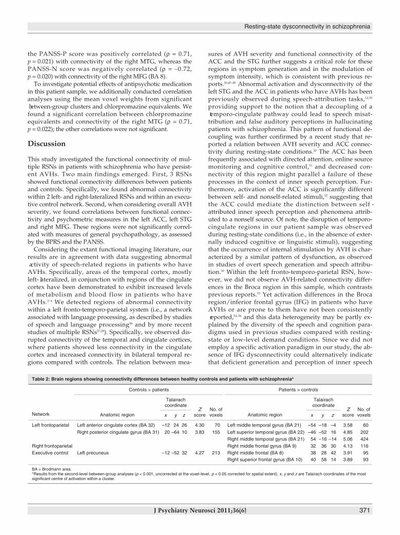
Table 2: Brain regions showing connectivity differences between healthy controls and patients with schizophrenia*
Controls > patients Patients > controls
Talairachcoordinate
Talairachcoordinate
Network Anatomic region x y zZ
scoreNo. ofvoxels Anatomic region x y z
Zscore
No. ofvoxels
Left frontoparietal Left anterior cingulate cortex (BA 32) –12 24 26 4.30 70 Left middle temporal gyrus (BA 21) –54 –18 –4 3.58 60
Right posterior cingulate gyrus (BA 31) 20 –64 10 3.83 155 Left superior temporal gyrus (BA 22) –46 –52 16 4.85 202
Right middle temporal gyrus (BA 21) 54 –16 –14 5.06 424
Right frontoparietal Right middle frontal gyrus (BA 9) 32 36 30 4.13 116
Executive control Left precuneus –12 –52 32 4.27 213 Right middle frontal (BA 8) 38 28 42 3.91 95Right superior frontal gyrus (BA 10) 40 58 14 3.89 93
BA = Brodmann area.*Results from the second-level between-group analyses (p < 0.001, uncorrected at the voxel-level, p < 0.05 corrected for spatial extent). x, y and z are Talairach coordinates of the mostsignificant centre of activation within a cluster.
dys-wolf_JPN template 11-10-14 8:29 AM Page 371

and decreased attentional control14,57,58 rather than speechexpression are among the primary disrupted functions inpatients with persistent AVHs. Thus, the left STG may lieat the core of a final common pathway under lying the generation and experience of AVHs, as proposed by otherresearchers.3,57
Interestingly, the speech-related RSN was not the only net-work showing connectivity abnormalities in this study. Con-nectivity differences in patients who have AVHs were alsodetected in the right frontoparietal and the executive controlnetwork. Two features of these findings are noteworthy: first,patients showed increased functional connectivity in pre-dominantly prefrontal regions compared with controls, andsecond, the pattern of dysconnectivity included mostly re-gions of the right hemisphere. Abnormal activation of righthemisphere regions is a frequently reported finding in pa-tients who have AVHs, where activation changes have beenmost consistently reported for areas of the prefrontal andtemporal cortices.45,55,59 These findings have been discussedwithin the context of a disrupted language lateralization inpatients who have AVHs.2,60 The data provided by our study,however, suggest the involvement of an RSN distinct fromthe network subserving language. For instance, a right later-alized ventral attentional system comprising middle and in-ferior prefrontal and posterior parietal regions has been de-scribed by numerous functional neuroimaging studies ofattention, including studies of intrinsic and phasic alertness61
and attentional control.62 Also, several resting-state fMRIstudies have consistently reported a right frontoparietal net-work similar to the RSN identified in our study, possibly reflecting baseline properties of a network subserving at -tentional processes that persists in the absence of externalevents.28,63 In contrast, the executive control network, as iden-tified by our study, has been previously referred to as corres -ponding to cognition paradigms of inhibitory control and af-fective processing.28 In conjunction with findings of abnormalconnectivity in a speech-related RSN, we speculate that aber-rant right middle and superior frontal connectivity within attention and executive networks could reflect increased attentional resources for salient internal events, such as theoccurrence of AVHs, paralleled by inefficient top–down suppression and executive control. A similar suggestionemerged from activation studies of dichotic listening in pa-tients who have AVHs, in whom a failure to recruit a gener-alized effort network4 (i.e., brain circuits necessary for ade-quate self-monitoring, inhibitory control and goal-directedbehaviour) has been proposed to underlie AVH in additionto deficient speech-processing networks.4,6 However, since weinvestigated participants under resting-state conditions, theprecise cognitive or affective functions of the RSN describedin our study cannot be solely inferred by their spatial patternnor fully delineated by circumscribed loci of dysfunction.Thus, the preliminary conclusions drawn from the presentresting-state data set clearly need further support from multi-modal neuropsychologic, task-based and resting-state fMRIstudies.Contrary to our prediction, we did not confirm DMN
dysconnectivity in this patient sample. This contrasts with
previous reports of disrupted DMN connectivity in patientswith schizophrenia.16,25 In this regard, it is worthy to note thatprevious studies of the DMN in patients with schizophreniahave also reported heterogeneous findings for specific loci ofDMN dysconnectivity,16,25 possibly related to clinical variationand symptom expression in a given sample. Moreover, DMNdysfunction in patients with schizophrenia has been moreconsistently reported within the context of cognitive tasks,25,26
where task demands and stimulus-driven interactions oftask-positive (i.e., frontoparietal networks) and task-negative(i.e., DMN) systems have to be considered.17 Further researchis needed to dissociate alterations of brain network connec-tivity during experimentally induced conditions and their interactions with baseline DMN connectivity, specifically inhallucinating patients.
Limitations
Our study has several strengths, such as the hypothesis- driven investigation of multiple functional RSNs not influ-enced by externally induced task-specific stimuli, the use ofmultivariate methods for fMRI data and the inclusion of apsychometrically well-characterized clinical population withpersistent AVHs and sufficient insight into their symptoms.However, we also acknowledge several limitations. First, al-though our data are in agreement with the extant neuroimag-ing evidence suggesting abnormal frontotemporal function inpatients who have AVHs, the generalizability of our results ispotentially limited by the relatively small sample size. How-ever, in contrast to most studies involving patients who haveAVHs so far, we used multivariate methods, which areknown to yield increased power compared with analysesconducted within the framework of the general linear model.In this regard, independent component analysis, in contrastto the general linear model, uses all scans to estimate spa-tially independent and temporally coherent networks, thusreducing on the first level the number of scans needed to ob-tain a significant response.32 We acknowledge that independ -ent component analysis may also create a potential multiplecomparison problem, since this method minimizes the mu-tual information among sources and may thus estimatenonorthogonal components. Currently, however, there are noavailable statistical tools to correct for this potential issue forfMRI data. Nevertheless, the independent component analy-sis method of exploratory fMRI analysis is regarded aspreferable to that of, for example, principal component analy-sis since the spatial independence enforced upon compon -ents by (spatial) independent component analysis dictatesonly that their time courses should not be highly colinear, re-sulting in a more biologically plausible systems model thanthat obtained from a principal component analysis decom -pos ition. With the latter approach, the analysis enforces or-thogonality between time courses, precluding the detectionof signals that partially correlate in the temporal domain.64
Second, we investigated a group of hallucinating patientsonly, and thus the specificity of our connectivity findings incontrast to patients who do not have AVHs must remainopen at this stage of research. We found different correlations
Wolf et al.
372 J Psychiatry Neurosci 2011;36(6)
dys-wolf_JPN template 11-10-14 8:29 AM Page 372

Resting-state dysconnectivity in schizophrenia
J Psychiatry Neurosci 2011;36(6) 373
for AVH measures than for scores rating general psycho -pathology (BPRS) or overall positive and negative symp-toms (PANSS-P and PANSS-N), suggesting that some re-gions could be specifically associated with the occurrence ofAVHs and with AVH severity. However, the correlation re-sults should be interpreted with caution given the limitedsample size.65 It is also noteworthy that a recent study ofmultiple RSNs in nonhallucinating patients with schizophre-nia reported a dysfunction of the DMN and the right fronto -parietal network,16 suggesting that actively hallucinating andnonhallucinating patients may show different patterns of in-trinsic dysconnectivity. Nevertheless, further research isneeded to elucidate the specificity of RSN dysfunction in pa-tients who have AVHs in comparison with those who do notor who have other syndromes within the schizophreniaspectrum. Third, we included patients with chronic schizophrenia
and, although our data are in agreement with results fromstudies of first-episode patients who have AVHs,54 patientswith a longer history of schizophrenia might show differentphenomenologic characteristics of AVH66 and possibly dif-ferent spatial patterns of neural dysfunction. Eventually, allof the patients who participated in the present study re-ceived treatment with antipsychotic medications. Althougha significant correlation was only found between connectiv-ity of the right MTG and chlorpromazine equivalents, wecannot fully rule out potential long-term medication effectsor effects of previous treatments with antipsychotics on RSNconnectivity.
Conclusion
The results of the present study are suggestive of abnormalconnectivity of multiple RSNs in patients with schizophreniawho have persistent, treatment-refractory AVHs. Our findingsindicate that functional changes of a left-lateralized fronto- temporoparietal RSN associated with language processing andspeech monitoring may underlie and modulate persistentAVHs and symptom intensity. In addition, the occurrence ofhallucinatory symptoms seems to affect other RSNs, possiblyinteracting with attentional and executive capacity.
Acknowledgements: This work was supported by a research grantfrom the University of Ulm, Germany (principal investigatorR.C.W.). The authors thank all participants and their families fortheir time and interest in this study. The authors are grateful to Miriam Ott and Petra Neumann for their assistance with data collection.
Competing interests:None declared from Drs. N.D. Wolf, Sambataro,Vasic, Schmid, Schönfeldt-Lecuona, Thomann and R.C. Wolf. Dr.Frasch declares having received grant support from AstraZeneca,lecture fees from Janssen and travel support from AstraZeneca,Janssen, Lilly and Pfizer; and holds stock in STADA.
Contributors: Drs. N.D. Wolf, Sambataro, R.C. Wolf, Vasic andSchönfeldt-Lecuona designed the study. Drs. N.D. Wolf, R.C. Wolf, Vasic, Frasch and Schmid acquired the data. Drs. N.D. Wolf, Sambataro,R.C. Wolf and Thomann analyzed the data. Drs. N.D. Wolf and R.C.Wolf wrote the article, which Drs. N.D. Wolf, Sambataro, R.C. Wolf,Vasic, Frasch, Schmid, Schönfeldt-Lecuona and Thomann reviewed.All authors approved publication.
References
1. Aleman A, de Haan EH. On redefining hallucinations. Am J Orthopsychiatry 1998;68:656-9.
2. Allen P, Laroi F, McGuire PK, et al. The hallucinating brain: a re-view of structural and functional neuroimaging studies of hallu -cin ations. Neurosci Biobehav Rev 2008;32:175-91.
3. Stephane M, Barton S, Boutros NN. Auditory verbal hallucinationsand dysfunction of the neural substrates of speech. Schizophr Res2001; 50:61-78.
4. Hugdahl K, Loberg EM, Nygård M. Left temporal lobe structuraland functional abnormality underlying auditory hallucinations inschizophrenia. Front Neurosci 2009;3:34-45.
5. McGuire PK, Silbersweig DA, Wright I, et al. Abnormal monitoringof inner speech: a physiological basis for auditory hallucinations.Lancet 1995;346:596-600.
6. Hugdahl K, Loberg EM, Specht K, et al. Auditory hallucinations inschizophrenia: the role of cognitive, brain structural and geneticdisturbances in the left temporal lobe. Front Hum Neurosci 2007;1:6.Epub 2008 Mar. 28.
7. Heinks-Maldonado TH, Mathalon DH, Houde JF, et al. Relation-ship of imprecise corollary discharge in schizophrenia to auditoryhallucinations. Arch Gen Psychiatry 2007;64:286-96.
8. Chen CM, Mathalon DH, Roach BJ, et al. The corollary dischargein humans is related to synchronous neural oscillations. J CognNeurosci 2010 Oct. 14. [Epub ahead of print]
9. Mathalon DH, Ford JM. Corollary discharge dysfunction in schizo-phrenia: evidence for an elemental deficit. Clin EEG Neurosci 2008;39: 82-6.
10. Ford JM, Mathalon DH. Corollary discharge dysfunction in schizo-phrenia: Can it explain auditory hallucinations? Int J Psychophysiol2005;58:179-89.
11. Walter H, Vasic N, Höse A, et al. Working memory dysfunction inschizophrenia compared to healthy controls and patients with depression: evidence from event-related fMRI. Neuroimage 2007; 35:1551-61.
12. Friston KJ, Herold S, Fletcher PC. Abnormal fronto-temporal interac-tions in schizophrenia. In: Watson SJ, editor. Biology of schizophreniaand affective diseases. New York (NY): Raven; 1995. p. 449-81.
13. Lawrie SM, Buechel C, Whalley HC, et al. Reduced frontotemporalfunctional connectivity in schizophrenia associated with auditoryhallucinations. Biol Psychiatry 2002;51:1008-11.
14. Mechelli A, Allen P, Amaro E Jr, et al. Misattribution of speechand impaired connectivity in patients with auditory verbal hallu -cinations. Hum Brain Mapp 2007;28:1213-22.
15. Calhoun VD, Maciejewski PK, Pearlson GD, et al. Temporal lobe and“default” hemodynamic brain modes discriminate between schizo-phrenia and bipolar disorder. Hum Brain Mapp 2008;29:1265-75.
16. Rotarska-Jagiela A, van de Ven V, Oertel-Knochel V, et al. Resting-state functional network correlates of psychotic symptoms inschizophrenia. Schizophr Res 2010;117:21-30.
17. Wolf RC, Vasic N, Sambataro F, et al. Temporally anticorrelatedbrain networks during working memory performance reveal pre-frontal and hippocampal dysconnectivity in patients with schizo-phrenia. Prog Neuropsychopharmacol Biol Psychiatry 2009;33:1464-73.
18. Fox MD, Greicius M. Clinical applications of resting state func-tional connectivity. Front Syst Neurosci 2010;4:19.
19. Hoffman RE, Fernandez T, Pittman B, et al. Elevated functionalconnectivity along a corticostriatal loop and the mechanism of auditory/verbal hallucinations in patients with schizophrenia. BiolPsychiatry 2011:69:407-14.
20. Vercammen A, Knegtering H, den Boer JA, et al. Auditory hallucina-tions in schizophrenia are associated with reduced functional con-nectivity of the temporo-parietal area. Biol Psychiatry 2010;67:912-8.
21. Fox MD, Corbetta M, Snyder AZ, et al. Spontaneous neuronal ac-tivity distinguishes human dorsal and ventral attention systems.
dys-wolf_JPN template 11-10-14 8:29 AM Page 373

374 J Psychiatry Neurosci 2011;36(6)
Wolf et al.
Proc Natl Acad Sci U S A 2006;103:10046-51.
22. Fox MD, Snyder AZ, Vincent JL, et al. The human brain is intrin -sically organized into dynamic, anticorrelated functional net-works. Proc Natl Acad Sci U S A 2005;102:9673-8.
23. Raichle ME, MacLeod AM, Snyder AZ, et al. A default mode ofbrain function. Proc Natl Acad Sci U S A 2001;98:676-82.
24. Gusnard DA, Akbudak E, Shulman GL, et al. Medial prefrontalcortex and self-referential mental activity: relation to a defaultmode of brain function. Proc Natl Acad Sci U S A 2001;98:4259-64.
25. Garrity AG, Pearlson GD, McKiernan K, et al. Aberrant “defaultmode” functional connectivity in schizophrenia. Am J Psychiatry2007; 164:450-7.
26. Sambataro F, Blasi G, Fazio L, et al. Treatment with olanzapine isassociated with modulation of the default mode network in pa-tients with schizophrenia. Neuropsychopharmacology 2010;35:904-12.
27. Damoiseaux JS, Rombouts SA, Barkhof F, et al. Consistent resting-state networks across healthy subjects. Proc Natl Acad Sci U S A2006; 103:13848-53.
28. Smith SM, Fox PT, Miller KL, et al. Correspondence of the brain’sfunctional architecture during activation and rest. Proc Natl AcadSci U S A 2009;106:13040-5.
29. Fox MD, Raichle ME. Spontaneous fluctuations in brain activityobserved with functional magnetic resonance imaging. Nat RevNeurosci 2007;8:700-11.
30. van den Heuvel MP, Hulshoff Pol HE. Exploring the brain net-work: a review on resting-state fMRI functional connectivity. EurNeuropsychopharmacol 2010;20:519-34.
31. Calhoun VD, Adali T, Pekar JJ. A method for comparing groupfMRI data using independent component analysis: application to visual, motor and visuomotor tasks. Magn Reson Imaging 2004;22:1181-91.
32. Calhoun VD, Kiehl KA, Pearlson GD. Modulation of temporallycoherent brain networks estimated using ICA at rest and duringcognitive tasks. Hum Brain Mapp 2008;29:828-38.
33. Williamson P. Are anticorrelated networks in the brain relevant toschizophrenia? Schizophr Bull 2007;33:994-1003.
34. Overall JE, Gorham DR. The Brief Psychiatric Rating Scale. PsycholRep 1962;10:790-812.
35. Kay SR, Fiszbein A, Opler LA. The positive and negative syndromescale (PANSS) for schizophrenia. Schizophr Bull 1987;13:261-76.
36. Oldfield RC. The assessment and analysis of handedness: The Edinburgh Inventory. Neuropsychologia 1971;9:97-113.
37. Haddock G, McCarron J, Tarrier N, et al. Scales to measure dimen-sions of hallucinations and delusions: the psychotic symptom rating scales (PSYRATS). Psychol Med 1999;29:879-89.
38. Correa N, Adali T, Yi-Ou L, et al. Comparison of blind source sep-aration algorithms for FMRI using a new Matlab toolbox: GIFT.Proceedings of the 2005 IEEE International Conference on Acoustics,Speech, and Signal Processing; 2005 Mar. 18–23; Philadelphia, Pa.2005; 5:401-4.
39. Li YO, Adali T, Calhoun VD. Estimating the number of independ -ent components for functional magnetic resonance imaging data.Hum Brain Mapp 2007;28:1251-66.
40. Tzourio-Mazoyer N, Landeau B, Papathanassiou D, et al. Auto-mated anatomical labeling of activations in SPM using a macro-scopic anatomical parcellation of the MNI MRI single-subjectbrain. Neuroimage 2002;15:273-89.
41. Talairach J, Tournoux P. Co-planar stereotaxic atlas of the humanbrain. New York: Thieme Medical Publishers; 1988.
42. Brett M, Anton J-L, Valabregue R, et al. Region of interest analysisusing an SPM toolbox [abstract]. Presented at the 8th InternationalConference on Functional Mapping of the Human Brain, June 2–6,2002, Sendai, Japan. Available on CD-ROM in NeuroImage, Vol16, No. 2, abstract 497.
43. Benjamini Y, Yekutieli D. The control of the false discovery rate inmultiple testing under dependency. Ann Stat 2001;29:1165-88.
44. Buckner RL, Andrews-Hanna JR, Schacter DL. The brain’s defaultnetwork: anatomy, function, and relevance to disease. Ann N YAcad Sci 2008;1124:1-38.
45. Allen P, Aleman A, McGuire PK. Inner speech models of auditoryverbal hallucinations: evidence from behavioural and neuroimag-ing studies. Int Rev Psychiatry 2007;19:407-15.
46. Price CJ. The anatomy of language: contributions from functionalneuroimaging. J Anat 2000;197:335-59.
47. Onitsuka T, Shenton ME, Salisbury DF, et al. Middle and inferiortemporal gyrus gray matter volume abnormalities in chronicschizophrenia: an MRI study. Am J Psychiatry 2004;161:1603-11.
48. Plaze M, Bartres-Faz D, Martinot JL, et al. Left superior temporalgyrus activation during sentence perception negatively correlateswith auditory hallucination severity in schizophrenia patients.Schizophr Res 2006;87:109-15.
49. Cleghorn JM, Garnett ES, Nahmias C, et al. Regional brain metabo-lism during auditory hallucinations in chronic schizophrenia. Br JPsychiatry 1990;157:562-70.
50. Allen P, Amaro E, Fu CH, et al. Neural correlates of the misattribu-tion of speech in schizophrenia. Br J Psychiatry 2007;190:162-9.
51. Carter CS, Braver TS, Barch DM, et al. Anterior cingulate cortex,error detection, and the online monitoring of performance. Science1998; 280:747-9.
52. Northoff G, Bermpohl F. Cortical midline structures and the self.Trends Cogn Sci 2004;8:102-7.
53. Diederen KM, Neggers SF, Daalman K, et al. Deactivation of thepara hippocampal gyrus preceding auditory hallucinations inschizo phrenia. Am J Psychiatry 2010;167:427-35.
54. Parellada E, Lomena F, Font M, et al. Fluordeoxyglucose-PETstudy in first-episode schizophrenic patients during the hallucina-tory state, after remission and during linguistic-auditory activa-tion. Nucl Med Commun 2008;29:894-900.
55. Sommer IE, Diederen KM, Blom JD, et al. Auditory verbal hallu -cinations predominantly activate the right inferior frontal area.Brain 2008; 131:3169-77.
56. Silbersweig DA, Stern E, Frith C, et al. A functional neuroanatomyof hallucinations in schizophrenia. Nature 1995;378:176-9.
57. Stephane M, Folstein M, Matthew E, et al. Imaging auditory verbalhallucinations during their occurrence. J Neuropsychiatry Clin Neurosci 2000;12:286-7.
58. Shergill SS, Brammer MJ, Williams SC, et al. Mapping auditoryhallucinations in schizophrenia using functional magnetic reson -ance imaging. Arch Gen Psychiatry 2000;57:1033-8.
59. Raij TT, Valkonen-Korhonen M, Holi M, et al. Reality of auditoryverbal hallucinations. Brain 2009;132:2994-3001.
60. Woodruff PW, Wright IC, Bullmore ET, et al. Auditory hallucina-tions and the temporal cortical response to speech in schizophrenia:a functional magnetic resonance imaging study. Am J Psychiatry1997; 154:1676-82.
61. Sturm W, Willmes K. On the functional neuroanatomy of intrinsicand phasic alertness. Neuroimage 2001;14:S76-84.
62. Sharp DJ, Bonnelle V, De Boissezon X, et al. Distinct frontal sys-tems for response inhibition, attentional capture, and error pro-cessing. Proc Natl Acad Sci U S A 2010;107:6106-11.
63. Napadow V, LaCount L, Park K, et al. Intrinsic brain connectivityin fibromyalgia is associated with chronic pain intensity. ArthritisRheum 2010;62:2545-55.
64. Cole DM, Smith SM, Beckmann CF. Advances and pitfalls in theanalysis and interpretation of resting-state FMRI data. Front SystNeurosci 2010;4:8.
65. Yarkoni T. Big correlations in little studies: inflated fMRI correl -ations reflect low statistical power. Perspect Psychol Sci 2009;4:294-8.
66. Nayani TH, David AS. The auditory hallucination: a phenomeno-logical survey. Psychol Med 1996;26:177-89.
dys-wolf_JPN template 11-10-14 8:29 AM Page 374
Copyright © 2022 FDOKUMEN