Effect of Voxel Size on 3D Micro-CT Analysis of Cortical Bone Porosity

Available online at www.sciencedirect.com
iological Psychiatry 32 (2008) 72–80www.elsevier.com/locate/pnpbp
Progress in Neuro-Psychopharmacology & B
Schizophrenia with auditory hallucinations: A voxel-basedmorphometry study
Gracián García-Martí a,⁎, Eduardo J. Aguilar b, Juan J. Lull a, Luis Martí-Bonmatí c,María J. Escartí b, José V. Manjón a, David Moratal a, Montserrat Robles a, Julio Sanjuán d
a Bioengineering, Electronic and Telemedicine Group, Polytechnic University of Valencia, Camino de Vera, s/n, ES-46022 Valencia, Spainb Psychiatric Unit, Clinic University Hospital, Avda. Blasco Ibáñez, 17, 46010 Valencia, Spain
c Department of Radiology, Dr Peset University Hospital, Avda. Gaspar Aguilar, 90, 46017 Valencia, Spaind Psychiatric Unit, University of Valencia School of Medicine, Avda. Blasco Ibáñez, 17, 46010 Valencia, Spain
Received 14 December 2006; received in revised form 11 July 2007; accepted 11 July 2007Available online 19 July 2007
Abstract
Many studies have shown widespread but subtle pathological changes in gray matter in patients with schizophrenia. Some of these studies haverelated specific alterations to the genesis of auditory hallucinations, particularly in the left superior temporal gyrus, but none has analysed the relationshipbetweenmorphometric data and a specific scale for auditory hallucinations. The present study aims to define the presence and characteristics of structuralabnormalities in relation with the intensity and phenomenology of auditory hallucinations by means of magnetic resonance voxel-based morphometry(MR-VBM) method applied on a highly homogeneous group of 18 persistent hallucinatory patients meeting DSM-IV criteria for schizophreniacompared to 19 healthy matched controls. Patients were evaluated using the PSYRATS scale for auditory hallucinations. Reductions of gray matterconcentration in patients to controls were observed in bilateral insula, bilateral superior temporal gyri and left amygdala. In addition, specificrelationships between left inferior frontal and right postcentral gyri reductions and the severity of auditory hallucinations were observed. All these areasmight be implicated in the genesis and/or persistence of auditory hallucinations through specific mechanisms. Precise morphological abnormalities mayhelp to define reliable MR-VBM biomarkers for the genesis and persistence of auditory hallucinations.© 2007 Elsevier Inc. All rights reserved.
Keywords: Auditory hallucinations; Magnetic resonance imaging; PSYRATS; Schizophrenia; Voxel-based morphometry
1. Introduction
Pathological changes in the gray matter (GM) have beenshown in patients with schizophrenia by means of magneticresonance (MR) imaging techniques. These morphologicchanges are widespread (Honea et al., 2005) but subtle and
Abbreviations: AAL, Automated Anatomical Labeling; AH, AuditoryHallucinations; BPRS, Brief Psychiatric Rating Scale; CSF, CerebrospinalFluid; DSM-IV, Diagnostic and Statistical Manual of Mental Disorders, ForthEdition; FDR, False Discovery Rate; GM, Gray Matter; MR, Magneticresonance; PSYRATS, Psychotic Symptom Rating Scale; RFT, Random FieldTheory; ROI, Region of Interest; SnPM, Statistical non-Parametric Mapping;SPM, Statistical Parametric Mapping; VBM, Voxel-based morphometry; WM,White Matter.⁎ Corresponding author. Tel.: +34 96 387 70 00x75275; fax: +34 96 387 90 09.E-mail address: [email protected] (G. García-Martí).
0278-5846/$ - see front matter © 2007 Elsevier Inc. All rights reserved.doi:10.1016/j.pnpbp.2007.07.014
with a variable level of evidence and expression (Shenton et al.,2001). Region of interest (ROI) analyses, which are landmark-based manual procedures, have been the gold standard forstructural MR examinations. While their advantages includeanatomical validity, definition of landmarks in native space andquantitative measures of the voxels in the regions under study,they are time consuming and are of varying reliability. FocusedROI procedures are mainly useful in situations where the ex-pected changes are small and the involved areas are clearlydefined. Even more, multi-ROI studies are tedious to perform,and need many inter- and intra-subject reliability tests, whichare not thoroughly performed. For this reason, several auto-mated whole-brain techniques have been developed.
Voxel-based morphometry (VBM) (Ashburner and Friston,2000; Good et al., 2001) is a fast and reliable technique,although less sensitive than focused ROI procedures. VBMprovides the relative changes in the concentration of GM within

73G. García-Martí et al. / Progress in Neuro-Psychopharmacology & Biological Psychiatry 32 (2008) 72–80
each voxel. Therefore, brain areas shown to be abnormal withVBM analysis represent specific points of maximal changesinstead of defined affected regions.
Both VBM and ROI measurement studies have tried to cor-relate volumetric changes with auditory hallucinations (AH) inpatients with schizophrenia (Tables 1 and 2).Most of these studieshave looked for specific relationship between superior temporalgyrus reductions and AH severity (Barta et al., 1990; Flaum et al.,1995; Levitan et al., 1999; Rajarethinam et al., 2000; Matsumotoet al., 2001; Onitsuka et al., 2004). Left Heschl's gyrus reductionhas also been specifically related to AH (Sumich et al., 2005).Structural reductions associated with AH have also been iden-tified in other areas as the left anterior cingulate (Noga et al.,1995), left anterior hippocampus (Rajarethinam et al., 2001), leftmiddle temporal gyrus (Onitsuka et al., 2004), and also a leftwardasymmetry of the sylvian fissure (Shapleske et al., 2001).
Also, voxel-based studies have found a possible relationshipbetween different structural abnormalities and AH. Shapleskeet al. (2002) compared prominent auditory verbal hallucinatoryand non-hallucinatory patients and described only one signif-icant cluster with a deficit in GM including the left insula andadjacent medial temporal lobe. A significant correlation wasfound between size of the left insula cluster and hallucinations.Milev et al. (2003) prospectively studied 123 psychotic patientsduring a period of 5 years using an automated stereotaxic-basedparcellation method showing a bilateral temporal lobe GMreduction associated with the persistence of AH. Using a similarmethod, Shin et al. (2005) studied 25 drug-naïve schizophrenicpatients showing that the hallucinatory group had right temporal
Table 1Summary of MR imaging studies reporting positive findings in schizophrenia and A
Author/year Sample Clinical evaluation
Barta et al. (1990) 15 M medicated Schiz/15 HC Retrospective SAPSFlaum et al. (1995) 166 Schiz spectrum patients CASHLevitan et al. (1999) 30 Schiz with history of AH SAPSRajarethinam et al. (2000) 20 M Schiz/20 HC BPRSMatsumoto et al. (2001) 40 recent-onset Schiz/40 HC PANSSRajarethinam et al. (2001) 20 M Schiz/20 HC BPRSShapleske et al. (2001) 74 M Schiz (30 without AH
and 44 with strong historyof AH)/32 HC
SAPS (for samplecharacterization)
Shapleske et al. (2002) 72 M Schiz (41 withprominent AH and 31without AH)
SAPS
Milev et al. (2003) 123 Schiz,Schizophreniformor Schizoaffect
Outcome at a meanof five years later
Gaser et al. (2004) 85 (56 non-hallucinatingand 29 hallucinating)Schiz
SAPS
Onitsuka et al. (2004) 23 M, chronic Schiz/28 HC SAPSShin et al. (2005) 25 drug-naïve Schiz (17 with
and 8 without AH)SCID (for samplecharacterization)
Sumich et al. (2005) 25 first-episodepsychotic patients
PANSS
Neckelmann et al. (2006) 12 Schiz/12 HC BPRS
Abbreviations: M, males; Schiz, patients with schizophrenia; HC, healthy controls;
white matter (WM), and frontal and temporal GM excess. Gaseret al. (2004) found, using deformation-based morphometry, thatthe severity of AH was significantly correlated with volume lossin the left Heschl's gyrus and in the left supramarginal gyrus,as well as middle and inferior right prefrontal gyri. Finally,Neckelmann et al. (2006) have recently published a VBM studyin which the superior (transverse) temporal gyrus, left thalamus,and left and right cerebellum density were negatively correlatedwith hallucinations.
Noteworthy, results are not completely in agreement. Clinicalheterogeneity is probably one of the main reasons for the dis-crepancies. Studying large and homogeneous patient samplesthroughVBManalysesmay reduce this bias (Kubicki et al., 2002;Shapleske et al., 2002). On the other hand, most ROI studieshave looked at the superior temporal gyrus and therefore, otherpotentially relevant areas have not been routinely evaluated.Although global analysis can be performed by whole-brainstudies, only five such studies have been focused on AH and dataare not completely in agreement (Tables 1 and 2). Finally, theavailable data show that some of the phenomenological di-mensions of AH have specific neural substrates (Stephane et al.,2003). This may be another source of disagreement and givessupport to the recommendation of using more accurate scales tomeasure unreality symptoms (Sumich et al., 2005).
The present study explores the relationship between AHand structural brain abnormalities through a VBM study. Theexamination was performed on a highly homogeneous group ofschizophrenic patients with persistent hallucinations by using aspecific clinical scale for assessing multiple dimensions of AH.
H
MRI technique Association with auditory hallucinations
ROIs Left STG reductionROIs Left STG reductionROIs Left anterior STG reductionROIs Left anterior STG reductionROIs Right STG reductionROIs Left anterior hippocampus reductionROIs Sylvian fissure asymmetry (the longer
the left Sylvian fissure, the more severe)
Voxel-based methodsimilar to VBM
Left insula and right medial temporal lobe reduction;right inferior parietal lobe and white matter in lefttemporal–parietal connecting tracts excess
Automated stereotaxicbased parcellationmethod
Bilateral temporal lobe gray matter volumes reduction(with persistence of AH)
Deformation-basedmorphometry
Left transverse temporal gyrus of Heschl, rightmiddle/inferior prefrontal gyri and left inferiorsupramarginal gyrus reduction
ROIs Left STG and MTGAutomatedstereotactic-basedparcellation method
Right temporal white matter, frontal gray matterand temporal gray matter excess
ROIs Left Heschl's gyrus reduction
VBM Left STG gyrus, bilateral cerebellum and leftthalamus reduction
STG, superior temporal gyrus; MTG, middle temporal gyrus.

Table 2Summary of MR imaging studies reporting negative or mixed findings in schizophrenia and AH
Author/year Sample Clinical evaluation MRItechnique
Association with auditory hallucinations
Delisi et al. (1994) 85 first-episode Schiz/40 HC SADS, chart review ROIs Not with size or lateralization of planum temporale orSTG
Zipursky et al. (1994) 22 M, chronic Schiz/20 HC BPRS ROIs Not with STG or hippocampusNoga et al. (1995) 14 Schiz/14 HC Retrospective SAPS ROIs Left anterior cingulated reduction, not significant after
Bonferroni correctionMarsh et al. (1997) 56 M, severely ill, Schiz inpatients/52 HC BPRS ROIs Not with STGHavermans et al. (1999) 30 Schiz (15 with chronic AH and
15 without AH)/17 HCBPRS (for samplecharacterization)
ROIs Not with STG and other temporal lobe structures
Rossell et al. (2001) 71 M (42 with and 29 without historyof AH) Schiz/33 HC
SAPS (for samplecharacterization)
ROIs Not with corpus callosum areas
Abbreviations: M, males; Schiz, patients with schizophrenia; HC, healthy controls; STG, superior temporal gyrus; MTG, middle temporal gyrus.
74 G. García-Martí et al. / Progress in Neuro-Psychopharmacology & Biological Psychiatry 32 (2008) 72–80
We hypothesized reductions of GM in these patients, correlatingwith the AH severity. If statistically validated, a reliable mor-phometric biomarker for persistent AH in this homogeneousphenomenological group of schizophrenic patients will beobtained.
2. Methods
2.1. Subjects
Thirty seven subjects were selected for this study, including18 patients with DSM-IV schizophrenia and persistent AH and19 healthy control subjects. Patients' ages ranged from 21 to42 years (mean 35.71±6.11) while the control group rangedfrom 20 to 48 years (mean 33.11±7.61). Both groups werealso matched by sex (all males), ethnic group (all Caucasian),laterality (all right handed), and educational level (all had asecondary school qualification). All participants gave writtenpermission in order to participate in the study, which was ap-proved by the local ethics committee. Exclusion criteria includedprevious electroconvulsive therapy or severe head trauma,present or past criteria for drug abuse (except for tobacco andcannabis), carrying a metallic prosthesis or suffering from ahearing impairment. All patients were in maintenance phase andmet the following selection criteria for persistent hallucinations:
a) Voices were not modified in any way by treatment over thecourse of a year.
Table 3Data from both groups of subjects
Control subjects(n=19)
Schizophrenic patients(n=17)
Age (years) 33.11±7.61 35.71±6.11Age first hallucinations (years) – 18.82±7.76GAFa – 41.29±9.51BPRSb – 53.41±8.92PSYRATS c – 30.56±4.26
Data are displayed as mean±SD.a Global Assesment of Function.b Brief Psychiatric Rating Scale.c Psychotic Symptom Rating Scale.
b) At least two antipsychotic drugs had been tried, at dosesequivalent to 600 mg/day of chlorpromazine, in the last year.
Patients were assessed with the Psychotic Symptom RatingScale (PSYRATS) for AH (Haddock et al., 1999) and the BriefPsychiatric Rating Scale (BPRS) (Ventura et al., 1993) by atrained evaluator, just before data acquisition. One of thesepatients had to be discarded because of having completelydisorganized behaviour, thinking and speech with an unmea-surable range of hallucinations. The PSYRATS scale rates sev-eral characteristics of AH in 11 domains on a five-point scale:frequency, duration, location, loudness, beliefs about the originof voices, amount of negative content, degree of negative con-tent, amount of distress, intensity of distress, disruption to lifeand controllability of voices. The BPRS consists of 18 items,each to be rated in a 7-point scale of severity ranging from‘not present’ to ‘extremely severe’. Table 3 shows clinical anddemographical variables.
2.2. Scanning protocol
Three dimensional high-resolution whole-brain images wereobtained with aspoiled gradient-echo T1-weighted sequence ona 1.5 T MR magnet (Intera, Philips Medical Systems, Best, TheNetherlands). The acquisition protocol parameters were: 96axial slices covering the whole brain, TR=7 ms, TE=1.9 ms,flip angle=8°, 1.25 mm slice thickness with no inter-slice gap,acquisition matrix=256×256 and field of view=220 mm,achieving a voxel size of 0.86×0.86×1.25 mm.
Table 4Areas with GM density reduction in schizophrenic patients vs. control subjectsshowed by VBM analysis
Coordinates (mm) Label L/R tvalue p(cluster)
X Y Z
−43 12 −10 Insula L 5.84 0.0045 16 −9 Insula R 5.10 0.0057 −35 8 Superior temporal gyrus R 3.87 0.11−23 4 −22 Amygdala L 3.56 0.03−37 8 −18 Superior temporal gyrus L 3.51 0.04
pb0.05 FDR corrected k=135. Corrected p values are shown at cluster-level.
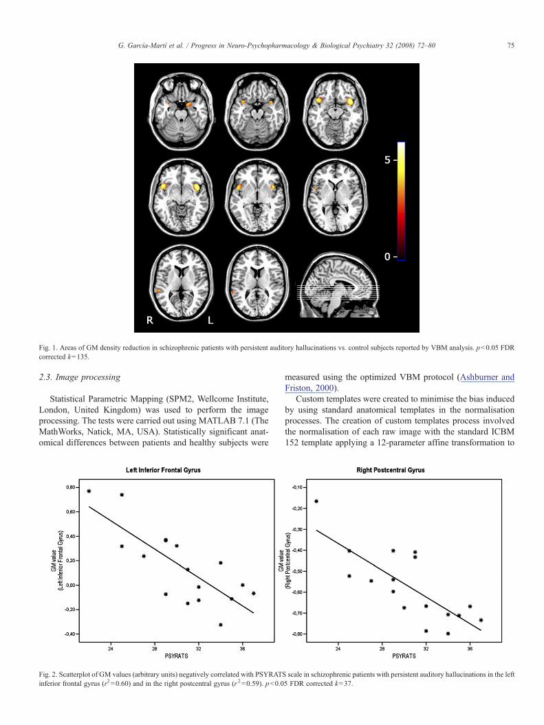
Fig. 1. Areas of GM density reduction in schizophrenic patients with persistent auditory hallucinations vs. control subjects reported by VBM analysis. pb0.05 FDRcorrected k=135.
75G. García-Martí et al. / Progress in Neuro-Psychopharmacology & Biological Psychiatry 32 (2008) 72–80
2.3. Image processing
Statistical Parametric Mapping (SPM2, Wellcome Institute,London, United Kingdom) was used to perform the imageprocessing. The tests were carried out using MATLAB 7.1 (TheMathWorks, Natick, MA, USA). Statistically significant anat-omical differences between patients and healthy subjects were
Fig. 2. Scatterplot of GM values (arbitrary units) negatively correlated with PSYRATSinferior frontal gyrus (r2=0.60) and in the right postcentral gyrus (r2=0.59). pb0.0
measured using the optimized VBM protocol (Ashburner andFriston, 2000).
Custom templates were created to minimise the bias inducedby using standard anatomical templates in the normalisationprocesses. The creation of custom templates process involvedthe normalisation of each raw image with the standard ICBM152 template applying a 12-parameter affine transformation to
scale in schizophrenic patients with persistent auditory hallucinations in the left5 FDR corrected k=37.

Table 5Areas negatively correlated between PSYRATS and GM values in schizophrenicpatients
Coordinates (mm) Label L/R tvalue p(cluster)
X Y Z
−49 18 −3 Inferior frontal gyrus L 4.93 0.0468 −7 19 Postcentral gyrus R 3.94 0.11
pb0.05 FDR corrected k=37. Corrected p values are shown at cluster-level.
76 G. García-Martí et al. / Progress in Neuro-Psychopharmacology & Biological Psychiatry 32 (2008) 72–80
place the data in a common stereotactic space (Good et al.,2001). The normalised images were then segmented to obtainGM maps, which were averaged and smoothed with an 8-mmGaussian smoothing filter obtaining a whole-brain template anda specific GM template.
To perform the optimized VBM analysis, each original T1-weighted MR image was normalised with the created whole-brain template and again segmented to obtain GM tissue maps.At this stage, segmentation involved a cleaning process thatremoved non-useful tissue such as scalp, skull and dural ve-nous sinus (Good et al., 2001). Non-linear spatial normalisa-tion (Ashburner and Friston, 1999) parameters between GMsegmented images and GM template maps were then estimatedand applied in order to warp the original T1 images. Finally,warped GM images were segmented and smoothed by a 12-mmGaussian kernel.
2.4. Statistical analysis
Statistical measurements were performed with the Statisticalnon-Parametric Mapping (SnPM) (Nichols and Holmes, 2002)software, which provides a suitable framework for analyseswhen conditions as low degrees of freedom (small size of thesample) make the variance estimation noisy and, therefore, theassumptions of the Random Field Theory (RFT) about fieldcontinuity not feasible.
Fig. 3. Scatterplot of GM values (arbitrary units) negatively correlated with BPRS sparahippocampal gyrus (r2=0.17) and in the right superior temporal gyrus (r2=0.40
The SnPM software takes into consideration the multiplecomparison problem and provides a smoothed varianceestimation procedure to inherently deal with this issue (Holmeset al., 1996). A significance criterion was established by using apb0.05, with correction for multiple comparisons following theFalse Discovery Rate (FDR) methodology (Genovese et al.,2002).
In order to measure the morphometric differences and eval-uate the anatomical correlations with BPRS and PSYRATSscales, two independent statistical models were generated. Themodel constructed for assessing structural inter-group variabilityincluded a group condition (patients vs. controls) and twonuisance variables (age and total intracranial volume). Totalintracranial volume was computed from the GM, WM andcerebrospinal fluid (CSF) maps obtained after segmenting theoriginal raw images. GM, WM and CSF tissue volumes werecalculated by integrating the probability value of each voxel withthe voxel size. These three values were added to derive the totalintracranial volume. Statistical parametric maps were obtainedby performing permutation tests over the GM maps, using one-tailed contrasts in order to measure differences among groups.The number of permutations was fixed to 30,000. This value waschosen in order to obtain a random sub-sample of all possiblepermutations with a confidence interval of 95% for a giventhreshold of pb0.05. Additionally, a spatial filter was applied toeliminate spurious findings by considering only those clusterswith a minimum size of k=135 voxels (expected number ofvoxels per cluster).
To perform the correlation analysis between PSYRATS andBPRS variables with structural variability, another statisticalmodel was built including the GM images of patients and thePSYRATS and BPRS scores. A correlation test was performedto evaluate the relationship between illness duration and brainvolume of each patient. As results were not significant (r2 =0.00;p=0.90), these variables were not included in the model. Es-timation parameters were adjusted looking for linear regressions
cale in schizophrenic patients with persistent auditory hallucinations in the left). pb0.05 FDR corrected k=37.

77G. García-Martí et al. / Progress in Neuro-Psychopharmacology & Biological Psychiatry 32 (2008) 72–80
between the PSYRATS and BPRS scales related to GM signalchanges. The significance threshold was fixed at pb0.05 FDRcorrected and the cluster filter was set to k=37 voxels (expectednumber of voxels per cluster). The model was estimated against30,000 permutations.
Both, morphometric and correlation results were labelled withthe Automated Anatomical Labeling (AAL) software (Tzourio-Mazoyer et al., 2001). Areas-identifying coordinates were deter-mined by the maximum Student-t value in the correspondingbrain area.
3. Results
3.1. Optimized VBM method
GM density reduction in patients vs. controls (patientsGMbcontrolsGM) was found in the areas shown in Table 4 andrepresented in Fig. 1. The results showed a significant (pb0.05FDR Corrected, k=135) GM decreased concentration in insula(bilateral), superior temporal gyrus (bilateral) and amygdala(left). PatientsGMNcontrolsGM comparison (GM concentrationincreases in patients vs. controls) did not produce anysignificant results.
3.2. PSYRATS and BPRS regression
Significant (pb0.05 FDR Corrected, k=37) negative correla-tions between each PSYRATS and BPRS scales related to focalGM density reduction were observed in patients. PSYRATSvariable was negatively correlated in the left inferior frontalgyrus (r2 =0.60; p=0.04) and in the right postcentral gyrus(r2 =0.59; p=0.11) (Fig. 2, Table 5), while negative BPRScorrelation affected the left parahippocampal (r2 = 0.17;p=0.21) and the right superior temporal gyrus (r2 =0.40;p=0.03) (Fig. 3, Table 6) regions.
3.3. Exploratory analyses: specific PSYRATS items and self-related areas
Exploratory correlations were also performed between threePSYRATS domains (items #2 or duration, #5 or beliefs about theorigin of voices, and #11 or controllability of voices) whichwe hypothesized as of particular relevance, and GM values ininsula. Significant (pb0.05 FDR corrected, k=37) negativecorrelations between ‘duration’ and bilateral insula and between‘controllability of voices’ and left insula GM values wereobserved. Significant (pb0.05 FDR corrected, k=37) positive
Table 6Areas with negative correlation between BPRS and GM values in schizophrenicpatients
Coordinates (mm) Label L/R tvalue p(cluster)
X Y Z
−17 −14 −25 Parahippocampal gyrus L 4.29 0.2170 −29 8 Superior temporal gyrus R 3.70 0.03
pb0.05 FDR corrected k=37. Corrected p values are shown at cluster-level.
correlations were also obtained between ‘beliefs about the originof voices’ and bilateral insula GM values.
4. Discussion
4.1. Differences between patients and controls
GM reductions in bilateral insula and superior temporalgyrus, altogether with a left amygdala decrease have been de-tected in our highly homogeneous sample of patients withpersistent AH compared to healthy controls. According to this,these areas seem to participate in the structural network involvedin the persistence of AH. Some functional studies also supportthe multiplicity of areas involved in AH (Shergill et al., 2000).Most studies using the ROI approach (Tables 1 and 2) havecentred on the superior temporal gyrus after the seminal paper byBarta et al. (1990), with other relevant brain areas being underrecognized until the implementation of automated techniques.
A literature review does not show a straightforward relationbetween GM density alterations, either reductions or enlarge-ments, and the severity or persistence of AH. Although the leftsuperior temporal gyrus has repeatedly been involved in theseverity of AH (Table 1), several negative reports (Table 2) haveto be considered. Moreover, some studies have reported largerfrontal and temporal GM volumes in patients with AH comparedwith patients without AH (Shin et al., 2005). It is to say thatpatients included in this study were first-episode psychoticpatients and predominantly female (Shin et al., 2005). On theother hand, severity may not imply persistence. Thus, the onlyother morphometric study on patients with chronic auditoryverbal hallucinations, including both genders, did not findvolume reductions of temporal lobe structures compared withcontrols, neither in the hallucinators compared with the non-hallucinators patients (Havermans et al., 1999). The influence ofthe gender might help to clarify these controversies. In fact,superior temporal gyrus reductions may be more severe in men,while amygdala volumes may exhibit a reverse pattern withreductions in men and enlargements in women (Gur et al., 2000).Due to our series definition, sex effects cannot be analysed in ourstudy. However, our bilateral findings for the superior temporalgyrus are somewhat concordant with a study byMatsumoto et al.(2001) that also gives support to the possible role of the rightsuperior temporal gyrus reduction in the severity of AH.
According to our results, right and left insula and left amygdalamay also be implicated on the persistence of AH. Shapleske et al.(2002) obtained results in agreement with these findings, sincethey found a significant cluster with a deficit in GM whichincluded the left insula and adjacent medial temporal lobe whencomparing hallucinators and non-hallucinators (Shapleske et al.,2002).
The insula is a key region of the emotional brain (Alemanand Kahn, 2005) and could also participate in conjunction withthe amygdala to foster the chronicity of positive symptomsthrough emotion-mediated mechanisms. However this structuremight be implicated through a different mechanism. One of themost accepted and an investigated cognitive model defends thatAH appear because of defective verbal self-monitoring (Frith

78 G. García-Martí et al. / Progress in Neuro-Psychopharmacology & Biological Psychiatry 32 (2008) 72–80
and Done, 1988). Neural correlates supporting this model arenow being investigated in imaging studies (Woodruff, 2004).The insula is involved in the perception of representations of theself (Farrer and Frith, 2002). Physiologically, a lesser sense ofagency (experience of causing an action) would be associated toa reduced activation of both anterior insula (Farrer and Frith,2002). Our finding on the insular GM reduction may be relatedto the persistence of AH through abnormalities in the senseof agency and, therefore, the self-monitoring. We obtainedpositive correlations for both insula GM values with the domain‘beliefs about the origin of voices’. However, the sign ofcorrelations was unexpected (the more GM the higher externalattribution) which raises questions on the role of insular cortexand the relationships between functional and structural findings.Thus, we cannot discard a coexistence of greater insular GMvolumes with reduced activation, as we have shown for otherbrain areas with chronically hallucinating patients (Martí-Bonmatí et al., 2007). The rest of our exploratory correlationsbetween specific AH domains and some brain areas also suggesta crucial role for the insula on the persistence of AH. A higherduration of voices was related to bilateral insular reductionswhile less controllability of voices was associated to left insulaGM reductions. Finally, the insula region has also been relatedto the production of hallucinations in some functional studies(Shergill et al., 2004).
Although its exact role is still unclear, the amygdala is a keycomponent in the emotional processing of patients with schizo-phrenia (Brunet-Gouet and Decety, 2006). Van Rijn et al. (2005)concluded in their review that amygdalar reduction is a riskfactor for schizophrenia and they even proposed amygdalaabnormalities as an endophenotype in schizophrenia. Alemanand Kahn (2005) have proposed a model supporting a role foramygdalar hyperactivation in the genesis of positive symptoms.In concordance with this model, in a previous functionalMR imaging study with chronic hallucinators, we found a clearoveractivation of the frontal lobe, temporal cortex, insula,cingulate, and amygdala in patients when hearing emotionalwords in comparison with controls (Sanjuán et al., 2007). Wehypothesized that the functional overactivation in these patientscould be directly related to the emotional response seen inpositive symptoms. Aleman and Kahn (2005) also suggestedthat prolonged hyperactivation of the amygdala during psychoticstates at the onset of schizophrenia could result in excitotoxicitydue to excessive glutamatergic activity and hence lesions of theamygdala. This hypothesis could explain amygdalar volumereductions in chronically hallucinating patients. Notwithstand-ing, we only found a reduction in the left amygdala. In this sense,two meta-analyses have suggested greater left than rightamygdalar activation to the cognitive processing of emotionalstimuli (Wager et al., 2003; Baas et al., 2004). Moreover, the leftamygdala seems to be more affected than the right amygdala inpatients with schizophrenia (Honea et al., 2005). It has also beenhypothesized a neurodevelopmentally mediated alteration of theleft amygdala that would predispose to schizophrenia. Keshavanet al. (2002) investigated unaffected, young offspring of schizo-phrenia patients and reported reduced volumes of the left an-terior and posterior amygdalo-hippocampal complex.
4.2. Areas related to the severity of auditory hallucinations
We have observed specific relationships between left inferiorfrontal and right postcentral gyri reductions and the severity ofAH. The left inferior frontal gyrus has previously been related tothe pathogenesis of AH. At this point, it can be considered areplicated finding (McGuire et al., 1993; Shergill et al., 2004).Hunter and Spence (2005) suggested that the activation offrontal area could be related not only to the hallucinations butalso to the intention to activate the motor area when the button ispressed in functional MRI studies. However, according to theresults in this study, it could be considered that the inferiorfrontal gyrus area is thought to participate in the first stages ofthe hallucination by the genesis of auditory verbal contents.
Our finding on the postcentral gyrus seems more counterin-tuitive since this area is known to be primary somatosensorycortex. There is not a clear explanation for a possible relationshipbetween postcentral gyrus and the severity of AH and reasoningon this issue has to be somewhat speculative. Generation ofinner speech, which has been linked to the pathogenesis of AH(McGuire et al., 1995), has been associated with activation in theleft inferior frontal, the right pre- and postcentral and bothsuperior temporal gyri (Shergill et al., 2002). GM reductions inpostcentral gyrus could imply alterations in the monitoring ofinner speech but this hypothesis cannot be tested in our study.
Against our predictions, differences in the superior temporalgyrus density were present in group comparisons but did notappear specifically related to AH severity. Note that a type IIerror cannot be ruled out due to the sample size and the statisticalprocedures aimed to avoid false positives. Besides, as expectedin a homogeneous sample, the variance in PSYRATS scores waslow which has surely taken down the intensity of the expectedcorrelations. Moreover, the transient character of AH posesanother problem when searching for correlations with presum-ably more stable GM densities. The existence of chronic anti-psychotic treatment, which is known to influence brainmorphology, is a possible source of bias. Reductions related tothe superior temporal gyrus can reverse after one year of treat-ment (Keshavan et al., 1998). As far as the sample compositionis concerned, two previous studies used comparable samplesand, interestingly, none has replicated the superior temporalgyrus findings on AH production (Havermans et al., 1999;Shapleske et al., 2002).
The comparability of studies could pose another majorproblem, both in regard to the neuroimaging technique and thesample composition. Several studies have compared ROI mea-surements with VBM (Wright et al., 1999). Volumes weresimilar for the caudate nuclei but not for temporal lobe mea-surements. However, Kubicki et al. (2002) showed consistenciesfor left superior temporal gyrus. Cluster analysis also showedsimilar results but could not reveal group differences in themedial temporal lobe selected with the ROI analysis. Job et al.'s(2002) results with VBM showed a relative rather than absolutereplication of the ROI findings. Two recent studies have rep-licated previous ROI findings in global terms (Job et al., 2003;Moorhead et al., 2004). Finally, Giuliani et al. (2005) replicatedthe results of greater left inferior and right superior frontal

79G. García-Martí et al. / Progress in Neuro-Psychopharmacology & Biological Psychiatry 32 (2008) 72–80
cortical GM deficits in schizophrenia. However VBM showed asignificantly lower concentration of GM in the middle andsuperior temporal gyrus and did not lead to replication of thedescribed changes.
4.3. Areas related to the severity of global symptoms
We have also found an association between symptom severityand both left parahippocampus and right superior temporal gyrusGM reductions. Although the parahippocampus has been mainlyassociated with cognitive abilities in patients with schizophrenia(Antonova et al., 2004), our finding is consistent with previousstudies. Prasad et al. (2004) examined the parahippocampal gyrusmorphology using structural MR imaging in 33 neuroleptic-naïvesubjects with first-episode psychoses and 43 matched healthysubjects. The parahippocampal gyrus volume negatively corre-lated with total positive symptom, delusion and conceptual dis-organization scores on BPRS. The association of right superiortemporal gyrus volume reduction with symptom severity but notwith AH severity was not expected. We do not have an expla-nation for this finding since this area is preferably related to AH.
Several study limitations and biases should be consideredbefore establishing definitive conclusions. As all the patients inour series had AH to be included, a group of patients withschizophrenia who had never experienced AH was not withinour sample. A study to clarify whether our observed findings arerelated specifically to the propensity to hallucinated, as opposedto the disorder of schizophrenia itself, should be investigated.Moreover, all patients were medicated with a wide range ofdrugs, both typical and atypical antipsychotics.
5. Conclusions
Our VBM study confirms the association between AH andstructural abnormalities by analysing a highly homogeneousgroup of persistent hallucinatory patients. We believe thisis clearly demonstrated by using a good phenomenologicalcharacterization of the AH symptom. Our results only partiallyconfirmed the superior temporal gyrus as being crucial forAH persistence. Other areas, particularly the insula and amyg-dala, seem to be implicated through yet unclear pathologicalmechanisms.
Acknowledgements
This study was supported by Spanish grants from Red deGenotipación y Psiquiatría Genética (ISCIII — G03/184),FIS P.I. 02/0018, P.I. 05/2332, Imagen Médica Molecular yMultimodalidad IM3 (ISCIII — G03/185) and INBIOMED(ISCIII — G03/160). The authors would also like to thankthe Asociación para el Desarrollo y la Investigación enResonancia Magnética (ADIRM) for their support.
References
Aleman A, Kahn RS. Strange feelings: do amygdala abnormalities dysregulatethe emotional brain in schizophrenia? Prog Neurobiol 2005;77:283–98.
Antonova E, Sharma T, Morris R, Kumari V. The relationship between brainstructure and neurocognition in schizophrenia: a selective review. SchizophrRes 2004;70:117–45.
Ashburner J, Friston KJ. Nonlinear spatial normalisation using basis functions.Hum Brain Mapp 1999;7:254–66.
Ashburner J, Friston KJ. Voxel-based morphometry — the methods. Neuro-image 2000;11:805–21.
Baas D, Aleman A, Kahn RS. Lateralization of amygdala function: a systematicreview of functional neuroimaging studies. Brain Res Rev 2004;45:96–103.
Barta PE, Pearlson GD, Powers RE, Richards SS, Tune LE. Auditoryhallucinations and smaller superior temporal gyral volume in schizophrenia.Am J Psychiatry 1990;147:1457–62.
Brunet-Gouet E, Decety J. Social brain dysfunctions in schizophrenia: a reviewof neuroimaging studies. Psychiatry Res 2006;148:75–92.
Delisi LE, Hoff AL, Neale C, Kushner M. Asymmetries in the superior temporallobe in male and female first-episode schizophrenic patients: measures of theplanum temporale and superior temporal gyrus by MRI. Schizophr Res1994;12:19–28.
Farrer C, Frith CD. Experiencing oneself vs another person as being the cause ofan action: the neural correlates of the experience of agency. Neuroimage2002;15:596–603.
Flaum M, O'Leary DS, Swayze II VW, Miller DD, Arndt S, Andreasen NC.Symptom dimensions and brain morphology in schizophrenia and relatedpsychotic disorders. J Psychiatr Res 1995;29:261–76.
Frith CD, Done DJ. Towards a neuropsychology of schizophrenia. Br J Psychiatry1988;153:437–43.
Gaser C, Nenadic I, Volz HP, Buchel C, Sauer H. Neuroanatomy of ‘‘hearingvoices”: a frontotemporal brain structural abnormality associated withauditory hallucinations in schizophrenia. Cereb Cortex 2004;14:91–6.
Genovese CR, Lazar NA, Nichols T. Thresholding of statistical maps in functionalneuroimaging using the false discovery rate. Neuroimage 2002;15:870–8.
Giuliani NR, Calhoun VD, Pearlson GD, Francis A, Buchanan RW. Voxel-basedmorphometry versus region of interest: a comparaison of two methods foranalyzing gray matter differences in schizophrenia. Schizophr Res 2005;74:135–47.
Good CD, Johnsrude IS, Ashburner J, Henson RN, Friston KJ, Frackowiak RS.A voxel-based morphometric study of ageing in 465 normal adult humanbrains. Neuroimage 2001;14:21–36.
Gur RE, Turetsky BI, Cowell PE, Finkelmann C, Maany V, Grossman RI, et al.Temporolimbic volume reductions in schizophrenia. Arch Gen Psychiatry2000;57:769–75.
Haddock G, McCarron J, Tarrier N, Faragher EB. Scales to measure dimensionsof hallucinations and delusions: the psychotic symptom rating scales(PSYRATS). Psychol Med 1999;29:879–89.
Havermans R, Honig A, Vuurman EF, Krabbendam L,Wilmink J, Lamers T, et al.A controlled study of temporal lobe structure volumes and P300 responses inschizophrenic patients with persistent auditory hallucinations. Schizophr Res1999;38:151–8.
Holmes AP, Blair RC, Watson JD, Ford I. Nonparametric analysis of statisticimages from functional mapping experiments. J Cereb Blood Flow Metab1996;16:7–22.
Honea R, Crow TJ, Passingham D, Mackay CE. Regional deficits in brainvolume in schizophrenia: a meta-analysis of voxel-based morphometrystudies. Am J Psychiatry 2005;162:2233–45.
Hunter MD, Spence SA. Left frontal activation. Br J Psychiatry 2005;187:89.Job DE, Whalley HC, McConnell S, Glabus M, Johnstone EC, Lawrie SM.
Structual gray matter differences between first-episode schizophrenicsand normal controls using voxel-based morphometry. Neuroimage 2002;17:880–9.
Job DE, Whalley HC, McConnell S, Glabus M, Johnstone EC, Lawrie SM.Voxel-based morphometry of grey matter densities in subjects at high risk ofschizophrenia. Schizophr Res 2003;64:1–13.
Keshavan MS, Haas GL, Kahn CE, Aguilar E, Dick EL, Schooler NR, et al.Superior temporal gyrus and the course of early schizophrenia: progressive,static, or reversible? J Psychiatr Res 1998;32:161–7.
Keshavan MS, Dick E, Mankowski I, Harenski K, Montrose DM, Diwadkar V,et al. Decreased left amygdala and hippocampal volumes in young offspringat risk for schizophrenia. Schizophr Res 2002;58:173–83.

80 G. García-Martí et al. / Progress in Neuro-Psychopharmacology & Biological Psychiatry 32 (2008) 72–80
Kubicki M, Shenton ME, Salibsbury DF, Hirayasu Y, Kasai K, Kikins R, et al.Voxel-based morphometry analysis of gray matter in first episode schizophre-nia. Neuroimage 2002;17:1711–9.
Levitan C, Ward PB, Catts SV. Superior temporal gyral volumes and lateralitycorrelates of auditory hallucinations in schizophrenia. Biol Psychiatry 1999;46:955–62.
Marsh L, Harris D, LimKO, BealM, Hoff AL,Minn K, et al. Structural magneticresonance imaging abnormalities in men with severe chronic schizophreniaand an early age at clinical onset. Arch Gen Psychiatry 1997;54:1104–12.
Martí-Bonmatí L, José Lull JJ, García-Martí G, Aguilar EJ, Moratal-Pérez D,Poyatos C, et al. MR analysis of the coincidence between functional andmorphological abnormalities in schizophrenic patients with chronic auditoryhallucinations. Radiology 2007;244:549–56.
Matsumoto H, Simmons A, Williams S, Hadjulis M, Pipe R, Murray R, et al.Superior temporal gyrus abnormalities in early-onset schizophrenia:similarities and differences with adult-onset schizophrenia. Am J Psychiatry2001;158:1299–304.
McGuire PK, Shah GM, Murray RM. Increased blood flow in Broca's areaduring auditory hallucinations in schizophrenia. Lancet 1993;342:703–6.
McGuire PK, Silbersweig DA, Wright I, Murray RM, David AS, FrackowiakRS, et al. Abnormal monitoring of inner speech: a physiological basis forauditory hallucinations. Lancet 1995;346:596–600.
Milev P, Ho BC, Arndt S, Nopoulos P, AndreasenNC. Initial magnetic resonanceimaging volumetric brain measurements and outcome in schizophrenia:a prospective longitudinal study with 5-year follow-up. Biol Psychiatry2003;54:608–15.
Moorhead TW, Job DE, Whalley HC, Sanderson TL, Johnstone EC, Lawrie SM.Voxel-based morphometry of comorbid schizoprhrenia and learningdisability: analyses in normalized and native spaces using parametric andnonparametric statistical methods. Neuroimage 2004;22:188–202.
Neckelmann G, Specht K, Lund A, Ersland L, Smievoll AI, Neckelmann D, et al.MRmorphometry analysis of greymatter volume reduction in schizophrenia:association with hallucinations. Int J Neurosci 2006;116:9–23.
Nichols TE, Holmes AP. Nonparametric permutation tests for functionalneuroimaging: a primer with examples. Hum Brain Mapp 2002;15(1):1–25.
Noga JT, Aylward E, Barta PE, Pearlson GD. Cingulate gyrus in schizophrenicpatients and normal volunteers. Psychiatry Res 1995;61:201–8.
Onitsuka T, Shenton ME, Salisbury DF, Dickey CC, Kasai K, Toner SK, et al.Middle and inferior temporal gyrus gray matter volume abnormalities inchronic schizophrenia: an MRI study. Am J Psychiatry 2004;161:1603–11.
Prasad KM, Rohm BR, Keshavan MS. Parahippocampal gyrus in first episodepsychotic disorders: a structural magnetic resonance imaging study. ProgNeuropsychopharmacol Biol Psychiatry 2004;28:651–8.
Rajarethinam RP, DeQuardo JR, Nalepa R, Tandon R. Superior temporal gyrusin schizophrenia: a volumetric magnetic resonance imaging study. SchizophrRes 2000;41:303–12.
Rajarethinam R, DeQuardo JR, Miedler J, Arndt S, Kirbat R, Brunberg JA, et al.Hippocampus and amygdala in schizophrenia: assessment of the relationshipof neuroanatomy to psychopathology. Psychiatry Res 2001;108:79–87.
Rossell SL, Shapleske J, Fukuda R, Woodruff PW, Simmons A, David AS.Corpus callosum area and functioning in schizophrenic patients withauditory–verbal hallucinations. Schizophr Res 2001;50:9–17.
Sanjuán J, Lull JJ, Aguilar EJ, Martí-Bonmatí L, Moratal-Perez D, Gonzales JC,et al. Emotional words induce brain overactivation in schizophrenic patientswith auditory hallucinations. Psychiatry Res 2007;154:21–9.
Shapleske J, Rossell SL, Simmons A, David AS, Woodruff PW. Are auditoryhallucinations the consequence of abnormal cerebral lateralization? Amorphometric MRI study of the sylvian fissure and planum temporale. BiolPsychiatry 2001;49:685–93.
Shapleske J, Rossell SL, Chitnis XA, Suckling J, SimmonsA, Bullmore ET, et al.A computational morphometric MRI study of schizophrenia: effects ofhallucinations. Cereb Cortex 2002;12:1331–41.
Shenton ME, Dickey CC, Frumin M, McCarley RW. A review of MRI findingsin schizophrenia. Schizophr Res 2001;49:1–52.
Shergill SS, Brammer MJ, Williams SC, Murray RM, McGuire PK. Mappingauditory hallucinations in schizophrenia using functional magnetic reso-nance imaging. Arch Gen Psychiatry 2000;57:1033–8.
Shergill SS, BrammerMJ, Fukuda R, Bullmore E, Amaro Jr E,Murray RM, et al.Modulation of activity in temporal cortex during generation of inner speech.Hum Brain Mapp 2002;16:219–27.
Shergill SS, Brammer MJ, Amaro E, Williams SC, Murray RM, McGuire PK.Temporal course of auditory hallucinations. Br J Psychiatry 2004;185:516–7.
Shin SE, Lee JS, Kang MH, Kim CE, Bae JN, Jung G. Segmented volumes ofcerebrum and cerebellum in first episode schizophrenia with auditoryhallucinations. Psychiatry Res 2005;38:33–42.
StephaneM, Thuras P, NasrallahH, Georgopoulos AP. The internal structure of thephenomenology of auditory verbal hallucinations. Schizophr Res 2003;61:185–93.
SumichA, Chitnis XA, FannonDG, O'Ceallaigh S, DokuVC, Faldrowicz A, et al.Unreality symptoms and volumetric measures of Heschl's gyrus and planumtemporal in first-episode psychosis. Biol Psychiatry 2005;57:947–50.
Tzourio-Mazoyer N, Landeau B, Papathanassiou D, Crivello F, Etard O,Delcroix N, et al. Automated anatomical labeling of activations in SPMusing a macroscopic anatomical parcellation of the MNI MRI single-subjectbrain. Neuroimage 2001;15:273–89.
VanRijn S,AlemanA, SwaabH, Kahn RS.Neurobiology of emotion and high riskfor schizophrenia: role of the amygdala. Neurosci Biobehav Rev 2005;29:385–97.
Ventura J, Green MF, Shaner A, Liberman RP. Training and quality assurancewith the Brief Psychiatric Rating Scale: ‘the drift busters’. Int J MethodsPsychiatr Res 1993;3:221–44.
Wager TD, Phan KL, Liberzon I, Taylor SF. Valence, gender, and lateralizationof functional brain anatomy in emotion: a meta-analysis of findings fromneuroimaging. Neuroimage 2003;19:513–31.
Woodruff PW. Auditory hallucinations: insights and questions from neuroima-ging. Cogn. Neuropsychiatry 2004;9:73–91.
Wright IC, Ellison ZR, Sharma T, Friston KJ, Murray RM,McGuire PK.Mappingof grey matter changes in schizophrenia. Schizophr Res 1999;35:1–14.
Zipursky RB, Marsh L, Lim KO, DeMent S, Shear PK, Sullivand EV, et al.Volumetric MRI assessment of temporal lobe structures in schizophrenia.Biol Psychiatry 1994;35:501–16.
Copyright © 2022 FDOKUMEN