Hallucinations as Superceptions: Adaptive Behavior, Aptic Structures, and the Neurology of Hearing...
80
DRAFT: Please Do Not Quote without Permission of Author Hallucinations as Superceptions: Adaptive Behavior, Aptic Structures, and the Neurology of Hearing Voices 1 Brian J. McVeigh, PhD When I was a young boy, grumbling that I couldn’t accomplish some difficult task, my mother would say “Stop your complaining. If your brain hears you saying you can’t do something, it will believe you.” The mind, I concluded, is not unitary despite the strange illusion that it is. This peaked my interest in how the mind is put together, but perhaps my brother, Billy, also has something to do with my interests. Diagnosed with autism from a very early age, Billy had an angelic countenance, was ordinarily shy, and hardly spoke. When he did, what he had to say was done in a telegraphic, soft-spoken, and gentle manner. But occasionally Billy would erupt, volcano-like, in extremely violent psychomotor seizures. These were ferocious but fortunately brief. Accompanying these attacks were guttural shouts, fearsome grunts, murderous threats, and language so foul and vile we could only wonder from where he had acquired such a formidable linguistic arsenal. It was as if an angry demon resided somewhere in his person. Surely, I thought, the individual psyche must be composed of different parts, perhaps ordinarily segregated from each other, that manifest themselves under the right conditions.
Transcript of Hallucinations as Superceptions: Adaptive Behavior, Aptic Structures, and the Neurology of Hearing...

DRAFT: Please Do Not Quote without Permission of Author
Hallucinations as Superceptions: Adaptive Behavior, Aptic Structures, and the
Neurology of Hearing Voices1
Brian J. McVeigh, PhD
When I was a young boy, grumbling that I couldn’t accomplish some
difficult task, my mother would say “Stop your complaining. If
your brain hears you saying you can’t do something, it will
believe you.” The mind, I concluded, is not unitary despite the
strange illusion that it is. This peaked my interest in how the
mind is put together, but perhaps my brother, Billy, also has
something to do with my interests. Diagnosed with autism from a
very early age, Billy had an angelic countenance, was ordinarily
shy, and hardly spoke. When he did, what he had to say was done
in a telegraphic, soft-spoken, and gentle manner. But
occasionally Billy would erupt, volcano-like, in extremely
violent psychomotor seizures. These were ferocious but
fortunately brief. Accompanying these attacks were guttural
shouts, fearsome grunts, murderous threats, and language so foul
and vile we could only wonder from where he had acquired such a
formidable linguistic arsenal. It was as if an angry demon
resided somewhere in his person. Surely, I thought, the
individual psyche must be composed of different parts, perhaps
ordinarily segregated from each other, that manifest themselves
under the right conditions.

2
Several other experiences, not nearly as influential as the
aforementioned and transpiring when I was older, have undoubtedly
peaked my interests. I had just gone to bed when I heard my
father’s voice sharply call out my name. I sat up and within a
second realized that something was strangely amiss, as I was the
only one in the house that night. The voice, clear,
unmistakable, and with an echo-like resonance, originated from
the living room down the hall from my bedroom. Though this was
the first and only time that I distinctly hallucinated a voice (I
was probably about eighteen years old), two other similar
experiences have stayed with me. A colleague was supposed to
pick me up for a dinner engagement at a hotel where I was staying
in Durham, North Carolina. Ten, twenty, forty-five minutes
passed. I gradually grew impatient and finally annoyed. And
then a sentence—“Your life is not your own”—popped into my head,
reminding me that in our daily lives our control over events is
limited. Not exactly spoken in an audible voice but with a force
that was more than a passing thought, the words felt as if they
came from somewhere other than my own mind. These very same
words would visit me again about a year later, but under very
different circumstances.
I must have been 35,000 feet over northern Canada, flying
from Tokyo to Newark, NJ. Turbulence was buffeting and rocking
an otherwise uneventful and mind-numbingly boring thirteen-hour
flight, when suddenly I could have sworn I heard banshee-like
noises—screaming winds—coming from outside. Then bumps turned

3
into forceful jolts and the plane began to violently jerk,
shudder, and vibrate. I had flown through choppy weather many
times before, even severe turbulence, but nothing prepared me for
the sickening feeling of being belted into an aircraft that was
plummeting like a rock out of the sky. It was as if my body
defied gravity; my stomach strained against the seatbelt attached
to a plane that was rapidly descending. Thuds, bangs, screams,
and other alarming noises filled the cabin. The shaking was so
intense that the oxygen masks dropped from the ceiling. And then
half-way through the ordeal, that quasi-vocalization—“Your life
is not your own”—again nonchalantly rambled across my mind. The
words were not reassuring. I suppose a part of my self was just
being philosophical under what seemed at the time a very dire
situation.
Purpose: Explaining the Mystery of Hallucinations
What does the saliency of hallucinations among the general, non-
clinical population indicate? Are hallucinations are a matter of
loose wires or vestigial neurostructures that under the right
conditions are reactivated? To answer these questions, I first
define hallucinations, distinguishing them from
pseudohallucinations and illusions. Next I comment on the
failure to explain hallucinations and how a root cause is too
often confused with accompanying conditions. I describe
hallucinations as produced by “aptic structures” or evolved,
innate neurological networks plus the results of experience that

4
afford certain aptitudes (Jaynes 1976). I integrate what we do
know about the basics of the neurology of language, the adaptive
advantages of the asymmetrical dual brain, and right-side brain
activity and hallucinations. I conclude by noting how some
therapists acknowledge that hallucinations are not inherently
pathopsychological, and that “voice-hearers” should be encouraged
to confront their voices as aspects of their selves. I also
suggest that to understand hallucinations, a new terminology is
required and that mental imagery, though in some respects quite
different from hallucinations, is essentially hallucinatory and
the successor to what Jaynes termed bicameral mentality. Taken
together, hallucinations and mental imagery (inner quasi-
perceptions) constitute “superperceptions.”
The Failure to Explain Hallucinations
Many assume that hallucinations in and of themselves are
pathopsychological, but we need to be cautious in confusing
causation with what might be called symptoms. The analysis of
hallucinations requires highlighting three issues: (1) their
prevalence among the non-clinical population; (2) confusing cause
with accompanying conditions or triggers; and (3) the innateness
of hallucinations. As I argue below, hallucinations are
expressions of “aptic structures” that once served a socially
adaptive function. But first, some basic facts about
hallucinations are in order.
5
Hallucinations, Pseudohallucinations, and Illusions
The word “hallucination” 2—or “waking sensory experience having
no identified external physical stimulus” (Stevenson 1983:1609)—
was coined by the physician Jean-Étienne Dominique Esquirol
(1772–1840) in 1817. He also distinguished between this
phenomena and illusions. The DSM-V defines hallucinations as a
“perception-like experience with the clarity and impact of a true
perception but without the external stimulation of the relevant
sensory organ” (American Psychiatric Association 2013). The
experient may or may not have insight into the non-veridical
nature of the hallucination.
Hallucinations and illusions are sometimes used
interchangeably in the literature. They should not be. The
latter transpire when an external stimulus is misinterpreted or
misperceived. Hallucinations, however, often occur without
perceptual triggers. They are not just sensory distortions and
do not require external objective sensation. Notably,
hallucinations are superimposed over sensory experience, i.e.,
they happen concurrently with real perceptions. The key features
of hallucinations include: (1) appearing as if in objective space
(i.e., in the physical world, not “in the mind”); they are
externalized or projected, as if they come from outside the
person; (2) having the same realistic qualities as actual
physical objects; (3) not subject to volitional manipulation; and
(4) they are a normal sensory experience from the experient’s
point of view, and the belief that something was veridically

6
perceived is not corrected even in light of other information
after the hallucinatory experience.3 The difference between
illusions and hallucinations is actually not always clear-cut,
and in some instances, hallucinations may be triggered by
illusions. Pseudohallucinations, sharing features of both
hallucinations and illusions, are an intermediate category of
experience. However, pseudohallucinations are recognized as not
being a veridical experience, i.e., the experient possesses
insight into the unreality of the hallucination. Most likely,
illusions, pseudohallucinations, and hallucinations form a
continuum. Indeed, some contend that hallucinations and
pseudohallucinations are not two separate entities but rather sit
on a spectrum (Weller and Wiedemann 1989) 4
Hallucinations may be elementary, composed of simple
percepts, or complex. They may be experienced through one
sensory mode (auditory, visual, haptic, gustatory) or
multimodal.5 Auditory verbal hallucinations (AVHs), the most
commonly reported in the research literature, may be perceived in
the distance, in one’s ears, or in one’s head or body. AVHs have
been characterized as “second person” (a voice directly address
the experient); “commanding” (gives authoritative instructions to
the experient); “running commentary”; “third person” (two or more
voices talk about the experient), or “thought echo” (voices
anticipate or repeat the experient’s thoughts). Besides
audiovisual superceptions, individuals have also described
“thought insertion,” “thought broadcasting” (or “thought

7
diffusion”), “thought blocking,” or “thought extraction.” These
may qualify as hallucinatory experiences.
The Ubiquity of Hallucinations among the General Population
A surprisingly large number of normals experience hallucinations
without any detrimental effects. Here we should note that it is
likely that many individuals who have hallucinations, due to its
association with stigma and mental illness, do not report their
experiences. Given that nonclinical populations commonly report
hallucinatory experiences, “perhaps the real question should be
why are a minority [of those reporting AVHs] pathological?”
(Pearson, Smalley, Ainsworth, Cook, Boyle, and Flury 2008:637).
Very much related to the ubiquity of hallucinations is their
saliency evident in ancient religious texts (Jaynes 1976). In
this essay I will not investigate the historical aspect of the
problem, but it should at least be mentioned as it involves how
pivotal and widespread hallucinations have been throughout human
history.
Investigating the prevalence of hallucinations among the
general population presents a host of challenges. For one thing,
due to association with mental illness, the very word
“hallucination” carries strong negative associations.
Consequently, those who have hallucinations may be disinclined
from reporting their experiences. Stevenson, who defines
hallucinations as “unshared sensory experiences” (1983), asks if
we need a new word to replace “hallucination” in order to
8
neutralize its negative meanings. He suggests “idiophany,” from
the Greek for “private” (idios) and “appear” (phainomai).6
Problems of definition and assessment plague investigations
of how common hallucinations are among nonclinical populations,7
but reported prevalence rates for hallucinations in the general
population range from 10% to as high as 25% or from 4.2% to
20.01% (Jardri, Cachia, Thomas and Pins, eds., 2012). Interest
in hallucinations has been a topic of modern psychological
research since the late 1800s. Francis Galton argued that
hallucinations occurred at the extreme end of a continuum, with
mental imagery in the middle, and a complete absence of imagery
at the other end (1883). Sidgwick, Johnson, Myers, Podmore, and
Sidgwick (1894) surveyed 17,000 adults in England, Russia, and
Brazil between 1889 to 1892 and reported prevalence rates of 8%
and 12% among men and women, respectively (9.9% and 6.9% adjusted
for sleep-related phenomena). 3.6% claimed they heard human
voices. It should be noted that Sidgwick’s study was under the
auspices of the Society for Psychical Research and sought to
prove the existence of telepathy. To their credit, the
researchers went to great efforts to eliminate false positives.
More modern studies have reported relatively high rates. West
noted 8.0% (N = 1519) (1948), and in what might be termed the
first modern study, McKellar found that 25% (N = 125) normals
experienced hallucinations at least once in their lives (1968).
In a more recent study, Johns, Nazroo, Bebbington, and Kuipers
9
determined that 4% of normal “white respondents” (N = 2,800)
hallucinated (1998).
In a review of seventeen surveys from nine countries,
Beavan, Read, and Cartwright (2011) note that hallucinations are
not only experienced by psychiatric patients; voice-hearing is
neither rare nor a meaningless symptom of mental illness. Though
making comparisons are problematic because of differences in
definitions and methodologies, Beavan, Read, and Cartwright
(2011) reviewed 17 studies conducted between 1894 and 2007.
Prevalence rates range from 0.6% to 84%, with a mean of 19.5% (SD
= 24.71). They suggest that their findings support the movement
away from pathological models of hallucinations to understanding
voice-hearing as occurring in the general population. They also
contend that hallucinations possess meaning for the voice-hearer.
In a large-scale study that investigated the prevalence of
visual, auditory, olfactory, and somatic hallucinations, Tien
(1991) used data from the National Institute of Mental Health’s
Epidemiologic Catchment Area Program (between 1980 and 1984).
The ECA surveyed New Haven, Baltimore, Durham, St. Louis, and Los
Angeles and relied on the NIMH Diagnostic Interview Schedule
(DIS) from the DSM-III. Prevalence rates were 13.0% (N =
18,572), 11.1% (follow-up data one year later, N = 15,258), and
4.6% (new incidence at follow-up, N = 13,622). 1.5% to 3.2%
claimed they experienced AVHs. Gender differences were apparent:
Visual hallucinations were higher in males (about 20 per 1000 per
year) than females (about 13 per 1000 per year). For males

10
auditory hallucinations peaked at age 25–30, while for females
the peak was at age 40–50. The most consistent finding was an
increase in hallucinations due to age-related disorders (e.g.,
sensory loss).
In another large-scale investigation, Ohayon sampled the
general population of the United Kingdom, Germany and Italy aged
15 years or older (N = 13,057) to delineate the variety of
experiences (visual, auditory, olfactory, haptic, gustatory, out-
of-body experiences, hypnagogic, and hypnopompic hallucinations)
(2000). Ohayon found that 38.7% reported hallucinatory
experiences (19.6% less than once in a month; 6.4% monthly; 2.7%
once a week; and 2.4% more than once a week). Though many
hallucinatory experiences were associated with mental illness or
other pathologies, the prevalence of hallucinations in
nonclinical populations is “not negligible.” Ohayon also notes
that daytime auditory and visual hallucinations are correlated
with a greater risk of psychiatric disorders (2000).
Hill and Linden (2013) examined 14 studies8 and found that
voices are more positive, less frequent, less disruptive, and
less distressing among nonclinical populations, and that in
clinical populations voice-hearing seems linked to negative life
experiences. They also suggest that the brain circuitry of
hallucinations involve the same specific sensory pathways that
are recruited for the analysis of external stimuli.
Hallucinations among University Students

11
Several studies indicate relatively high numbers of university
students experiencing hallucinations. Some studies report
students who heard voices were not troubled by the experiences,
though those diagnosed with schizophrenia were greatly distressed
by the voices (Bentall and Slade 1985; Young et al. 1986; Kendell
1985). Posey and Losch (1983) claim that their study of college
students supports Jaynes’s hypotheses about bicamerality. They
report that 71% (N = 374) claimed to have had brief auditory
hallucination during wakeful situations (interviews and MMPI
results suggested these accounts are not pathological). They
describe five types of hallucinatory experiences: (1) hypnagogic
and hypnopompic events; (2) hearing a voice call one’s name when
alone; (3) hearing one’s thoughts spoken aloud; (4) hearing a
comforting or advising voice; and (5) holding a conversation with
a voice.
Feelgood and Rantzen (1994) administered the Launay-Slade
Hallucination Scale (LSHS; designed to measure the predisposition
to hallucinate among nonclinical popualtions) to university
students (N = 136) who were also requested to complete an
auditory task that utilized non-hypnotic suggestion and ambiguous
stimuli. The “high LSHS group” reported a significantly greater
number of meaningful visual and auditory experiences in response
to ambiguous stimulation. Though one could argue that higher
vividness of imagery among the high LSHS scorers may not be
genuine hallucinations, Feelgood and Rantzen (1994) contend that
they are in fact hallucinations.

12
Barrett and Etheridge (1992) conducted two studies. They
used the verbal hallucination scale developed by Posey and Losch
(1983) plus several instruments to assess social conformity. The
first study was among 19 male and 586 females college students (N
= 605), of which a “large minority” reported hallucinations that
were not related to psychopathology. On some of the questions
their findings were consistent with Posey and Losch’s (1983) as
well as Pearson et al.’s (2008) research (N = 496) (Table 1).
Table 1. Comparison of Research Findings on Two Items.
Barrett &Etheridge (1992)
Posey & Losch(1983)
Pearson et al.(2008)
Heard one’s own thoughts spoken aloud
37.1% 38.9% 41.1% & 49.1%, adolescent & adults, respectively
Heard one’s own name when alone inthe house
32.8%. 36% 31.2% & 30.8%, adolescent & adults, respectively
López Rodrigo, Paíno Piñeiro, Martínez Suárez, Caro, Lemos
Giráldez (1997) contend that hallucinations exists on one end of
a continuum of normal conscious experience that include vivid
imagery and daydreams. They asked college students (N = 222) to
fill-in the Hallucination Questionnaire (Barrett and Etheridge,
1994), the Betts QMI Vividness of Imagery Scale (Richardson
1969), and Millon's Clinical Multiaxial Inventory (MCMI-II;
Millon 1983). Compared to non-hallucinators, hallucinators

13
experience more vivid imagery and score higher on most Millon's
Inventory scales. However, a normal distribution of the
hallucinatory experiences was not apparent, which raises
questions about the dimensionality of hallucinations.
Using the Revised Hallucination Scale (RHS), Cangas, Langer,
and Moriana (2011) analyzed a non-clinical population of Spanish
university students (N = 265) in order to describe the
participants’ meaning of their associated beliefs. Four factors,
made up of six types of beliefs (personal difficulties,
psychological explanations, dreamlike experiences, vivid
thoughts, perceptive distortions; and personal desires), explain
52.8% of the variance.
The figures in Table 2, which lists percentages of
individuals from the general population who experienced
hallucinations, are borrowed from Beavan, Read, and Cartwright
(2011) with two more studies added. The mean for 19 studies is
20.95% (SD = 22.96). 7888.22 individuals from all studies
combined (72,347) experienced hallucinations (10.90%). If six
studies with particularly high percentages are removed
(italicized in Table 2), the mean becomes 8.02% (N = 13; SD =
5.34), which is still a salient figure for an anomalous
phenomenon.
Table 2. Studies of Hallucinations in the General Population.
Study Numberof
Particip
% of thoseWho
Hallucinated
Number ofthose
Hallucinated

14
antsSidgwick, Johnson, Myers, Podmore, & Sidgwick (1894)
17,000 3.6% 612
West (1948) 1519 8.0% 121.52McKellar (1968) 125 25.0% 31.25Johns, Nazroo, Bebbington, & Kuipers (1998)
2800 4.0% 112
Tien (1991) 18,572 1.5%–3.2% [2.4%]*
445.73
Ohayon (2000) 13,057 38.7% 5053.05Posey & Losch (1983) 374 71.0% 265.54Rees (1971) 293 13.3% 38.97Jocano (1971) 2000 13.3% 266Bentall & Slade (1985) 136 15.4% 20.94Young, Bentall, Slade, & Dewey (1986) 204 13.2% 26.93Barrett & Etheridge (1992) 345 45.0% 155.25Grimby (1993) 50 30.0% 15Verdoux et al. (1998) 462 16.0% 73.92Millham & Easton (1998) 55 84.0% 46.2Johns, Nazroo, Bebbington, & Kuipers (2002)
7849 1.1% 86.34
Dhossche, Ferdinand, van der Ende, Hofstra, & Verhulst (2002)
796 2.3% 18.31
Caspi et al. (2005) 803 3.4% 27.30Shevlin, Dorahy, & Adamson (2007) 5907 8.3% 490.28
= ∑72,347
N = 19; Mean = 20.95%; SD = 22.96
= 7888.22∑
Italicized studies removed: N = 13; Mean = 8.02%; SD = 5.34
* Mean of 1.5% and 3.2% = 2.4%.
Hallucinations and Imaginary Playmates
Some researchers discern a possible linkage between
hallucinations and imaginary playmates, though commentary on this
linkage deserves another essay. But here it suffices to mention

15
the connections and point to a number of recent studies.9 Bass
(1983) adopts a psychoanalytic approach and focuses on the normal
development of ego in an adult’s imaginary companion.10 Taylor,
Cartwright, and Carlson (1993) point out that though many
recognize the unreality of their imaginary (hallucinated)
playmates, some do not, and these may constitute full-blown
hallucinations. Trujillo, Lewis, Yeager, and Gidlow (1996)
discern a normal to pathologic continuum of boys with imaginary
companions and dissociative identity disorder/multiple
personality disorder. Pearson, Rouse, Doswell, Ainsworth,
Dawson, Simms, Edwards, and Faulconbridge (2001) found that 46.2%
of normal children aged 5 to 12 years (N = 1800) had imaginary
companions, a phenomenon that is more common among girls than
boys and not restricted to the very young.11 Pearson, Burrow,
FitzGerald, Green, Lee, Wise (2001) also suggest that
hallucinatory experiences may be linked to imaginary companions.
Confusing Cause with Accompanying Conditions and Triggers
Various causes have been given for hallucinations: schizophrenia,
schizoaffective disorder, dementia; affective and mood disorders;
borderline personality; dissociative disorders and other mental
illnesses; disorders of sense organs (e.g., negative scotoma);
sensory deprivation (e.g., black patch disease in cataract
surgery, enucleation); structural lesions of the visual pathways;
lesions (tumors; infarcts); epilepsy; migraines; disorders of the
central nervous system (e.g., temporal lobe epilepsy);

16
hallucinogens, alcohol, and abuse of other substances;
Parkinson’s disease; hearing impairment; and strong suggestion
while under hypnosis.12 Intense emotions; fatigue, exhaustion
have also been associated with hallucinations.
Bentall (1990) reviews various explanations of
hallucinations: Conditioning (learned behavior); seepage of
normally preconscious material into consciousness; failure of
disinhibitory process; abnormally vivid mental imagery;
subvocalization theories (“inner speech” is experienced as alien
experience). He suggests that hallucinations result from a
failure of the metacognitive skills involved in discriminating
between self-generated and external sources of information or
between imaginary and real events (Bentall 1990). Laroi, Sommer,
Blom, Fernyhough, Ffytche, and Hugdhal (2012) enumerate the
characteristic features of AVHs in clinical and nonclinical
groups, noting that they are not a unitary entity and
heterogeneous. Therefore, multiple models are needed and they
call for careful phenomenological investigations. Jones (2010)
reviews different theories of AVHs, such as inner speech and
intrusions from memory. He examines the phenomenological fit of
cognitive and neurological models and asks if we need multiple
models and subcategorizations of AVHs since different
neurocognitive mechanisms might underlie their presentation.
McCarthy-Jones, Trauer, Mackinnon, Sims, Thomas, and Copolov
(2012) provide a phenomenological survey of the types, qualities,
and content of hallucinated voices among a psychiatric population

17
diagnosed with schizophrenia (N = 199; 81%). They note that
different underlying neurocognitive mechanisms demand different
clinical interventions and therapies.
Diederen et al. (2011:1079–1080) list four theoretical
models. The first is the most influential: AVHs result when the
experient fails to recognize that such phenomona are self-
generated “inner speech.” The focus for this hypothesis is the
activation of left frontal and temporoparietal areas during AVHs,
which has consistently been implicated in linguistic production
and perception. For example, in a thorough review of methods,
subjects, experimental designs, and neurological activation
patterns associated with hallucinations, Allen, Laroi, McGuire,
and Aleman (2008) present a neuroanatomic model that accounts for
“erroneous precepts” and dysfunction in the prefrontal premotor
cingulate, subcortical, and cerebellar regions that seem to
contribute to hallucinatory experiences. This dysfunction
involves “top-down” and “bottom-down” networks that lead to an
“experience of externality.” However, they do not explain why
the human neurological apparatus is able to produce
hallucinations in the first place. At a more general level, the
“failure-to-recognize-self-generated-inner-speech” model cannot
explain activation of right hemisphere frontal and
temporoparietal regions which are regularly observed in AVHs.
Moreover, what exactly is “inner speech”?
A second model hypothesizes that AVHs result from the
release of language acitivity in the right hemisphere, which is

18
usually inhibited in the normal brain. While right hemisphere
frontal and temporoparietal areas are not considered classical
language regions, studies have shown that the right hemisphere
can generate nonpropositional or “automatic” language, such as
overly learned sequences of low linguistic complexity. In a
third model AVHs result from aberrant activation of the primary
auditory cortex (in their own study, Diederen et al. [2011] found
no support for this theory). In the final model, AVHs emerge
from memory, leading to the re-experience of previously encoded
information. Perhaps memory recollections that precede AVHs
trigger activations in language-related areas responsible for the
experience of the actual AVHs. However, no activation of regions
implicating memory processing has been evident, at least
according to Diederen et al. (2011).
Loose Wires or Innate Structures?
I submit that the aforementioned accounts of the “why” of
hallucinations fall under the category of “loose-wiring.”
Besides confusing hallucinations with distorted sensory
perceptions, many of the aforementioned “causes” are just
descriptions or accounts of circumstances or accompanying
conditions under which hallucinations transpire or are triggered;
they are not explanations.13 Not a few studies acknowledge that non-
clinical populations do experience hallucinations, but the
underlying assumption seems to be that while hallucinations may
be benign in certain circumstances, they result from some type of

19
dysfunction.14 Or other researchers utilize a non-clinical
population as a control group to better understand the
pathological aspects of hallucinations. In principle there is
nothing wrong with this. After all, to the extent that
hallucinations interfere with daily functioning or harass an
individual they are, of course, abnormal (see Chapter Appendix:
Hallucinations as Abnormal).
I propose that we must carefully differentiate between (1)
elicitors (which may or may not be psychopathological) and (2)
the innate ability to experience hallucinations. An analogy
might be assuming that since speech is part of coprolalia, speech
itself is abnormal. I contend that hallucinations in and of
themselves are not abnormal and can be understood as operating in
two modes. In the first or “potential” mode they are culturally
“turned off”: Latent, dormant, and inactive, the sociocultural
environment discourages their expression. In the second or
“engaged” mode, they become, due to any number of reasons,
“turned on.” When activated, they usually manifest themselves
without harmful effects. However, pathopsychological conditions
can exacerbate the presentations of engaged hallucinations.
However, such negatory hallucinations are not the cause of
dysfunction, but rather a symptom.
Hallucinations are most likely engaged neurological networks
that result from “aptic structures.” These are the basis of
innate, evolved aptitudes plus the results of experience (i.e.,
encultration). For Jaynes these organizations of the brain are

20
“meant to replace such problematic words as instincts.” Aptic
structures “make the organism apt to behave in a certain way
under certain conditions” (Jaynes 1976:31). Language is probably
an array of aptic structures.
The Basics of the Neurology of Language
Neurologically human language is produced and processed in the
“dominant” left hemisphere. Left-hemisphere dominance for
language is evident in 95% of right-handed people. Matters are
more complicated for left-handed people, among whom 60% have
left-hemisphere dominance but about 19% of have right-hemisphere
dominance for language, while about 20% of have bilateral
language functions. The corpus callosum links the two
hemispheres, while the anterior commissure connects the two
temporal lobes. A number of brain regions are involved in
language production and comprehension, the best known being
Broca’s area, Wernicke’s area (the arcuate fasciculus links
Broca’s and Wernicke’s areas), the supplementary motor cortex,
and the auditory cortex.
The first, Broca’s area, is involved in articulation,
vocabulary, inflection, grammar, and directs muscle movements
involved in speech. The second, Wernicke’s area, is involved in
vocabulary, syntax, meaning, and speech comprehension. There is
some debate concerning the exact location of Wernicke’s area, but
generally this region adjoins the auditory complex on the Sylvian
fissure (where the temporal and parietal lobes meet and

21
represented as the posterior part of Brodmann area 22). The
third area is the supplementary motor cortex and is mostly
involved in articulation (Chart).
Chart. Key Language Areas of the Brain.
Major Area Name of Regions Brodmann’s AreasBroca’s Left frontal lobe,
in inferior frontal gyrus
Pars opercularis
Pars triangularis
44: afferent connections from motor, somatosensory, inferiorparietal regions
45: receives afferent connections from prefrontal cortex, superior temporal gyrus, superior temporal sulcus
Wernicke’s Left temporal lobe, posterior section of superior temporal gyrus; encircles auditory cortex onSylvian fissure
22 (posterior part): some branches extend around posterior section of lateral sulcus in parietal lobe
Auditory Cortex Bilateral, superior temporal plane, comprising parts of Heschl’s gyrus & superior temporal gyrus
41, 42, partially 22
Supplementary Motor Cortex Bilateral, top of left frontal lobe
6
Research by Binder, Frost, Hammeke, Cox, Rao, and Prieto
(1997) demonstrate the complexity of pinning down the language
areas of the brain. They found evidence of localization that is
less consistent with the classical models of language production:

22
(1) the existence of left hemisphere temporoparietal language
areas outside the traditional “Wernicke area,” namely, in the
middle temporal, inferior temporal, fusiform, and angular gyri
(approximately Brodmann areas 21, 20, 36, 37, and 39,
respectively); (2) extensive left prefrontal language areas
outside the classical “Broca area”; and (3) clear participation
of these left frontal areas in a task emphasizing “receptive”
language functions. The large temporoparietal regions in the
left hemisphere, then, probably play a significant role in
comprehension at a linguistic–semantic level. These areas
include, but may not be limited to the angular gyrus, middle
temporal gyrus, inferior temporal gyrus, and fusiform gyrus
For our purposes it is the homologs of Broca’s and
Wernicke’s areas in the right hemispheres that deserve focus.
Note that while both hemispheres can understand language, only
the left can speak (moreover, the right hemisphere is superior to
the left for processing aspects of linguistics that are
indispensable for understanding emotional intent, e.g.,
inflectional nuances such as intensity, stress, melody,
intonation, timbre, and cadence. “Could it be that these silent
‘speech’ areas on the right hemisphere had some function” in an
earlier stage that they now lack? (Jaynes 1976:103).
It is at this point that some attention to cerebral
asymmetry will provide perspective on the function of language
and hallucinations.

23
Adaptive Advantages of the Asymmetrical Dual Brain
Studies investigating AVHs typically utilize functional magnetic
resonance imaging (fMRI) to detect activation in the language
production/perception regions of the left hemisphere during
hallucinations. However, activation of right hemispheric regions
should be contextualized within what is known about the duality
of the brain. For centuries it has been recognized that the
human brain possesses two parts. But in 1844 the English
physician Arthur Wigan (1785‒1847) published The Duality of the Mind
(1985) in which he argued that if we have two brains
(hemispheres), then we have two minds. These two neurological
components “must occasionally be discrepant when influenced by
disease, either direct, sympathetic, or reflex” (1884:201‒202).
It would not be until the pioneering “split-brain” research of
the 1950s (Gazzaniga, 1970; Sperry, 1974) that the significance
of Wigan’s thinking became appreciated. It was experimentally
shown that if the corpus callosum was severed (the neurological
bridge that transfers sensory, perceptual, motor, and gnostic
information between the two hemispheres), surprising aspects of
interhemispheric communication were revealed—the left specializes
in logical, analytic functions, while the right concentrates on
analogical, holistic processes.
One person, one brain, one mind. The relation between these
three entities seems obviously straightforward. But in fact,
given that input from paired external organs which is dually
processed by two hemispheres, there is a mystery as to why we do

24
not perceive objects doubly (Puccetti 1993:675). Though usually
it certainly seems that two hemispheres subserve a single mind,
experiments on those who have had their corpus callosum severed
demonstrate that, for certain purposes, it makes little sense to
assume that we only have one mind. Split-brain research shows
that each hemisphere, despite having access to sensory
information from only the contralateral half of space,
nevertheless somehow perceives a whole stimulus and functions as
if it is programmed to “believe” that it possesses rules to guide
its construction of a model of the world. Each hemisphere, then,
completes the stimulus it receives by hallucinating the missing
portion of the perceptual field (Levy 1977:267).
We are a “complex minded entity” (Puccetti 1993:679). Could
it be that the two hemispheres are the “biological substrate of
two persons, each of which has one mind” (Puccetti 1993:679, emphasis
in original)? Was there a time when each individual possessed
two “persons” who communicated with each other? Note that some
neuroscientists and philosophers of mind postulate a non-unitary
concept of self, conceived as a “society of mind,” “modularity,”
and “cognitive homunculi” (Cavanna, A.E., M. Trimble, F. Cinti, &
F. Monaco 2007).
Why might different parts of our neurological apparatus be
dedicated to certain tasks, especially given Mother Nature’s
predilection to build redundancy into organisms? The answer is
to be found in the evolutionary and comparative study of animal
behavior and the role of the nervous system (i.e.,

25
neuroethology). As Vallortigara and Rogers point out, capacities
are generally not lateralized in the animal kingdom. However,
for some species lateralization enhances cognitive capacities.
More specifically, asymmetrical brains can increase the chances
of survival for a species.15 For neuroethological reasons, the
preferential use of the left or right visual hemifield during
activities such as the search for food, agonistic responses, and
escape from predators can be advantageous (2005). Such
specialization has been found in fish, reptiles, birds, as well
as mammals. The left hemisphere focuses on classifying
information and governs mundane, routine activities, while the
right hemisphere controls responses to emergencies, novelty, and
unexpected events.
In his discussion of the adaptive consequences of cerebral
asymmetry, Levy (1977) argues that the functional asymmetry of
the human brain is a biological adaptation that developed in a
social milieu. Beginning with the very basics, he notes that in
the brains of bilateria—organisms with two-sided symmetry, i.e.,
they have a front and back, as well as an upside and downside—
bimorphic structures mirror organs on the left and right,
reflecting the general bilateral symmetry of the body as a whole.
More specifically in the case of higher-order mammalians, Levy
believes that cerebral asymmetry is capable of dealing not just
with the immediate environment, but with unseen distal spatial
regions, as well as being able to plan better since not only can
the organism learn from the past, but the future can be imagined

26
(1997: 266‒267). Even more specifically, human brain asymmetry
appears to be a recent evolutionary attempt at “de-duplication”
of function. This allows the utilization of all 1300 cc of the
brain mass, instead of just half that much, to construct our
spatiotemporal worlds (Levy 1997: 269). In other words, the
lateral asymmetry of the brain almost doubles its cognitive
power. Evolutionary forces have reduced unnecessary redundancy
and extended space for new functions (Gazzaniga 2005). This
asymmetry arose within a social environment as a response to
specifically human selective pressures. Mutually interdependent,
each hemisphere served to increase the fitness of the species by
the contribution of specific skills (Levy 1997:271).
Despite cerebral specialization, interhemispheric
communication must transpire in order for the organism to
function as one entity. The corpus callosum plays an integrative
role, serving as the great communication link between redundant
systems. “Lateral specialization reflects the emergence of new
skills” and the retention of others; in other words, pre-existing
capabilities could be jettisoned as new functions developed in
one hemisphere (Gazzaniga 2005:1294).
Right-Side Brain Activity and Hallucinations
If Jaynes is correct that the right hemisphere houses aptic
structures evolved to produce hallucinations now disengaged by
the force of socialization, then we should still be able to
detect cerebral activity on the right side of the brain during

27
hallucinations. Cavanna, Trimble, Cinti, and Monaco (2007) note
that functional neuroimaging findings seem to confirm the theory
that the right middle temporal gyrus is the source of
hallucinations in at least some schizophrenic patients. The
findings are suggestive but inconsistent. This may be due to
individual variation, timing of the scan to the exact onset of
the hallucinations, motion artifacts, the effects of anti-
psychotic medication, or other confounding variables (Kuijsten
2009). In this section I look at the evidence that
hallucinations and right-side brain activity correlate. But
first, to set the stage, some discussion of the relation between
the two hemispheres.
Neurocultural Organization and Interhemispheric Integration
In a piece on neuroimaging, auditory hallucinations, and the
bicameral mind, Sher cites Olin to support his views on Jaynes,
who argued that neuroimaging studies have confirmed Jaynes’s
theories. Olin borrowed findings from Lennox et al. (1999) and
Dierks et al. (1999) to make his case. Building upon split-brain
research and inspired by Jaynes’s theories, Nasrallah offered
evidence for how unintegrated right hemispheric cognition is
interpreted as an “alien intruder” in schizophrenics (1985). His
arguments concern, I contend, how culture can socialize us to
either inhibit or disinhibit communication among different
components of our mental machinery, thereby forming different

28
patterns of neurocultural organization adapted to a historical
period.
In bicameral times, right hemispheric speech areas sent
voice‒volitions, via the anterior commissure, to the
corresponding areas in the left hemisphere. The default relation
between the two hemispheres was one of disinhibition. Due to
sociopolitical forces, this disinhibitory state had to be
overcome, and individuals gradually learned a new, more efficient
post-bicameral neurocultural arrangement. In post-bicameral
settings individuals are raised to believe that within their
bodies (usually the chest or head) exists a “space” occupied by
an executive, unitary self. An important part of this belief
system is how culture trains the left hemisphere to not register
the reception and transmission of thoughts, intentions, and
feelings between itself and the right hemisphere. This
inhibition of aptic structures designed to produce hallucination
maintains the unity of the right and left hemispheres. The
result of this new mentality is that the left hemisphere (for
most of us) is linguistically-dominant; it is the verbally
expressive “spokesperson” self that has been taught that it alone
is in charge (note that the right hemisphere can understand but
cannot produce speech). In fact perceptuo-cognitive inputs from
both the left and right hemispheres constitute part of the same
self, regardless of what the “spokesperson” left hemisphere may
tell itself.

29
Under certain conditions, the culturally-imposed inhibition
of communication of right-hemispheric activity breaks down. In
schizophrenia (and in benign dissociative states, such as most
forms of spirit possession), defective interhemispheric
integration occurs (which is probably neurochemical in nature).
This leads to disinhibition of the awareness by the left
hemisphere that it is being “influenced” by an unknown “external
force,” which is in fact the right hemisphere. An individual can
function with “two anatomically communicating but neurochemically
unintegrated” spheres of consciousness (Nasrallah 1985:275)
(though note that the two hemispheres, even in split-brain
patients, are extensively connected at the subcortical level).
Nasrallah argues that schizophrenia is associated with a
left-hemispheric dysfunction, a weakening of hemisphereic
dominance, and a shift of laterality. It involves the experience
of “nonself” and “alien” information from an alien source. The
consequences are Schneiderian first rank symptoms:16 (1) delusion
of influence from outside; (2) somatic passivity; (3) thought
withdrawal; (4) thought insertion; (5) thought broadcasting; (6)
“made” feelings; (7) “made” impulses; (8) “made” volitional acts;
(9) delusional perception; and (10) specific auditory
hallucinations (audible thoughts, voices arguing, voices
commenting on actions) (cited in Nasrallah 1985:275). Abnormal
lateralization implicates vestiges of an earlier, adaptive
mentality. Interestingly, in findings that correspond with
Jaynes’s views, Diederen, de Weijer, Daalman, Blom, Neggers,

30
Khan, and Sommer (2010) found that decreased language
lateralization characterizes psychosis, not auditory
hallucinations.
Mitchell and Crow (2005) review evidence that supports the
view that right hemisphere language functions are central to
social communication and argue that the primary problem in
psychosis is the de-segregation of right and left hemispheric
functions.17 As they point out, schizophrenics perform poorly on
tests of discourse planning/comprehension, understanding humor,
sarcasm, metaphors, indirect requests, and the
generation/comprehension of emotional prosody. These right-
hemispheric specializations are crucial to appreciating
communicative intent and thus facilitate social interaction.
Speculating that schizophrenia and language have a common origin
in genetic changes some 100,000 to 150,000 years ago that
configured this feature, they offer a bi-hemispheric theory of
the neurology of language, stressing the part played by the Homo
sapiens-specific “cerebral torque” in which the right frontal and
left posterior poles of the two hemispheres are larger. This
neuroanatomical feature of the human brain has implications for
the origins of language and the typical symptoms of
schizophrenia. According to this model, each of the four
quadrants possesses a distinct function: The “phonological loop
has motor and sensory components focusing on Broca’s and
Wernicke’s areas, respectively, and the visuospatial sketchpad
likewise has a sensory component in right occipito-temporo-

31
parietal cortex and a motor component in right dorso-lateral
prefrontal cortex” (Mitchell and Crow, 2005:973). Information
typically flows from left occipito-temporo-parietal to right
occipito-temporo-parietal to right dorso-lateral prefrontal to
left dorso-lateral prefrontal. But any “back-flow” or leakage of
neural activity from the right dorso-lateral prefrontal cortex
into right occipito-temporo-pareital cortex possibly ends up
presenting abnormal input to the left hemisphere.
In an examination of cognitive and neural processes in non-
clinical auditory hallucinations, Barkus, Sterling, Hopkins,
McKie, and Lewis (2007) administered the Oxford Liverpool
Inventory of Feelings and Experiences (O-LIFE) to assess
schizotypy and the Launay-Slade Hallucination Scale (LSHS) to
1206 individuals. From this group were selected 63 participants
who completed the Schizotypal Personality Questionnaire (SPQ) and
a semi-structured interview to assess substance use and the
presence or history of major depression or psychotic disorder.
The participants, based on their scores, were divided into three
groups: high (N = 30), medium (N = 15), or low (N = 18) on
hallucination proneness. The groups then completed an auditory
signal detection task designed to elicit verbal hallucinations.
From the high-hallucination prone group subjects who produced a
large number of false alarm responses (interpreted as non-
clinical hallucinations) (N = 8) were selected to repeat the task
during functional MRI.

32
For the signal detection experiment, four results were
determined for each participant: (1) hit—positive responses when
a voice as present; (2) correct rejections—negative responses
when a voice was not present; (3) misses—negative responses when
a voice was presented; (4) false alarms—positive responses when a
voice was not present (the phenomena under investigation).
Results showed that participants highly prone to hallucinations
reported more false perceptions of voices under conditions of
stimulus ambiguity during the signal detection test. Two
subtractions were made: (1) false alarms minus correct rejections
—to examine areas activated by hallucination-like phenomena; (2)
false alarms minus hits—to examine areas activated by
hallucination-like phenomena in addition to areas activated by
hearing a voice which was present. One-sample t-test was used.
Z scores above 3.09 were taken to be significant (approximating
to p = 0.001 level of uncorrected significance). For false
alarms minus correct rejections, right side activations were 75%;
for false alarms minus hits, right side activations were 37.5%.
If false alarms minus correct rejections and false alarms minus
hits are added together, activation on the right side is 50%.
Barkus et al. (2007) concluded that those in the non-
clinical population who reported AVHs during functional MRI
displayed cerebral activity patterns similar to those of
schizophrenics experiencing AVHs. The four areas of activation
included: Right middle temporal gyrus, bilateral fusiform gyrus,
and the right putamen. Significantly, areas on the right side of

33
the brain were activated 50% of the time, though this number
increases to 75% for “false alarms minus correct rejections”
(Table 3). Barkus et al. (2007) also noted that for individuals
to be prone to false perceptions more than high schizotypy scores
are required. As they point out, from the study in question it
was not possible to determine what the additional factor might
be. Could it be something we all share? Aptic structures that
have been culturally disengaged?
Table 3. Activity in Brain Regions and False Alarms.*
Brian Region R = Right, L=Left
False alarms minus correct rejections
Middle temporal gyrus RFusiform gyrus † L, RPutamen ‡ R
False alarms minus hits
Superior frontal gyrus R, LMiddle frontal gyrus LCingulate gyrus RSuperior temporal gyrus L, LMiddle temporal gyrus LCerebellum R
* Borrowed and modified from Barkus, Sterling, Hopkins, McKie, and Lewis (2007:79). † Part of the temporal lobe and occipital lobe (Brodmann area 37). ‡ A round structure located at the base of the forebrain (telencephalon).

34
Using PET (positron-emission tomography) scans, Buchsbaum et
al. (1982) examined cerebral glucography (local cerebral uptake
of deoxyglucose labeled with fluorine 18) in schizophrenics (N =
8) and in age-matched normal volunteers (N = 6). The
schizophrenics showed lower ratios in the frontal cortex
(suggesting relatively lower glucose use than the control
subjects) as well as increased glucose uptake in the right
temporal lobe, indicating increased brain activity in this part
of the brain among schizophrenics with AVHs.
While it is assumed that AVHs are a key characteristic of
schizophrenia, they also occur in nonpsychotic individuals in the
absence of a psychiatric or neurological disorder or substance
abuse. In order to discern if AVHs elicit similar brain
activation in psychotic and nonpsychotic individuals, Diederen et
al. (2011) utilized a conjunction analysis using 3T (fMRI)
scanning. Subjects included nonpsychotic subjects with AVHs (N =
21) and matched psychotic patients (N = 21) (10 = schizophrenia;
2 = schizoaffective disorders; 9 = psychosis Not Otherwise
Specified). During the experience of AVHs a number of areas of
activation were observed for the psychotic and nonpsychotic
subjects: Bilateral inferior frontal gyri, insula, superior
temporal gyri, supramarginal gyri and postcentral gyri, left
precentral gyrus, inferior parietal lobule, superior temporal
pole, and right cerebellum. Interestingly, significant
differences in AVH-related neural activity were not evident
between the groups. A 2-sample t-test was employed to discover

35
possible differences in AVH-related activation between the
groups. No significant differences in activation during AVHs
between the groups was revealed. Also, no signfiicnat
differences in lateralization indices were observed between
nonpsychotic individuals (mean = ‒0.09; SD = 0.29; range = 1.36)
and the psychotic individuals (mean = ‒0.04; SD = 0.18; range =
1.67). The researchers concluded that the AVH-related activation
of multiple common areas in psychotic and nonpsychotic
individuals implicates the involvement of the same cortical
network in both groups. 3 out of the 7 brain regions that
displayed activation were on the right side (42.85%) during AVHs
(Table 4).
Table 4. Brain Region Activated during AVHs.*
Side Brain Region ActivatedLeft
Left
Left
Right
Left
Right
Right
Postcentral gyrus/supramarginal gyrus
Precentral gyrus/superior temporal gyrus
Inferior parietal lobule
Superior temporal gyrus/inferior frontal gyrus/insula
Inferior frontal gyrus/insula/superior temporal gyrus/superior temporal pole
Postcentral gyrus/supramarginal gyrus
Cerebellum
* From Diederen et al. (2011:1079).
36
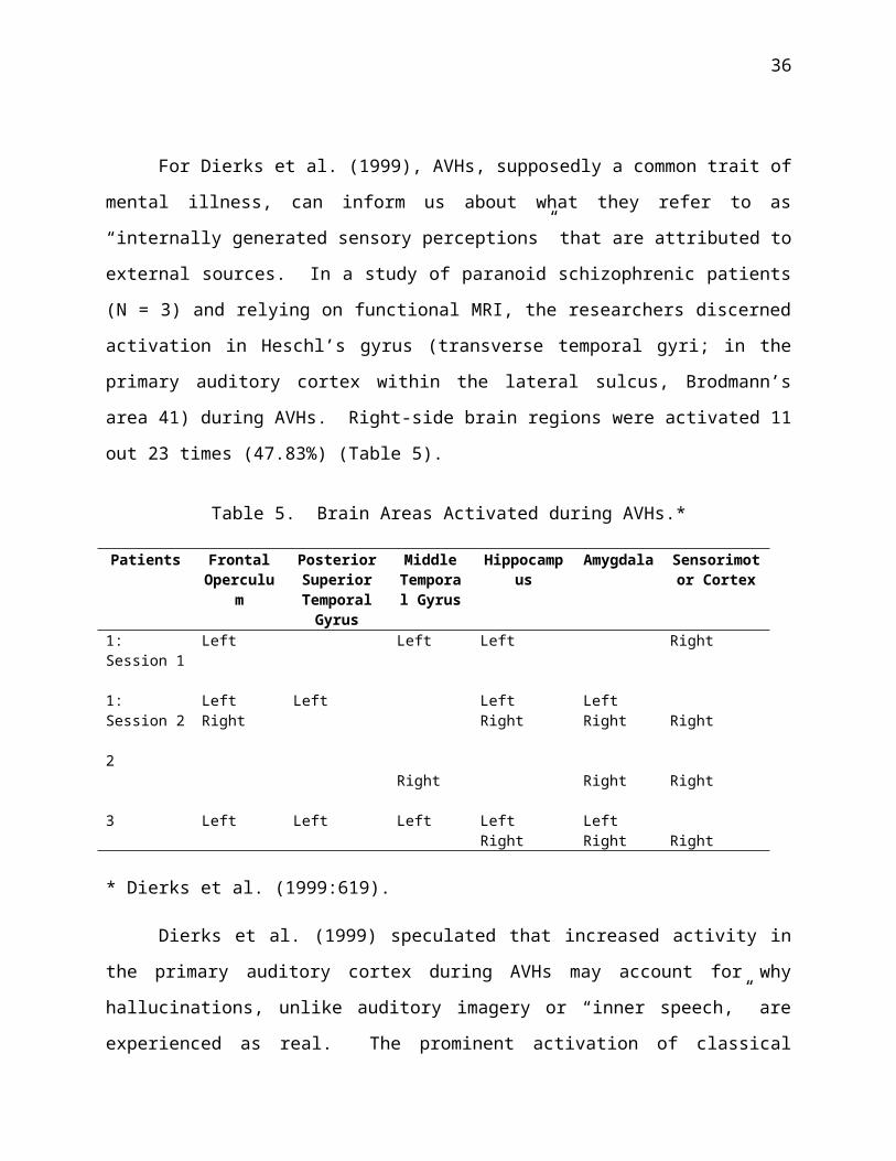
For Dierks et al. (1999), AVHs, supposedly a common trait of
mental illness, can inform us about what they refer to as
“internally generated sensory perceptions” that are attributed to
external sources. In a study of paranoid schizophrenic patients
(N = 3) and relying on functional MRI, the researchers discerned
activation in Heschl’s gyrus (transverse temporal gyri; in the
primary auditory cortex within the lateral sulcus, Brodmann’s
area 41) during AVHs. Right-side brain regions were activated 11
out 23 times (47.83%) (Table 5).
Table 5. Brain Areas Activated during AVHs.*
Patients FrontalOperculu
m
PosteriorSuperiorTemporalGyrus
MiddleTemporal Gyrus
Hippocampus
Amygdala Sensorimotor Cortex
1: Session 1
Left Left Left Right
1: Session 2
LeftRight
Left LeftRight
LeftRight Right
2Right Right Right
3 Left Left Left LeftRight
LeftRight Right
* Dierks et al. (1999:619).
Dierks et al. (1999) speculated that increased activity in
the primary auditory cortex during AVHs may account for why
hallucinations, unlike auditory imagery or “inner speech,” are
experienced as real. The prominent activation of classical

37
speech production areas is suggestive of processes related to
inner speech. However, important differences should be noted:
Inner speech is not perceived to arise from external influences
and does not activate Heshcl’s gyrus.
Using fMRI scans to determine areas of hallucinatory
activation, Sommer et al. (2007) applied low-frequency repetitive
transcranial magnetic stimulation (rTMS) to the left
temporoparietal areas of a medication-resistant group (effect
size of 0.76).18 The group consisted of male schizophrenia
patients (N = 15). Twelve treatments were successful.
Hallucinatory activation was predominantly within the left
temporoparietal areas for four patients. For five it was in the
right-sided temporopareital areas, and for three it was located
deep within the contralateral homolog of Broca’s area. Thus,
most patients (8 out of 12; 66.7%) had predominantly right-sided
hallucinatory activity.
In an article published in Schizophrenia Bulletin (Sommer et al.,
2010) scores from around 4,000 respondents to a website
questionnaire (a version of the Launay-Slade Hallucination Scale)
led to the identification of a group of 103 people who had
genuine voice-hearing experiences but no psychopathology. The 42
people who were scanned in the new study were selected from this
larger sample, and matched to data from an existing fMRI dataset
from psychiatric patients with a range of disorders (including
schizophrenia, schizoaffective disorder and psychosis not
otherwise specified). To be included, participants had to have

38
had at least four AVH experiences during the 8-minute scan
period, with a minimum total duration of fifty seconds.
Participants indicated when they were hearing a voice by
squeezing a balloon and releasing it when the voice stopped. The
results of these analyses were rather simple: In both groups, the
areas that were expected to activate mostly did activate. There
were no differences in activation between the groups, however,
leading the researchers to conclude that nonclinical and clinical
AVHs do not differ in terms of their underlying neural
activation.
In an article whose title succinctly expresses their
findings—“Auditory verbal hallucinations predominantly activate
the right inferior frontal area”—Sommer et al. (2008, emphasis in
original) used fMRI to measure cerebral activation among
psychotic patients (N = 24) (schizophrenia, schizoaffective
disorder or psychotic disorder not otherwise specified). The
researchers carried out tow experiments. In the first, scanning
occurred while subjects experienced AVHs. Activation was evident
in the right homologue of Broca's area, bilateral insula,
bilateral supramarginal gyri and right superior temporal gyrus.
10 out of 21 activations (47.6%) were on the right side.
Interestingly, Broca's area and the left superior temporal gyrus
were not activated. In the second experiment, scanning occurred
while they silently generated words. Activation occurred in
Broca's and Wernicke's areas and to a lesser degree their right-

39
sided homologues, bilateral insula and anterior cingulate gyri.
13 out of 27 activations (48.1%) were on the right side.
The main difference that Sommer et al. (2008) found between
cerebral activity during AVHs and activity during normal inner
speech appears to be lateralization. The predominant engagement
of the right inferior frontal area during AVH may be related to
the typical low semantic complexity and negative emotionality.
The mean lateralization index was –0.11 (SD 0.41) during AVHs and
0.14 (SD 0.34) for the word generation task. Paired sample t-
test revealed significantly lower lateralization (t(23) = –2.4, p
< 0.02). In a conjunction analysis brain areas on the right side
were activated 9 out of 15 times (60.0%).
Activity during AVHs was uncorrelated with language
lateralization; rather, lateralization was related to the degree
to which the content of the AVHs possessed negative emotionality.
Sommer et al. (2008) note that the predominant engagement of the
right inferior frontal area during AVHs may be related to low
semantic complexity and negative emotionality.
Van de Ven et al. (2005) used spatial independent component
analysis (sICA)19 to extract the activity patterns associated
with AVHs in schizophrenia patients (N = 6). Viewed bilaterally,
activation of the auditory cortex (components of interest)
occurred 60.0% on the right side during hallucinations (not
including the posterior temporal plane) (Table 6). If bilateral
activations are eliminated (i.e., only unilateral instances are

40
considered), right side activation occurred 66.7% of the time
during AVHs.
Table 6. Activation of Auditory Cortex and the PosteriorTemporal Plane.*
Patient
Components of Interests Other Areas
1 HG = Left, RightSMC = Right
PTP = Left
2 SMC = Right3 HG = Left, Right
SMC = RightPTP = Left
4 HG/STG = RightSMC = Left
5 SMC = Left
* Based on Van de Ven et al. (2005:651); abbreviated. HG = Heschl’s gyrus; SMC= sensorimotor cortex; STG = superior temporal gyrus; PTP = posterior temporal plane.
Table 7 summarizes the frequency of right side activity
during AVHs as reported in a number of selected studies.
Table 7. Summary: Frequency of Right Side Activity during AVHsReported in Selected Studies.
Study Numberof
Participants
Frequency of RightSide Activity
Barkus, Sterling, Hopkins, McKie, & Lewis (2007)
8 50.0%
Buchsbaum et al. (1982) 14 100.0%Diederen et al. (2011) 42 42.9%Dierks et al. (1999) 3 47.8%Sommer et al. (2007) 15 66.7%Sommer et al. (2008) 24 47.6%

41
60.0% (conjunction analysis)
Van de Ven et al. (2005) 6 66.7%Mean = 60.21% (SD = 17.26)If conjunction analysis not included, Mean = 60.24% (SD = 18.45)
Other Relevant Studies
Ćurčić-Blake et al. (2012) attempted to discern connectivity
models between left hemispheric speech-processing areas and their
right hemispheric homologs. They investigated reduced
information flow to Broca’s area in schizophrenia patients with
AVHs. Besides Broca’s area, they examined the interaction among
three other linguistic regions: Wernicke’s area (in left
hemisphere) and the homologs of Broca’s and Wernicke’s areas in
the right hemisphere (Figure 1). Subjects included a healthy
control group (presumably those who do not experience
hallucinations) (N = 18), two groups of schizophrenics without (N
= 14) and with hallucinations (N = 21). The subjects were asked
to perform a task requiring inner speech processing during
functional brain scanning (fMRI). The researchers used dynamic
causal modeling to track the flow of information (this allows for
directional inference). Bayesain model averaging was used to
estimate the connectivity strengths and evaluate group
differences.
Results indicated that those with AVHs had activation of the
left superior temporal gyrus20 (including Wernicke’s area), the
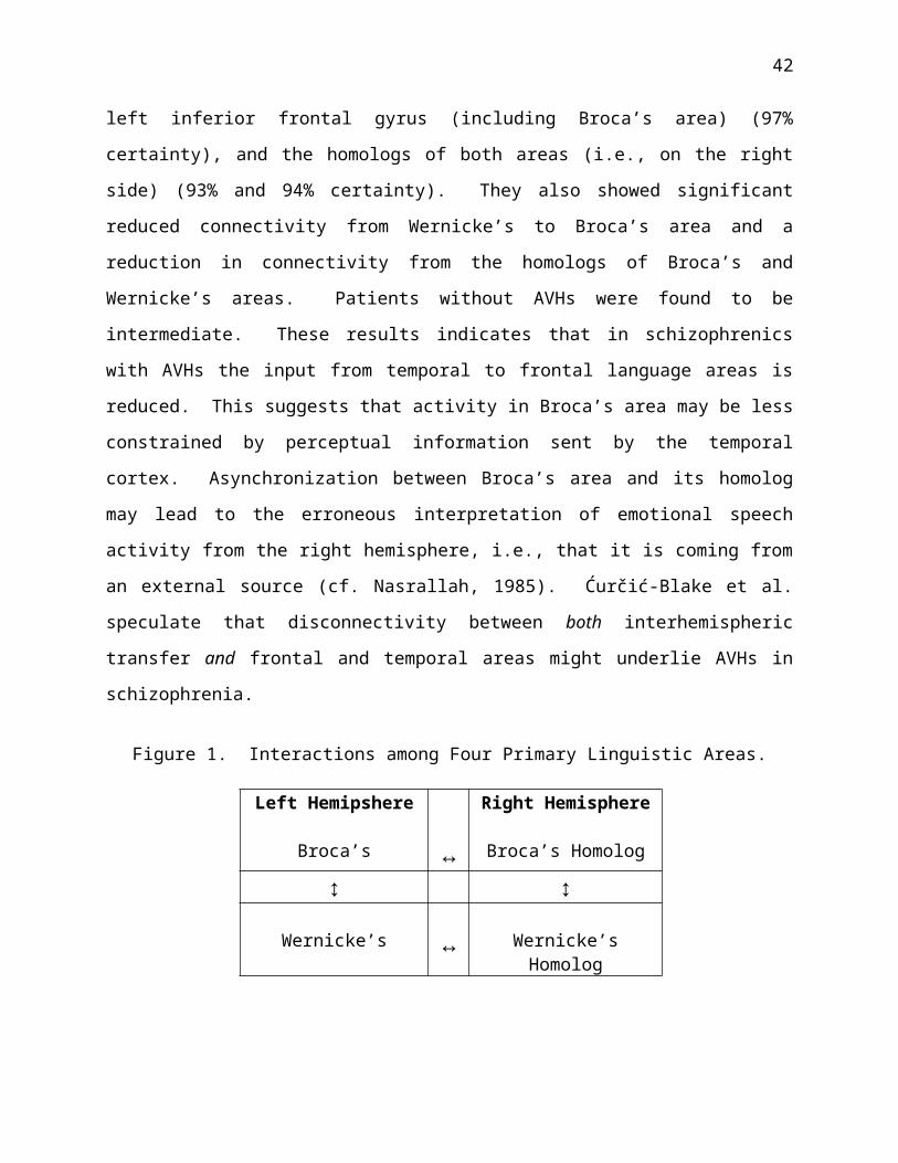
42
left inferior frontal gyrus (including Broca’s area) (97%
certainty), and the homologs of both areas (i.e., on the right
side) (93% and 94% certainty). They also showed significant
reduced connectivity from Wernicke’s to Broca’s area and a
reduction in connectivity from the homologs of Broca’s and
Wernicke’s areas. Patients without AVHs were found to be
intermediate. These results indicates that in schizophrenics
with AVHs the input from temporal to frontal language areas is
reduced. This suggests that activity in Broca’s area may be less
constrained by perceptual information sent by the temporal
cortex. Asynchronization between Broca’s area and its homolog
may lead to the erroneous interpretation of emotional speech
activity from the right hemisphere, i.e., that it is coming from
an external source (cf. Nasrallah, 1985). Ćurčić-Blake et al.
speculate that disconnectivity between both interhemispheric
transfer and frontal and temporal areas might underlie AVHs in
schizophrenia.
Figure 1. Interactions among Four Primary Linguistic Areas.
Left Hemipshere
Broca’s ↔
Right Hemisphere
Broca’s Homolog
↕ ↕
Wernicke’s ↔ Wernicke’sHomolog

43
Bentaleb, Beauregard, Liddle, and Stip (2002) note that two
theories for AVHs have received empirical support: (1) they arise
from misinterpreted inner speech or (2) from aberrant activation
of the primary auditory cortex. To test these hypotheses, they
used fMRI to measure the brain activity of a schizophrenic woman
in the temporal and inferior frontal regions during AVHs and
while listening to external speech. This woman usually
experienced continuous AVHs but these disappeared when she
listened to loud external speech. A matched control subject’s
bran activity was recorded under the same conditions. They found
higher metabolic activity in the left primary auditory cortex21
and the right middle temporal gyrus during AVHs and believe that
they these areas possibly interact during AVHs. They concluded
that the two theories are not mutually exclusive.
Psychotic patients typically display decreased language
lateralization. However, Diederen et al. (2010) investigated
whether dysfunction is associated psychosis in general or with
certain symptoms of psychosis, such as AVHs. Subjects included
patients with a psychotic disorder (N = 35), 35 non-psychotic
subjects with AVHs (N = 35), and healthy control subjects (N =
35). All groups covertly performed a paced verbal fluency task
while being scanned (3T fMRI). In order to measure performance,
subjects were asked to generate words overtly in an additional
task. Language lateralization indices were calculated and group-
wise brain activation during verbal fluency was compared for the
three groups. Task performance did not differ significantly

44
among the groups. Lateralization indices are defined as the
difference in “thresholded” signal intensity changes in the left
versus the right hemisphere (in the selected language areas)
divided by the sum of “thresholded” signal intensity changes. +1
indicates strong left-hemispheric dominance, while –1 indicates
strong right-hemispheric dominance. As expected, relative to the
other two groups, decreased language lateralization was
significantly reduced for the patient group. The latter group
also presented significantly more activity in the right
precentral gyrus, left insula, and the right superior parietal
lobule compared to the other groups. Interestingly,
lateralization indices were not very different between the AVHs-
non-psychotic group and the healthy control groups, and no
significant differences in neural activity during verbal fluency
between the two non-psychotic groups were discerned. Diederen et
al. (2010) conclude that language lateralization was not
significantly reduced in the AVHs-non-psychotic group and
presently a relationship between AVHs and redueced language
lateralization cannot be demonstrated.
Ocklenburg, Westerhausen, Hirnstein, and Hugdahl (2013), in
an effort to integrate studies investigating language
lateralization in schizophrenics, provide indirect evidence of
right side activity during AVHs. Using dichotic listening
testing, they conducted two meta-analyses. The first looked at
21 different studies, comparing schizophrenics (N = 700) with
healthy controls (N = 707). The results showed schizophrenics

45
had weaker language lateralization compared to healthy controls.
However, the effect size was small: g = –0.26 (95% confidence
interval –0.36 to –0.15), significantly different from zero (Z =
–4.69; p < .00001). The schizophrenic population, then, cannot
be considered a homogenous group in relation to language
lateralization. In the second analyses of 8 different studies,
schizophrenics experiencing AVHs (N = 179) were compared with
non-hallucinating controls (N = 228). In this analyses the
effect size was significantly larger: g = –0.45 (95% confidence
interval –0.65 to –0.25), significantly different from zero (Z =
–4.45; p < .00001). Compared to non-hallucinating controls,
schizophrenics who have AVHs show a significantly reduced right
ear advantage in the dichotic listening tasks.22 The researchers
conclude that reduced language lateralization is a weak trait
marker for schizophrenia. However, reduced language
lateralization is a strong trait marker for those experiencing
AVHs within the schizophrenic population.
Concluding Thoughts
Affinities between Hallucinations and Introspection
I have attempted to show that though redundancy characterizes
many crucial biological features, the lateralization of human
speech is consistent with a common trait of bilateria, i.e.,
capabilities on one side of the brain are freed up so as to allow
its focus on pressing environmental matters. In the case of
preconscious humans, this would involve social situations in

46
which predictable routines for whatever reason broke down and a
guiding, commanding, admonishing or inspiring divine voices
intervened. Though not completely convincing, suggestive
evidence exist that auditory hallucinations originate in the
right temporal lobe in regions corresponding to the language
areas in the left temporal lobe. The hemispheres are
“communicating” but in surprising ways they are “unintegrated”
neurological components. About three thousand years ago massive
sociocultural changes redesigned our socioneurology, replacing
voice–volitions with another form of hallucinatory experience,
i.e., the quasi-perception of mental imagery (conscious
interiority). Even patterns of inhibition and disinhibition are
matters of culture and socialization.
It is easy to overlook the almost magical properties of
mental imagery, often described as quasi-perception. Many
mistakenly assume that the ability to call up mental sceneries
—“unshared sensory experiences” (Stevenson 1983)—is a type of
sensory perception, a mirroring of the surrounding environment,
and then conclude that introspective experiences can be explained
away or reduced to perceptual processes. Unfortunately, the role
of conscious interior experience is gravely misunderstood. In
fact, the mysterious nature of quasi-perception comes from some
important affinities it shares with hallucinations, i.e., both
types of experiences cannot be pointed to in the physical world
and yet exert influence over behavior. The cultural invention of
a privately-viewable place to “introceive” (not perceive) allows

47
us to excerpt scenes and objects and weave “should’ve-could’ve-
would’ve” narratives necessary for planning in increasingly
complex societies. Introception is a type of adaptive
hallucination, i.e., mental imagery is employed in the
“selection, rehearsal, planning, and perfecting of adaptive
activities,” thereby providing the “means to guide experimentally
and transform experience by running off activity cycles as mental
simulations of the real thing.” In short, a “primary function of
consciousness is the mental rehearsal of adaptive, goal-directed
action through the experimental manipulation of perceptual-motor
imagery” (Marks 1999:579, 567). Introception conveniently
conjures up a “viewable” cognitive map of the world but one
infused with a menu of maybe’s, perhaps’s, and possibilities. It
is a post-bicameral adaptive upgrading of our mental machinery
suited to more complex sociopolitical circumstances, just as
hallucinatory as divine visions or disembodied voices
(extraception) were in bicameral times. In this sense,
introception is the counterpart to exteriorized hallucinations
(McVeigh 2013). Introceptions and extraceptions (audiovisual
hallucinations interpreted as divine voices and visitations in
ancient times) can be called superceptions. Vestigial
extraceptions are anomalous behaviors, e.g., hallucinations still
experienced by schizophrenics, while coceptions describe how
perceptions and introceptions coincide (such overlapping deludes
us into assuming that interior experiences are sensory
reflections of reality).

48
Practical and Therapeutic Implications
Numerous articles have been written on AVHs as if they are
inherently pathopsychological.23 However, enough evidence exist
to argue that AVHs in themselves may not be neurologically
pathological (i.e., they arise built-in aptic structures), though
they can be psychologically pathological (they might accompany or
indicate serious dysfunction). If so, what are the therapeutic
implications acknowledging that AVHs in and of themselves are not
pathological? A number of others have acknowledged that AVHs can
be potentially positive: Beavan (2011); Chin, Hayward, and Drinna
(2008); Hayward, Oveton, Dorey, and Denney (2008); and Pérez-
Álvarez, Garcia-Montes, Perona-Garcelán, and Vallina-Fernández
(2008).
Romme and Escher (1996) submit that hearing voices has a
functional role in aiding people to cope with the problems of
daily life. They noticed key differences between good copers and
bad copers. The latter experienced more negative “imperative
voices,” i.e., a power structure existed between the voices and
person, with the voices viewed as stronger than one’s self.
Romme and Escher also found that non-patients were more like to
be married, received more support than patients, and were more
likely to discuss their voices with other people than patients.
They found no connection between the characteristics of hearing
voices and specific psychiatric illnesses, indicating that voice-
hearing is not the result only of pathopsychology.

49
Romme, Honig, Noorthhoorn, and Escher (1992) sent
questionnaires to 460 people with chronic hallucinations who
responded to a request on a television show. They received 254
replies, of which 186 could be used for analysis (it was
determined 13 were probably not experiencing hallucinations).
They found that 115 reported an inability to cope with voices, 97
were in psychiatric care, and copers were significantly less
often in psychiatric care (24%) than non-copers (49%). They
discovered that successful copers used four strategies for
dealing with AVHs: (1) distraction; (2) ignoring the voices; (3)
selective listening; and (4) setting limits on their influence.
Corstens, Longden, and May (2112) introduce a therapeutic
approach called “Talking with Voices” which is derived from the
theory and practice of Voice Dialogue (Stone and Stone, 1989),
authors of Embracing our selves: The voice dialogue training manual. The
Stones (1989) postulated that selves are composed of “sub-
personalities” that require re-integration. Corstens, Longden,
and May (2o12) explain how the therapist acts a “facilitator” who
directly engages with the different selves (e.g., primary or
dominant selves that push away “disowned selves”) in order to
raise awareness of the meaning and origin of their voices. As
Jones and Coffey (2012), in their thematic analysis of the
personal meanings behind AVHs, contend that a biologically-based
psychiatry with its medico-physical explanations is inadequate
for acknowledging the actual experiences of voice-hearers.

50
Chapter Appendix: Hallucinations as Abnormal
Among medical and mental health practitioners, hallucinations
have been deeply associated with psychopathology of one sort or
another, such as Parkinson’s disease. Barnes and David (2001)
report 8% to 40% of those with Parkinson's disease experience
visual hallucinations (N = 44) (see also Barnes, Connelly, Wiggs,
Boubert, and Maravic [2010]). Barnes, Koch, Wilford, and Boubert
(2011) investigated the linkages among personality, stress,
sleep, and hallucinations in a normal student population (N =
127). They found significant associations between stress and
sleep, with stress being a significant predictor of
hallucinations. Also a predictive relationship between
hallucinations and schizotypal personality traits was found (age
did not seem to play a noticeable role). Nettle (2006) sees a
linkage between schizotypy and mental illness amongst poets,
visual artists, and mathematicians (N = 501).
But hallucinations have been particularly linked to
schizophrenia and dissociative disorders.24 For example,
Morrison and Haddock (1997) asked schizophrenics experiencing
auditory hallucinations, schizophrenics not experiencing them,
and normal controls to complete the private self-consciousness
subscale of the Self Consciousness Scale. It was found that
hallucinators exhibited significantly higher levels of self-focus
than non-hallucinators. The latter did not differ from normal
subjects. The level of self-focus predicted whether or not
51
subjects experienced hallucinations (N = 30). Johns and Van Os
(2001) explore the continuity of psychotic experiences in the
general population. They contend that schizophrenia is not a
discrete illness and that the distribution of psychosis, with
symptoms of hallucinations and delusions, would not be
represented by a normal curve nor a bimodal or dichotomous curve,
but rather a continuous but only half-normal distribution. They
see a need for a “psychosis continuum” (which might have
implications for improved therapy).
Ross, Joshi, and Currie (1990) administered the 28-item
Dissociative Experiences Scale respondents (N = 1,055) in
Winnipeg and found that 26% of the general population experienced
dissociative experiences. Using factor analysis, they found that
three factors accounted for 47.1% of the combined variance of the
score: (1) absorption‒imaginative involvement (benign: Ignoring
pain, missing part of a conversation, etc.); (2) dissociated
states; and (3) depersonalization‒derealization (not recognizing
friends or family members, not recognizing one’s reflection in a
mirror, etc.). “Hearing voices in one’s head” (prevalence of
26%) was included in the third factor.
Hallucinations may not be directly caused by or related to
psychopathology, but studies typically find what are taken to be
significant correlations. Barrett and Etheridge (1994) selected
hallucinators (N = 18) and non-hallucinators (N = 24) among
normals using the Verbal Halluciantion Questionnaire and had them
take the Millon’s Clinical Multiaxial Inventory (MCMI; measures

52
dysfunctional personality correlates). They found that
hallucinators experience heightened negative affect, feelings of
rejection, and incompetence in social situations. In their
analysis of the links between hallucinations and imagery in a
normal population (N = 24), Jakes and Hemsley (1987) found that
psychoticism (Eysenck Personality Questionnaire) was
significantly related to reports of hallucinatory experiences,
and a measure of hallucinatory predisposition was significantly
associated with strong hypnogogic/hypnopompic imagery. Launay
and Slade (1981), in a study of male and female prisoners (N =
297), used Eysencks’ psychoticism (P) scale to discern a link
between aggressive‒paranoid tendencies and hallucinatory
predisposition. Using the Sleep-EVAL system, Ohayon and
Schatzberg (2002) analyzed the general population (N = 18,980) in
the UK, Germany, Italy, Spain, and Portugal. They found that
12.5% of those with depressive episodes had psychotic features
(either delusions or hallucinations). Feelings of worthlessness
or guilt were also associated with high rates of hallucinations
(9.7%).
Young, Bentall, Slade, and Dewey (1987) investigated the
relation between suggestibility and the elicitation of auditory
and visual hallucinations in normal and psychiatric subjects.
One experiment, using the LSHS-A, assessed if normal subjects
were predisposed to hallucinate. The second experiment assessed
hallucinating psychiatric and non-hallucinating psychiatric
patients. High LSHS-A scorers and hallucinating psychiatric

53
patients were significantly more likely to hear suggested sounds
and see suggested objects than were their respective controls
(but this finding was not replicated in the psychiatric
patients). No significant differences on measures of
suggestibility were found between the groups, although for the
psychiatric group the Barber Test Suggestion and Subjective
Involvement scores correlated positively with LSHS-A scores (N =
203).
Using the South African Stress and Health Study, Temmingh,
Stein, Seedat, and Williams (2011) utilized the Composite
International Diagnostic Interview (CIDI) in a survey of the
general population of South African adults (N = 4,250). They
found a prevalence rate of 12.7% of people reporting
hallucinations, which is comparable to reports in other parts of
the world. They also found correlations between reported
auditory or visual hallucinations and role impairment, service
utilization, and suicidality. In an investigation of a non-
psychiatric population of Caribbean descent residing in the US,
Izquierdo (2000) found that culture shapes the way reality is
interpreted and that hallucinations are to be taken as a weak
symptom of mental instability (at least among those of Caribbean
descent).
Basing their methodology upon that used by Feelgood and
Rantzen (1994), Pearson, Burrow, FitzGerald, Green, Lee, Wise
(2001) exposed child participants (aged 9–11 years old) to
ambiguous stimuli which then generated hallucinations. Their

54
findings indicated a “developmental continuum” of hallucination
experiences between children and adults. Pearson, Smalley,
Ainsworth, Cook, Boyle, and Flury (2008) propose a two-axis
model: (1) child/adolescent versus adult; and (2) nonclinical
versus pathological continua. This developmental model, based on
adults (N = 496), links child abuse to pathological
hallucinations and possible later mental illness. Pearson,
Smalley, Ainsworth, Cook, Boyle, and Flury (2008), in their study
of AVHs among adolescent and adults, see the need for early
intervention for psychosis.
Several studies have examined the connection between
emotions and hallucinations, but again, the underlying premise
seems to be that hallucinations are inherently abnormal. Freeman
and Garety (2003), in the context of neurosis and psychosis,
investigated the influence of emotion on delusions and
hallucinations. Laroi, van der Linden, and Marczewski (2004)
looked at how emotionality and meta-cognitive beliefs impacted
monitoring tasks in hallucination-prone subjects. Davies,
Griffin, and Vice (2001) looked at reactions to auditory
hallucinations in psychotic, evangelical and control groups: 27%
of the control group (non-psychotic, non-evangelical) heard
voices, while 59% of evangelical Christians and 100% of the
psychotic group did, respectively (N = 102). Not surprisingly,
the experiences of the evangelical group were more positive than
those of the control group (which were significantly more
positive than those of the psychotic group). Delespaul, deVries,

55
and van Os (2002) found anxiety to be the most prominent emotion
and the strongest predictor of hallucinations intensity, though
an array of contextual influences play a role for schizophrenics.
Death is of course an emotinally stressful event. Osis and
Haraldsson (1977) researched deathbed observations by physicians
and nurses in the US and India of mostly terminally ill patients
(N = 877). The most frequently reported phenomena were
hallucinations of human figures (471 cases; 53.7%). Four-fifths
of the hallucinations were of deceased persons and religious
figures.
According to Kumari et al. (2008), previous small-sample
studies have shown altered frontotemporal activity in
schizophrenia patients with auditory hallucinations. Utilizing
fMRI, Kumari et al. (2008) attempted to study monitoring of self-
and externally generated speech in two samples: Among
schizophrenics (N = 63) and a group of healthy controls (N = 20).
Participants were asked to perform a self-monitoring task while
undergoing fMRI using verbal feedback: (1) their own voice (self-
undistorted); (2) their own voice lowered in pitch by 4 semitones
(self-distorted); (3) voice of another person matched on a
participant’s sex (other-undistorted); and (4) another person’s
voice with the pitch lowered by 4 semitones (other-distorted).
Participants indicated the origin of voices by pressing buttons
marked “self” for their own voices, “other” for “other” voice,
and “unsure.” Kumari et al. (2008) did not scan during AVHs, so
their findings are indirect evidence of the neurological

56
correlations of hallucinations. Moreover, given that the focus
of my review is to discern the level of right-sided neural
activation, the use of subjects’ own voice, whether distorted or
not, may introduce an unnecessary confounding variable.
Kumari et al. (2008) detected activation of the thalamus
(medial geniculate nucleus, MGN) and frontotemporal regions.
Among poorly performing patients (1 standard deviation below
controls’ mean; N = 36), less activation of the thalamus (MGN,
pulvinar) and superior-middle temporal and inferior frontal gyri
was evident (relative to the combined group of controls and well-
performing patients). They found greater deactivation of the
ventral striatum and hypothalamus occurred when hearing one’s own
voice among schizophrenics. This was combined with
nonsignificant activation of the same regions to the voice of
others and was associated with negative symptoms, e.g., blunted
affect, emotional withdrawal, poor rapport, passive social
avoidance, regardless of performance. They also found that
heightened activation of the right superior-middle temporal gyrus
during undistorted feedback [their own voice or the voice of
another] was associated with both positive symptoms
(hallucinations, persecution) and poor performance.25 For all
participants, increased brain activity associated with individual
tasks conditions were reported 34 times, 20 of which were on the
right side (52.94%). Decrease in activity associated with
individual tasks conditions were reported 24 times, 12 of which

57
were on the right side (50.00%). Tables 8, 9, and 10 summarize
these results.
Table 8. Increases and Decreases of Neural Activation inAssociation with Individual Tasks Conditions.*
Side ofBrain
Decrease Increase
Left 50.00% 43.75%Right 50.00% 56.25%
* From Kumari et al. (2008:745); abbreviated.
Table 9. Increases in Association with Individual TaskConditions.
VerbalFeedback
Brian Region Brodmann Area Side
Self-undistorted
Superior temporal gyrus
Lingual gyrus/cuneus
222222181818
RLRRLL
Self-distorted Superior temporal gyrus
Postcentral gyrusHeschl’s gyrusSuperior temporal gyrusThalamusCuneus
Middle occipital gyrus
2222404122n/a1918191919
RRRLLRLLRRR
Other- Superior temporal 22 R
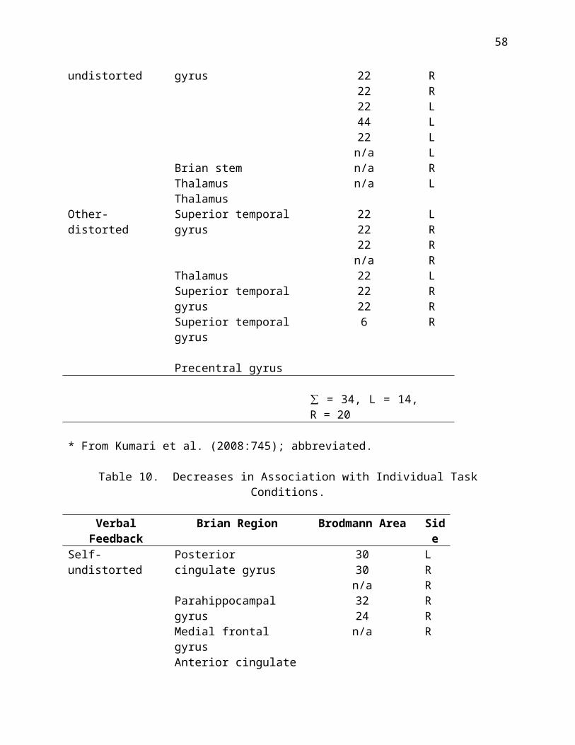
58
undistorted gyrus
Brian stemThalamusThalamus
2222224422n/an/an/a
RRLLLLRL
Other-distorted
Superior temporal gyrus
ThalamusSuperior temporal gyrusSuperior temporal gyrus
Precentral gyrus
222222n/a2222226
LRRRLRRR
= 34, L = 14,∑R = 20
* From Kumari et al. (2008:745); abbreviated.
Table 10. Decreases in Association with Individual TaskConditions.
VerbalFeedback
Brian Region Brodmann Area Side
Self-undistorted
Posterior cingulate gyrus
Parahippocampal gyrusMedial frontal gyrusAnterior cingulate
3030n/a3224n/a
LRRRRR

59
gyrusCaudate nucleus
Self-distorted Parahippocampal gyrusCalcarine sulcusParahippocampal gyrusCaudate nucleusAnterior cingulategyrus
n/an/an/an/a2424
LLRLLR
Other-undistorted
Parahippocampal gyrusAngular gyrusPosterior cingulate gyrusAnterior cingulategyrus
n/a3930
24/323232
LLRLRR
Other-distorted
Posterior cingulate gyrus
Angular gyrusAnterior cingulategyrusMedial frontal gyrus
3023392410
10/32
RLRLLL
= 24, L = 12,∑R = 12
* From Kumari et al. (2008:746); abbreviated.
Kumari et al. (2008) found that if brain areas showing
activation changes for “source” and “distortion effects” are
measured (for all participants), 5 out of 7 (71.4%) instances
were on the right side. If neural activity, differentiated by
“good” and “poor performers,” is measured (regardless of
diagnosis), 21 out of 42 (50.0%) instances were on the right

60
side. If neural activity, differentiated by healthy participants
and schizophrenic patients, is measured (regardless of
performance), 8 out of 17 (47.1%) instances were on the right
side.
In conclusion, Kumari et al. (2008) surmise that
hypoactivation of a neural network, consisting of the thalamus
and frontotemporal regions, is involved in the impaired speech
monitoring in schizophrenia. They also believe that a common
activation abnormality is shared by positive symptoms and poor
monitoring in the right superior temporal gyrus during processing
of degraded speech.
In a review of relevant studies,26 Diederen, van Lutterveld,
and Sommer (2012) found that in non-psychotic individuals AVHs
were associated with increased sensitivity of auditory areas to
auditory stimulation as well as aberrant connectivity for frontal
and temporoparietal language regions, and of language production
and perception, particularly in the anterior cingulate regions
(Brodmann’s areas 24, 32, and 33). Hallucinators appear to
possess an attention bias to auditory stimuli that may be related
to aberrant activation of the anterior cingulate regions.27
Diederen, van Lutterveld, and Sommer (2012) also discovered that
decreased cerebral dominance for language as well as dopamine
dysfunction (commonly associated with schizophrenia) are probably
not in themselves related to AVHs (these abnormalities were not
apparent in non-psychotic voice hearers). Finally, specific

61
aspects of AVH, such as voluntary control, may be related to the
timing of the supplementary motor area and language areas.
Sommer et al. (2010) studied 103 nonclinical individuals
with AVHs. These were compared to a control group of 60
individuals matched for sex, age, and education. They did find
that, after a battery of tests (including the Schizotypal
Personality Questionnaire [SPQ] and the Peters Delusion
Inventory), that the hallucinating individuals had a lower level
of global functioning and a “general increased schizotypal and
delusional tendency.” Evidence of childhood trauma among those
who experienced hallucinations was also apparent. They concluded
that AVHs in otherwise healthy individuals are not an “isolated
phenomenon” but rather part of a “general vulnerability for
schizophrenia.”

1 The germ for this chapter was a graduate school paper for a coursein research methods and assessment entitled “Auditory VerbalHallucinations and Right Hemispheric Activity: Evidence andImplications.” I am grateful for Prof. Donald Graves (Sage GraduateSchool) for offering invaluable advice. 2 From the Latin “to wander in the mind,” which came from the Greek, alusso, “to be uneasy.” 3 Cultural traditions, of course, play a role in how hallucinationsare defined. See Luhrmann (2011), Luhrmann, Padmavati, Tharoor, andOsei (2014), and Larøi, Luhrmann, Bell, Christian, Deshpande ,Fernyhough, Jenkins, and Woods (2014). Al-Issa (1995) examines thedifferences between “Western” and “non-Western” views ofhallucinations. In the former, or “rational” societies, a cleardistinction between reality and fantasy encourages negative attitudestoward hallucinations resulting in people being unfamiliar with theirown imagination. But in “less rational” cultures the distinctionbetween reality and fantasy is more “flexible” and individuals aresocialized to accept their hallucinations. An appreciation of suchviews towards anomalous experiences might aid mental-health providersin distinguishing pathological from culturally-sanctionedhallucinations. 4 Much of my treatment of the categorization of hallucinations andillusions relies on Kaplan and Sadock’s Synopsis of Psychiatry: BehavioralSciences and Clinical Psychiatry (1991). 5 Some are “reflex” in which a stimulus in one sensory modality causes a hallucination in another, while others are “functional,” i.e., a stimulus causes a hallucination in the same sensory modality,but both the stimulus and the hallucination are experienced. 6 Suggested to Stevenson by David Kovacs, a scholar of classics at the University of Virginia. 7 E.g., see Kokoszka (1992) who investigated “altered states ofconsciousness” in normal populations, e.g., hallucinations, peakexperiences, mystical states, hypnotic-like experiences, and out-of-body experiences. Kokoszka pointed out that such phenomena are toocommon to be ignored by psychology. 8 Andrew et al. (2008); Daalman et al. (2011); Davies et al. (2001);Diederen et al. (2011); Hill et al. (in preparation a); Hill et al.(in preparation b); Honig et al. (1998); Johns et al. (2002a, 2002b);Jones et al. (2003); Leudar et al. (1997); Linden et al. (2011);Romme and Escher (1989); Sommer et al. (2010); Sorrell et al. (2010).

9 For a review of studies on imaginary playmates, see Klausen and Passman (2006). 10 See also Connolly (1991). 11 On children who hear voices, see Escher, Romme, and Buiks (1998) and Pearson (1998). 12 See Weller and Wiedemann (1989). 13 In the case of visual hallucinations, for example, causes may beirritative phenomena, release phenomena, or processing disturbanceswithin the visual pathways (Weller and Wiedermann 1989). 14 Two useful works that provide an overview of the subject are Watkins, Hearing voices: A Common Human Experience (1998) and Romme and Escher, Making Sense of Voices: A Guide for Mental Health Professionals Working with Voice-Hearers (2000). 15 Lateralization is evident in vertebrate and even some invertebrate species (Halpern, Güntürkün, Hopkins, and Rogers 2005). 16 From the German psychiatrist, Kurt Schneider (1887–1967). 17 They also point out that studies of stroke patients with lesions and dichotic listening and functional imaging studies of healthy people show that certain language functions are mediated by the righthemisphere rather than the left. 18 The Auditory Hallucination Scale (AHRS) and Positive and Negative Symptom Scale (PANSS) were utilized to make assessments. 19 sICA decomposes the functional data set into a set of spatial maps without the use of any input function. 20 In study by Jardri (2007) of a schizophrenic child, bilateral activation of the superior temporal gyri was evident (predominantly on the left hemisphere). 21 The primary auditory cortex is bilaterally located, near the upper sides of the temporal lobes on the superior temporal plane within thelateral fissure and comprising parts of Heschl’s gyrus and the superior temporal gyrus, It includes planum polare and planum temporale and roughly corresponds to Brodmann’s areas 41, 42, and part of 22. 22 The “right ear advantage” in dichotic listening tests is usually defined as the difference between the number of correctly identified stimuli presented to the right ear compared to the number of correctly identified stimuli presented to the left ear. 23 Bentall, Haddock, and Slade (1994). 24 Numerous studies investigate such matters. For a general treatment, see Slade (1973) and Slade and Bentall (1988).

25 Thalamic abnormality characterized schizophrenics regardless of performance and symptoms. 26 Barkus et al. (2007); Diederen et al. (2010); de Weijers et al.(2011); Howes et al. (2013); Linden et al. 92011); Lewis-Hanna et al.(2011); Szechtman et al. (1998); van Lutterveld et al. (2010). 27 Not specifically related to AVHs are decreased cerebral dominancefor language and dopamine dysfunction (typically evident inschizophrenia). Such abnormalities were absent in nonclinical voicehearers. Also, voluntary control may be related to the timing of thesupplementary motor area and language areas in AVHs.
References
Al-Issa, I. (1995). The illusion of reality or the reality of illusion: Hallucinations and culture. British Journal of Clinical Psychology, 166, 368–373.
Allen, P., Laroi, F., McGuire, P.K., and Aleman, A. (2008). The hallucinating brain: a review of structural and functional neuroimaging studies of hallucinations. Neuroscience Review, 32, 175–191.
American Psychiatric Association (2013). Diagnostic and statistical manual of mental disorders (5th ed.). Washington, D.C.: American Psychiatric Association.
Andrew, E.M., Gray, N.S., and Snowden, R.J. (2008). The relationshipbetween trauma and beliefs about hearing voices: A study of psychiatric and non-psychiatric voice-hearers. Psychological Medicine, 38, 1409–1417.
Barkus, E.J., Sterling, J., Hopkins, R., McKie, S., and Lewis, S. (2007). Cognitive and neural processes in non-clinical auditory

hallucinations. British Journal of Psychiatry, 51, 76–81.
Barnes, J. and David, A.S. (2001). Visual hallucinations in Parkinson's disease: a review and phenomenological survey. Journal of Neurology, Neurosurgery, & Psychiatry, 70:727–733.
Barnes, J., Connelly, V., Wiggs, L., Boubert, L., and Maravic, K. (2010). Sleep patterns in Parkinson’s disease patients with visual hallucinations. International Journal of Neuroscience, 120, 564–569.
Barnes, J., Koch, L., Wilford, C., Boubert, L. (2011). An investigation into personality, stress and sleep with reports of hallucinations in a normal population. Psychology, 2, 4, 371–375.
Barrett, T.R. and Etheridge, J.B. (1992). Verbal hallucinations in normals: I. People who hear ‘voices.’ Applied Cognitive Psychology 6, 379–387.
Barrett, T.R. and Etheridge, J.B. (1994). Verbal hallucinations in normal III: Dysfunctional personality correlates. Personality and Individual Differences, 16, 57–62.
Bass, H. (1983). The development of an adult’s imaginary companion. Psychoanalytic Review, 70, 519-533.
Battro, A.M. 2001. Half a Brain is Enough: The Story of Nico. Cambridge University Press.
Beavan, V. (2011). Towards a definition of ‘hearing voices’: A phenomenological approach. Psychosis: Psychological, Social and Integrative Approaches, 3, 63–73.
Beavan, V., Read, J., and Cartwright, C. (2011). The prevalence of voice-hearers in the general population: A literature review. Journal of Mental Health, 20, 3, 281–292.
Bentaleb, L.A. Beauregard, M. Liddle, P. Stip, E. (2002). Cerebral activity associated with auditory verbal hallucinations. Journal of Psychiatry & Neuroscience, 27 (2): 110.

Bentall, R. P. and Slade, P. D. (1985). Reality testing and auditoryhallucinations: A signal detection analysis. British Journal of Clinical Psychology, 24, 159–169.
Bentall, R.P. (1990). The illusion of reality: A review and integration of psychological research on hallucinations. Psychological Bulletin, 107, 82–95.
Bentall, R.P., Haddock, G., and Slade, P.D. (1994). Cognitive behaviour therapy for persistent auditory hallucinations—from theory to therapy. Behaviour Therapy, 25(1), 51–66.
Binder, J.R., Frost, J.A., Hammeke, T.A., Cox, R.W., Rao, S.M., and Prieto T. (1997). Human Brain Language Areas Identified by Functional Magnetic Resonance Imaging. The Journal of Neuroscience, 17, 1,353–362.
Buchsbaum, M.S., Ingvar, D.H, Kessler, R., Waters, R.N.; Cappelletti,J., van Kammen, D.P, King, A.C., Johnson, J.L., Manning, R.G., Flynn,R.W., Mann, L.S., Bunney Jr, W.E., Sokoloff, L. (1982). Cerebralglucography with positron tomography: Use in normal subjects and inpatients with schizophrenia. Archives of General Psychiatry, 39:251–259.
Cangas, A.J. Langer, A.I. Moriana, J.A. (2011). Hallucinations andrelated perceptual disturbance in an non-clinical Spanish population.International Journal of Social Psychiatry, 57, 2, 120–131.
Caspi, A., Moffit, T., Cannon, M., McClay, J., Murray, R., Harrington, H., et al. (2005). Moderation of the effect of adolescent-onset cannibis use on adult psychosis by a functional polymorphism in the catechol-o-methyltransferase gene: Longitudinal evidence of a gene x environment interaction. Biological Psychiatry, 57, 1117–1127.
Cavanna, A.E., Trimble, M., Cinti, F., and Monaco, F. (2007). The “bicameral mind” 30 years on: A critical reappraisal of Julian Jaynes’ hypothesis. Functional Neurology . 22 (1): 11–15.
Chin, J., Hayward, M., and Drinna, A. (2008). ‘Relating’ to voices: Exploring the relevance of this concept to people who hear voices. Psychology and Psychotherapy: Theory, Research and Practice, 82, 1–17.

Cochran, G. 2007. In J. Brockman (ed.) What Is Your Dangerous Idea? Harper Perennial.
Connolly, J.F. (1991). Adults who had imaginary playmates as children. In Mental imagery, Kunzendorf, R.G. (ed.), pp. 113–120. New York, NY: Plenum Press.
Corstens, D., Longden, E., and May, R. (2012). Talking with voices:Exploring what is expressed by the voices people hear. Psychosis, 4, 2, 95–104.
Ćurčić-Blake, B., Liemburg, E., Vercammen, A., Swart, M., Knegtering,H., Bruggeman, R., and Aleman, A. (2012). When Broca goes uninformed: Reduced information flow to Broca’s area in schizophreniapatients with auditory hallucinations. Schizophrenia Bulletin, 39(5):1087–95.
Daalman, K., Boks, M.P.M., Diederen, K.M.J., de Weijer, A.D., Blom, J.D., Kahn, R.S., et al. (2011). Same or different? Auditory hallucinations in healthy and psychotic individuals. The Journal of Clinical Psychiatry, 72, 3, 320–325.
David, A. & J.C. Cutting. (eds.) 1994. The Neuropsychology of Schizophrenia.Lawrence Erlbaum Associates.
Davies, M.F., Griffin, M., and Vice, S. (2001). Affective reactions to auditory hallucinations in psychotic, evangelical and control groups. British Journal of Clinical Psychology, 40, 361–370.
de Weijer, A.D., Neggers, S.F.W., Diederen, K.M., Mandl, R.C., Kahn, R.S., Hulshoff Pol, H.E., Sommer I.E. (2011). Aberrations in the arcuate fasciculus are associated with auditory verbal hallucinationsin psychotic and in non-psychotic individuals. Human Brain Mapping, 130, 68–77.
Delespaul, P., deVries, M., and van Os, J. (2002). Determinants of occurrence and recovery from hallucinations in daily life. Social Psychiatry and Psychiatric Epidemiology, 37, 97–104.

Dennett, D. 1986. Julian Jaynes’s software archeology. Canadian Psychology, 27 (2).
Desmond, J. E. & Glover, G. H. (2002). Estimating sample size in functional MRI (fMRI) neuroimaging studies: Statistical power analyses. Journal of Neuroscience Methods, 118, 115–128.
Dewhurst, K & A.W. Beard. 1970. Sudden religious conversions in temporal lobe epilepsy. British Journal of Psychiatry, 117:497–507.
Dhossche, D., Ferdinand, R., van der Ende, J., Hofstra, M., & Verhulst, F. (2002). Diagnostic outcome of self-reported hallucinations in a community sample of adolescents. Psychological Medicine, 32, 619–627.
Diederen, K.M., Daalman, K., de Weijer, A.D., Neggers, S.F., van Gastel, W., Blom, J.D., Khan, R.S., Sommer, I.E.C. (2011). Auditory hallucinations elicit similar brain activation in psychotic and nonpsychotic individuals. Schizophrenia Bulletin, 38, 5:1074–1082.
Diederen, K.M., de Weijer, A.D., Daalman, K., Blom, J.D., Neggers, S.F., Khan, R.S., and Sommer, I.E.C. (2010). Decreased language lateralization is characteristic of psychosis, not auditory hallucinations. Brain, 133, 3734–3744.
Diederen, K.M.J., van Lutterveld, R., Sommer, I.E.C. (2012). Neuroimaging of voice hearing in non-psychotic individuals: a mini-review. Frontiers in human Neuroscience 6, Vol. 111, 1–5.
Dierks, T., Linden, D.E.L, Jandi, M., Formisano, E., Goebel, R., Lanfermann, H., Singer, W. (1999). Activation of Heschl’s gyrus during auditory hallucinations. Neuron 22:615–621.
Escher, S., Romme, M. and Buiks, A. (1998). Small talk: voice hearing in children. Open Mind, 92, 12–14.
Esquirol, J.E.D. (1832). Sur les illusions des sens chez les alienes. Archives Generales de Medicine, 2, 5–23.
Feelgood, S.R. and Rantzen, A.J. (1994). Auditory and visual hallucinations in university students. Personality and Individual Differences,

17, 2, 293–296.
Freeman, D. and Garety, P.A. (2003). Connection neurosis and psychosis: The direct influence of emotion on delusions and hallucinations. Behaviour Research and Therapy, 41, 923–947.
Friston, K. J., Holmes, A. P., and Worsley, K. J. (1999). Comments and controversies: How many subjects constitute a study? NeuroImage 10, 1–5.
Galton, F. (1883). Inquiries into human faculty and its development. London: Macmillan.
Gazzaniga, M.S. (1970). The Bisected Brain. New York, NY: Appleton-Century-Crofts.
Gazzaniga, M.S. (2005). Cerebral specialization and interhemisphericcommunication: Does the corpus callosum enable the human condition? Brain, 123, 1293–1326.
Gazzaniga, M.S. (2005). Forty-five years of split-brain research andstill going strong. Nature Reviews: Neuroscience, 6, 653–659.
Grimby, A. (1993). Bereavement among elderly people: Grief reactions, pos-bereavement hallucinations and quality of life. Acta Psychiatrica Scandinavica, 87, 72–80.
Halpern, M.E., Güntürkün, O., Hopkins, W.D., and Rogers, L.J. (2005).Lateralization of the Vertebrate Brian: Taking the side of model systems. The Journal of Neuroscience, 25, 4, 10351–10357.
Hamilton, J. 1985. Auditory hallucinations in non-verbal quadriplegics. Psychiatry. 48 (4):382–92.
Hayward, M., Oveton, J., Dorey, T., and Denney, J. (2008). Relatingtherapy for people who hear voices: A case series. Clinical Psychology and Psychotherapy, 16, 216–227.
Hill, K. and Linden, D.E.J. (2013). Hallucinatory experiences in non-clinical populations. In Jardri, R. et al. eds., The Neuroscience of Hallucinations. New York, NY: Springer, pp. 21–41.
Hill, K., Jackson, M.C., and Linden, D.E.J. (in preparation a). An interpersonal phenomenological analysis of clinical and non-clinical voice-hearers’ beliefs and experiences.
Hill, K., Jackson, M.C., and Linden, D.E.J. (in preparation b). Early adversity, beliefs about voices and distress: A comparison of clinical and non-clinical voice-hearers.
Honig, A., Romme, M.A., Ensink, B.J., Escher, S.D., Pennings, M.H., and Devries, M.W. 91998). Auditory hallucinations: A comparison between patients and non-patients. The Journal of Nervous and Mental Disease, 186, 646–651.
Howes, O.D., Montgomery, A. J., Asselin, M.C., Murray, R.M., Grasby, P.M., McGuire, P.K. (2007). Molecular imaging studies of the striatal dopaminergic system in psychosis and predictions for the prodromal phase of psychosis. British Journal of Psychiatry, 191, s13–s18.
Howes, O.D., Shotbolt, P., Bloomfield, M., Daalman, K., Demjaha, A., Diederen, K.M.J., Ibrahim, K., Kim, E., McGuire, P., Kahn, R.S., and Sommer, I.E. (2013). Dopaminergic function in the psychosis spectrum: An [18F]-DOPA Imaging Study in Healthy Individuals with Auditory Hallucinations. Schizophrenia Bulletin, 39, 4, 807–14.
Izquierdo A.M. (2000). A study of manifestations of hallucinations in a non-psychiatric population of Caribbean descent. Dissertation Abstracts International: Section B: The Sciences & Engineering 2000; 61(5-B):2764.
Jakes, S. and Hemsley, D.R. (1987). Personality and reports of hallucination and imagery in a normal population. Perceptual and Motor Skills, 64, 765-766.
Jardri, R. Cachia, A., Thomas, P., Pins, D. (eds.) (2012). The Neuroscience of Hallucinations. New York, NY: Springer.
Jardri, R., Pins, D., Delmaire, C., Goeb, J.L., and Thomas, P. (2007). Activation of bilateral auditory cortex during verbal hallucinations in a child with schizophrenia. Molecular Psychiatry, 12, 319.

Jaynes, J. (1976). The origin of consciousness in the breakdown of the bicameral mind. Boston, MA: Houghton Mufflin.
Jaynes, J. 1986. Consciousness and the voices of the mind. Canadian Psychology, 27 (2): 128–148.
Jocano, F. (1971). Varieties of supernatural experiences among Filipino peasants: Hallucinations or idiom of cultural cognition? Transcultural Psychiatry Research Review, 8, 43–45.
Johns, L., Hemsley, D., and Kuipers, E. (2002). A comparison of auditory hallucinations in a psychiatric and non-psychiatric group. British Journal of Clinical Psychology, 41, 81–86.
Johns, L.C. and Van Os, J. (2001). The continuity of psychotic experiences in the general population. Clinical Psychology Review, 21, 1125–1141.
Johns, L.C., Hemsley, D., and Kuipers, E. (2002). A comparison of auditory hallucinations in a psychiatric and non-psychiatric group. The British Journal of Clinical Psychology, 41, 81–86.
Johns, L.C., Nazroo, J.Y., Bebbington, P., and Kuipers, E. (1998). Occurrence of hallucinations in a community sample. Schizophrenia Research, 29, 23.
Johns, L.C., Nazroo, J.Y., Bebbington, P., and Kuipers, E. (2002). Occurrence of hallucinatory experiences in a community sample and ethnic variations. The British Journal of Psychiatry, 180, 174–178.
Jones, M. and Coffey, M. (2012). Voice hearing: A secondary analysis of talk by people who hear voices. International Journal of Mental Health Nursing, 21, 50–59.
Jones, S., Guy, A., and Ormond, J.A. (2003). A Q-methodological study of hearing voices: A preliminary exploration of voice-hearers’ understanding of their experiences. Psychology and Psychotherapy: Theory Research and Practice, 76, 189–209.

Jones, S.R. (2010). Do we need multiple models of auditory verbal hallucinations? Examining the phenomenological fit of cognitive and neurological models. Schizophrenia Bulletin 36, 3, 566–575.
Kaplan, H.I. and Sadock, B.J. (1991). Synopsis of Psychiatry: BehavioralSciences and Clinical Psychiatry (6th edition). Baltimore, Maryland: Williamsand Wilkins.
Kendell, R.E. (1985). Schizophrenia: Clinical features. In R. Michaels and Cavenar, J.O., eds. Psychiatry, Vol. 1. London: Basic Books.
Klausen, E., Passman, R.H. (2006). Pretend companions (imaginary playmates): The emergence of a field. The Journal of Genetic Psychology: Research and Theory on Human Development, 167, 4, 349–364.
Kokoszka, A. (1992). Occurrence of altered states of consciousness: An overview. Imagination, Cognition and Personality, 12, 1, 89–96.
Kuijsten, M. (ed.). (2007). Reflections on the Dawn of Consciousness. Julian Jaynes Society.
Kuijsten, M. (ed.). (2009). New Evidence for Jaynes’s Neurological Model: A Research Update. The Jaynesian: Newsletter of the Julian Jaynes Society, 3(1):1–5.
Kumari, V., Fannon, D., Ffytche, D.H., Raveendran, V., Antonova, E. ,Premkumar, P., Cooke, M.A. , Anilkumar, P. P., Williams, S.C.R.,Andrew, C., Johns, L.C., Fu, H.Y., Mcguire, P.K., Kuipers, E.,(2008). Functional MRI of verbal self-monitoring in schizophrenia.Schizophrenia Bulletin, 36, 740–755.
Kumari, V., Fannon, D., Ffytche, D.H., Raveendran, V., Antonova, E. et al. (2010). Functional MRI of verbal self-monitoring in schizophrenia: Performance and Illness-Specific Effects. Schizophrenia Bulletin, 36 (4): 740–755.
Lampl, Y., Lorberboym, M., Gilad, R., Boaz, M., Sadeh, M. (2005).Auditory hallucinations in acute stroke. Behavioural Neurology, 16 (4),

211–216.
Lampl, Y., M. Lorberboym, R. Gilad, et al. (2005). Auditory hallucinations in acute stroke. Behavioural Neurology, 16 (4): 211–216.
Laroi, F., Sommer, I.E., Blom, J.D., Fernyhough, C., Ffytche, D., Hugdahl, K. (2012). The characteristic features of auditory verbal hallucinations in clinical and nonclinical groups: State-of-the-art overview and future directions. Schizophrenic Bulletin, 38, 4, 724–733.
Laroi, F., van der Linden, M., and Marczewski, P. (2004). The effectsof emotional salience, cognitive effort and meta-cognitive beliefs ona reality monitoring task in hallucination-prone subjects. British Journal of Clinical Psychology, 43, 221–233.
Launay, G. and Slade, P. (1981). The measurement of hallucinatory predisposition in male and female prisoners. Personality and Individual Differences, 2, 221–234.
Lennox, B.R., Bert S., Park, G., Jones, P.B., and Morris, P.G. (1999). Spatial and temporal mapping of neural activity associated with auditory hallucinations. Lancet 353:644.
Leudar, I., Thomas, P., McNally, D., and Glinski, A. (1997). What voices can do with words: Pragmatics of verbal hallucinations. Psychological Medicine, 27, 885–898.
Levy, Jerre (1977). The mammalian brain and the adaptive advantage of cerebral asymmetry. Annals of New York Academy of Science, 299, 264–272.
Lewis-Hanna, L.L., Hunter, M.D., Farrow, T.F., Wilkinson, I.D., Woodruff, P.W. (2011). Enhanced cortical effects of auditory stimulation and auditory attention in healthy individuals prone to auditory hallucinations during partial wakefulness. Neuroimage, 57, 1154–1161.
Linden, D.E.J., Thornton, K., Kuswanto, C.N., Johnston, S.J., and Jackson, M.C. (2011). The brain’s voices: Comparing nonclinical auditory hallucinations and imagery. Cerebral Cortex, 21, 330–337.

López Rodrigo, A.M., Paíno Piñeiro, M.M., Martínez Suárez, P.C. Caro,M.I., Lemos Giráldez, S. (1997). Hallucinations in a normal population: Imagery and personality influences. Psychology in Spain, 1, 1, 10–16.
Marks, D.F. (1999). Consciousness, Mental Imagery and Action.British Journal of Psychology 90:567–85.
McCarthy-Jones, S., Trauer, T., Mackinnon, A., Sims, E., Thomas, N., and Copolov, D. (2012). A new phenomenological survey of auditory hallucinations: Evidence for subtypes and implications for theory and practice. Schizophrenia Bulletin, 40, 1, 231–235.
McKellar, P. (1968). Experience and behavior. London: Penguin.
Millham, A. and Easton, S. (1998). Prevalence of auditory hallucinations in nurses in mental health. Journal of Psychiatric and Mental Health Nursing, 5, 95–99.
Moffic, H.D. 1987. What about the bicameral mind? American Journal of Psychiatry, 144: 696.
Morrison, A.P., Haddock, G. (1997). Self-focused attention in schizophrenic patients with and without auditory hallucinations and normal subjects: A comparative study. Personality and Individual Differences, 23, 937–941.
Nasrallah, H.A. 1985. The unintegrated right hemispheric consciousness as alien intruder. Comparative Psychiatry, 26: 273–82.
Nettle, D. (2006). Schizotypy and mental health amongst poets, visual artists, and mathematicians. Journal of Research in Personality, 40, 876–890.
Ocklenburg, S., Westerhausen, R., Hirnstein, M., and Hugdahl, K. (2013). Auditory hallucinations and reduced language lateralization in schizophrenia: A Meta-analysis of dichotic listening studies. Journal of the international Neuropsychological Society, 19, 410–418.
Ohayon, M.M. (2000). Prevalence of hallucinations and their pathological associations in the general population. Psychiatry Research,

97, 153–164.
Ohayon, M.M. and Schatzberg, A.F. (2002). Prevalence of depressive episodes with psychotic features in the general population. American Journal of Psychiatry, 159, 11, 1855–1861.
Olin, R. (1999). Auditory hallucinations and the bicameral mind. Lancet, 354, 166.
on the census of hallucination. Proceedings of Society for Psychical Research, 10, 25–422.
Osis, K. and Haraldsson, E. (1977). Deathbed observations by physicians and nurses: A cross-cultural survey. Journal of the American Society for Psychical Research, 71, 3, 237–259.
Pearson, D. (1998). The social acceptability of children hearing voices. Doctor of clinical psychology thesis, University of Leicester.
Pearson, D., Rouse, H., Doswell, S., Ainsworth, C., Dawson, O., Simms, K., Edwards, L., and Faulconbridge, J. (2001). The prevalenceof imaginary companions in a normal child population. Child: Care, Health and Development, 27, 1, 13–22.
Pearson, D., Smalley, M., Ainsworth, C., Cook, M., Boyle, J., and Flury, S. (2008). Auditory Hallucinations in Adolescent and Adult Students: Implications for Continuums and Adult Pathology Following Child Abuse. The Journal of Nervous and Mental Disease, 196, 8, 634–638.
Pearson, D.B., Burrow, A., FitzGerald, A., Green, K., Lee, G., Wise, N. (2001). Auditory hallucinations in normal child populations. Personality and Individual Differences, 31, 3, 401–407.
Pérez-Álvarez, M., Garcia-Montes, J., Perona-Garcelán, S., and Vallina-Fernández, O. (2008). Changing relationships with voices: New therapeutic perspectives for treating hallucinations. Clinical Psychology and Psychotherapy, 15, 75–85.
Perry, B. 2002. Childhood experience and the expression of genetic potential. Brain and Mind, 3 (1).

Persinger, M. 1987. Neuropsychological Bases of God Beliefs. New York, NY: Praeger.
Posey, T.B. and Losch, M.E. (1983). Auditory hallucinations of hearing voices in 375 normal subjects. Imagination, Cognition and Personality, 3, 99–113.
Puccetti, R. (1993). Mind with a Double Brain. The British Journal for the Philosophy of Science, 44, 4, 675–691.
Ramachandran, V.S. & S. Blakeslee. 1998. Phantoms in the Brain. William Morrow & Company.
Rees, W. (1971). The hallucinations of widowhood. British Medical Journal,210, 37–41.
Romme, E. and Escher, S. 2000. Making sense of voices: A guide for mental health professionals working with voice-hearers. London, UK: Mind Publications.
Romme, M. A. and Escher, S. (1996). Empowering people who hear voices. In G. Haddock, G. and Slade, P.D., eds., Cognitive-behavioural interventions with psychotic disorders (pp. 137–150). London: Routledge.
Romme, M.A. and Escher, A.D. (1989). Hearing voices. Schizophrenia Bulletin, 15, 209–216.
Romme, M.A., Honig, A., Noorthhoorn, E.O., and Escher, A.D. (1992). Coping with hearing voices: an emancipatory approach. British Journal of Psychiatry, 16, 99–103.
Ross, C. A., Joshi, S., and Currie, R. (1990). Dissociative experiences in the general population: A factor analysis. American Journal of Psychiatry, 147, 1547–1552.
Sher, L. (2000). Neuroimaging, auditory hallucinations, and the bicameral mind. Journal of Psychiatry & Neuroscience, 25(3), 239–240.
Shevlin, M., Dorahy, M., and Adamson, G. (2007). Childhood traumas and hallucinations: An analysis of the National Comorbidity Survey. Journal of Psychiatric Research, 41, 222–228.

Siddle, R., G. Haddock, N. Tarrier, E.B. Faragher. 2002. Religious delusions in patients admitted to hospital with schizophrenia. Social Psychiatry and Psychiatric Epidemiology, 37 (3):130-138.
Sidgwick, H., Johnson, A., Myers, F. W. H., Podmore, F., Sidgwick, E.M. (1894). Report
Sidgwick, H.A., Johnson, A., Myers, F.W.H., et al. (1894). Report ofthe census of hallucinations. Proceedings of the Society Psychical Research, 26,259–394.
Slade, P.D. (1973). The psychological investigation and treatment ofauditory hallucinations: A second case report. British Journal of Medical Psychology, 46, 293–296.
Slade, P.D. and Bentall, R. P. (1988). Sensory Deception: A Scientific Analysis of Hallucination. London, UK: Croom Helm.
Sommer, I. E. C., Daalman, K., Rietkerk, T., Diederen, K. M., Bakker,S., Wijkstra, J., & Boks, M. P. M. (2010). Healthy individuals with auditory verbal hallucinations: Who are they? Psychiatric assessmentsof a selected sample of 103 subjects. Schizophrenia Bulletin, 36, 633–641.
Sommer, I.E., de Weijer, A.D., Daalman, K., Neggers, S.F., Somers, M., Kahn, R.S., Slotema, C.W., Blom, J.D., Hoek, H.W., Aleman, A. (2007). Can fMRI-guidance improve the efficacy of rTMS treatment forauditory verbal hallucinations? Schizophrenia Research, 93: 406–8.
Sommer, I.E.C., Daalman, K., Rietkerk, T., Diederen, K.M., Bakker, S., Wijkstra, J., et al. (2010). Health individuals with auditory verbal hallucinations: Who are they? Schizophrenia Bulletin, 36, 3, 633–641.
Sommer, I.E.C., Diederen, K.M.J., Blom, J.D., Willems, A. et al. (2008). Auditory verbal hallucinations predominantly activate the right inferior frontal area. Brain. October 13.
Sommer, I.E.C., Kelly, M.J., Diederen, K.M.J., Blom, J.D., Willems, A., Kushan, L., Slotema, K., Boks, M.P.M., Daalman, K.M., Hoek, H.W.,Neggers, S.F.W., Kahn, R.S. (2008). Auditory verbal hallucinations

predominantly activate the right inferior frontal area. Brain 131, 12, 3169–3177.
Sorrell, E., Hayward, M. and Meddings, S. (2010). Interpersonal process and hearing voices: A study of the association between relating to voices and distress in clinical and non-clinical hearers.Behavioural and Cognitive Psychotherapy, 38, 2, 127–140.
Sperry, R.W. (1974). Lateral specialization in the surgically separated hemispheres. In Schmidt, F.O. and Worden, F.G. (eds.). Neuroscience: Third Study Program, pp. 3–19. Cambridge, Mass: MIT Press.
Stephens, G.L. & G. Graham. 2000. When Self-Consciousness Breaks: Alien Voices and Inserted Thoughts. The MIT Press.
Stevenson, I. (1983). Do we need a new word to supplement “hallucination”? American Journal of Psychiatry, 140, 12, 1609–1611.
Stone, H. and Stone, S. (1989). Embracing our selves: The voice dialogue training manual. New York, NY: Nataraj Publishing.
Szechtman, H., Woody, E., Bowers, K., and Nahmias, C. (1998). Where the imaginal appears real: A positron emission tomography study of auditory hallucinations. Proceedings of the National Academy of Science, 95, 1956–1960.
Taylor, M., Cartwright, B.S., and Carlson, S.M. (1993). A developmental investigation of children’s imaginary companions. Developmental Psychology, 39, 737–754.
Temmingh, H., Stein, D.J., Seedat, S., and Williams, D.R. 2011. Theprevalence and correlates of hallucinations in a general population sample: Findings from the South African Stress and Health Study. African Journal of Psychiatry, July, 211–217.
Tien, A.Y. (1991). Distribution of hallucinations in the population.Social Psychiatry and Psychiatric Epidemiology, 26, 287–292.
Trujillo, K., Lewis, D.O., Yeager, C.A., Gidlow, B. (1996). Imaginary companions of school boys and boys with dissociative identity disorder/multiple personality disorder: A normal to

pathologic continuum. Child and Adolescent Psychiatric Clinics of North America, 4,2, 375–391.
Vallortigara, G., and Rogers, L.J. (2005). Survival with an asymmetrical brain: advantages and disadvantages of cerebral lateralization. Behavioral and Brian Sciences, 28, 575–633.
van de Ven, V.G., Formisanoa, E. Röderc, C.H., Prvulovicb, D., Bittnerb, R.A., Dietzd, M.G., Huble, D., Dierkse, T., Federspiele, A., Espositof, F., Di Sallef, F., Jansmaa, B., Goebela, R., Lindenb, D.E.J. (2005). The spatiotemporal pattern of auditory cortical responses during verbal hallucinations. NeuroImage 27, 644–655.
Van Lutterveld, R., Oranje, B., Kemner, C., Abramovic, L., Willems, A.E., Boks, M.P., Glenthojo, B.Y., Kahn, R.S., and Sommer, I.E. (2010). Increased psychophysiological parameters of attention in non-psychotic individuals with auditory verbal hallucinations. Schizophrenia Research, 121, 153–159.
Verdoux, H., Maurice-Tison, S., Gay, B., van Os, J., Salamon, R., andBourgeois, M. (1998). A survey of delusional ideation in primary-care patients. Psychological Medicine, 28, 27–34.
Watkins, J. 1998. Hearing voices: A common human experience. Melbourne, Australia: Hill of Content.
Weller, M. and Wiedemann, P. (1989). An outline of etiological and pathogenetic concepts. International Ophthalmology, 13, 193–199.
West, D.J. (1948). A Mass observation questionnaire on hallucinations. Journal of the Society Psychical Research, 34, 187–196.
Wigan, A.L. 1985 [1844]). The Duality of the Mind. Malibu, CA: Joseph Simon.
Young, H.F., Bentall, R.P., Slade, P.D., & Dewey, B.A. (1986). Disposition towards hallucinations, gender and EPQ scores: A brief report. Personality and Individual Differences, 7, 247-249.
Young, H.F., Bentall, R.P., Slade, P.D., & Dewey, B.A. (1987). The role of brief instructions and suggestibility in the elicitation of

auditory and visual hallucinations in normal and psychiatric subjects. The Journal of Nervous and Mental Disease, 175, 41–48.