Functional interaction between RAFT1/FRAP/mTOR and protein kinase Cδ in the regulation of...
11
The EMBO Journal Vol.19 No.5 pp.1087–1097, 2000 Functional interaction between RAFT1/FRAP/mTOR and protein kinase Cδ in the regulation of cap-dependent initiation of translation 1996; Beretta et al., 1996; von Manteuffel et al., 1996). Vijay Kumar, Pramod Pandey, Pretreatment of cells with rapamycin affects mitogen- David Sabatini 1 , Madhur Kumar 2 , stimulated phosphorylation of certain modulators of pro- Pradip K.Majumder, Ajit Bharti, tein translation that include the ribosomal S6 protein, Gordon Carmichael 2 , Donald Kufe and p70s6k (Chung et al., 1992; Kuo et al., 1992; Price et al., Surender Kharbanda 3 1992; Lane et al., 1993; Reinhard et al., 1994; Pearson et al., 1995; Lin and Lawrence, 1996), 4E-binding protein 1 Dana-Farber Cancer Institute, Harvard Medical School, Boston, MA 02115, 1 Whitehead Institute for Biomedical Research, (4E-BP1)/PHAS-I (Hu et al., 1994; Beretta et al., 1996; 9 Cambridge Center, Cambridge, MA 02140 and 2 Department of von Manteuffel et al., 1996), 4E-BP2 (Lin and Lawrence, Microbiology, University of Connecticut Health Center, Farmington, 1996) and elongation factor 2 (Redpath et al., 1996). In CT 06030, USA yeast, rapamycin is a potent inhibitor of translation 3 Corresponding author (90%) (Barbet et al., 1996), but causes only partial e-mail: [email protected] inhibition of translation in various mammalian cells (Jefferies et al., 1994, 1997; Terada et al., 1994; Beretta Hormones and growth factors induce protein trans- et al., 1996; Jefferies and Thomas, 1996). Rapamycin lation in part by phosphorylation of the eukaryotic inhibits G 1 cell cycle progression as a consequence of initiation factor 4E (eIF4E) binding protein 1 (4E-BP1). decreases in mRNA translation (Brown et al., 1994; Brown The rapamycin and FK506-binding protein (FKBP)- and Schreiber, 1996). In concert with these findings, the target 1 (RAFT1, also known as FRAP) is a mammalian yeast target of rapamycin (TOR) regulates G 1 progression homolog of the Saccharomyces cerevisiae target of through a translational mechanism (Barbet et al., 1996). rapamycin proteins (mTOR) that regulates 4E-BP1. The rapamycin–FKBP complex binds to FKBP– However, the molecular mechanisms involved in growth rapamycin-associated protein (FRAP) in humans, factor-initiated phosphorylation of 4E-BP1 are not rapamycin and FKBP12 target (RAFT1) in rats and TOR well understood. Here we demonstrate that protein in yeast (Heitman et al., 1991; Kunz et al., 1993; Brown kinase Cδ (PKCδ) associates with RAFT1 and that et al., 1994; Sabatini et al., 1994; Stan et al., 1994; Sabers PKCδ is required for the phosphorylation and inactiva- et al., 1995). RAFT1 is a 220 kDa polypeptide that tion of 4E-BP1. PKCδ-mediated phosphorylation of contains at the C-terminus a protein and/or lipid kinase 4E-BP1 is wortmannin resistant but rapamycin sensit- catalytic domain, most closely related to those of the ive. As shown for serum, phosphorylation of 4E-BP1 DNA-dependent protein kinase (DNA-PK) and the ataxia- by PKCδ inhibits the interaction between 4E-BP1 telangectasia mutated (ATM), MEC1 and Tel1 checkpoint and eIF4E and stimulates cap-dependent translation. gene products, and somewhat distantly related to that of the Moreover, a dominant-negative mutant of PKCδ phosphoinositide 3-kinase (PI3-K) (Keith and Schreiber, inhibits serum-induced phosphorylation of 4E-BP1. 1995). Mammalian TOR (mTOR) is an upstream regulator These findings demonstrate that PKCδ associates with of 4E-BP1 phosphorylation (Brunn et al., 1997; Hara RAFT1 and thereby regulates phosphorylation of et al., 1997; Burnett et al., 1998; Gingras et al., 1999). 4E-BP1 and cap-dependent initiation of protein trans- Moreover, recent studies have shown that the PI3-K– lation. Akt(PKB) signaling pathway induces phosphorylation of Keywords: cap-dependent translation/eukaryotic 4E-BP1 (Gingras et al., 1998). PI3-K-mediated phospho- initiation factor 4E/PKCδ/mTOR/4E-BP1 rylation of 4E-BP1 is wortmannin- and rapamycin-sensit- phosphorylation ive, but Akt-mediated phosphorylation is wortmannin insensitive (Gingras et al., 1998). These findings demon- strate that PI3-K, Akt and FRAP/mTOR are key regulators Introduction in the signaling pathway that confers phosphorylation of 4E-BP1. Growth factors and hormones induce translation of certain The protein kinase C (PKC) family, a group of 11 known proteins that are essential for the proliferation and survival members that contain phospholipid-dependent serine/ responses of cells (Sonenberg, 1994, 1996; Brown et al., threonine kinase activity, plays a key role in cellular 1995; Nielsen et al., 1995; Brown and Schreiber, 1996). signal transduction and is involved in the regulation of However, little is known about signaling events that various cellular processes (Nishizuka, 1988, 1995; Dekker connect growth stimuli to activation of the protein syn- and Parker, 1994). PKC isoenzymes are differentially thesis machinery. Studies with rapamycin, an immuno- expressed and respond differently to physiological suppressive macrolide that interacts with the cytosolic inducers in diverse tissues and cell types (Dekker et al., 12 kDa FK506-binding protein (FKBP) (Kunz and Hall, 1995). PKCδ is a novel PKC (nPKC) that is activated by 1993), have elucidated a signaling pathway that regulates protein synthesis in both animals and yeast (Barbet et al., diacylglycerol (DAG) or 12-O-tetradecanoylphorbol 13- © European Molecular Biology Organization 1087
-
Upload
independent -
Category
Documents
-
view
3 -
download
0
Transcript of Functional interaction between RAFT1/FRAP/mTOR and protein kinase Cδ in the regulation of...
The EMBO Journal Vol.19 No.5 pp.1087–1097, 2000
Functional interaction between RAFT1/FRAP/mTORand protein kinase Cδ in the regulation ofcap-dependent initiation of translation
1996; Beretta et al., 1996; von Manteuffel et al., 1996).Vijay Kumar, Pramod Pandey,Pretreatment of cells with rapamycin affects mitogen-David Sabatini1, Madhur Kumar2,stimulated phosphorylation of certain modulators of pro-Pradip K.Majumder, Ajit Bharti,tein translation that include the ribosomal S6 protein,Gordon Carmichael2, Donald Kufe andp70s6k (Chung et al., 1992; Kuo et al., 1992; Price et al.,Surender Kharbanda3
1992; Lane et al., 1993; Reinhard et al., 1994; Pearsonet al., 1995; Lin and Lawrence, 1996), 4E-binding protein 1Dana-Farber Cancer Institute, Harvard Medical School, Boston,
MA 02115, 1Whitehead Institute for Biomedical Research, (4E-BP1)/PHAS-I (Hu et al., 1994; Beretta et al., 1996;9 Cambridge Center, Cambridge, MA 02140 and 2Department of von Manteuffel et al., 1996), 4E-BP2 (Lin and Lawrence,Microbiology, University of Connecticut Health Center, Farmington,
1996) and elongation factor 2 (Redpath et al., 1996). InCT 06030, USAyeast, rapamycin is a potent inhibitor of translation
3Corresponding author (�90%) (Barbet et al., 1996), but causes only partiale-mail: [email protected] of translation in various mammalian cells(Jefferies et al., 1994, 1997; Terada et al., 1994; BerettaHormones and growth factors induce protein trans-et al., 1996; Jefferies and Thomas, 1996). Rapamycinlation in part by phosphorylation of the eukaryoticinhibits G1 cell cycle progression as a consequence ofinitiation factor 4E (eIF4E) binding protein 1 (4E-BP1).decreases in mRNA translation (Brown et al., 1994; BrownThe rapamycin and FK506-binding protein (FKBP)-and Schreiber, 1996). In concert with these findings, thetarget 1 (RAFT1, also known as FRAP) is a mammalianyeast target of rapamycin (TOR) regulates G1 progressionhomolog of the Saccharomyces cerevisiae target ofthrough a translational mechanism (Barbet et al., 1996).rapamycin proteins (mTOR) that regulates 4E-BP1.
The rapamycin–FKBP complex binds to FKBP–However, the molecular mechanisms involved in growthrapamycin-associated protein (FRAP) in humans,factor-initiated phosphorylation of 4E-BP1 are notrapamycin and FKBP12 target (RAFT1) in rats and TORwell understood. Here we demonstrate that proteinin yeast (Heitman et al., 1991; Kunz et al., 1993; Brownkinase Cδ (PKCδ) associates with RAFT1 and thatet al., 1994; Sabatini et al., 1994; Stan et al., 1994; SabersPKCδ is required for the phosphorylation and inactiva-et al., 1995). RAFT1 is a 220 kDa polypeptide thattion of 4E-BP1. PKCδ-mediated phosphorylation ofcontains at the C-terminus a protein and/or lipid kinase4E-BP1 is wortmannin resistant but rapamycin sensit-catalytic domain, most closely related to those of theive. As shown for serum, phosphorylation of 4E-BP1DNA-dependent protein kinase (DNA-PK) and the ataxia-by PKCδ inhibits the interaction between 4E-BP1telangectasia mutated (ATM), MEC1 and Tel1 checkpointand eIF4E and stimulates cap-dependent translation.gene products, and somewhat distantly related to that of theMoreover, a dominant-negative mutant of PKCδphosphoinositide 3-kinase (PI3-K) (Keith and Schreiber,inhibits serum-induced phosphorylation of 4E-BP1.1995). Mammalian TOR (mTOR) is an upstream regulatorThese findings demonstrate that PKCδ associates withof 4E-BP1 phosphorylation (Brunn et al., 1997; HaraRAFT1 and thereby regulates phosphorylation ofet al., 1997; Burnett et al., 1998; Gingras et al., 1999).4E-BP1 and cap-dependent initiation of protein trans-Moreover, recent studies have shown that the PI3-K–lation.Akt(PKB) signaling pathway induces phosphorylation ofKeywords: cap-dependent translation/eukaryotic4E-BP1 (Gingras et al., 1998). PI3-K-mediated phospho-initiation factor 4E/PKCδ/mTOR/4E-BP1rylation of 4E-BP1 is wortmannin- and rapamycin-sensit-phosphorylationive, but Akt-mediated phosphorylation is wortmannininsensitive (Gingras et al., 1998). These findings demon-strate that PI3-K, Akt and FRAP/mTOR are key regulators
Introduction in the signaling pathway that confers phosphorylation of4E-BP1.Growth factors and hormones induce translation of certain
The protein kinase C (PKC) family, a group of 11 knownproteins that are essential for the proliferation and survivalmembers that contain phospholipid-dependent serine/responses of cells (Sonenberg, 1994, 1996; Brown et al.,threonine kinase activity, plays a key role in cellular1995; Nielsen et al., 1995; Brown and Schreiber, 1996).signal transduction and is involved in the regulation ofHowever, little is known about signaling events thatvarious cellular processes (Nishizuka, 1988, 1995; Dekkerconnect growth stimuli to activation of the protein syn-and Parker, 1994). PKC isoenzymes are differentiallythesis machinery. Studies with rapamycin, an immuno-expressed and respond differently to physiologicalsuppressive macrolide that interacts with the cytosolicinducers in diverse tissues and cell types (Dekker et al.,12 kDa FK506-binding protein (FKBP) (Kunz and Hall,1995). PKCδ is a novel PKC (nPKC) that is activated by1993), have elucidated a signaling pathway that regulates
protein synthesis in both animals and yeast (Barbet et al., diacylglycerol (DAG) or 12-O-tetradecanoylphorbol 13-
© European Molecular Biology Organization 1087
V.Kumar et al.
acetate (TPA), but is unresponsive to Ca2� (Osada et al.,1992). Recent work has demonstrated that insulin inducesits proliferative effects in part through a PKCδ-dependentpathway (Reks et al., 1998). Treatment of H4 hepatomacells with insulin is associated with translocation of PKCδfrom cytosol to membrane and thereby activation of PKCδ(Reks et al., 1998).
Recent studies have shown that phosphoinositide-dependent kinase 1 (PDK1) is at the hub of many signalingpathways, activating Akt and PKC isoenzymes (Belhamet al., 1999). The activity of PDK1 expressed in mamma-lian cells is unaffected by stimuli that strongly activateAkt through PI3-K (Alessi et al., 1997). Furthermore,recent studies have shown that PDK1 associates with
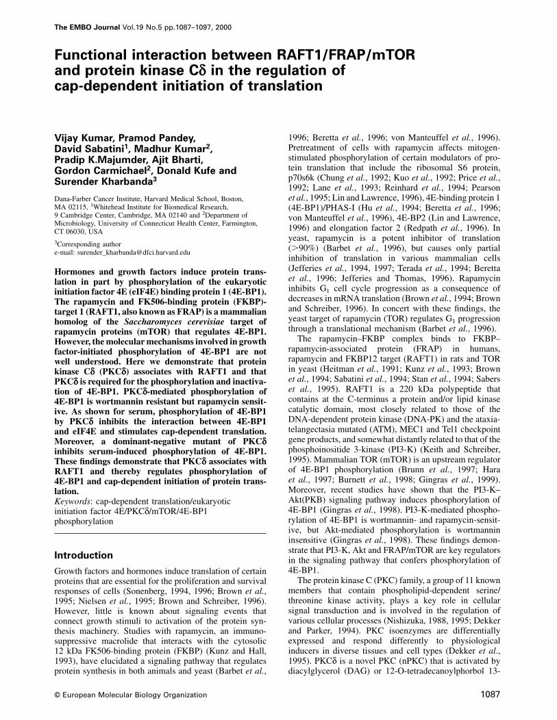
Fig. 1. Association of PKCδ with RAFT1. (A) Total lysates fromPKCδ in vivo and directly phosphorylates the activation293T cells were subjected to immunoprecipitation with anti-PKCδ orloop of PKCδ in vitro (Le Good et al., 1998). The pre-immune rabbit serum (PIRS). The precipitates and total lysate
findings that serum-stimulated phosphorylation of PKCδ were separated by SDS–PAGE and analyzed by immunoblotting withanti-RAFT1. (B) 293T cells were transiently transfected with HA-is enhanced by coexpression of PDK1 and that thisRAFT1. Total lysates were subjected to immunoprecipitation withresponse is also sensitive to wortmannin indicate thatanti-PKCδ, anti-HA or PIRS. The precipitates and the lysates werePKCδ plays a key role in regulating the serum-inducedanalyzed by immunoblotting with anti-HA. (C) 293T cells were
signaling pathway and acts downstream to PI3-K. More- transiently transfected with HA-RAFT1. Lysates were subjected toover, a recent study has shown that, like PDK1, phospho- immunoprecipitation with anti-HA. The precipitates and lysates were
analyzed by immunoblotting with anti-PKCδ.rylation of PKCδ is independently regulated by a pathwayinvolving mTOR/RAFT1 (Parekh et al., 1999). Takentogether, these findings demonstrate that serum-inducedphosphorylation and activation of PKCδ is regulated bymultiple upstream effectors.
Since phosphorylation and activation of PKCδ by serumand insulin is mediated by PI3-K→PDK1 signaling, wesought to determine whether PKCδ contributes to theregulation of the FRAP/mTOR→→4E-BP1 phosphoryl-ation pathway and thereby mediates cap-dependent transla-tion. The results demonstrate that PKCδ interacts withFRAP/mTOR and is required for phosphorylation of4E-BP1 in vivo. The functional significance of the PKCδ–mTOR interaction is supported by the finding that PKCδstimulates eukaryotic initiation factor 4E (eIF4E)-
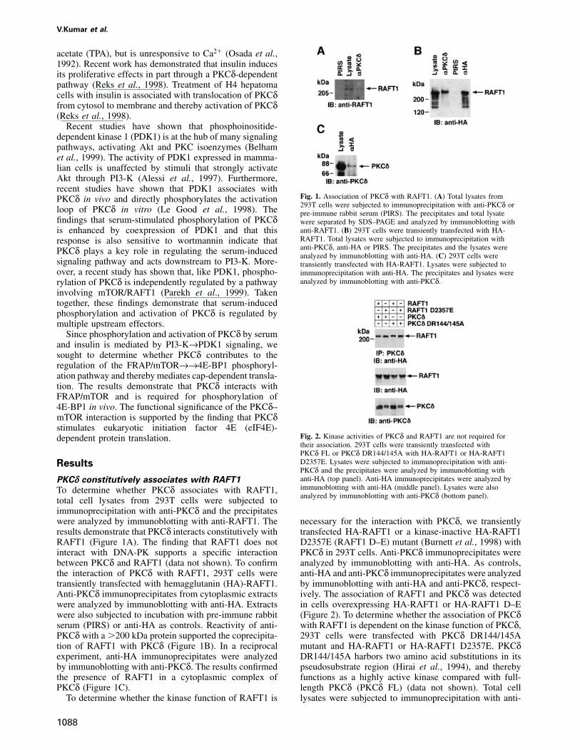
Fig. 2. Kinase activities of PKCδ and RAFT1 are not required fordependent protein translation.their association. 293T cells were transiently transfected withPKCδ FL or PKCδ DR144/145A with HA-RAFT1 or HA-RAFT1D2357E. Lysates were subjected to immunoprecipitation with anti-ResultsPKCδ and the precipitates were analyzed by immunoblotting withanti-HA (top panel). Anti-HA immunoprecipitates were analyzed byPKCδ constitutively associates with RAFT1immunoblotting with anti-HA (middle panel). Lysates were alsoTo determine whether PKCδ associates with RAFT1,analyzed by immunoblotting with anti-PKCδ (bottom panel).total cell lysates from 293T cells were subjected to
immunoprecipitation with anti-PKCδ and the precipitateswere analyzed by immunoblotting with anti-RAFT1. The necessary for the interaction with PKCδ, we transiently
transfected HA-RAFT1 or a kinase-inactive HA-RAFT1results demonstrate that PKCδ interacts constitutively withRAFT1 (Figure 1A). The finding that RAFT1 does not D2357E (RAFT1 D–E) mutant (Burnett et al., 1998) with
PKCδ in 293T cells. Anti-PKCδ immunoprecipitates wereinteract with DNA-PK supports a specific interactionbetween PKCδ and RAFT1 (data not shown). To confirm analyzed by immunoblotting with anti-HA. As controls,
anti-HA and anti-PKCδ immunoprecipitates were analyzedthe interaction of PKCδ with RAFT1, 293T cells weretransiently transfected with hemagglutanin (HA)-RAFT1. by immunoblotting with anti-HA and anti-PKCδ, respect-
ively. The association of RAFT1 and PKCδ was detectedAnti-PKCδ immunoprecipitates from cytoplasmic extractswere analyzed by immunoblotting with anti-HA. Extracts in cells overexpressing HA-RAFT1 or HA-RAFT1 D–E
(Figure 2). To determine whether the association of PKCδwere also subjected to incubation with pre-immune rabbitserum (PIRS) or anti-HA as controls. Reactivity of anti- with RAFT1 is dependent on the kinase function of PKCδ,
293T cells were transfected with PKCδ DR144/145APKCδ with a �200 kDa protein supported the coprecipita-tion of RAFT1 with PKCδ (Figure 1B). In a reciprocal mutant and HA-RAFT1 or HA-RAFT1 D2357E. PKCδ
DR144/145A harbors two amino acid substitutions in itsexperiment, anti-HA immunoprecipitates were analyzedby immunoblotting with anti-PKCδ. The results confirmed pseudosubstrate region (Hirai et al., 1994), and thereby
functions as a highly active kinase compared with full-the presence of RAFT1 in a cytoplasmic complex ofPKCδ (Figure 1C). length PKCδ (PKCδ FL) (data not shown). Total cell
lysates were subjected to immunoprecipitation with anti-To determine whether the kinase function of RAFT1 is
1088
Regulation of cap-dependent translation by PKCδ
PKCδ and analyzed by immunoblotting with anti-HA.The results demonstrate that PKCδ DR144/145A associ-ates both with wild-type as well as the D2357E mutantof RAFT1 (Figure 2). Taken together, these findingsindicate that the interaction of PKCδ and RAFT1 isindependent of the kinase functions of RAFT1 and PKCδ.
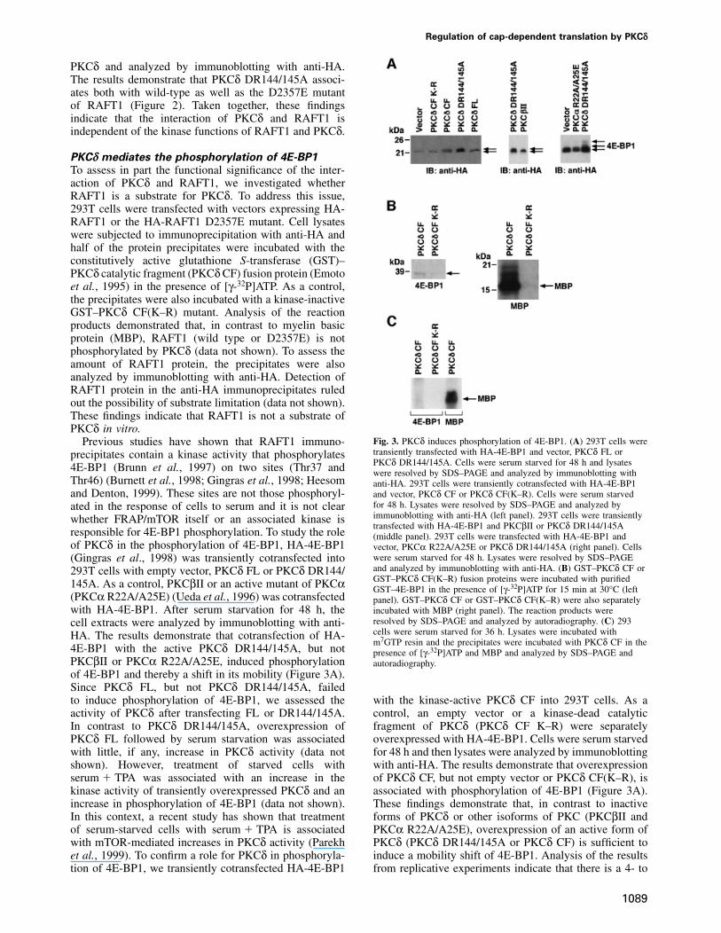
PKCδ mediates the phosphorylation of 4E-BP1
To assess in part the functional significance of the inter-action of PKCδ and RAFT1, we investigated whetherRAFT1 is a substrate for PKCδ. To address this issue,293T cells were transfected with vectors expressing HA-RAFT1 or the HA-RAFT1 D2357E mutant. Cell lysateswere subjected to immunoprecipitation with anti-HA andhalf of the protein precipitates were incubated with theconstitutively active glutathione S-transferase (GST)–PKCδ catalytic fragment (PKCδ CF) fusion protein (Emotoet al., 1995) in the presence of [γ-32P]ATP. As a control,the precipitates were also incubated with a kinase-inactiveGST–PKCδ CF(K–R) mutant. Analysis of the reactionproducts demonstrated that, in contrast to myelin basicprotein (MBP), RAFT1 (wild type or D2357E) is notphosphorylated by PKCδ (data not shown). To assess theamount of RAFT1 protein, the precipitates were alsoanalyzed by immunoblotting with anti-HA. Detection ofRAFT1 protein in the anti-HA immunoprecipitates ruledout the possibility of substrate limitation (data not shown).These findings indicate that RAFT1 is not a substrate ofPKCδ in vitro.
Fig. 3. PKCδ induces phosphorylation of 4E-BP1. (A) 293T cells werePrevious studies have shown that RAFT1 immuno-transiently transfected with HA-4E-BP1 and vector, PKCδ FL orprecipitates contain a kinase activity that phosphorylatesPKCδ DR144/145A. Cells were serum starved for 48 h and lysates4E-BP1 (Brunn et al., 1997) on two sites (Thr37 and were resolved by SDS–PAGE and analyzed by immunoblotting with
Thr46) (Burnett et al., 1998; Gingras et al., 1998; Heesom anti-HA. 293T cells were transiently cotransfected with HA-4E-BP1and Denton, 1999). These sites are not those phosphoryl- and vector, PKCδ CF or PKCδ CF(K–R). Cells were serum starved
for 48 h. Lysates were resolved by SDS–PAGE and analyzed byated in the response of cells to serum and it is not clearimmunoblotting with anti-HA (left panel). 293T cells were transientlywhether FRAP/mTOR itself or an associated kinase istransfected with HA-4E-BP1 and PKCβII or PKCδ DR144/145A
responsible for 4E-BP1 phosphorylation. To study the role (middle panel). 293T cells were transfected with HA-4E-BP1 andof PKCδ in the phosphorylation of 4E-BP1, HA-4E-BP1 vector, PKCα R22A/A25E or PKCδ DR144/145A (right panel). Cells
were serum starved for 48 h. Lysates were resolved by SDS–PAGE(Gingras et al., 1998) was transiently cotransfected intoand analyzed by immunoblotting with anti-HA. (B) GST–PKCδ CF or293T cells with empty vector, PKCδ FL or PKCδ DR144/GST–PKCδ CF(K–R) fusion proteins were incubated with purified145A. As a control, PKCβII or an active mutant of PKCα GST–4E-BP1 in the presence of [γ-32P]ATP for 15 min at 30°C (left
(PKCα R22A/A25E) (Ueda et al., 1996) was cotransfected panel). GST–PKCδ CF or GST–PKCδ CF(K–R) were also separatelywith HA-4E-BP1. After serum starvation for 48 h, the incubated with MBP (right panel). The reaction products were
resolved by SDS–PAGE and analyzed by autoradiography. (C) 293cell extracts were analyzed by immunoblotting with anti-cells were serum starved for 36 h. Lysates were incubated withHA. The results demonstrate that cotransfection of HA-m7GTP resin and the precipitates were incubated with PKCδ CF in the
4E-BP1 with the active PKCδ DR144/145A, but not presence of [γ-32P]ATP and MBP and analyzed by SDS–PAGE andPKCβII or PKCα R22A/A25E, induced phosphorylation autoradiography.of 4E-BP1 and thereby a shift in its mobility (Figure 3A).Since PKCδ FL, but not PKCδ DR144/145A, failedto induce phosphorylation of 4E-BP1, we assessed the with the kinase-active PKCδ CF into 293T cells. As a
control, an empty vector or a kinase-dead catalyticactivity of PKCδ after transfecting FL or DR144/145A.In contrast to PKCδ DR144/145A, overexpression of fragment of PKCδ (PKCδ CF K–R) were separately
overexpressed with HA-4E-BP1. Cells were serum starvedPKCδ FL followed by serum starvation was associatedwith little, if any, increase in PKCδ activity (data not for 48 h and then lysates were analyzed by immunoblotting
with anti-HA. The results demonstrate that overexpressionshown). However, treatment of starved cells withserum � TPA was associated with an increase in the of PKCδ CF, but not empty vector or PKCδ CF(K–R), is
associated with phosphorylation of 4E-BP1 (Figure 3A).kinase activity of transiently overexpressed PKCδ and anincrease in phosphorylation of 4E-BP1 (data not shown). These findings demonstrate that, in contrast to inactive
forms of PKCδ or other isoforms of PKC (PKCβII andIn this context, a recent study has shown that treatmentof serum-starved cells with serum � TPA is associated PKCα R22A/A25E), overexpression of an active form of
PKCδ (PKCδ DR144/145A or PKCδ CF) is sufficient towith mTOR-mediated increases in PKCδ activity (Parekhet al., 1999). To confirm a role for PKCδ in phosphoryla- induce a mobility shift of 4E-BP1. Analysis of the results
from replicative experiments indicate that there is a 4- totion of 4E-BP1, we transiently cotransfected HA-4E-BP1
1089
V.Kumar et al.
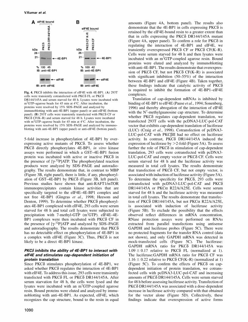
amounts (Figure 4A, bottom panel). The results alsodemonstrate that the 4E-BP1 in cells expressing PKCδ isretained by the eIF4E-bound resin to a greater extent thanthat in cells expressing the PKCδ DR144/145A mutant(Figure 4A, upper panel). To confirm a role for PKCδ inregulating the interaction of 4E-BP1 and eIF4E, wetransiently overexpressed PKCδ CF or PKCδ CF(K–R).Cells were serum starved for 48 h and then lysates wereincubated with an m7GTP-coupled agarose resin. Boundproteins were eluted and analyzed by immunoblottingwith anti-4E-BP1. The results demonstrate that overexpres-sion of PKCδ CF, but not PKCδ CF(K–R) is associatedwith significant inhibition (50–55%) of the interactionbetween 4E-BP1 and eIF4E (Figure 4B). Taken together,these findings indicate that catalytic activity of PKCδis required to inhibit the formation of 4E-BP1–eIF4E
Fig. 4. PKCδ inhibits the interaction of eIF4E with 4E-BP1. (A) 293T complexes.cells were transiently cotransfected with PKCδ FL or PKCδTranslation of cap-dependent mRNAs is inhibited byDR144/145A and serum starved for 48 h. Lysates were incubated with
m7GTP–agarose beads for 45 min at 4°C. After incubation, the binding of 4E-BP1 to eIF4E (Pause et al., 1994; Sonenberg,proteins were resolved by 15% SDS–PAGE and analyzed by 1996) and thereby abrogation of the interaction of eIF4Eimmunoblotting with anti-4E-BP1 (upper panel) or anti-eIF4E (bottom with the N7-methylguanosine cap structure. To determinepanel). (B) 293T cells were transiently transfected with PKCδ CF or
whether PKCδ regulates cap-dependent translation, wePKCδ CF(K–R) and serum starved for 48 h. Lysates were incubatedtransfected 293T cells with the pcDNA3-LUC-pol-CATwith m7GTP–agarose beads for 45 min at 4°C. After incubation, the
proteins were resolved by 15% SDS–PAGE and analyzed by immuno- vector that exhibits cap-dependent translation of luciferaseblotting with anti-4E-BP1 (upper panel) or anti-eIF4E (bottom panel). (LUC) (Craig et al., 1998). Cotransfection of pcDNA3-
LUC-pol-CAT with PKCβII had no effect on luciferaseactivity. In contrast, PKCδ DR144/145A induced the5-fold increase in phosphorylation of 4E-BP1 by over-
expressing active mutants of PKCδ. To assess whether expression of luciferase by �2-fold (Figure 5A). To assessfurther the role of PKCδ in stimulation of cap-dependentPKCδ directly phosphorylates 4E-BP1, in vitro kinase
assays were performed in which a GST–4E-BP1 fusion translation, 293 cells were cotransfected with pcDNA3-LUC-pol-CAT and empty vector or PKCδ CF. Cells wereprotein was incubated with active or inactive PKCδ in
the presence of [γ-32P]ATP. The phosphorylated reaction serum starved for 48 h and the luciferase activity wasmeasured in total cell lysates. The results demonstrateproducts were analyzed by SDS–PAGE and autoradio-
graphy. The results demonstrate that, in contrast to MBP that transfection of PKCδ CF, but not empty vector, isassociated with induction of luciferase activity (Figure 5A).(Figure 3B, right panel), there is little, if any, phosphoryl-
ation of GST–4E-BP1 by PKCδ (Figure 3B, left panel). To determine the specificity for PKCδ, 293 cells werecotransfected with pcDNA3-LUC-pol-CAT and PKCδPrevious studies have shown that anti-RAFT1/mTOR
immunoprecipitates contain kinase activities that are DR144/145A or PKCα R22A/A25E. Cells were serumstarved for 48 h and the luciferase activity was measuredspecifically targeted to the eIF4E–4E-BP1 complex and
not free 4E-BP1 (Gingras et al., 1998; Heesom and in total cell lysates. The results demonstrate that transfec-tion of PKCδ DR144/145A, but not PKCα R22A/A25E,Denton, 1999). To determine whether PKCδ phosphoryl-
ates 4E-BP1 complexed with eIF4E, 293 cells were serum is associated with induction of luciferase activity(Figure 5B). To exclude the possibility that the effectsstarved for 48 h and total cell lysates were subjected to
precipitation with 7-methyl-GTP (m7GTP). eIF4E–4E- observed reflect differences in mRNA concentration,RNase protection assays were performed on RNAsBP1 complexes were then incubated with PKCδ CF in
the presence of [γ-32P]ATP and analyzed by SDS–PAGE extracted from parallel transfections using antisenseGAPDH and luciferase probes (Figure 5C). There wereand autoradiography. The results demonstrate that PKCδ
has no detectable effect on phosphorylation of 4E-BP1 in no protected fragments for the transfer RNA control (datanot shown), and only GAPDH mRNA was detected ina complex with eIF4E (Figure 3C). Thus, PKCδ is not
likely to be a direct 4E-BP1 kinase. mock-transfected cells (Figure 5C). The luciferase:GAPDH mRNA ratio for PKCδ DR144/145A was1.09 � 0.17 relative to PKCδ FL (normalized at 1).PKCδ inhibits the ability of 4E-BP1 to interact with
eIF4E and stimulates cap-dependent initiation of The luciferase:GAPDH mRNA ratio for PKCδ CF was1.16 � 0.22 relative to PKCδ CF(K–R) (normalized at 1)protein translation
Since PKCδ stimulates phosphorylation of 4E-BP1, we (Figure 5C). To confirm the effects of PKCδ on cap-dependent initiation of protein translation, we cotrans-asked whether PKCδ regulates the interaction of 4E-BP1
with eIF4E. To address this issue, 293 cells were transiently fected cells with pcDNA3-LUC-pol-CAT and increasingamounts of PKCδ DR144/145A. Cells were serum starvedtransfected with PKCδ FL or PKCδ DR144/145A. After
serum starvation for 48 h, the cells were lysed and the for 48 h before assessing luciferase activity. Transfection ofPKCδ DR144/145A was associated with a dose-dependentlysates were incubated with an m7GTP-coupled agarose
resin. Bound proteins were eluted and analyzed by immu- increase in luciferase activity compared with that obtainedfor the vector alone (Figure 5D). Collectively, thesenoblotting with anti-4E-BP1. As expected, eIF4E, which
recognizes the cap structure, bound to the resin in equal findings indicate that overexpression of active forms
1090
Regulation of cap-dependent translation by PKCδ
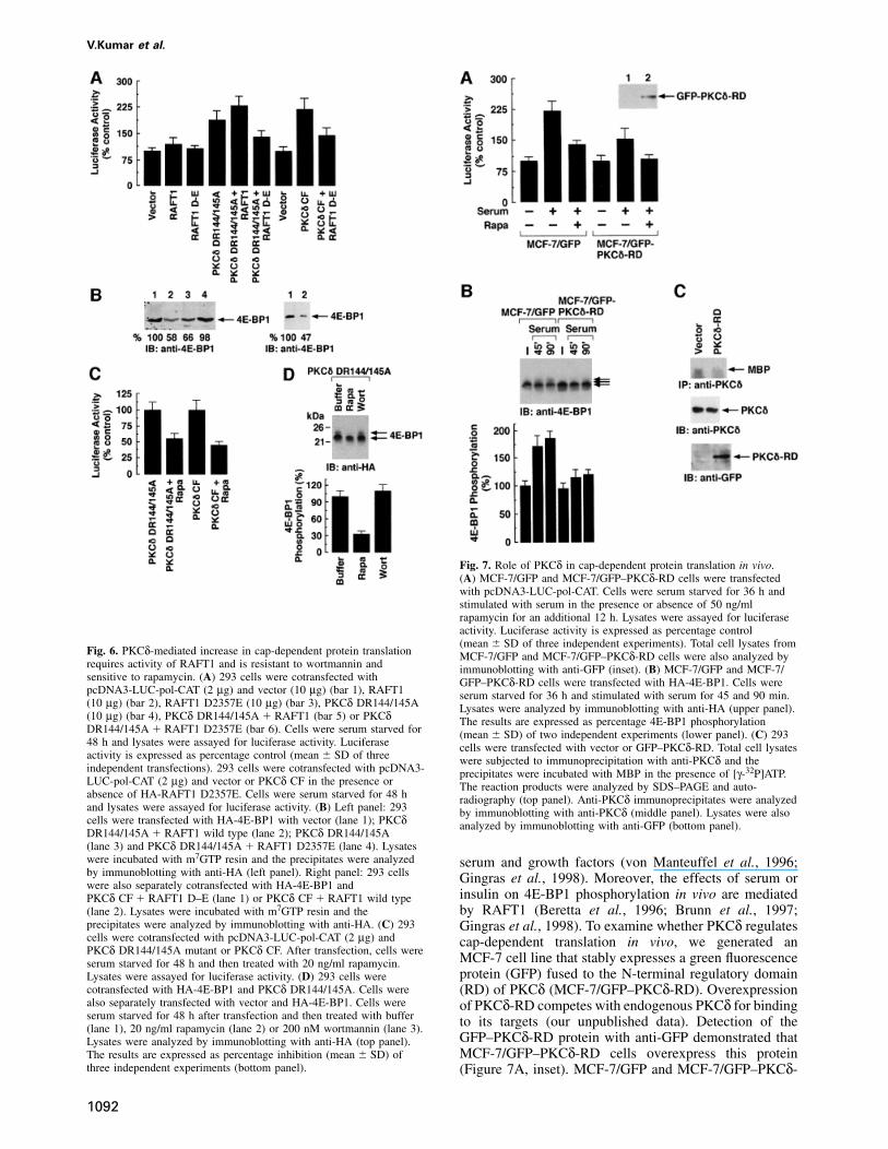
starved for 48 h and total lysates were analyzed byluciferase activity assays. The results demonstrate thatoverexpression of PKCδ DR144/145A with RAFT1increases cap-dependent translation by ~2.5-fold comparedwith vector (Figure 6A). Importantly, this increase inprotein translation was inhibited by �50% in cells co-expressing PKCδ DR144/145A and RAFT1 D2357E(Figure 6A). Similar experiments were performed in whichcells were transfected with PKCδ CF with or withoutRAFT1 D2357E. Cells were cotransfected with pcDNA3-LUC-pol-CAT, serum starved for 48 h and luciferaseactivity was measured in total lysates. The results demon-strate that the PKCδ CF-mediated increase in luciferaseactivity was significantly inhibited by overexpression ofRAFT1 D2357E (Figure 6A). To confirm a role forPKCδ–RAFT1 complexes in regulating the interaction of4E-BP1 and eIF4E, we transiently cotransfected PKCδDR144/145A, PKCδ CF or PKCδ CF(K–R) with RAFT1or RAFT1 D2357E. Cells were serum starved for 48 hand lysates were incubated with m7GTP-coupled agaroseresin. Bound proteins were eluted and analyzed byimmunoblotting with anti-4E-BP1. The results demon-strate that, in contrast to RAFT1 D2357E, overexpressionof PKCδ DR144/145A or PKCδ CF with RAFT1 wildtype is associated with significant inhibition (45–55%) of
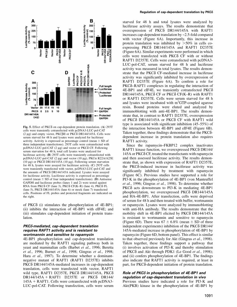
Fig. 5. Effect of PKCδ on cap-dependent protein translation. (A) 293T the interaction between 4E-BP1 and eIF4E (Figure 6B).cells were transiently cotransfected with pcDNA3-LUC-pol-CAT
Taken together, these findings demonstrate that the PKCδ-(2 µg) and empty vector, PKCβII or PKCδ DR144/145A. Cells weredependent increase in protein translation also requiresserum starved for 48 h and lysates were analyzed for luciferase
activity. Activity is expressed as percentage control (mean � SD of RAFT1 activity.three independent transfections). 293T cells were cotransfected with Since the rapamycin–FKBP12 complex inactivatespcDNA3-LUC-pol-CAT (2 µg) and vector or PKCδ CF. Following RAFT1 kinase function, we overexpressed PKCδ DR144/serum starvation for 48 h, total cell lysates were analyzed for
145A or PKCδ CF, treated the transfectants with rapamycinluciferase activity. (B) 293T cells were transiently cotransfected withpcDNA3-LUC-pol-CAT (2 µg) and vector (10 µg), PKCα R22A/A25E and then assessed luciferase activity. The results demon-(10 µg) or PKCδ DR144/145A (10 µg). Following serum starvation strate that, as shown with expression of RAFT1 D2357E,for 48 h, lysates were assayed for luciferase activity. (C) 293T cells the PKCδ-induced increase in luciferase activity waswere transiently transfected with vector, pcDNA3-LUC-pol-CAT and
significantly inhibited by treatment with rapamycinthe amounts of PKCδ DR144/145A indicated. Lysates were assayed(Figure 6C). Previous studies have supported a role forfor luciferase activity. Luciferase activity is expressed as percentage
control (mean � SD of four independent transfections). (D) Antisense PI3-K in the phosphorylation of 4E-BP1 (von ManteuffelGAPDH and luciferase probes (lanes 1 and 2) were tested against et al., 1996; Gingras et al., 1998). To determine whetherRNA from PKCδ CF (lane 3), PKCδ CF(K–R) (lane 4), PKCδ FL PKCδ acts downstream to PI3-K in mediating 4E-BP1(lane 5), PKCδ DR144/145A (lane 6) or mock (lane 7) transfected
phosphorylation, we overexpressed PKCδ DR144/145Acells. Positions of FL probes and protected fragments are indicated onthe right. and HA-4E-BP1. After transfection, cells were deprived
of serum for 48 h and then treated with buffer, wortmanninor rapamycin. Lysates were analyzed by immunoblottingof PKCδ (i) stimulates the phosphorylation of 4E-BP1;
(ii) inhibits the interaction of 4E-BP1 with eIF4E; and with anti-HA antibody. The results demonstrate that themobility shift in 4E-BP1 elicited by PKCδ DR144/145A(iii) stimulates cap-dependent initiation of protein trans-
lation. is resistant to wortmannin and sensitive to rapamycin(Figure 6D). There was 67 � 6.8% (mean � SD of threeindependent experiments) inhibition of the PKCδ DR144/PKCδ-mediated, cap-dependent translation
requires RAFT1 activity and is resistant to 145A-mediated increase in phosphorylation of 4E-BP1 byrapamycin (Figure 6D, bottom panel). This effect is similarwortmannin and sensitive to rapamycin
4E-BP1 phosphorylation and cap-dependent translation to that observed previously for Akt (Gingras et al., 1998).Taken together, these findings support a pathway thatare mediated by the RAFT1 signaling pathway both in
yeast and mammalian cells (Barbet et al., 1996; Beretta (i) involves activation of PI3-K and thereby stimulationof PKCδ and Akt through PDK1 (Le Good et al., 1998);et al., 1996; Burnet et al., 1998; Gingras et al., 1998;
Hara et al., 1997). To determine whether a dominant- and (ii) confers phosphorylation of 4E-BP1. The findingsalso indicate that RAFT1 activity is required, at least innegative mutant of RAFT1 (RAFT1 D2357E) inhibits
PKCδ DR144/145A-mediated increases in cap-dependent part, for PKCδ-dependent initiation of protein translation.translation, cells were transfected with vector, RAFT1wild type, RAFT1 D2357E, PKCδ DR144/145A, PKCδ Role of PKCδ in phosphorylation of 4E-BP1 and
regulation of cap-dependent translation in vivoDR144/145A � RAFT1 D2357E or PKCδ DR144/145A � RAFT1. Cells were cotransfected with pcDNA3- Previous studies have indicated a role for PI3-K and
Akt(PKB) kinase in the phosphorylation of 4E-BP1 byLUC-pol-CAT. Following transfection, cells were serum
1091
V.Kumar et al.
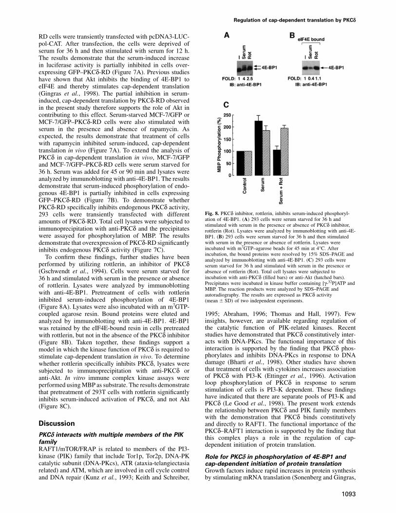
Fig. 7. Role of PKCδ in cap-dependent protein translation in vivo.(A) MCF-7/GFP and MCF-7/GFP–PKCδ-RD cells were transfectedwith pcDNA3-LUC-pol-CAT. Cells were serum starved for 36 h andstimulated with serum in the presence or absence of 50 ng/mlrapamycin for an additional 12 h. Lysates were assayed for luciferaseactivity. Luciferase activity is expressed as percentage control(mean � SD of three independent experiments). Total cell lysates from
Fig. 6. PKCδ-mediated increase in cap-dependent protein translation MCF-7/GFP and MCF-7/GFP–PKCδ-RD cells were also analyzed byrequires activity of RAFT1 and is resistant to wortmannin and immunoblotting with anti-GFP (inset). (B) MCF-7/GFP and MCF-7/sensitive to rapamycin. (A) 293 cells were cotransfected with GFP–PKCδ-RD cells were transfected with HA-4E-BP1. Cells werepcDNA3-LUC-pol-CAT (2 µg) and vector (10 µg) (bar 1), RAFT1 serum starved for 36 h and stimulated with serum for 45 and 90 min.(10 µg) (bar 2), RAFT1 D2357E (10 µg) (bar 3), PKCδ DR144/145A Lysates were analyzed by immunoblotting with anti-HA (upper panel).(10 µg) (bar 4), PKCδ DR144/145A � RAFT1 (bar 5) or PKCδ The results are expressed as percentage 4E-BP1 phosphorylationDR144/145A � RAFT1 D2357E (bar 6). Cells were serum starved for (mean � SD) of two independent experiments (lower panel). (C) 29348 h and lysates were assayed for luciferase activity. Luciferase cells were transfected with vector or GFP–PKCδ-RD. Total cell lysatesactivity is expressed as percentage control (mean � SD of three were subjected to immunoprecipitation with anti-PKCδ and theindependent transfections). 293 cells were cotransfected with pcDNA3- precipitates were incubated with MBP in the presence of [γ-32P]ATP.LUC-pol-CAT (2 µg) and vector or PKCδ CF in the presence or The reaction products were analyzed by SDS–PAGE and auto-absence of HA-RAFT1 D2357E. Cells were serum starved for 48 h radiography (top panel). Anti-PKCδ immunoprecipitates were analyzedand lysates were assayed for luciferase activity. (B) Left panel: 293 by immunoblotting with anti-PKCδ (middle panel). Lysates were alsocells were transfected with HA-4E-BP1 with vector (lane 1); PKCδ analyzed by immunoblotting with anti-GFP (bottom panel).DR144/145A � RAFT1 wild type (lane 2); PKCδ DR144/145A(lane 3) and PKCδ DR144/145A � RAFT1 D2357E (lane 4). Lysateswere incubated with m7GTP resin and the precipitates were analyzed serum and growth factors (von Manteuffel et al., 1996;by immunoblotting with anti-HA (left panel). Right panel: 293 cells Gingras et al., 1998). Moreover, the effects of serum orwere also separately cotransfected with HA-4E-BP1 and
insulin on 4E-BP1 phosphorylation in vivo are mediatedPKCδ CF � RAFT1 D–E (lane 1) or PKCδ CF � RAFT1 wild typeby RAFT1 (Beretta et al., 1996; Brunn et al., 1997;(lane 2). Lysates were incubated with m7GTP resin and the
precipitates were analyzed by immunoblotting with anti-HA. (C) 293 Gingras et al., 1998). To examine whether PKCδ regulatescells were cotransfected with pcDNA3-LUC-pol-CAT (2 µg) and cap-dependent translation in vivo, we generated anPKCδ DR144/145A mutant or PKCδ CF. After transfection, cells were
MCF-7 cell line that stably expresses a green fluorescenceserum starved for 48 h and then treated with 20 ng/ml rapamycin.protein (GFP) fused to the N-terminal regulatory domainLysates were assayed for luciferase activity. (D) 293 cells were
cotransfected with HA-4E-BP1 and PKCδ DR144/145A. Cells were (RD) of PKCδ (MCF-7/GFP–PKCδ-RD). Overexpressionalso separately transfected with vector and HA-4E-BP1. Cells were of PKCδ-RD competes with endogenous PKCδ for bindingserum starved for 48 h after transfection and then treated with buffer to its targets (our unpublished data). Detection of the(lane 1), 20 ng/ml rapamycin (lane 2) or 200 nM wortmannin (lane 3).
GFP–PKCδ-RD protein with anti-GFP demonstrated thatLysates were analyzed by immunoblotting with anti-HA (top panel).MCF-7/GFP–PKCδ-RD cells overexpress this proteinThe results are expressed as percentage inhibition (mean � SD) of
three independent experiments (bottom panel). (Figure 7A, inset). MCF-7/GFP and MCF-7/GFP–PKCδ-
1092
Regulation of cap-dependent translation by PKCδ
RD cells were transiently transfected with pcDNA3-LUC-pol-CAT. After transfection, the cells were deprived ofserum for 36 h and then stimulated with serum for 12 h.The results demonstrate that the serum-induced increasein luciferase activity is partially inhibited in cells over-expressing GFP–PKCδ-RD (Figure 7A). Previous studieshave shown that Akt inhibits the binding of 4E-BP1 toeIF4E and thereby stimulates cap-dependent translation(Gingras et al., 1998). The partial inhibition in serum-induced, cap-dependent translation by PKCδ-RD observedin the present study therefore supports the role of Akt incontributing to this effect. Serum-starved MCF-7/GFP orMCF-7/GFP–PKCδ-RD cells were also stimulated withserum in the presence and absence of rapamycin. Asexpected, the results demonstrate that treatment of cellswith rapamycin inhibited serum-induced, cap-dependenttranslation in vivo (Figure 7A). To extend the analysis ofPKCδ in cap-dependent translation in vivo, MCF-7/GFPand MCF-7/GFP–PKCδ-RD cells were serum starved for36 h. Serum was added for 45 or 90 min and lysates wereanalyzed by immunoblotting with anti-4E-BP1. The resultsdemonstrate that serum-induced phosphorylation of endo-genous 4E-BP1 is partially inhibited in cells expressingGFP–PKCδ-RD (Figure 7B). To demonstrate whetherPKCδ-RD specifically inhibits endogenous PKCδ activity,
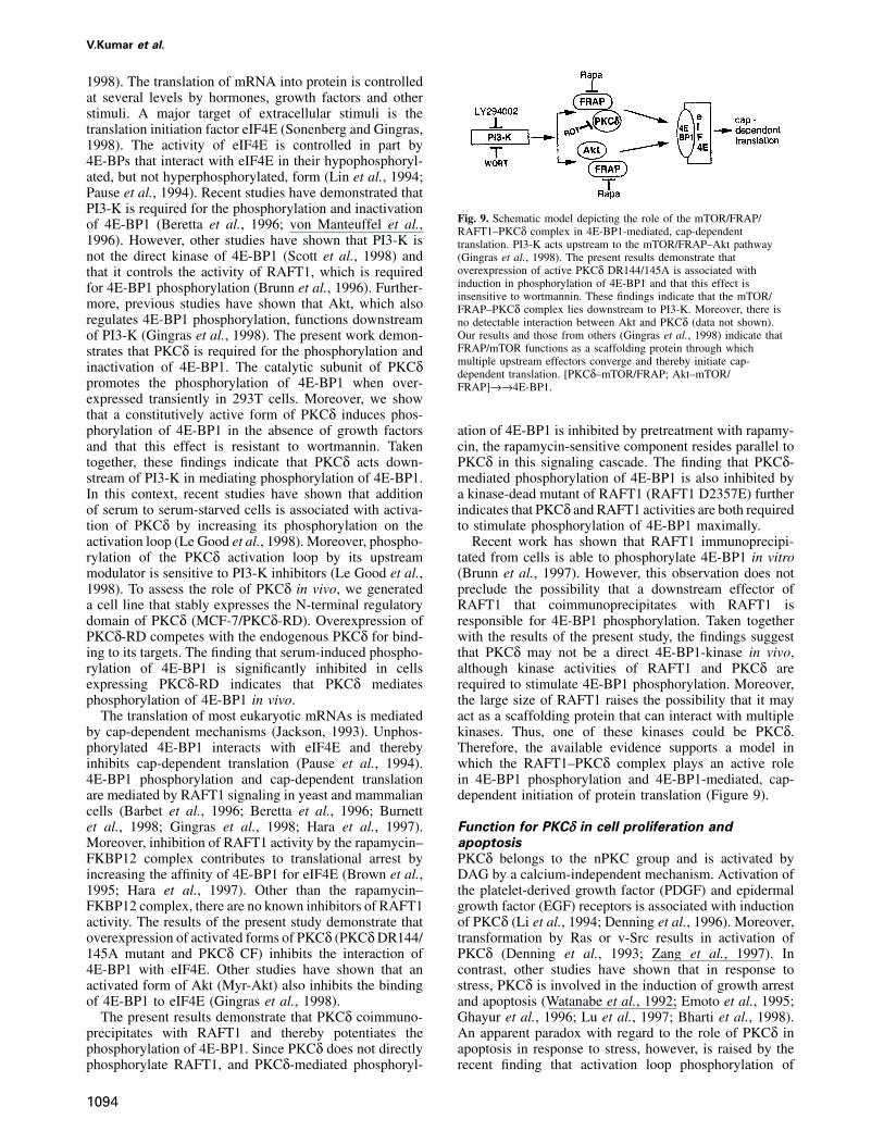
Fig. 8. PKCδ inhibitor, rottlerin, inhibits serum-induced phosphoryl-293 cells were transiently transfected with differentation of 4E-BP1. (A) 293 cells were serum starved for 36 h andamounts of PKCδ-RD. Total cell lysates were subjected tostimulated with serum in the presence or absence of PKCδ inhibitor,
immunoprecipitation with anti-PKCδ and the precipitates rottlerin (Rot). Lysates were analyzed by immunoblotting with anti-4E-were assayed for phosphorylation of MBP. The results BP1. (B) 293 cells were serum starved for 36 h and then stimulated
with serum in the presence or absence of rottlerin. Lysates weredemonstrate that overexpression of PKCδ-RD significantlyincubated with m7GTP–agarose beads for 45 min at 4°C. Afterinhibits endogenous PKCδ activity (Figure 7C).incubation, the bound proteins were resolved by 15% SDS–PAGE andTo confirm these findings, further studies have been analyzed by immunoblotting with anti-4E-BP1. (C) 293 cells were
performed by utilizing rottlerin, an inhibitor of PKCδ serum starved for 36 h and stimulated with serum in the presence orabsence of rottlerin (Rot). Total cell lysates were subjected to(Gschwendt et al., 1994). Cells were serum starved forincubation with anti-PKCδ (filled bars) or anti-Akt (hatched bars).36 h and stimulated with serum in the presence or absencePrecipitates were incubated in kinase buffer containing [γ-32P]ATP andof rottlerin. Lysates were analyzed by immunoblottingMBP. The reaction products were analyzed by SDS–PAGE and
with anti-4E-BP1. Pretreatment of cells with rottlerin autoradiography. The results are expressed as PKCδ activityinhibited serum-induced phosphorylation of 4E-BP1 (mean � SD) of two independent experiments.(Figure 8A). Lysates were also incubated with an m7GTP-coupled agarose resin. Bound proteins were eluted and 1995; Abraham, 1996; Thomas and Hall, 1997). Few
insights, however, are available regarding regulation ofanalyzed by immunoblotting with anti-4E-BP1. 4E-BP1the catalytic function of PIK-related kinases. Recentwas retained by the eIF4E-bound resin in cells pretreatedstudies have demonstrated that PKCδ constitutively inter-with rottlerin, but not in the absence of the PKCδ inhibitoracts with DNA-PKcs. The functional importance of this(Figure 8B). Taken together, these findings support ainteraction is supported by the finding that PKCδ phos-model in which the kinase function of PKCδ is required tophorylates and inhibits DNA-PKcs in response to DNAstimulate cap-dependent translation in vivo. To determinedamage (Bharti et al., 1998). Other studies have shownwhether rottlerin specifically inhibits PKCδ, lysates werethat treatment of cells with cytokines increases associationsubjected to immunoprecipitation with anti-PKCδ orof PKCδ with PI3-K (Ettinger et al., 1996). Activationanti-Akt. In vitro immune complex kinase assays wereloop phosphorylation of PKCδ in response to serumperformed using MBP as substrate. The results demonstratestimulation of cells is PI3-K dependent. These findingsthat pretreatment of 293T cells with rottlerin significantlyhave indicated that there are separate pools of PI3-K andinhibits serum-induced activation of PKCδ, and not AktPKCδ (Le Good et al., 1998). The present work extends(Figure 8C).the relationship between PKCδ and PIK family memberswith the demonstration that PKCδ binds constitutively
Discussion and directly to RAFT1. The functional importance of thePKCδ–RAFT1 interaction is supported by the finding that
PKCδ interacts with multiple members of the PIK this complex plays a role in the regulation of cap-family dependent initiation of protein translation.RAFT1/mTOR/FRAP is related to members of the PI3-kinase (PIK) family that include Tor1p, Tor2p, DNA-PK Role for PKCδ in phosphorylation of 4E-BP1 andcatalytic subunit (DNA-PKcs), ATR (ataxia-telangiectasia cap-dependent initiation of protein translationrelated) and ATM, which are involved in cell cycle control Growth factors induce rapid increases in protein synthesis
by stimulating mRNA translation (Sonenberg and Gingras,and DNA repair (Kunz et al., 1993; Keith and Schreiber,
1093
V.Kumar et al.
1998). The translation of mRNA into protein is controlledat several levels by hormones, growth factors and otherstimuli. A major target of extracellular stimuli is thetranslation initiation factor eIF4E (Sonenberg and Gingras,1998). The activity of eIF4E is controlled in part by4E-BPs that interact with eIF4E in their hypophosphoryl-ated, but not hyperphosphorylated, form (Lin et al., 1994;Pause et al., 1994). Recent studies have demonstrated thatPI3-K is required for the phosphorylation and inactivation
Fig. 9. Schematic model depicting the role of the mTOR/FRAP/of 4E-BP1 (Beretta et al., 1996; von Manteuffel et al., RAFT1–PKCδ complex in 4E-BP1-mediated, cap-dependent1996). However, other studies have shown that PI3-K is translation. PI3-K acts upstream to the mTOR/FRAP–Akt pathwaynot the direct kinase of 4E-BP1 (Scott et al., 1998) and (Gingras et al., 1998). The present results demonstrate that
overexpression of active PKCδ DR144/145A is associated withthat it controls the activity of RAFT1, which is requiredinduction in phosphorylation of 4E-BP1 and that this effect isfor 4E-BP1 phosphorylation (Brunn et al., 1996). Further-insensitive to wortmannin. These findings indicate that the mTOR/
more, previous studies have shown that Akt, which also FRAP–PKCδ complex lies downstream to PI3-K. Moreover, there isregulates 4E-BP1 phosphorylation, functions downstream no detectable interaction between Akt and PKCδ (data not shown).
Our results and those from others (Gingras et al., 1998) indicate thatof PI3-K (Gingras et al., 1998). The present work demon-FRAP/mTOR functions as a scaffolding protein through whichstrates that PKCδ is required for the phosphorylation andmultiple upstream effectors converge and thereby initiate cap-inactivation of 4E-BP1. The catalytic subunit of PKCδ dependent translation. [PKCδ–mTOR/FRAP; Akt–mTOR/
promotes the phosphorylation of 4E-BP1 when over- FRAP]→→4E-BP1.expressed transiently in 293T cells. Moreover, we showthat a constitutively active form of PKCδ induces phos-phorylation of 4E-BP1 in the absence of growth factors ation of 4E-BP1 is inhibited by pretreatment with rapamy-
cin, the rapamycin-sensitive component resides parallel toand that this effect is resistant to wortmannin. Takentogether, these findings indicate that PKCδ acts down- PKCδ in this signaling cascade. The finding that PKCδ-
mediated phosphorylation of 4E-BP1 is also inhibited bystream of PI3-K in mediating phosphorylation of 4E-BP1.In this context, recent studies have shown that addition a kinase-dead mutant of RAFT1 (RAFT1 D2357E) further
indicates that PKCδ and RAFT1 activities are both requiredof serum to serum-starved cells is associated with activa-tion of PKCδ by increasing its phosphorylation on the to stimulate phosphorylation of 4E-BP1 maximally.
Recent work has shown that RAFT1 immunoprecipi-activation loop (Le Good et al., 1998). Moreover, phospho-rylation of the PKCδ activation loop by its upstream tated from cells is able to phosphorylate 4E-BP1 in vitro
(Brunn et al., 1997). However, this observation does notmodulator is sensitive to PI3-K inhibitors (Le Good et al.,1998). To assess the role of PKCδ in vivo, we generated preclude the possibility that a downstream effector of
RAFT1 that coimmunoprecipitates with RAFT1 isa cell line that stably expresses the N-terminal regulatorydomain of PKCδ (MCF-7/PKCδ-RD). Overexpression of responsible for 4E-BP1 phosphorylation. Taken together
with the results of the present study, the findings suggestPKCδ-RD competes with the endogenous PKCδ for bind-ing to its targets. The finding that serum-induced phospho- that PKCδ may not be a direct 4E-BP1-kinase in vivo,
although kinase activities of RAFT1 and PKCδ arerylation of 4E-BP1 is significantly inhibited in cellsexpressing PKCδ-RD indicates that PKCδ mediates required to stimulate 4E-BP1 phosphorylation. Moreover,
the large size of RAFT1 raises the possibility that it mayphosphorylation of 4E-BP1 in vivo.The translation of most eukaryotic mRNAs is mediated act as a scaffolding protein that can interact with multiple
kinases. Thus, one of these kinases could be PKCδ.by cap-dependent mechanisms (Jackson, 1993). Unphos-phorylated 4E-BP1 interacts with eIF4E and thereby Therefore, the available evidence supports a model in
which the RAFT1–PKCδ complex plays an active roleinhibits cap-dependent translation (Pause et al., 1994).4E-BP1 phosphorylation and cap-dependent translation in 4E-BP1 phosphorylation and 4E-BP1-mediated, cap-
dependent initiation of protein translation (Figure 9).are mediated by RAFT1 signaling in yeast and mammaliancells (Barbet et al., 1996; Beretta et al., 1996; Burnettet al., 1998; Gingras et al., 1998; Hara et al., 1997). Function for PKCδ in cell proliferation and
apoptosisMoreover, inhibition of RAFT1 activity by the rapamycin–FKBP12 complex contributes to translational arrest by PKCδ belongs to the nPKC group and is activated by
DAG by a calcium-independent mechanism. Activation ofincreasing the affinity of 4E-BP1 for eIF4E (Brown et al.,1995; Hara et al., 1997). Other than the rapamycin– the platelet-derived growth factor (PDGF) and epidermal
growth factor (EGF) receptors is associated with inductionFKBP12 complex, there are no known inhibitors of RAFT1activity. The results of the present study demonstrate that of PKCδ (Li et al., 1994; Denning et al., 1996). Moreover,
transformation by Ras or v-Src results in activation ofoverexpression of activated forms of PKCδ (PKCδ DR144/145A mutant and PKCδ CF) inhibits the interaction of PKCδ (Denning et al., 1993; Zang et al., 1997). In
contrast, other studies have shown that in response to4E-BP1 with eIF4E. Other studies have shown that anactivated form of Akt (Myr-Akt) also inhibits the binding stress, PKCδ is involved in the induction of growth arrest
and apoptosis (Watanabe et al., 1992; Emoto et al., 1995;of 4E-BP1 to eIF4E (Gingras et al., 1998).The present results demonstrate that PKCδ coimmuno- Ghayur et al., 1996; Lu et al., 1997; Bharti et al., 1998).
An apparent paradox with regard to the role of PKCδ inprecipitates with RAFT1 and thereby potentiates thephosphorylation of 4E-BP1. Since PKCδ does not directly apoptosis in response to stress, however, is raised by the
recent finding that activation loop phosphorylation ofphosphorylate RAFT1, and PKCδ-mediated phosphoryl-
1094
Regulation of cap-dependent translation by PKCδ
(Santa Cruz) antibodies. The protein precipitates were assayed for kinasePKCδ in response to serum stimulation of cells is PI3-Kactivity as described (Kharbanda et al., 1996).dependent and mediated by PDK1 (Le Good et al., 1998).
Thus, PKCδ may be functional in both apoptotic andPhosphorylation of 4E-BP1proliferative pathways. In this setting, PKCδ could repres-293T cells were transiently cotransfected with HA-4E-BP1 with pCDNA3ent a switch that determines cell fate, such that PKCδ PKCδ FL, pCDNA3 PKCδ DR144/145A, pCDNA3 PKCδ CF or
effects proliferative signals following growth-mediated pCDNA3 PKCδ CF(K–R). Cells were also separately cotransfected withHA-4E-BP1, RAFT1 and different amounts of pCDNA3 PKCδ DR144/phosphorylation and apoptotic signals as a consequence145A. Total cell lysates were analyzed by immunoblotting with anti-of stress-mediated phosphorylation. Since 4E-BP1 is aHA. 293 cells were serum starved for 36 h. Lysates were subjected tonegative regulator of cell growth (Rousseau et al., 1996),protein precipitation with m7GTP resin. The precipitates were then
it is possible that the growth-mediated effects of PKCδ incubated with active PKCδ CF in the presence of [γ-32P]ATP and kinaseare governed, at least in part, by phosphorylation of 4E- buffer at 30°C for 20 min. The reaction products were analyzed by
SDS–PAGE and autoradiography.BP1 and thereby activation of eIF4E. Because of potentialroles for PKCδ in both cell proliferation and survival, its
m7GTP and eIF4E affinity chromatographydownstream targets in the proliferative response might beTo purify endogenous eIF4E, 50 µl of a 50% slurry of m7GTP Sepharosedifferent from its targets in response to stress.were added to the lysate and the mixture was incubated for 45 min at4°C. After washing the resin twice with 50 mM HEPES pH 7.4, 50 mMNaCl, 2 mM EDTA, 0.1% Triton X-100, bound proteins were elutedMaterials and methodsand analyzed by SDS–PAGE and immunoblotting with anti-HA oranti-4E-BP1.Cell culture and reagents
293, 293T, MCF-7, MCF-7/GFP and MCF-7/PKCδ-RD cells weregrown as described (Kharbanda et al., 1995b). Cells were treated with Luciferase activity assays20–50 ng/ml rapamycin (Sigma), 200 nM wortmannin (Sigma) or 10 µM 293T cells were transiently transfected with the plasmid pcDNA3-LUC-rottlerin (Calbiochem). pol-CAT (Beretta et al., 1996, Craig et al., 1998) using the calcium
phosphate method as described (Bharti et al., 1998). After transfection,Antibodies and other reagents cells were serum starved for 24 h. Serum was added for 12 h with orThe antibodies used were from the following sources: anti-HA, without 20 ng/ml rapamycin. Cell extracts were prepared and assayedBoehringer Mannheim; anti-PKCδ and anti-PKCβ, Santa Cruz Biotech- for luciferase activity in a luminometer (Turner) using an Enhancednology (Santa Cruz, CA); anti-P-Tyr, Upstate Biotechnology Inc., (UBI, Luciferase Assay Kit (Analytical Luminescence Laboratories, AnnUpstate, NY); anti-4E-BP1, clone 11208 (Gingras et al., 1996). m7GTP Arbor, MI).coupled to agarose resin was purchased from Pharmacia Biotech(Uppsala, Sweden).
Isolation of RNA and RNase protection assaysForty-eight hours after cell transfection, total RNA was isolated using
Plasmidsthe single-step guanidinium isothiocyanate method as described (KumarFL, CF and CF(K–R) of PKCδ were as described (Bharti et al., 1998).and Carmichael, 1997). Internally labeled RNA probes were made bypGEX-PKCδ FL, pGEX-PKCδ CF, pGEX-PKCδ CF(K–R), pEGFP-in vitro transcription by T3 or T7 RNA polymerase in the presence ofPKCδ CF and pEGFP-PKCδ CF(K–R) were as described (Bharti et al.,[α-32P]UTP as described (Kumar and Carmichael, 1997). In brief, DNA1998). HA-RAFT1 and HA-RAFT1 D2357E (Sabatini et al., 1994;templates were removed by RQ1 DNase digestion followed by phenol/Burnett et al., 1998), 4E-BP1 cDNA (Gingras et al., 1998) and pCDNA3-chloroform extraction. RNA was hybridized overnight to 32P-labeledLuc-Pol-CAT were as described (Craig et al., 1998). PKCδ DR144/antisense RNA probes specific for GAPDH (Pharmingen, San Diego,145A and PKCα R22A/A25E expression vectors were provided byCA) and luciferase as described (Donze et al., 1995; Kumar andS.Ohno (Ueda et al., 1996). The pE1-PKCβII expression plasmid wasCarmichael, 1997).constructed as described (Kaneki et al., 1999).
Transient transfectionsCells were grown in 100-mm cell culture dishes and were transiently Acknowledgementstransfected by SuperFect™ or calcium phosphate as described
We thank Drs Joseph Avruch and Robert T.Abraham for anti-mTOR(Kharbanda et al., 1997; Kumar et al., 1998). The transfectionantibody and S.Ohno for the PKCδ DR144/145A and PKCα R22A/efficiency, as determined by analysis of β-galactosidase activity and byA25E. We also thank Drs Nahum Sonenberg and Ann-Claude GingrasGFP immunofluorescence, was 60–70%. After 12 h of incubation atfor critical reading of the manuscript and excellent suggestions. This37°C, the medium was replaced and the cells were incubated for anotherinvestigation was supported by PHS grant CA75216 (S.K.) awarded by24–36 h.the National Cancer Institute, DHHS.
Immunoprecipitation and immunoblot analysisPreparation of cell lysates and immunoprecipitations was performed asdescribed (Kharbanda et al., 1995a,b). Soluble proteins (150 µg) were Referencesincubated with anti-HA, anti-PKCδ or anti-PKCβ as indicated for2–3 h and precipitated with protein A–Sepharose for an additional 1 h. Abraham,R.T. (1996) Phosphoinositol 3-kinase related kinases. Curr.The resulting immune complexes were analyzed by SDS–PAGE and Opin. Immunol., 8, 412–418.immunoblotting. Signal intensities were determined by densitometric Alessi,D.R. et al. (1997) Phosphoinositide-dependent protein kinase-1analysis. (PDK1): structural and functional homology with the Drosophila
DSTPK61 kinase. Curr. Biol., 7, 776–789.PKCδ activity assays Barbet,N., Schneider,U., Helliwell,S.B., Stansfield,I., Tuite,M.F. and293T cells were transiently transfected with HA-RAFT1. Total cell Hall,M.N. (1996) TOR controls translation initiation and early G1lysates were subjected to immunoprecipitation with anti-HA. The immune progression in yeast. Mol. Cell. Biol., 7, 25–42.complex kinase assays were performed using MBP as a substrate as Belham,C., Wu,S. and Avruch,J. (1999) Intracellular signaling: PDK1–described (Bharti et al., 1998). a kinase at the hub of things. Curr. Biol., 9, R93–R96.
Beretta,L., Gingras,A.C., Svitkin,Y.V., Hall,A.N. and Sonenberg,N.(1996) Rapamycin blocks the phosphorylation of 4E-BP1 and inhibitsKinase assayscap-dependent initiation of translation. EMBO J., 15, 658–664.293 cells were serum starved for 36 h. Following serum starvation,
Bharti,A. et al. (1998) Inactivation of DNA-dependent protein kinasemedia containing 15% fetal bovine serum was added in the presence orby protein kinase Cδ: implications for apoptosis. Mol. Cell. Biol., 18,absence of 10 µM rottlerin for different time intervals. Total cell lysates
were subjected to immunoprecipitation with anti-PKCδ or anti-Akt 6719–6728.
1095
V.Kumar et al.
Brown,E.J. and Schreiber,S.L. (1996) A signaling pathway to translational Jefferies,H.B.J. and Thomas,G. (1996) Ribosomal protein S6control. Cell, 86, 517–520. phosphorylation and signal transduction. In Hershey,J.W.B.,
Brown,E.J., Albers,M.W., Shin,T.B., Ichikawa,K., Keith,C.T., Lane,W.S. Mathews,M.B. and Sonenberg,N. (eds), Translational Control. Coldand Schreiber,S.L. (1994) A mammalian protein targeted by G1- Spring Harbor Laboratory Press, Cold Spring Harbor, NY, pp. 389–409.arresting rapamycin–receptor complex. Nature, 369, 756–758. Jefferies,H.B.J., Reinhard,C., Kozma,S.C. and Thomas,G. (1994)
Brown,E.J., Beal,P.A., Keith,C.T., Chen,J., Shin,T.B. and Schreiber,S.L. Rapamycin selectively represses translation of the ‘polypyrimidine(1995) Regulation of p70 S6 kinase activity of FRAP in vivo. Nature, tract’ mRNA family. Proc. Natl Acad. Sci. USA, 91, 4441–4445.377, 441–446. Jefferies,H.B.J., Fumagalli,S., Dennis,P.B., Reinhard,C., Pearson,R.B.
Brunn,G.J., Hudson,C.C., Sekulic,A., Williams,J.M., Hosoi,H., and Thomas,G. (1997) Rapamycin suppresses 5� TOP mRNAHoughton,P.J., Lawrence,J.C.,Jr and Abraham,R.T. (1997) translation through inhibition of p70s6k. EMBO J., 15, 3693–3704.Phosphorylation of the translational repressor PHAS-I by the Kaneki,M., Kharbanda,S., Pandey,P., Yoshida,K., Takekawa,M.,mammalian target of rapamycin. Science, 277, 99–101. Liou,J.R., Stone,R. and Kufe,D. (1999) Functional role for protein
Burnett,P.E., Barrow,R.K., Cohen,N.A., Snyder,S.H. and Sabatini,D.M. kinase Cβ as a regulator of stress-activated protein kinase activation(1998) RAFT1 phosphorylation of the translational regulators p70S6 and monocytic differentiation of myeloid leukemia cells. Mol. Cell.kinase and 4E-BP1. Proc. Natl Acad. Sci. USA, 95, 1432–1437. Biol., 19, 461–470.
Chung,J., Kuo,C.J., Crabtree,G.R. and Blenis,J. (1992) Rapamycin– Keith,C.T. and Schreiber,S.L. (1995) PIK-related kinases: DNA repair,FKBP specifically blocks growth-dependent activation of and signaling recombination and cell cycle checkpoints. Science, 270, 50–51.by the 70kd S6 protein kinases. Cell, 69, 1–20. Kharbanda,S., Ren,R., Pandey,P., Shafman,T.D., Feller,S.M.,
Craig,A.W.B., Haghighat,A., Yu,A.T.K. and Sonenberg,N. (1998) Weischlbaum,R. and Kufe,D. (1995a) Activation of the c-Abl tyrosineInteraction of polyadenylate-binding protein with the eIF4G kinase in the stress response to DNA-damaging agents. Nature, 376,homologue PAIP enhances translation. Nature, 392, 520–523. 785–788.
Dekker,L.V. and Parker,P.J. (1994) Protein kinase C–a question of Kharbanda,S., Pandey,P., Ren,R., Meyer,B., Zon,L. and Kufe,D. (1995b)specificity. Trends Biochem. Sci., 19, 73–77. The c-Abl tyrosine kinase is activated by ara-C. J. Biol. Chem., 270,
Dekker,L.V., Palmer,R.H. and Parker,P.J. (1995) The protein kinase C 30278–30281.and protein kinase C related families. Curr. Opin. Struct. Biol., 5, Kharbanda,S., Bharti,A., Wang,J., Pandey,P., Pei,D., Ren,R., Walsh,C.396–402. and Kufe,D. (1996) The stress response to ionizing radiation involves
Denning,M.F., Dlugosz,A.A., Howett,M.K. and Yushpa,S.H. (1993) c-Abl-dependent phosphorylation of SHPTP1. Proc. Natl Acad. Sci.Expression of an oncogenic rasHa gene in murine keratinocytes USA, 93, 6898–6901.induces tyrosine phosphorylation and reduced activity of protein Kharbanda,S., Pandey,P., Jin,S., Inoue,S., Bharti,A., Yuan,Y.M.,kinase C δ. J. Biol. Chem., 268, 26069–26081. Weichselbaum,R., Weaver,D. and Kufe,D. (1997) Functional
Denning,M.F., Dlugosz,A.A., Threadgill,T., Magnuson,T. and interaction between DNA-PK and c-Abl in response to DNA damage.Yushpa,S.H. (1996) Activation of the epidermal growth factor receptor Nature, 386, 732–735.signal transduction pathways stimulates tyrosine phosphorylation of Kumar,M. and Carmichael,G.G. (1997) Nuclear antisense RNA inducesprotein kinase C δ. J. Biol. Chem., 271, 5325–5331. extensive adenosine modifications and nuclear retention of target
Donze,O., Damy,P. and Spahr,P.F. (1995) The first and third uORFs in transcripts. Proc. Natl Acad. Sci. USA, 94, 3542–3547.RSV leader RNA are efficiently translated: implications for Kumar,S., Pandey,P., Bharti,A., Jin,S., Weichselbaum,R., Weaver,D.,translational regulation and viral RNA packaging. Nucleic Acids Res., Kufe,D. and Kharbanda,S. (1998) Regulation of DNA-dependent23, 861–868. protein kinase by the Lyn tyrosine kinase. J. Biol. Chem., 273,
Emoto,Y. et al. (1995) Proteolytic activation of protein kinase C δ by 25654–25658.an ice-like protease in apoptotic cells. EMBO J., 14, 6148–6156. Kunz,J. and Hall,M.N. (1993) Cyclosporin A, FK506 and rapamycin:
Ettinger,S.L., Lauener,R.W. and Duronio,V. (1996) Protein kinase C δ more than just immunosuppression. Trends Biol. Sci., 18, 334–338.specifically associates with phosphatidyl 3-kinase following cytokine Kunz,J., Henriquez,R., Schneider,U., Deuter-Reinhard,M., Movva,M.R.stimulation. J. Biol. Chem., 271, 14514–14518. and Hall,M.N. (1993) Target of rapamycin in yeast, TOR2, is an essential
Ghayur,T. et al. (1996) Proteolytic activation of protein kinase C δ by phosphatidylinositol kinase homolog required for G1 progression. Cell,an ICE/CED3-like protease induces characteristics of apoptosis. J. Exp. 73, 585–596.Med., 184, 2399–2404.
Kuo,C.J., Chung,J., Fiorntino,D.F., Flanagan,W.M., Blenis,J. andGingras,A., Svitkin,Y., Belsham,G., Pause,A. and Sonenberg,N. (1996)
Crabtree,G.R. (1992) Rapamycin selectively inhibits IL-2 activationActivation of the translational suppressor 4E–BP1 following infectionof p70 S6 kinase. Nature, 358, 70–73.with encephalomyocarditis virus and poliovirus. Proc. Natl Acad. Sci.
Lane,H.A., Fernandez,A., Lamb,N.J.C. and Thomas,G. (1993) p70s6kUSA, 93, 5578–5583.function is essential for G1 progression. Nature, 363, 170–172.Gingras,A.-C., Gygi,S.P., Raught,B., Polakiewicz,R.D., Abraham,R.T.,
Le Good,J.A., Ziegler,W.H., Parekh,D.B., Alessi,D.R, Cohen,D.P. andHoekstra,M.F., Aebersold,R. and Sonenberg,N. (1999) Regulation ofParker,P.J. (1998) Protein kinase C isotypes controlled by4E-BP1 phosphorylation: a novel two-step mechanism. Genes Dev.,phosphoinositide 3-kinase through the protein kinase PDK1. Science,13, 1422–1437.281, 2042–2045.Gschwendt,M., Muller,H.J., Kielbassa,K., Zang,R., Kittstein,W.,
Li,W., Yu,J.C., Michieli,P., Beeler,J.F., Ellmore,N., Heidaran,M.A. andRincke,G. and Marks,F. (1994) Rottlerin, a novel protein kinasePierce,J.H. (1994) Stimulation of the PDGF β receptor signalinginhibitor. Biochem. Biophys. Res. Comm. 199, 93–98.pathway activates protein kinase-C δ. Mol. Cell. Biol., 14, 6727–6735.Hara,K., Yonezawa,K., Kozlowski,M.T., Sugimoto,T., Andrabi,T.,
Lin,T.A. and Lawrence,J.C.,Jr (1996) Control of the translation regulatorsWeng,Q.P., Kauga,M., Nishimoto,I. and Avruch,J. (1997) RegulationPHAS-I and PHAS-II by insulin and cAMP in 3T3-L1 adipocytes.of eIF-4E BP1 phosphorylation by mTOR. J. Biol. Chem., 272,J. Biol. Chem., 271, 30199–30204.26457–26463.
Lin,T.A., Kong,X., Haystead,T.A.J., Pause,A., Belsham,G., Sonenberg,N.Heesom,K.J. and Denton,R.M. (1999) Dissociation of the eukaryoticand Lawrence,J.C.,Jr (1994) PHAS-I as a link between mitogen-initiation factor-4E/4E-BP1 complex involves phosphorylation ofactivated protein kinase and translation initiation. Science, 266, 653–4E-BP1 by an mTOR-associated kinase. FEBS Lett., 457, 489–493.656.Heitman,J., Movva,N.R. and Hall,M.N. (1991) Targets for cell cycle
Lu,Z., Hornia,H., Jiang,Y-W., Zang,Q., Ohno,S. and Foster,D.A. (1997)arrest by the immunosuppressant rapamycin in yeast. Science, 253,Activation of protein kinase C triggers its ubiquitination and905–909.degradation. Mol. Cell. Biol., 17, 3418–3428.Hirai,S., Izumi,Y., Higa,K., Kaibuchi,K., Mizuno,K., Osada,S., Suzuki,K.
Nielsen,F.C., Ostergaard,L., Nielsen,J. and Christiansen,J. (1995)and Ohno,S. (1994) Ras-dependent signal transduction is indispensableGrowth-independent translation of IGF-II mRNA by a rapamycin-but not sufficient for the activation of AP1/JUN by PKCδ. EMBO J.,sensitive pathway. Nature, 377, 358–362.13, 2331–2340.
Nishizuka,Y. (1988) The heterogeneity and differential expression ofHu,C., Pang,S., Kong,X., Velleca,M. and Lawrence,J.C.,Jr (1994)multiple species of the protein kinase family. Biofactors, 1, 17–20.Molecular cloning and tissue distribution of PHAS-I, an intracellular
Nishizuka,Y. (1995) Protein kinase C and lipid signaling for sustainedtarget for insulin and growth factors. Proc. Natl Acad. Sci. USA, 91,cellular responses. FASEB J., 9, 484–496.3730–3734.
Osada,S., Mizuno,K., Saido,T.C., Suzuki,K., Kuroki,T. and Ohno,S.Jackson,R.J. (1993) Cytoplasmic regulation of mRNA function: theimportance of the 3� untranslated region. Cell, 74, 9–14. (1992) A new member of the protein kinase C family, nPKC θ
1096
Regulation of cap-dependent translation by PKCδ
predominantly expressed in skeletal muscle. Mol. Cell. Biol., 12,3930–3938.
Parekh,D., Ziegler,W., Yonezawa,K., Hara,K. and Parker,P.J. (1999)Mammalian TOR controls one of two kinase pathways acting uponnPKCδ and nPKCε. J. Biol. Chem., 274, 34758–34764.
Pause,A., Belsham,G.J., Gingras,A.-C., Dounze,O., Lin,T.A.,Lawrence,J.C.,Jr and Sonenberg,N. (1994) Insulin-dependentstimulation of protein synthesis by phosphorylation of a regulator of5�-cap function. Nature, 371, 762–767.
Pearson,R.B., Dennis,P.B., Han,J.W., Williamson,N.A., Kozma,S.C.,Wettenhall,R.E.H. and Thomas,G. (1995) The principal target ofrapamycin-induced p70s6k inactivation is a novel phosphorylationsite within a conserved hydrophobic domain. EMBO J., 21, 5279–5287.
Price,D.J., Grove,J.R., Calvo,V., Avruch,J. and Bierer,B.E. (1992)Rapamycin-induced inhibition of the 70 kd S6 protein kinase. Science,257, 973–977.
Redpath,N.T., Price,N.T. and Proud,C.G. (1996) Cloning and expressionof cDNA encoding protein synthesis elongation factor-2 kinase. J. Biol.Chem., 271, 17547–17554.
Reinhard,G., Fernandez,A., Lamb,N.J.C. and Thomas,G. (1994) Nuclearlocalization of p85s6k: functional requirement for entry into S phase.EMBO J., 1, 1557–1565.
Reks,S.E., Smith,P.H., Messina,J.L. and Weinstock,R.S. (1998)Translocation PKCδ by insulin in a rat hepatoma cell line. Endocrine,8, 161–167.
Rousseau,D., Kasper,R., Rosenwald,I., Gehrke,L. and Sonenberg,N.(1996) Translation initiation of ornithine decarboxylase andnucleocytoplasmic transport of cyclin D1 mRNA are increased incells overexpressing eukaryotic initiation factor 4E. Proc. Natl Acad.Sci. USA, 93, 1065–1070.
Sabatini,D.M., Erdjument-Bromage,H., Lui,M., Tempst,P. andSnyder,S.H. (1994) RAFT1: A mammalian protein that binds toFKBP12 in a rapamycin-dependent fashion and is homologous toyeast TORs. Cell, 78, 35–43.
Sabers,C.J., Wiederrecht,G., Williams,J.M., Martin,M.M., Dumont,F.J.and Abraham,R.T. (1995) Isolation of a protein target of the FKBP12–rapamycin complex in mammalian cells. J. Biol. Chem., 270, 815–822.
Scott,P.H., Brunn,G.J., Kohn,A.D., Roth,R.A. and Lawrence,J.C. (1998)Evidence of insulin-stimulated phosphorylation and activation of themammalian target of rapamycin mediated by a protein kinase Bsignaling pathway. Proc. Natl Acad. Sci. USA, 95, 7772–7777.
Sonenberg,N. (1994) mRNA translation: influence of the 5� and 3�untranslated regions. Curr. Opin. Genet. Dev., 4, 310–315.
Sonenberg,N. (1996) mRNA 5� cap-binding protein eIF-4E and controlof cell growth. In Hershey,J.W., Mathews,M.B. and Sonenberg,N.(eds), Translational Control. Cold Spring Harbor Laboratory Press,Cold Spring Harbor, NY, pp. 245–269.
Sonenberg,N. and Gingras,A.C. (1998) The mRNA 5� cap-binding eIF4Eand control of cell growth. Curr. Opin. Cell Biol., 10, 268–275.
Stan,R., McLaughlin,M.M., Cafferkey,R., Johnson,R.K., Rosenberg,M.and Livi,G.P. (1994) Interaction between FKBP12–rapamycin andmTOR involves a conserved serine residue. J. Biol. Chem., 269,32027–32030.
Terada,N., Patel,H.R., Takase,K., Kohno,K., Nairns,A.C. andGelfand,E.W. (1994) Rapamycin selectively inhibits translation ofmRNAs encoding elongation factors and ribosomal proteins. Proc.Natl Acad. Sci. USA, 91, 11477–11481.
Thomas,G. and Hall,M.N. (1997) TOR signalling and control of cellgrowth. Curr. Opin. Cell Biol., 9, 782–787.
Ueda,Y., Hirai,S., Osada,S., Suzuki,A., Mizuno,K. and Ohno,S. (1996)Protein kinase C δ activates the MEK–ERK pathway in a mannerindependent of Ras and dependent on Raf. J. Biol. Chem., 271,23512–23519.
von Manteuffel,S.R., Gingras,A.-C., Ming,X.F., Sonenberg,N. andThomas,G. (1996) 4E-BP1 phosphorylation is mediated by the FRAP–p70s6k pathway and is independent of mitogen-activated proteinkinase. Proc. Natl Acad. Sci. USA, 93, 4070–4080.
Watanabe,T., Ono,Y., Taniyama,Y., Hazama,K., Igarashi,K., Ogita,K.,Kikkawa,U. and Nishizuka,Y. (1992) Cell division arrest induced byphorbol ester in CHO cells overexpressing PKCδ subspecies. Proc.Natl Acad. Sci. USA, 89, 10159–10163.
Zang,Q., Lu,Z., Curto,M., Barile,N., Shalloway,D. and Foster,D.A.(1997) Association between v-Src and protein kinase C δ in v-Src-transformed fibroblasts. J. Biol. Chem., 272, 13275–13280.
Received September 28, 1999; revised January 10, 2000;accepted January 18, 2000
1097