Functional Anatomy of Nonvisual Feedback Loops during Reaching: A Positron Emission Tomography Study
10
Functional Anatomy of Nonvisual Feedback Loops during Reaching: A Positron Emission Tomography Study Michel Desmurget, 1,2 Helena Gre ´ a, 1,2 Jeff S. Grethe, 3 Claude Prablanc, 2 Garret E. Alexander, 1 and Scott T. Grafton 3,4 1 Emory University School of Medicine, Department of Neurology, Atlanta, Georgia 30322, 2 Institut National de la Sante ´ et de la Recherche Me ´ dicale U534, Espace et Action, 69500 Bron, France, and 3 Center for Cognitive Neuroscience and 4 Department of Psychological and Brain Science, Dartmouth College, Hanover, New Hampshire 03755 Reaching movements performed without vision of the moving limb are continuously monitored, during their execution, by feedback loops (designated nonvisual). In this study, we inves- tigated the functional anatomy of these nonvisual loops using positron emission tomography (PET). Seven subjects had to “look at” (eye) or “look and point to” (eye–arm) visual targets whose location either remained stationary or changed unde- tectably during the ocular saccade (when vision is suppressed). Slightly changing the target location during gaze shift causes an increase in the amount of correction to be generated. Func- tional anatomy of nonvisual feedback loops was identified by comparing the reaching condition involving large corrections (jump) with the reaching condition involving small corrections (stationary), after subtracting the activations associated with saccadic movements and hand movement planning [(eye–arm– jumping minus eye–jumping) minus (eye–arm–stationary minus eye–stationary)]. Behavioral data confirmed that the subjects were both accurate at reaching to the stationary targets and able to update their movement smoothly and early in response to the target jump. PET difference images showed that these corrections were mediated by a restricted network involving the left posterior parietal cortex, the right anterior intermediate cerebellum, and the left primary motor cortex. These results are consistent with our knowledge of the functional properties of these areas and more generally with models emphasizing pa- rietal–cerebellar circuits for processing a dynamic motor error signal. Key words: error correction; feedback; reaching; cerebellum; parietal; double step; PET; human Imaging studies using positron emission tomography (PET) have identified a large set of cortical and subcortical areas activated during the execution of goal-directed movements. During the last decade, several attempts have been made to partition this extended reach related network into separate functional subcircuits mediating, for instance, visuomotor ad- aptation (Clower et al., 1996) or the control of movement velocity (Turner et al., 1998). The present study is in line with this approach. Our goal was to identif y the f unctional anatomy of nonvisual feedback loops, i.e., of feedback loops that do not rely on the vision of the moving limb (for review, see Desmur- get and Grafton, 2000). The existence of these loops was demonstrated initially in simple psychophysical experiments showing that reaching movements performed without vision of the moving limb were significantly less accurate when the target was turned off after hand movement onset (Prablanc et al., 1986). Further evidence was provided by subliminal target displacement experiments (Goodale et al., 1986; Prablanc and Martin, 1992; Desmurget et al., 1999a). In these “double-step” experiments, subjects were required to “look and point,” in the dark, to visual targets displayed in the peripheral visual field. During saccadic gaze displacement (when vision is sup- pressed) the target location was slightly modified. This modi- fication triggered a change in hand trajectory that deviated early and smoothly from its initial path to reach the new target location. The occurrence and characteristics of these devia- tions were identical whether or not vision of the moving limb was allowed, indicating that nonvisual feedback loops repre- sent the key mechanism for early hand trajectory control, even when vision of the moving limb is available (Prablanc and Martin, 1992). Functionally, subliminal double-step paradigms do mimic the organization of single-step movements directed at stationary tar- gets (Desmurget et al., 1999a; Desmurget and Grafton, 2000). Indeed, when a subject is required to point “quickly and accu- rately” to a stationary target located in the peripheral visual field, muscle activation starts nearly simultaneously for eyes and arm (Biguer et al., 1982), indicating that the motor command initially sent to the upper limb is based on the initial peripheral visual signal. As reported in several studies, this signal is not entirely accurate (Prablanc et al., 1979; Bock, 1993). At the end of the ocular saccade, which roughly corresponds to hand movement onset (Prablanc and Martin, 1992), the target location is recom- puted on the basis of perifoveal information. The updated visual signal is then used by the nervous system to adjust the ongoing trajectory (Prablanc et al., 1986). Modifying slightly the target location during gaze shift simply increases an error that is already present in the system. In this study, we took advantage of this functional similarity to investigate the motor network mediating nonvisual feedback loops during reaching movements. Received Nov. 6, 2000; revised Jan. 25, 2001; accepted Feb. 5, 2001. This project was supported by National Institutes of Health Grant NS 33504 to S.G. We thank Michael White and Delicia Votaw for their technical assistance and Roger Woods for providing image analysis software. We also thank Miranda Lim for her contribution during data collection and Laura Payne for editing this manuscript. Correspondence should be addressed to Scott T. Grafton, Center for Cognitive Neuroscience, 6162 Moore Hall, Dartmouth College, Hanover, NH 03755. E-mail: [email protected]. Copyright © 2001 Society for Neuroscience 0270-6474/01/212919-10$15.00/0 The Journal of Neuroscience, April 15, 2001, 21(8):2919–2928
-
Upload
univ-lyon1 -
Category
Documents
-
view
2 -
download
0
Transcript of Functional Anatomy of Nonvisual Feedback Loops during Reaching: A Positron Emission Tomography Study
Functional Anatomy of Nonvisual Feedback Loops duringReaching: A Positron Emission Tomography Study
Michel Desmurget,1,2 Helena Grea,1,2 Jeff S. Grethe,3 Claude Prablanc,2 Garret E. Alexander,1 andScott T. Grafton3,4
1Emory University School of Medicine, Department of Neurology, Atlanta, Georgia 30322, 2Institut National de la Sante etde la Recherche Medicale U534, Espace et Action, 69500 Bron, France, and 3Center for Cognitive Neuroscience and4Department of Psychological and Brain Science, Dartmouth College, Hanover, New Hampshire 03755
Reaching movements performed without vision of the movinglimb are continuously monitored, during their execution, byfeedback loops (designated nonvisual). In this study, we inves-tigated the functional anatomy of these nonvisual loops usingpositron emission tomography (PET). Seven subjects had to“look at” (eye) or “look and point to” (eye–arm) visual targetswhose location either remained stationary or changed unde-tectably during the ocular saccade (when vision is suppressed).Slightly changing the target location during gaze shift causes anincrease in the amount of correction to be generated. Func-tional anatomy of nonvisual feedback loops was identified bycomparing the reaching condition involving large corrections(jump) with the reaching condition involving small corrections(stationary), after subtracting the activations associated withsaccadic movements and hand movement planning [(eye–arm–
jumping minus eye–jumping) minus (eye–arm–stationary minuseye–stationary)]. Behavioral data confirmed that the subjectswere both accurate at reaching to the stationary targets andable to update their movement smoothly and early in responseto the target jump. PET difference images showed that thesecorrections were mediated by a restricted network involving theleft posterior parietal cortex, the right anterior intermediatecerebellum, and the left primary motor cortex. These results areconsistent with our knowledge of the functional properties ofthese areas and more generally with models emphasizing pa-rietal–cerebellar circuits for processing a dynamic motor errorsignal.
Key words: error correction; feedback; reaching; cerebellum;parietal; double step; PET; human
Imaging studies using positron emission tomography (PET)have identified a large set of cortical and subcortical areasactivated during the execution of goal-directed movements.During the last decade, several attempts have been made topartition this extended reach related network into separatefunctional subcircuits mediating, for instance, visuomotor ad-aptation (Clower et al., 1996) or the control of movementvelocity (Turner et al., 1998). The present study is in line withthis approach. Our goal was to identif y the functional anatomyof nonvisual feedback loops, i.e., of feedback loops that do notrely on the vision of the moving limb (for review, see Desmur-get and Grafton, 2000). The existence of these loops wasdemonstrated initially in simple psychophysical experimentsshowing that reaching movements performed without vision ofthe moving limb were significantly less accurate when thetarget was turned off after hand movement onset (Prablanc etal., 1986). Further evidence was provided by subliminal targetdisplacement experiments (Goodale et al., 1986; Prablanc andMartin, 1992; Desmurget et al., 1999a). In these “double-step”experiments, subjects were required to “look and point,” in thedark, to visual targets displayed in the peripheral visual field.
During saccadic gaze displacement (when vision is sup-pressed) the target location was slightly modified. This modi-fication triggered a change in hand trajectory that deviatedearly and smoothly from its initial path to reach the new targetlocation. The occurrence and characteristics of these devia-tions were identical whether or not vision of the moving limbwas allowed, indicating that nonvisual feedback loops repre-sent the key mechanism for early hand trajectory control, evenwhen vision of the moving limb is available (Prablanc andMartin, 1992).
Functionally, subliminal double-step paradigms do mimic theorganization of single-step movements directed at stationary tar-gets (Desmurget et al., 1999a; Desmurget and Grafton, 2000).Indeed, when a subject is required to point “quickly and accu-rately” to a stationary target located in the peripheral visual field,muscle activation starts nearly simultaneously for eyes and arm(Biguer et al., 1982), indicating that the motor command initiallysent to the upper limb is based on the initial peripheral visualsignal. As reported in several studies, this signal is not entirelyaccurate (Prablanc et al., 1979; Bock, 1993). At the end of theocular saccade, which roughly corresponds to hand movementonset (Prablanc and Martin, 1992), the target location is recom-puted on the basis of perifoveal information. The updated visualsignal is then used by the nervous system to adjust the ongoingtrajectory (Prablanc et al., 1986). Modifying slightly the targetlocation during gaze shift simply increases an error that is alreadypresent in the system. In this study, we took advantage of thisfunctional similarity to investigate the motor network mediatingnonvisual feedback loops during reaching movements.
Received Nov. 6, 2000; revised Jan. 25, 2001; accepted Feb. 5, 2001.This project was supported by National Institutes of Health Grant NS 33504 to
S.G. We thank Michael White and Delicia Votaw for their technical assistance andRoger Woods for providing image analysis software. We also thank Miranda Lim forher contribution during data collection and Laura Payne for editing this manuscript.
Correspondence should be addressed to Scott T. Grafton, Center for CognitiveNeuroscience, 6162 Moore Hall, Dartmouth College, Hanover, NH 03755. E-mail:[email protected] © 2001 Society for Neuroscience 0270-6474/01/212919-10$15.00/0
The Journal of Neuroscience, April 15, 2001, 21(8):2919–2928
MATERIALS AND METHODSSubjectsSeven right-handed naive subjects (one female, six males) ranging from19 to 37 years (mean, 25.4; SD, 6.6) participated in the study. All subjectsgave informed consent, and the study was approved by the institutionalHuman Investigation Committee of Emory University. All subjects un-derwent a brief neurological examination to ensure they were healthy anddevoid of visual deficits.
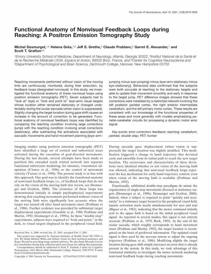
ApparatusThroughout the study, subjects were supine in the scanner. Their headswere immobilized with a thermoplastic mask. The experimental appara-tus was similar to one used in previous psychophysical studies (Desmur-get et al., 1999a,b) (Fig. 1 A). It consisted of a pointing board placed infront of the subjects. An array of light-emitting diodes (LEDs) wasarranged orthogonally to the pointing board. A half-reflecting mirror waspositioned at an angle of 45° with respect to both the pointing board and
the array of LEDs. The subjects saw the virtual images of the LEDsthrough the mirror, in the plane of the board. Consequently, the reachinghand could not occlude the virtual image of the LEDs, which preventedthe subjects from gaining an indirect feedback of their reaching accuracy.The targets were located on a circle centered on the hand starting point(S; radius, 25 cm). A direct orthogonal reference frame was defined fordata analysis and target location definition (Fig. 1 B). S was the origin ofthis reference frame. The z-axis was orthogonal to the pointing boardand oriented toward the subject. The x-axis was horizontal and orientedrightward. The y was orthogonal to x-z and oriented upward. Nine targetswere used in the present experiment. One green diode was located in theleft hemispace at minus30° (with respect to the y-axis). Eight red diodeswere located in the right hemispace with a 5° increment from 10 to 45°.The orientation of the pointing board was adjusted so that the z-axispassed through the subject cyclopean eye (center of mass of the two eyeballs). The distance between the cyclopean eye and the hand startingpoint was 45 cm. During the experiment, movement of an infraredemitting diode located on the subject’s index fingertip was recorded withan Elite motion analysis system at a sampling frequency of 100 Hz. Eyemovements were recorded binocularly using DC electro-oculography(EOG).
Experimental conditionsLight was turned off at injection time (i.e., 10 sec before the start of thescan; see below), and all scans were recorded in a totally dark room. Lightwas turned on between scans. The protocol resulted from the combina-tion of two experimental variables. The first variable was related to theinstruction given to the subject before the session. Subjects were in-structed either to “look and point to the target” (eye 1 arm: eye–arm),or to only “look at the targets, without pointing” (eye). The secondvariable was related to the type of trial defined by the target response. Inhalf of the sessions, the target remained stationary during the whole trial(stationary). In the other half, the target location was modified during thesaccadic response ( jump). The combination of these two variables re-sulted in four experimental conditions: eye–arm–stationary, eye–arm–jump, eye–stationary, and eye–jump. Each of these four conditions wasreplicated three times leading to a total number of 12 scans per subject(four conditions, three replications). The different conditions and differ-ent replications were randomly ordered across subjects. The sequence oftarget presentation was strictly balanced with respect to the effectorfactor (for a given subject, the three sequences of target presentationused for the three repetitions of the eye condition were also used for theeye–arm condition). For the perturbation factor ( jump vs stationary) thesequence of target presentation was balanced with respect to the move-ment final location. In the stationary condition the subjects pointed 12times to each target. In the jump condition the same targets werepresented after either a leftward or a rightward jump. The 10° target(extreme left) was always presented after a leftward jump (15° 3 10°).The 45° target (extreme right) was always presented after a rightwardjump (40° 3 45°). The other targets were presented after either aleftward (six times) or a rightward jump (six times).
All scans involved the same sequence of events. (1) The green visualfixation point (left LED) was turned on for 1.4 sec. (2) Visual fixation wasturned off while one of the red targets (right LEDs) was simultaneouslyturned on. (3) Depending on the experimental instruction, the subjectshad to “look at” (eye) or “look and point to” (eye–arm) the target. (4)The target location either remained stationary or was modified duringthe ocular saccade (because of saccadic suppression this displacementwas not consciously detected by the subjects). The target presentationphase lasted 1.4 sec. (5) The green target was turned on again for visualfixation. In the eye–arm conditions, the subjects used proprioceptiveinformation from the contralateral left hand to return the right handquickly to the starting point. Their left index finger was placed, as atactile mark, just below the hand starting location while their left armrested comfortably on a large pillow placed on their abdomen. In thereaching condition when the right hand was at the starting point, theelbow was slightly flexed (;140° if 180 describes a fully extended arm)with the plane of the arm (wrist–elbow–shoulder) making an angle of;30° with respect to the sagittal plane. In the eye condition both the rightand left hands rested passively on the pillow.
During the experiment, eye velocity was extracted on-line from theposition signal, using a two-point central difference derivative algorithm(Bahill and McDonald, 1983). The change in target location occurred, inthe jump conditions, when eye velocity reached a level roughly equal tohalf of its the peak value. The threshold for target jump was set manually
Figure 1. A, Schematic representation of the experimental apparatus.Subjects were supine with their head immobilized in the scanner. Apointing board was placed in front of them. An array of LEDs and ahalf-reflecting mirror were suspended over the pointing board. The sub-jects saw the virtual image of the LEDs (targets) through the mirror, inthe plane of the board. B, Schematic representation of the pointing board.Nine LEDs were used in the present experiment. They were located on acircle (radius, 250 mm; center, the hand starting point). One green diode(white circle) was located in the left hemispace at 230° (visual fixationpoint). Eight red diodes (black circles) were located in the right hemispacewith a 5° increment from 10 to 45° (targets).
2920 J. Neurosci., April 15, 2001, 21(8):2919–2928 Desmurget et al. • Guiding Hand Movements in the Dark
on an oscilloscope at the beginning of the experiment while the subjectwas required to perform a series of saccades. It was adjusted during thescans if necessary.
Behavioral analysesFor arm movements, the x, y, and z position signals were filtered at 10 Hzwith a second-order Butterworth dual pass filter. Movement velocity wascomputed from the filtered position signal, using a least square second-order polynomial method (window 6 4 points). The same method wasused to compute the acceleration of the hand from the velocity signal.The main arm-related parameters analyzed in this experiment were thehand reaction time (RThand), the hand movement duration (MDhand), theindex finger final location, and the hand path linearity. The index fingerfinal location was defined by the x and y coordinates of the index fingertiplocation at the end of the trial. Hand path linearity was defined as theratio of the largest deviation of arm trajectory from the line connectingthe start and end points of the movement to the length of this line(Atkeson and Hollerbach, 1985). It accounted for the global movementcurvature (Desmurget et al., 1999b). The hand path linearity index isequal to 0 when the movement is perfectly straight and to 0.5 when themovement is semicircular. In addition to the previous parameters, wealso determined the movement direction (Mdir) at the time of peakacceleration and time of peak velocity. Mdir was determined by comput-ing the azimuth and elevation angles of the tangential velocity vector. Asshown by earlier studies (Prablanc and Martin, 1992; Desmurget andPrablanc, 1997), Mdir is the most accurate indicator of the motorreaction time to the perturbation. The onset and the end of the move-ments were computed automatically using the following thresholds: handvelocity 5 80 mm/sec and hand acceleration 5 150 mm/sec 2.
Two-way ANOVA (perturbation 3 target location) was used to deter-mine significant differences between experimental conditions for armmovement parameters. Two different ANOVA were conducted to con-trast the stationary condition with each of the jump conditions (leftwardor rightward). This was done because rightward and leftward targetjumps are expected to have opposite kinematic effects and because theexperimental design was not complete with respect to the initial targetlocation factor (the extreme left target was never the initial targetlocation for the leftward jump condition; the extreme right target wasnever the initial target location for the rightward jump condition). Forthe comparisons involving leftward jumps, the 10° target was removedfrom the stationary dataset. For the comparisons involving the rightwardjumps, the 45° target was removed from the stationary dataset. Bidimen-sional parameters such as the index final location (x and y coordinates)were compared using two way MANOVAs. In this case, the F value wasdetermined from the Wilk’s lambda, using Rao’s approximation (Max-well and Delaney, 1990).
The calibration of the EOG signal was performed in two steps. First,the eccentricity of the different targets was redefined with respect to thecyclopean eye (target eccentricity was initially defined with respect tothe hand starting point). When expressed in eye-centered coordinates,the target eccentricities were: 215.5° (230° fixation point /sec), 5.5°(10°/sec), 8.2° (15°/sec), 10.8° (20°/sec), 13.2° (25°/sec), 15.5° (30°/sec),17.7° (35°/sec), 19.7° (40°/sec), and 21.4° (45°/sec). Second, the EOGsignal was measured while the subject looked at the different targets. Acalibration curve was then computed from these measurements by fittinga polynomial through the data. This curve was used to transform theEOG signal into a calibrated eye position signal. Once calibrated, the eyeposition signal was numerically filtered at 30 Hz with a second-orderButterworth dual pass filter. The velocity signal was computed from thefiltered position signal, using a least square second-order polynomialmethod (window 6 4 points). The main saccadic parameters analyzed inthis experiment were the eye reaction time (RTeye), the eye movementduration (MDeye), and the amplitude of the primary saccade. The latterparameter was expressed as a percentage of the initial required displace-ment. The beginning and the end of the primary saccade were automat-ically detected using a velocity threshold procedure (20°/sec). The resultsof this procedure were checked off-line and corrected if necessary.
Three-way ANOVA was used to determine significant differencesbetween experimental conditions for eye movements [effector (eye alonevs eye–arm); perturbation; target location]. As for arm movements (seeabove), we conducted two different ANOVAs to contrast the stationarycondition with each of the jump conditions (leftward or rightward).Because the primary saccadic response is known to be unmodifiedon-line on the basis of peripheral visual information (Deubel et al., 1986;Desmurget et al. 2000), we also conducted a single three way ANOVA in
which the saccades related to leftward or rightward trials were averagedtogether. Results were identical in both analyses. As a consequence, onlythe first one will be reported here for the sake of consistency.
The statistical threshold was set at p 5 0.05 for all behavioral analyses.
ImagingImaging methods have been described in previous publications (Desmur-get et al., 1998, 2000). In brief, regional cerebral blood flow (rCBF)images were acquired with a Siemens ECAT Exact scanner, by using amodified autoradiographic method in three-dimensional mode. Ninetysecond scans were recorded every 8 min. The series of scans was made,from each subject, using bolus intravenous injections of H2
15O (25 mCi)that were delivered into the left arm 10 sec before the start of the scan.Performance of the designated task began at the same time as thescanning. Images were reconstructed by using calculated attenuationcorrection.
Image processing was performed on a SunSparc5 workstation. Forspatial normalization, a within-subject alignment of PET scans wasperformed by using an automated registration algorithm (Woods et al.,1998a,b). For each subject, the mean PET image was then coregistered toa population-based PET reference atlas centered in Talairach coordi-nates (Talairach and Tournoux 1988), using affine and nonlinear trans-forms with 60 degrees of freedom (Woods et al., 1998a,b). CoregisteredPET images were smoothed to a final isotropic resolution of 15 mm fullwidth at half maximum and normalized to each other by using propor-tionate global scaling. ANOVA for a randomized complete block designwas used, for all contrasts, to identify significant task effects (Neter et al.,1990; Woods et al., 1996). The effects (and source of variance) in thestatistical model were subject, task, and repetition. Given the perfor-mance consistency of the behavioral paradigm under investigation andthe randomization procedure, repetition could be treated as replication,resulting in a two-way ANOVA (Turner et al., 1998). For all the contrastsevaluated in this study, the statistical threshold was initially set at p 50.005. The following five contrasts were evaluated.
Overall hand-reaching effect. [(eye–arm–jump plus eye–arm-stationary) minus (eye–jump plus eye–stationary)] (df 5 70; t $ 2.65).This contrast allows removal of the brain activation specifically related tothe visual capture of the target. As a consequence it should primarilyidentify the areas involved in movement planning and movement control.However, one may not exclude the possibility that areas associatedspecifically with motor correction (response to the jump) or eye–handcoordination (interaction) contribute to the overall effect observed. No apriori hypothesis was formulated about the areas that might be activatedin this contrast (unplanned contrast). To adjust for multiple comparisons,the t statistic image was corrected using the method developed by Fristonet al. (1994). This method takes into account the size of the activation(359 resolving elements), the search volume, and the degree of imagesmoothness. Correction for multiple comparison was conducted at a finalcertainty of p , 0.01.
Strict hand-reaching effect. [eye–arm–stationary minus eye-stationary](df 5 28; t $ 2.76). This contrast is similar to the previous one except thatonly the movements to stationary targets were taken into account. If it iscorrect that the jump and no jump trials involve similar functionalmechanisms, the strict and overall reaching contrast should give compa-rable results. No a priori hypothesis was formulated about the areas thatmight be activated in this contrast (unplanned contrast). As a conse-quence, t statistic image was corrected for multiple comparisons (359resolving elements) to a final certainty of p , 0.01 using the methoddeveloped by Friston et al. (1994).
Overall jump effect. [(eye–arm–jump plus eye–jump) minus (eye–arm–stationary plus eye–stationary)] (df 5 56; t $ 2.67). In this contrast wedetermined the effect of jumping the target location, irrespective of theeffector. The overall jump effect should give information about the globalnetwork activated when the estimation of the target location by theperipheral retina is erroneous (see introductory remarks). No a priorihypothesis was formulated about the areas that might be activated in thiscontrast (unplanned contrast). As a consequence, t statistic image wascorrected for multiple comparisons (359 resolving elements) to a finalcertainty of p , 0.01 using the method developed by Friston et al. (1994).
Eye error correction effect. [eye–jump minus eye–stationary] (df 5 56;t $ 2.67). In this contrast we determined the effect of jumping the targetlocation on the oculomotor system. This contrast should give informationabout the network activated when the initial saccadic response is incor-rect. No a priori hypothesis was formulated about the areas that might beactivated in this contrast (unplanned contrast). As a consequence, t
Desmurget et al. • Guiding Hand Movements in the Dark J. Neurosci., April 15, 2001, 21(8):2919–2928 2921
statistic image was corrected for multiple comparisons (359 resolvingelements) to a final certainty of p , 0.01 using the method developed byFriston et al. (1994).
Hand error correction effect. [(eye–arm–jump minus eye–jump) minus(eye–arm–stationary minus eye–stationary)] (df 5 56; t $ 2.67). In thiscontrast we determined the subcircuit mediating on-line hand trajectoryadjustments. To this end, we contrasted the jump and stationary condi-tions after subtraction of the oculomotor-related activity. It is worthemphasizing that this double difference amounts, in fact, to an interac-tion. The areas identified by this contrast are the areas that increase theirresponsiveness when larger corrections have to be performed. As alreadyemphasized, what we compare in this experiment is a condition involvingsmall corrections (stationary) with a condition involving large corrections( jump). Consequently, the interpretation of the present contrast can beframed in terms of modulation of the underlying executive system by aperturbation that increases the error processing. A potential difficultywith this design, and more precisely with the fact that similar feedbackloops are engaged in both the jump and stationary conditions, is that thehand error correction effect might be very subtle and hard to detect.Stringent statistical procedures involving strict corrections for multiplecomparisons might be excessively conservative in this context. At thesame time, however, increasing the statistical p value or abolishingcorrections for multiple comparisons might abnormally increase the riskof type I errors (declaring significant an activation that is not). Toaccommodate these contradictory exigencies, (i.e., increasing statisticalsensitivity while minimizing type I errors) a two-step analysis was con-ducted. First, a nonplanned contrast was evaluated. For this contrast, noa priori hypothesis was formulated about the areas that might be acti-vated. As a consequence, the t statistic image was corrected for multiplecomparisons (359 resolving elements) to a final certainty of p , 0.01using the method developed by Friston et al. (1994). Second, a plannedcontrast was evaluated. For this contrast, the search volume was re-stricted a priori to the structures showing an unequivocal reach relatedeffect, i.e., to the structures activated in the strict hand-reaching contrast.No correction for multiple comparisons was applied within this restrictedset of functionally plausible structures. To avoid an overly conservativerestriction of the search volume “the reach-related network” was definedat a relaxed threshold, with no corrections for multiple comparisons(strict hand-reaching effect at p , 0.01; 39 resolving elements).
RESULTSBehavioral observations: saccade characteristicsFor both the stationary and jump conditions, the saccadic re-sponse consisted of two phases (Fig. 2A): an initial saccadeundershooting the initial target position and covering on average96% (63.7) of the initially required displacement and a correc-tive saccade achieving accurate target acquisition. The amplitudeof the primary saccade did not vary significantly as a function ofthe perturbation [stationary, 96.3% (63.9); rightward jump,95.1% (63.8); leftward jump, 95.7% (64.1); p . 0.10] or effectorfactors [eye, 95.6% (64.1); eye–arm, 95.9% (63.9); p . 0.35].The number of trials involving more than one corrective saccadewas marginal, even in the jump condition. This latter observationwas expected considering the modest amplitude of the targetjump (between 1.7° and 2.7°; see Materials and Methods) (Des-murget et al., 2000).
RTeye was equal to 212 msec (636). This parameter did notvary significantly as a function of the perturbation [stationary, 215msec (643); rightward jump, 209 msec (631); leftward jump, 211msec (631); p . 0.35] or effector factors [eye, 215 msec (636);eye–arm, 209 msec (635); p . 0.25]. MDeye was equal to 88 msec(615). It was not affected by the perturbation factor [stationary,87 msec (614); rightward jump, 85 msec (614); leftward jump, 92msec (616); p . 0.10] but was found to be significantly shorter ineye–arm than in eye [86 msec (613) versus 90 msec (616); p ,0.01]. The meaning of this heretofore undescribed effect is un-clear. On the one hand, this slight difference may represent a falsepositive inference. On the other hand, it may reflect the func-
tional capacity of the motor system to update the target locationmore quickly to allow early hand path adjustments.
None of the subjects reported the existence of a change intarget position during the saccadic response, even when ques-tioned explicitly at the end of the study. The absence of consciousperception of the target jump is coherent with the absence ofsignificant variation of the kinematic characteristics of the pri-mary saccadic response as a function of the perturbation factor.
Behavioral observations: armmovement characteristicsRThand was independent of the perturbation factor [stationary,277 msec (650); rightward jump, 269 msec (648); leftward jump,263 msec (640); p . 0.25]. On average RThand was 58 msec longerthan RTeye [270 msec (646) vs 212 msec (636)]. This indicatesthat arm movement started around the end of the ocular saccade(for eye–arm, RTeye 1 MDeye 5 300 msec; Fig. 2A,B), or in otherwords that the arm motor command was issued on the basis of aperipheral retinal input. If one considers that the onset of theagonist muscle contraction occurs 50–100 msec before the actualmotion for reaching movements (Biguer et al., 1982; Turner et al.,1995), the latencies observed in this study are compatible withprevious observations showing that arm muscle contraction issynchronous with eye movement onset during fast reaching move-ments directed at peripheral targets (Biguer et al., 1982).
Peak hand acceleration occurred on average at 125 msec (622msec) and was independent of the perturbation factor ( p . 0.30).No significant variations of the movement direction were ob-served at the time of peak hand acceleration ( p . 0.90), support-ing the idea that the subjects did not develop a specific strategy inthe jump sessions. Peak hand velocity occurred on average at 251msec (637), i.e., at 41% of the total movement duration. Likepeak hand acceleration, peak hand velocity did not vary signifi-cantly with the perturbation factor ( p . 0.50). Interestingly,significant variations of Mdir were observed at the time of peak
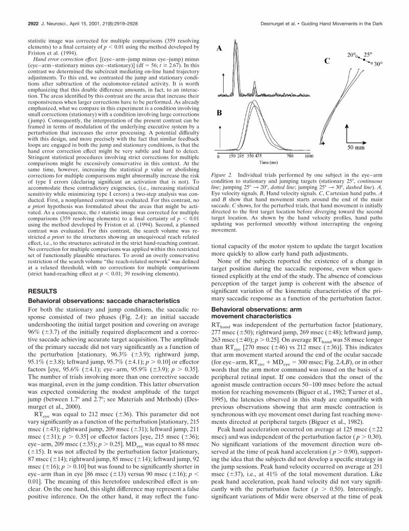
Figure 2. Individual trials performed by one subject in the eye–armcondition to stationary and jumping targets (stationary 25°, continuousline; jumping 25° 3 20°, dotted line; jumping 25° 3 30°, dashed line). A,Eye velocity signals. B, Hand velocity signals. C, Cartesian hand paths. Aand B show that hand movement starts around the end of the mainsaccade. C shows, for the perturbed trials, that hand movement is initiallydirected to the first target location before diverging toward the secondtarget location. As shown by the hand velocity profiles, hand pathsupdating was performed smoothly without interrupting the ongoingmovement.
2922 J. Neurosci., April 15, 2001, 21(8):2919–2928 Desmurget et al. • Guiding Hand Movements in the Dark
hand velocity ( p , 0.04). At this instant, Mdir was found to berotated to the left by 2.7° on average in the leftward jumpcondition and rotated to the right by 3.3° on average in therightward jump condition. This result demonstrates the existenceof early path corrections in response to the target jump.
Arm trajectory amendments were clearly visible in the handpath linearity index, which varied significantly as a function of theperturbation factor [stationary, 0.120 (60.021); rightward jump,0.130 (60.024); leftward jump, 0.108 (60.019); p , 0.005] (Fig.2C). They were also reflected in the index fingertip final location.This parameter was significantly different in the control trialsdirected to a given target and in the jump trials initially directedto the same target ( p , 0.001). Interestingly, when the controltrials directed to a given target T were contrasted with the jumptrials for which T was the final target, no significant difference wasobserved for the final hand location ( p . 0.20). This resultindicates that the path corrections observed in the jump conditionwere nearly complete. Trajectory amendments occurred withoutsignificant increase of the mean movement duration [stationary,618 msec (664); rightward jump, 609 msec (681); leftward jump,627 msec (688); p . 0.60], as observed in previous reports(Goodale et al., 1986; Desmurget et al., 1999a).
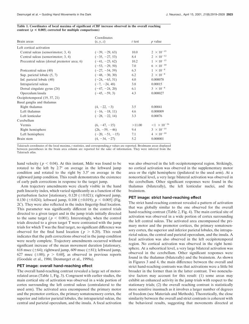
PET image: overall hand-reaching effectThe overall hand-reaching contrast revealed a large set of motor-related areas (Table 1, Fig. 3). Congruent with earlier studies, themain cortical site of activation was observed in a wide portion ofcortex surrounding the left central sulcus (contralateral to theused arm). The activated area encompassed the primary motorand the premotor cortices, the primary somatosensory cortex, thesuperior and inferior parietal lobules, the intraparietal sulcus, thecentral and parietal operculum, and the insula. A focal activation
was also observed in the left occipitotemporal region. Strikingly,no cortical activation was observed in the supplementary motorarea or the right hemisphere (ipsilateral to the used arm). At anoncortical level, a very large bilateral activation was observed inthe cerebellum. Other significant responses were found in thethalamus (bilaterally), the left lenticular nuclei, and thebrainstem.
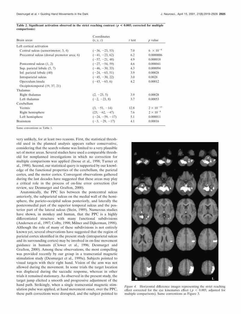
PET image: strict hand-reaching effectThe strict hand-reaching contrast revealed a pattern of activationthat was globally similar to the one observed for the overallhand-reaching contrast (Table 2, Fig. 4). The main cortical site ofactivation was observed in a wide portion of cortex surroundingthe left central sulcus. The activated area encompassed the pri-mary motor and the premotor cortices, the primary somatosen-sory cortex, the superior and inferior parietal lobules, the intrapa-rietal sulcus, the central and parietal operculum, and the insula. Afocal activation was also observed in the left occipitotemporalregion. No cortical activation was observed in the right hemi-sphere. At a subcortical level, a very large bilateral activation wasobserved in the cerebellum. Other significant responses werefound in the thalamus (bilaterally) and the brainstem. As shownin Figures 3 and 4, the main difference between the overall andstrict hand-reaching contrasts was that activations were a little bitbroader in the former than in the latter contrast. Two nonexclu-sive factors may account for this result: (1) some areas mayexhibit an enhanced activity in the jump trials with respect to thestationary trials; (2) the overall reaching contrast is statisticallymore sensitive inasmuch as it involves a larger number of degreesof freedom (see Materials and Methods). Theoretically, the closesimilarity between the overall and strict contrasts is coherent withthe behavioral results, suggesting that movements directed at
Table 1. Coordinates of local maxima of significant rCBF increase observed in the overall reachingcontrast (p < 0.005; corrected for multiple comparisons)
Brain areasCoordinates(x, y, z) t test p value
Left cortical activationCentral sulcus (sensorimotor; 3, 4) (239, 229, 63) 10.0 2 3 10215
Central sulcus (sensorimotor; 3, 4) (235, 227, 53) 8.4 2 3 10212
Precentral sulcus (dorsal premotor area; 6) (241, 223, 62) 10.2 1 3 10215
(253, 229, 50) 7.0 6 3 10210
Postcentral sulcus (40) (227, 254, 59) 6.3 1 3 1028
Sup. parietal lobule (5, 7) (248, 230, 30) 6.2 2 3 1028
Inf. parietal lobule (40) (224, 263, 51) 4.0 0.000078Intraparietal sulcus (27, 224, 48) 3.8 0.00015Dorsal cingulate gyrus (24) (247, 224, 20) 6.1 3 3 1028
Operculum/insula (245, 259, 3) 4.3 0.000027Occipitotemporal (19, 37, 21)Basal ganglia and thalamus
Right thalamus (4, 222, 23) 3.5 0.00041Left thalamus (216, 218, 11) 4.6 0.000009Left lenticular (228, 222, 14) 3.3 0.00076
CerebellumVermis (6, 247, 215) .11.00 ,1 3 10216
Right hemisphere (26, 259, 246) 9.4 3 3 10214
Left hemisphere (220, 251, 215) 7.1 4 3 10210
Brain stem (4, 234, 227) 5.2 0.000001
Talairach coordinates of the local maxima, t statistics, and corresponding p values are reported. Brodmann areas displayedbetween parentheses in the brain area column are reported for the sake of information. They were inferred from theTalairach atlas.
Desmurget et al. • Guiding Hand Movements in the Dark J. Neurosci., April 15, 2001, 21(8):2919–2928 2923
jumping and stationary targets involve similar functional pro-cesses (see introductory remarks). Further argument supportingthis view will be presented in the next section.
PET image: overall jump effect, eye error correctioneffect, and hand error correction effectThese three contrasts failed to reveal any significant activation ateither the cortical or subcortical levels after correction for multi-ple comparisons. These results confirm and extend the conclu-sions of a previous study showing that jumping the target locationrandomly during the course of the saccade does not generate anysignificant activation in the main oculomotor areas with respect toa condition in which the target location remains stationary (Des-murget et al., 2000). The present negative finding shows that themetabolic response induced by the target jump is not substantialenough to be identified with stringent nonplanned statisticalprocedures.
PET image: hand error correction effect andplanned analysisThis analysis revealed a restricted motor network engaging threeareas previously postulated to have a role in nonvisual feedback
loops (see Discussion), namely the parietal cortex, the frontalcortex, and the cerebellum (Table 3, Fig. 5). The parietal activa-tion was located in the left intraparietal sulcus, in a region that isgenerally considered the rostral part of the posterior parietalcortex (PPC). The cerebellar activation was found in the rightanterior parasagittal cerebellar cortex, in a region associated withthe production of arm movements. The frontal activation waslocated in the arm-related area of the primary motor cortex.Figure 6 displays rCBF values for these three regions of interest,in each experimental condition.
DISCUSSIONWe identified a distributed cerebral network activated duringvisually directed movements performed without vision of themoving limb. Then, we describe within this network, for the firsttime, a restricted subset of areas specifically involved in on-linemovement guidance.
Reaching in the darkAlthough the reach-related cerebral network observed in thepresent study is generally coherent with previous observationsperformed with and without vision of the moving limb(Colebatch et al., 1991; Grafton et al., 1992, 1996; Deiber et al.,1996; Lacquaniti et al., 1997; Inoue et al., 1998; Turner et al.,1998; Winstein et al., 1997), our results differ from earlier reportsin two ways. First, we observed a larger noncortical contribution,especially within the cerebellum and pontine nuclei. Second, wenoted a less distributed cortical activation. In particular, we didnot identify any activation within the motor, premotor, and pa-rietal cortices of the hemisphere ispilateral to the moving arm.Similarly, we did not observe any rCBF increase within thesupplementary motor area or occipital visual areas. These differ-ences may be related to the fact that the motor reaching task weinvestigated here was more “rudimentary” than the tasks consid-ered in earlier experiments. The present study differs, indeed,from earlier studies by at least one of the following aspects: (1)vision of the moving limb was never allowed, preventing visualfeedback loops from operating; (2) no estimation of the reachingerror was provided during or at the end of trial, prohibiting motorlearning (Jordan 1990; Redding and Wallace 1996); (3) the sub-jects reached to the target directly without the mediation of amanipulandum or a joystick, avoiding the need for complex visuo-motor transformations; (4) targets were seen in binocular visionand not through a virtual display system that provides conflictingvergence and accommodation signals, thus requiring adaptivebehavior by preventing real depth perception; (5) oculomotoractivity was strictly controlled allowing precise evaluation of armreaching-related changes in rCBF. Our data suggest that basicreaching movements performed without visual guidance involve aless distributed cortical network and rely more consistently oncerebellar structures than the visually more complex motor tasksusually studied.
Functional anatomy of movement guidanceFunctional anatomy of nonvisual feedback loops was determined,within the reach-related motor network, by identifying the brainareas that increase their responsiveness when the error to becorrected is larger. Because this planned analysis was performedwithout stringent corrections for multiple comparisons (see Ma-terials and Methods), one might wonder whether the areas weidentified may represent a series of false-positive inferences.Although this possibility cannot be rejected categorically it seems
Figure 3. Horizontal difference images representing the overall reachingeffect corrected for the eye kinematics effect ( p , 0.005; adjusted formultiple comparisons). Activations are shown superimposed on a meanmagnetic resonance image (MRI) in Talairach coordinates. The anatomicright side is shown on the lef t side of the figure. The first section (top lef t)is 49 mm below the anteroposterior commissural line (Z 5 249). The lastsection (bottom right) is 65 mm above the anteroposterior commissuralline (Z 5 165). Sections are presented every 6 mm.
2924 J. Neurosci., April 15, 2001, 21(8):2919–2928 Desmurget et al. • Guiding Hand Movements in the Dark
very unlikely, for at least two reasons. First, the statistical thresh-old used in the planned analysis appears rather conservative,considering that the search volume was limited to a very plausibleset of motor areas. Several studies have used a comparable thresh-old for nonplanned investigations in which no correction formultiple comparisons was applied (Inoue et al., 1998; Turner etal., 1998). Second, our statistical query is supported by our knowl-edge of the functional properties of the cerebellum, the parietalcortex, and the motor cortex. Convergent observations gatheredduring the last decades have suggested that these areas may playa critical role in the process of on-line error correction (forreview, see Desmurget and Grafton, 2000).
Anatomically, the PPC lies between the postcentral sulcusanteriorly, the subparietal sulcus on the medial wall of the hemi-sphere, the parieto-occipital sulcus posteriorly, and laterally theposteromedial part of the superior temporal sulcus and the pos-terior part of the lateral sulcus (Stein, 1989). Numerous studieshave shown, in monkey and human, that the PPC is a highlydifferentiated structure with many functional subdivisions(Andersen et al., 1997; Colby, 1998; Milner and Dijkerman, 1998).Although the role of many of these subdivisions is not entirelyknown yet, several observations have suggested that the region ofparietal cortex identified in the present study (intraparietal sulcusand its surrounding cortex) may be involved in on-line movementguidance in humans (Clower et al., 1996; Desmurget andGrafton, 2000). Among these observations, the most compellingwas provided recently by our group in a transcranial magneticstimulation study (Desmurget et al., 1999a). Subjects pointed tovisual targets with their right hand. Vision of the arm was notallowed during the movement. In some trials the target locationwas displaced during the saccadic response, whereas in othertrials it remained stationary. As observed in the present study, thetarget jump elicited a smooth and progressive adjustment of thehand path. Strikingly, when a single transcranial magnetic stim-ulation pulse was applied, at hand movement onset, over the PPC,these path corrections were disrupted, and the subject pointed to
Figure 4. Horizontal difference images representing the strict reachingeffect corrected for the eye kinematics effect ( p , 0.005; adjusted formultiple comparisons). Same conventions as Figure 3.
Table 2. Significant activation observed in the strict reaching contrast (p < 0.005; corrected for multiplecomparisons)
Brain areasCoordinates(x, y, z) t test p value
Left cortical activationCentral sulcus (sensorimotor; 3, 4) (236, 223, 53) 7.0 6 3 1028
Precentral sulcus (dorsal premotor area; 6) (241, 223, 62) 6.2 0.0000006(257, 221, 48) 4.9 0.000018
Postcentral sulcus (1, 2) (227, 254, 59) 4.6 0.000041Sup. parietal lobule (5, 7) (246, 230, 33) 4.3 0.000094Inf. parietal lobule (40) (224, 263, 51) 3.9 0.00028Intraparietal sulcus (245, 230, 22) 3.0 0.0028Operculum/insula (243, 263, 6) 4.2 0.00012Occipitotemporal (19, 37, 21)
ThalamusRight thalamus (2, 223, 5) 3.9 0.00028Left thalamus (22, 223, 8) 3.7 0.00053
CerebellumVermis (3, 251, 214) 12.8 2 3 10213
Right hemisphere (23, 262, 247) 7.6 2 3 1028
Left hemisphere (224, 259, 217) 5.1 0.000011Brainstem (23, 229, 217) 4.1 0.00016
Same conventions as Table 1.
Desmurget et al. • Guiding Hand Movements in the Dark J. Neurosci., April 15, 2001, 21(8):2919–2928 2925

the first target location. This result was recently replicated in aclinical study involving a patient presenting with bilateral isch-emic lesions of the PPC (Pisella et al., 2000). Although thispatient was able to accurately point to stationary targets, shepresented a dramatic inability to correct her ongoing movementswhen the target location was slightly modified at movement onset.
Functionally, it has been suggested that a major role of PPC inmovement guidance is to determine whether and to what extentthe current motor response is inadequate (Desmurget et al.,1999a; Desmurget and Grafton, 2000). This hypothesis is basedon the observation that PPC displays three main properties thatwould be expected from an error detection module. First, it hasaccess to a representation of the target and current hand location
through afferent information coming from many sensory modal-ities (visual, proprioceptive, vestibular), and the main motorstructures (Andersen et al., 1997; Brodal and Bjaalie, 1997).Second, it is critical for establishing stable relationships betweenheterogeneous information, i.e., for merging arm and target-related signals into a common frame of reference (Clower et al.,1996; Carey et al., 1997; Colby, 1998; Binkofski et al., 1999; Xingand Andersen, 2000). Third, it modulates its neural activity as thehand approaches the target, i.e., as the motor error varies(MacKay, 1992). In a recent paper, Desmurget and Grafton(2000) have suggested that dynamic error detection was achievedby the PPC through forward modeling. According to this view, aforward model of the arm’s dynamics is generated during themovement. This forward model, which requires integration ofboth afferent and efferent information, allows prediction of themovement end point. When a discrepancy is detected betweenthe predicted movement final location and the target location, anerror signal is generated. This error signal has then to be trans-formed into an actual motor command. The cerebellum is aprimary candidate for this task.
Like PPC, the cerebellum has long been associated with feed-back control (Miall et al., 1993; Stein, 1986). As shown in severalstudies involving reaching (Day et al., 1998) and tracking tasks(Miall et al., 1993; Haggard et al., 1995), cerebellar lesions do notprevent on-line trajectory adjustments from occurring. However,the motor corrections generated by patients presenting with alesion of the cerebellum are characterized by excessive deviationsand ill-tuned muscle activation patterns. Anatomically, it wasshown that the cerebellum receives abundant input from PPC viathe pontine nuclei (Brodal and Bjaalie, 1997; Middleton andStrick, 1998). Functionally, it was suggested that one of the maincontributions of the cerebellum to movement control is to per-form the inverse computations allowing transformation of a de-sired displacement into an actual muscle command. In support ofthis idea, it was demonstrated that inverse models are representedwithin the cerebellum (Wolpert et al., 1998; Kawato, 1999;Imamizu et al., 2000) and that patients with cerebellar lesionsdisplay a chronic inability to accurately define the pattern of
Figure 5. Horizontal, sagittal, and coronal difference images represent-ing the hand error correction effect corrected for the eye kinematics andhand movement planning effects ( p , 0.005; planned comparison). Ac-tivations are shown superimposed on a mean MRI in Talairach coordi-nates. On the horizontal and coronal images, the anatomic right side isshown on the lef t side. On the sagittal images, positive values of xdesignate the right hemisphere (ipsilateral to the reaching arm), andnegative values designate the left hemisphere (contralateral to the reach-ing arm). The top row is centered on the cerebellar activation site (11,245, 220). The middle row is centered on the posterior parietal activationsite (241, 244, 58). The bottom row is centered on the precentralactivation site (230, 226, 57).
Figure 6. rCBF mean values (SD vertical bars), in each experimentalcondition, for the three cerebral regions showing significant activation inthe hand error correction contrast (E&A, eye and arm pointing; E, eyealone; statio, stationary trial; jump, jump trial). These regions are theprimary motor cortex (black triangles, lef t curve; Talairach coordinates:230, 226, 57), the posterior parietal cortex (black circles, middle curve;241, 244, 58), and the anterior parasagittal cerebellar cortex (blacksquares, right curve; 11, 245, 220).
Table 3. Significant activation observed in the hand error correctioncontrast (p < 0.005; uncorrected for multiple comparisons)
Brain areasCoordinates(x, y, z) t test p value
Left cortical activationPrecentral gyrus (4) (230,226,57) 2.9 0.0027Intraparietal sulcus (241,244,58) 3.0 0.002
CerebellumRight anterior parasagittal (11,245,220) 3.0 0.002
Same conventions as Table 1.
2926 J. Neurosci., April 15, 2001, 21(8):2919–2928 Desmurget et al. • Guiding Hand Movements in the Dark
muscle activation required to direct the hand along a specific path(Bastian et al., 1996; Day et al., 1998).
The previous observations suggest that the cerebellar contri-bution to on-line movement guidance may be to convert thedynamic motor error signal computed by PPC into an appropriatecorrective command. Within this context, the precentral gyrusactivation we observed, concurrently with the cerebellar activa-tion, may be accounted for by assuming that the cerebellar signalinfluence the ongoing motor command by modulating the neuralsignal issued by the primary motor cortex. In agreement with thisview, it has been shown that the primary motor cortex receivessubstantial input from the cerebellum via the ventrolateral thal-amus (Asanuma et al., 1983; Brodal and Bjaalie, 1997; Hooverand Strick, 1999). Also, it has been suggested that the motorsystem is organized in a relative hierarchy such that the primarymotor cortex is mainly involved in the low-level aspects of motorcontrol. Consistent with this idea, it was shown that purely kine-matic and dynamical aspects of the movement are more com-monly represented in the motor cortex than, for instance, in theparietal or premotor areas that seem to encode more abstractvariables (Alexander and Crutcher, 1990; Scott et al., 1997; Shenand Alexander, 1997; Turner et al., 1998). The activation of M1during error correction may appear to be in contradiction with aprevious transcranial magnetic stimulation (TMS) study in whichwe observed no effect, on movement correction, when the motorcortex was stimulated (Desmurget et al., 1999). However, the levelof stimulation we used in our previous TMS study was not highenough to generate any EMG response in the primary armmovers. This indicates that the stimulation did not interfere withthe ongoing movement. It is likely that higher level of stimulationwould have resulted in more dramatic effects.
In conclusion, the present experiment provides new evidence insupport of the hypothesis that parietal-cerebellar circuits arecritical for hand movement guidance. We have shown that non-visual feedback loops involve a limited network including themotor cortex, the cerebellum, and the PPC. Based on recentneurophysiological and clinical observations, we hypothesize thatPPC computes a dynamic motor error by comparing the updatedlocation of the visual target and the estimated movement endpoint. This dynamic motor error is sent to the cerebellum, whichconverts it into a corrective motor command. The correctivesignal influence finally the ongoing motor command by modulat-ing the neural signal issued by the primary motor cortex.
REFERENCESAlexander GE, Crutcher MD (1990) Neural representations of the tar-
get (goal) of visually guided arm movements in three motor areas of themonkey. J Neurophysiol 64:164–178.
Andersen RA, Snyder LH, Bradley DC, Xing J (1997) Multimodalrepresentation of space in the posterior parietal cortex and its use inplanning movements. Annu Rev Neurosci 20:303–330.
Asanuma C, Thach WR, Jones EG (1983) Anatomical evidence forsegregated focal groupings of efferent cells and their terminal ramifi-cations in the cerebellothalamic pathway of the monkey. Brain Res286:267–297.
Atkeson CG, Hollerbach JM (1985) Kinematic features of unrestrainedvertical arm movements. J Neurosci 5:2318–2330.
Bahill AT, McDonald JD (1983) Frequency limitations and optimal stepsize for the two-points central difference derivative algorithm withapplication to human eye data. IEEE Trans Biomed Eng 30:191–194.
Bastian AJ, Martin TA, Keating JG, Thach WT (1996) Cerebellar atax-ia: abnormal control of interaction torques across multiple joints.J Neurophysiol 76:492–509.
Biguer B, Jeannerod M, Prablanc C (1982) The coordination of eye,head and arm movements during reaching at a single visual target. ExpBrain Res 46:301–304.
Binkofski F, Buccino G, Dohle C, Seitz RJ, Freund HJ (1999) Mirror
agnosia and mirror ataxia constitute different parietal lobe disorders.Ann Neurol 46:51–61.
Bock O (1993) Localization of objects in the peripheral visual field.Behav Brain Res 56:77–84.
Brodal P, Bjaalie JG (1997) Salient anatomic features of the cortico-ponto-cerebellar pathway. Prog Brain Res 114:227–249.
Carey DP, Coleman RJ, Della Sala S (1997) Magnetic misreaching.Cortex 33:639–652.
Clower DM, Hoffman JM, Votaw JR, Faber TL, Woods RP, Alexander G(1996) Role of posterior parietal cortex in the recalibration of visuallyguided reaching. Nature 383:618–621.
Colby CL (1998) Action-oriented spatial reference frames in cortex.Neuron 20:15–24.
Colebatch JG, Deiber MP, Passingham RE, Friston KJ, Frackowiak RS(1991) Regional cerebral blood flow during voluntary arm and handmovements in human subjects. J Neurophysiol 65:1392–1401.
Day BL, Thompson PD, Harding AE, Marsden CD (1998) Influence ofvision on upper limb reaching movements in patients with cerebellarataxia. Brain 121:357–372.
Deiber MP, Ibanez V, Sadato N, Hallett M (1996) Cerebral structuresparticipating in motor preparation in humans: a positron emissiontomography study. J Neurophysiol 75:233–247.
Desmurget M, Grafton ST (2000) Forward modeling allows feedbackcontrol for fast reaching movements. Trends Cognit Sci 4:423–431.
Desmurget M, Prablanc C (1997) Postural control of three dimensionalprehension movements. J Neurophysiol 77:452–464.
Desmurget M, Pelisson D, Urquizar C, Prablanc C, Alexander GE,Grafton ST (1998) Functional anatomy of saccadic adaptation in hu-mans. Nat Neurosci 1:524–528.
Desmurget M, Epstein CM, Turner RS, Prablanc C, Alexander GE,Grafton ST (1999a) Role of the posterior parietal cortex in updatingreaching movements to a visual target. Nat Neurosci 2:563–567.
Desmurget M, Prablanc C, Jordan MI, Jeannerod M (1999b) Are reach-ing movements planned to be straight and invariant in the extrinsicspace: kinematic comparison between compliant and unconstrainedmotions. Q J Exp Psychol 52A:981–1020.
Desmurget M, Pelisson D, Grethe JS, Alexander GE, Urquizar C, Pra-blanc C, Grafton ST (2000) Functional adaptation of reactive saccadesin humans: a PET study. Exp Brain Res 132:243–259.
Deubel H, Wolf W, Hauske G (1986) Adaptive gain control of saccadiceye movements. Hum Neurobiol 5:245–253.
Friston KJ, Worsley KJ, Frakowiak RSJ, Mazziotta JC (1994) Assessingthe significance of focal activations using their spatial extent. HumBrain Mapp 1:214–220.
Goodale MA, Pelisson D, Prablanc C (1986) Large adjustments in visu-ally guided reaching do not depend on vision of the hand and percep-tion of target displacement. Nature 320:748–750.
Grafton ST, Mazziotta JC, Woods RP, Phelps ME (1992) Human func-tional anatomy of visually guided finger movements. Brain 115:565–587.
Grafton ST, Fagg AH, Woods RP, Arbib MA (1996) Functional anat-omy of pointing and grasping in humans. Cereb Cortex 6:226–237.
Haggard P, Miall RC, Wade D, Fowler S, Richardson A, Anslow P, SteinJ (1995) Damage to cerebellocortical pathways after closed head in-jury: a behavioural and magnetic resonance imaging study. J NeurolNeurosurg Psychiatry 58:433–438.
Hoover JE, Strick PL (1999) The organization of cerebellar and basalganglia outputs to primary motor cortex as revealed by retrogradetransneuronal transport of herpes simplex virus type 1. J Neurosci19:1446–1463.
Imamizu H, Miyauchi S, Tamada T, Sasaki Y, Takino R, Putz B, Yo-shioka T, Kawato M (2000) Human cerebellar activity reflecting anacquired internal model of a new tool. Nature 403:192–195.
Inoue K, Kawashima R, Satoh K, Kinomura S, Goto R, Koyama M,Sugiura M, Ito M, Fukuda H (1998) PET study of pointing with visualfeedback of moving hands. J Neurophysiol 79:117–125.
Jordan MI (1990) Motor learning and the degrees of freedom problem.In: Attention and performance XIII: Motor representation and control(Jeannerod M, ed), pp 796–836. HillsDale, NJ: Erlbaum.
Kawato M (1999) Internal models for motor control and trajectory plan-ning. Curr Opin Neurobiol 9:718–727.
Lacquaniti F, Perani D, Guigon E, Bettinardi V, Carrozzo M, Grassi F,Rossetti Y, Fazio F (1997) Visuomotor transformations for reachingto memorized targets: a PET study. NeuroImage 5:129–146.
MacKay WA (1992) Properties of reach-related neuronal activity in cor-tical area 7A. J Neurophysiol 67:1335–1345.
Maxwell SE, Delaney HD (1990) Designing experiments and analysingdata. A model comparison perspective. Belmont, CA: Wadsworth.
Miall RC, Weir DJ, Wolpert DM, Stein JF (1993) Is the cerebellum aSmith predictor. J Mot Behav 25:203–216.
Middleton FA, Strick PL (1998) The cerebellum: an overview. TrendsNeurosci 21:367–369.
Milner AD, Dijkerman HC (1998) Visual processing in the primateparietal lobe. In: Comparative neuropsychology (Milner AD, ed), pp70–94. Oxford: Oxford UP.
Desmurget et al. • Guiding Hand Movements in the Dark J. Neurosci., April 15, 2001, 21(8):2919–2928 2927
Neter J, Wasserman W, Kutner MH (1990) Applied linear models. Bos-ton, MA: Irwin.
Pisella L, Grea H, Tilikete C, Vighetto A, Desmurget M, Rode G, BoissonD, Rossetti Y (2000) An “automatic pilot” for the hand in humanposterior parietal cortex: toward reinterpreting optic ataxia. Nat Neu-rosci 3:729–736.
Prablanc C, Martin O (1992) Automatic control during hand reaching atundetected two-dimensional target displacements. J Neurophysiol67:455–469.
Prablanc C, Echallier JF, Komilis E, Jeannerod M (1979) Optimal re-sponse of eye and hand motor system in pointing at visual target. I.Spatio-temporal characteristics of eye and hand movements and theirrelationships when varying the amount of visual information. BiolCybern 35:113–124.
Prablanc C, Pelisson D, Goodale MA (1986) Visual control of reachingmovements without vision of the limb. I. Role of extraretinal feedbackof target position in guiding the hand. Exp Brain Res 62:293–302.
Redding GM, Wallace B (1996) Adaptative spatial alignment and stra-tegic perceptual-motor control. J Exp Psychol Hum Percept Perform22:379–394.
Scott SH, Sergio LE, Kalaska JF (1997) Reaching movements with sim-ilar hand paths but different arm orientations. II. Activity of individualcells in dorsal premotor cortex and parietal area 5. J Neurophysiol78:2413–2426.
Shen L, Alexander GE (1997) Neural correlates of a spatial sensory-to-motor transformation in primary motor cortex. J Neurophysiol77:1171–1194.
Stein JF (1986) Role of the cerebellum in the visual guidance of move-ment. Nature 323:217–221.
Stein JF (1989) Representation of egocentric space in the posteriorparietal cortex. Q J Exp Physiol 74:583–606.
Talairach J, Tournoux P (1988) Co-planar stereotaxic atlas of the humanbrain. Stuttgart, Germany: Thieme.
Turner RS, Owens Jr JW, Anderson ME (1995) Directional variation ofspatial and temporal characteristics of limb movements made by mon-keys in a two-dimensional work space. J Neurophysiol 74:684–697.
Turner RS, Grafton ST, Votaw JR, Delong MR, Hoffman JM (1998)Motor subcircuits mediating the control of movement velocity: a PETstudy. J Neurophysiol 80:2162–2176.
Winstein CJ, Grafton ST, Pohl PS (1997) Motor task difficulty and brainactivity: investigation of goal-directed reciprocal aiming using positronemission tomography. J Neurophysiol 77:1581–1594.
Wolpert DM, Miall RC, Kawato M (1998) Internal models in the cere-bellum. Trends Cognitive Sci 2:338–347.
Woods RP, Iacobini M, Grafton ST, Mazziotta JC (1996) Three-wayanalysis of variance. In: Quantification of brain function using PET(Myers R, Cunningham V, Bailey D, eds), pp 353–358. New York:Academic.
Woods RP, Grafton ST, Holmes CJ, Cherry SR, Mazziotta JC (1998a)Automated image registration: 1. General methods and intra- subjectvalidation. J Comput Assist Tomogr 22:139–152.
Woods RP, Grafton ST, Watson JDG, Sicotte NL, Mazziotta JC (1998b)Automated image registration: 2. Intersubject validation of linear andnonlinear models. J Comput Assist Tomogr 22:155–165.
Xing J, Andersen RA (2000) Models of the posterior parietal cortexwhich perform multimodal integration and represent space in severalcoordinate frames. J Cognit Neurosci 12:601–614.
2928 J. Neurosci., April 15, 2001, 21(8):2919–2928 Desmurget et al. • Guiding Hand Movements in the Dark