From sequence to molecular pathology, and a mechanism driving the neuroendocrine phenotype in...
12
ORIGINAL PAPER Journal of Pathology J Pathol 2012; 227: 286–297 Published online in Wiley Online Library (wileyonlinelibrary.com) DOI: 10.1002/path.4047 From sequence to molecular pathology, and a mechanism driving the neuroendocrine phenotype in prostate cancer Anna V Lapuk, 1† Chunxiao Wu, 1† Alexander W Wyatt, 1 Andrew McPherson, 2,3 Brian J McConeghy, 1 Sonal Brahmbhatt, 1 Fan Mo, 1 Amina Zoubeidi, 1 Shawn Anderson, 1 Robert H Bell, 1 Anne Haegert, 1 Robert Shukin, 1 Yuzhuo Wang, 1 Ladan Fazli, 1 Antonio Hurtado-Coll, 1 Edward C Jones, 4 Faraz Hach, 3 Fereydoun Hormozdiari, 3 Iman Hajirasouliha, 3 Paul C Boutros, 5 Robert G Bristow, 6 Yongjun Zhao, 2,7 Marco A Marra, 7 Andrea Fanjul, 8 Christopher A Maher, 9 Arul M Chinnaiyan, 9 Mark A Rubin, 10 Himisha Beltran, 11 S Cenk Sahinalp, 3 Martin E Gleave, 1 Stanislav V Volik 1 and Colin C Collins 1 * 1 Vancouver Prostate Centre and Department of Urologic Sciences, University of British Columbia, Vancouver, BC, Canada 2 Centre for Translational and Applied Genomics, BC Cancer Agency, Vancouver, BC, Canada 3 School of Computing Science, Simon Fraser University, Burnaby, BC, Canada 4 Department of Pathology and Laboratory Medicine and Department of Urologic Sciences, University of British Columbia, Vancouver, BC, Canada 5 Informatics and Biocomputing Platform, Ontario Institute for Cancer Research, Toronto, ON, Canada 6 Division of Applied Molecular Oncology, Ontario Institute for Cancer Research, Toronto, ON, Canada 7 Canada’s Michael Smith Genome Sciences Centre, BC Cancer Agency, Vancouver, BC, Canada 8 Pfizer Global Research and Development, La Jolla Laboratories, California, USA 9 Michigan Center for Translational Pathology, Ann Arbor, Michigan, USA 10 Department of Pathology and Laboratory Medicine, Weill Cornell Cancer Center, New York, NY, USA 11 Department of Medicine, Weill Cornell Cancer Center, New York, NY, USA *Correspondence to: Colin C Collins, 2660 Oak St, Vancouver, BC, V6H 3Z6, Canada. e-mail: [email protected] † These authors contributed equally to this work. Data availability: Illumina sequence data are available from the Vancouver Prostate Centre website at http://www.lagapc.ca/FTP_Lapuk.html Abstract The current paradigm of cancer care relies on predictive nomograms which integrate detailed histopathology with clinical data. However, when predictions fail, the consequences for patients are often catastrophic, especially in prostate cancer where nomograms influence the decision to therapeutically intervene. We hypothesized that the high dimensional data afforded by massively parallel sequencing (MPS) is not only capable of providing biological insights, but may aid molecular pathology of prostate tumours. We assembled a cohort of six patients with high- risk disease, and performed deep RNA and shallow DNA sequencing in primary tumours and matched metastases where available. Our analysis identified copy number abnormalities, accurately profiled gene expression levels, and detected both differential splicing and expressed fusion genes. We revealed occult and potentially dormant metastases, unambiguously supporting the patients’ clinical history, and implicated the REST transcriptional complex in the development of neuroendocrine prostate cancer, validating this finding in a large independent cohort. We massively expand on the number of novel fusion genes described in prostate cancer; provide fresh evidence for the growing link between fusion gene aetiology and gene expression profiles; and show the utility of fusion genes for molecular pathology. Finally, we identified chromothripsis in a patient with chronic prostatitis. Our results provide a strong foundation for further development of MPS-based molecular pathology. Copyright 2012 Pathological Society of Great Britain and Ireland. Published by John Wiley & Sons, Ltd. Keywords: molecular pathology; massively parallel sequencing; neuroendocrine prostate cancer; REST repressor; chromothripsis Received 27 February 2012; Revised 24 April 2012; Accepted 24 April 2012 Conflict of interest statement: There is a possible conflict of interest as Andrea Fanjul is a Pfizer employee. Introduction Prostate cancer remains the second most common cause of male cancer-related death [1]. Currently, patient stratification and therapy selection are based on histopathology and clinical history. Arguably, the pinnacles of this approach are the pre- and post- operative nomograms [2–5], which predict patient prognosis with high accuracy [2]. However, for many patients nomograms fail, usually with catastrophic con- sequences, underscoring the need for the development of technologies to aid risk stratification. Although most of the advanced prostate cancer patients respond to the initial androgen deprivation treatment with surgical or chemical castration, the pro- gression to the fatal castrate-resistant prostate cancer Copyright 2012 Pathological Society of Great Britain and Ireland. J Pathol 2012; 227: 286–297 Published by John Wiley & Sons, Ltd. www.pathsoc.org.uk www.thejournalofpathology.com
-
Upload
independent -
Category
Documents
-
view
1 -
download
0
Transcript of From sequence to molecular pathology, and a mechanism driving the neuroendocrine phenotype in...

ORIGINAL PAPERJournal of PathologyJ Pathol 2012; 227: 286–297Published online in Wiley Online Library(wileyonlinelibrary.com) DOI: 10.1002/path.4047
From sequence to molecular pathology, and a mechanism drivingthe neuroendocrine phenotype in prostate cancerAnna V Lapuk,1† Chunxiao Wu,1† Alexander W Wyatt,1 Andrew McPherson,2,3 Brian J McConeghy,1Sonal Brahmbhatt,1 Fan Mo,1 Amina Zoubeidi,1 Shawn Anderson,1 Robert H Bell,1 Anne Haegert,1Robert Shukin,1 Yuzhuo Wang,1 Ladan Fazli,1 Antonio Hurtado-Coll,1 Edward C Jones,4 Faraz Hach,3Fereydoun Hormozdiari,3 Iman Hajirasouliha,3 Paul C Boutros,5 Robert G Bristow,6 Yongjun Zhao,2,7 MarcoA Marra,7 Andrea Fanjul,8 Christopher A Maher,9 Arul M Chinnaiyan,9 Mark A Rubin,10 Himisha Beltran,11
S Cenk Sahinalp,3 Martin E Gleave,1 Stanislav V Volik1 and Colin C Collins1*
1 Vancouver Prostate Centre and Department of Urologic Sciences, University of British Columbia, Vancouver, BC, Canada2 Centre for Translational and Applied Genomics, BC Cancer Agency, Vancouver, BC, Canada3 School of Computing Science, Simon Fraser University, Burnaby, BC, Canada4 Department of Pathology and Laboratory Medicine and Department of Urologic Sciences, University of British Columbia, Vancouver, BC, Canada5 Informatics and Biocomputing Platform, Ontario Institute for Cancer Research, Toronto, ON, Canada6 Division of Applied Molecular Oncology, Ontario Institute for Cancer Research, Toronto, ON, Canada7 Canada’s Michael Smith Genome Sciences Centre, BC Cancer Agency, Vancouver, BC, Canada8 Pfizer Global Research and Development, La Jolla Laboratories, California, USA9 Michigan Center for Translational Pathology, Ann Arbor, Michigan, USA10 Department of Pathology and Laboratory Medicine, Weill Cornell Cancer Center, New York, NY, USA11 Department of Medicine, Weill Cornell Cancer Center, New York, NY, USA
*Correspondence to: Colin C Collins, 2660 Oak St, Vancouver, BC, V6H 3Z6, Canada. e-mail: [email protected]
†These authors contributed equally to this work.
Data availability: Illumina sequence data are available from the Vancouver Prostate Centre website at http://www.lagapc.ca/FTP_Lapuk.html
AbstractThe current paradigm of cancer care relies on predictive nomograms which integrate detailed histopathology withclinical data. However, when predictions fail, the consequences for patients are often catastrophic, especially inprostate cancer where nomograms influence the decision to therapeutically intervene. We hypothesized that thehigh dimensional data afforded by massively parallel sequencing (MPS) is not only capable of providing biologicalinsights, but may aid molecular pathology of prostate tumours. We assembled a cohort of six patients with high-risk disease, and performed deep RNA and shallow DNA sequencing in primary tumours and matched metastaseswhere available. Our analysis identified copy number abnormalities, accurately profiled gene expression levels,and detected both differential splicing and expressed fusion genes. We revealed occult and potentially dormantmetastases, unambiguously supporting the patients’ clinical history, and implicated the REST transcriptionalcomplex in the development of neuroendocrine prostate cancer, validating this finding in a large independentcohort. We massively expand on the number of novel fusion genes described in prostate cancer; provide freshevidence for the growing link between fusion gene aetiology and gene expression profiles; and show the utility offusion genes for molecular pathology. Finally, we identified chromothripsis in a patient with chronic prostatitis.Our results provide a strong foundation for further development of MPS-based molecular pathology.Copyright 2012 Pathological Society of Great Britain and Ireland. Published by John Wiley & Sons, Ltd.
Keywords: molecular pathology; massively parallel sequencing; neuroendocrine prostate cancer; REST repressor; chromothripsis
Received 27 February 2012; Revised 24 April 2012; Accepted 24 April 2012
Conflict of interest statement: There is a possible conflict of interest as Andrea Fanjul is a Pfizer employee.
Introduction
Prostate cancer remains the second most commoncause of male cancer-related death [1]. Currently,patient stratification and therapy selection are basedon histopathology and clinical history. Arguably, thepinnacles of this approach are the pre- and post-operative nomograms [2–5], which predict patient
prognosis with high accuracy [2]. However, for manypatients nomograms fail, usually with catastrophic con-sequences, underscoring the need for the developmentof technologies to aid risk stratification.
Although most of the advanced prostate cancerpatients respond to the initial androgen deprivationtreatment with surgical or chemical castration, the pro-gression to the fatal castrate-resistant prostate cancer
Copyright 2012 Pathological Society of Great Britain and Ireland. J Pathol 2012; 227: 286–297Published by John Wiley & Sons, Ltd. www.pathsoc.org.uk www.thejournalofpathology.com

MPS diagnostics and mechanism driving neuroendocrine prostate cancer 287
(CRPC) is inevitable [6]. Although neuroendocrinecancers (small cell carcinoma of the prostate) orig-inating from neuroendocrine cells are rare at initialdiagnosis, 40–100% of CRPCs demonstrate evidenceof neuroendocrine differentiation (NED) [7], a pro-cess whereby adenocarcinoma cells acquire a neuroen-docrine phenotype. The neuroendocrine phenotype isintrinsically resistant to androgen deprivation therapiesand therefore may confer a poor prognosis; however,the mechanisms underlying its acquisition are onlystarting to be characterized [8].
Adenocarcinomas can be classified on the molecu-lar level by their genomic landscape and gene fusionsinvolving ETS family members [9]. For example,TMPRSS2–ERG occurs early in 15–50% of tumoursand persists through disease progression [9–11]. Whiletranscriptome complexity beyond ETS fusions andgene expression signatures is largely unexplored, recentstudies point to the importance of transcriptome struc-tural variation arising from fusions [12] and alterna-tive splicing (AS) [13] in prostate cancer. Androgenreceptor (AR) splice variants in CRPC provide a sen-tinel example [14]. Indeed, splice signatures show greatpromise for improving tumour classification and pro-viding a novel source for candidate biomarkers andtherapeutic targets [15].
The combined massively parallel sequencing (MPS)of genomes (DNA-Seq) and transcriptomes (RNA-Seq)allows comprehensive discovery of deleterious cancervariants. We hypothesized that this approach wouldnot only be valuable for the systematic molecularcharacterization of prostate cancer, but also serve asan effective molecular diagnostic tool. This reportprovides compelling support for the validity of thishypothesis.
Materials and methods
Patients’ samplesAll patients signed a consent form approved by theEthics Board (UBC Ethics Board No: H09-01 628;VCHRI Nos: V09-0320 and V07-0058). Surgical sam-ples were collected and snap-frozen (FF) at the Van-couver General Hospital (VGH). The remaining tissuewas fixed in formalin and used for histopathologicalevaluation by three independent pathologists from theVGH pathology department and Vancouver ProstateCentre (VPC). Snap-frozen and formalin-fixed blocksare stored at the VPC Tissue Bank. Tumour blockswith tumour cellularity of at least 30% were usedfor molecular profiling (for details see the Supportinginformation, Supplementary methods).
Tumour cell linesLNCaP and C4-2 cells were kindly provided by DrLeland WK Chung (1992, MDACC, Houston, TX,USA). The human PCa cell lines PC-3 and DU145
were purchased from the American Type CultureCollection (Manassas, VA, USA; 2008, ATCC authen-tication by isoenzyme analysis). LNCaP.AI cells usedfor sequencing were provided by Pfizer (La Jolla, CA,USA). These are hormone refractory cells engineeredto express three- to five-fold higher levels of the humanAR by stably transducing the cells using viral infectionwith a cDNA encoding for the hAR. All cell lines wereauthenticated using aCGH, expression microarrays, andwhole-genome and whole-transcriptome sequencing onan Illumina Genome Analyzer IIx platform (Illumina,San Diego, CA, USA) at the time of data collection forthe current study.
Molecular profilingFor DNA and RNA isolation, tumour cell lines andFF tumour sections were processed as before [16].Initially, we performed genome copy number (CN) pro-filing on an Agilent HD-CGH Microarray (Design ID014 698; Agilent Technologies, Santa Clara, CA, USA)to assess DNA quality prior to sequencing. 0.5 µg ofgenomic DNA was used for hybridization accordingto the manufacturer’s standard protocols, as previouslydescribed [16]. Biodiscovery Nexus Copy Number v5.1was used for CGH data quality assessment, visualiza-tion, and analysis. As we gained confidence in therobustness and sensitivity of DNA-Seq of prostatetumours, this practice was abandoned in order to min-imize tissue expenditure.
Genome and transcriptome sequencing was per-formed at BCCA Michael Smith Genome SciencesCentre, Vancouver, BC according to established proto-cols [17]. For DNA-Seq analyses, reads were mappedto the NCBI 36.1 (hg18) human genome referencesequence using MAQ 0.7.174. Genomic CN changeswere derived as described in ref 18 and are shown inSupplementary Figure 3 of the Supporting information.
For RNA-Seq analyses, we used the ALEXA ref-erence sequence database [19], containing sequencesof all human exons and exon junctions (Ensemblrelease 54). RNA-Seq reads for each transcriptomelibrary were aligned to the ALEXA using MAQ0.7.174. Genes and exon expression were quantifiedas described in ref 20, transformed into log2 space, andquantile-normalized [21]. Alternative splicing detectionwas performed using the strategy described in ref 20(for details see the Supporting information, Supplemen-tary methods). All Illumina sequence mapping data areavailable from the Vancouver Prostate Centre websiteat http://www.lagapc.ca/FTP_Lapuk.html.
Differential gene and splice variants expressionanalysesGenes were ranked by their s.d. of expression acrosstumours and the top 1000 were used for unsuper-vised hierarchical clustering (GenePattern [22]). Todetect genes and splice variants differentially expressedbetween subsets of tumour samples, we applied a two-tailed Student’s t-test with equal variances with a
Copyright 2012 Pathological Society of Great Britain and Ireland. J Pathol 2012; 227: 286–297Published by John Wiley & Sons, Ltd. www.pathsoc.org.uk www.thejournalofpathology.com

288 AV Lapuk et al
significance level cut-off of 0.05 (Supporting infor-mation, Supplementary methods). Pathway and func-tion enrichment analysis was performed using theIngenuity (IPA) Knowledge Base 9 (Ingenuity
Sys-
tems; http://www.ingenuity.com) (Supporting informa-tion, Supplementary methods).
Identification of fusion genes
Gene fusions were identified from matched DNA-Seq and RNA-Seq profiles using Comrad [23] withthe default parameters, the NCBI 36 (hg18) humanreference genome, and Ensembl release 54 geneannotations.
Experimental validation
To validate predicted fusion transcripts and alterna-tive splice events, we amplified the site of fusion orexon inclusion by RT-PCR from cDNA and genomicbreakpoints by PCR from genomic DNA using stan-dard techniques (PCR primers provided in the Support-ing information, Supplementary Tables 2 and 3). Allamplification products were validated by sequencingon an ABI PRISM
310 Genetic Analyzer (Applied
Biosystems, Foster City, CA, USA) using standardtechniques.
siRNA experimentsREST knockdown was performed using LNCaP cellsand two independent siRNAs. For the first experiment,LNCaP cells were transfected twice with 10 nM scram-bled (Scr) or REST siRNA (Dharmacon, Lafayette,CO, USA). For the second experiment, LNCaP cellswere transfected twice with 20 nM Scr siRNA (Dhar-macon) or REST siRNA (Santa Cruz Biotechnol-ogy, Santa Cruz, CA, USA). RNA was isolated fromcells pre- and post-transfection and used for RT-PCR. Immunoblotting was performed as previouslydescribed [24]. Forty-eight hours post-transfection,total proteins were extracted and 30 µg was submit-ted to western blot using anti-REST antibody (Sigma-Aldrich, St. Louis, MO, USA). The staining with vin-culin antibody (Sigma-Aldrich) was used as a loadingcontrol.
Results
Tumour cohortWe assembled a cohort of six high-risk patients har-bouring tumours of pathological stage T3B or greater,Gleason score ≥7, with different serum PSA levelsand outcome (Figure 1). Patient 945 was diagnosedwith T3BN0 prostate cancer with PSA of 25 ng/mland had undergone neoadjuvant treatment. The PSA
Figure 1. Summary of the prostate tumour cohort. Sample origin indicated in the top panel with histopathology status (red = malignant;blue = benign). The depth of RNA-Seq is presented as the sum of read lengths mapped to the human genome (Gb = gigabases).The tumour cellularity was estimated by histopathology. PCa = prostate adenocarcinoma; ? = unknown primary origin; hPCa = dualsignature adenocarcinoma–neuroendocrine prostate cancer; NEPCa = neuroendocrine prostate cancer (small cell carcinoma of theprostate); Dx = diagnosis; 1◦ = primary tumour; LN = lymph node; U = urethral metastasis; Pn = penile metastasis; X = xenograftderived from urethral metastasis. Therapy prior to sample collection: NHT = neoadjuvant hormone therapy; IAS = intermittent androgensuppression (four cycles); CHT = continuous hormone therapy. CPRC = castrate = resistant prostate cancer. ∗The patient did not reachnadir post-surgery (PSA did not drop to 0.2 ng/dl). # Negative pathological surgical margins.
Copyright 2012 Pathological Society of Great Britain and Ireland. J Pathol 2012; 227: 286–297Published by John Wiley & Sons, Ltd. www.pathsoc.org.uk www.thejournalofpathology.com
MPS diagnostics and mechanism driving neuroendocrine prostate cancer 289
level dropped to 0.47 ng/ml 6 months post-surgery,after which the patient suffered rapid recurrence witha PSA doubling time of less than 3 months. Togetherwith high Gleason grade and negative surgical mar-gins, the clinical features of this patient were consis-tent with the metastatic recurrence. Patient 890 withT3BN1 disease and a history of chronic prostatitis andlow diagnostic PSA responded well to treatment andremained recurrence-free for 30 months. Patient 963was diagnosed with a clinically localized adenocar-cinoma that rapidly progressed to metastatic diseasewith low serum PSA (see details in ref 25). The patientwas put on the maximum androgen blockade therapyshowing a rapid response, but the disease recurred in13 months, as indicated by rising PSA. Patient 946was diagnosed with neuroendocrine prostate cancer(NEPCa) with small-cell carcinoma morphology, andhad previously undergone a salvage cystoprostatec-tomy. We collected two metastatic samples from theurethral and penile sites. The urethral specimen wasused to establish a patient-derived mouse xenograft asdescribed in refs 26 and 27. It was profiled togetherwith the other tumours to determine if it maintainedthe salient features of the donor tumour. From patient961 who was diagnosed with LN-positive disease, wecollected a primary tumour sample. The sample frompatient 1005, previously treated for prostate cancer withradiation therapy, was collected from the underarmarea. Histopathology evaluation indicated an architec-turally poorly differentiated non-small cell carcinoma.Immunohistochemistry showed equivocal staining forACPP and negative staining for PSA, breast carci-noma markers (eg GCDFP, ER), KRT7, KRT20, TTF1,and p63. It was therefore classified as carcinoma ofunknown primary (CUP). In total, we collected 11tumour specimens from primary and metastatic sitesfor molecular profiling, including three matched lymphnodes (Figure 1).
Integrated sequence-based characterizationof prostate tumoursWe sequenced the genomes (2–7× coverage relativeto haploid genome) and transcriptomes (to an averageof 33-fold coverage assuming 200 MB transcriptomesize) of each tumour using Illumina GA-IIx paired-end technology (Figure 1 and Supporting information,Supplementary Table 1 and Supplementary methods).From DNA-Seq data we derived CN profiles (Sup-porting information, Supplementary Figure 2), whichwere highly similar to aCGH profiles derived fromthe same tumour block (data not shown). In theinstance where direct comparison was possible, thecorrelation between aCGH and DNA-Seq CN pro-files was high (Supporting information, SupplementaryFigure 3). Low genome coverage and lack of matchednormal samples precluded the detection of sequencevariants. However, integrative analysis of genome andtranscriptome sequence data using the Comrad algo-rithm [23] allowed the detection of numerous novel
fusions that were private to patients; 53 of those pre-dicted with highest confidence were experimentallyvalidated by PCR (Supporting information, Supple-mentary Table 2). The average fusion transcript burdenwas 8 per tumour with the exception of patient 961’stumour, where the absence of fusions was consistentwith minimal CN changes (Supporting information,Supplementary Table 1). One of the fusions involvedan ETS family gene (ETV1 ). None of sequencedsamples expressed the TMPRSS2–ERG fusion tran-script. The lack of evidence for this fusion tran-script in sequencing data was supported by aCGHshowing absence of the characteristic deletion in the21q22 region that gives rise to the fusion event(Supporting information, Supplementary Figure 2) andabsence of ERG up-regulation even in the sampleswith high TMPRSS2 expression. The frequency ofthe TMPRSS2–ERG gene fusion is estimated to bebetween 18% and 50%, depending on the cohort[9,28,29]. Therefore, the probability of not having itrepresented in a cohort of six patients would rangefrom 0.30 to 0.02 (for 18% and 50% frequency, respec-tively). Additionally, the TMPRSS2–ERG fusion isassociated with a lower Gleason score (<7) [29]. Thetumours in our cohort, however, were of all high risk,with Gleason scores ≥7.
RNA-Seq was used to estimate the expression lev-els of all human genes, exons, and exon junctions (seeMethods). Analysis of exon and exon-junction expres-sion resulted in the identification of approximately1000 genes differentially spliced between the tumours,a subset of which was validated by RT-PCR (Figure 2aand Supporting information, Supplementary Table 3).Some of the observed differential splicing events arelocated within functional protein domains (Figure 2b);others showed a specific splice pattern in NEPCa ver-sus PCa (Figures 2c and 2d). Overall, concordancebetween RNA-Seq predictions and RT-PCR was high,with an 85% validation rate (Supporting information,Supplementary Table 3), demonstrating the robustnessof our approach.
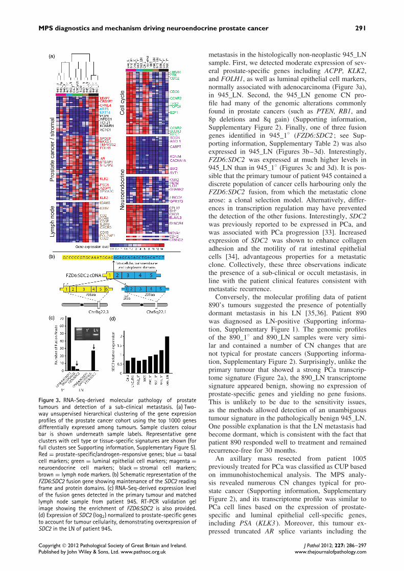
High-resolution molecular pathology and detectionof sub-clinical metastasisMPS allows genome/transcriptome-wide copy numberand expression profiling and the identification of fusiontranscripts that may represent unique tumour-specificbiomarkers [30]. We explored the global gene expres-sion signatures of the tumours by performing unsuper-vised hierarchical clustering of the genes differentiallyexpressed among samples. This resulted in sam-ple clusters accurately reflecting the tumour subtypeand/or sample composition (Figure 3a and Support-ing information, Supplementary Figure 5). The clusterof primary adenocarcinomas was distinct from that ofadenocarcinoma cell lines. As expected, the cluster-ing of PCa tumours as a group was driven by highexpression of prostate-specific stromal and basal cellmarkers and by the expression of androgen-responsive
Copyright 2012 Pathological Society of Great Britain and Ireland. J Pathol 2012; 227: 286–297Published by John Wiley & Sons, Ltd. www.pathsoc.org.uk www.thejournalofpathology.com

290 AV Lapuk et al
Figure 2. Alternative splicing in prostate tumours. (a) RT-PCR validation of alternatively spliced genes detected in prostate tumours andcell lines. Primers designed for the flanking constitutive exons with expected PCR product sizes are shown in the cartoons below each gelelectrophoresis image. RNA-Seq-based splicing data for inclusion isoform (corresponding to the upper PCR band) are shown below eachgel as splicing index bar charts for corresponding exons/junctions. (b) Possible functional consequences of alternative splicing. Proteindomains (Pfam [61]) are shown for three genes, where alternatively spliced regions fall within functional domains. Alternatively splicedregion of a gene is shown below the protein domain structure (blue = alternative exons; grey = constitutive exons) and its portion codingfor functional domains are indicated with dashed lines. (c) Heat map of the splicing index data for exons/junctions within 12 genes showingdifference in splicing in neuroendocrine samples versus primary adenocarcinoma tumours. Genes labelled on the right with magenta areinvolved in nervous system development [58–60]. Exon/junction IDs are shown on the right from gene symbols. (d) Alternative splicing ofthe PHF21A gene. The expression of the mutually exclusive exons of PHF21A by RNA-Seq is shown as a bar chart. PCR validation gel isshown underneath.
genes [eg KLK3 (PSA), KLK2, TMPRSS2 ]. LN sam-ples from patients 890 and 945 each had a substantialnormal LN tissue component (Figure 1 and Support-ing information, Supplementary Figure 1) and conse-quently exhibited high expression of LN-specific genes,including those associated with T cells, B cells, den-dritic cells, and macrophage cells (Figure 3a). NEPCatumours from patient 946 formed a separate clustertogether with those of patient 963, driven by the expres-sion of neuroendocrine markers. The xenograft 946_Xclustered closely with the corresponding tumours frompatient 946, suggesting that the xenograft retained thesalient features of the donor tumour. However, unlikethe donor NEPCa tumour, the xenograft expressed sev-eral androgen-responsive genes at low level. This canbe explained by the fact that the xenograft was grownin intact male mice supplemented with testosterone.
The mechanism of this, however, is unclear, since thexenograft does not express detectable AR protein [31].Interestingly, patient 963’s tumours exhibited a dualneuroendocrine and adenocarcinoma gene expressionphenotype and represented a distinct sub-cluster witha unique signature (Figure 3a). Immunohistochemistryruled out classical NED and adenocarcinoma, and indi-cated the presence of a hybrid luminal-neuroendocrinetumour in this patient, which is characterized in depthin ref 16.
Lymph node (LN) metastases are important deter-minants of disease progression and are challenging todetect by conventional histopathology. NHT treatmentbrings an additional challenge, as tumour foci shrinkand remaining deposits are even harder to detect [32].However, three lines of evidence from DNA- andRNA-Seq data suggested the presence of an occult
Copyright 2012 Pathological Society of Great Britain and Ireland. J Pathol 2012; 227: 286–297Published by John Wiley & Sons, Ltd. www.pathsoc.org.uk www.thejournalofpathology.com
MPS diagnostics and mechanism driving neuroendocrine prostate cancer 291
Figure 3. RNA-Seq-derived molecular pathology of prostatetumours and detection of a sub-clinical metastasis. (a) Two-way unsupervised hierarchical clustering of the gene expressionprofiles of the prostate cancer cohort using the top 1000 genesdifferentially expressed among tumours. Sample clusters colourbar is shown underneath sample labels. Representative geneclusters with cell type or tissue-specific signatures are shown (forfull clusters see Supporting information, Supplementary Figure 5).Red = prostate-specific/androgen-responsive genes; blue = basalcell markers; green = luminal epithelial cell markers; magenta =neuroendocrine cell markers; black = stromal cell markers;brown = lymph node markers. (b) Schematic representation of theFZD6:SDC2 fusion gene showing maintenance of the SDC2 readingframe and protein domains. (c) RNA-Seq-derived expression levelof the fusion genes detected in the primary tumour and matchedlymph node sample from patient 945. RT-PCR validation gelimage showing the enrichment of FZD6:SDC2 is also provided.(d) Expression of SDC2 (log2) normalized to prostate-specific genesto account for tumour cellularity, demonstrating overexpression ofSDC2 in the LN of patient 945.
metastasis in the histologically non-neoplastic 945_LNsample. First, we detected moderate expression of sev-eral prostate-specific genes including ACPP, KLK2,and FOLH1, as well as luminal epithelial cell markers,normally associated with adenocarcinoma (Figure 3a),in 945_LN. Second, the 945_LN genome CN pro-file had many of the genomic alterations commonlyfound in prostate cancers (such as PTEN, RB1, and8p deletions and 8q gain) (Supporting information,Supplementary Figure 2). Finally, one of three fusiongenes identified in 945_1◦ (FZD6:SDC2 ; see Sup-porting information, Supplementary Table 2) was alsoexpressed in 945_LN (Figures 3b–3d). Interestingly,FZD6:SDC2 was expressed at much higher levels in945_LN than in 945_1◦ (Figures 3c and 3d). It is pos-sible that the primary tumour of patient 945 contained adiscrete population of cancer cells harbouring only theFZD6:SDC2 fusion, from which the metastatic clonearose: a clonal selection model. Alternatively, differ-ences in transcription regulation may have preventedthe detection of the other fusions. Interestingly, SDC2was previously reported to be expressed in PCa, andwas associated with PCa progression [33]. Increasedexpression of SDC2 was shown to enhance collagenadhesion and the motility of rat intestinal epithelialcells [34], advantageous properties for a metastaticclone. Collectively, these three observations indicatethe presence of a sub-clinical or occult metastasis, inline with the patient clinical features consistent withmetastatic recurrence.
Conversely, the molecular profiling data of patient890’s tumours suggested the presence of potentiallydormant metastasis in his LN [35,36]. Patient 890was diagnosed as LN-positive (Supporting informa-tion, Supplementary Figure 1). The genomic profilesof the 890_1◦ and 890_LN samples were very simi-lar and contained a number of CN changes that arenot typical for prostate cancers (Supporting informa-tion, Supplementary Figure 2). Surprisingly, unlike theprimary tumour that showed a strong PCa transcrip-tome signature (Figure 2a), the 890_LN transcriptomesignature appeared benign, showing no expression ofprostate-specific genes and yielding no gene fusions.This is unlikely to be due to the sensitivity issues,as the methods allowed detection of an unambiguoustumour signature in the pathologically benign 945_LN.One possible explanation is that the LN metastasis hadbecome dormant, which is consistent with the fact thatpatient 890 responded well to treatment and remainedrecurrence-free for 30 months.
An axillary mass resected from patient 1005previously treated for PCa was classified as CUP basedon immunohistochemical analysis. The MPS analy-sis revealed numerous CN changes typical for pro-state cancer (Supporting information, SupplementaryFigure 2), and its transcriptome profile was similar toPCa cell lines based on the expression of prostate-specific and luminal epithelial cell-specific genes,including PSA (KLK3 ). Moreover, this tumour ex-pressed truncated AR splice variants including the
Copyright 2012 Pathological Society of Great Britain and Ireland. J Pathol 2012; 227: 286–297Published by John Wiley & Sons, Ltd. www.pathsoc.org.uk www.thejournalofpathology.com

292 AV Lapuk et al
oncogenic variant AR3(V7) [37,38], which, in thecohort, was present at the highest ratio relative to thefull length AR (Supporting information, Supplemen-tary Figure 7). Constitutively-active truncated isoformsof AR, including AR3(V7), are associated with pooroutcome [14] and are likely to represent an adaptiveresponse to castration [39]. These observations are con-sistent with the fact that patient 1005 had been exposedto several cycles of androgen deprivation therapy, andallowed unambiguous classification of patient’s 1005tumour as adenocarcinoma of prostatic origin.
Molecular signatures of castrate-resistantneuroendocrine prostate cancerIn the NEPCa tumours from patient 946, we ob-served a strong and distinct gene expression signature(Figure 2), with high expression of genes involved incellular proliferation, the cell cycle, and mitosis, as wellas genes important for endocrine biology (enrichmentanalysis, Fisher’s exact test, adjusted p value ≤0.0001;Supporting information, Supplementary Table 3). Wethen compiled a targeted, literature-driven panel ofup-regulated genes (compared with adenocarcinomas)which comprehensively defined the neuronal pheno-type in the tumours from patient 946 (Figure 4). Detec-tion of part of this gene signature in patient 963led to the identification of a novel hybrid luminal-neuroendocrine cancer subtype [16].
Significantly, in the NEPCa and hybrid PCa/NEPCatumours, we observed reduced expression of REST, atranscription factor considered to be the master re-pressor of neuronal differentiation [40] (Supportinginformation, Supplementary Figure 6). REST bindsto target sites [41] within genes important for aneuronal phenotype and prevents transcription. Inter-estingly, 28/50 of the transcriptionally active neu-roendocrine phenotype genes described in Figure 4harbour experimentally validated REST binding site(s).To determine the clinical relevance of this pattern,we mined publically available data. Taylor et al pro-filed 218 prostate tumours [9], and we have foundREST down-regulation and associated up-regulationof the neuronal signature in 50% of tumours with aNED/NEPCa component (Figure 4b). In contrast, only3% of other tumours from that cohort exhibited thispattern. The difference in the frequency of this patternin the two groups of tumours was highly significant(p < 0.0001, Fisher’s exact test).
Most if not all hallmarks of cancer are associ-ated with perturbed exon splicing [42]. To explorepossible contributions of AS to the neuroendocrinephenotype, we explored NEPCa-specific AS eventsand found a number of genes associated with neu-ronal functions (Figure 2c and Supporting information,Supplementary Tables 5 and 6). Of particular inter-est, a component of the REST transcriptional complex,PHF21A [43], is differentially spliced in NEPCa ver-sus PCa (Figure 2d). The PHF21A isoform expressedin NEPCa tumours was found to lack an exon, encoding
an AT-hook protein motif necessary for DNA binding(Figure 2b).
To experimentally validate the influence of RESTdown-regulation on associated gene and exon expres-sion changes, we performed an siRNA knockdown ofREST in LNCaP cells (Figure 4c). We observed an up-regulation of a number of NEPCa markers along withincreased exclusion of the PHF21A AT-hook encod-ing exon, consistent with observations in the clinicalsamples (Figure 4c).
The analysis also suggested a possible role ofglobal splicing regulation in NED and epithelial-to-mesenchymal transition (EMT). Genes differentiallyspliced between NEPCa and other tumours showedmoderate enrichment of cell morphology functions(Fisher’s exact test, adjusted p value ≤0.004; Support-ing information, Supplementary Table 5). Interestingly,we found alternatively spliced genes important for cellshape and invasion, which were reported by others toundergo alternative splicing in breast cancer cells uponinduction of EMT [44] (Supporting information, Sup-plementary Table 6). NED of PCa cells was previouslylinked to EMT [45], a process accompanied by mor-phological transformation of organized epithelial cellsinto isolated, migratory mesenchymal cells [46]. In linewith this, we noticed an up-regulation of the mesenchy-mal marker N-cadherin, and a number of cell adhesionand motility genes (Figures 3a and 4).
Prostatic adenocarcinomas are known to containrecurrent fusions [47], but the spectrum of NEPCafusions is largely unexplored. We identified 11 fusiontranscripts in NEPCa samples from patient 946 (Sup-porting information, Supplementary Table 2 and Sup-plementary Figure 4), which were shared by the urethraand penile metastases and the xenograft. Half of thefusions involved genes with restricted expression inneuroendocrine cells (Figure 4), thereby recapitulatingthe gene expression results. A similar situation wasobserved in the tumours of patient 963, where thefusion gene set mirrored the dual PCa/NEPCa geneexpression signature [16]. This finding supports recentreports that chromosomal translocations are cell-type-specific as they occur preferentially in transcriptionallyactive genes [12,48].
Identification of chromothripsis in prostate cancerWhile the existing paradigm dictates that chromosomalrearrangements occur gradually over time, recent evi-dence suggests that in at least 2–3% of cancers, tensto hundreds of genomic rearrangements involving onlyone or a few chromosomes can occur in a one-off cellu-lar crisis resulting in cancer-causing lesions [49]. Thisphenomenon, known as chromothripsis [18], was notpreviously described in prostate cancer and its associa-tion with outcome is unclear. We found evidence of itin the primary tumour genome from one of our high-risk patients (890) with a history of chronic prostatitis.The 890_1◦ genome contained fewer amplificationsthan other tumours. Two chromosome arms (2p and
Copyright 2012 Pathological Society of Great Britain and Ireland. J Pathol 2012; 227: 286–297Published by John Wiley & Sons, Ltd. www.pathsoc.org.uk www.thejournalofpathology.com

MPS diagnostics and mechanism driving neuroendocrine prostate cancer 293
Figure 4. The acquisition and maintenance of a neuroendocrine phenotype. (a) The bottom panel shows the neuronal phenotype identifiedin the tumours of patients 946 and 963. For patient 946, we used the mean expression from all three samples. Stars within the heatmap illustrate statistically different expression from other adenocarcinoma PCa samples. Red circles by the gene name indicate anexperimentally validated REST binding site within the gene loci. Rec–receptor. The top-left panel demonstrates a panel of regulatory genesup-regulated in the same tumour samples which we hypothesize to act synergistically to maintain expression of the neuronal phenotype.The top-right panel is a Circos plot [62] of the 946_Pn sample demonstrating the CN and fusion gene profile exhibited by the tumoursof patient 946. Chromosomes are arranged circularly end-to-end with each chromosome’s cytobands marked in the outer ring. The innerring displays copy number data inferred from genome sequencing, with red indicating gains and green indicating losses. Within the circle,genomic rearrangements are shown as arcs with the resultant validated fusion transcript annotated. Function of genes involved in fusionsare indicated by colour and provided in the key. (b) Recurrence of the REST down-regulation and the neuronal signature up-regulationfrom a in the prostate cancer cohort published by Taylor et al [9] showing 50% frequency in the NED/NEPCa subset of tumours (n = 16)versus 3% in the PCa subset (n = 134). The NED/NEPCa subset was defined by overexpression of at least one of the neuroendocrinemarkers (CHGA, CHGB or SYP ; Supporting information, Supplementary methods). (c) siRNA knockdown of REST in LNCaP cells. RT-PCR gelshows down-regulation of REST using two independent siRNA pools and concomitant up-regulation of neuronal signature genes as wellas change in PHF21A splicing. An arrow indicates the isoform lacking the AT-hook binding domain. Sc = scrambled RNA control. Westernblot using REST and vinculin antibodies is shown underneath RT-PCR, demonstrating significant down-regulation of REST protein uponsiRNA transfection.
Copyright 2012 Pathological Society of Great Britain and Ireland. J Pathol 2012; 227: 286–297Published by John Wiley & Sons, Ltd. www.pathsoc.org.uk www.thejournalofpathology.com

294 AV Lapuk et al
SLC35D2-LPPR1-MRPL50ZNF638-KCNS3-PPM1GABL1-ANXA4
ZNF638
KCNS3
PPM1G
ANXA4 EXOC6B
C2orf28
LCLAT1
Chr2p
Chr8
Chr9q
DENND1A
RALGPS1
SLC35D2
LPPR1
MRPL50 GPR107
Unique gene expression signature
DNA fragmentation
>8 fold higherexpression levels
than other tumours890 Primary tumour
Mean of all other tumours
Gene expression levels(log2 scale)
0 2 >5
Activation of survival mechanisms / salvage repair + chromothripsis phenotype including triple gene fusions
Chr2p Chr9q
Copy NumberLoss
S100A
9C
ST
2K
CN
S1
RE
G4*
CE
AC
AM
5**S
TM
N2
SH
ISA
3P
YD
C1
CE
AC
AM
7P
LAC
1LC
TS
LL1A
C104812.1B
3GN
T3
TC
P11
C11orf90N
RIP
3N
OX
1**D
KK
1P
IPS
OX
30A
C105446.4C
YP
4Z2P
SE
RP
INA
11S
ST
MU
C12
AC
CN
5G
PR
110A
SC
L2C
D177
CS
T6
BC
L2L10C
OL10A
1K
RT
20**G
PR
88P
TX
4M
MP
10LR
RC
31E
RP
27F
AP
IAP
P*
NO
X4
AL158140.9S
LC22A
16S
LC5A
1U
GT
2A3
AC
HE
SU
LT1C
4R
EG
1A*
AC
110619.2U
NC
93AH
TR
2BB
CL2L15
No Amplifications
ABL1
GPR107-C2orf28RALGPS1-EXOC6BTMEM55A-LCLAT1 DENND1A-ANXA4
Figure 5. Chromothripsis, inflammatory response, and fusion genes in patient 890. The top panel indicates multiple putative genomicbreakpoints between 2p and 9q resulting in validated fusion transcripts. The second panel demonstrates the top 50 uniquely expressedgenes in the primary tumour of patient 890 compared with all other tumour samples, showing enrichment (small arrows) of genes linkedto response to inflammation and apoptosis. ∗Pancreas-specific genes; ∗∗colon-specific genes. The third panel shows the fusion transcriptsderived from genomic rearrangements between chromosomes 2 and 9, including triple gene fusions. The bottom panel provides genomicsequence-derived copy number profiles, indicating the lack of amplifications and the high number of focal deletions, a hallmark ofchromothripsis [18,49].
9q) were found to harbour more deletions than otherchromosome arms and to possess a more segmentedCN profile (Supporting information, SupplementaryTable 1 and Supplementary Figure 2). Furthermore,nine out of 11 validated fusion events in 890_1◦
involved genes on 2p or 9q and two fusions involvedthree genes, providing evidence of complex chromo-somal rearrangements involving multiple breakpoints(Figure 5) (for details see the Supporting informa-tion, Supplementary data). On the transcriptome level,this tumour exhibited a unique expression signatureof genes with highly restricted expression patterns innormal tissues other than prostate (Figure 5). A com-prehensive literature search revealed that this panelincluded genes associated with resistance to bacterialinfection, response to inflammation, anti-apoptosis, andcell stress (indicated by arrows in Figure 5).
Discussion
We previously used MPS to identify an aggressivehybrid luminal-neuroendocrine tumour [16] and havenow expanded the study to six high-risk prostate cancerpatients. We demonstrated that integrative analysis ofshallow genome and deep transcriptome sequencing isa powerful tool for specific and sensitive molecularpathology, which can improve diagnosis and possiblyhave an impact on therapy. Moreover, we identifieda mechanism that may drive the emergence of theneuroendocrine phenotype that is frequently found inCRPC cancers.
Combined DNA- and RNA-Seq-based molecularprofiling of prostate tumours led to findings that haveimportant clinical implications. Firstly, we unambigu-ously identified a LN metastasis not detected by stan-dard histopathology; suggested a potentially dormantLN metastasis; classified a tumour of unknown ori-gin; and identified a new hybrid subtype of PCa [18].Identification of an occult LN metastasis in patient945 is associated with a poorer outcome and maywarrant more aggressive treatment [50]. The presenceand higher expression of gene fusion FZD6:SDC2in the LN compared with the primary tumour maysuggest clonal selection and supports the idea thatsuch tumour- and patient-specific markers may playa role in molecular pathology, as well as in mon-itoring disease progression and response to therapy,eg by measuring their levels in patient plasma sam-ples [30]. Conversely, the absence of a tumour tran-scriptome signature in the LN sample of LN-positivepatient 890 potentially suggests that this patient couldbenefit from a less aggressive therapeutic interven-tion. Thus, although based only on two patients, theseresults provide hope that MPS may ultimately aidin differentiating quiescent from aggressive metastaticprostate cancer. Secondly, in the US alone 31 000 peo-ple will be diagnosed with cancers of unknown primary(CUP) in 2012 (approximately 2% of all cancers diag-nosed), and identification of a primary site is impor-tant for therapeutic management of CUP patients [51].Although assignment to the primary site is possiblewith microarrays, MPS profiling significantly broadensthe spectrum of tumour-specific biomarkers, enabling
Copyright 2012 Pathological Society of Great Britain and Ireland. J Pathol 2012; 227: 286–297Published by John Wiley & Sons, Ltd. www.pathsoc.org.uk www.thejournalofpathology.com

MPS diagnostics and mechanism driving neuroendocrine prostate cancer 295
the identification of fusion transcripts, splice variants,and genomic breakpoints. Finally, the identification ofa novel hybrid luminal-neuroendocrine phenotype inaggressive conventional adenocarcinoma may imply apoorer outcome for patient 963, who should be man-aged accordingly.
Molecular characterization of NEPCa and hybridPCa/NEPCa tumours is a high-priority research area,given the prevalence of NED in advanced PCa andits association with resistance to current therapies [7].Our findings indicate that the REST transcriptionalcomplex, a master repressor of the neuronal pheno-type, may be one of the important determinants of theneuroendocrine phenotype in PCa. We observed down-regulation of REST with associated up-regulation ofneuronal genes, a pattern also found in 50% of tumourswith a NED/NEPCa component in a large indepen-dent cohort [9]. Simultaneously, the REST co-factorPHF21A appears to lose the AT-hook domain impor-tant for DNA binding through alternative splicing.Additionally, NED has been previously linked to EMT[45], and our observation of splicing regulation ofEMT-related functions in NEPCa supports this link.Finally, we found that the fusion genes that we iden-tified preferentially involved transcriptionally activegenes, consistent with previous reports [12,48]. Thus,we hypothesize that the fusion transcript spectrum mayreflect the evolutionary history of a tumour and provideinsight into cancer progression.
The presence of genome copy number aberra-tions and complex genome rearrangements is typicalfor high-risk prostate tumours [9,52]. However, wedetected evidence of chromothripsis in the primarytumour of patient 890 with a history of chronic prostati-tis and low PSA. Chromothripsis, recently describedin other cancers, results in many genomic aberrationslocalized to a few genomic regions [18]. Several mech-anisms were proposed to explain this phenomenon,one of which is an aborted apoptosis that may havebeen triggered by a noxious stimulus, such as radi-ation or infection [53]. Interestingly, this tumour’stranscriptome exhibited a signature of non-prostate-specific genes associated with an inflammatory immuneresponse, anti-apoptosis, and cell stress. A growingbody of evidence points to a possible role of prostaticinflammation in the aetiology of prostate cancer; how-ever, a causal link is yet to be established [54]. Ourobservation of chromothripsis in PCa and its poten-tial association with chronic prostatitis warrant furtherinvestigation. Furthermore, we detected the presenceof triple fusion genes, potentially arising from chro-mothripsis. Although the clinical relevance of this phe-nomenon is yet to be elucidated, it may represent adistinct mechanism of prostate carcinogenesis.
It is well established that PCa is biologically andclinically heterogeneous. Current methods of patientstratification based on integration of clinical historywith tumour histopathology still fail for a significantnumber of cancer patients, resulting in both over-and under-treatment. The possible benefits of MPS
for understanding of the biological underpinnings ofprostate cancer are undisputed. Moreover, a recentpilot study demonstrated that whole-genome, targeted-exome, and transcriptome sequencing is possible withina time frame relevant for cancer patients [55]. Sequenc-ing costs are expected to reach US $1000 or less pergenome in the near future. Thus, MPS has the potentialto improve on rational disease intervention and lowerhealthcare costs [56,57]. It is reasonable to expect thatMPS will ultimately complement conventional pathol-ogy in the future, resulting in improved patient man-agement.
Acknowledgment
This study was supported by the Canadian Institutesof Health Research (YZW, MG), Centres of Excel-lence for Commercialization and Research (MG), PNWProstate Cancer SPORE P50 CA097186, Prostate Can-cer Canada, Genome BC (CC), Prostate Cancer Foun-dation (AL, CC), and the Ontario Institute for CancerResearch (through funding provided by the Govern-ment of Ontario) to RB, PB, and CC via the CPC-GENE prostate cancer genome-network. The workin the AC laboratory was supported by NIH P50CA69568, U01 CA111275, and R01CA132874 grants.
Author contribution statement
CCC, SVV, AL, CW, and AWW conceived experi-ments and analysed data. CCC funded and directedthe project. SVV coordinated the project, data man-agement, and analysis. AVL, SVV, AM, SA, and FMconducted bioinformatics analyses and RHB coordi-nated statistical analyses. CW, BJM, RS, AH, SB,and AZ carried out experiments. MEG, LF, AH-C,and ECJ identified tumours and performed histopathol-ogy analysis. YW provided prostate tumour mousexenografts. YZ and MAM coordinated the next-generation sequencing. FHa, FHo, IH, SCS, CAM, andAMC assisted in bioinformatics analyses. MAR andHB provided mouse xenograft sequencing data. MEGco-directed the project and reviewed the manuscript.AZ, PCB, and RGB aided data analyses and reviewedthe manuscript. AVL, AWW, SVV, and CCC wrote themanuscript.
References
Note: References 63–65 are cited in the Supportinginformation to this article.
1. Siegel R, Naishadham D, Jemal A. Cancer statistics, 2012. Ca
Cancer J Clin 2012; 62: 10–29.2. Graefen M, Karakiewicz PI, Cagiannos I, et al . Validation study
of the accuracy of a postoperative nomogram for recurrence afterradical prostatectomy for localized prostate cancer. J Clin Oncol
2002; 20: 951–956.
Copyright 2012 Pathological Society of Great Britain and Ireland. J Pathol 2012; 227: 286–297Published by John Wiley & Sons, Ltd. www.pathsoc.org.uk www.thejournalofpathology.com

296 AV Lapuk et al
3. Graefen M, Karakiewicz PI, Cagiannos I, et al . International vali-
dation of a preoperative nomogram for prostate cancer recurrence
after radical prostatectomy. J Clin Oncol 2002; 20: 3206–3212.
4. Kattan MW, Eastham JA, Stapleton AM, et al . A preoperative
nomogram for disease recurrence following radical prostatectomy
for prostate cancer. J Natl Cancer Inst 1998; 90: 766–771.
5. Kattan MW, Wheeler TM, Scardino PT. Postoperative nomogram
for disease recurrence after radical prostatectomy for prostate
cancer. J Clin Oncol 1999; 17: 1499–1507.
6. Beltran H, Beer TM, Carducci MA, et al . New therapies for
castration-resistant prostate cancer: efficacy and safety. Eur Urol
2011; 60: 279–290.
7. Vashchenko N, Abrahamsson PA. Neuroendocrine differentiation
in prostate cancer: implications for new treatment modalities. Eur
Urol 2005; 47: 147–155.
8. Beltran H, Rickman DS, Park K, et al . Molecular characterization
of neuroendocrine prostate cancer and identification of new drug
targets. Cancer Discov 2011; 1: 487–495.
9. Taylor BS, Schultz N, Hieronymus H, et al . Integrative genomic
profiling of human prostate cancer. Cancer Cell 2010; 18:
11–22.
10. Tomlins SA, Laxman B, Varambally S, et al . Role of the
TMPRSS2–ERG gene fusion in prostate cancer. Neoplasia 2008;
10: 177–188.
11. Miyagi Y, Sasaki T, Fujinami K, et al . ETS family-associated
gene fusions in Japanese prostate cancer: analysis of 194 radical
prostatectomy samples. Mod Pathol 2010; 23: 1492–1498.
12. Berger MF, Lawrence MS, Demichelis F, et al . The genomic com-
plexity of primary human prostate cancer. Nature 2011; 470:
214–220.
13. Friedlander TW, Roy R, Tomlins SA, et al . Common structural
and epigenetic changes in the genome of castration resistant
prostate cancer. Cancer Res 2012; 72: 616.
14. Dehm SM, Tindall DJ. Alternatively spliced androgen receptor
variants. Endocr Relat Cancer 2011; 18: R183–R196.
15. Lee DJ, Cha EK, Dubin JM, et al . Novel therapeutics for the
management of castration-resistant prostate cancer (CRPC). BJU
Int 2012; 109: 968–985.
16. Wu C, Wyatt AW, Lapuk AV, et al . Integrated genome and tran-
scriptome sequencing identifies a novel form of hybrid and aggres-
sive prostate cancer. J Pathol 2012; 227: 53–61.
17. Shah SP, Morin RD, Khattra J, et al . Mutational evolution in a
lobular breast tumour profiled at single nucleotide resolution.
Nature 2009; 461: 809–813.
18. Maher CA, Wilson RK. Chromothripsis and human disease: piec-
ing together the shattering process. Cell 2012; 148: 29–32.
19. Griffith M, Griffith OL, Mwenifumbo J, et al . Alternative expres-
sion analysis by RNA sequencing. Nature Methods 2010; 7:
843–847.
20. Lapuk A, Marr H, Jakkula L, et al . Exon-level microarray anal-
yses identify alternative splicing programs in breast cancer. Mol
Cancer Res 2010; 8: 961–974.
21. Bullard JH, Purdom E, Hansen KD, et al . Evaluation of statistical
methods for normalization and differential expression in mRNA-
Seq experiments. BMC Bioinformatics 2010; 11: 94.
22. Reich M, Liefeld T, Gould J, et al . GenePattern 2.0. Nature Genet
2006; 38: 500–501.
23. McPherson A, Wu C, Hajirasouliha I, et al . Comrad: a novel
algorithmic framework for the integrated analysis of RNA-Seq and
WGSS data. Bioinformatics 2011; 27: 1481–1488.
24. Zoubeidi A, Ettinger S, Beraldi E, et al . Clusterin facilitates
COMMD1 and I-kappaB degradation to enhance NF-kappaB
activity in prostate cancer cells. Mol Cancer Res 2010; 8:
119–130.
25. McShane LM, Altman DG, Sauerbrei W, et al . REporting recom-mendations for tumor MARKer prognostic studies (REMARK).J Natl Cancer Inst 2005; 97: 1180–1184.
26. Cutz JC, Guan J, Bayani J, et al . Establishment in severe com-bined immunodeficiency mice of subrenal capsule xenografts andtransplantable tumor lines from a variety of primary human lungcancers: potential models for studying tumor progression-relatedchanges. Clin Cancer Res 2006; 12: 4043–4054.
27. Collins CC, Volik SV, Lapuk A, et al . Next generation sequencingof prostate cancer from a patient identifies a deficiency of methylth-ioadenine phosphorylase (MTAP), an exploitable tumor target. Mol
Cancer Ther 2012; 11: 775–783.28. Markert EK, Mizuno H, Vazquez A, et al . Molecular classification
of prostate cancer using curated expression signatures. Proc Natl
Acad Sci U S A 2011; 108: 21276–21281.29. Fine SW, Gopalan A, Leversha MA, et al . TMPRSS2–ERG gene
fusion is associated with low Gleason scores and not with high-grade morphological features. Mod Pathol 2010; 23: 1325–1333.
30. Leary RJ, Kinde I, Diehl F, et al . Development of personalizedtumor biomarkers using massively parallel sequencing. Sci Transl
Med 2010; 2: 20ra14.31. Tung WL, Wang Y, Gout PW, et al . Use of irinotecan for treat-
ment of small cell carcinoma of the prostate. Prostate 2011; 71:675–681.
32. Gomella LG, Singh J, Lallas C, et al . Hormone therapy in themanagement of prostate cancer: evidence-based approaches. Ther
Adv Urol 2010; 2: 171–181.33. Popovic A, Demirovic A, Spajic B, et al . Expression and prognos-
tic role of syndecan-2 in prostate cancer. Prostate Cancer Prostatic
Dis 2010; 13: 78–82.34. Choi S, Kim Y, Park H, et al . Syndecan-2 overexpression regu-
lates adhesion and migration through cooperation with integrinalpha2. Biochem Biophys Res Commun 2009; 384: 231–235.
35. Aguirre-Ghiso JA. Models, mechanisms and clinical evidence forcancer dormancy. Nature Rev Cancer 2007; 7: 834–846.
36. Shiozawa Y, Pedersen EA, Havens AM, et al . Human prostatecancer metastases target the hematopoietic stem cell niche toestablish footholds in mouse bone marrow. J Clin Invest 2011;121: 1298–1312.
37. Guo Z, Yang X, Sun F, et al . A novel androgen receptor splicevariant is up-regulated during prostate cancer progression andpromotes androgen depletion-resistant growth. Cancer Res 2009;69: 2305–2313.
38. Sun S, Sprenger CC, Vessella RL, et al . Castration resistance inhuman prostate cancer is conferred by a frequently occurring andro-gen receptor splice variant. J Clin Invest 2010; 120: 2715–2730.
39. Watson PA, Chen YF, Balbas MD, et al . Constitutively activeandrogen receptor splice variants expressed in castration-resistantprostate cancer require full-length androgen receptor. Proc Natl
Acad Sci U S A 2010; 107: 16759–16765.40. Ooi L, Wood IC. Chromatin crosstalk in development and disease:
lessons from REST. Nature Rev Genet 2007; 8: 544–554.41. Mortazavi A, Leeper Thompson EC, Garcia ST, et al . Compara-
tive genomics modeling of the NRSF/REST repressor network:from single conserved sites to genome-wide repertoire. Genome
Res 2006; 16: 1208–1221.42. Venables JP. Unbalanced alternative splicing and its significance
in cancer. BioEssays 2006; 28: 378–386.43. Hakimi MA, Bochar DA, Chenoweth J, et al . A core-BRAF35
complex containing histone deacetylase mediates repression ofneuronal-specific genes. Proc Natl Acad Sci U S A 2002; 99:7420–7425.
44. Shapiro IM, Cheng AW, Flytzanis NC, et al . An EMT-drivenalternative splicing program occurs in human breast cancer andmodulates cellular phenotype. PLoS Genet 2011; 7: e1002218.
Copyright 2012 Pathological Society of Great Britain and Ireland. J Pathol 2012; 227: 286–297Published by John Wiley & Sons, Ltd. www.pathsoc.org.uk www.thejournalofpathology.com

MPS diagnostics and mechanism driving neuroendocrine prostate cancer 297
45. McKeithen D, Graham T, Chung LW, et al . Snail transcriptionfactor regulates neuroendocrine differentiation in LNCaP prostatecancer cells. Prostate 2010; 70: 982–992.
46. Kalluri R, Weinberg RA. The basics of epithelial–mesenchymaltransition. J Clin Invest 2009; 119: 1420–1428.
47. Clark JP, Cooper CS. ETS gene fusions in prostate cancer. NatureRev Urol 2009; 6: 429–439.
48. Lin C, Yang L, Tanasa B, et al . Nuclear receptor-induced chromo-somal proximity and DNA breaks underlie specific translocationsin cancer. Cell 2009; 139: 1069–1083.
49. Stephens PJ, Greenman CD, Fu B, et al . Massive genomic rear-rangement acquired in a single catastrophic event during cancerdevelopment. Cell 2011; 144: 27–40.
50. Pagliarulo V, Hawes D, Brands FH, et al . Detection of occultlymph node metastases in locally advanced node-negative prostatecancer. J Clin Oncol 2006; 24: 2735–2742.
51. Pavlidis N, Briasoulis E, Hainsworth J, et al . Diagnostic and thera-peutic management of cancer of an unknown primary. Eur J Cancer2003; 39: 1990–2005.
52. Paris PL, Andaya A, Fridlyand J, et al . Whole genome scanningidentifies genotypes associated with recurrence and metastasis inprostate tumors. Hum Mol Genet 2004; 13: 1303–1313.
53. Tubio JM, Estivill X. Cancer: when catastrophe strikes a cell.Nature 2011; 470: 476–477.
54. Sfanos KS, De Marzo AM. Prostate cancer and inflammation: theevidence. Histopathology 2012; 60: 199–215.
55. Roychowdhury S, Iyer MK, Robinson DR, et al . Personalizedoncology through integrative high-throughput sequencing: a pilotstudy. Sci Transl Med 2011; 3: 111ra121.
SUPPORTING INFORMATION ON THE INTERNET
The following supporting information may be found in the online version of this article.
Supplementary data and methods
Figure S1. Haematoxylin and eosin (H&E) staining images of patient samples from fresh frozen sections of primary tumours, axillary massand invasion of urethra (A), and FFPE sections of lymph node samples at 2× and 20× magnification (B).
Figure S2. Whole-genome CN profile of 11 samples from the prostate tumour cohort, derived from DNA-Seq.
Figure S3. Concordance of CN profiles derived from aCGH and DNA-Seq.
Figure S4. Representative results of fusion gene validations.
Figure S5. Unsupervised hierarchical clustering of the top 1000 differentially expressed genes (by s.d. of expression across samples) in theprostate cancer cohort.
Figure S6. RNA-Seq-derived expression of the REST gene showing reduced expression in NEPCa compared with PCa samples.
Figure S7. Alternative splicing of the androgen receptor gene (AR) detected in the high-risk prostate tumours.
Table S1. Summary of tumour genome sequencing and CN aberrations.
Table S2. Fifty-three novel fusion genes identified in prostate tumours.
Table S3. PCR validation of predicted alternative splicing events.
Table S4. The list of genes differentially expressed between PCa and NEPCa.
Table S5. Top enriched pathways and functions identified with Ingenuity enrichment analysis.
Table S6. The list of exons/junctions alternatively spliced between PCa and NEPCa.
56. Armstrong K. Can genomics bend the cost curve? J Am Med Assoc
2012; 307: 1031–1032.57. Drmanac R. The advent of personal genome sequencing. Genet
Med 2011; 13: 188–190.58. Dunah AW, Hueske E, Wyszynski M, et al . LAR receptor protein
tyrosine phosphatases in the development and maintenance ofexcitatory synapses. Nature Neurosci 2005; 8: 458–467.
59. Maric D, Fiorio Pla A, Chang YH, et al . Self-renewing and dif-ferentiating properties of cortical neural stem cells are selectivelyregulated by basic fibroblast growth factor (FGF) signaling viaspecific FGF receptors. J Neurosci 2007; 27: 1836–1852.
60. Zhang H, Webb DJ, Asmussen H, et al . Synapse formation isregulated by the signaling adaptor GIT1. J Cell Biol 2003; 161:131–142.
61. Finn RD, Mistry J, Tate J, et al . The Pfam protein familiesdatabase. Nucleic Acids Res 2010; 38: D211–D222.
62. Krzywinski M, Schein J, Birol I, et al . Circos: an informationaesthetic for comparative genomics. Genome Res 2009; 19:1639–1645.
63. Wang XS, Prensner JR, Chen G, et al . An integrative approachto reveal driver gene fusions from paired-end sequencing data incancer. Nature Biotechnol 2009; 27: 1005–1011.
64. Gasi D, van der Korput HA, Douben HC, et al . Overexpression offull-length ETV1 transcripts in clinical prostate cancer due to genetranslocation. PLoS One 2011; 6: e16332.
65. Attard G, Clark J, Ambroisine L, et al . Duplication of the fusionof TMPRSS2 to ERG sequences identifies fatal human prostatecancer. Oncogene 2008; 27: 253–263.
Copyright 2012 Pathological Society of Great Britain and Ireland. J Pathol 2012; 227: 286–297Published by John Wiley & Sons, Ltd. www.pathsoc.org.uk www.thejournalofpathology.com