Free Energetics and the Role of Water in the Permeation of Methyl Guanidinium across the...
15
Free Energetics and the Role of Water in the Permeation of Methyl Guanidinium across the Bilayer−Water Interface: Insights from Molecular Dynamics Simulations Using Charge Equilibration Potentials Shuching Ou, † Timothy R. Lucas, † Yang Zhong, †,§ Brad A. Bauer, ‡ Yuan Hu, † and Sandeep Patel* ,† † Department of Chemistry and Biochemistry, University of Delaware, Newark, Delaware 19716, United States ‡ Department of Physical and Biological Sciences, The College of Saint Rose, Albany, New York 12203, United States § Genomics Institute of the Novartis Research Foundation, 10675 John J. Hopkins Drive, San Diego, California 92121, United States * S Supporting Information ABSTRACT: Combining umbrella sampling molecular dynamics (MD) simulations, the weighted histogram analysis method (WHAM) for unbiasing probabilities, and polarizable charge equilibration force fields, we compute the potential of mean force for the reversible transfer of methyl guanidinium from bulk solution to the center of a model DPPC bilayer. A 5 kcal/mol minimum in the potential of mean force profile for membrane permeation suggests that the analogue will preferentially reside in the headgroup region of the lipid, qualitatively in agreement with previously published results. We find the potential of mean force for permeation to be approximately 28 kcal/mol (relative to the minimum in the headgroups), within the range of values reported for similar types of simulations using fixed-charge force fields. From analysis of the lipid structure, we find that the lipid deformation leads to a substantial destabilizing contribution to the free energy of the methyl guanidinium as it resides in the bilayer center, though this deformation allows more efficient stabilization by water defects and transient pores. Water in the bilayer core stabilizes the charged residue. The role of water in stabilizing or destabilizing the solute as it crosses the bilayer depends on bulk electrolyte concentration. In 1 M KCl solution, the water contribution to the potential of mean force is stabilizing over the entire range of the permeation coordinate, with the sole destabilizing force originating from the anionic species in solution. Conversely, methyl guanidinium experiences net destabilization from water in the absence of electrolyte. The difference in solvent contributions to permeation free energy is traced to a local effect arising from differences in water density in the bilayer−water solution interface, thus leading to starkly opposite net forces on the permeant. The origin of the local water density differential rests with the penetration of hydrated chloride anions into the solution−bilayer interface. Finally, water permeation into the bilayer is required for the deformation of individual lipid molecules and permeation of ions into the membrane. From simulations where water is first excluded from the bilayer center where methyl guanidinium is restrained and then, after equilibration, allowed to enter the bilayer, we find that in the absence of any water defects/permeation into the bilayer, the lipid headgroups do not follow the methyl guanidinium. Only when water enters the bilayer do we see deformation of individual lipid molecules to associate with the amino acid analogue at bilayer center. I. INTRODUCTION Recent years have witnessed great interest in the understanding of the molecular origins of the presence of charged and polar amino acid residues in ostensibly hydrophobic lipid bilayer environments. 1−13 The motivation for such a microscopic understanding stems from the broad range of biophysical processes predicated on the interactions between such protein residues and hydrophobic lipid chains. These processes range from voltage gating in select ion channels, 14−16 permeation of cationic residue enriched cell-penetrating peptides for trans- porting cargo across the cellular membrane, 17−23 and the action of antimicrobial peptides upon interaction with native cellular membranes. Understanding of these protein−lipid interactions has sought recourse in hydrophobicity scales quantifying relative partitioning propensities of different amino acid side chains from aqueous to bilayer-like environments. 24 Elaborating upon ideas of partitioning of functional chemical groups between hydrophilic and hydrophobic environments, recent work has broadened the palette of hydrophobicity scales attempting to address the relative free energetics of Received: July 22, 2012 Revised: February 8, 2013 Published: February 14, 2013 Article pubs.acs.org/JPCB © 2013 American Chemical Society 3578 dx.doi.org/10.1021/jp400389z | J. Phys. Chem. B 2013, 117, 3578−3592
-
Upload
independent -
Category
Documents
-
view
2 -
download
0
Transcript of Free Energetics and the Role of Water in the Permeation of Methyl Guanidinium across the...

Free Energetics and the Role of Water in the Permeation of MethylGuanidinium across the Bilayer−Water Interface: Insights fromMolecular Dynamics Simulations Using Charge EquilibrationPotentialsShuching Ou,† Timothy R. Lucas,† Yang Zhong,†,§ Brad A. Bauer,‡ Yuan Hu,† and Sandeep Patel*,†
†Department of Chemistry and Biochemistry, University of Delaware, Newark, Delaware 19716, United States‡Department of Physical and Biological Sciences, The College of Saint Rose, Albany, New York 12203, United States§Genomics Institute of the Novartis Research Foundation, 10675 John J. Hopkins Drive, San Diego, California 92121, United States
*S Supporting Information
ABSTRACT: Combining umbrella sampling molecular dynamics (MD)simulations, the weighted histogram analysis method (WHAM) forunbiasing probabilities, and polarizable charge equilibration force fields,we compute the potential of mean force for the reversible transfer of methylguanidinium from bulk solution to the center of a model DPPC bilayer. A 5kcal/mol minimum in the potential of mean force profile for membranepermeation suggests that the analogue will preferentially reside in theheadgroup region of the lipid, qualitatively in agreement with previouslypublished results. We find the potential of mean force for permeation to beapproximately 28 kcal/mol (relative to the minimum in the headgroups),within the range of values reported for similar types of simulations usingfixed-charge force fields. From analysis of the lipid structure, we find that thelipid deformation leads to a substantial destabilizing contribution to the freeenergy of the methyl guanidinium as it resides in the bilayer center, thoughthis deformation allows more efficient stabilization by water defects and transient pores. Water in the bilayer core stabilizes thecharged residue. The role of water in stabilizing or destabilizing the solute as it crosses the bilayer depends on bulk electrolyteconcentration. In 1 M KCl solution, the water contribution to the potential of mean force is stabilizing over the entire range ofthe permeation coordinate, with the sole destabilizing force originating from the anionic species in solution. Conversely, methylguanidinium experiences net destabilization from water in the absence of electrolyte. The difference in solvent contributions topermeation free energy is traced to a local effect arising from differences in water density in the bilayer−water solution interface,thus leading to starkly opposite net forces on the permeant. The origin of the local water density differential rests with thepenetration of hydrated chloride anions into the solution−bilayer interface. Finally, water permeation into the bilayer is requiredfor the deformation of individual lipid molecules and permeation of ions into the membrane. From simulations where water isfirst excluded from the bilayer center where methyl guanidinium is restrained and then, after equilibration, allowed to enter thebilayer, we find that in the absence of any water defects/permeation into the bilayer, the lipid headgroups do not follow themethyl guanidinium. Only when water enters the bilayer do we see deformation of individual lipid molecules to associate with theamino acid analogue at bilayer center.
I. INTRODUCTION
Recent years have witnessed great interest in the understandingof the molecular origins of the presence of charged and polaramino acid residues in ostensibly hydrophobic lipid bilayerenvironments.1−13 The motivation for such a microscopicunderstanding stems from the broad range of biophysicalprocesses predicated on the interactions between such proteinresidues and hydrophobic lipid chains. These processes rangefrom voltage gating in select ion channels,14−16 permeation ofcationic residue enriched cell-penetrating peptides for trans-porting cargo across the cellular membrane,17−23 and the actionof antimicrobial peptides upon interaction with native cellular
membranes. Understanding of these protein−lipid interactionshas sought recourse in hydrophobicity scales quantifyingrelative partitioning propensities of different amino acid sidechains from aqueous to bilayer-like environments.24 Elaboratingupon ideas of partitioning of functional chemical groupsbetween hydrophilic and hydrophobic environments, recentwork has broadened the palette of hydrophobicity scalesattempting to address the relative free energetics of
Received: July 22, 2012Revised: February 8, 2013Published: February 14, 2013
Article
pubs.acs.org/JPCB
© 2013 American Chemical Society 3578 dx.doi.org/10.1021/jp400389z | J. Phys. Chem. B 2013, 117, 3578−3592

partitioning; this has been possible due to novel experiments onwell-characterized integral-membrane protein systems8 as wellas elucidation of structural aspects of the machinery implicatedin the synthesis and insertion of membrane proteins uponsynthesis in the ribosome.25 More recently, there appears to bea convergence of molecular modeling based predictions ofrelative free energetics of different amino acid side chains aspart of a macromolecular assembly and experiment.7 Furtherfactors possibly contributing to interactions of charge and polarspecies in lipid bilayers include bilayer thickness, nonadditivityof interactions between nonbilayer components,26,27 specificityof protein sequence to specific bilayer composition,28−31
interactions of a particular amino side chain in the bilayerwith lipid headgroups and water, and lipid deformation(coupled with the ease of deformability of the lipid).10,18,32
In conjunction with recent experimental advances in studyingthe relative stability of charged amino acid residues in bilayerenvironments (within several contexts including cell-penetrat-ing peptides, the presence of charged voltage gating and sensingdomains in potassium channels, and other biochemicaltransformations), there has been a steady push for developingnovel molecular modeling methods for treating the heteroge-neous environments presented by lipid bilayers of variouscompositions. In particular, nonadditive force fields are beingpursued for modeling lipid bilayers. Though nonadditive forcefields have been pursued for over two decades now, onlyrecently have there been substantial, published studiesdocumenting the application of such models to the study ofbiophysical problems of general interest.33−46,46−54 Recently,we, along with others, have published results of the firstapplications of polarizable, nonadditive force fields for treatinglipid bilayers in a fully atomistic molecular simulation. We havebeen developing a class of nonadditive, polarizable force fieldsbased on the charge equilibration (CHEQ) or fluctuatingcharge (FQ) formalism. One of the first attempts to applyCHEQ-based force fields to lipid bilayers was by Shimizu andco-workers;55 in their study, the authors explored theconsequence of the extent of charge transfer in extendedsystems defining the nature of charge transfer effects andpossible hyper-polarization in such models. The authorsdemonstrated the need for some approach to control thesuperlinear polarizability in applying charge equilibrationmodels in a manner that does not constrain charge transferin a systematic and physically meaningful way. Recently, Vachaet al.56 applied a fully polarizable force field (lipid, water, andions) to the study of monovalent ion interactions withphosphatidylcholine membranes and associated effects at themembrane−water interface. Furthermore, Harder et al.45
examined the difference in the interfacial dipole potentialbetween the pure liquid−vapor interface of water and that of aDPPC monolayer. The authors presented the case forpolarization helping to accurately predict the potentialdifference between these two systems compared to existingnonpolarizable force fields. Over the last several years, ourgroup has been involved in a substantial effort to developnonadditive electrostatic models for lipid bilayers to be used inconjunction with molecular dynamics simulations. We havepresented a systematic parametrization of a first-generationcharge equilibration force field based on the DPPC lipid.54,57 Inthis study, we continue further application of our models toexplore the free energetics of an analogue of arginine, methylguanidinium, in a model DPPC bilayer. This complementsearlier studies using both polarizable and nonpolarizable force
fields in conjunction with molecular dynamics simulations tostudy this canonical system. We do not claim that polarizableforce fields are a panacea to all deficiencies of the current stateof the art force fields; this study further explores applications ofcurrent polarizable force field technology to systems ofbiochemical interest.In this work, we continue to explore the free energetics of
charged species, in this case methyl guanidinium, permeatingacross a model lipid bilayer. We consider a model DPPC bilayeras the oft-used proxy for a physiological membrane. Weconsider pure water and 1 M KCl systems, the saltconcentration being higher than physiological conditions tosufficiently sample configurations of the ions in the vicinity ofthe membrane−water interface. We further explore the natureof water and its ability to mediate deformations of the lipidbilayer in stabilizing a charged species at the center of thismodel bilayer. There has been much literature on thepermeation of polar and charged solutes into lipid membraneswith an accompanying hydration shell of water molecules.12,13
We address the idea that this accompanying water alsomediates the interactions between the lipid headgroups andpermeating solute. We attempt to study this effect byperforming molecular simulations where water is expelledfrom the core of the bilayer. We then proceed to remove thisconstraint to observe any deformations that accompany thepermeation of water into the bilayer. Section II discussesgeneral aspects of the charge equilibration force field and itsimplementation. Section IIC addresses the validation of themethyl guanidinium force field through the calculation ofhydration enthalpies and comparison to previous literature data.Section IID discusses results of the potential of mean force(PMF) for methyl guanidinium permeation. Section IIIpresents extended discussion of the results of hydration freeenergy calculations, the potential of mean force, decompositionof the potential of mean force into contributions from systemcomponents, an explanation of the differences of the PMFscomputed in salt solution versus pure aqueous environments,and finally discussion of the role of water in potentiallymediating membrane deformation. Section IV concludes with asummary and final thoughts.
II. METHODS
A. Charge Equilibration Force Fields. We next considerdetails of the charge equilibration (CHEQ) method andconsiderations in our specific implementation of this method.An additive (or nonpolarizable) formalism for force fields isbased on the construct that all atomic partial charges are fixedthroughout the course of the simulations. Alternatively, we canconsider the variation of atomic partial charge using the CHEQformalism.46,58−67 The CHEQ formalism is based on Sander-son’s idea of electronegativity equilibration66,67 in which thechemical potential is equilibrated via the redistribution ofcharge density. In a classical sense, charge density is reduced topartial charges, Qα, on each atomic site α. The charge-dependent energy for a system of M molecules containing Ni
atoms per molecule is then expressed as
The Journal of Physical Chemistry B Article
dx.doi.org/10.1021/jp400389z | J. Phys. Chem. B 2013, 117, 3578−35923579

∑ ∑
∑ ∑ ∑ ∑ ∑ ∑
∑ ∑
χ
πε
λ
=
+ + ′ ′
+ −
αα α
α βα β α β
= =
= = = = = =
= =
E R Q Q
J Q QQ Q
r
Q Q
( , )
12
12 4
( )
i
M N
i i
i
M
j
M N N
i j i ji
MN
j
MNi j
ij
j
M
ji
N
ji j
CHEQ1 1
1 1 1 1 1 1 0
1 1
Total
i j
(1)
where the χ terms represent the atomic electronegativitieswhich control the directionality of electron flow and J termsrepresent the atomic hardnesses which control the resistance toelectron flow to or from the atom. Although these parametersare derived from the definitions of electron affinity andionization potential, they are treated as empirical parameters forindividual atom types. Heterogeneous hardness elements thatdescribe the interaction between two different atom types arecalculated using the combining rule68 on the parametrizedhomogeneous hardness elements (Jii°)
° ° =° + °
+ ° + °J R J J
J J
J J R( , , )
( )
1.0 ( )ij ij ii jj
ii jj
ii jj ij
12
14
2 2(2)
where Rij is the distance between atoms i and j. Thiscombination locally screens Coulombic interactions butprovides the correct limiting behavior at atomic separationsgreater than approximately 2.5 Å. The standard Coulombinteraction between sites not involved in the dihedral, angle, orbonded interactions (denoted by primed summation) with eachother is included as the third term in eq 1. The second term ineq 1 represents the local charge transfer interaction, which isusually restricted to within a molecule or an appropriate chargenormalization unit. Currently, we do not consider intermo-lecular charge transfer, although approaches to incorporate thiseffect have recently been developed and applied to liquidwater.69 Charge is constrained via a Lagrange multiplier, λ,which is included for each molecule as indicated in the last termof eq 1. We remark that use of multiple charge normalizationunits can modulate molecular polarizability by limitingintramolecular charge transfer to physical distances. Such anapproach controls previously observed superlinear polarizabilityscaling,53,70,71 which also manifests as the polarizationcatastrophe (in point polarizable force fields),53,72 while alsodeveloping a construct for piecing together small molecularentities into macromolecules. The molecular polarizability inthe CHEQ formalism can be calculated as53
α = ′γβ β γ−R J RT 1
(3)
where J′ is the atomic hardness matrix augmented with theappropriate rows and columns to treat the charge conservationconstraints. Rγ and Rβ are the γ and β Cartesian coordinates ofthe atomic position vectors; these vectors are augmented toappropriately match the dimensions of the atomic hardnessmatrix. Representative values for the molecular polarizability fora wide variety of small molecule analogues of biomolecularfunctional groups are given in ref 58.Charge degrees of freedom are propagated via an extended
Lagrangian formulation, thus providing for electronegativityequilibration at each dynamics step. The system Lagrangian is
∑ ∑ ∑ ∑
∑ ∑λ
= +
− − −
αα
α
αα
α
αα
= = = =
= =
⎛⎝⎜
⎞⎠⎟
⎛⎝⎜
⎞⎠⎟L m
drdt
mdQ
dt
E Q r Q Q
12
12
( , ) (( ) )
i
M N
ii
i
M N
Q ii
i
M
i
N
i i
1 1
2
1 1,
2
1 1
total
(4)
where the first two terms represent the nuclear and chargekinetic energies; the third term is the potential energy; and thefourth term is the molecular charge neutrality constraintenforced on each molecule i via a Lagrange multiplier λi. Thefictitious charge dynamics are determined using a charge “mass”with units of (energy time2/charge2), which is analogous to theuse of an adiabaticity parameter in fictitious wave functiondynamics in Car−Parrinello (CP) type methods.61,73 Chargesare propagated based on the forces arising from differencesbetween the average electronegativity of a molecule and theinstantaneous electronegativity at an atomic site.
B. Simulation Setup. The membrane system consists of 72DPPC lipid molecules, arranged in a bilayer (36 molecules perleaflet), solvated with an approximately 1 M potassium chloridesolution composed of 3203 TIP4P-FQ water molecules,61 57K+, and 58 Cl− ions and containing a single positively chargedmethyl guanidinium (mguanH+) molecule. We performedmolecular dynamics simulations using the CHEQ formalismin the constant particle, normal pressure, lateral surface area,and temperature (NPAT) ensemble at a pressure of 1 atm, atemperature of 323 K, and an area per lipid of 63.0 Å2, abovethe temperature of the liquid−gel phase transition74 and withinthe experimental range for the area per lipid.75 The pressurewas maintained using the Langevin piston method76 with apiston mass of 2025 amu in the z-direction, normal to thebilayer. The temperature of the simulation was controlled usingthe Nose−Hoover method76,77 with a thermal piston mass of3000 kcal/mol·ps2. Long-range electrostatics were accountedfor by the particle mesh Ewald (PME) summation78,79 with a48 × 48 × 80 Å3 fast Fourier transform (FFT) grid, fourth-order B-spline interpolation, and screening parameter κ = 0.32.Dynamics were propagated using the Leapfrog Verletintegrator76 with a 0.5 fs time step.The CHEQ lipid force field has been developed and
validated based on NVT ensemble simulations of hydratedbilayer and monolayer systems.50,54,57 In the currentsimulations we use an intermediate, revised version of theCHEQ lipid force field in which (1) the Lennard-Jones (LJ)nonbonded parameters between headgroup atoms were variedto facilitate surface tensionless MD simulations in constantNPT simulations and (2) selected dihedral parameters wereadjusted to better match ab initio torsion profiles. The details ofthe lipid force field modification are given in the SupportingInformation (SI) with area per lipid and torsion fitting resultspresented in Figures S1 and S2 (SI), respectively. The CHEQpolarizable force field model for methyl guanidinium wasconstructed by adapting the CHARMM CHEQ force field forproteins,46,58 extending the electrostatic parameters, hardnesses,and electronegativities from the protein force field for anarginine side chain. We stress that we perform all simulations inthe NPAT ensemble.The methyl guanidinium was partitioned into two charge
normalization units to control polarizability scaling. The firstunit encompasses the methyl (−CH3) and secondary amine(−NH) groups, while the second unit includes the centralcarbon and two primary amine groups (−C(NH2)2). This
The Journal of Physical Chemistry B Article
dx.doi.org/10.1021/jp400389z | J. Phys. Chem. B 2013, 117, 3578−35923580

partitioning allows for consistency with the existing CHARMMCHEQ protein force field and permits use of the existingnonbond (LJ and electrostatic) parameters for arginine fromthe polarizable force field. The partitioning approach has beenapplied previously by our group in the treatment of proteins,lipids, and carbohydrates.52−54,57,71,80,81 The isotropic, gas-phase molecular polarizability for this molecule was determinedto be 6.4 Å3 compared to the value of 7.0 Å3 from MP2/aug-cc-pVTZ calculations. A reduction in the force field molecularpolarizability is consistent with the decrease in the intrinsicmolecular polarizability observed, in theory, when going fromthe gas to the condensed phase. However, the exact magnitudeof this reduction is currently unknown and poses a challengefor force field development. We consider only the protonatedmethyl guanidinium model compound for our studies. Recentstudies2,9,82 have explored the shift in stability of the protonatedand unprotonated species, as manifested in pKa shifts, along thebilayer normal. For thin membranes, it has been suggested thatthe protonated state is plausible and sustained by acombination of lipid membrane deformation and long-livedpolar (water and lipid) pores across the bilayer stabilizing thecharge. Acknowledging these studies, we focus here on the freeenergetics of partitioning of the charged species.The nonbonded (Lennard-Jones) interactions between
methyl guanidinium and lipid headgroup analogues dimethylphosphate (DMP) and tetramethyl ammonium (TMA), as wellas with solvent species (water and chloride), were validatedthrough the comparison of gas-phase small-molecule geo-metries and interaction energies calculated using the CHEQforce field with those calculated using high-level quantummechanics. All quantum mechanical (QM) dimer structureswere optimized at the MP2/aug-cc-pVTZ level with theexception of the mguanH+/DMP dimer, which was optimizedusing the 6-31++g(2d,p) basis set. Interaction energies were
calculated at the MP2/aug-cc-pVTZ level using the previouslyoptimized structures. Hydrogen bond distances and interactionenergies are shown in Table 1, and although overestimated, thehydrogen bond distances exhibit suitable agreement betweenthe force field and MP2/aug-cc-pVTZ optimized structures.Interaction energies are consistently underestimated but are infair agreement with QM values. In the absence of laboriousreparameterization of the methyl guanidinium interactions withall system species, we consider it sufficient that for the purposesof the present work the trend of relative interaction strengthsbased on QM and force field calculations is equivalent, with thestrongest interactions occurring between mguanH+ and DMP.
C. Methyl Guanidinium Hydration Free Energy viaThermodynamic Integration. To further describe thequality of the present methyl guanidinium interactions withsolvent, as well as to compare with existing literature data fromearlier computational and experimental studies, hydration freeenergies of methyl guanidinium in dilute aqueous and hexanesolutions were calculated using a cubic box of 988 TIP4P-FQwater molecules (or 216 hexane molecules) and one mguanH+
molecule via thermodynamic integration (TI). Following thetwo-step decoupling procedure described by Warren et al. forsingle ion hydration,83 the mguanH+ is first electrostaticallydecoupled from the solvent followed by a decoupling of thenonbonded Lennard-Jones (LJ) interactions.
∫
∫
λλ
λ
λλ
λ
Δ = Δ + Δ
=
+
λ λ λ λ
λ
λ
+
=
=
G G G
dHd
dHd
d( )
d( )
TI TI TI
0
1
11
1 0
0
1
22
2 1
1 2 1 2
2
1 (5)
Table 1. Hydrogen Bond Distances and Interaction Energies between Methyl Guanidinium and Lipid Headgroup AnalogueMolecules Dimethyl Phosphate (DMP) and Tetramethyl Ammonium (TMA), Water, and Chloride Calculated Using theCHARMM CHEQ Force Field (FF)a
molecule q (e) H-bond type RH−XFF (Å) RH−X
QM (Å) EinteractionFF (kcal/mol) Einteraction
QM (kcal/mol)
DMP −1 H2NH−O 1.69 1.57 −113.73 −120.33H2NH−O 1.69 1.60
TMA +1 − − − 58.67−14.46 51.94water 0 H2NH−O 2.18 2.02 −12.82 −16.57
H2NH−O 2.18 2.00NH−O 2.31 2.00 −12.00 −16.42H2NH−O 2.10 2.02H2NH−O 2.02 1.83 −11.80 −13.39H2CH−O 2.83 2.76H2CH−O 2.83 3.00
chloride −1 H2NH−Cl 2.30 1.96 −93.62 −114.55H2NH−Cl 2.31 2.01NH−Cl 2.36 1.92 −92.19 −112.32H2NH−Cl 2.26 2.05H2NH−Cl 2.18 1.70 −91.63 −115.63H2CH−Cl 2.92 3.07H2CH−Cl 2.92 2.74
aQuantum mechanical (QM) interaction energies were calculated at the MP2/aug-cc-pvtz level of theory using counterpoise correction. All QMcalculations were carried out using the Gaussian 03 package.93 The H-bond type indicates the specific atoms involved to differentiate between methylguanidinium hydrogens belonging to NH3, NH, and CH3 groups. The range in values given for the mguan/TMA FF interaction energy representsthe variation in the interactions found given distance separations (measured between the central carbon of mguanH+ and the central nitrogen ofTMA) from 5.7 to 22.7 Å between the two like-charged species, while the QM interaction is for an analogous structure with a separation of 6.1 Å.The three values given for the interaction energies between mguanH+ and both water and chloride represent three unique and stable geometriesaround methyl guanidinium.
The Journal of Physical Chemistry B Article
dx.doi.org/10.1021/jp400389z | J. Phys. Chem. B 2013, 117, 3578−35923581

where H(λ) is the Hamiltonian for the system, while λ1 and λ2are path coordinates for the electrostatic and LJ decoupling,respectively. This scheme allows for a complete decoupling ofsolute−solvent interactions while avoiding any errors arisingfrom an exposed charge in the absence of any LJ repulsion.Double wide sampling (forward and reverse) is employed forboth decoupling schemes to reduce sampling bias. Each λwindow was sampled for a total of 225 ps of moleculardynamics, with the first 25 ps taken as equilibration and the last200 ps considered for averaging. Simulations were performed inthe NPT ensemble with a pressure of 1 atm and temperature of323 K.On the basis of well-established work regarding hydration
free energies of ions in condensed phases using periodic Ewald-type methods, we consider corrections for the finite size of thesimulation cells used in this work. We follow the results ofLynden-Bell.84 For a cubic periodic simulation cell of edgelength L, the reversible work to create a periodic lattice ofcharges (in this case the lattice of charges of the ion) in vacuum(relative dielectric of unity, ε = 1) is
ξπε
Δ =q e
L8
2 2
0 (6)
This equation represents the work in the potential of theperiodic images of the central ion. The same process in acondensed-phase dielectric medium of relative dielectricconstant ε is
ξπεε
Δ = −q e
L8
2 2
0 (7)
Since we adopt the method of Warren and Patel for use withcharge equilibration models, we dynamically remove the firstcontribution during the TI simulation process;83 this leavesonly the second term for consideration as the finite sizecorrection in our case. We use ξ = −2.837297 for the latticeparameter, and the average box lengths (for NPT TIsimulations) for water and hexane are Lwater = 31.1 Å andLhexane = 37.0 Å, respectively.To account for nonbonded LJ interactions beyond the finite
cutoff distance, a long-range correction is estimated, asdescribed previously by Zhong et al.,52 using an analyticrepulsion−dispersion correction suggested by Shirts85
∫∑ ∑ πρεσ σ
= −
−
=
∞⎡
⎣⎢⎢⎛⎝⎜⎜
⎞⎠⎟⎟
⎛⎝⎜⎜
⎞⎠⎟⎟
⎤
⎦⎥⎥E r
r r
S r r r
( ) 16
(1 ( )) d
iji j
ijr r
ij
ij
ij
ij
ij ij
LRC
12 6
2
on
(8)
where i and j run over the solute and solvent atoms,respectively; ρ is the number density of the solvent molecules;εij is the LJ well-depth; σij is the solute−solvent effective LJdiameter; ron is the separation at which the switching functionactivates; and roff is the separation distance at which the LJcutoff potential is zero (the switching function if zero). Theswitching function, S(rij), is defined in CHARMM as
=
≤
− + −
−< ≤
>
⎧
⎨⎪⎪⎪
⎩⎪⎪⎪
S r
r r
r r r r r
r rr r r
r r
( )
1,
( ) ( 2 3 )
( ),
0,
ij
ij
ij ijij
ij
on
off2 2 2
off2 2
on2
off2
on2 3 on off
off
(9)
We also add an a posteriori correction to account forcontributions from the interfacial potentials of the air−waterand air−-hexane interfaces. The interfacial potential of a systemcan be calculated through double integration of charge densityas a function of distance from the center of the solvent layeralong the interface normal86
∫ ∫ερ= − ″ ″ ′
∞ ∞
′V z z z z( )
1( )d d
z z
0 (10)
Here, ε0 is the permittivity of the vacuum, and ρ(z) is thecharge density achieved by segmenting the system into slices ofwidth dz and summing the charges within each slice.
D. Potential of Mean Force. To calculate the equilibriumpotential of mean force (PMF) for methyl guanidiniumtraversing the lipid membrane, umbrella sampling windowswere constructed along a reaction coordinate representing thez-component of the difference between the centers of mass ofmguanH+ and the lipid bilayer. Windows were constructed at 1Å intervals in bulk solution (−35 Å ≤ z ≤ −33 Å) and 0.5 Åintervals at the water−lipid interface and in the interior of themembrane (−32.5 Å ≤ z ≤ 0 Å) for a total of 69 independentsimulation windows. Each window was sampled for 10.0 ns.The weighted histogram analysis method (WHAM)87 was usedfor postsimulation unbiasing. The center of mass restraint wasimposed using the CHARMM miscellaneous mean fieldpotential (MMFP) utility88 and was continuously monitoredto ensure sufficient overlap between adjacent windows.MguanH+ motion in the lateral plane was not restrained;once within the bilayer interface, the lateral motion is effectivelydamped. To maintain the system geometry an additional planarrestraint was enforced on the center of mass of the bilayer toprevent drift of the lipid in the z-direction. Initially, themguanH+ was placed in the bulk solvent region of a previouslyequilibrated DMPC/water simulation cell. From moleculardynamics simulations of unrestrained mguanH+, we selectedsnapshots spanning a range of positions between the mguanH+
and bilayer center of mass; this generated starting config-urations at various z-positions in bulk (far from the bilayer−water interface) to positions in the vicinity of the interface.From umbrella sampling simulations in windows close to theinterface, we selected a snapshot where the position of themguanH+ was in the window closer to the bilayer center andthen began an umbrella sampling simulation in that window.After 1 ns of simulation in this window, we repeated theprocess for the remaining windows moving into the bilayercenter. For each window, we allowed one nanosecond ofequilibration before considering the rest of the simulation dataas production data.To independently confirm the PMF computed via umbrella
sampling and WHAM unbiasing of probability distributions aswell as establish a method to allow decomposition of the overallPMF into contributions from various system components (suchas water, lipid, ions, etc.), we use a somewhat ad hoc force-based approach as follows. The force decomposition methodand caveats related to the use of a lipid reference and cutoff
The Journal of Physical Chemistry B Article
dx.doi.org/10.1021/jp400389z | J. Phys. Chem. B 2013, 117, 3578−35923582

versus PME electrostatics have been discussed in detail by Li etal.,2 and we refer the reader to that reference and the associatedSI for relevant details. We compute the PMF defined as
∫ ζ ζΔ = − ⟨ ⟩ζ
ζ
=−
=W F ( ) dz
35
0
(11)
where ζ is the z-component of the center of mass distancebetween mguanH+ and the lipid bilayer; ⟨Fz⟩ is the averageforce in the z-direction (normal or the bilayer) experienced bythe mguanH+ when it is at position ζ. The PMF represents thereversible work associated with changing the relative center ofmass distance from a value of −35 Å (methyl guanidinium inbulk solution) to 0 Å (methyl guanidinium in bilayer center).Since we are using data from the harmonically restrained MDsimulations (which allow a narrow Gaussian distribution ofpositions centered at the unique positions selected along thereaction coordinate), the average force along the reactioncoordinate, ⟨Fz⟩, is not exactly associated with a unique value ofthe reaction coordinate but averaged over this narrow Gaussiandistribution of positions; in this sense, we consider this an adhoc approach. On the basis of the current results (and thosepublished previously49), we see that both approaches lead toself-consistent results. Finally, we consider the total force actingon the methyl guanidinium as well as the decomposition of thetotal force into constituent contributions
∫ ∫∫
ζ ζ ζ
ζ ζ ζ
Δ = Δ + Δ + Δ
= − ⟨ ⟩ − ⟨ ⟩
− ⟨ ⟩ζ
ζ
ζ
ζ
ζ
ζ=−
=
=−
=
=−
=
+
+
W W W W
F F
F
( ) d ( ) d
( ) d
z z
z
Lipid MguanH Solvent
35
0
,Lipid35
0
,MguanH
35
0
,Solvent
(12)
where the solvent contribution can be further decomposed intowater and inorganic ion (potassium and chloride ion)contributions
∫∫∫
ζ ζ
ζ ζ
ζ ζ
Δ = Δ + Δ + Δ
= − ⟨ ⟩
− ⟨ ⟩
− ⟨ ⟩
ζ
ζ
ζ
ζ
ζ
ζ
=−
=
=−
=
=−
=
W W W W
F
F
F
( ) d
( ) d
( ) d
z
z
z
Solvent Water Potassium Chloride
35
0
,Water
35
0
,Potassium
35
0
,Chloride
(13)
To investigate the effect of the inorganic ion concentrationon the PMF, we construct a KCl-free mguanH+−lipid system inpure water. This No Salt system is similar to the 1 M KCl Saltsystem described previously except that all of the inorganic ionshave been removed (except one Cl− to maintain chargeneutrality). A series of umbrella sampling windows wereconstructed analogous to those of the Salt system (69 total),and MD simulations were run for 10 ns.To study the importance of continuous solvation on the
ability of mguanH+ to penetrate the bilayer and its ramificationson the PMF for permeation, we construct a Water Excludedsystem, analogous to the 1 M KCl Salt system, with an addedrestraint to prevent water from entering the center of themembrane. The planar restraint was added using the MMFPutility which imposes a penalty function to prevent water frompenetrating the lipid further than ±12 Å from the membranecenter in the z-direction. This effectively dehydrates the lipidbelow the carbonyl groups and prevents mguanH+ frombringing any solvating water molecules into the membrane.Umbrella sampling windows were constructed similar to the
Figure 1. Decomposition of the potential of mean force for the Salt, No Salt, and Water Excluded systems. (a, d, g) The PMFs calculated viaumbrella sampling with WHAM analysis as well as through integration of forces. The integrated force profiles have been offset by −5 Å for clarity. (b,e, h) Decomposition of the total PMF into solvent (red, dashed curves) and lipid contributions (solid, black curves). (c, f, i) Decomposition of thesolvent contributions into constituent water (solid, blue curves), chloride ion (red, dashed curves), and potassium ion (black, dotted curves)contributions. Dotted lines at 0 were added to clarify positive and negative PMF contributions.
The Journal of Physical Chemistry B Article
dx.doi.org/10.1021/jp400389z | J. Phys. Chem. B 2013, 117, 3578−35923583

previous systems but with additional windows spaced at 0.25 Åintervals added in the lipid interior (−4.75 Å ≤ z ≤ 0 Å).Higher harmonic force constants were required due to thedifficulty of generating adjacent windows at distances furtherthen 0.25 Å while approaching the center of the membrane.Each window of the Water Excluded system (79 total) was runfor 10 ns of production.Uncertainty estimates for the WHAM-based PMF are
determined using eq 1 of Zhu and Hummer;89 variancesneeded for this method were obtained using the blockingmethod of Flyvbjerg et al.90 The PMF was consideredconverged once the uncertainty of the PMF at various positionsalong the reaction coordinate reached a plateau value.
III. RESULTS AND DISCUSSIONA. Hydration Free Energies. Correction factors for the
long-range LJ interactions (past the finite cutoff) for themguanH+/water and mguanH+/hexane systems were calculatedto be −0.90 and −5.52 kcal/mol, respectively. The interfacialpotential of TIP4P-FQ water (at 323 K) was previously foundto be −12.45 kcal/mol,50 while that of hexane (at 298 K) wasfound to be −4.12 kcal/mol.91 We assume that the interfacialpotential of hexane will not change significantly within thetemperature range of 298−323 K. Taking into account thecorrection factors results in a hydration free energy formguanH+ in pure TIP4P-FQ (air to water) to be −57.91 ±0.09 kcal/mol and that of mguanH+ in pure hexane (air tohexane) to be −23.64 ± 0.05 kcal/mol which results in apartitioning free energy (water to hexane) of 34.27 ± 0.07 kcal/mol. These values compare fairly well with those of Vorobyovet al. using Drude polarizable force fields in which the authorsfind the hydration free energies for mguanH+ in water and inhexane to be −61.72 ± 0.09 and −27.54 ± 0.16 kcal/mol,respectively, and an overall partitioning free energy (water tohexane) of 34.69 ± 0.19.3 The lower value we find for thehydration free energy of mguanH+ in TIP4P-FQ water may berelated to the underestimated interaction energy found usingthe CHEQ force field versus quantum mechanical calculations.B. Salt PMF. Figure 1a shows the total PMF based on
umbrella sampling (red, solid curve) and integrated force(black, dashed curve) for mguanH+ traversing the lipid bilayer;the dashed curve is shifted by −5 kcal/mol for clarity.Uncertainties are shown in panels a, d, and g (see also SIFigure S10 and discussion of uncertainty calculations andFigure S11 for assessment of convergence of PMF). The PMFshows a minimum of −4.6 kcal/mol (relative to bulk water) inthe region of the lipid headgroups. The stability of methylguanidinium near the lipid headgroups has also been observedin simulations of an unrestrained mguanH+ which crosses thebilayer−water interface and preferentially resides in theheadgroup region (see SI and Figure S3 therein). The barrierreaches a peak of around 28 kcal/mol (relative to the globalminimum in the headgroup region) at the center of themembrane. This barrier is about 5−10 kcal/mol higher than,but reasonably consistent with, previous results using polar-izable, nonpolarizable, and coarse-grained force fields tosimulate the transfer of guanidinium, methyl guanidinium,propyl guanidinium, and an exposed arginine attached to apolyleucine α helix across a lipid membrane.1−5 Thoughplausible to suspect that polarizability of the lipids, leading to adielectric constant of 2 (versus 1 for additive force fields),92
would give rise to a smaller difference between the bulk/minimum and bilayer center, we draw attention to other factors
contributing to the free energetics. First, the balance betweensolvent−mguan and headgroup−mguan interactions may notbe accurate (though compared here to quantum mechanicalcalculations at the correlated level). However, based on theunderestimation of the interaction of the mguanH with thenegatively charged component of the headgroup, and therepulsive interaction between the like-charged tetramethylguanidinium group, we believe that an imbalance would workto lower the barrier. Second, the dipole potential for our chargeequilibration model has been determined to be higher by about200 mV compared to fixed-charge, additive force fields,54,57
though the underlying assumptions invoked in determination ofcanonical experimental values remain ambiguous;50 this wouldlead to a slightly higher barrier for a positively charged moleculeas in this case. Finally, the difficult issue of the deformationenergy of the membrane may also be at play. As discussedbelow, the bilayer is deformed to some extent in thepermeation process, and the various properties of the bilayer(rigidity, headgroup−headgroup interactions, etc.) are possiblydifferent in this model compared to the general additive, fixed-charge model. Thus, we do not consider that the polarization ofthe lipid chains is a sole contributor to any effects on the barrierheight; this is a result of several contributions, only a few ofwhich have been addressed in the preceding discussion.To elucidate in more detail specific contributions from
system components to the total PMF (i.e., water, ions, lipids,solvent, and lipid buried in the bilayer center in the vicinity ofthe permeating cation, etc.), we consider individual contribu-tions next. This analysis also provides information useful incomparing contributions from system elements based onsimulations using nonpolarizable force fields or continuumdielectric based methods (implicit solvent or lipid models). Thedecomposition of the integrated force PMF (Figure 1b) showsthat the stabilizing contribution of the lipid (solid, black curve)is offset by the slightly more destabilizing contribution from thesolvent (red, dashed curve). After further decomposition(Figure 1c) of the solvent, the destabilizing contribution arisessolely from chloride (red, dashed curve). The water (solid, bluecurve) and potassium (dotted, black curve) contributions arestabilizing but not sufficiently adequate to balance the large(about 560 kcal/mol) chloride contribution. The slopes of theprofiles are indicative of the sign (direction) of the z-component forces acting on mguanH+ at varying distancesfrom the center of the bilayer. The total lipid contribution isincreasingly stabilizing as the mguanH+ travels into themembrane headgroup region from the bulk, reaching a plateaufrom about −15 to −10 Å (in the area of the lipid carbonyls),and becomes less stabilizing as mguanH+ approaches themembrane center; a large destabilization (positive slope leadingto negative z-direction forces) arises from the large electrostaticforces of the headgroups and carbonyl moieties of distant lipidsattracting the positively charged cation. Similar but oppositebehavior is seen with the solvent contribution which isincreasingly destabilizing moving from the bulk and into themembrane interface, plateauing in the carbonyl region, andbecoming less destabilizing toward the center of the bilayer dueto the increased stabilization from water solvating mguanH+ atthe center of the membrane. The ion contributions show fairlylinear behavior from the bulk and into the lipid, reaching aplateau toward the center of the membrane where the force ofthe ions on mguanH+ will be minimal. We observe over thecourse of the simulation that, for the window in which themguanH+ is in the center of the bilayer (z = 0), there is at least
The Journal of Physical Chemistry B Article
dx.doi.org/10.1021/jp400389z | J. Phys. Chem. B 2013, 117, 3578−35923584

one lipid phosphate group strongly associating with themguanH+ 94% of the time as well as several water molecules(1 to 7 on average, and up to 12 in rare instances) surroundingit. We define a lipid or water molecule as strongly associating ifthe lipid phosphate or water oxygen atoms are within 5 Å of themguanH+ center of mass. Coordination profiles and histogramsare presented in Figure 2. The profile showing the number of
coordinating water molecules as a function of time (top, redprofile) has been offset by 4 for clarity with a dashed red lineadded to indicate zero. Our results are in qualitative agreementwith the solvation profiles observed for mguanH+ in the workof Li et al.2 We will consider the nature of the watercontributions in the various systems (as shown in Figure 3)further below.
C. No Salt PMF. The large and sole destabilizingcontribution of the chloride anions to the total PMF leads usto explore the effects of ion concentration on the freeenergetics of mguanH+ permeation into the bilayer; we thusconsider the extreme case of zero salt concentration. Figure 1dshows the total PMF profiles (WHAM-based and integratedforces) for the No Salt system, which is similar in shape andmagnitude with the PMF of the Salt system; the black dashedline is the result of the force-based PMF, shifted by −5 kcal/mol for clarity. A global minimum of −5.7 kcal/mol is seen inthe headgroup region of the lipid, similar to the −4.6 kcal/molminimum found in the Salt system. This slight (1.1 kcal/mol)difference may be caused by the accessibility of additionalmguanH+ bonding sights in the lipid that had previously beenoccupied by potassium ions. Taking into account the slightlydeeper minimum we see a barrier peak of about 28 kcal/mol atthe center of the membrane, consistent with the Salt system. Todemonstrate that an absence of ions will have no affect on theoverall lipid structure, we compare heavy atom number densityprofiles between the Salt and No Salt systems, with details andresults shown in the Supporting Information, Figure S4.The similarities between Salt and No Salt systems are
intriguing due to the very large contributions that the ions havein the total Salt PMF. To further explore this we decomposethe PMF into constituent contributions, as done for the Saltsystem, via eqs 12 and 13 with results shown in Figure 1e and f.As in the Salt system, the favorable lipid contribution (solid,black curve) is offset by an unfavorable solvent contribution(red, dashed line). While the overall shape and sign of thecontribution profiles resemble that of the Salt system we notethat the magnitude of the lipid and solvent interactions is muchlower at the center of the bilayer in the system without salt.Further decomposition of the solvent contributions (Figure 1f)shows that the single chloride ion (red, dashed line) destabilizesthe system by 12.5 kcal/mol. That the forces from the singlechloride are indeed destabilizing (i.e., in the negative z-direction) while the methyl guanidinium is asymmetricallypositioned in the negative side of the bilayer is demonstrated inFigure S5 of the SI. Water (solid, blue line), having beenstabilizing for the Salt system (−81 kcal/mol), now exhibits adestabilizing contribution of about 20.5 kcal/mol at the centerof the bilayer. The slopes of the lipid and solvent profiles aresimilar with those of the Salt system with the lipid contributionincreasingly stabilizing from the bulk to the headgroup region,reaching a plateau from around −17.5 to −12.5 Å (slightlycloser to the bulk region than in the Salt system) and becomingless stabilizing toward the center of the membrane. The solventcontribution is increasingly destabilizing until reaching a plateauand becoming less destabilizing approaching the membranecenter. The water contribution, which comprises the majorityof the solvent contribution, is increasingly destabilizing, movingfrom the bulk toward the lipid and upon entering themembrane reaching a plateau (similar to that of the solventand lipid contributions) before becoming less destabilizingtoward the center of the membrane. This behavior is starklydifferent from the water contribution in the system with salt;we address possible origins of this difference in Section IIIE.
D. Water Excluded PMF. We have seen that mguanH+ isable to deform the membrane while remaining solvated byseveral water molecules which serves to stabilize the cation inthe center of the membrane. We investigate the effects ofremoving this stabilizing water by excluding water from thecenter of the membrane and effectively dehydrating mguanH+
Figure 2. Number of coordinating lipid phosphate (black) and water(red) molecules surrounding methyl guanidinium (within 5 Å of themguanH+ center of mass) at the center of the lipid membrane as afunction of simulation time (top) and as a distribution over all data(bottom) for the Salt system. The number of coordinating watermolecules (top, red profile) has been offset by 4 for clarity with thedashed red line indicating zero.
Figure 3. Comparison of the water contributions to the total PMFprofiles of the Salt, No Salt, and Water Excluded systems. Numberdensity profiles for select lipid and salt atoms, taken from the Saltsystem in which methyl guanidinium is in the bulk (z = −35 Å), areshown to give a sense of the lipid environment. Vertical dotted lineswere added to distance values of −30, −27.5, and −25 Å to guide thereader during the discussion in the main text.
The Journal of Physical Chemistry B Article
dx.doi.org/10.1021/jp400389z | J. Phys. Chem. B 2013, 117, 3578−35923585

and the lipid headgroups that form the lipid deformation. Theresulting umbrella sampling and force-based PMFs for theWater Excluded system are shown in Figure 1g, the shape ofwhich shows a much steeper rise, starting at around z = −15 Å(near the lipid carbonyl groups), than the PMF profiles for theSalt and No Salt systems. The PMF reaches a peak value ofabout 66 kcal/mol (relative to the global minimum of −4.7kcal/mol in the headgroup region) at the center of the bilayer,more than twice as high as those found for the Salt and No Saltsystems in which water is allowed to permeate the membraneand continuously solvate mguanH+ and any lipid headgroupsaccompanying the cation into the bilayer.Decomposition of the PMF into solvent and lipid
contributions shows that, as in the Salt and No Salt systems,the solvent contribution is strongly destabilizing while the lipidcontribution is stabilizing but not strong enough to offset thesolvent. Decomposition of the solvent contributions showssimilarities with the Salt system, in that the chloridecontribution is the only destabilizing species, while the lipid,water, and potassium all stabilize the mguanH+ in the center ofthe bilayer.E. Water Contribution to Total PMFs. After decomposing
the PMFs of the three systems above we find that the watercontribution profiles for the 1 M systems (Salt and WaterExcluded) are quite similar with one another (at least until theregion where the planar water constraint takes effect), whilethat of the No Salt system is markedly different in magnitudeand somewhat in shape. In particular, the water contribution forthe Salt system shows that the water is initially stabilizing as themguanH+ enters the bilayer, a behavior which is not observedfor the system devoid of salt. This is apparent in the directcomparison of the three water contribution profiles shown inFigure 3. Number density profiles for select lipid atoms (takenfrom a Salt system window where methyl guanidinium was inthe bulk) are shown to help illustrate the various regions of themembrane. We show in the SI (Figure S4) that the averagelipid component distributions are similar for the Salt and NoSalt systems. It is plausible to consider that differences in localwater density around mguanH+ entering the bilayer may lead todifferent net forces, stabilizing the cation in one high saltconcentration case. We next consider local hydration and itsconsequences. Since the biggest difference in the watercontributions to the PMF occur at −30 Å (Figure 3, theslopes are of opposite sign) we consider this position in detail.
To investigate the difference in the water contributions to thetotal PMF of the Salt and No Salt systems we analyze the forcesthat individual water molecules in the local hydrationenvironment (within 10 Å from the mguanH+ center ofmass) of mguanH+ exert on methyl guanidinium. We areinterested in mapping the direction and magnitude of the z-component of force on mguanH+ from a water molecule at aparticular position around the cation. The reference coordinatesystem we choose to represent the relative position is aspherical polar coordinate system. We first define a vectorrcation,water between the center of mass of the methylguanidinium and the water molecule under consideration.The angle between the z-axis of the overall system and thisvector is taken to be θ, and the angle between the x-axis and theprojection of rcation,water onto the x−y plane is taken to be ϕ. Wecan thus consider the z-direction force on methyl guanidiniumarising from a water molecule at a relative position defined by |rcation,water|,θ,ϕ as Fz(|rcation,water|,θ,ϕ). For visualization of theposition-dependent force on methyl guanidinium fromhydration water molecules, we choose to plot in ensuingfigures Fz as a function of the angle θ by essentially averagingover the x−y plane angle ϕ. We use two more mappings relatedto the angle θ. First, the z-position of the water moleculerelative to the methyl guanidinium center of mass is simply z = |rcation,water| cos(θ). Thus, plotting with |rcation,water| cos(θ) as oneaxis of the figure gives information on the z-position of thewater molecule. Second, the distance in the x−y plane from thecenter of mass of methyl guanidinium to the water molecule issimply |rcation,water| sin(θ); thus, we choose this distance as thesecond axis in our ensuing figures.Forces were calculated using CHARMM with PME (the
same grid size used for full bilayer simulations). Coordinate fileswere extracted from trajectories, and all lipid, salt, and water(with any atoms outside of 10 Å from the methyl guanidiniumcenter of mass) were deleted. The force from individual watermolecules was calculated by looping through the remainingwater molecules and calculating the position of the water andthe total force on the cation. We correct for the force of methylguanidinium on itself by deleting all water molecules andcalculating the force on the cation in isolation; this captures theforces on the single cation in the central simulation cell fromintramolecular interactions as well as it periodic images.Extracting the forces exerted by water molecules surroundingmethyl guanidinium allows us to map the variation betweencontributions from first and higher solvation shell waters as well
Figure 4. Panel a shows the force from water molecules at different positions around a centered methyl guanidinium when the cation is in theumbrella sampling window at −30.0 Å relative separation. Shown are results for the Salt system, with analogous profiles for the No Salt systems(shown in the Supporting Information). Positive forces (lighter color) bias the cation toward the bilayer center (positive z-direction), and negativeforces (darker color) bias the cation toward the bulk solution (negative z-direction). Panel b shows difference in local water density between the NoSalt and Salt systems where positive values correspond to a higher local water density in the No Salt system relative to the Salt system; negativevalues correspond to the opposite. Panel c shows the difference in density-weighted force, ΔFρ = Fρ(No Salt) − Fρ(Salt) at −30 Å.
The Journal of Physical Chemistry B Article
dx.doi.org/10.1021/jp400389z | J. Phys. Chem. B 2013, 117, 3578−35923586

as show at which positions water will have a stabilizing ordestabilizing effect. We analyze the final 3 ns of data fromseveral umbrella sampling windows for both the Salt and NoSalt systems. To account for differences in water densitybetween windows and between Salt and No Salt systems, weweight the forces by the density of water (relative to the totalnumber of water molecules sampled surrounding mguanH+), Fρ= (F(z))(ρwater(z)).To address local water density differences and the resulting
differences in net force from local water molecules on mguanH+
at z = 30 Å, we compute maps of the water density difference(Δρ) and density-weighted force difference (ΔFρ) between thetwo systems in Figure 4. Explicitly, Δρ = ρ(No Salt) − ρ(Salt)and ΔFρ = Fρ(No Salt) − Fρ(Salt) .Figure 4a shows the force from water molecules at different
positions around a centered methyl guanidinium when thecation is in the umbrella sampling window at −30.0 Å relativeseparation. Shown are results for the Salt system, withanalogous profiles for the No Salt systems (shown in theSupporting Information). Positive forces (lighter color) bias thecation toward the bilayer center (positive z-direction); negativeforces (darker color) bias the cation toward the bulk solution(negative z-direction). We note some general characteristics. Atpoints closest to the cation (regardless of the ϕ angle in the x−y plane), water contributes a repulsive force. Thus, a first-solvation layer water molecule on the bulk solution side of thecation center of mass (COM) will stabilize the cation towardthe center (as evidenced by the light color), and a first-solvationlayer water molecule on the bilayer center side will push thecation toward the bulk (dark color). Water molecules in secondand higher solvation shells generally attract the cation inopposition to first solvation layer molecules.The difference in local water density between the No Salt
and Salt systems is shown in Figure 4b where positive valuescorrespond to a higher local water density in the No Saltsystem relative to the Salt system; negative values correspondto the opposite. At a position of −30.0 Å, the Salt system hashigher water density on the bilayer-center side of the methylguanidinium center of mass (circle on right); this region wouldcontribute stabilizing forces on the cation, and the position of−30.0 Å corresponds to the biggest difference in the watercontribution behavior; that is, the slopes of the watercontributions to the PMF are opposite.Figure 4c shows the difference in density-weighted force, ΔFρ
= Fρ(No Salt) − Fρ(Salt). At −30 Å, the majority of regions ofnegative values suggests that the Salt system is contributingmore stabilizing forces (net force that is more positive than inthe No Salt system). This corresponds to the negative slope inthe Salt system’s water contribution to the PMF in this region(Figure 3). The origin of this singular difference comes fromthe interplay of water density differences and position-dependent forces around the cation in this region. FiguresS6b and S7b in the Supporting Information show that theposition-dependent z-direction forces (F(z)) for the Salt andNo Salt system are similar. The densities (Figures S8b and S9b,SI) exhibit differences in the regions corresponding to bothstabilizing and destabilizing forces in both systems. ComparingFigures S8b and S9b (SI) shows that in the Salt system localwater density is higher at positions where water contributesforces that tend to stabilize the cation in the bilayer as reflectedin the density difference, Figure 4b. In Figure 4b, the Saltsystem has higher density for z-positions between −3 and −4 Åfrom the center of mass of the cation (left circle); there is also
higher water density in the Salt system for positions from 3 to10 Å and |rcation,water| sin(θ) values of ±5 Å. From Figure 4 andFigures S6b and S7b in the SI, water molecules in these regionsexert stabilizing forces (toward the bilayer center) except forthe small region between |rcation,water| cos(θ) values of 3 and 5 Å.Effectively, the reduced density of water in the No Salt systemin the locations where water exerts forces to pull the cationtoward the center leads to the dramatic differences in the watercontributions to the PMF. Moreover, this effect is local.The higher local water density in the Salt system correlates
with the position of the chloride anion density profile. Becausethe chloride prefers to be hydrated by either water or polarheadgroups, it is reasonable that chloride anions bring withthem water molecules to some extent. The chloride aniondensity reaches a maximum around −27.5 Å which correspondsto the minimum in the water contribution to the PMF. Thus, asmethyl guanidinium enters the bilayer, as long as there is a localstabilizing asymmetry in water density, brought about by theasymmetry in the chloride anion density, the water contributionto the PMF is stabilizing. For methyl guanidinium residing atpositions between 0 and −27.5 Å, since the chloride aniondensity is increasing toward the bilayer, thus increasing thestabilizing water density, the water contribution to the PMF isstabilizing. Moving further into the bilayer, the same watermolecules now become destabilizing, thus giving rise to thebarrier before finally becoming stabilizing at the bilayer center.
F. Core Lipid Headgroup and Water Contributions. Toaddress the change in the slopes of the profiles for the lipid andwater contributions to the total PMF (Figure 1) as the coreregion of the bilayer is encountered, we explore the energeticstoward the center of the bilayer, in the “core” of the membrane.A water or lipid headgroup is considered to be in the core if awater oxygen or lipid phosphorus has a magnitude of |z|-position ≤ 13 Å. In Figure 5, for the Salt and No Salt systemswe find similarly shaped profiles. The core water contribution isnegligible, while mguanH+ is in the bulk and becomesdestabilizing as it approaches the carbonyl groups, with watermolecules forced into the membrane center interacting withmguanH+ presenting an effective “barrier” as these water
Figure 5. Decomposition of the potential of mean force into “core”lipid and water contributions for the Salt (black, solid curve), No Salt(red, dashed curve), and Water Excluded (blue, dotted curve) systems.Water or lipid molecules are defined as being in the membrane “core”if the water oxygen or lipid phosphorus has a |z|-position ≤ 13 Å.Scaled number density profiles for select lipid and salt atoms, takenfrom the Salt system in which methyl guanidinium is in the bulk (z =−35), are included to give a sense of the lipid environment, similar tothose in Figure 3.
The Journal of Physical Chemistry B Article
dx.doi.org/10.1021/jp400389z | J. Phys. Chem. B 2013, 117, 3578−35923587
molecules prefer to remain in the polar environment of thecarbonyl region. As mguanH+ moves past the carbonyl groups,dragging lipid phosphates into the center of the membrane, thesolvating water mainly exists in the region of the lipiddeformation and yields an increasingly stabilizing contributionof about 140 kcal/mol. This effect has been observed by Li etal.2 though to a lesser extent based on Figure 7 of ref 2 as wellas Figures S4A and S4B of the associated SupportingInformation. The lipid contribution is slightly stabilizing asthe lipid phosphate groups pull mguanH+ into the membranebut becomes increasingly destabilizing as mguanH+ moves tothe other side of the phosphate groups, pulling them into themembrane, and forming a deformation of the membrane. Thisis qualitatively similar to what is observed by Li et al.2 Theoverall profiles for the core lipid molecules are stronglydestabilizing (about 80 kcal/mol), while core water moleculesare strongly stabilizing (about −140 kcal/mol).Our results compare with the results of Li et al.2 using
nonpolarizable force fields. They find the core lipid, while stilldestabilizing (nearly 50 kcal/mol), is more than offset by thecontribution from core water of about −90 kcal/mol leading toabout 60 kcal/mol barrier from the partitioning of the cationinto the unperturbed low dielectric bilayer and electrostatics ofbulk electrolyte. We obtain a remainder contribution (result ofsubtracting the core lipid and water contributions from the totalPMF) of about 80 kcal/mol. The higher barrier may arise fromthe higher dipole potential contribution of the polarizablebilayer model (1 V) as well as membrane properties such asrigidity and headgroup−headgroup interactions being differentacross force fields. Finally, in the Water Excluded system, wefind that water within 13 Å of the bilayer center stabilizes thecation to a lesser degree, naturally due to the lesser number ofwater molecules in this region due to the restraint. The WaterExcluded system shows a core water contribution strictly due tothe definition we adopt (to be consistent with previousstudies); had we chose the “core” water molecules to be within12 Å of the bilayer center, then there would be no contributionsince the restraints would not allow any water in this region;thus the “core” water contribution in the Water Excludedsystem arises solely from water molecules in a region from[−13,−12] and [12,13] Å from the bilayer center.G. Water Defect Mediated Lipid Deformation. Finally,
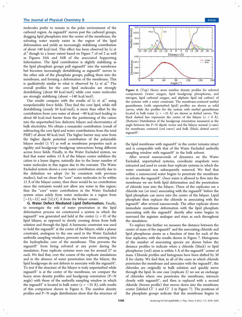
to investigate the role of water penetration in the lipiddeformation process we constructed a system in which themguanH+ was generated and held at the center (z = 0) of thelipid bilayer, as opposed to slowly crossing down from bulksolution and through the lipid. A harmonic constraint was usedto hold the mguanH+ at the center of the bilayer, while a planarconstraint, analogous to the one used in the Water Excludedumbrella sampling windows, prevents water from entering intothe hydrophobic core of the membrane. This prevents themguanH+ from being solvated at any point during thesimulation. Four replicate systems were run for around 27 nseach. We find that, over the course of the replicate simulationsand in the absence of water penetration into the bilayer, thelipid headgroups do not deform to associate with the mguanH+.To test if the structure of the bilayer is truly unperturbed whilemguanH+ is at the center of the membrane, we compare theheavy atom density profiles and headgroup orientation (P−Nangle) with those of the umbrella sampling window in whichthe mguanH+ is located in bulk water (z = −35 Å), with resultsof this comparison shown in Figure 6. The number densityprofiles and P−N angle distributions show that the structure of
the lipid membrane with mguanH+ in the center remains intactand is comparable with that of the Water Excluded umbrellasampling window with mguanH+ in the bulk solvent.After several nanoseconds of dynamics on the Water
Excluded, unperturbed systems, coordinate snapshots wereextracted and used to create four replicate systems in which thewater constraint was subsequently removed. We find thatwithin a nanosecond water begins to penetrate the membraneto solvate the mguanH+. Once water is allowed to flow into themembrane we see both lipid deformation and the penetrationof chloride ions into the bilayer. Three of the replicates see achloride ion (or ions) associating with the mguanH+ before thelipid phosphate can move into the center of the bilayer. Lipidphosphate then replaces the chloride in associating with themguanH+ after several nanoseconds. The other replicate showsimmediate membrane deformation with the lipid phosphateassociating with the mguanH+ shortly after water begins tosurround the arginine analogue and stays as such throughoutthe simulation.To explore this further we measure the distance between the
center of mass of the mguanH+ and the associating chloride andlipid phosphorus atoms as a function of time for each of thefour replicates, with the results shown in Figure 7. Histogramsof the number of associating species are shown below thedistance profiles to indicate when a chloride (black) or lipidphosphorus (red) atom is within 5 Å of the mguanH+ center ofmass. Chloride profiles and histograms have been shifted by 30Å for clarity. We find that, in all of the cases in which chloridepenetrates the membrane and associates with the mguanH+, thechlorides are originally in bulk solution and quickly movethrough the lipid. In one case (replicate 2) we see an exchangeof chlorides where one penetrates the membrane, interactsclosely with mguanH+, and then is replaced with a secondchloride (brown profile) that moves down into the membranecenter (labeled Cl− 1 and Cl− 2 in Figure 7). The positions ofthe phosphate groups indicate that the membrane begins to
Figure 6. (Top) Heavy atom number density profiles for selectedcomponents (water oxygen, lipid headgroup phosphorus, andnitrogen, lipid carbonyl oxygen, and aliphatic lipid tail carbon) ofthe systems with a water constraint. The membrane-centered methylguanidinium (with unperturbed lipid) profiles are shown as solidcurves, while the profiles for the system with methyl guanidiniumlocated in bulk water (z = −35 Å) are shown as dotted curves. Theblack dashed line represents the center of the bilayer (z = 0 Å).(Bottom) Distribution of the headgroup orientation measured as theangle between the P−N dipole vector and the bilayer normal (z-axis)for membrane centered (red curve) and bulk (black, dotted curve)mguanH+.
The Journal of Physical Chemistry B Article
dx.doi.org/10.1021/jp400389z | J. Phys. Chem. B 2013, 117, 3578−35923588

deform from a starting separation of 20 Å from the center massof the mguanH+ at the beginning of the simulation (consistentwith the average position of the unperturbed lipid phosphateheadgroups in Figure 6) and slowly moves toward the center ofthe membrane where it competes with chloride to interactdirectly with mguanH+. In replicate 3, however, we see themembrane deform quickly, and the phosphate groups are ableto associate with mguanH+ before any chlorides can enter thecenter of the membrane. In the cases where chloride penetratesinto the membrane, it does so quickly, on the order of 1 ns orless. Moreover, the chloride is solvated by varying watermolecules as it permeates from the bulk and through themembrane, as opposed to moving with a larger, constantsolvation structure. Analyzing a sampling of the water thatsurrounds the chloride atoms in the first hydration shell (within3.8 Å), we find that, although the chloride stays continuouslyhydrated throughout the simulation, the water molecules thatcomprise the hydration shell vary rapidly and are relativelytransient. The results of this exercise demonstrate theimportance of water in mediating the deformation of phosphategroups, even when the methyl guanidinium is already at thecenter of the bilayer. This seems to suggest that thedeformation of the lipid in terms of one or two phosphategroups associating with the charged species in the bilayer centeris the inherent state when the arginine resides in the center.That is, the deformation of the lipid headgroups is not anartifact of the simulation protocol that involves “pulling” alonglipid molecules by their headgroups as the methyl guanidiniumis slowly (reversibly) transferred into the bilayer via somebiasing potential.
IV. SUMMARY AND CONCLUSIONS
We investigated the thermodynamics of permeation of anamino acid side-chain analogue, methyl guanidinium, through a
model lipid bilayer, DPPC. Using both umbrella sampling andreweighting in conjunction with average force decomposition,we obtained the potential of mean force for the reversibletransfer of methyl guanidinium through the bilayer. We furtherdecomposed the overall PMF into contributions from varioussystem components including water, lipid, ions, and “core”water and lipids. We explored the effects of high saltconcentrations in the bulk solution, particularly with respectto the influence of ions on the water contribution to the totalPMF. We also investigated the impact of water permeation intothe bilayer on lipid deformability. Finally, we performed ourcalculations using a charge equilibration force field developed inour laboratory (and recently modified as discussed in theSupporting Information of this paper). This is a furtherapplication of a new class of force fields that can allow fordifferences in charge distributions, in a very gross sense, whenparticular molecular species encounter widely varying electro-chemical environments.With respect to the overall free energetics of methyl
guanidinium permeation through the bilayer, our results arequalitatively in agreement with a range of previous simulationstudies. We find a free energy minimum in the headgroupregion, which is corroborated by molecular dynamicssimulations of the unrestrained methyl guanidinium (asshown in Figure S3 of the Supporting Information). We findthe potential of mean force for permeation to be approximately28 kcal/mol (relative to the minimum in the headgroups),within the range of values reported for similar types ofsimulations using fixed-charge force fields. By decomposing theoverall free energy profile into contributions from variouscomponents, we find that the lipid in total confers astabilization of the charge cation in a range from −12 to−100 kcal/mol depending on the ionic strength of the bulksolution. For systems with 1 M KCl salt concentration in the
Figure 7. Distance profiles of chloride and lipid phosphate atoms closest to methyl guanidinium for four replicate systems in which mguanH+ is atthe center of an initially unperturbed bilayer in which the water constraint has been removed at the beginning of the simulation (time = 0 ns).Profiles are measured as the distance from the mguanH+ center of mass to the chloride and phosphorus atoms. Histograms of the number ofassociating species are shown below the graphs to indicate when a chloride (black) or lipid phosphorus (red) atom is within 5 Å of the mguanH+
center of mass. Chloride distance profiles and histograms have been offset by 30 Å for clarity. Replicate (2) shows two chloride atoms (the second ofwhich is shown in brown) which are able to move toward the center of the bilayer and associate with mguanH+ at different times during thesimulation.
The Journal of Physical Chemistry B Article
dx.doi.org/10.1021/jp400389z | J. Phys. Chem. B 2013, 117, 3578−35923589

bulk solution, the chloride anion is the sole species destabilizingmethyl guanidinium in the bilayer center, with the potassium,water, and lipids all contributing stabilizing forces. In theabsence of bulk electrolyte (except for a single chloride anionincluded in the simulation to maintain charge neutrality), wefind that the overall water contribution becomes destabilizing,in stark contrast to the contributions when 1 M KCl is includedin the bulk solution. Upon examining differences in local waterdensity and density-weighted forces acting on the permeatingcation, we find that the interplay between these twocomponents leads to differences in the asymmetry of waterforces on the cation that manifests in two starkly differentbehaviors. The presence of chloride anions in one case givesrise to an enhancement of local water density as the methylguanidinium enters the bilayer. These results reinforce currentviews on the importance of surface active ions of theHofmeister series and their interactions with phosphatidylcho-line-based lipid bilayers, particularly with the penetration ofsuch ions along with hydration layers into the solution−bilayerinterface.56 This enhanced water density in regions around themethyl guanidinium conferring stabilizing forces, relative to thecase where no ions are included in the simulation, leads to theobserved opposite slope in the water contribution to the totalPMF as the methyl guanidinium enters the bilayer. The natureof water contributions to the free energetics of the permeantare similar to the case where no salt is included in the bath inregions along the reaction coordinate where no chloride ionsare present. At the core of the bilayer, both systems displaystabilizing water contributions arising from a marked waterdistribution asymmetry.Finally, we find that water permeation into the bilayer is
required for the deformation of individual lipid molecules andpermeation of ions into the membrane. Through a series ofsimulations in which water is first prevented from entering thebilayer center where methyl guanidinium is restrained and afterequilibration allowed to enter the bilayer, we find that in theabsence of any water defects/permeation into the bilayer thelipid headgroups do not follow the methyl guanidinium. Onlyafter water is allowed to enter the bilayer do we seedeformation of individual lipid molecules allowing associationwith the amino acid analogue at the bilayer center. Moreover,using the force field we have, we find that it is possible forchloride anions to enter the bilayer center very quickly after thewater is allowed to enter, thus associating with the methylguanidinium initially. However, due to the relative energetics ofthe interaction models used in this work, we observe that theanions in time are displaced by the lipid headgroup phosphatesthat deform (with the aid of water defects) to associate with themethyl guanidinium. We believe that this suggests that thedeformation of the lipid in terms of one or two phosphategroups associating with the charged species in the bilayer centeris the inherent state when the arginine resides in the center.Bekker et al.27 have elegantly demonstrated that the cation, indeforming the membrane, effectively creates an interface thatprecludes it from experiencing the full positive dipole potentialexpected for an unperturbed bilayer.3 The deformationmaintains an interface between high and low potentialenvironments, implying that entry of a second charged specieswould be facilitated by the entry of the first. This nonadditivityhas also been demonstrated by MacCallum et al.26 Thus, thedeformation of the lipid headgroups is not an artifact of thesimulation protocol that involves “pulling” along lipidmolecules by their headgroups as the methyl guanidinium is
slowly (reversibly) transferred into the bilayer via some biasingpotential. This further supports numerous earlier studiespointing to the importance of water defects on a local scalein facilitating structural and dynamic aspects of membranebiophysics.12,13,27
■ ASSOCIATED CONTENT*S Supporting InformationAspects of simulation protocol, refined parameters for the lipidforce field and recent modifications, assessment of theconvergence of the potentials of mean force, description ofuncertainty analysis, further information of position-dependentforces and densities for the various systems discussed in themain text, detailed analysis of effects of water restraint forceconstants, and details of the core water and lipid decompositionanalysis. This material is available free of charge via the Internetat http://pubs.acs.org.
■ AUTHOR INFORMATIONCorresponding Author*E-mail: [email protected] authors declare no competing financial interest.
■ ACKNOWLEDGMENTSThe authors acknowledge support from the National ScienceFoundation (CAREER:MCB:1149802). Computational resour-ces are acknowledged via support from National Institutes ofHealth (COBRE:P20-RR015588) in the Chemical EngineeringDepartment at the University of Delaware. S.P. thanks N. Patelfor fruitful discussion and encouragement for the duration ofthis work.
■ REFERENCES(1) Dorairaj, S.; Allen, T. W. On the Thermodynamic Stability of aCharged Arginine Sidechan in a Transmembrane Helix. Proc. Natl.Acad. Sci. U.S.A. 2007, 104, 4943−4948.(2) Li, L.; Vorobyov, I.; Allen, T. W. Potential of Mean Force andpKa Profile Calculation for the Lipid Membrane-Exposed ArginineSide Chain. J. Phys. Chem. B 2008, 112, 9574−9587.(3) Vorobyov, I.; Li, L.; Allen, T. W. Assessing Atomistic and Coarse-Grained Force Fields for Protein-Lipid Interactions: The FormidableChallenge of an Ionizable Side Chain in a Membrane. J. Phys. Chem. B2008, 112, 9588−9602.(4) Li, L.; Vorobyov, I.; Dorairaj, S.; Allen, T. W. Charged ProteinSide Chain Movement in Lipid Bilayers Explored with Free EnergySimulation. Curr. Top. Membr. 2008, 60, 505−459.(5) Schow, E. V.; Freites, J. A.; Cheng, P.; Bernsel, A.; von Heijne, G.;White, S. H.; Tobias, D. J. Arginine in Membranes: The ConnectionBetween Molecular Dynamics Simulations and Translocon-mediatedInsertion Experiments. J. Membr. Biol. 2011, 239, 35−48.(6) Gumbart, J.; Roux, B. Determination of Membrane-InsertionFree Energies by Molecular Dynamics Simulations. Biophys. J. 2012,102, 795−801.(7) Fleming, P. J.; Freites, J. A.; Moon, C. P.; Tobias, D. J.; Fleming,K. G. Outer Membrane Phospholipase A in Phospholipid Bilayers: AModel System for Concerted Computational and ExperimentalInvestigations of Amino Acid Side Chain Partitioning Into LipidBilayers. Biochim. Biophys. Acta, Biomembr. 2012, 1818, 126−134.(8) Moon, C. P.; Fleming, K. G. Side-chain hydrophobicity scalederived from transmembrane protein folding into lipid bilayers. Proc.Natl. Acad. Sci. U.S.A. 2011, 108, 10174−10177.(9) Yoo, J.; Cui, Q. Does Arginine Remain Protonated in the LipidMembrane? Insights from Microscopic pKa Calculations. Biophys. J.2008, 94, L61−L63.
The Journal of Physical Chemistry B Article
dx.doi.org/10.1021/jp400389z | J. Phys. Chem. B 2013, 117, 3578−35923590

(10) Hristova, K.; Wimley, W. C. A Look at Arginine in Membranes.J. Membr. Biol. 2011, 239, 253−259.(11) Tieleman, D. P.; MacCallum, J. L.; Ash, W. L.; Kandt, C.; Xu, Z.;Monticelli, L. M. Membrane Protein Simulations with a United AtomLipid and All Atom Protein Model: Side Chain Transfer Free Energiesand Model Proteins. J. Phys.: Condens. Matter 2006, 18, S1221−S1234.(12) MacCallum, J. L.; Bennett, W. F. D.; Tieleman, D. P.Distribution of Amino Acids in a Lipid Bilayer from ComputerSimulations. Biophys. J. 2008, 94, 3393−3404.(13) MacCallum, J. L.; Bennett, W. F. D.; Tieleman, D. P.Partitioning of Amino Acid Side Chains into Lipid Bilayers: Resultsfrom Computer Simulations and Comparison to Experiment. J. Gen.Physiol. 2007, 129, 371−377.(14) Jiang, Y.; Lee, A.; Chen, J.; Ruta, V.; Cadene, M.; Chait, B. T.;MacKinnon, R. X-ray Structure of a Voltage-Dependent K+ Channel.Nature 2003, 423, 33−41.(15) Jiang, Y.; Lee, A.; Chen, J.; Cadene, M.; Chait, B. T.;MacKinnon, R. The Open Pore Conformation of Potassium Channels.Nature 2002, 417, 523−526.(16) Jensen, M. O.; Jogini, V.; Borhani, D. W.; Leffler, A. E.; Dror, R.O.; Shaw, D. E. Mechanism of Voltage Gating in Potassium Channels.Science 2012, 336, 229−233.(17) Herce, H. D.; Garcia, A. E. Cell Penetrating Peptides: How DoThey Do It? J. Biol. Phys. 2007, 33, 345−356.(18) Marks, J.; Placone, J.; Hristova, K.; Wimley, W. C. SpontaneousMembrane-Translocating Peptides by Orthogonal High-ThroughputScreening. J. Am. Chem. Soc. 2011, 133, 8995−9004.(19) Herce, H. D.; Garcia, A. E.; Litt, J.; Kane, R. S.; Martin, P.;Enrique, N.; Rebolledo, A.; Milesi, V. Arginine-Rich PeptidesDestabilize the Plasma Membrane, Consistent with a Pore FormationTranslocation Mechanism of Cell-Penetrating Peptides. Biohys. J.2009, 97, 1917−1925.(20) Jarver, P.; Langel, U. Cell-Penetrating Peptides: A BriefIntroduction. Biochim. Biophys. Acta, Biomembr. 2006, 1758, 260−263.(21) Vives, E.; Schmidt, J.; Pelegrin, A. Cell-Penetrating and Cell-Targeting Peptides in Drug Delivery. Biochim. Biophys. Acta, Revi.Cancer 2008, 1786, 126−138.(22) Futaki, S. Oligorarginine Vectors for Intracelular Delivery:Design and Cellular - Uptake Mechanisms. Biopolymers 2006, 84,241−249.(23) Lattig-Tunnemann, G.; Prinz, M.; Hoffmann, D.; Behlke, J.;Palm-Apergi, C.; Morano, I.; Herce, H. D.; Cardoso, M. C. BackboneRigidity and Static Presentation of Guanidinium Groups IncreasesCellular Uptake of Arginine-Rich Cell-Penetrating Peptides. Nat.Commun. 2011, 2, 453−458.(24) MacCallum, J. L.; Tieleman, D. P. Hydrophobicity Scales: AThermodynamic Looking Glass Into Lipid-Protein Interactions. TrendsBiochem. Sci. 2011, 36, 653−662.(25) Hessa, T.; White, S. H.; von Heijne, G. Membrane Insertion of aPotassium Channel Voltage Sensor. Science 2005, 307, 1427−1427.(26) MacCallum, J. L.; Bennett, W. F. D.; Tieleman, D. P. Transfer ofArginine Into Lipid Bilayers is Nonadditive. Biophys. J. 2011, 101,110−117.(27) Vorobyov, I.; Bekker, B.; Allen, T. W. Electrostatics ofDeformable Lipid Membranes. Biophys. J. 2010, 98, 2904−2913.(28) Bogdanov, M.; Xie, J.; Dowhan, W. Lipid−Protein InteractionsDrive Membrane Protein Topogenesis in Accordance with the PositiveInside Rule. J. Biol. Chem. 2009, 284, 9637−9641.(29) Bogdanov, M.; Heacock, P. N.; Dowhan, W. A PolytopicMembrane Protein Displays a Reversible Topology Dependent onMembrane Lipid Composition. EMBO J. 2002, 21, 2107−2116.(30) Bogdanov, M.; Xie, J.; Heacock, P. N.; Dowhan, W. To Flip orNot to Flip: Lipid-Protein Charge Interactions are a Determinant ofthe Final Membrane Protein Topology. J. Cell Biol. 2008, 182, 925−935.(31) Zhang, W.; Bogdanov, M.; Pi, J.; Pittard, A. J.; Dowhan, W.Reversible Topological Organization Within a Polytopic MembraneProtein is Governed by a Change in Membrane PhospholipidComposition. J. Biol. Chem. 2003, 278, 50128−50135.
(32) Wimley, W. C.; Hristova, K. Antimicrobial Peptides: Successes,Challenges, and Unanswered Questions. J. Membr. Biol. J. Membr.Biol. 2011, 239, 27−34.(33) Gresh, N.; Cisneros, G. A.; Darden, T. A.; Piquemal, J.-P.Anisotropic, Polarizable Molecular Mechanics Studies of Inter- andIntramolecular Interactions and Ligand-Macromolecule Complexes. Abottom-up Strategy. J. Chem. Theory Comput. 2007, 3, 1960−1986.(34) Piquemal, J.-P.; Perera, L.; Cisneros, G. A.; Ren, P.; Pedersen, L.G.; Darden, T. A. Towards Accurate Solvation Dynamics of DivalentCations in Water Using the Polarizable Amoeba Force Field: FromEnergetics to Structure. J. Chem. Phys. 2006, 125 (054511), 1−7.(35) de Courcy, B.; Piquemal, J.-P.; Garbay, C.; Gresh, N. PolarizableWater Molecules in Ligand-Macromolecule Recognition. Impact onthe Relative Affinities of Competing Pyrrolopyrimidine Inhibitors forFAK Kinase. J. Am. Chem. Soc. 2010, 132, 3312−3320.(36) Jiao, D.; King, C.; Grossfield, A.; Darden, T. A.; Ren, P.Simulation of Ca2+ and Mg2+ Solvation Using Polarizable AtomicMultipole Potential. J. Phys. Chem. B 2006, 110, 18553−18559.(37) Ponder, J. W.; Wu, C.; Ren, P.; Pande, V. S.; Chodera, J. D.;Schnieders, M. J.; Haque, I.; Mobley, D. L.; Lambrecht, D. S.; DiStasio,R. A., Jr.; Head-Gordon, M.; Clark, G. N. I.; Johnson, M. E.; Head-Gordon, T. Current Status of the AMOEBA Polarizable Force Field. J.Phys. Chem. B 2010, 114, 2549−2564.(38) Jiao, D.; Golubkov, P. A.; Darden, T. A.; Ren, P. Calculation ofProtein-Ligand Binding Free Energy by Using a Polarizable Potential.Proc. Natl. Acad. Sci. 2008, 105, 6290−6295.(39) Anisimov, V. M.; Lamoureux, G.; Vorobyov, I. V.; Huang, N.;Roux, B.; MacKerell, A. D., Jr. Determination of ElectrostaticParameters for a Polarizable Force Field Based on the ClassicalDrude Oscillator. J. Chem. Theory Comput. 2005, 1, 153−168.(40) Lamoureux, G.; Roux, B. Modeling Induced Polarization withClassical Drude Oscillators: Theory and Molecular DynamicsSimulations Algorithm. J. Chem. Phys. 2003, 119 (3025), 1−15.(41) Lamoureux, G.; MacKerell, A. D., Jr.; Roux, B. A SimplePolarizable Model of Water Based on Classical Drude Oscillators. J.Chem. Phys. 2003, 119 (5185), 1−13.(42) Lamoureux, G.; Harder, E.; Vorobyov, I. V.; Roux, B.;MacKerell, A. D., Jr. Polarizable Model of Water for MolecularDynamics Simulations of Biomolecules. Chem. Phys. Lett. 2006, 418,245−249.(43) Lopes, P. E. M.; Lamoureux, G.; Roux, B.; MacKerell, A. D., Jr.Polarizable Empirical Force Field for Aromatic Compounds Based onthe Classical Drude Oscillator. J. Phys. Chem. B 2007, 111, 2873−2885.(44) Yu, H.; Whitfield, T. W.; Harder, E.; Lamoureux, G.; Vorobyov,I.; Anisimov, V. M.; MacKerell, A. D., Jr.; Roux, B. SimulatingMonovalent and Divalent Ions in Aqueous Solution Using a DrudePolarizable Force Field. J. Chem. Theory Comput. 2010, 6, 774−786.(45) Harder, E.; MacKerell, A. D., Jr.; Roux, B. Many-BodyPolarization Effects and the Membrane Dipole Potential. J. Am.Chem. Soc. 2009, 131, 2760−2761.(46) Patel, S.; MacKerell, A. D., Jr.; Brooks, C. L., III CHARMMFluctuating Charge Force Fields for Proteins: II. Protein/SolventProperties from Molecular Dynamics Simulations Using a NonadditiveElectrostatic Model. J. Comput. Chem. 2004, 25, 1504−1514.(47) Patel, S.; Brooks, C. L., III A Nonadditive Methanol Force Field:Bulk Liquid and Liquid-Vapor Interfacial Properties via MolecularDynamics Simulations Using a Fluctuating Charge Model. J. Chem.Phys. 2005, 122 (024508), 1−10.(48) Patel, S.; Brooks, C. L., III Structure, Thermodynamics, andLiquid-Vapor Equilibrium of Ethanol from Molecular DynamicsSimulations Using Nonadditive Interactions. J. Chem. Phys. 2005,123 (164502), 1−12.(49) Bauer, B. A.; Lucas, T. R.; Meninger, D.; Patel, S. WaterPermeation Through DMPC Lipid Bilayers Using Polarizable ChargeEquilibration Force Fields. Chem. Phys. Lett. 2011, 508, 289−294.(50) Lucas, T. R.; Bauer, B. A.; Davis, J. E.; Patel, S. MolecularDynamics Simulation of Hydrated DPPC Monolayers Using ChargeEquilibration Force Fields. J. Comput. Chem. 2012, 33, 141−152.
The Journal of Physical Chemistry B Article
dx.doi.org/10.1021/jp400389z | J. Phys. Chem. B 2013, 117, 3578−35923591

(51) Davis, J. E.; Patel, S. Revised Charge Equilibration Parametersfor More Accurate Hydration Free Energies of Alkanes. Chem. Phys.Lett. 2010, 484, 173−176.(52) Zhong, Y.; Patel, S. Nonadditive Empirical Force Fields forShort-Chain Linear Alcohols: Methanol to Butanol. Hydration FreeEnergies and Kirkwood-Buff Analysis Using Charge EquilibrationModels. J. Phys. Chem. B 2010, 114, 11076−11092.(53) Warren, G. L.; Davis, J. E.; Patel, S. Origin and Control ofSuperlinear Polarizability Scaling in Chemical Potential EquilizationMethods. J. Chem. Phys. 2008, 128 (144110), 1−14.(54) Davis, J. E.; Rahaman, O.; Patel, S. Molecular DynamicsSimulations of a DMPC Bilayer Using Nonadditive InteractionModels. Biophys. J. 2009, 96, 385−402.(55) Shimizu, K.; Chaimovich, H.; Farah, J. P. S.; Dias, L. G.; Bostick,D. L. Calculation of the Dipole Moment for Polypeptides Using theGeneralized Born-Electronegativity Equalization Method: Results inVacuum and Continuum-Dielectric Solvent. J. Phys. Chem. B 2004,108, 4171−4177.(56) Vacha, R.; Jurkiewicz, P.; Petrov, M.; Berkowitz, M. L.;Bockmann, R. A.; Barucha-Kraszewska, J.; Hof, M.; Jungwirth, P.Mechanism of Interaction of Monovalent Ions with Phosphatidylcho-line Lipid Membranes. J. Phys. Chem. B 2010, 114, 9504−9509.(57) Davis, J. E.; Patel, S. Charge Equilibration Force Fields for LipidEnvironments: Applications to Fully Hydrated DPPC Bilayers andDMPC-Embedded Gramicidin A. J. Phys. Chem. B 2009, 113, 9183−9196.(58) Patel, S.; Brooks, C. L., III CHARMM Fluctuating Charge ForceFields for Proteins: I Parameterization and Application to BulkOrganic Liquid Simulations. J. Comput. Chem. 2004, 25, 1−15.(59) Patel, S.; Brooks, C. L., III Fluctuating Charge Force Fields:Recent Developments and Applications from Small Molecules toMacromolecular Biological Systems. Mol. Simul. 2006, 32, 231−249.(60) Rappe, A. K.; Goddard, W. A. Charge Equilibration forMolecular Dynamics Simulations. J. Phys. Chem. 1991, 95, 3358−3363.(61) Rick, S. W.; Stuart, S. J.; Berne, B. J. Dynamical FluctuatingCharge Force Fields: Application to Liquid Water. J. Chem. Phys. 1994,101, 6141−6156.(62) Rick, S. W.; Stuart, S. J.; Bader, J. S.; Berne, B. J. FluctuatingCharge Force Fields for Aqueous Solutions. J. Mol. Liq. 1995, 65/66,31−40.(63) Rick, S. W.; Berne, B. J. Dynamical Fluctuating Charge ForceFields: The Aqueous Solvation of Amides. J. Am. Chem. Soc. 1996, 118,672−679.(64) Rick, S. W. Simulations of Ice and Liquid Water Over a Range ofTemperatures Using the Fluctuating Charge Model. J. Chem. Phys.2001, 114, 2276−2283.(65) Rick, S. W.; Stuart, S. J. Reviews of Computational Chemistry;John Wiley & Sons: New York, 2002; pp 89−146.(66) Sanderson, R. T. An Interpretation of Bond Lengths and aClassification of Bonds. Science 1951, 114, 670−672.(67) Sanderson, R. T. Chemical Bonds and Bond Energy; AcademicPress: New York, 1976.(68) Nalewajski, R. F.; Korchowiec, J.; Zhou, Z. Molecular Hardnessand Softness Parameters and Their Use in Chemistry. Int. J. QuantumChem. 1988, 22, 349−366.(69) Lee, A. J.; Rick, S. W. The Effects of Charge Transfer on theProperties of Liquid Water. J. Chem. Phys. 2011, 134 (184507), 1−9.(70) Chelli, R.; Procacci, P.; Righini, R.; Califano, S. ElectricalResponse in Chemical Potential Equalization Schemes. J. Chem. Phys.1999, 111, 8569−8575.(71) Davis, J. E.; Warren, G. L.; Patel, S. Revised ChargeEquilibration Potential for Liquid Alkanes. J. Phys. Chem. B 2008,112, 8298−8310.(72) Ren, P.; Ponder, J. W. Polarizable Atomic Multipole WaterModel for Molecular Mechanics Simulation. J. Phys. Chem. B 2003,107, 5933−5947.(73) Car, R.; Parrinello, M. Unified Approach for MolecularDynamics and Density Functional Theory. Phys. Rev. Lett. 1985, 55,2471−2474.
(74) Biltonen, R. L.; Lichtenberg, D. The Use of DifferentialScanning Calorimetry as a Tool to Characterize LiposomePreparations. Chem. Phys. Lipid 1993, 64, 129−142.(75) Kucerka, N.; Nagle, J. F.; Sachs, J. N.; Feller, S. E.; Pencer, J.;Jackson, A.; Katsaras, J. Lipid Bilayer Structure Determined by theSimultaneous Analysis of Neutron and X-ray Scattering Data. Biophys.J. 2008, 95, 2356−2367.(76) Allen, M. P.; Tildesley, D. J. Computer Simulations of Liquids;Clarendon: U.K., 1987.(77) Nose, S. A Molecular Dynamics Methods for Simulations in theCanonical Ensemble. Mol. Phys. 1984, 52, 255−268.(78) Darden, T.; York, D.; Pedersen, L. Particle Mesh Ewald: An N ·log (N) Method for Ewald Sums in Large Systems. J. Chem. Phys.1993, 98, 10089−10092.(79) Essmann, U.; Darden, T.; Lee, H.; Perera, L.; Berkowitz, M. L.;Pederson, L. A Smooth Particle Mesh Ewald Method. J. Chem. Phys.1995, 103, 8577−8593.(80) Patel, S.; Davis, J. E.; Bauer, B. A. Exploring Ion PermeationEnergetics in Gramicidin A Using Polarizable Charge EquilibrationForce Fields. J. Am. Chem. Soc. 2009, 131, 13890−13891.(81) Zhong, Y.; Bauer, B. A.; Patel, S. Solvation Properties of N-Acetyl-β-glucosamine: A Molecular Dynamics Study IncorporatingElectrostatic Polarization. J. Comput. Chem. 2011, 32, 3339−3353.(82) Li, L. B.; Vorobyov, I.; Allen, T. W. The Role of MembraneThickness in Charged Protein-Lipid Interactions. Biochim. Biophys.Acta, Biomembr. 2012, 1818, 135−145.(83) Warren, G. L.; Patel, S. Hydration Free Energies of MonovalentIons in Transferable Intermolecular Potential Four Point FluctuatingCharge Water: An Assessment of Simulation Methodology and ForceField Performance and Transferability. J. Chem. Phys. 2007, 127(064509), 1−19.(84) Lynden-Bell, R. M.; Rasaiah, J. C. From Hydrophobic toHydrophillic Behaviour: A Simulation Study of Solvation Entropy andFree Energy of Simple Solutes. J. Chem. Phys. 1997, 107, 1981−1991.(85) Shirts, M. R.; Pande, V. S. Solvation Free Energies of AminoAcid Side Chain Analogs for Common Molecular Mechanics WaterModels. J. Chem. Phys. 2005, 122 (134508), 1−13.(86) Harder, E.; Roux, B. On the Origin of the Electrostatic PotentialDifference at a Liquid-Vacuum Interface. J. Chem. Phys. 2008, 129(234706), 1−9.(87) Kumar, S.; Bouzida, D.; Swendsen, R. H.; Kollman, P. A.;Rosenberg, J. M. The Weighted Histogram Analysis Method for Free-Energy Calculations on Biomolecules. I. The Method. J. Comput.Chem. 1992, 13, 1011−1021.(88) Beglov, D.; Roux, B. Finite Representation of an Infinite BulkSystem: Solvent Boundary Potential for Computer Simulations. J.Chem. Phys. 1994, 100 (9050), 1−14.(89) Zhu, F.; Hummer, G. Convergence and Error Estimation in FreeEnergy Calculations Using the Weighted Histogram Analysis Method.J. Comput. Chem. 2011, 33, 453−465.(90) Flyvbjerg, H.; Petersen, H. G. Error Estimates on Averages ofCorrelated Data. J. Chem. Phys. 1989, 91, 461−466.(91) Patel, S.; Brooks, C. L., III Revisiting the Hexane-WaterInterface via Molecular Dynamics Simulations Using NonadditiveAlkane-Water Potentials. J. Chem. Phys. 2006, 124 (204706), 1−14.(92) Vorobyov, I. V.; Anisimov, V. M.; A. D. MacKerell, J. PolarizableEmpirical Force Field for Alkanes Based on the Classical DrudeOscillator Model. J. Phys. Chem. B 2005, 109, 18988−18999.(93) Frisch, M. J. et al. Gaussian 03, revision D.02; Gaussian, Inc.:Wallingford, CT, 2004.
The Journal of Physical Chemistry B Article
dx.doi.org/10.1021/jp400389z | J. Phys. Chem. B 2013, 117, 3578−35923592