Fragmented ruling coalitions and economic developments in ...
Upload
khangminh22Category
view
0download
0
genesG C A T
T A C G
G C A T
Article
Fragmented Nuclear DNA Is the PredominantGenetic Material in Human Hair Shafts
Michael D. Brandhagen †, Odile Loreille † and Jodi A. Irwin *
DNA Support Unit, FBI Laboratory, 2501 Investigation Parkway, Quantico, VA 22135, USA;[email protected] (M.D.B.); [email protected] (O.L.)* Correspondence: [email protected]† These authors contributed equally to this work.
Received: 1 November 2018; Accepted: 10 December 2018; Published: 18 December 2018 �����������������
Abstract: While shed hairs are one of the most commonly encountered evidence types, they areamong the most limited in terms of DNA quantity and quality. As a result, nuclear DNA short tandemrepeat (STR) profiling is generally unsuccessful and DNA testing of shed hair is instead performed bytargeting the mitochondrial DNA control region. Although the high copy number of mitochondrialDNA relative to nuclear DNA routinely permits the recovery of mitochondrial DNA (mtDNA) datain these cases, mtDNA profiles do not offer the discriminatory power of nuclear DNA profiles.In order to better understand the total content and degradation state of DNA in single shed hairsand assess the feasibility of recovering highly discriminatory nuclear DNA data from this commonevidence type, high throughput shotgun sequencing was performed on both recently collected andaged (approximately 50-year-old) hair samples. The data reflect trends that have been demonstratedpreviously with other technologies, namely that mtDNA quantity and quality decrease along thelength of the hair shaft. In addition, the shotgun data reveal that nuclear DNA is present in shed hairand surprisingly abundant relative to mitochondrial DNA, even in the most distal fragments. NuclearDNA comprised, at minimum, 88% of the total human reads in any given sample, and generallymore than 95%. Here, we characterize both the nuclear and mitochondrial DNA content of shed hairsand discuss the implications of these data for forensic investigations.
Keywords: hair shaft; mitochondrial DNA; mtDNA; mtGenome; nuclear DNA; next-generationsequencing
1. Introduction
Shed hairs are one of the most commonly encountered evidence types [1], but also among themost limited in terms of DNA quantity and quality. By some estimates, shed hair represents up to 90%of the hair samples collected at crime scenes [2,3]. Unfortunately, nuclear DNA (nuDNA) is generallytoo low in quantity and quality to permit successful short tandem repeat (STR) typing.
The difficulty of successful STR typing of shed hairs is attributed primarily to the keratinizationprocess, which degrades cellular organelles and nucleic acids [4]. Not only do the general enzymaticactivities associated with keratinization result in DNA degradation, but also nuclear DNA, in particular,is specifically targeted for destruction [5–7]. Nevertheless, some nuclear DNA is known to persist intelogen hairs, albeit at low quantities and highly variable qualities [4]. Though a number of studiesdescribe the presence, and successful PCR amplification, of autosomal STR markers from telogenhairs [2,8–12], STR typing is not routinely pursued in forensic laboratories for a number of reasons.For one, reduced size STR amplicons, ranging from ~60–150 bp, are generally required to achieveamplification success. Yet, since only a relatively small number of reduced sized amplicons can bemultiplexed with currently employed capillary electrophoresis technologies, data recovery is somewhat
Genes 2018, 9, 640; doi:10.3390/genes9120640 www.mdpi.com/journal/genes

Genes 2018, 9, 640 2 of 20
limited. Second, elevated cycle numbers, often between 30 and 40, are generally required to amplifythe low quantities of nuclear DNA to detectable levels. However, even when reduced size ampliconsand elevated cycle numbers are employed, amplification success is still inconsistent, and resulting STRprofiles are often incomplete. In addition, commonly employed quantitative PCR (qPCR) assays rarelyyield enough information to adequately inform downstream analysis. The qPCR assays routinelyimplemented are simply not sensitive enough for the low levels of nuclear DNA present in telogenhairs [12]. More recent studies have shown that direct amplification of telogen hairs (i.e., amplificationwithout preliminary DNA extraction), when combined with elevated cycles, can result in full STRprofiles approximately 20% of the time [12]. However, it is difficult or impossible to predict a priori if ahair will yield probative DNA data.
It is also the case that success rates in routine forensic practice are likely lower. Most researchstudies are necessarily based on freshly, or relatively recently, collected hair samples. While generalpatterns can obviously be ascertained from such samples, the variability and difficulty of aged and/ordamaged casework samples is nearly impossible to accurately represent. Indeed, in those studiesfor which hairs recovered from actual crime scenes were included, the evidence hairs performedworse than the recently collected hairs [10,13]. Given that amplification success cannot be predictedin advance, and that amplification success rates are generally low, it simply does not make sensefrom a practical perspective to exhaust limited sample material on a testing modality unlikely to yieldprobative results. Instead, mitochondrial DNA (mtDNA) is routinely sought in these cases due to itsabundance relative to nuclear DNA. Though mtDNA profiles do not offer the discriminatory power ofnuclear DNA profiles, mtDNA is recovered in 92.5% of telogen hair cases [14].
Because of the difficulty of recovering nuclear DNA from hair, studies conducted to characterizeand better understand the state of DNA in hair have largely focused on the more accessible mtDNAmolecule. At a broad level, Melton et al., [14] showed that with increasing age of the hair specimen,the likelihood of obtaining a full hypervariable region I/hypervariable region II (HVI/HVII) profiledecreased. The same pattern was observed in a systematic study by Gilbert et al. [15]. Additionalstudies, based on the size of recoverable PCR amplicons, have shown progressive degradation ofmtDNA along the hair shaft [16–18] with DNA quality deteriorating rapidly within a few millimetersof the root [10].
Some of the most recent information on the overall quantity and quality of DNA in telogen hairshafts has come from studies employing next-generation sequencing (NGS). Because NGS-basedshotgun sequencing is not dependent on pre-defined amplicons, the sequence data reflect theendogenous size of the DNA. Generally speaking, these studies show that DNA preservation inaged hair is overwhelmingly poor [19–22]. For example, the average mtDNA size of a 4000-year-oldpaleo Eskimo sample was 76 bp [19], and mtDNA averaged only 61 bp (range between 48 to 73 bp) [22]in 111 human hairs collected between 1920 and 1970.
Preliminary studies in our laboratory showed that in shotgun libraries of two freshly collected singleshed hairs, 99.93 and 99.88% of the reads mapping to the human genome were nuclear DNA, and theremaining 0.07% and 0.12% were mitochondrial DNA [23]. These results are consistent with those of othershotgun sequencing studies of aged hairs. In particular, Rasmussen et al. [20] found that ~80% of thereads produced from a 1.5 g sample of 4000-year-old hair were human sequences and that only 0.13% ofthe human reads were mtDNA sequences. The remainder, 99.87%, were nuclear DNA sequences.
Here, we aim to confirm that, despite a high level of degradation, nuclear DNA comprises thevast majority of total human DNA in hair shafts. In addition, we further characterize the quantity andquality of both mitochondrial and nuclear DNA that can be recovered from single shed telogen hairsregularly encountered in forensic casework.
2. Materials and Methods
All tested samples were rootless hair shafts collected with informed consent from the donorsunder FBI Institutional Review Board approved project #417-17 (Tables 1 and 2).

Genes 2018, 9, 640 3 of 20
Table 1. Description of donors and hair samples used in this study. RT = Room Temperature (20–25 ◦C).§ Refers to permanent hair coloring.
Donors Characteristics Time between Collection/Cutand DNA Analysis
TemperatureStorage Treated
I Adult, female, recent 4 years 4 ◦C NoII Adult, male, recent 2 months RT NoII Child, male, aged 53 years RT NoIII Adult, female, recent 2 months 4 ◦C §YesIV Adult, female, recent 2.5 years 4 ◦C NoV Adult, female, recent 1.5 year 4 ◦C NoVI Adult, female, recent 1 h RT §YesVII Child, male, aged 60 years RT NoVIII Adult, female, aged >40 years RT NoIX Child, female, aged 30 years RT No
Table 2. Description of each hair sample. The asterisk denotes extracts that were obtained withpurification protocol B.
Recent Hairs
Donor Sample Names Hair Portions Hair Size (cm) Purification Protocol
IR1 single hair 5
AR1*eluate C
IIR2
five 1.3 cm hairs 6.5A
R2*eluate C
IIIR3 single hair 5
AR3* eluate C
IV R4 single hair 5 AV R5 single hair 5 AVI R6* single hair 6 B
I
Seg1-R7 segment 1 5A
Seg1-R7* BSeg2-R7 segment 2 5
ASeg2-R7* BSeg3-R7 segment 3 5
ASeg3-R7* BSeg4-R7 segment 4 5
ASeg4-R7* BSeg5-R7 segment 5 5
ASeg5-R7* B
Aged Hairs
IIA1
two 2.5 cm hairs 5A
A1* BII A6* five 1.5 cm hairs 7.5 B
VIIA2
three 2.5 cm hairs 7.5A
A2* BVIII A7* three 2.5 cm hairs 7.5 B
IX
Seg1-A4* segment 1 5 BSeg2-A4* segment 2 5 BSeg3-A4* segment 3 5 BSeg4-A4* segment 4 5 B
IX
Seg1-A8* Segment 1 1.7 BSeg2-A8* Segment 2 1.7 BSeg3-A8* Segment 3 1.7 BSeg4-A8* Segment 4 1.7 BSeg5-A8* Segment 5 1.7 B
2.1. Types of Hair
2.1.1. Recent hairs
The hairs referred to as recent hairs (R series) were cut or collected less than 6 years before thedate of the DNA testing and stored primarily at 4 ◦C. Recent hairs were collected from hairbrushes or

Genes 2018, 9, 640 4 of 20
by finger combing from random portions of the head. Prior to DNA extraction, the root (proximal)ends of these hairs were cut (~1 cm) and removed. Six recently collected hair samples were used tocharacterize total DNA (mitochondrial and nuclear) content in rootless shed hairs and approximately5 to 6.5 cm was extracted for any given hair (Table 2).
2.1.2. Aged Hairs
The aged hairs (A series) were taken from hair cuttings that were ~40 to 60 years old and had beenstored at room temperature. Because the samples were hair cuttings, the lengths of the hairs at the timeof cutting, and thus the distances from the scalp of the tested segments, were unknown. Furthermore,it was not possible to easily identify the proximal and distal end of these hairs. Samples A1, A2, A6 andA7 were children’s hairs that originated from three individuals whose mtDNA genome (mtGenome)profiles were known. Both hair types were used to evaluate: (1) mtDNA and nuclear DNA content,(2) mtDNA quantity and quality along the length of the shaft, (3) the possibility of complete mtGenomesequence recovery and 4) nuclear DNA quantity and quality along the length of the shaft.
2.1.3. Segmented Hairs
To assess total DNA quantity and quality along the length of individual hair shafts, a recentuntreated hair that had been stored refrigerated for 4 years (R7) and two separate aged hairs (A4 &A8) were tested. Following removal of the root (if necessary), the hairs were cut into 5 cm segments.For R7, the mtGenome profile of the donor was known and five segments were tested (see Figure 1),with Segment 1 representing the segment closest to the root end. For A4, seven segments were cut butonly four were tested. For A8, five continuous segments were tested. Samples A4 and A8 are from a>50 cm adult braid that was cut at least 40 years ago and for which no reference profile was available.
Genes 2018, 9, 640 4 of 21
Figure 1. Segmentation of hairs. Hairs R7, A4, and A8 were long hairs cut in segments used to assess total DNA quantity and quality along the length of individual hair shafts.
2.2. Extraction
The protocols for washing and digesting the hair samples are presented in Protocols, Supplementary Materials. Once the hairs were fully digested, lysates were purified with one of three protocols (Figure 2). The purification methods were being tested for a different study, but essentially served as replicate extractions for those hairs purified with multiple protocols. The protocols are summarized below:
(1) A protocol adapted from a Qiagen user-developed method [24,25]) that has been employed in the FBI Laboratory’s routine casework since 2014 (referred to from here on as protocol A; see Protocols in the Supplementary Materials).
(2) A protocol based on Allentoft et al., [26] that employs a binding buffer that favors the recovery of small (<100 bp) DNA fragments.
(3) A combined protocol that follows protocol A until the step at which the lysate and silica beads are on the magnet. For this protocol, (protocol C in Protocols, Supplementary Materials), the bead binding waste solution was retained and mixed with the binding buffer from protocol B. It was subsequently purified and eluted with MinElute columns (Qiagen, Germantown, MD, USA).
Table 2 summarizes the purification protocol used for each hair sample tested in this study. Generally speaking, protocol A was used for the purification of recently collected hairs while protocol B was used with aged hairs for which DNA was expected to be degraded.
Figure 1. Segmentation of hairs. Hairs R7, A4, and A8 were long hairs cut in segments used to assesstotal DNA quantity and quality along the length of individual hair shafts.
2.2. Extraction
The protocols for washing and digesting the hair samples are presented in Protocols,Supplementary Materials. Once the hairs were fully digested, lysates were purified with one of threeprotocols (Figure 2). The purification methods were being tested for a different study, but essentially

Genes 2018, 9, 640 5 of 20
served as replicate extractions for those hairs purified with multiple protocols. The protocols aresummarized below:
(1) A protocol adapted from a Qiagen user-developed method [24,25]) that has been employedin the FBI Laboratory’s routine casework since 2014 (referred to from here on as protocol A;see Protocols in the Supplementary Materials).
(2) A protocol based on Allentoft et al., [26] that employs a binding buffer that favors the recovery ofsmall (<100 bp) DNA fragments.
(3) A combined protocol that follows protocol A until the step at which the lysate and silica beadsare on the magnet. For this protocol, (protocol C in Protocols, Supplementary Materials), the beadbinding waste solution was retained and mixed with the binding buffer from protocol B. It wassubsequently purified and eluted with MinElute columns (Qiagen, Germantown, MD, USA).
Table 2 summarizes the purification protocol used for each hair sample tested in this study.Generally speaking, protocol A was used for the purification of recently collected hairs while protocolB was used with aged hairs for which DNA was expected to be degraded.Genes 2018, 9, 640 5 of 21
Figure 2. Schematic representation of the three purification methods used in this study. For details on the protocols, see Protocols, in the Supplementary Materials.
2.3. Quantification
Quantity of mtDNA was assessed by qPCR of the DNA extracts. For the recent hair extracts, quality of the mtDNA was assessed in one of two ways: by a mtDNA qPCR assay developed by Kavlick [27] on a 7500 Real Time PCR system (Thermo Fisher Scientific, Waltham, MA, USA) or by sequence read length for the aged hair extracts.
For the mtDNA quantification, two microliters of each extract and reagent blank (RB) were amplified in duplicate with a qPCR assay that incorporates a DNA degradation index. With this assay, degradation is assessed based on the ratio of large (≥316 bp) and small (≥105 bp) mtDNA fragments. The larger the degradation index, the lower the number of mtDNA fragments ≥316 bp relative to fragments between 105 bp–316 bp. For example, a degradation index of 1 or lower indicates no mtDNA degradation (all quantified fragments ≥316 bp), while a degradation index >1 indicates that fewer 316 bp or larger fragments are present in the extract than 105 bp–316 bp fragments [27]. An undetermined degradation index indicates that all the human mtDNA is degraded to a size smaller than 316 bp.
Sequence read lengths could not be used to assess endogenous mtDNA quality in the recent hair extracts because, following qPCR, the DNA required shearing to ensure successful library preparation and sequencing. This is due to the presence of large mtDNA fragments that have been shown to exist in freshly collected hair shafts [23]. Conversely, qPCR could not be used to assess mtDNA quality in the aged hairs because the degradation index, which is dependent on the amplification of DNA fragments of 105 and 316 bp in size, could not be recovered from most of the aged hair segments.
Nuclear DNA quantification of the recent hair extracts using the Quantifiler Trio DNA quantification kit (Thermo Fisher Scientific) was attempted for a number of samples, but the results were too low to be useful (<0.5 pg/µL).
Figure 2. Schematic representation of the three purification methods used in this study. For details onthe protocols, see Protocols, in the Supplementary Materials.
2.3. Quantification
Quantity of mtDNA was assessed by qPCR of the DNA extracts. For the recent hair extracts,quality of the mtDNA was assessed in one of two ways: by a mtDNA qPCR assay developed byKavlick [27] on a 7500 Real Time PCR system (Thermo Fisher Scientific, Waltham, MA, USA) or bysequence read length for the aged hair extracts.
For the mtDNA quantification, two microliters of each extract and reagent blank (RB) wereamplified in duplicate with a qPCR assay that incorporates a DNA degradation index. With this assay,degradation is assessed based on the ratio of large (≥316 bp) and small (≥105 bp) mtDNA fragments.The larger the degradation index, the lower the number of mtDNA fragments ≥316 bp relative tofragments between 105 bp–316 bp. For example, a degradation index of 1 or lower indicates no mtDNAdegradation (all quantified fragments ≥316 bp), while a degradation index >1 indicates that fewer316 bp or larger fragments are present in the extract than 105 bp–316 bp fragments [27]. An undetermineddegradation index indicates that all the human mtDNA is degraded to a size smaller than 316 bp.
Genes 2018, 9, 640 6 of 20
Sequence read lengths could not be used to assess endogenous mtDNA quality in the recent hairextracts because, following qPCR, the DNA required shearing to ensure successful library preparationand sequencing. This is due to the presence of large mtDNA fragments that have been shown to existin freshly collected hair shafts [23]. Conversely, qPCR could not be used to assess mtDNA qualityin the aged hairs because the degradation index, which is dependent on the amplification of DNAfragments of 105 and 316 bp in size, could not be recovered from most of the aged hair segments.
Nuclear DNA quantification of the recent hair extracts using the Quantifiler Trio DNAquantification kit (Thermo Fisher Scientific) was attempted for a number of samples, but the resultswere too low to be useful (<0.5 pg/µL).
2.4. Library Preparation
Following quantitation, aged hair DNA extracts were used for library preparation. For recenthairs, to ensure successful downstream library preparation and sequencing, DNA was fragmentedprior to library preparation. Shearing was performed with the Fragmentase enzyme present in theKAPA HyperPlus Library Preparation Kit (Kapa Biosystems, Wilmington, MA, USA) for 30 min.Extracts were then purified with a Qiagen MinElute PCR purification kit, and eluted in 50 µL of H2O.
50 µL of each extract or RB was then converted to an Illumina library using the NEBNext Ultra IIkit (NEB, Ipswich, MA, USA) and looped adapters from the NEBNext Multiplex oligos for Illumina kit(NEB). The libraries of the aged hairs were prepared according to the manufacturer’s instructions withthe exception of the ligation which was performed overnight at 7 ◦C. Following ligation, the loopedadapters were converted into Y-shaped adapters and the libraries purified with Ampure XP beads(Beckman coulter, Sykesville, MD, USA). All libraries were dual-indexed with indexed primers fromthe NEBNext Multiplex oligos for Illumina kit and subsequently amplified for 25 cycles with theNEBNext Ultra II Q5 PCR kit. Purification was performed using Ampure XP beads.
2.5. Sequencing
All libraries were shotgun sequenced on an Illumina MiSeq FGX instrument with a 300 cycles v2cartridge and 2 × 150 cycles + 2 × 8 cycles for the indexes for the aged hairs and a v3 2 × 300 cycles +2 × 8 cycles for the recent hairs.
2.6. Data Analysis
Read mapping and consensus variant calling were performed with the CLC Genomics Workbenchsoftware, version 10.0.1 (CLC Bio, Qiagen). All reads were trimmed and overlapping pairs merged (seedetails in CLC workflow, Supplementary Materials). The default Genomics Workbench mapping andalignment parameters, which included a length fraction of 85% and a similarity fraction of 97%, as wellas insertion/deletion (indel) and mismatch costs of 3, were used for all samples. Alignments to themtGenome were performed using the revised Cambridge Reference Sequence (rCRS, [28]). MtGenomevariant calling was performed using the Fixed Ploidy variant caller.
Alignments to the human genome were performed using the human reference genome sequencebuild hg38. The percentages of reads mapping to both the mitochondrial and nuclear genomes weredetermined based on summary data and mapping statistics produced by the CLC software.
3. Results
Comprehensive overviews of all data can be found in Tables S1–S3, Supplementary Materials.
3.1. Mitochondrial DNA in Recent Hairs
To assess the quantity and quality of DNA in recent hair, five 5 cm segments of a single recent hairshaft (R7) were tested with two purification methods that served as replicates (Protocol A and ProtocolB) and then quantified via qPCR (Table 3 and to see mtDNA copies/ml (see Table S1, Supplementary

Genes 2018, 9, 640 7 of 20
Materials). The absolute quantity of mtDNA recovered from any given hair segment differed based onthe purification method used. Protocol A, which targets DNA fragments generally larger than 100 bp,yielded between 1.5 and 5.4 times more mtDNA fragments than protocol B for any given hair segment.However, the overall patterns of mtDNA quantity and quality observed within a single hair were thesame regardless of DNA purification method.
Table 3. Mitochondrial DNA (mtDNA) quantitation values and degradation index values for onerecent segmented hair (R7) purified using protocols A and B. mtGenomes: mtDNA genome.
ExtractProtocol A Protocol B
mtGenomes/cm Degradation Index mtGenomes/cm Degradation Index
Segment 1 (proximal end) 43,992 1.53 17,261 1.50Segment 2 17,836 2.33 11,185 1.90Segment 3 11,568 2.53 3497 2.87Segment 4 8,034 2.96 1414 2.91
Segment 5 (distal end) 10,296 3.44 2802 3.69
Results from both purification methods showed that segments more distal to the root containedlower mtDNA quantities than segments more proximal to the root (Figure 1). For protocol A,the quantity of mtDNA fragments in the segment closest to the root (1 to 6 cm from root, as thefirst 1 cm with root had been removed) was 43,992 mtG/cm, while the quantity of mtDNA fragmentsin the most distal segment (approximately 21–26 cm from root) was 10,296 mtG/cm (Table 3). Overall,a 4-fold decrease in DNA quantity was observed along the length of the hair. Similarly, for protocol B,the quantity of mtDNA fragments in the segment most proximal to the root was 17,261 mtG/cm, withonly 2802 mtG/cm recovered from the segment 21–26 cm from the root.
This trend in the reduction in quantity of mtDNA moving from the proximal to the distal end ofthe hair was also observed in the sequencing data of the protocol B DNA extracts (Table 4). Not onlydid the number of mtDNA reads decrease from 4255 to 738 along the length of the shaft, but also thepercentage of mtGenome data recovered declined (Tables 4 and 5). At a read depth of 2x, coverage of99.94% of the mtGenome was recovered at the proximal end, but only 42.27% and 55.88% at the distalends. Similarly, at a read depth of 10x, mtGenome coverage fell from 80.16% to 4.97%.
Table 4. Sequencing statistics for recent hair R7* purified with protocol B. This mapping used buildhg38 as a reference. nuDNA: nuclear DNA.
ExtractAveragemtDNA
Length (bp)
AverageNuDNA
Length (bp)
Number ofmtDNA Reads
Number ofNuDNA
Reads
PercentagemtDNA/nuDNA
Seg1 (proximal) 168 81 4255 103,015 4.0/96.0Seg2 130 43 2066 76,713 2.6/97.4Seg3 94 42 1271 62,481 2.0/98.0Seg4 96 37 586 67,519 0.9/99.1
Seg5 (distal) 91 39 738 69,935 1.0/99.0
Table 5. mtGenome coverage for small fragments in recent hair R7*. The asterisk denotes extractionusing protocol B. This mapping used revised Cambridge Reference Sequence (rCRS) as a referenceto avoid gaps due to short reads that could either map to the mtGenome or to the nuclear genome(pseudogenes).
Extract Coverage of atLeast 2 Reads
Coverage of atLeast 5 Reads
Coverage of atLeast 10 Reads
AverageCoverage
CoverageRange
Seg1 99.94% 96.10% 80.16% 26x 1x–184xSeg2 86.96% 56.30% 31.01% 7.82x 0–100xSeg3 73.85% 37.80% 15.09% 5.36x 0–77xSeg4 42.27% 15.36% 5.79% 2.6x 0–33xSeg5 55.88% 20.76% 4.97% 2.74x 0–21x

Genes 2018, 9, 640 8 of 20
Regardless of the purification method used, the declines in quantity generally followed a linearpattern (Table 3). However, with both purification protocols, the most distal segment reflected moremtDNA than the segment immediately proximal. While this could reflect a true increase in DNAquantity in the distal segment of the tested hair (as the increase is seen in both purification replicatesand the replicates were created after hair lysis), something about the particular hair segment or lysatewould seem to explain the results (e.g., less efficient binding to silica, stochastic amplification, etc.) asthe DNA degradation indices and average fragment sizes of the distal segments did not follow thesame pattern. Overall, and depending on purification method, between four and six times less DNAwas recovered from the distal segments than the proximal segments.
The two replicates, or purification methods, also revealed consistent patterns when DNA qualitywas assessed. For protocol A, a degradation index (DI) of 1.53 was obtained from the Segment 1 extract,and the DI increased to 3.44 in Segment 5 (Table 3). For protocol B, the degradation factor increasedfrom 1.50 to 3.69. In both cases, and similar to the reduction in mtDNA quantity along the length ofthe shaft, the degradation indices suggested a consistent reduction in the quality of mtDNA along thelength of the shaft.
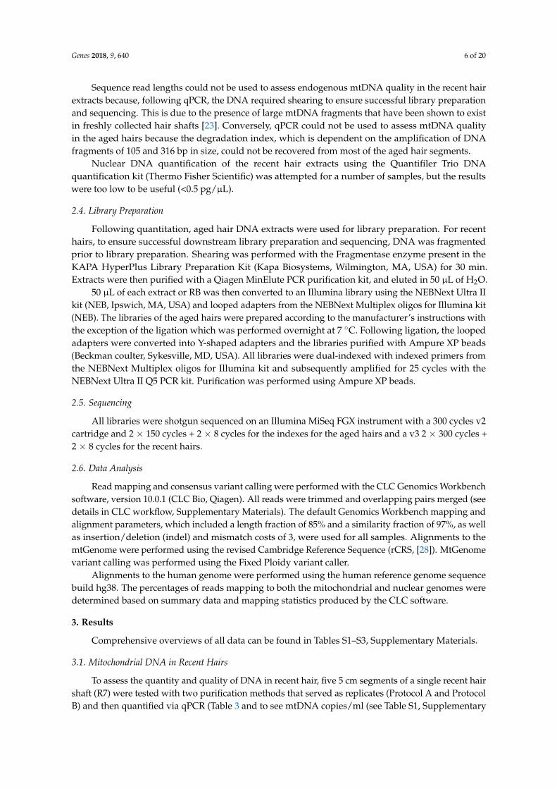
When the individual DIs from each replicate were plotted, R2 values of 0.9514 and 0.96641 wereobtained for Protocols A and B, respectively (Figure 3). In both cases, the trend was not only consistentalong the length of the shaft, but also statistically significant (Replicate 1: p < 0.05; Replicate 2: p < 0.05).This reduction in quality was confirmed in the sequencing data from protocol B DNA extracts of thehair segments (DNA was not fragmented before sequencing in these cases). The data showed that theaverage mtDNA size decreased from 168 bp at the proximal end to 91 bp at the distal end (Table 4).
Genes 2018, 9, 640 9 of 21
as the DNA degradation indices and average fragment sizes of the distal segments did not follow the same pattern. Overall, and depending on purification method, between four and six times less DNA was recovered from the distal segments than the proximal segments.
The two replicates, or purification methods, also revealed consistent patterns when DNA quality was assessed. For protocol A, a degradation index (DI) of 1.53 was obtained from the Segment 1 extract, and the DI increased to 3.44 in Segment 5 (Table 3). For protocol B, the degradation factorincreased from 1.50 to 3.69. In both cases, and similar to the reduction in mtDNA quantity along the length of the shaft, the degradation indices suggested a consistent reduction in the quality of mtDNA along the length of the shaft.
When the individual DIs from each replicate were plotted, R2 values of 0.9514 and 0.96641 were obtained for Protocols A and B, respectively (Figure 3). In both cases, the trend was not only consistent along the length of the shaft, but also statistically significant (Replicate 1: p < 0.05; Replicate 2: p < 0.05). This reduction in quality was confirmed in the sequencing data from protocol B DNAextracts of the hair segments (DNA was not fragmented before sequencing in these cases). The data showed that the average mtDNA size decreased from 168 bp at the proximal end to 91 bp at the distal end (Table 4).
Figure 3. Mitochondrial DNA quality in five segments of recently collected hair. Two replicates (orange series and blue series) were performed for each segment.
The potential of recovering complete mtGenome sequence from shotgun sequence data of recently collected hairs was tested using 5 cm–6 cm of six rootless hairs (R1-R6, Table 2). Cuttings from hairs R1, R3, R4, R5, and R6 were each composed of the first 1–6 cm proximal segment of an approximately 20–25 cm length hair. R2 was composed of several ~1.3 cm cuttings from the most distal portion of several ~5 cm hairs (from a haircut). The six DNA extracts varied in mtDNA quantity between 400 mtG/cm to 103,181 mtG/cm and reflected degradation indices ranging from 1.93–5.86 (Table 6). The quantity and quality values appeared to show no correspondence to hair treatment or time of storage. The differences may be due simply to the variability known to exist between hairs [16,18]. Despite the wide variation in mtDNA quantity and quality of the various hair shafts tested, complete mtGenome sequences were recovered from five of the six samples with at least 2x coverage. Depths of coverage in these cases averaged 44x and ranged between 2x and 844x (Table 6). The sixth sample (R2), which was also the sample with the lowest mtDNA yield, produced data covering
R² = 0.9664
R² = 0.9514
0
0.5
1
1.5
2
2.5
3
3.5
4
1 2 3 4 5
Degr
adat
ion
Inde
x
Hair Segment
mtDNA Quality Fresh Hair Segments
Protocol A Protocol B
Figure 3. Mitochondrial DNA quality in five segments of recently collected hair. Two replicates (orangeseries and blue series) were performed for each segment.
The potential of recovering complete mtGenome sequence from shotgun sequence data of recentlycollected hairs was tested using 5 cm–6 cm of six rootless hairs (R1–R6, Table 2). Cuttings from hairsR1, R3, R4, R5, and R6 were each composed of the first 1–6 cm proximal segment of an approximately20–25 cm length hair. R2 was composed of several ~1.3 cm cuttings from the most distal portionof several ~5 cm hairs (from a haircut). The six DNA extracts varied in mtDNA quantity between400 mtG/cm to 103,181 mtG/cm and reflected degradation indices ranging from 1.93–5.86 (Table 6).The quantity and quality values appeared to show no correspondence to hair treatment or time of

Genes 2018, 9, 640 9 of 20
storage. The differences may be due simply to the variability known to exist between hairs [16,18].Despite the wide variation in mtDNA quantity and quality of the various hair shafts tested, completemtGenome sequences were recovered from five of the six samples with at least 2x coverage. Depthsof coverage in these cases averaged 44x and ranged between 2x and 844x (Table 6). The sixth sample(R2), which was also the sample with the lowest mtDNA yield, produced data covering approximately73% of the mtGenome (Table 6). In this case, the average read coverage was 10x, with a minimum of0 reads and a maximum of 163 reads. In an attempt to recover DNA fragments that may be lost duringProtocol A extraction from recent hairs R1–R3, the bead binding waste solution was retained andpurified/extracted with the binding buffer from Protocol B (Protocol C). An additional 5902 mtG/cmfor R1, 13,410 mtG/cm for R2, and 11,765 mtG/cm for R3 were recovered.
Table 6. mtDNA quantitation values, degradation indexes, and coverage over the mtGenome for sixsingle recent hairs.
Extract mtGenomes/cm DegradationIndex
Coverageof at Least
2 Reads
Coverageof at Least
5 Reads
Coverageof at Least10 Reads
AverageCoverage
CoverageRange
R1 8190 2.22 100% 99% 84% 18x 2x–47xR2 400 1.93 73% 55% 39% 10x 0–163xR3 44,447 2.39 100% 98% 82% 16x 2x–69xR4 49,647 3.74 100% 100% 100% 30x 8x–122xR5 103,181 2.14 100% 100% 100% 88x 38x–153xR6 29,240 5.86 100% 100% 100% 67x 8x–844x
3.2. Mitochondrial DNA in Aged Hairs
To better understand total mtDNA quantity and quality in the types of degraded hair samplesoften encountered in forensic casework, four aged hair samples were evaluated via both mtDNA qPCRand sequencing (Table 7). The hairs had been cut in 1962 (A1, A6), in 1958 (A2), and in 1978 (A7)and stored at room temperature. Based on quantitative PCR data from Table 7, the samples exhibitedmtDNA yields substantially lower than the yields from the recent hair samples, even when quantitationvalues from the aged hairs were compared to the two most distal segments of the recently collectedhair (17–22 and 22–27 cm from the root; quantitation values ranged between 1414 and 10,296 mtG/cm).As with the recent hairs, the purification method used on the lysates had a noticeable effect on theabsolute quantity of mtDNA recovered (only protocol B used for A6 and A7). In the case of the aged hairsA1 and A2, however, the protocol B extracts—which targeted smaller fragments—yielded more DNAthan the protocol A extracts. For A1, mtDNA yields were 20 mtG/cm for protocol A and 100 mtG/cmfor protocol B, while for A2 the mtDNA yields were, respectively 67 mtG/cm and 947 mtG/cm. Thoughbased on only a handful of data points, the lower calculated mtDNA quantities (as measured by the105 bp qPCR molecule) but recoverable degradation indices (which reflects amplification of at leastsome ≥ 316 bp fragments) for only protocol A extracts is likely due to the preferential recovery of largerDNA fragments with protocol A. Protocol B, which preferentially recovers smaller DNA fragments,consistently yielded higher quantitation values, but no degradation values.
Table 7. mtDNA quantitation values, degradation indexes, and coverage over the mtGenome for agedhairs. The asterisk denotes DNA extraction using protocol B. NR: no result.
Extract mtGenomes/cm DegradationIndex
Coverageof at Least
2 Reads
Coverageof at Least
5 Reads
Coverageof at Least10 Reads
AverageCoverage
CoverageRange
A1 20 3 8.28% 1.99% 0% 0.4x 0–9xA1* 100 NR 100% 100% 100% 92x 20x–247xA2 67 4.35 36.54% 11.9% 2.12% 2x 0–20xA2* 947 NR 100% 100% 100% 84x 19x–232xA6* 95 NR 100% 99.9% 98.98% 27x 3x–79xA7* 2784 1.97 100% 100% 98.93% 27x 6x–715x

Genes 2018, 9, 640 10 of 20
The increased recovery of smaller DNA fragments from protocol B was directly observed in thesequencing results. Depending on the hair, between 58 and 309 times more sequencing reads wererecovered from the protocol B extracts than the protocol A extracts, resulting in only 0–8.28% and2.12–36.54% mtGenome coverage with Protocol A and 98.93–100% mtGenome coverage with protocolB (Table 7). This suggests that the majority of DNA in the aged hairs was extremely fragmented andlost with purification protocol A. Regardless of this recovery difference, however, the degradation stateof the DNA as indicated by both purification methods was consistent. For A2 (cut and collected in1958), the average size of the mtDNA reads from protocols A and B were 87 bp and 70 bp, respectively(Table S3 in Supplementary Materials). For A1 (cut and collected in 1965), the mtDNA reads averaged71 and 55 bp.
Because of the substantially improved data recovery from the aged hairs with protocol B, mtDNAquantity and quality along the length of the shaft was assessed with protocol B only. For this evaluation,DNA was extracted from four, 5 cm segments of a single ~40-year-old hair shaft (Seg 1 to 4-A4*) andthen assessed via both qPCR and sequencing. Similar to the results from the recently collected hairs,the qPCR results from the aged hairs showed a consistent decrease in mtDNA quantity as segmentsmore distal to the root were tested (Table 8). While the most proximal segment (1–6 cm from the root)yielded 3330 mtG/cm, the most distal segment (31–36 cm from the root) yielded only 510 mtG/cm(Table 8).
Table 8. mtDNA quantitation values and degradation Index values for two single aged segmentedhairs. The asterisk denotes DNA extraction using protocol B. No degradation index was obtained forany of the samples.
Samples Segment mtGenomes/cm
A4*
Seg1 3330Seg2 2900Seg3 1950Seg4 510
A8*
Seg1 2401Seg2 952Seg3 678Seg4 379Seg5 49
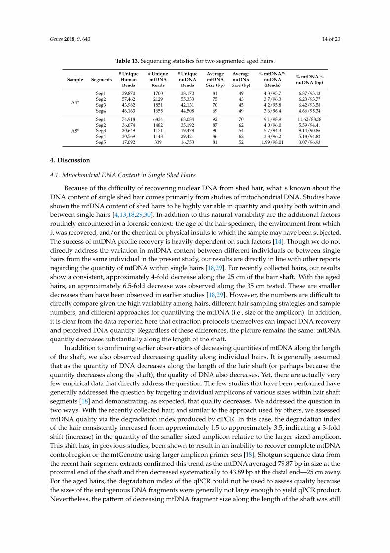
Degradation indices were not recovered from any of the aged hair segments, most likely due to thefact that DNA fragments of the size required to derive the degradation index (≥316 bp) were simplynot present in the tested segments. MtDNA quality was instead assessed based on the read lengthsof the shotgun sequencing data. Again, similar to the recent hairs, a reduction in mtDNA fragmentsize was observed as segments from the proximal to distal end of the hair were tested (Table S3,Supplementary Materials). MtDNA read lengths (and, by proxy, endogenous mtDNA fragment size)in the most proximal segment of the hair averaged 81 bp, with read lengths decreasing to an averageof 69 bp in the most distal fragment (Table 13). When mtDNA quantity and quality were plottedagainst hair segment for Sample A4, R2 values of 0.9455 and 0.9951 were obtained for mtDNA quantityand quality, respectively (Figure 4). In both cases, the trends were not only very consistent along thelength of the shaft, but also statistically significant (Quantity: p < 0.05; Quality: p < 0.05). A similarlyconsistent trend was observed with the mtDNA quantities of the five segments of hair A8*, but notnecessarily with the mtDNA qualities. Though the average size of the most distal A8* segment wassmaller than the most proximal end, the decline was not consistent from segment to segment (Figure 4,Table 13). This may be a result of the fact that the A8* segments spanned a shorter length of hair thanthe A4* segments (see Figure 1).

Genes 2018, 9, 640 11 of 20
Genes 2018, 9, 640 12 of 21
Figure 4. Mitochondrial DNA quality and quantity measures for two single, aged segmented hairs. For hair A4*, the four 5 cm segments spanned 35 cm of hair. For A8, the five 5 cm segments spanned25 cm of hair. Both came from the same donor.
In terms of complete mtGenome sequence recovery, and not surprisingly given the observed mtDNA quantities and degradation states, the aged hairs were less successful in producing fullmtGenome data than the recent hairs (Table 7). The aforementioned difference in DNA yields from the two different purification protocols made a clear difference in the recovery of complete mtGenome data. Only two aged hairs were tested with both protocols (A1 and A2), and complete mtGenome data were recovered only from the lysates purified with protocol B. More than likely, the small endogenous DNA fragments were simply washed away with protocol A, leaving close to nothing to be sequenced in these extracts. In the two cases for which sequencing was successful,complete mtGenomes were recovered despite low quantification values (100 mtG/cm for A1* and 947 mtG/cm for A2*). For A1*, the complete genome sequence was based on 31,762 unique reads and anaverage coverage of 92x, while the A2* genome was based on 44,826 unique reads and an average coverage of 84x (Tables 7 and 9).
Table 9. Sequencing statistics for single aged hairs. The asterisk denotes DNA extraction using protocol B [26].
Extract Average
mtDNA Length (bp; Ref hg38)
Average mtDNA
Length (bp;Ref rCRS)
Average nuDNA Length (bp; Ref
hg38)
# of mtDNA Reads
# of nuDNA Reads
Percentage mtDNA/nuDNA
Reads
A1 71 65 88 145 131,100 0.1/99.9A1* 55 54 54 44,826 5,132,784 0.9/99.1A2 87 78 58 548 449,782 0.1/99.9A2* 70 68 57 31,762 14,926,177 0.2/99.8A6* 58 57 55 12,291 1,718,590 0.7/99.3A7* 80 79 49 9,352 112,190 7.7/92.3
R² = 0.9455
R² = 0.8437
R² = 0.9979 R² = 0.7472
50
55
60
65
70
75
80
85
90
95
100
0
500
1000
1500
2000
2500
3000
3500
4000
1 2 3 4 5
mtD
NA q
ualit
y (b
ase
pairs
)
mtD
NA q
uant
ity (m
tG/c
m)
Hair Segment
mtDNA Quantity and Quality Aged Hair Segments
Hair A4 mtDNA quantity
Hair A8 mtDNA quantity
Hair A4 mtDNA quality
Hair A8 mtDNA quality
Figure 4. Mitochondrial DNA quality and quantity measures for two single, aged segmented hairs.For hair A4*, the four 5 cm segments spanned 35 cm of hair. For A8, the five 5 cm segments spanned25 cm of hair. Both came from the same donor.
In terms of complete mtGenome sequence recovery, and not surprisingly given the observedmtDNA quantities and degradation states, the aged hairs were less successful in producing fullmtGenome data than the recent hairs (Table 7). The aforementioned difference in DNA yields from thetwo different purification protocols made a clear difference in the recovery of complete mtGenome data.Only two aged hairs were tested with both protocols (A1 and A2), and complete mtGenome data wererecovered only from the lysates purified with protocol B. More than likely, the small endogenous DNAfragments were simply washed away with protocol A, leaving close to nothing to be sequenced in theseextracts. In the two cases for which sequencing was successful, complete mtGenomes were recovereddespite low quantification values (100 mtG/cm for A1* and 947 mtG/cm for A2*). For A1*, the completegenome sequence was based on 31,762 unique reads and an average coverage of 92x, while the A2*genome was based on 44,826 unique reads and an average coverage of 84x (Tables 7 and 9).
Table 9. Sequencing statistics for single aged hairs. The asterisk denotes DNA extraction using protocol B [26].
Extract
AveragemtDNA
Length (bp;Ref hg38)
AveragemtDNA
Length (bp;Ref rCRS)
AveragenuDNA
Length (bp;Ref hg38)
# of mtDNAReads
# of nuDNAReads
PercentagemtDNA/nuDNA
Reads
A1 71 65 88 145 131,100 0.1/99.9A1* 55 54 54 44,826 5,132,784 0.9/99.1A2 87 78 58 548 449,782 0.1/99.9A2* 70 68 57 31,762 14,926,177 0.2/99.8A6* 58 57 55 12,291 1,718,590 0.7/99.3A7* 80 79 49 9352 112,190 7.7/92.3
Though A1* and A2* yielded 100% mtGenome coverage at 10 reads, and A6* & A7* yielded ~99%coverage, the segments of aged hairs A4* and A8* yielded less (Table 10). At a depth of 10 reads,zero to 15% of the genome was covered for all segment extracts except Seg1-A8 (93.6%). At a depth ofcoverage (DOC) of five reads, however, approximately half of the genome was covered for all samples

Genes 2018, 9, 640 12 of 20
(except Seg1-A8 at 99.05%), and at a depth of two reads, with the exception of Seg5-A8*, over 80% ofthe genome was covered for all samples (Table 10). Interestingly, even in shotgun data, the controlregion generally had the highest DOC compared to the rest of the mtGenome. This may be due to theGC content of the CR (Figure S1, Supplementary Materials). In all cases for which the mtDNA profileof the donor was known, the sequence data corresponded to the known profile.
Table 10. mtGenome coverage of one segmented aged hair segments extracted with Protocol B.
Sample Extract Coverage of atLeast 2 Reads
Coverage of atLeast 5 Reads
Coverage of atLeast 10 Reads
AverageCoverage
CoverageRange
A4*
Seg1 90.69% 43.47% 7.94% 5x 0–28xSeg2 96.18% 66.56% 15.2% 6x 0–37xSeg3 90.18% 46.93% 4.62% 5x 0–25xSeg4 85.53% 40.22% 1.7% 4x 0–26x
A8*
Seg1 100% 99.05% 93.6% 22x 2x–70xSeg2 85.17% 45.54% 6.44% 5x 0–21xSeg3 80.12% 30.89% 2.22% 4x 0–14xSeg4 80.85% 30.48% 1.89% 4x 0–16xSeg5 27% 0.85% 0% 1x 0–7x
3.3. Nuclear DNA from Recent Hairs
The potential of nuclear DNA recovery from rootless shed hairs was assessed with six recentlycollected hairs (R1-R6). For all hairs, nuclear DNA quantification values could not be recovered viatraditional nuclear qPCR, and thus nuclear DNA content was assessed based solely on the ratio ofnuclear to mitochondrial sequence reads in the final data. For these six recently collected samples,between 34,909 and 952,728 unique reads mapped to the human genome. Of these, 33,044 to 927,153mapped to the human nuclear genome, with the remainder in any given sample mapping to the humanmitochondrial genome (Table 11). In total, the percentage of reads mapping to the human nucleargenome for any particular recently collected hair ranged between 88.4–99.5% of the total DNA reads(Table 11).
Table 11. Sequencing statistics for recent single hairs.
Extracts # UniqueHuman Reads
# mtDNAUnique Reads
# UniquenuDNA Reads
% mtDNA/%nuDNA (bp)
% mtDNA/%nuDNA (Reads)
R1 34,909 1865 33,044 11.95/88.05 5.3/94.7R2 327,165 1707 325,458 0.59/99.41 0.5/99.5R3 42,308 2287 40,021 9.29/90.71 5.4/94.6R4 196,737 4790 191,947 2.88/97.12 2.4/97.6R5 94,969 10,997 83,972 14.08/85.92 11.6/88.4R6 952,728 25,575 927,153 3.56/96.44 2.7/97.3
3.4. Nuclear DNA in Aged Hairs
For almost all of the aged hairs, more than 99% of the shotgun reads that mapped to the humangenome were nuclear DNA sequences (between 99.1 and 99.9%) while the remaining 0.1–0.9% mappedto the mtGenome. Sample A7 had 92.3% align to nuDNA and 7.7% align to mtDNA. These highpercentages of nuclear DNA were observed regardless of the purification protocol (both of which wereused for the assessment of A1 and A2). The average size of the nuclear DNA reads varied between49 and 88 bp, and for five of the six extracts, the average read length of the nuclear DNA fragmentswas smaller than the average read length of the mtDNA fragments from the same extract (Table 12).For sample A2*, the mtDNA fragments averaged 69 bp in length, while the nuclear DNA fragmentsaveraged only 57 bp. The same trend held true for samples A2 and A1* where the mtDNA fragmentsaveraged, respectively, 87 bp and 55 bp and the nuclear DNA fragments averaged 58 bp and 54 bp.The only exception to this pattern was sample A1, for which mtDNA reads averaged 71 bp, while nuclearDNA reads averaged 88 bp. This result could be authentic, or, given the small number of mtDNA

Genes 2018, 9, 640 13 of 20
reads (145) upon which the read length average is based, the larger size of the recovered nuclear DNAfragments could simply be the result of read sampling. Given that the four segments of aged hair(samples A4 Seg1–Seg4 and A8 Seg1–Seg5) also showed a pattern of the nuclear DNA being smallerthan the mitochondrial DNA, it seems likely that the sample A1 results are a sampling issue (Figure 4).
Table 12. Sequencing statistics for single aged hairs. The asterisk denotes DNA extraction usingprotocol B.
Extracts # UniquemtDNA Reads
# UniquenuDNA Reads
Average mtDNASize (bp)
Average nuDNASize (bp)
% mtDNA/%nuDNA (Reads)
A1 145 131,100 71 88 0.1/99.9A1* 44,826 5132,784 55 54 0.9/99.1A2 548 449,782 87 58 0.1/99.9A2* 31,762 14,926,177 69 57 0.2/99.8A6* 12,291 1718,590 58 55 0.7/99.3A7* 9352 112,190 80 49 7.7/92.3
For the four aged hair shaft segments Seg-A4*, nuclear DNA read lengths ranged between 43and 49 bp while mtDNA fragments averaged between 69 and 81 bp. Interestingly, however, while themtDNA read length seemed to consistently decline along the length of the hair shaft (Figure 5) thereappeared to be no clear pattern of a decrease in quality (i.e., size) for the nuclear DNA. Five segmentsfrom a second aged hair (Seg-A8*) did not show a similar consistent decline in mtDNA read length butdid repeat the overall trend of the mtDNA fragments (81–92 bp) being larger than nuDNA fragments(52 to 70 bp) in any given segment. For both sets of samples, the total nuclear DNA content mirroredobservations from the other recently collected and aged hair samples, with between 94.3 and 98.9% ofthe human DNA reads mapping to the nuclear DNA genome (Table 13).
Genes 2018, 9, 640 15 of 21
Figure 5. Nuclear and mitochondrial DNA quality (i.e. read length) for segments of two aged hairs.For hair A4*, the four 5 cm segments spanned 35 cm of hair. For A8*, the five 5 cm segments spanned 25 cm of hair. For each segment, and regardless of the hair sample, the average size of the mtDNAfragments was larger than the average size of the nuclear DNA fragments. In addition, with theexception of the hair A4 nuclear DNA, fragment sizes tended to decrease along the length of the hair shaft.
4. Discussion
4.1. Mitochondrial DNA Content in Single Shed Hairs
Because of the difficulty of recovering nuclear DNA from shed hair, what is known about theDNA content of single shed hair comes primarily from studies of mitochondrial DNA. Studies have shown the mtDNA content of shed hairs to be highly variable in quantity and quality both within and between single hairs [4,13,18,29,30]. In addition to this natural variability are the additional factors routinely encountered in a forensic context: the age of the hair specimen, the environment from which it was recovered, and/or the chemical or physical insults to which the sample may havebeen subjected. The success of mtDNA profile recovery is heavily dependent on such factors [14]. Though we do not directly address the variation in mtDNA content between different individuals or between single hairs from the same individual in the present study, our results are directly in linewith other reports regarding the quantity of mtDNA within single hairs [18,29]. For recently collectedhairs, our results show a consistent, approximately 4-fold decrease along the 25 cm of the hair shaft. With the aged hairs, an approximately 6.5-fold decrease was observed along the 35 cm tested. These are smaller decreases than have been observed in earlier studies [18,29]. However, the numbers aredifficult to directly compare given the high variability among hairs, different hair sampling strategiesand sample numbers, and different approaches for quantifying the mtDNA (i.e. size of the amplicon). In addition, it is clear from the data reported here that extraction protocols themselves can impact DNA recovery and perceived DNA quantity. Regardless of these differences, the picture remains the same: mtDNA quantity decreases substantially along the length of the shaft.
In addition to confirming earlier observations of decreasing quantities of mtDNA along the length of the shaft, we also observed decreasing quality along individual hairs. It is generally assumed that as the quantity of DNA decreases along the length of the hair shaft (or perhaps because the quantity decreases along the shaft), the quality of DNA also decreases. Yet, there are actually very
R² = 0.9262
R² = 0.0074
R² = 0.7472
R² = 0.6231
0
10
20
30
40
50
60
70
80
90
100
1 2 3 4 5
Aver
age
Read
Leng
th/D
NA Si
ze (b
ase
pairs
)
Hair Segment
Nuclear and Mitochondrial DNA Quality Aged Hair Segments
Hair A4 mtDNA quality
Hair A4 nucDNA quality
Hair A8 mtDNA quality
Hair A8 nucDNA quality
Figure 5. Nuclear and mitochondrial DNA quality (i.e., read length) for segments of two aged hairs.For hair A4*, the four 5 cm segments spanned 35 cm of hair. For A8*, the five 5 cm segments spanned25 cm of hair. For each segment, and regardless of the hair sample, the average size of the mtDNAfragments was larger than the average size of the nuclear DNA fragments. In addition, with the exceptionof the hair A4 nuclear DNA, fragment sizes tended to decrease along the length of the hair shaft.
Genes 2018, 9, 640 14 of 20
Table 13. Sequencing statistics for two segmented aged hairs.
Sample Segments# UniqueHumanReads
# UniquemtDNAReads
# UniquenuDNAReads
AveragemtDNASize (bp)
AveragenuDNASize (bp)
% mtDNA/%nuDNA(Reads)
% mtDNA/%nuDNA (bp)
A4*
Seg1 39,870 1700 38,170 81 49 4.3/95.7 6.87/93.13Seg2 57,462 2129 55,333 75 43 3.7/96.3 6.23/93.77Seg3 43,982 1851 42,131 70 45 4.2/95.8 6.42/93.58Seg4 46,163 1655 44,508 69 49 3.6/96.4 4.66/95.34
A8*
Seg1 74,918 6834 68,084 92 70 9.1/98.9 11.62/88.38Seg2 36,674 1482 35,192 87 62 4.0/96.0 5.59/94.41Seg3 20,649 1171 19,478 90 54 5.7/94.3 9.14/90.86Seg4 30,569 1148 29,421 86 62 3.8/96.2 5.18/94.82Seg5 17,092 339 16,753 81 52 1.99/98.01 3.07/96.93
4. Discussion
4.1. Mitochondrial DNA Content in Single Shed Hairs
Because of the difficulty of recovering nuclear DNA from shed hair, what is known about theDNA content of single shed hair comes primarily from studies of mitochondrial DNA. Studies haveshown the mtDNA content of shed hairs to be highly variable in quantity and quality both within andbetween single hairs [4,13,18,29,30]. In addition to this natural variability are the additional factorsroutinely encountered in a forensic context: the age of the hair specimen, the environment from whichit was recovered, and/or the chemical or physical insults to which the sample may have been subjected.The success of mtDNA profile recovery is heavily dependent on such factors [14]. Though we do notdirectly address the variation in mtDNA content between different individuals or between singlehairs from the same individual in the present study, our results are directly in line with other reportsregarding the quantity of mtDNA within single hairs [18,29]. For recently collected hairs, our resultsshow a consistent, approximately 4-fold decrease along the 25 cm of the hair shaft. With the agedhairs, an approximately 6.5-fold decrease was observed along the 35 cm tested. These are smallerdecreases than have been observed in earlier studies [18,29]. However, the numbers are difficult todirectly compare given the high variability among hairs, different hair sampling strategies and samplenumbers, and different approaches for quantifying the mtDNA (i.e., size of the amplicon). In addition,it is clear from the data reported here that extraction protocols themselves can impact DNA recoveryand perceived DNA quantity. Regardless of these differences, the picture remains the same: mtDNAquantity decreases substantially along the length of the shaft.
In addition to confirming earlier observations of decreasing quantities of mtDNA along the lengthof the shaft, we also observed decreasing quality along individual hairs. It is generally assumedthat as the quantity of DNA decreases along the length of the hair shaft (or perhaps because thequantity decreases along the shaft), the quality of DNA also decreases. Yet, there are actually veryfew empirical data that directly address the question. The few studies that have been performed havegenerally addressed the question by targeting individual amplicons of various sizes within hair shaftsegments [18] and demonstrating, as expected, that quality decreases. We addressed the question intwo ways. With the recently collected hair, and similar to the approach used by others, we assessedmtDNA quality via the degradation index produced by qPCR. In this case, the degradation indexof the hair consistently increased from approximately 1.5 to approximately 3.5, indicating a 3-foldshift (increase) in the quantity of the smaller sized amplicon relative to the larger sized amplicon.This shift has, in previous studies, been shown to result in an inability to recover complete mtDNAcontrol region or the mtGenome using larger amplicon primer sets [18]. Shotgun sequence data fromthe recent hair segment extracts confirmed this trend as the mtDNA averaged 79.87 bp in size at theproximal end of the shaft and then decreased systematically to 43.89 bp at the distal end—25 cm away.For the aged hairs, the degradation index of the qPCR could not be used to assess quality becausethe sizes of the endogenous DNA fragments were generally not large enough to yield qPCR product.Nevertheless, the pattern of decreasing mtDNA fragment size along the length of the shaft was still

Genes 2018, 9, 640 15 of 20
clearly evident from the shotgun sequence data. This fine-scale view of the data from sample A4*showed that the mtDNA averaged 81 bp in size at the proximal end of the shaft and then decreasedsystematically to 69 bp at the distal end, 30 cm away.
Given the extremely small size of the mtDNA fragments recovered from the aged hairs, it isnot surprising that probative mtDNA data are more difficult to recover in forensic cases involvingolder, and damaged or degraded hair samples. Melton et al., [14] describe that while over 90% of hairsamples aged 20 years and younger produce some mtDNA data using standard Sanger sequencingtechniques, successful mtDNA recovery drops to 60% of samples aged 30 years or older. Not onlyare the DNA extraction and purification protocols routinely employed in forensic laboratories notoptimized for the recovery of fragments <50 bp in size, but also the vast majority of downstream DNAtyping assays are designed for DNA fragments >100 bp [13,31,32]. Thus, even if the extremely smallfragments recovered from the aged samples tested here were routinely recovered at the extractionstage, DNA typing via traditional targeted PCR would still be unsuccessful. These factors are alsolikely why complete control region sequences could only be recovered in approximately 50% of hairsegments tested in Desmyter et al., [18] where the decline in sequence recovery corresponded with thedecline in mtDNA quantity towards the distal ends of the hairs.
For all single recent hairs but one (R2), the shotgun sequencing strategy employed in the currentstudy, produced between 98 to 100% of the mtGenome (DOC of 5x), regardless of mtDNA quantityor quality. For the incomplete sample, 55% of the genome, was still recovered with a DOC of 5x.Interestingly, the single recently collected hair sample that yielded incomplete data behaved, byall measures, more like the aged hair samples than the recent hair samples. Not only did it yieldvery little DNA when the purification protocol favoring larger DNA fragments was used (unlikethe other recent hairs), it also showed levels of DNA damage that exceeded levels observed in theaged hairs. It is possible that these observations demonstrate the extreme variability in hairs betweenindividuals [14,16,18].
We have previously reported the recovery of complete mtGenome sequence information fromshotgun data of two recent shed hairs [23]. In those cases, the hair fragments tested were immediatelyproximal to the root, included the root bulb (but no tissue), and were approximately 10 cm long.Here, we have extended those findings by demonstrating that complete mtGenome sequences can berecovered using shotgun sequencing from smaller hair fragments (2.5 cm versus 10 cm) and fragmentsthat do not include the root end. Given that qPCR quantification and degradation index values couldbe recovered from most of the recent hairs, it is likely that mtDNA fragments of a size sufficient fortargeted PCR amplification are present, and that commercial assays targeting small amplicons couldalso be successful in producing complete mtGenome data.
The same is likely not true for the aged hair samples, however. In the case of the hairs testedhere, mtDNA fragment sizes averaged only between 55 and 87 bp, depending on the sample. This isconsistent with other NGS-based studies on mtDNA from clumps of shed hair that found DNAfragments of approximately 61 bp in samples between 50 and 100 years old [22]. In all cases, these sizesare approximately 100 bp smaller than the average amplicon size in commercially available mtDNAassays [33]. Nevertheless, in all of the single hair shafts tested here (unsegmented rootless hairs greaterthan 40 years old), complete or near complete mtGenomes were recovered even when no mtDNAquant values were produced. While we have shown here that complete mtGenomes can be recoveredfrom extremely old hair even without enrichment, hybridization capture assays that enrich for mtDNAwould almost certainly improve efficiency. Indeed, when several recent hair segments and a numberof aged hairs not included in this study were enriched for mtDNA with a hybridization capture assay,complete mtGenomes were routinely recovered (Table S4 in Supplementary Materials).
4.2. Nuclear DNA Content in Single Shed Hairs
With the recovery of complete mtGenomes, we were also interested in the potential to both recoverand characterize the nuclear DNA content of single shed hairs. Though autosomal short tandem repeat

Genes 2018, 9, 640 16 of 20
profiles have been shown to be recoverable from such samples, success rates for producing informativeprofiles (e.g., 8 loci or greater) are understood to be low [8–12,34]. As a result, it is generally assumedthat nuclear DNA is simply not present in high enough copy number and/or is too degraded to berecovered in shed hairs. In our own laboratory, attempts to recover STR profiles from shed telogenhairs using the capillary electrophoresis-based protocols routinely employed in operational caseworkproduced full STR profiles in only 4.4% of the samples tested; and both of these samples (out of 45 total)included the root end.
Unfortunately, because of the library preparation for the recent hair, which necessarily requiredDNA shearing to permit successful downstream sequencing, the native fragmentation state of thenuclear DNA could not be assessed in the recent samples. However, data from the aged hairs showedextreme degradation, with average nuclear DNA fragment sizes of just 43 bp–88 bp sizes far smallerthan those required for targeted PCR amplification with the typical commercially available assays.The nuclear DNA was also consistently found to be more heavily fragmented than the mtDNA. In allbut one of the extracts tested, the average nuclear DNA length was smaller than the average mtDNAlength, with the only exception to this pattern likely a data sampling issue. It should be noted herethat the purification protocol employed had a clear and direct impact on the absolute length of nuclearand mtDNA fragment sizes recovered (with smaller average sizes observed for both the mtDNA andnuclear DNA fragments when the protocol favoring smaller fragments—B- was employed). However,the length of the nuclear relative to the mitochondrial DNA was the same regardless of the purificationprotocol. In both cases, the average mtDNA fragment size was consistently larger than the averagenuclear DNA fragment size.
Though the nuclear DNA was found to be more degraded than the mtDNA, it was found in farhigher quantity in any given hair or hair sample. In fact, for all of the hairs or hair segments tested,nuclear DNA comprised the vast majority of the human reads (Table S3, Supplementary Materials).This was the case regardless of the sample’s age (recently collected or >50 years old) or the proximityof the tested hair segment to the root. For the six recent hairs tested, and in line with previous studieson freshly collected single shed hair [23], nuclear DNA reads comprised, at minimum, 88% of the totalhuman reads, and generally more than 95%. For the aged hairs, the nuclear DNA read content hoveredaround 94–99% for the hair segments (samples A4* and A8*), and all but one of the aged hairs reflectednuclear DNA contents of >99%. Nuclear DNA recovery has been reported previously for hair samplesof 20 mg to 2 g in weight [19–22]. The findings here come from less than 1 mg of sample material.
Because of the size of the nuclear genome, the overwhelmingly large percentage of nuclear DNAreads was not sufficient to provide any reasonable depth of coverage across the genome. This wasin contrast to the mtDNA data where the relatively small percentage of reads was often adequate toprovide complete mtGenome coverage at an average read depth >10 when all samples were considered.Again, these data were produced with an Illumina MiSeq FGx—a low throughput/output instrument,relatively speaking. A higher throughput sequencer (such as a NextSeq or a HiSeq) would likely allowfor a substantial improvement in the recovery of both mtDNA and nuclear DNA data. Experimentswith these instruments are ongoing. Regardless, a preliminary analysis of 1,201 Y-SNPs based on verylow coverage of the nuclear genome with the MiSeq was still sufficient to provide a Y-chromosomehaplogroup prediction for hair A2* (Table S5 in Supplementary Materials).
4.3. Forensic Applications
A number of commercially available assays are available for next-generation sequencing offorensically relevant markers [33,35,36]. However, all of these assays are based on targeted PCRamplicons. In some cases, the amplicons are as small as 70 bp [37], but generally they are larger than100 bp. As a result, their utility for samples harboring heavily degraded DNA, such as shed hairs,is limited.
The utility of NGS approaches that do not require targeted PCR amplification is becomingincreasingly recognized in forensics, as it is only with such approaches that DNA from the most

Genes 2018, 9, 640 17 of 20
degraded specimens can be characterized. Though most applications to date have focused onmitochondrial genome typing from old skeletal remains [38–40], a handful of recent studies havealso described the recovery of nuclear DNA data from other types of limited and degradedspecimens [23,41,42]. The results of these studies, together with the data reported here describing thehigh content but low quality of nuclear DNA in shed hair, support the hypothesis that a key factor,perhaps the key factor, in the inability to recover common STR markers from such samples is thedegradation state of the DNA. DNA degradation has long been understood to be a factor in STRrecovery from shed hair, with mini-amplicon approaches often proving more successful than standardapproaches. However, the persistence and availability of nuclear DNA has also been a question.Studies have suggested that because keratinization involves the breakdown of the nucleus, includingthe DNA, nuclear DNA is, by extension, simply not present in telogen hair shafts [4,5]. Our datasuggest otherwise. Nuclear DNA is present in high quantity, but in extremely small fragments.
To get a sense of the type of forensically relevant data that could be gleaned from nuclear DNArecovered from rootless shed hair, data from one of the hair samples (A2*) were mapped against the163 ancestry/phenotype and 104 identity single nucleotide polymorphisms (SNPs) included in theVerogen ForenSeq assay [36]. SNP genotypes were developed for 11 and 14 SNPs, respectively, albeitat low coverage. However, when preliminary tests of a hybridization capture assay that targets thosesame SNPs were performed, SNP recovery increased to 104/163 for the ancestry/phenotype SNPsand 69/104 for the identity SNPs. Even assuming dropout at all loci exhibiting just one allele, theidentity SNP data resulted in a random match probability of one in 1014 (the calculation also assumedindependence of all SNPs). Though more developmental work will be required to optimize the assayto meet strict forensic guidelines for profile accuracy and reliability, the recovery of nuclear DNA fromsingle telogen hair fragments of less than 1 mg and that yield neither nuclear nor mtDNA quantitationvalues is promising.
The fact that DNA data was most effectively recovered from the aged hair specimens whena purification step that preferentially targets small fragments suggests that the DNA of interest inmany forensic cases may be inadvertently eliminated at the DNA extraction step. For these types ofhighly degraded specimens for which routine STR typing is simply not possible due to the quality ofendogenous DNA, our results suggest that modified extraction protocols that preferentially recoversmaller over larger fragments—and, therefore, recover rather than wash away the target DNA—can becritical to the recovery of probative DNA data in such cases. For most forensic casework, standardextraction protocols that recover DNA fragments of the size required for autosomal STR typing aresufficient. However, if and when the recovery of nuclear DNA is important, and DNA fragment sizesrequired for STR typing no longer exist, extraction protocols that specifically target smaller fragmentsmay be preferable. In addition, a combination of protocol types, as demonstrated here with Protocol C,may be a viable method to recover both large and small DNA fragments from the same sample.
5. Conclusions
In an effort to directly characterize the quantity and quality of mtDNA and nuclear DNA presentin the types of limited, aged, and degraded shed hair specimens often encountered in forensic casework,shotgun sequencing was performed on rootless telogen hairs. The results, based on direct observationof the endogenous molecules, revealed that:
1. mtDNA quantity and quality decline along the length of the hair shaft,2. mtDNA fragments are generally larger than nuclear DNA fragments in the same hair or
hair segment,3. complete mtGenomes can be recovered from aged hair shafts with shotgun sequencing data
along (i.e., no enrichment)4. nuDNA quality tends to decrease along the length of the hair shaft,

Genes 2018, 9, 640 18 of 20
5. both nuclear and mitochondrial DNA fragment sizes in the aged hairs were generally <80 bp (toosmall for routinely employed targeted PCR amplicons) and
6. nuclear DNA was not only recovered but comprised the vast majority of DNA in any givenhair sample.
Clearly, the relative sizes and copy numbers of the two genomes play a critical role in the recoveryof informative DNA profiles from telogen hair samples (for reference, the percentage of nuclear DNAin a cell with 1000 mtGenomes is 99.997%), but our results show that in the types of specimens thathave historically failed to yield mtDNA control region data with existing Sanger protocols, not onlycould complete mitochondrial genomes be developed, but also informative nuclear DNA data couldbe recovered. Further development of assays that accommodate the small size of the nuclear DNA mayallow for more routine recovery of discriminatory nuclear DNA profiles from such samples. A betterunderstanding of DNA quantity and quality, for nuclear DNA in particular, should promote furtherdevelopment of cost-effective forensic assays that can generate more discriminatory information fromnot only hair, but also other samples harboring extremely degraded DNA.
Supplementary Materials: The following are available online at http://www.mdpi.com/2073-4425/9/12/640/s1,Figure S1: Comparison between the control region and the mtGenome coverage; Table S1: Global mtDNA qPCRresults; Table S2: Coverage of the mtGenome for recent and aged hairs and hair segments; Table S3: Sequencingstatistics for all recent and aged hairs; Table S4: Difference between shotgun sequencing and sequencing oflibraries enriched for mtDNA; Table S5: The derived and ancestral Y haplogroup diagnostic SNPs that wereobserved for Sample A2*. Protocols: description of protocols A, B and C; CLC Workflow: workflow used to mapreads to the mtGenome.
Author Contributions: M.D.B., O.L. and J.A.I. conceived the experiments. M.D.B. and O.L. performedexperiments. O.L. did the data analyses. J.A.I., M.D.B. and O.L. wrote and approved the manuscript.
Acknowledgments: The authors would like to thank Rebecca Just and Thomas Callaghan for critical review ofthe manuscript, and Danielle Daniels, Michelle Galusha, and Anthony Onorato for helpful discussion.
Conflicts of Interest: Names of commercial manufacturers are provided for identification purposes only andinclusion does not imply endorsement of the manufacturer, or its products or services by the FBI. The viewsexpressed are those of the authors and do not necessarily reflect the official policy or position of the FBI. This isFBI Laboratory publication #19-04.
References
1. Pfeiffer, H.; Huhne, J.; Ortmann, C.; Waterkamp, K.; Brinkmann, B. Mitochondrial DNA typing from humanaxillary, pubic and head hair shafts—Success rates and sequence comparisons. Int. J. Legal Med. 1999, 112,287–290. [CrossRef] [PubMed]
2. Bender, K.; Schneider, P.M. Development of a new multiplex assay for STR typing of telogen hair roots.Int. Congr. Ser. 2006, 1288, 654–656. [CrossRef]
3. Graham, E.A. DNA reviews: Hair. Forensic Sci. Med. Pathol. 2007, 3, 133–137. [CrossRef] [PubMed]4. Bengtsson, C.F.; Olsen, M.E.; Brandt, L.O.; Bertelsen, M.F.; Willerslev, E.; Tobin, D.J.; Wilson, A.S.; Gilbert, M.T.
DNA from keratinous tissue. Part I: Hair and nail. Ann. Anat. 2012, 194, 17–25. [CrossRef] [PubMed]5. McNevin, D.; Wilson-Wilde, L.; Robertson, J.; Kyd, J.; Lennard, C. Short tandem repeat (STR) genotyping of
keratinised hair. Part 1. Review of current status and knowledge gaps. Forensic Sci. Int. 2005, 153, 237–246.[CrossRef] [PubMed]
6. Fischer, H.; Eckhart, L.; Mildner, M.; Jaeger, K.; Buchberger, M.; Ghannadan, M.; Tschachler, E. DNase1L2degrades nuclear DNA during corneocyte formation. J. Investig. Dermatol. 2007, 127, 24–30. [CrossRef][PubMed]
7. Fischer, H.; Szabo, S.; Scherz, J.; Jaeger, K.; Rossiter, H.; Buchberger, M.; Ghannadan, M.; Hermann, M.;Theussl, H.C.; Tobin, D.J.; et al. Essential role of the keratinocyte-specific endonuclease DNase1L2 in theremoval of nuclear DNA from hair and nails. J. Investig. Dermatol. 2011, 131, 1208–1215. [CrossRef]
8. Hellmann, A.; Rohleder, U.; Schmitter, H.; Wittig, M. STR typing of human telogen hairs—A new approach.Int. J. Legal Med. 2001, 114, 269–273. [CrossRef] [PubMed]
9. Grubwieser, P.; Muhlmann, R.; Parson, W. New sensitive amplification primers for the STR locus D2S1338for degraded casework DNA. Int. J. Legal Med. 2003, 117, 185–188.

Genes 2018, 9, 640 19 of 20
10. Muller, K.; Klein, R.; Miltner, E.; Wiegand, P. Improved STR typing of telogen hair root and hair shaft DNA.Electrophoresis 2007, 28, 2835–2842. [CrossRef]
11. Opel, K.L.; Fleishaker, E.L.; Nicklas, J.A.; Buel, E.; McCord, B.R. Evaluation and quantification of nuclearDNA from human telogen hairs. J. Forensic Sci. 2008, 53, 853–857. [CrossRef] [PubMed]
12. Ottens, R.; Taylor, D.; Abarno, D.; Linacre, A. Successful direct amplification of nuclear markers from a singlehair follicle. Forensic Sci. Med. Pathol. 2013, 9, 238–243. [CrossRef] [PubMed]
13. Berger, C.; Parson, W. Mini-midi-mito: Adapting the amplification and sequencing strategy of mtDNA to thedegradation state of crime scene samples. Forensic Sci. Int. Genet. 2009, 3, 149–153. [CrossRef]
14. Melton, T.; Dimick, G.; Higgins, B.; Lindstrom, L.; Nelson, K. Forensic mitochondrial DNA analysis of 691casework hairs. J. Forensic Sci. 2005, 50, 73–80. [CrossRef] [PubMed]
15. Gilbert, M.T.; Janaway, R.C.; Tobin, D.J.; Cooper, A.; Wilson, A.S. Histological correlates of post mortemmitochondrial DNA damage in degraded hair. Forensic Sci. Int. 2006, 156, 201–207. [CrossRef] [PubMed]
16. Linch, C.A.; Whiting, D.A.; Holland, M.M. Human hair histogenesis for the mitochondrial DNA forensicscientist. J. Forensic Sci. 2001, 46, 844–853. [CrossRef] [PubMed]
17. Almeida, M.; Betancor, E.; Fregel, R.; Suarez, N.M.; Pestano, J. Efficient DNA extraction from hair shafts.Forensic Sci. Int. Genet. 2011, 3, 319–320. [CrossRef]
18. Desmyter, S.; Bodner, M.; Huber, G.; Dognaux, S.; Berger, C.; Noel, F.; Parson, W. Hairy matters: MtDNAquantity and sequence variation along and among human head hairs. Forensic Sci. Int. Genet. 2016, 25, 1–9.[CrossRef]
19. Gilbert, M.T.; Kivisild, T.; Gronnow, B.; Andersen, P.K.; Metspalu, E.; Reidla, M.; Tamm, E.; Axelsson, E.;Gotherstrom, A.; Campos, P.F.; et al. Paleo-Eskimo mtDNA genome reveals matrilineal discontinuity inGreenland. Science 2008, 320, 1787–1789. [CrossRef]
20. Rasmussen, M.; Li, Y.; Lindgreen, S.; Pedersen, J.S.; Albrechtsen, A.; Moltke, I.; Metspalu, M.; Metspalu, E.;Kivisild, T.; Gupta, R.; et al. Ancient human genome sequence of an extinct Palaeo-Eskimo. Nature 2010, 463,757–762. [CrossRef]
21. Rasmussen, M.; Guo, X.; Wang, Y.; Lohmueller, K.E.; Rasmussen, S.; Albrechtsen, A.; Skotte, L.; Lindgreen, S.;Metspalu, M.; Jombart, T.; et al. An Aboriginal Australian genome reveals separate human dispersals intoAsia. Science 2011, 334, 94–98. [CrossRef] [PubMed]
22. Tobler, R.; Rohrlach, A.; Soubrier, J.; Bover, P.; Llamas, B.; Tuke, J.; Bean, N.; Abdullah-Highfold, A.; Agius, S.;O’Donoghue, A.; et al. Aboriginal mitogenomes reveal 50,000 years of regionalism in Australia. Nature 2017,544, 180–184. [CrossRef] [PubMed]
23. Parson, W.; Huber, G.; Moreno, L.; Madel, M.B.; Brandhagen, M.D.; Nagl, S.; Xavier, C.; Eduardoff, M.;Callaghan, T.C.; Irwin, J.A. Massively parallel sequencing of complete mitochondrial genomes from hairshaft samples. Forensic Sci. Int. Genet. 2015, 15, 8–15. [CrossRef] [PubMed]
24. Burnside, E.S.; Bintz, B.J.; Wilson, M.R. Improved extraction efficiency of human mitochondrial DNAfrom hair shafts and its implications for sequencing of the entire mtGenome from a single hair fragment.In Proceedings of the American Academy of Forensic Sciences 65th Annual Meeting, Atlanta, GA, USA,20–25 February 2012.
25. Gallimore, J.M.; McElhoe, J.A.; Holland, M.M. Assessing heteroplasmic variant drift in the mtDNA controlregion of human hairs using an MPS approach. Forensic Sci. Int. Genet. 2018, 32, 7–17. [CrossRef] [PubMed]
26. Allentoft, M.E.; Sikora, M.; Sjogren, K.G.; Rasmussen, S.; Rasmussen, M.; Stenderup, J.; Damgaard, P.B.;Schroeder, H.; Ahlstrom, T.; Vinner, L.; et al. Population genomics of Bronze Age Eurasia. Nature 2015, 522,167–172. [CrossRef]
27. Kavlick, M.F. Development of a triplex mtDNA qPCR assay to assess quantification, degradation, inhibition,and amplification target copy numbers. Mitochondrion 2018. [CrossRef] [PubMed]
28. Andrews, R.M.; Kubacka, I.; Chinnery, P.F.; Lightowlers, R.N.; Turnbull, D.M.; Howell, N. Reanalysis andrevision of the Cambridge reference sequence for human mitochondrial DNA. Nat. Genet. 1999, 23, 147.[CrossRef]
29. Andreasson, H.; Nilsson, M.; Budowle, B.; Lundberg, H.; Allen, M. Nuclear and mitochondrial DNAquantification of various forensic materials. Forensic Sci. Int. 2006, 164, 56–64. [CrossRef]
30. Nilsson, M.; Norlin, S.; Allen, M. Sequencing of mtDNA in shed hairs: A retrospective analysis of materialfrom forensic cases and a pre-screening. Open Forensic Sci. J. 2012, 5, 13–22. [CrossRef]

Genes 2018, 9, 640 20 of 20
31. Gabriel, M.N.; Huffine, E.F.; Ryan, J.H.; Holland, M.M.; Parsons, T.J. Improved MtDNA sequence analysis offorensic remains using a “mini-primer set” amplification strategy. J. Forensic Sci. 2001, 46, 247–253. [CrossRef]
32. Edson, S.M.; Ross, J.P.; Coble, M.D.; Parsons, T.J.; Barritt, S.M. Naming the dead—Confronting the realitiesof rapid identification of degraded skeletal remains. Forensic Sci. Rev. 2004, 16, 63–90. [PubMed]
33. Precision ID mtDNA Whole Genome Panel; Thermo Fisher Scientific: Waltham, MA, USA, 2018; Cataloguenumber: A30938.
34. Grisedale, K.S.; Murphy, G.M.; Brown, H.; Wilson, M.R.; Sinha, S.K. Successful nuclear DNA profiling ofrootless hair shafts: a novel approach. Int. J. Legal Med. 2018, 132, 107–115. [CrossRef] [PubMed]
35. PowerSeqTM CRM Nested System; Promega corp.: Madison, WI, USA, 2018; Catalogue number: AX5810.36. ForenSeqTM DNA Siganture Prep Kit (TG-450–1001); Verogen/Illumina, Inc.: San Diego, CA, USA, 2017.37. Fazi, A.; Gobeski, B.; Foran, D. Development of two highly sensitive forensic sex determination assays based
on human DYZ1 and Alu repetitive DNA elements. Electrophoresis 2014, 35, 3028–3035. [CrossRef] [PubMed]38. Templeton, J.E.; Brotherton, P.M.; Llamas, B.; Soubrier, J.; Haak, W.; Cooper, A.; Austin, J.J. DNA capture and
next-generation sequencing can recover whole mitochondrial genomes from highly degraded samples forhuman identification. Investig. Genet. 2013, 4, 26. [CrossRef] [PubMed]
39. Eduardoff, M.; Xavier, C.; Strobl, C.; Casas-Vargas, A.; Parson, W. Optimized mtDNA control region primerextension capture analysis for forensically relevant samples and highly compromised mtDNA of differentage and origin. Genes 2017, 8, 237. [CrossRef]
40. Marshall, C.; Sturk-Andreaggi, K.; Daniels-Higginbotham, J.; Oliver, R.S.; Barritt-Ross, S.; McMahon, T.P.Performance evaluation of a mitogenome capture and Illumina sequencing protocol using non-probative,case-type skeletal samples: Implications for the use of a positive control in a next-generation sequencingprocedure. Forensic Sci. Int. Genet. 2017, 31, 198–206. [CrossRef] [PubMed]
41. Shih, S.Y.; Bose, N.; Goncalvez, A.B.R.; Erlich, H.A.; Calloway, C.D. Application of probe capture enrichmentnext generation sequencing assay for whole mitochondrial genome and 426 nuclear SNPs for forensicallychallenging samples. Genes 2018, 9, 49. [CrossRef] [PubMed]
42. Loreille, O.; Ratnayake, S.; Bazinet, A.L.; Stockwell, T.B.; Sommer, D.D.; Rohland, N.; Mallick, S.;Johnson, P.L.F.; Skoglund, P.; Onorato, A.; et al. Biological sexing of a 4000-year-old Egyptian mummy headto assess the potential of nuclear DNA recovery from the most damaged and limited forensic specimens.Genes 2018, 9, 135. [CrossRef]
© 2018 by the authors. Licensee MDPI, Basel, Switzerland. This article is an open accessarticle distributed under the terms and conditions of the Creative Commons Attribution(CC BY) license (http://creativecommons.org/licenses/by/4.0/).
Copyright © 2022 FDOKUMEN