Formation of protein nanoparticles by controlled heat treatment of lactoferrin: Factors affecting...
7
Formation of protein nanoparticles by controlled heat treatment of lactoferrin: Factors affecting particle characteristics Carlos Bengoechea b, * , Irene Peinado c , David Julian McClements a a Biopolymers and Colloids Research Laboratory, Department of Food Science, University of Massachusetts, Amherst, MA 01003, USA b Departamento de Ingeniería Química, Universidad de Sevilla, Facultad de Química, Profesor García González 1, 41012 Sevilla, Spain c Institute of Food Engineering for Development, Polytechnic University of Valencia, Camino de Vera s/n, 46022 Valencia, Spain article info Article history: Received 10 September 2010 Accepted 24 December 2010 Keywords: Lactoferrin Nanoparticles DSC z-Potential Aggregation Denaturation abstract Lactoferrin is a globular protein from milk that has considerable potential as a functional ingredient in food, cosmetic and pharmaceutical applications. In this study, we examined the possibility of preparing food-grade bovine lactoferrin (bLf) nanoparticles using a simple thermal processing method. Differential Scanning Calorimetry (DSC), light scattering, and z-potential techniques were used to provide infor- mation about the conformational changes, aggregation, and electrical charge of bLf in aqueous solutions. DSC studies indicated that the protein had two thermal denaturation temperatures (61 and 93 C), which were associated with two different lobes of the protein. Protein denaturation was found to be irre- versible, which was attributed to the formation of protein nanoparticles, whose size depended on the temperature and duration of the thermal treatment. Higher holding temperatures produced faster protein aggregation and larger protein nanoparticles: 85 > 80 > 75 > 70 C. The protein nanoparticles produced by thermal treatment were resistant to subsequent changes in pH (from 3 to 11) and to salt addition (0e200 mM NaCl). The lactoferrin nanoparticles produced in this study may be useful as function ingredients in commercial products. Ó 2010 Elsevier Ltd. All rights reserved. 1. Introduction Bovine lactoferrin, bLf, is a single-chain glycoprotein with a reported molecular weight around 80 kDa and approximately 700 amino acid residues (Levay & Viljoen, 1995; Öztas ¸ , Yes ¸ im, & Özgünes ¸, 2005). Lactoferrin is an iron transporter and may be found in both the iron-saturated (holo-lactoferrin) and the iron- depleted (apo-lactoferrin) forms. Its structure consists of two lobes with a reversible iron-binding capacity of two atoms per lactoferrin molecule (one for each lobe), with one HCO 3 ion being synergistically bound with each Fe 3þ ion. One of the main interests in lactoferrin resides in its various biological properties, such as antimicrobial, anti-inflammatory, anti-tumor, immune- modulatory, and enzymatic activities (Brock, 2002). It seems that when iron is bound into the lactoferrin molecule, a more closed conformation is adopted (Levay & Viljoen, 1995). It has been extensively reported that the isoelectric point (pI) of lactoferrin is around 8e9(Brock, 2002; Levay & Viljoen, 1995; Moguilevsky, Retegui, & Masson, 1985). This relatively high pI is explained by a unique basic region in the N terminal region of lactoferrin, which can bind acidic molecules (Brock, 2002). There has been considerable interest in the formation of biopolymer particles from proteins and/or polysaccharides (Benichou, Aserin, & Garti, 2002; McClements, 2006; Tolstoguzov, 2002, 2003; Turgeon, Schmitt, & Sanchez, 2007), as they may be used for protection and delivery of bioactive compounds in food systems (Chen, Remondetto, & Subirade, 2006; Emerich & Thanos, 2007; Goldberg, Langer, & Jia, 2007; Ubbink & Krueger, 2006). Biopolymer particles may be formed through complexation of proteins, polysaccharides and other compounds. Alternatively, recent studies have shown that protein nanoparticles and micro- particles can be formed by controlled thermal denaturation of globular protein solutions (Bromley, Krebs, & Donald, 2006; Donato, Schmitt, Bovetto, & Rouvet, 2009; Hoffmann, Roefs, Verheul, VanMil, & DeKruif, 1996; Jones, Decker, & McClements, 2010; Santipanichwong, Suphantharika, Weiss, & McClements, 2008). Some authors have combined both approaches to form nanoparticles through heat denaturation of globular proteins fol- lowed by electrostatic complexation with polysaccharides (Hong & McClements, 2007; Jones, Lesmes, Dubin, & McClements, 2010; Kelly, Gudo, Mitchell, & Harding, 1994; Sanchez & Paquin, 1997; Schmitt, Sanchez, Desobry-Banon, & Hardy, 1998; Yu, Hu, Pan, * Corresponding author. Tel.: þ34 954557179; fax: þ34 954556447. E-mail address: [email protected] (C. Bengoechea). Contents lists available at ScienceDirect Food Hydrocolloids journal homepage: www.elsevier.com/locate/foodhyd 0268-005X/$ e see front matter Ó 2010 Elsevier Ltd. All rights reserved. doi:10.1016/j.foodhyd.2010.12.014 Food Hydrocolloids 25 (2011) 1354e1360
Transcript of Formation of protein nanoparticles by controlled heat treatment of lactoferrin: Factors affecting...
lable at ScienceDirect
Food Hydrocolloids 25 (2011) 1354e1360
Contents lists avai
Food Hydrocolloids
journal homepage: www.elsevier .com/locate/ foodhyd
Formation of protein nanoparticles by controlled heat treatmentof lactoferrin: Factors affecting particle characteristics
Carlos Bengoechea b,*, Irene Peinado c, David Julian McClements a
aBiopolymers and Colloids Research Laboratory, Department of Food Science, University of Massachusetts, Amherst, MA 01003, USAbDepartamento de Ingeniería Química, Universidad de Sevilla, Facultad de Química, Profesor García González 1, 41012 Sevilla, Spainc Institute of Food Engineering for Development, Polytechnic University of Valencia, Camino de Vera s/n, 46022 Valencia, Spain
a r t i c l e i n f o
Article history:Received 10 September 2010Accepted 24 December 2010
Keywords:LactoferrinNanoparticlesDSCz-PotentialAggregationDenaturation
* Corresponding author. Tel.: þ34 954557179; fax:E-mail address: [email protected] (C. Bengoeche
0268-005X/$ e see front matter � 2010 Elsevier Ltd.doi:10.1016/j.foodhyd.2010.12.014
a b s t r a c t
Lactoferrin is a globular protein from milk that has considerable potential as a functional ingredient infood, cosmetic and pharmaceutical applications. In this study, we examined the possibility of preparingfood-grade bovine lactoferrin (bLf) nanoparticles using a simple thermal processing method. DifferentialScanning Calorimetry (DSC), light scattering, and z-potential techniques were used to provide infor-mation about the conformational changes, aggregation, and electrical charge of bLf in aqueous solutions.DSC studies indicated that the protein had two thermal denaturation temperatures (61 and 93 �C), whichwere associated with two different lobes of the protein. Protein denaturation was found to be irre-versible, which was attributed to the formation of protein nanoparticles, whose size depended on thetemperature and duration of the thermal treatment. Higher holding temperatures produced fasterprotein aggregation and larger protein nanoparticles: 85 > 80 > 75 > 70 �C. The protein nanoparticlesproduced by thermal treatment were resistant to subsequent changes in pH (from 3 to 11) and to saltaddition (0e200 mM NaCl). The lactoferrin nanoparticles produced in this study may be useful asfunction ingredients in commercial products.
� 2010 Elsevier Ltd. All rights reserved.
1. Introduction
Bovine lactoferrin, bLf, is a single-chain glycoprotein witha reported molecular weight around 80 kDa and approximately700 amino acid residues (Levay & Viljoen, 1995; Öztas, Yesim, &Özgünes, 2005). Lactoferrin is an iron transporter and may befound in both the iron-saturated (holo-lactoferrin) and the iron-depleted (apo-lactoferrin) forms. Its structure consists of twolobes with a reversible iron-binding capacity of two atoms perlactoferrin molecule (one for each lobe), with one HCO3
� ion beingsynergistically bound with each Fe3þ ion. One of the maininterests in lactoferrin resides in its various biological properties,such as antimicrobial, anti-inflammatory, anti-tumor, immune-modulatory, and enzymatic activities (Brock, 2002). It seems thatwhen iron is bound into the lactoferrin molecule, a more closedconformation is adopted (Levay & Viljoen, 1995). It has beenextensively reported that the isoelectric point (pI) of lactoferrin isaround 8e9 (Brock, 2002; Levay & Viljoen, 1995; Moguilevsky,Retegui, & Masson, 1985). This relatively high pI is explained by
þ34 954556447.a).
All rights reserved.
a unique basic region in the N terminal region of lactoferrin,which can bind acidic molecules (Brock, 2002).
There has been considerable interest in the formation ofbiopolymer particles from proteins and/or polysaccharides(Benichou, Aserin, & Garti, 2002; McClements, 2006; Tolstoguzov,2002, 2003; Turgeon, Schmitt, & Sanchez, 2007), as they may beused for protection and delivery of bioactive compounds in foodsystems (Chen, Remondetto, & Subirade, 2006; Emerich & Thanos,2007; Goldberg, Langer, & Jia, 2007; Ubbink & Krueger, 2006).Biopolymer particles may be formed through complexation ofproteins, polysaccharides and other compounds. Alternatively,recent studies have shown that protein nanoparticles and micro-particles can be formed by controlled thermal denaturation ofglobular protein solutions (Bromley, Krebs, & Donald, 2006;Donato, Schmitt, Bovetto, & Rouvet, 2009; Hoffmann, Roefs,Verheul, VanMil, & DeKruif, 1996; Jones, Decker, & McClements,2010; Santipanichwong, Suphantharika, Weiss, & McClements,2008). Some authors have combined both approaches to formnanoparticles through heat denaturation of globular proteins fol-lowed by electrostatic complexation with polysaccharides (Hong &McClements, 2007; Jones, Lesmes, Dubin, & McClements, 2010;Kelly, Gudo, Mitchell, & Harding, 1994; Sanchez & Paquin, 1997;Schmitt, Sanchez, Desobry-Banon, & Hardy, 1998; Yu, Hu, Pan,
C. Bengoechea et al. / Food Hydrocolloids 25 (2011) 1354e1360 1355
Yao, & Jiang, 2006). Previous studies have primarily been carriedout using b-lactoglobulin to form the nanoparticles, which has anisoelectric point of around pH 5 and a single thermal denaturationtemperature. In this study, we examined whether protein nano-particles could also be formed using lactoferrin, which has a muchhigher isoelectric point than b-lactoglobulin (and is thereforepositively charged over a wider pH range) and has two thermaldenaturation temperatures. The stability of lactoferrin particlesformed through heating at different pH and ionic strength valueswill be evaluated. It should be noted, that Sreedhara, et al. (2010)recently studied the pH-induced changes in the structure ofa bovine lactoferrin similar to the one used in this work, finding anunfolded structure for the protein under acidic conditions, andfolded at higher pH values.
Lactoferrin nanoparticles may be useful functional ingredientsin food, cosmetic and pharmaceutical applications, e.g., to modifythe optical or rheological properties of products, or to encapsulateand deliver bioactive ingredients. Our purpose was to demonstratethat protein nanoparticles could be formed from lactoferrin, and tohighlight some of the factors that influence their properties.Further work would be required to translate this approach into anindustrial method of producing lactoferrin nanoparticles.
2. Experimental
2.1. Materials
Purified lactoferrin powder (Lot 10410479) obtained from cow’smilk was kindly donated by DMV International (BA Veghel, TheNetherlands). The reported composition expressed as a dry weightpercent was: 97.7% protein; and 0.12% ash; the remaining watercontent expressed on the wet basis of the powder was 2.8%. Themanufacturer reported that the iron content of the lactoferrinpowder was reported to be less than 120 ppm (0.012 wt%), whichcorresponds to a molar ratio of iron-to-protein of<0.18. It was useddirectly from the sample container without further purification. Allsolutions were prepared with double-distilled/de-ionized waterobtained from a filtration unit on-site.
2.2. Biopolymer solution preparation
Protein solutions were prepared by adding water to a weighedamount of lactoferrin powder and then stirring constantly atambient temperature for 1 h. Protein solutions were initiallyadjusted to pH 7.0, which is the most stable form of lactoferrin(Brock, 1997), using a 0.1 N sodium hydroxide solution. Lactoferrinconcentrations are reported as weight percentages (wt%) based onthe initial masses of the powders. A fixed protein concentration of0.2 wt% was used in these experiments so as to form solutions thatgave a convenient signal in the light scattering and z-potentialinstrument. Biopolymer solutions were adjusted to pH valuesbelow 7.0 using 1.0, 0.1 and/or 0.01 N hydrochloric acid solutions.All solutions were equilibrated at the desired pH for 1e3min beforesample aliquots were removed. All solutions were prepared withdouble-distilled/de-ionized water obtained from a filtration uniton-site.
2.3. Creation of protein particles through thermal treatment
The formation of biopolymer particles by thermal treatmentwas carried out by first adjusting 0.2% lactoferrin solutions to pH 7,then pouring them into glass test tubes with plastic screw-caps,and then subjecting them to a heat treatment by placing the testtubes in a temperature-controlled water bath at different temper-atures (70e90 �C) for different holding times (1e60 min). Jones,
Decker, and McClements (2009) previously used this procedure tocreate protein-based particulate-suspensions. Following thermaltreatments, solutions were removed from the water bath andallowed to cool in a water bath at room temperature.
2.4. pH stability of protein particles
The effect of pH on the properties of the protein particlesprepared by thermal treatment (75 �C for 20 min) was evaluatedusing two different approaches: (i) modifying the pH of the solu-tions (from 2 to 11) before thermal treatment; (ii) modifying the pHof the solutions (from 2 to 11) after thermal treatment. Measure-ments were performed 24 h after sample preparation.
2.5. Salt concentration
The effect of salt on the stability of the protein particles preparedby thermal treatment (75 �C for 20 min) was also evaluated usingtwo different approaches: (i) adding 0e200 mM NaCl beforethermal treatment; (ii) adding 0e200 mM NaCl after thermaltreatment. Measurements when performed 24 h after samplepreparation.
2.6. Particle characterization
2.6.1. Turbidity measurementsThe turbidity of 0.2% lactoferrin solutions was analyzed using
a UV/visible spectrophotometer at 600 nm (Ultraspec 3000 pro,Biochrom Ltd., Cambridge, UK). Samples measured at roomtemperature were analyzed in disposable plastic cuvettes witha cell path length of 1.0 cm. Solutions were vortexed for 2e3 s priorto measurement to ensure the samples were homogeneous.Distilled water was used as a blank reference.
The change in turbidity with time at various holding hightemperatures (75, 80, 85, 90 �C) and the change in turbidity withtemperature were also analyzed using the same UV/visible spec-trophotometer. In this case, samples were placed in cuvettescomposed of quartz with a path length of 1.0 cm. Sample solutionswere placed into the cuvettes, followed by 3 drops of hexadecane toprevent excessive evaporation. Distilled water was used as a blankreference. In the temperature scanning measurements, theturbidity was measured as the sample temperature was increasedfrom 25 to 95 �C at a rate of 0.6 �C perminute. Certain samples werealso scanned during cooling (95e25 �C) at a rate of 0.8 �C perminute to determine whether the transitions observed werereversible.
2.6.2. Particle size and charge measurementsParticle sizes and charges of biopolymer particles were
determined using a commercial dynamic light scattering andmicro-electrophoresis device (Nano-ZS, Malvern Instruments,Worcestershire, UK). The particle size data is reported as theZ-average mean diameter, while the particle charge data isreported as the x-potential (Jones & McClements, 2008; Joneset al., 2009).
2.6.3. Differential scanning calorimetryAbsorption or release of thermal energy from samples was
monitored using a differential scanning calorimeter (VP-DSC;MicroCal, Northampton, MA). Sample solutions were degassed ina small degassing unit for 15e20 min at room temperature.Degassed samples and distilled water as a blank were injected intothe sample and reference cells of the DSC, respectively, and thentemperature scans were carried out from 25 to 100 �C at a rate of1.0 �C per minute.
C. Bengoechea et al. / Food Hydrocolloids 25 (2011) 1354e13601356
2.6.4. Statistical analysisAll measurements were performed on at least three freshly
prepared samples and are reported as means and standarddeviations.
3. Results and discussion
3.1. Characterization of thermal denaturation and aggregationof lactoferrin
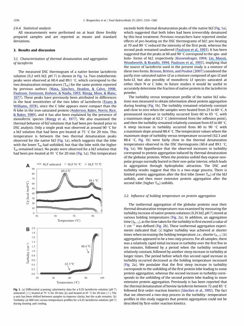
The measured DSC thermogram of a native bovine lactoferrinsolution (0.2 wt% bLf, pH 7) is shown in Fig. 1a. Two endothermicpeaks were observed at 60.4 and 89.1 �C, which correspond to thetwo denaturation temperatures (Tm) for the same protein reportedby previous authors (Mata, Sánchez, Headon, & Calvo, 1998;Paulsson, Svensson, Kishore, & Naidu, 1993; Rüegg, Moor, & Blanc,1977). These peaks have previously been attributed to differencesin the heat sensitivities of the two lobes of lactoferrin (Evans &Williams, 1978), since the C lobe appears more compact than theN lobe in the iron-saturated protein (Anderson, Baker, Norris, Rice,& Baker, 1989), and it has also been explained by the presence ofmonoferric species (Rüegg et al., 1977). We also examined thethermal behavior of bLf solutions that had been pre-heated prior toDSC analysis. Only a single peak was observed at around 90 �C fora bLf solution that had been pre-heated at 75 �C for 20 min. Thistemperature is between the two thermal denaturation peaksobserved for the native bLf (Fig. 1a), which suggests that the lobewith the lower Tm had unfolded, but that the lobe with the higherTm remained intact. No peaks were observed for a bLf solution thathad been pre-heated at 91 �C for 20 min (Fig. 1a). This temperature
0
10
20
30
40
50
60
70
80
90
30 50 70 90
Cp
(kca
l /m
ol/o C
)
Temperature (oC)
AbLF unheated bLF 91 ºC bLF 75 ºC
60.4 ºC
89.1 ºC
0.0
0.2
0.4
0.6
0.8
1.0
1.2
1.4
1.6
1.8
2.0
20 40 60 80 100
Tur
bidi
ty (
cm-1
)
Temperature (oC)
B
62.3 ºC
88.4 ºC
Fig. 1. (a) Differential scanning calorimetry data for a 0.2% lactoferrin solution (pH 7)unheated (,), heated at 75 �C for 20 min (D) and heated at 91 �C for 20 min (B). They-axis has been shifted between samples to improve clarity, but the scale remains; (b)Turbidity (at 600 nm) versus temperature profiles for a 0.2% lactoferrin solution (pH 7)during heating and cooling.
exceeds both thermal denaturation peaks of the native bLf (Fig. 1a),which suggested that both lobes had been irreversibly denaturedby this heat treatment. Previous researchers have reported similareffects of pre-heating on the DSC thermograms of bLf; pre-heatingat 70 and 80 �C reduced the intensity of the first peak, whereas thesecond peak remained unaltered (Paulsson et al., 1993). It has beensuggested that the peaks at 60 and 90 �C correspond to the apo- andholo- forms of bLf, respectively (Kussendrager, 1994; Lin, Mason,Woodworth, & Brandts, 1994; Paulsson et al., 1993), implying thatthe source of lactoferrin used in the present study is a mixture ofthese two forms. Brisson, Britten, and Pouliot (2007) considered thepartly iron-saturated native-Lf as amixture composed of apo-Lf andholo-Lf, but also possibly of monoferric Lf species saturated ateither their N or C lobe. In future studies it would be useful toaccurately determine the fraction of native protein in the lactoferrinpowder.
The turbidity versus temperature profile of the native bLf solu-tions wasmeasured to obtain information about protein aggregationduring heating (Fig. 1b). The turbidity remained relatively constantand close to zero when the samples were heated from 25 to 60 �C. Apronounced increase in turbidity occurred from 60 to 65 �C, witha maximum slope at 62.3 �C (determined from the inflexion point),and then the turbidity remained relatively constant from 65 to 80 �C.A steep increase in turbidity occurred from 80 to 90 �C withamaximum slope around 88.4 �C. The temperature values where themaximum slope of turbidity versus temperature occurred (62.3 and88.4 �C, Fig. 1b) were fairly close to the thermal denaturationtemperatures observed in the DSC thermograms (60.4 and 89.1 �C,Fig. 1a). We hypothesize that the observed increases in turbiditycorrespond to protein aggregation induced by thermal denaturationof the globular proteins. When the proteins unfold they expose non-polar groups normally buried in their non-polar interior, which leadsto aggregation through hydrophobic attraction. The DSC andturbidity results suggest that this is a two-stage process. There islimited protein aggregation after the first lobe (lower Tm) of the bLfunfolds, and then more extensive protein aggregation after thesecond lobe (higher Tm) unfolds.
3.2. Influence of holding temperature on protein aggregation
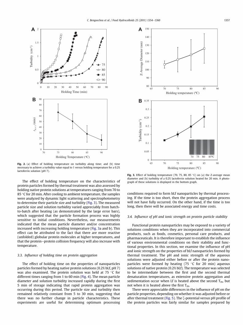
The isothermal aggregation of the globular proteins near theirthermal denaturation temperatures was examined bymeasuring theturbidity increase of native protein solutions (0.2% bLf, pH7) stored atvarious holding temperatures (Fig. 2a). In addition, an aggregationtime (tA¼1), as the time taken for the turbidity tofirst exceed avalueof1 cm�1 was defined (Fig. 2b). These isothermal aggregation experi-ments indicated that: (i) higher turbidity was achieved at shortertimes when increasing the holding temperature, i.e., shorter tA¼1; (ii)aggregation appeared to be a two-step process. For all samples, therewas a relatively rapid initial increase in turbidity over the first five toten minutes, followed by a period when the turbidity remainedrelatively constant, followed by another steep increase in turbidity atlonger times. The period before which this second rapid increase inturbidity occurred decreased as the holding temperature increased(Fig. 2a). We postulate that the first steep increase in turbiditycorresponds to the unfolding of the first protein lobe leading to someprotein aggregation, whereas the second increase in turbidity corre-sponds to the unfolding of the second protein lobe leading to moreextensive protein aggregation. Previously is has been reported thatthe thermal denaturation of bovine lactoferrin between 72 and 85 �Cfollowed first-order reaction kinetics (Sánchez et al., 1992). The factthat we observed a two-step process in the turbidityetemperatureprofiles in this study suggests that protein aggregation could not bedescribed by first-order reaction kinetics.
0
0.5
1
1.5
2
0 10 20 30 40 50 60 70 80 90
Tur
bidi
ty (
cm-1
)
Holding Time (min)
75
80
85
90
A
0
10
20
30
40
50
60
70
80
90
70 75 80 85 90
t A =
1(m
in)
Holding Temperature (oC)
B
Fig. 2. (a) Effect of holding temperature on turbidity along time; and (b) timenecessary to achieve a turbidity value equal to 1 versus holding temperature for a 0.2%lactoferrin solution (pH 7).
80
90
100
110
120
130
140
150
65 70 75 80 85 90
Z-A
vera
ge D
iam
eter
(nm
)
Holding temperature (ºC)
A
0
0.05
0.1
0.15
0.2
0.25
0.3
0.35
65 70 75 80 85 90
Tur
bidi
ty (
cm-1
)
Holding temperature (ºC)
B
70 75 80 85ºC70 75 80 85ºC
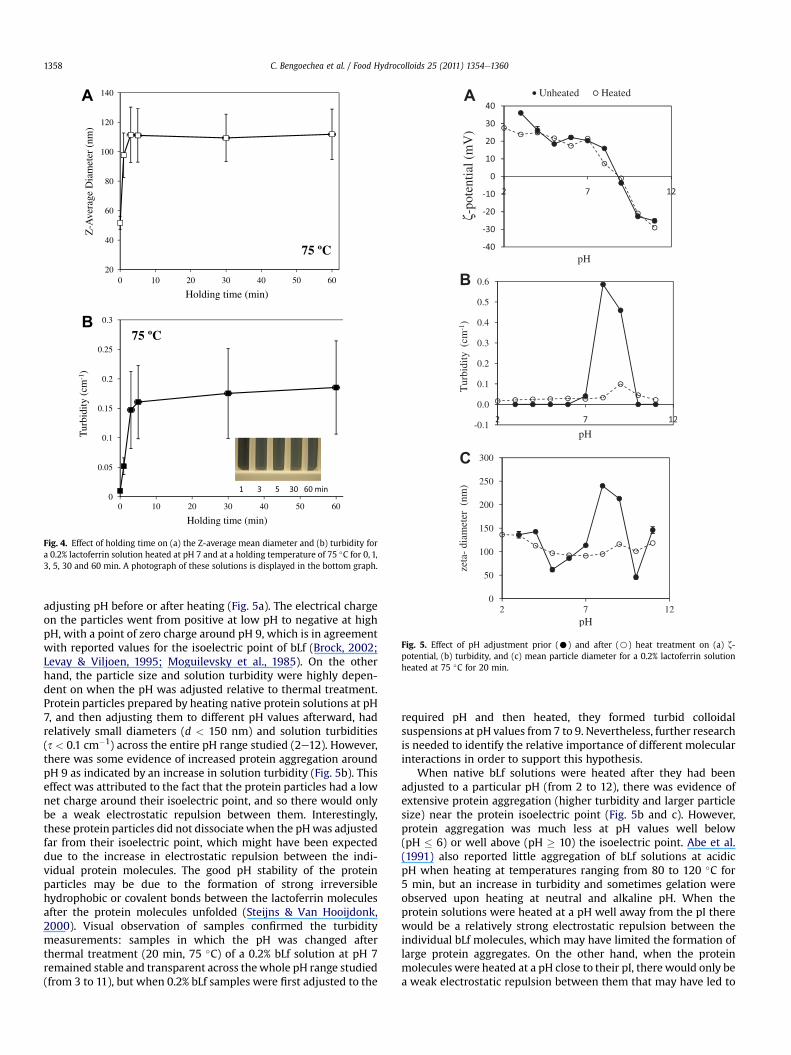
Fig. 3. Effect of holding temperature (70, 75, 80, 85 �C) on (a) the Z-average meandiameter and (b) turbidity of a 0.2% lactoferrin solution heated for 20 min. A photo-graph of these solutions is displayed in the bottom graph.
C. Bengoechea et al. / Food Hydrocolloids 25 (2011) 1354e1360 1357
The effect of holding temperature on the characteristics ofprotein particles formed by thermal treatment was also assessed byholding native protein solutions at temperatures ranging from 70 to85 �C for 20min. After cooling to ambient temperature, the sampleswere analyzed by dynamic light scattering and spectrophotometryto determine their particle size and turbidity (Fig. 3). The measuredparticle size and solution turbidity varied appreciably from batch-to-batch after heating (as demonstrated by the large error bars),which suggested that the particle formation process was highlysensitive to initial conditions. Nevertheless, our measurementsindicated that the mean particle diameter and/or concentrationincreased with increasing holding temperature (Fig. 3a and b). Thiseffect can be attributed to the fact that there are more reactive(unfolded) globular protein molecules at higher temperatures, andthat the proteineprotein collision frequency will also increase withtemperature.
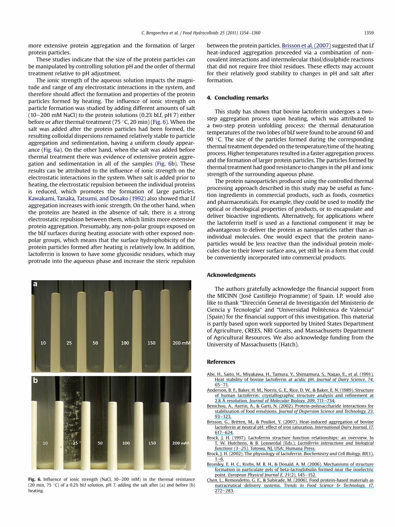
3.3. Influence of holding time on protein aggregation
The effect of holding time on the properties of nanoparticlesparticles formed by heating native protein solutions (0.2% bLf, pH 7)was also examined. The protein solution was held at 75 �C fordifferent times ranging from 1 to 60 min (Fig. 4). The mean particlediameter and solution turbidity increased rapidly during the first5 min of storage indicating that rapid protein aggregation wasoccurring during this period. The particle size and turbidity thenremained relatively constant from 5 to 30 min, suggesting thatthere was no further change in particle characteristics. Theseexperiments are useful for determining optimum processing
conditions required to form bLf nanoparticles by thermal process-ing. If the time is too short, then the protein aggregation processwill not have fully occurred. On the other hand, if the time is toolong, then there will be associated energy and time costs.
3.4. Influence of pH and ionic strength on protein particle stability
Functional protein nanoparticles may be exposed to a variety ofsolutions conditions when they are incorporated into commercialproducts, such as foods, cosmetics, personal care products, andpharmaceuticals. It is therefore important to establish the influenceof various environmental conditions on their stability and func-tional properties. In this section, we examine the influence of pHand ionic strength on the properties of bLf nanoparticles formed bythermal treatment. The pH and ionic strength of the aqueoussolutions were adjusted either before or after the protein nano-particles were formed by heating (75 �C for 20 min) aqueoussolutions of native protein (0.2% bLf). The temperature was selectedto be intermediate between the first and the second thermaldenaturation temperatures, as extensive protein aggregation andsedimentation occur when Lf is heated above the second Tm, butnot when it is heated above the first Tm.
There were appreciable differences in the influence of pH on theparticle properties, depending onwhether it was adjusted before orafter thermal treatment (Fig. 5). The z-potential versus pH profile ofthe protein particles was fairly similar for samples prepared by
20
40
60
80
100
120
140
0 10 20 30 40 50 60
Z-A
vera
ge D
iam
eter
(nm
)
Holding time (min)
A
75 ºC
0
0.05
0.1
0.15
0.2
0.25
0.3
0 10 20 30 40 50 60
Tur
bidi
ty (
cm-1
)
Holding time (min)
B
75 ºC
Fig. 4. Effect of holding time on (a) the Z-average mean diameter and (b) turbidity fora 0.2% lactoferrin solution heated at pH 7 and at a holding temperature of 75 �C for 0, 1,3, 5, 30 and 60 min. A photograph of these solutions is displayed in the bottom graph.
A
B
C
Fig. 5. Effect of pH adjustment prior (C) and after (B) heat treatment on (a) z-potential, (b) turbidity, and (c) mean particle diameter for a 0.2% lactoferrin solutionheated at 75 �C for 20 min.
C. Bengoechea et al. / Food Hydrocolloids 25 (2011) 1354e13601358
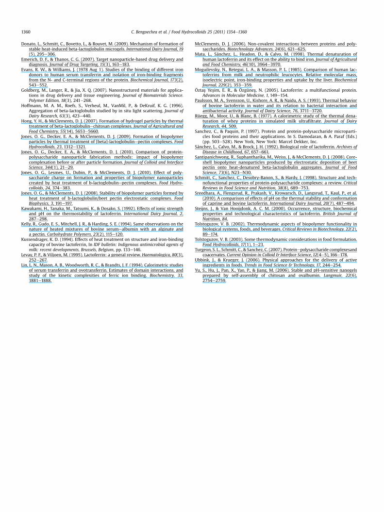
adjusting pH before or after heating (Fig. 5a). The electrical chargeon the particles went from positive at low pH to negative at highpH, with a point of zero charge around pH 9, which is in agreementwith reported values for the isoelectric point of bLf (Brock, 2002;Levay & Viljoen, 1995; Moguilevsky et al., 1985). On the otherhand, the particle size and solution turbidity were highly depen-dent on when the pH was adjusted relative to thermal treatment.Protein particles prepared by heating native protein solutions at pH7, and then adjusting them to different pH values afterward, hadrelatively small diameters (d < 150 nm) and solution turbidities(s < 0.1 cm�1) across the entire pH range studied (2e12). However,there was some evidence of increased protein aggregation aroundpH 9 as indicated by an increase in solution turbidity (Fig. 5b). Thiseffect was attributed to the fact that the protein particles had a lownet charge around their isoelectric point, and so there would onlybe a weak electrostatic repulsion between them. Interestingly,these protein particles did not dissociate when the pHwas adjustedfar from their isoelectric point, which might have been expecteddue to the increase in electrostatic repulsion between the indi-vidual protein molecules. The good pH stability of the proteinparticles may be due to the formation of strong irreversiblehydrophobic or covalent bonds between the lactoferrin moleculesafter the protein molecules unfolded (Steijns & Van Hooijdonk,2000). Visual observation of samples confirmed the turbiditymeasurements: samples in which the pH was changed afterthermal treatment (20 min, 75 �C) of a 0.2% bLf solution at pH 7remained stable and transparent across thewhole pH range studied(from 3 to 11), but when 0.2% bLf samples were first adjusted to the
required pH and then heated, they formed turbid colloidalsuspensions at pH values from 7 to 9. Nevertheless, further researchis needed to identify the relative importance of different molecularinteractions in order to support this hypothesis.
When native bLf solutions were heated after they had beenadjusted to a particular pH (from 2 to 12), there was evidence ofextensive protein aggregation (higher turbidity and larger particlesize) near the protein isoelectric point (Fig. 5b and c). However,protein aggregation was much less at pH values well below(pH � 6) or well above (pH � 10) the isoelectric point. Abe et al.(1991) also reported little aggregation of bLf solutions at acidicpH when heating at temperatures ranging from 80 to 120 �C for5 min, but an increase in turbidity and sometimes gelation wereobserved upon heating at neutral and alkaline pH. When theprotein solutions were heated at a pH well away from the pI therewould be a relatively strong electrostatic repulsion between theindividual bLf molecules, which may have limited the formation oflarge protein aggregates. On the other hand, when the proteinmolecules were heated at a pH close to their pI, there would only bea weak electrostatic repulsion between them that may have led to
C. Bengoechea et al. / Food Hydrocolloids 25 (2011) 1354e1360 1359
more extensive protein aggregation and the formation of largerprotein particles.
These studies indicate that the size of the protein particles canbe manipulated by controlling solution pH and the order of thermaltreatment relative to pH adjustment.
The ionic strength of the aqueous solution impacts the magni-tude and range of any electrostatic interactions in the system, andtherefore should affect the formation and properties of the proteinparticles formed by heating. The influence of ionic strength onparticle formation was studied by adding different amounts of salt(10e200 mM NaCl) to the protein solutions (0.2% bLf, pH 7) eitherbefore or after thermal treatment (75 �C, 20 min) (Fig. 6). When thesalt was added after the protein particles had been formed, theresulting colloidal dispersions remained relatively stable to particleaggregation and sedimentation, having a uniform cloudy appear-ance (Fig. 6a). On the other hand, when the salt was added beforethermal treatment there was evidence of extensive protein aggre-gation and sedimentation in all of the samples (Fig. 6b). Theseresults can be attributed to the influence of ionic strength on theelectrostatic interactions in the system. When salt is added prior toheating, the electrostatic repulsion between the individual proteinsis reduced, which promotes the formation of large particles.Kawakami, Tanaka, Tatsumi, and Dosako (1992) also showed that Lfaggregation increases with ionic strength. On the other hand, whenthe proteins are heated in the absence of salt, there is a strongelectrostatic repulsion between them, which limits more extensiveprotein aggregation. Presumably, any non-polar groups exposed onthe bLf surfaces during heating associate with other exposed non-polar groups, which means that the surface hydrophobicity of theprotein particles formed after heating is relatively low. In addition,lactoferrin is known to have some glycosidic residues, which mayprotrude into the aqueous phase and increase the steric repulsion
Fig. 6. Influence of ionic strength (NaCl, 10e200 mM) in the thermal resistance(20 min, 75 �C) of a 0.2% bLf solution, pH 7, adding the salt after (a) and before (b)heating.
between the protein particles. Brisson et al. (2007) suggested that Lfheat-induced aggregation proceeded via a combination of non-covalent interactions and intermolecular thiol/disulphide reactionsthat did not require free thiol residues. These effects may accountfor their relatively good stability to changes in pH and salt afterformation.
4. Concluding remarks
This study has shown that bovine lactoferrin undergoes a two-step aggregation process upon heating, which was attributed toa two-step protein unfolding process: the thermal denaturationtemperatures of the two lobes of bLf were found to be around 60 and90 �C. The size of the particles formed during the correspondingthermal treatment depended on the temperature/time of the heatingprocess. Higher temperatures resulted in a faster aggregationprocessand the formation of larger protein particles. The particles formed bythermal treatment had good resistance to changes in the pHand ionicstrength of the surrounding aqueous phase.
The protein nanoparticles produced using the controlled thermalprocessing approach described in this study may be useful as func-tion ingredients in commercial products, such as foods, cosmeticsand pharmaceuticals. For example, they could be used to modify theoptical or rheological properties of products, or to encapsulate anddeliver bioactive ingredients. Alternatively, for applications wherethe lactoferrin itself is used as a functional component it may beadvantageous to deliver the protein as nanoparticles rather than asindividual molecules. One would expect that the protein nano-particles would be less reactive than the individual protein mole-cules due to their lower surface area, yet still be in a form that couldbe conveniently incorporated into commercial products.
Acknowledgments
The authors gratefully acknowledge the financial support fromthe MICINN (José Castillejo Programme) of Spain. I.P. would alsolike to thank “Dirección General de Investigación del Ministerio deCiencia y Tecnología” and “Universidad Politécnica de Valencia”(Spain) for the financial support of this investigation. This materialis partly based upon work supported by United States Departmentof Agriculture, CREES, NRI Grants, and Massachusetts Departmentof Agricultural Resources. We also acknowledge funding from theUniversity of Massachusetts (Hatch).
References
Abe, H., Saito, H., Miyakawa, H., Tamura, Y., Shimamura, S., Nagao, E., et al. (1991).Heat stability of bovine lactoferrin at acidic pH. Journal of Dairy Science, 74,65e71.
Anderson, B. F., Baker, H. M., Norris, G. E., Rice, D. W., & Baker, E. N. (1989). Structureof human lactoferrin: crystallographic structure analysis and refinement at2.8 Å resolution. Journal of Molecular Biology, 209, 711e734.
Benichou, A., Aserin, A., & Garti, N. (2002). Protein-polysaccharide interactions forstabilization of food emulsions. Journal of Dispersion Science and Technology, 23,93e123.
Brisson, G., Britten, M., & Pouliot, Y. (2007). Heat-induced aggregation of bovinelactoferrin at neutral pH: effect of iron saturation. International Dairy Journal, 17,617e624.
Brock, J. H. (1997). Lactoferrin structure function relationships: an overview. InT. W. Hutchens, & B. Lonnerdal (Eds.), Lactoferrin interactions and biologicalfunctions (3e25). Totowa, NJ, USA: Humana Press.
Brock, J. H. (2002). The physiology of lactoferrin. Biochemistry and Cell Biology, 80(1),1e6.
Bromley, E. H. C., Krebs, M. R. H., & Donald, A. M. (2006). Mechanisms of structureformation in particulate gels of beta-lactoglobulin formed near the isoelectricpoint. European Physical Journal E, 21(2), 145e152.
Chen, L., Remondetto, G. E., & Subirade, M. (2006). Food protein-based materials asnutraceutical delivery systems. Trends in Food Science & Technology, 17,272e283.
C. Bengoechea et al. / Food Hydrocolloids 25 (2011) 1354e13601360
Donato, L., Schmitt, C., Bovetto, L., & Rouvet, M. (2009). Mechanism of formation ofstable heat-induced beta-lactoglobulin microgels. International Dairy Journal, 19(5), 295e306.
Emerich, D. F., & Thanos, C. G. (2007). Target nanoparticle-based drug delivery anddiagnosis. Journal of Drug Targeting, 15(3), 163e183.
Evans, R. W., & Williams, J. (1978 Aug 1). Studies of the binding of different irondonors to human serum transferrin and isolation of iron-binding fragmentsfrom the N- and C-terminal regions of the protein. Biochemical Journal, 173(2),543e552.
Goldberg, M., Langer, R., & Jia, X. Q. (2007). Nanostructured materials for applica-tions in drug delivery and tissue engineering. Journal of Biomaterials Science.Polymer Edition, 18(3), 241e268.
Hoffmann, M. A. M., Roefs, S., Verheul, M., VanMil, P., & DeKruif, K. G. (1996).Aggregation of beta-lactoglobulin studied by in situ light scattering. Journal ofDairy Research, 63(3), 423e440.
Hong, Y. H., & McClements, D. J. (2007). Formation of hydrogel particles by thermaltreatment of beta-lactoglobulinechitosan complexes. Journal of Agricultural andFood Chemistry, 55(14), 5653e5660.
Jones, O. G., Decker, E. A., & McClements, D. J. (2009). Formation of biopolymerparticles by thermal treatment of [beta]-lactoglobulinepectin complexes. FoodHydrocolloids, 23, 1312e1321.
Jones, O. G., Decker, E. A., & McClements, D. J. (2010). Comparison of protein-polysaccharide nanoparticle fabrication methods: impact of biopolymercomplexation before or after particle formation. Journal of Colloid and InterfaceScience, 344(1), 21e29.
Jones, O. G., Lesmes, U., Dubin, P., & McClements, D. J. (2010). Effect of poly-saccharide charge on formation and properties of biopolymer nanoparticlescreated by heat treatment of b-lactoglobulinepectin complexes. Food Hydro-colloids, 24, 374e383.
Jones, O. G., & McClements, D. J. (2008). Stability of biopolymer particles formed byheat treatment of b-lactoglobulin/beet pectin electrostatic complexes. FoodBiophysics, 3, 191e197.
Kawakami, H., Tanaka, M., Tatsumi, K., & Dosako, S. (1992). Effects of ionic strengthand pH on the thermostability of lactoferrin. International Dairy Journal, 2,287e298.
Kelly, R., Gudo, E. S., Mitchell, J. R., & Harding, S. E. (1994). Same observations on thenature of heated mixtures of bovine serumealbumin with an alginate anda pectin. Carbohydrate Polymers, 23(2), 115e120.
Kussendrager, K. D. (1994). Effects of heat treatment on structure and iron-bindingcapacity of bovine lactoferrin, In IDF bulletin: Indigenous antimicrobial agents ofmilk: recent developments, Brussels, Belgium, pp. 133e146.
Levay, P. F., & Viljoen, M. (1995). Lactoferrin: a general review. Haematologica, 80(3),252e267.
Lin, L. N., Mason, A. B., Woodworth, R. C., & Brandts, J. F. (1994). Calorimetric studiesof serum transferrin and ovotransferrin. Estimates of domain interactions, andstudy of the kinetic complexities of ferric ion binding. Biochemistry, 33,1881e1888.
McClements, D. J. (2006). Non-covalent interactions between proteins and poly-saccharides. Biotechnology Advances, 24(6), 621e625.
Mata, L., Sánchez, L., Headon, D., & Calvo, M. (1998). Thermal denaturation ofhuman lactoferrin and its effect on the ability to bind iron. Journal of Agriculturaland Food Chemistry, 46(10), 3964e3970.
Moguilevsky, N., Retegui, L. A., & Masson, P. L. (1985). Comparison of human lac-toferrins from milk and neutrophilic leucocytes. Relative molecular mass,isoelectric point, iron-binding properties and uptake by the liver. BiochemicalJournal, 229(2), 353e359.
Öztas Yesim, E. R., & Özgünes, N. (2005). Lactoferrin: a multifunctional protein.Advances in Molecular Medicine, 1, 149e154.
Paulsson, M. A., Svensson, U., Kishore, A. R., & Naidu, A. S. (1993). Thermal behaviorof bovine lactoferrin in water and its relation to bacterial interaction andantibacterial activity. Journal of Dairy Science, 76, 3711e3720.
Rüegg, M., Moor, U., & Blanc, B. (1977). A calorimetric study of the thermal dena-turation of whey proteins in simulated milk ultrafiltrate. Journal of DairyResearch, 44, 509.
Sanchez, C., & Paquin, P. (1997). Protein and protein-polysaccharide microparti-cles food proteins and their applications. In S. Damodaran, & A. Paraf (Eds.)(pp. 503e528). New York, New York: Marcel Dekker, Inc.
Sánchez, L., Calvo, M., & Brock, J. H. (1992). Biological role of lactoferrin. Archives ofDisease in Childhood, 67, 657e661.
Santipanichwong, R., Suphantharika, M., Weiss, J., & McClements, D. J. (2008). Core-shell biopolymer nanoparticles produced by electrostatic deposition of beetpectin onto heat-denatured beta-lactoglobulin aggregates. Journal of FoodScience, 73(6), N23eN30.
Schmitt, C., Sanchez, C., Desobry-Banon, S., & Hardy, J. (1998). Structure and tech-nofunctional properties of protein-polysaccharide complexes: a review. CriticalReviews in Food Science and Nutrition, 38(8), 689e753.
Sreedhara, A., Flengsrud, R., Prakash, V., Krowarsch, D., Langsrud, T., Kaul, P., et al.(2010). A comparison of effects of pH on the thermal stability and conformationof caprine and bovine lactoferrin. International Dairy Journal, 20(7), 487e494.
Steijns, J., & Van Hooijdonk, A. C. M. (2000). Occurrence, structure, biochemicalproperties and technological characteristics of lactoferrin. British Journal ofNutrition, 84.
Tolstoguzov, V. B. (2002). Thermodynamic aspects of biopolymer functionality inbiological systems, foods, and beverages. Critical Reviews in Biotechnology, 22(2),89e174.
Tolstoguzov, V. B. (2003). Some thermodynamic considerations in food formulation.Food Hydrocolloids, 17(1), 1e23.
Turgeon, S. L., Schmitt, C., & Sanchez, C. (2007). Proteinepolysaccharide complexesandcoacervates. Current Opinion in Colloid & Interface Science, 12(4e5), 166e178.
Ubbink, J., & Krueger, J. (2006). Physical approaches for the delivery of activeingredients in foods. Trends in Food Science & Technology, 17, 244e254.
Yu, S., Hu, J., Pan, X., Yao, P., & Jiang, M. (2006). Stable and pH-sensitive nanogelsprepared by self-assembly of chitosan and ovalbumin. Langmuir, 22(6),2754e2759.