Formation and characterization of core-sheath nanofibers through electrospinning and...
7
Formation and characterization of core-sheath nanofibers through electrospinning and surface-initiated polymerization Liwen Ji, Zhan Lin, Ying Li, Shuli Li, Yinzheng Liang, Ozan Toprakci, Quan Shi, Xiangwu Zhang * Fiber and Polymer Science Program, Department of Textile Engineering, Chemistry and Science, North Carolina State University, Raleigh, NC 27695-8301, USA article info Article history: Received 3 June 2010 Received in revised form 20 July 2010 Accepted 24 July 2010 Available online 1 August 2010 Keywords: Polyacrylonitrile Polypyrrole Electrospinning abstract Novel core-sheath nanofibers, composed of polyacrylonitrile (PAN) core and polypyrrole (PPy) sheath with clear boundary between them, were fabricated by electrospinning PAN/FeCl 3 $6H 2 O bicomponent nanofibers and the subsequent surface-initiated polymerization in a pyrrole-containing solution. By adjusting the concentration of FeCl 3 $6H 2 O, the surface morphology of PPy sheath changed from isolated agglomerates or clusters to relatively uniform thin-film structure. Thermal properties of PAN-PPy core- sheath nanofibers were also characterized. Results indicated that the PPy sheath played a role of inhibitor and retarded the complex chemical reactions of PAN during the carbonization process. Ó 2010 Elsevier Ltd. All rights reserved. 1. Introduction In the last decade, electrospinning has revived as a fascinating choice to assemble polymer nanofibers with various morphologies and functionalities due to its simplicity and cost effectiveness, along with environmental benignity [1e5]. The unique structural features, such as high surface-area-to-volume ratio, extremely long length, and complex porous structure, etc., of electrospun nano- fibers make them suitable for various applications, including but not limited to tissue engineering scaffolds [6], drug delivery systems [7], sensors [8], environmental protection [9,10], nanoscale electronic and optoelectronic devices [11], catalysts [12,13], and electrode materials for energy storage and conversion systems [14,15]. Nanofibers made from simple electrospinning usually exhibit a solid interior and smooth surface. However, electrospun nanofibers with unique secondary structures can also be prepared in order to obtain exceptional functionalities [3,16]. Especially, electrospun core-sheath nanofibers have gained extraordinary attention due to the combination of the characteristics of two different components into one integrity in the axial or radial direction [16,17]. For example, Wei et al. observed the formation of core-sheath polyaniline (PANI)-polycarboniate (PC) nanofibers, derived from the micro-phase separation after the electrospinning of PANI/PC blend solutions [16,17]. Core-sheath nanofibers can also be fabricated by other methods, such as co-electrospinning two different polymer solutions via a spinneret comprising two coaxial capillaries [18,19], template-directed growth [20], surface-initiated atom transfer radical polymerization (ATRP) [21], and the so-called ‘emulsion electrospinning’ [22]. Electrospun core-sheath nanofibers containing conductive polymers, such as PPy, are fundamentally important and have wide applications because of their high electrical conductivity and good stability under ambient conditions [23e28]. Most conductive polymers are expensive and hard to be electrospun because of their poor solubility in most solvents. The preparation of core-sheath nanofibers through indirect methods combined with electro- spinning technique is a feasible approach to obtain conductive polymer nanofiber materials, which may have wide technological applications in electrical, optical, thermal, and magnetic materials and devices. In this paper, we report the generation of PAN-PPy core-sheath nanofibers through electrospinning of PAN and the subsequent surface-initiated polymerization of pyrrole. The surface morphologies and the thermal properties of these fibers are also characterized. 2. Experimental 2.1. Materials PAN was purchased from Fisher Scientific. FeCl 3 $6H 2 O, pyrrole, N, N-dimethylformamide (DMF) and tetrahydrofuran (THF) were purchased from SigmaeAldrich. All these chemicals were used without further purification. DMF solutions of PAN (8 wt %) con- taining different amount of FeCl 3 $6H 2 O (0, 1, 2, 5, and 10 wt %) were prepared at 60 C with mechanical stirring for at least 48 h. * Corresponding author. Tel.: þ1 919 515 6547. E-mail address: [email protected] (X. Zhang). Contents lists available at ScienceDirect Polymer journal homepage: www.elsevier.com/locate/polymer 0032-3861/$ e see front matter Ó 2010 Elsevier Ltd. All rights reserved. doi:10.1016/j.polymer.2010.07.042 Polymer 51 (2010) 4368e4374
Transcript of Formation and characterization of core-sheath nanofibers through electrospinning and...

lable at ScienceDirect
Polymer 51 (2010) 4368e4374
Contents lists avai
Polymer
journal homepage: www.elsevier .com/locate/polymer
Formation and characterization of core-sheath nanofibers throughelectrospinning and surface-initiated polymerization
Liwen Ji, Zhan Lin, Ying Li, Shuli Li, Yinzheng Liang, Ozan Toprakci, Quan Shi, Xiangwu Zhang*
Fiber and Polymer Science Program, Department of Textile Engineering, Chemistry and Science, North Carolina State University, Raleigh, NC 27695-8301, USA
a r t i c l e i n f o
Article history:Received 3 June 2010Received in revised form20 July 2010Accepted 24 July 2010Available online 1 August 2010
Keywords:PolyacrylonitrilePolypyrroleElectrospinning
* Corresponding author. Tel.: þ1 919 515 6547.E-mail address: [email protected] (X. Zha
0032-3861/$ e see front matter � 2010 Elsevier Ltd.doi:10.1016/j.polymer.2010.07.042
a b s t r a c t
Novel core-sheath nanofibers, composed of polyacrylonitrile (PAN) core and polypyrrole (PPy) sheathwith clear boundary between them, were fabricated by electrospinning PAN/FeCl3$6H2O bicomponentnanofibers and the subsequent surface-initiated polymerization in a pyrrole-containing solution. Byadjusting the concentration of FeCl3$6H2O, the surface morphology of PPy sheath changed from isolatedagglomerates or clusters to relatively uniform thin-film structure. Thermal properties of PAN-PPy core-sheath nanofibers were also characterized. Results indicated that the PPy sheath played a role of inhibitorand retarded the complex chemical reactions of PAN during the carbonization process.
� 2010 Elsevier Ltd. All rights reserved.
1. Introduction
In the last decade, electrospinning has revived as a fascinatingchoice to assemble polymer nanofibers with various morphologiesand functionalities due to its simplicity and cost effectiveness,along with environmental benignity [1e5]. The unique structuralfeatures, such as high surface-area-to-volume ratio, extremely longlength, and complex porous structure, etc., of electrospun nano-fibers make them suitable for various applications, including butnot limited to tissue engineering scaffolds [6], drug deliverysystems [7], sensors [8], environmental protection [9,10], nanoscaleelectronic and optoelectronic devices [11], catalysts [12,13], andelectrode materials for energy storage and conversion systems[14,15]. Nanofibers made from simple electrospinning usuallyexhibit a solid interior and smooth surface. However, electrospunnanofibers with unique secondary structures can also be preparedin order to obtain exceptional functionalities [3,16]. Especially,electrospun core-sheath nanofibers have gained extraordinaryattention due to the combination of the characteristics of twodifferent components into one integrity in the axial or radialdirection [16,17]. For example, Wei et al. observed the formation ofcore-sheath polyaniline (PANI)-polycarboniate (PC) nanofibers,derived from the micro-phase separation after the electrospinningof PANI/PC blend solutions [16,17]. Core-sheath nanofibers can alsobe fabricated by other methods, such as co-electrospinning twodifferent polymer solutions via a spinneret comprising two coaxial
ng).
All rights reserved.
capillaries [18,19], template-directed growth [20], surface-initiatedatom transfer radical polymerization (ATRP) [21], and the so-called‘emulsion electrospinning’ [22].
Electrospun core-sheath nanofibers containing conductivepolymers, such as PPy, are fundamentally important and have wideapplications because of their high electrical conductivity and goodstability under ambient conditions [23e28]. Most conductivepolymers are expensive and hard to be electrospun because of theirpoor solubility in most solvents. The preparation of core-sheathnanofibers through indirect methods combined with electro-spinning technique is a feasible approach to obtain conductivepolymer nanofiber materials, which may have wide technologicalapplications in electrical, optical, thermal, and magnetic materialsand devices. In this paper, we report the generation of PAN-PPycore-sheath nanofibers through electrospinning of PAN and thesubsequent surface-initiated polymerization of pyrrole. The surfacemorphologies and the thermal properties of these fibers are alsocharacterized.
2. Experimental
2.1. Materials
PAN was purchased from Fisher Scientific. FeCl3$6H2O, pyrrole,N, N-dimethylformamide (DMF) and tetrahydrofuran (THF) werepurchased from SigmaeAldrich. All these chemicals were usedwithout further purification. DMF solutions of PAN (8 wt %) con-taining different amount of FeCl3$6H2O (0, 1, 2, 5, and 10 wt %) wereprepared at 60 �C with mechanical stirring for at least 48 h.
L. Ji et al. / Polymer 51 (2010) 4368e4374 4369
2.2. Electrospinning of PAN/FeCl3$6H2O nanofibers
Avariable high voltage power supply (Gamma ES40P-20W)wasused to provide a high voltage (around 10 kV) for electrospinningwith 0.5 ml h�1
flow rate and 15 cm needle-to-collector distance.The electrospun PAN/FeCl3$6H2O nanofibers were accumulated onan aluminum foil surface and collected as a fibrous mat.
2.3. Fabrication of PAN-PPy core-sheath nanofibers
PPy was obtained on electrospun PAN nanofibers by surface-initiated polymerization of pyrrole monomers using Fe3þ as anoxidant. PAN nanofibers (about 2 g) were immersed in 20 ml THFsolution of pyrrole (5ml), followedby adding about 20mlHCl (0.1M)aqueous solution. The mixture was shaken vigorously for 0.5 h. Thepolymerization of pyrrole was initiated by the Fe3þ in electrospunPAN nanofibers at room temperature. During the polymerization,PAN nanofibers changed appearance fromwhite to dark as the resultof the formation of black PPy on the surfaces. Nanofibers with PPysheath were then washed with THF and acetone for several times toremove the non-reacted pyrrole monomers.
2.4. Carbonization of PAN-PPy core-sheath nanofibers
PAN-PPy core-sheath bicomponent nanofibers were first stabi-lized in air environment at 280 �C for 6 h (heating rate was5 �C min�1) and then carbonized at 600 �C for 8 h in argon atmo-sphere (heating rate was 2 �C min�1).
2.5. Morphologies of nanofibers
The morphology of electrospun pure PAN, PAN/FeCl3$6H2O,PAN-PPy core-sheath nanofibers, and the corresponding carbonized
Fig. 1. SEM images of PAN/FeCl3$6H2O nanofibers with differen
nanofibers were evaluated using scanning electron microscopy(JEOL 6400F Field Emission SEM at 5 kV). PAN-PPy core-sheathnanofibers were also characterized using transmission electronmicroscopy (TEM) (FEI Tecnai G2 Twin)with an accelerating voltageof 120 kV.
2.6. ATR-FTIR spectroscopy
Pure PAN, PAN/10 wt% FeCl3$6H2O and the corresponding PAN-PPy core-sheath nanofibers were evaluated using attenuated totalreflection fourier transform infrared spectroscopy (ATR-FTIR). Thespectra were collected from an FTIR spectrometer (Nicolet 560) inthe wavenumber range of 3700e700 cm�1 at room temperature.Sixty-four scans were conducted to achieve an adequate signal-to-noise ratio.
2.7. Thermal analysis
Thermal properties of pure PAN, PAN/10 wt% FeCl3$6H2O andthe corresponding PAN-PPy core-sheath nanofibers were evaluatedusing differential scanning calorimetery (DSC) from 25 to 400 �C ata heating rate of 10 �C min�1 in a nitrogen atmosphere (ThermalInstrumentsDSC-Q2000). Thermo-gravimetric analysis (TGA) wasalso used to determine the weight loss of these nanofibers at10 �C min�1 from 25 to 800 �C in air environment (ThermalInstruments TGA-Q500).
3. Results and discussion
3.1. Surface morphologies of the prepared nanofibers
Fig. 1 presents the SEM images of electrospun PAN/FeCl3$6H2Ocomposite nanofibers with different FeCl3$6H2O contents. All
t FeCl3$6H2O contents: (A) 1, (B) 2, (C) 5 and (D) 10 wt%.
L. Ji et al. / Polymer 51 (2010) 4368e43744370
nanofibers present relatively uniform morphology and randomlyoriented structure. With the increase of FeCl3$6H2O content, thefiber diameter increases, which may be the result of the changesin solution viscosity, surface tension, and conductivity caused bythe addition of ions [3,10,29,30]. The uniform fiber morphology is
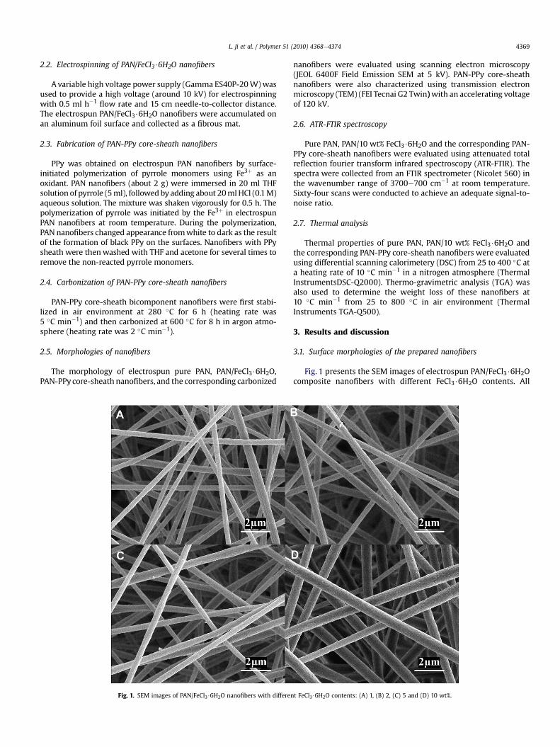
Fig. 2. SEM images of PAN-PPy core-sheath nanofibers made from electrospun PAN/FeCl3$6
totally changed after the surface-initiated polymerization ofpyrrole (Fig. 2). From Fig. 2, it is seen that after the polymeriza-tion, PAN/1 wt% FeCl3$6H2O nanofibers show some non-unifor-mities and irregularities. When the FeCl3$6H2O content increasesto 2 wt%, distinct PPy clusters or agglomerates appear on fiber
H2O with different FeCl3$6H2O contents: (A, B) 1, (C, D) 2, (E, F) 5 and (G, H) 10 wt%.

Fig. 4. ATR-FTIR images of (a) pure PAN, (b) electrospun PAN/10 wt% FeCl3$6H2O, and(c) corresponding PAN-PPy core-sheath nanofibers.
L. Ji et al. / Polymer 51 (2010) 4368e4374 4371
surface. At 5 wt % FeCl3$6H2O content, the PPy phase turns toa continuous and elongated fibrillar sheath structure wrappingthe electrospun PAN/FeCl3$6H2O nanofiber surface. The PPysheath can also been seen from the PAN/10 wt % FeCl3$6H2Onanofibers after the polymerization. The formation of core-shellnanofiber structure is further confirmed by Fig. 3, which showsthe TEM images of PAN/FeCl3$6H2O nanofibers (5 and 10 wt %FeCl3$6H2O) after the polymerization of pyrrole.
3.2. ATR-FTIR of nanofibers
In order to confirm the presence of PPy sheath, ATR-FTIRmeasurements were performed on the electrospun PAN/10 wt%FeCl3$6H2O nanofibers before and after the surface-initiated poly-merization, and the results are exhibited in Fig. 4. For comparison, theFTIR spectrumof pure PANnanofibers is also shown. It is seen that allof the spectra contain three prominent peaks at around 2930, 2245,and 1450 cm�1, which can be assigned to the stretching vibration ofthe methylene (eCH2e) group, the stretching vibration of nitrilegroups (eCN), and the bending vibration of methylene (eCH2e) ofPAN, respectively [4,10]. In addition, the intensities of these charac-teristic peaks increase after the surface-initiated polymerization ofpyrrole. Before polymerization, there are intermolecular interactionsbetween PAN and Fe3þ ions in electrospun PAN/10 wt% FeCl3$6H2Onanofibers, such as the coordination of Fe3þ ions with eCN, or thecomplex formedby Fe3þ ionswithbothDMFandPAN[30e32],which
Fig. 3. TEM images of PAN-PPy core-sheath nanofibers made from electrospun P
mayleadto relativelyweakcharacteristicpeaks ataround2930, 2245,and 1450 cm�1, comparedwith pure PAN. However, after the surface-initiated polymerization, the PPy sheath is formed and significantlyamount of Fe3þ ions may be reduced. In addition, the PPy phase mayalso affect or even inhibit the possible interactions between reducedFe3þ ions and PAN cores. As a result, the former interactions betweenPAN chains and Fe3þ ions decrease, and the intensities of the PANcharacteristic peaks increase. It should be noted that the similarreport about the interactions betweenmetal ions and PAN/PPychains
AN/FeCl3$6H2O with different FeCl3$6H2O contents: (A, B) 5, (C, D) 10 wt%.

L. Ji et al. / Polymer 51 (2010) 4368e43744372
is relatively deficient. Further research along with other character-izations is necessary to identify this issue.
From Fig. 4, it is also seen that after the surface-initiated poly-merization, a new peak appears at 1547 cm�1, which may beassigned to the typical PPy ring vibrations. In addition, the peaks at1178, 1040, and 900 cm�1 may correspond to the NeC stretchingand the ¼ CeH out-of-plane and in-plane vibrations in PPy,respectively [24,32e35]. The changes in ATR-FTIR spectrum afterthe surface-initiated polymerization demonstrate that PPy sheathhas been formed on the nanofiber surface.
Fig. 6. TGA thermograms of (a) pure PAN, (b) electrospun PAN/10 wt% FeCl3$6H2O, and(c) corresponding PAN-PPy core-sheath nanofibers.
3.3. Thermal analysis of nanofibers
In order to investigate the thermal properties of electrospunpure PAN, PAN/FeCl3$6H2O nanofibers, and the corresponding PAN-PPy core-sheath nanofibers, DSC and TGA characterizations wereperformed. Fig. 5 compares DSC thermograms of pure PAN,PAN/10 wt% FeCl3$6H2O, and PAN-PPy core-sheath nanofibers innitrogen atmosphere. Electrospun pure PAN nanofibers exhibita relatively large exothermic peak at about 290 �C, which derivesfrom the complex and multiple chemical reactions (i.e., dehydro-genation, instantaneous cyclization, and crosslinking) of PANduring the process of thermal treatment via the free radicalmechanism [25,29,30,36,37]. In the presence of Fe3þ ions, theexothermic peak shifts to a higher temperature, and the peak alsobecomes broader compared to that of pure PAN nanofibers. Inaddition, the peak intensity decreases after the addition ofFeCl3$6H2O. The broadening of the exothermic peak in the presenceof Fe3þ ions suggests that Fe3þ ions modify the activity of the freeradicals involved in the above-mentioned complex chemical reac-tions. The decreased peak intensity is due to the interactionsbetween PAN and Fe3þ ions (Fig. 4), which inhibit the formation offree radicals on the nitrile groups and subsequent reactions. ForPAN-PPy core-sheath nanofibers, the exothermic peak becomessharper again, and the peak position shifts to a higher temperatureof about 320 �C. Similar to the case of PAN/FeCl3$6H2O nanofibers,the reduced Fe3þ ions in PAN-PPy core-sheath nanofibers can alsomodify and reduce the formation of free radicals involved in thecomplex chemical reactions. In addition, the coated PPy sheathmayalso have an inhibit effect on the above-mentioned complexchemical reactions. As a result, the combined contribution ofreduced Fe3þ ions and PPy sheath retards the formation of freeradicals on the nitrile groups of PAN and subsequent re-combina-tions between the radicals inter- or intra-molecularly, leading tothe increase in the exothermic peak temperature for PAN-PPy core-sheath nanofibers [36].
Fig. 6 shows the TGA thermograms of electrospun pure PAN,PAN/10 wt% FeCl3$6H2O, and the corresponding PAN-PPy core-
Fig. 5. DSC curves of (a) pure PAN, (b) electrospun PAN/10 wt% FeCl3$6H2O, and (c)corresponding PAN-PPy core-sheath nanofibers.
sheath nanofibers under air environment. The TGA thermogramsindicate that the thermal stability of PAN/10 wt% FeCl3$6H2Onanofibers is reduced compared with that of pure PAN nano-fibers. For example, the majority of weight loss of pure PANnanofibers in air occurs in two steps at about 320 and 500 �C,respectively, while PAN/10 wt% FeCl3$6H2O nanofibers havemajor weight losses at about 260 and 350 �C, respectively. Thereduced stability can be explained by the interactions formedbetween Fe3þ ions and PAN, which accelerate the oxidativestabilization reactions that are known to occur when PAN isthermally treated in an air environment [14,36,38]. In the case ofPAN-PPy core-sheath nanofibers, the weight loss temperaturesupshift to about 300 and 500 �C, respectively. This may be theresult of the combined effects of both PPy and the reduced Fe3þ
ions. The former is easy to be degraded when thermal treatmentin air environment, while the presence of the later can reduce theinteractions between Fe3þ ions and PAN. These combined effectsmake the PAN-PPy core-sheath nanofibers decompose at highertemperatures compared with the corresponding PAN/10 wt%FeCl3$6H2O. However, these temperatures are still lower thanthose of pure PAN.
3.4. Surface morphologies of carbonized nanofibers
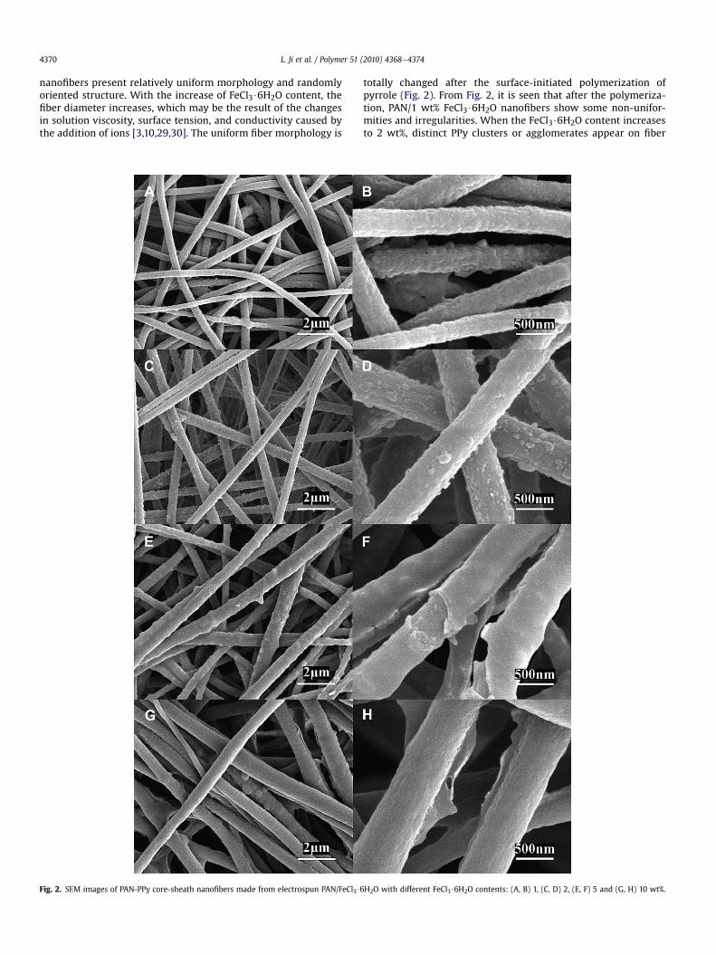
Fig. 7 shows SEM images of carbon nanofibers obtained by thecarbonization of PAN-PPy core-sheath nanofibers. It is seen thatthese core-sheath nanofiber-driven carbon nanofibers haveundulated and wrinkled surface morphology and their diametersare slightly smaller than those of corresponding PAN-PPy core-sheath nanofibers (Fig. 2), which may be due to the liberation ofhetero-atoms and the densification of carbon atoms in polymerchains during the thermal treatment [25,29,30,37]. In addition,when PPy/PAN ratio changes in PAN-PPy core-sheath nanofibers,the microstructures of the carbonized nanofibers also change. Itshould be noticed that PPy is also one important carbon sourcealthough it is relatively easy to be degraded when thermallytreated in air environment. As a result, these fibers keep theirfibrous morphology after the two-step carbonization processes.The prepared CNFs should come from both PPy and PANcomponents [25]. It is well-known that carbon materials can beused as electrodes in energy storage devices, such as recharge-able lithium-ion batteries and supercapacitors, in which theimprovement of the energy density along with other electro-chemical performance are strongly dependant on the crystallinephase microstructure and micro-morphology of the carbona-ceous material [39]. One-dimensional nanostructured compositeshave been demonstrated to have suitable structures and func-tionalities for energy storage [40,41]. We therefore anticipate
Fig. 7. SEM images of thermally treated PAN/FeCl3$6H2O composite nanofibers through stabilization and carbonization processes with different FeCl3$6H2O contents: (A, B) 1, (C, D)2, (E, F) 5, and (G, H) 10 wt%.
L. Ji et al. / Polymer 51 (2010) 4368e4374 4373
that the special surface morphology and microstructures of thecarbon nanofibers derived from PAN-PPy core-sheath nanofiberwith different PPy component may have promising electro-chemical properties when used as electrodes in energy storagesystems.
4. Conclusions
PAN-PPy core-sheath composite nanofibers were prepared bya judicious combination of electrospinning technique and surface-initiated polymerization processes using Fe3þ as an oxidant. It was

L. Ji et al. / Polymer 51 (2010) 4368e43744374
found that with increase in Fe3þ content, the PPy sheath structurechanges from particle-like, fibrillar, to continuous coating. As-formed core-sheath nanofibers can be thermally treated to preparecarbon nanofibers. Therefore, the combination of electrospinningand surface-initiated polymerization is a feasible approach toprepare conducting core-sheath nanofibers and carbon nanofibersfor various applications, including electrodes for energy storageand conversion systems, catalyst supports, sensors, and waterpurification, etc.
Acknowledgement
This study was supported by the Department of Energy (DE-EE0001177), the U.S. National Science Foundation (No. 0833837),and the ERC Program of the National Science Foundation underAward Number EEC-08212121. The authors would like to thank Mr.Christopher Bonino and Professor Saad A Khan in Chemical andBiomolecular Engineering Department at North Carolina StateUniversity for their help in sample characterizations.
References
[1] Ge JJ, Hou HQ, Li Q, Graham MJ, Greiner A, Reneker DH, et al. J Am Chem Soc2004;126:15754e61.
[2] Hou HQ, Ge JJ, Zeng J, Li Q, Reneker DH, Greiner A, et al. Chem Mater 2005;17:967e73.
[3] Li D, Xia YN. Adv Mater 2004;16:1151e70.[4] Zhang D, Karki AB, Rutman D, Young DR, Wang A, Cocke D, et al. Polymer
2009;50:4189e98.[5] Reneker DH, Chun I. Nanotechnology 1996;7:216e23.[6] Agarwal S, Wendorff JH, Greiner A. Polymer 2008;49:5603e21.[7] Hong YL, Chen XS, Jing XB, Fan HS, Guo B, Gu ZW, Zhang XD. Adv Mater
2010;22:754e8.[8] Liu Y, Teng H, Hou HQ, You TY. Biosens Bioelectron 2009;24:3329e34.[9] Thavasi V, Singh G, Ramakrishna S. Energy Environ Sci 2008;1:205e21.
[10] Ji LW, Medford AJ, Zhang XW. Polymer 2009;50:605e12.[11] Liu HQ, Reccius CH, Craighead HG. Appl Phys Lett 2005;87:253106.[12] Formo E, Peng ZM, Lee E, Lu XM, Yang H, Xia YN. J Phys Chem C 2008;112:
9970e5.[13] Huang JS, Hou HQ, You TY. Electrochem Commun 2009;11:1281e4.[14] Ji LW, Medford AJ, Zhang XW. J Mater Chem 2009;19:5593e601.[15] Le Viet A, Reddy MV, Jose R, Chowdari BVR, Ramakrishna S. J Phys Chem C
Nanomater Interfaces 2010;114:664e71.[16] Wei M, Lee J, Kang BW, Mead J. Macromol Rapid Commun 2005;26:1127e32.[17] Wei M, Kang BW, Sung CM, Mead J. Macro Mater Eng 2006;291:1307e14.[18] Sun ZC, Zussman E, Yarin AL, Wendorff JH, Greiner A. Adv Mater 2003;15:
1929e32.[19] Li D, Xia YN. Nano Lett 2004;4:933e8.[20] Bognitzki M, Hou HQ, Ishaque M, Frese T, Hellwig M, Schwarte C, et al. Adv
Mater 2000;12:637.[21] Fu GD, Lei JY, Yao C, Li XS, Yao F, Nie SZ, et al. Macromolecules 2008;41:6854e8.[22] Xu XL, Zhuang XL, Chen XS, Wang XR, Yang LX, Jing XB. Macromol Rapid
Commun 2006;27:1637e42.[23] Dong H, Jones WE. Langmuir 2006;22:11384e7.[24] Li XF, Hao XF, Yu HB, Na H. Mater Lett 2008;62:1155e8.[25] Ji LW, Yao YF, Toprakci O, Lin Z, Liang YZ, Shi Q, et al. J Power Sources
2010;195:2050e6.[26] Xie JW, MacEwan MR, Willerth SM, Li XR, Moran DW, Sakiyama-Elbert SE,
Xia YN. Adv Func Mater 2009;19:2312e8.[27] Chen R, Zhao SZ, Han GY, Dong JH. Mater Lett 2008;62:4031e4.[28] Laforgue A, Robitaille L. Chem Mater 2010;22:2474e80.[29] Ji LW, Zhang XW. Nanotechnology 2009;20:155705.[30] Ji LW, Medford AJ, Zhang XW. J Polym Sci B Polym Phys 2009;47:493e503.[31] Dong YC, Han ZB, Liu CY, Zhang BH. In: International Conference on Energy
Environment Technology (iceet); 2009, vol. 3. p. 218e221.[32] Sharma R, Lamba S, Annapoorni S. J Phys D Appl Phys 2005;38:3354e9.[33] Chen W, Li XW, Xue G, Wang ZQ, Zou WQ. Appl Surf Sci 2003;218:215e21.[34] Nicho ME, Hu H. Sol Energy Mater Sol Cells 2000;63:423e35.[35] Zhong WB, Liu SM, Chen XH, Wang YX, Yang WT. Macromolecules 2006;39:
3224e30.[36] Kim J, Kim YC, Ahn W, Kim CY. Polym Eng Sci 1993;33:1452e7.[37] Kim C, Jeong YI, Ngoc BTN, Yang KS, KojimaM, Kim YA, et al. Small 2007;3:91e5.[38] Dalton S, Heatley F, Budd PM. Polymer 1999;40:5531e43.[39] ZhouHS, Zhu SM,HibinoM, Honma I, IchiharaM. AdvMater 2003;15:2107e11.[40] Zhang Y, Suenaga K, Colliex C, Iijima S. Science 1998;281:973e5.[41] Schoen DT, Meister S, Peng HL, Chan C, Yang Y, Cui YJ. Mater Chem 2009;19:
5879e90.