Electrospun Core-Sheath Bicomponent Nanostructures for Soft Tissue Engineering
Evolution and Development of Hertwig’s Epithelial Root Sheath
Xianghong Luan, Yoshihiro Ito, and Thomas G.H. DiekwischBrodie Laboratory for Craniofacial Genetics and Department of Oral Biology, The University ofIllinois College of Dentistry, Chicogo, IL
AbstractPeriodontal regeneration and tissue engineering has re-awakened interest in the role of Hertwig’sEpithelial Root Sheath (HERS), an epithelial tissue layer first discovered in amphibians more thana century ago. Using developmental, evolutionary, and cell biological approaches we havetherefore performed a careful analysis of the role of HERS in root formation and compared ourdata with clinical findings. Our developmental studies revealed HERS as a transient structureassembled in the early period of root formation and elongation and subsequently fenestrated andreduced to epithelial rests of Malassez (ERM). Our comparative evolutionary studies indicatedthat HERS fenestration was closely associated with the presence of a periodontal ligament and agomphosis-type attachment apparatus in crocodilians and mammals. Based on these studies, weare proposing that HERS plays an important role in the regulation and maintenance of periodontalligament space and function. Additional support for this hypothesis was rendered by our meta-analysis of recent clinical reports related to HERS function.
KeywordsPeriodontium; Development; Cementum; Apoptosis; Darwinian Medicine
A. Hertwig’s Epithelial Root Sheath – from Comparative Anatomy to ClinicalRelevance
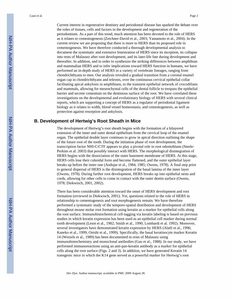
Hertwig’s Epithelial Root Sheath (HERS) was first discovered as a bi-layered cell sheathsurrounding the roots of amphibian teeth (e.g. in Triturus and Salamandra)(Hertwig, 1874).HERS descends from the oral epithelium and the enamel organ to form a collar around thecervical part of the amphibian tooth root (Fig. 1). The image of a continuous epithelial celllayer covering the newly formed root surface like a glove or a second skin and seeminglylogically contributing to the secretion of mineralized tissue on the dentinous root surface hasfound entry into many textbooks and also turned into a welcome scientific explanation forone of dentistry’s first biotech enterprises (Hammarström et al., 1996; Hammarström, 1997).Yet, many current concepts of the function of HERS during root formation ignore theprofound differences between amphibian and mammalian tooth roots. While Hertwig’soriginal report faithfully recounted root formation in amphibian teeth, mammalian HERS ismore or less a transitory entity, loosing its integrity already at the very moment of tooth rootelongation.
Author for correspondence: Thomas G.H. Diekwisch, D.M.D., Ph.D. (sc.), Ph.D. (phil.), Professor and Head, Department of OralBiology, Director, Brodie Laboratory for Craniofacial Genetics, Allan G. Brodie Endowed Chair for Orthodontic Research, Professorof Anatomy/Cell Biology, Bioengineering, Orthodontics, Periodontics, UIC College of Dentistry, 801 South Paulina Street, MC 841,Chicago, Illinois 60612, Phone: (312) 413 9683, FAX: (312) 996 6044, e-mail: [email protected], Web:http://dentistry.uic.edu/CraniofacialGenetics, Oral Biol: http://dentistry.uic.edu/Depts/oralb/welcome.htm.
NIH Public AccessAuthor ManuscriptDev Dyn. Author manuscript; available in PMC 2009 August 28.
Published in final edited form as:Dev Dyn. 2006 May ; 235(5): 1167–1180. doi:10.1002/dvdy.20674.
NIH
-PA Author Manuscript
NIH
-PA Author Manuscript
NIH
-PA Author Manuscript
Current interest in regenerative dentistry and periodontal disease has sparked the debate overthe roles of tissues, cells and factors in the development and regeneration of theperiodontium. As a part of this trend, much attention has been devoted to the role of HERSas it relates to cementogenesis (Zeichner-David et al., 2003; Yamamoto et al., 2004). In thecurrent review we are proposing that there is more to HERS than its proposed role incementogenesis. We have therefore conducted a thorough developmental analysis todocument the systematic and extensive fenestration of HERS since its inception, its collapseinto rests of Malassez after root development, and its later-life fate during development andthereafter. In addition, and in order to synthesize the striking differences between amphibianand mammalian HERS and to infer implications toward HERS function in humans, we haveperformed an in-depth study of HERS in a variety of vertebrate lineages, ranging fromchondrichthyans to men. Our analysis revealed a gradual transition from a coronal enamelorgan cap in chondrichthyans and teleosts, over the continuous cervical epithelial collarfacilitating apical ankylosis in amphibians, to the transient epithelial network of crocodiliansand mammals, allowing for mesenchymal cells of the dental follicle to trespass the epithelialbarrier and secrete cementum on the dentinous surface of the root. We have correlated theseinvestigations on the developmental and evolutionary biology of HERS with several clinicalreports, which are supporting a concept of HERS as a regulator of periodontal ligamentbiology as it relates to width, blood vessel homeostasis, and cementogenesis, as well asprotection against resorption and ankylosis.
B. Development of Hertwig’s Root Sheath in MiceThe development of Hertwig’s root sheath begins with the formation of a bilayeredextension of the inner and outer dental epithelium from the cervical loop of the enamelorgan. The epithelial double layer continues to grow in apical direction outlining the shapeof the future root of the tooth. During the initiation phase of root development, thetranscription factor NHI-C/CTF appears to play a pivotal role in root odontoblasts (Steele-Perkins et al. 2003) that possibly interact with HERS. The morphological disintegration ofHERS begins with the dissociation of the outer basement membrane of HERS. At this stage,HERS cells lose their cuboidal form and become flattened, and the outer epithelial layerbreaks up before the inner one (Andujar et al., 1984, 1985; Owens, 1978). A final step priorto general dispersal of HERS is the disintegration of the basal lamina of the inner layer(Owens, 1978). During further root development, HERS breaks up into epithelial nests andcords, allowing for other cells to come in contact with the outer dentin surface (Owens,1978; Diekwisch, 2001, 2002).
There has been considerable attention toward the onset of HERS development and rootformation (reviewed in Diekwisch, 2001). Yet, questions related to the role of HERS inrelationship to cementogenesis and root morphogenesis remain. We have thereforeperformed a systematic study of the temporo-spatial distribution and development of HERSthroughout mouse molar root formation using keratin as a marker for epithelial cells alongthe root surface. Immunohistochemical cell-tagging via keratin labeling is based on previousstudies in which keratin expression has been used as an epithelial cell marker during normaltooth development (Lesot et al., 1982; Smith et al., 1990, Lombardi et al. 1992). Moreover,several investigators have demonstrated keratin expression by HERS (Alatli et al., 1996;Kaneko et al., 1999; Onishi et al., 1999). Specifically, the basal keratinocyte marker Keratin14 (Wetzels et al., 1989) has been documented in rests of Malassez usingimmunohistochemistry and monoclonal antibodies (Gao et al., 1988). In our study, we haveperformed immunoreactions using an anti-pan-keratin antibody as a marker for epithelialcells along the root surface (Figs. 2 and 3). In addition, we have generated Keratin 14transgenic mice in which the K14 gene served as a powerful marker for Hertwig’s root
Luan et al. Page 2
Dev Dyn. Author manuscript; available in PMC 2009 August 28.
NIH
-PA Author Manuscript
NIH
-PA Author Manuscript
NIH
-PA Author Manuscript
sheath and epithelial rests of Malassez (Figs. 4 and 10). In these studies, the K14 transgenewas detected using the lacZ reporter gene and stained with β-galactosidase.
Onset of HERS development in mice as a bilayer extension of the outer and inner enamelepithelium
We have analyzed the beginning of HERS development in mice using bothimmunohistochemical and K14 transgenic keratin-based marker systems (Figs. 2 and 4). Atthe onset of HERS elongation from the cervical loop, cells from the ameloblast layer andfrom the outer enamel epithelium extended in apical direction (Fig. 2). Subsequently, HERSformed a continuous bi-layer of flat, cuboidal cells, at the interface between dental follicleand dental pulp (Fig. 4C).
During mouse molar root development HERS epithelial cells became increasinglydissociated
In order to follow the fate and distribution of HERS cells along the developing roots surface,we have marked epithelial cells along the developing root surface with anti-keratinantibodies to follow the fate of HERS cells during root formation. The set of figures frommouse molar tooth organs between 1day and 20 days postnatal illustrates the increasingdistance between individual HERS cells as a consequence of root elongation (Fig. 2). From5 days to 20 days postnatal there was a five-fold increase of the distance between individualHERS cells. While the distance between individual HERS cells increased progressively,mesenchymal cells populated the spaces between the epithelial cells. The epithelialdiaphragm as the most apical portion of HERS remained intact throughout all stagesinvestigated.
In 10 days postnatal mouse molars HERS formed a network of cells in proximity to but notin contact with the developing root surface
In order to understand the three-dimensional relationship between HERS cells and thedeveloping root surface we performed three-dimensional reconstructions of anti-keratinlabeled serial sections of a 10 days postnatal developing mouse first mandibular molar (Fig.3). The parasagittal sections in Fig. 3B illustrate a line of epithelial cells in close proximityto the root surface. A 30° horizontal rotation of the parasagittal montage (Fig. 3A) revealedthat the row of epithelial cells apparent in the parasagittal view was the lateral view of afenestrated network of epithelial cells in close proximity to the root surface.
Keratin 14 as a marker for Hertwig’s root sheath in miceOur studies established the Keratin 14 transgene as a highly specific marker gene for cells ofHertwig’s epithelial root sheath (HERS) and epithelial rests of Malassez (ERM). Ourfindings indicate that the K14 transgene was expressed by many epithelial tissues such asthe oral epithelium or the enamel organ (Fig. 4). However, HERS and ERM were the onlystructures identified as carriers of the K14 transgene along the root surface and in itsperiphery (Figs. 4 and 10). Connective tissues such as root dentin, alveolar bone, pulp,odontoblasts, and periodontal ligament were not marked for the K14 transgene (Figs. 4 and10). Moreover, all epithelial tissues in the root area contained the K14 transgene (Figs. 4 and10). We thus suggest that the K14 transgene is a viable marker for HERS and ERMepithelial cells along the root surface.
Specifially, our K14 transgene expression studies visualized the defined K14 expressionpattern of the HERS bilayer and the epithelial diaphragm (Fig. 4) in one week postnatalmice (Fig. 4). At this stage, the HERS bilayer was continuous (Fig. 4). Subsequent stages attwo and four week postnatal revealed the fenestration of HERS into a network of HERS
Luan et al. Page 3
Dev Dyn. Author manuscript; available in PMC 2009 August 28.
NIH
-PA Author Manuscript
NIH
-PA Author Manuscript
NIH
-PA Author Manuscript

cells (Fig. 4). In six months postnatal mice, the K14 transgene identified the aggregated cellrests of Malassez along the root surface (Figs. 4 and 10).
Mammalian HERS as a transient tissue layer in developmentIn summary, our data indicate that in mice, there is only a short period during which HERSemerges from the cervical loop and forms a continuous sheath tightly surrounding the newlydeveloped root surface. In humans, there is considerable distance between HERS and theroot surface already at the onset of root formation (Diekwisch, 2001). Both, in mice and inhumans, and in all other mammals to our knowledge as well, HERS breaks up into epithelialcords at the very beginning of cementogenesis (Diekwisch, 2001) and allows formesenchymal cells of the dental follicle to access the root surface (Diekwisch, 2002).Throughout the period of root elongation, HERS continues to dissociate while windows ofmesenchymal cells in between cords of the ever thinning HERS network increase in sizeuntil HERS’ final collapse into epithelial rests of Malassez. We are proposing that based onour marker studies, mammalian HERS is a fleeting, transient structure and profoundlydifferent from the highly organized dense layers of mineral secreting cells such asameloblasts and odontoblasts. We further argue that a morphological mineral layer pendantto the reticulated structure of the HERS network as a mineralized deposit on the root surfacehas yet to be described. We have therefore applied an evolutionary developmental approachto further reveal the function of HERS in mammals.
C. Evolution of HERS from Fish to HumanHertwig’s root sheath is one of the few tissues that were first discovered in amphibians andnot in mammals. The reason behind Hertwig’s early discovery of HERS in amphibians waslikely related to the longevity of HERS in the amphibian dentition – mid-saggital sections ofamphibian jaws reveal ample opportunities to view root sheaths at all stages of development.In the current study, we have investigated trends in HERS patterning from fish to human asa means to deduce clues about its function and involvement in root formation. The approachwe have taken is not unlike an evolutionary medicine (Darwinian medicine) approach as ithas been suggested by others (Kaifu et al., 2003; Trevathan et al., 1999) to detect generaltrends in evolutionary biology to the benefit of developmental biology or medicine. Asummary of the species and lineages investigated for the present study is presented in Fig.12.
In non-mammalian vertebrates, HERS continuity was not disrupted during developmentallowing for cementum deposition on the root surface apical of HERS
An extensive analysis of HERS position and fate in non-mammalian vertebrates wasperformed to provide further information about the origins of the tissues involved in rootformation. The results from our studies of non-mammalian vertebrates were compared to theposition and fate of HERS in mammalian periodontia of mice and men. Here we aresummarizing six common features of root development in basal vertebrates as they relate tothis study: (i) In all basal species investigated, keratin-labeled dental epithelium extendedonly over the coronal aspect of the entire tooth length, including the coronal portion of thetooth root (Figs. 5–7). The apical portion of the root surface was always free from epithelialcells, even though HERS covered the coronal part of the root surface in some species suchas the axolotl (Figs. 5–7). The extend to which HERS descended toward the root apex variedin different species. In gecko and iguana reptiles as well as in guppy fish and in thechondrichthyans shark and ray, the keratin-labeled dental epithelium formed an epithelialcap mostly covering the tooth crown (Figs. 5 and 7). In contrast, amphibian teeth weredistinctly separated into three portions: a coronal enamel organ cap, a cervical extension ofHERS, and an apical portion of the root surface that was not covered by HERS (Fig. 6). The
Luan et al. Page 4
Dev Dyn. Author manuscript; available in PMC 2009 August 28.
NIH
-PA Author Manuscript
NIH
-PA Author Manuscript
NIH
-PA Author Manuscript

dimensions of the enamel organ, HERS, and the apical root end in relationship to the entiretooth length varied significantly between axolotl and frog. Axolotl teeth were characterizedby a root sheath that covered most of the developing root surface and left only the lowerfifth to one third of the root accessible to mesenchymal tissues (Fig. 6). Many Urodeliandentitions also feature islands of epithelial cells in the non-mineralized interproximalconnective tissue between teeth (Fig. 6). In opposite, frog teeth featured a diminutive enamelorgan and an overextended apical root end (Fig. 6). (ii) Basal vertebrate HERS formed acontinuous layer throughout development; i.e. HERS in fishes, amphibians, and non-crocodilian reptiles was not fenestrated and did not form an epithelial diaphragm (Fig. 5–7).As a consequence of HERS continuity, there were no epithelial rests of Malassez in basalvertebrates (Figs. 5–7). (iii) In fishes, amphibians, and non-crocodilian reptiles, the apicalroot surface was covered by mesenchymal cells, cementum or bone (Figs. 5–7). (iv) In mostof the non-mammalian vertebrates investigated, a bridge-like cementoid tissue connectionwas detected between individual teeth amongst each other as well as between individualteeth and mandibular bone (Figs. 6–7). The cementoid tissue between individual teeth wasvisible in the bone-carrying species gecko, iguana, frog, axolotl, and guppy (Figs. 5–7)while it was missing in the continuously erupting teeth of the chrondrichthyans ray andshark (Fig. 5). (v) The enamel organs of individual teeth were connected with each other aswell as with the oral epithelium through a band of epithelium, the general lamina (e.g. Figs.6,7).
Rare human specimen of the Gottlieb collection revealed characteristic reticulated shapesof human HERS in close association with nervous ganglia
The numerous preparations of HERS in developing human teeth belong to the most preciousitems of the Gottlieb collection at Baylor University in Dallas/Texas. In a previous study, wehave described the significant distance of HERS from the developing root surface in humans(Diekwisch, 2001). Here, we are using Gottlieb’s large size thick sections of human HERSto reveal the fine network of human epithelial cells spanning many areas of the root surface(Fig. 8). Our micrographs illustrate filigree epithelial cords measuring one or two cell layersin thickness and surrounding mesenchymal windows of 100μm in width (Fig. 8). Gangliacells and nervous plexus were detected in close proximity of the “knots” of the cellular netspun by cords of HERS (Fig. 8). Our documentation of ganglia cells in close associationwith HERS is corroborated by earlier studies on periodontal neural endings intimatelyrelated to epithelial rest of Malassez in humans (Lambrichts et al., 1993). We suggest thatthe highly organized network structure of HERS in adult human teeth in tandem with therich innervation indicate that HERS remains as an active tissue layer in the humanperiodontium.
Evolutionary stages in HERS evolution: cap, sheath, and netTogether, these studies reveal a gradual progression of HERS morphology and functionfrom fish to human. Our studies allow for the distinction of three stages of HERS invertebrates: (i) A narrow epithelial cap laterally and apically confined by the cervical loop inteleosts and chondrichthyans (Fig. 5). In chondrichthyans, tooth root attachment occurs viafibrous ligaments basal of the cervical loop. Teleosts feature deposition of cementoid tissuesapical of the enamel organ, which anchor the teeth in the jaw. (ii) Continuous and elongatedepithelial sheaths covering the coronal portion of the root shaft throughout the life of thetooth in amphibians and non-crocodilian reptiles (Figs. 6 and 7). At this stage, deposition ofcementoid tissues occurs apical of HERS which in turn facilitates ankylotic attachment tothe jaw bone. Moreover, islands of epithelial rests in the interproximal space further definenon-mineralized regions in the coronal zone of the interdental space. (iii) In crocodilians andmammals, HERS has emerged as a transient structure evolving from a brief shaft associatedwith the initiation of root formation to a filigree network at later stages of root development
Luan et al. Page 5
Dev Dyn. Author manuscript; available in PMC 2009 August 28.
NIH
-PA Author Manuscript
NIH
-PA Author Manuscript
NIH
-PA Author Manuscript

and a subsequent collapse of the network into epithelial rests of Malassez. At this stage,there is an intimate association between the penetration of HERS’ epithelial cell barrier bydental follicle derived connective tissue cells and the subsequent establishment of aperiodontal ligament replacing HERS as the principle tissue occupying the root surface. Thesimilarities between mammalian and crocodilian dentition have been thoroughlydocumented in a previous study on the crocodilian attachment apparatus in Caimancrocodilus in which we have reported on the presence of a periodontal ligament togetherwith a fenestrated HERS in the caiman (McIntosh et al., 2002). Together, our analysisclosely links HERS morphology, evolution, and development with the evolution of thedental attachment apparatus from a simple ligamentous or ankylotic anchorage characteristicfor basal vertebrates to the complex gomphosis-type embedding of the tooth organ featuringperiodontal ligament and alveolar bone socket in mammals.
D. Fate of HERSCell proliferation assays have indicated significant proliferation of the cervical loop andHERS cells during initial root formation (Harada et al., 2002; Kawano et al., 2004; Ohshimaet al., 2005). Moreover, it has been demonstrated that the number of HERS cells decreasesthroughout human development (Tertel-Kalweit and Donath, 1985; Yamamoto et al., 2004).It is therefore conceivable that only a portion of the cells that set out to form HERSremained viable and stayed within the root sheath. Four possible mechanisms for thereduction of the number of HERS cells are generally discussed, either that HERS cellsundergo apoptosis (Kaneko et al., 1999; Cerri et al., 2000; Cerri and Katchburian, 2005),that they are incorporated into the advancing cementum front (Lester, 1969), that theyundergo epithelial-mesenchymal transformation (Thomas, 1995), and/or that they migrateaway from the root surface (Andujar et al., 1985).
Limitation of apoptosis signals to only a few cells in HERS and in the dental follicleIn our studies, we have used the TUNEL technique to detect indicators of apoptosis alongthe developing root surface (Fig. 9). Our studies revealed only sporadic labeling of isolatednuclei of HERS, of the developing dental follicle and of the periodontal ligament at a rate of1–5 nuclei per section (Fig. 9). In previous studies, we have also documented the presenceof crescent-shaped spaces indiciative of apoptosis in HERS cells using transmission electronmicroscopy (Diekwisch, 2001;Fig. 3E). These findings are in congruence with earlierreports who reported that only a few (Kaneko et al., 1999) or some (Cerri et al., 2000) HERScells underwent apoptosis or cell death during root formation. Together, these studiesindicate that while some HERS cells may undergo apoptosis at a rate representative fordeveloping tissues, many HERS cells remain vital and become part of the adult periodontalligament as rests of Malassez.
Incorporation of HERS cells into the developing cementum matrixOur studies using K14 as a marker for HERS cells and rests of Malassez revealedincorporation of epithelial cells along the root surface as well as at the apical tip of the root(Fig. 10). Our micrographs indicate that prior to incorporation into the cementum layer,epithelial cells became encapsulated and were engulfed by the mineralizing cementummatrix. In our studies, K14 emerged as an exclusive marker for HERS epithelial cells and/orrests of Malassez along the root surface. There were no un-stained epithelial cells detected inthe periodontal ligament, and K14 did not label mesenchymal cells. Nevertheless, not allK14-labeled HERS cells or rests of Malassez were incorporated by the advancing cementumfront - the majority of rests of Malassez remained in close proximity to the root surface (Fig.4). Our labeling studies provide strong support for Lester’s original 1969 discovery thatepithelial cells of the root surface may be incorporated by cementum (Lester, 1969).
Luan et al. Page 6
Dev Dyn. Author manuscript; available in PMC 2009 August 28.
NIH
-PA Author Manuscript
NIH
-PA Author Manuscript
NIH
-PA Author Manuscript
Where did all the epithelial cells go?Our studies of mouse molar root development document the continuous growth of HERSduring initial root formation (Figs. 2 and 4). The growth of HERS occurs by directedproliferation of the epithelial cells of the root sheath (Cho and Garant, 2000). At later stagesof root development however, the distance between cords of HERS cells and the meshdiameter in the epithelial network covering the root surface increased (Figs. 3 and 4),lending support to the argument that during terminal stages of root growth and development,HERS proliferation rates do not match the proliferation rates of connective tissue cellscontributing to root growth. It thus appears as if there were fewer HERS cells on thematuring root surface than on the early-onset root surface. Apoptosis occurred in all tissuesin the proximity of the developing root (Fig. 9), supporting earlier studies by Kaneko et al.(1999) and Cerri et al. (2000). Yet, in light of the established role of apoptosis during normaldevelopment (Adams, 2002), our findings suggested that apoptosis rates of HERS did notexceed those of other tissues in the periphery of the developing root. Incorporation of HERSby the thickening cementum layer does occur (Fig. 10), but only in the mature root. Thus,epithelial-mesenchymal transformation as originally suggested by Thomas (1995) remains aviable alternative to explain the fate of HERS. Another possibility has been suggested byAndujar et al. (1985) and Wentz et al. (1950) who documented the migratory capacity ofHERS cells and proposed that some of them migrate away from the root surface to form therests of Malassez. Based on our studies of preparations of human HERS (Fig. 8; andDiekwisch 2001) which is removed several cell layers from the advancing cementum front,we are supportive of Andujar’s and Wentz’s concept, even though Yamamoto et al. (2004)dispute the possibility of HERS migration. Yet, it is not clear whether HERS cells simplymove away from the root surface or whether other mechanisms contribute to their reductionand displacement over time.
E. Functional ImplicationsHERS and cementogenesis
It was Isaac Schour, who in his classic textbook wrote: “As soon as the dentin of the rootbegins to form, while the developing tooth is still within its bony crypt, connective tissuecells of the dental sac break through Hertwig’s epithelial sheath, and arrange themselvesalong the dentinal surface” (Schour/Noyes, 1938). Using fluorescent dyes and transmissionelectron microscopy, we have provided experimental evidence visualizing the massivemigration of dental follicle cells (Diekwisch, 2002) and their perforation of HERS(Diekwisch, 2001) in support of Schour’s original concept. In sites of initial cementogenesiswe have also documented that dental follicle cells accessed the root surface subsequent topenetration of the HERS barrier while HERS cells remained confined through a basallamina, indicating that dental follicle cells and not HERS cells secrete initial cementum(Diekwisch, 2001). In the present study, we are adding evidence for the continuousfenestration of HERS and its collapse as rests of Malassez providing access for dentalfollicle/periodontal ligament cells to attach to the root surface. Together, our studies indicatethat during root formation, HERS acts as a barrier that establishes root shape and maymediate cementum formation, but does not secrete cementum itself. Our results togetherwith the clinical data presented above confirm Heretier’s hypothesis that the absence ratherthan the presence of HERS epithelial cells is an essential requirement for the onset ofcementogenesis (Heretier, 1982), yet, we are not excluding the possibility of an inductiverole of HERS toward the initiation of acellular cementogenesis.
The possibility that HERS may in fact secrete cementum, or that HERS-derived productsmight be related to enamel-related molecules, and that these proteins might initiate acellularcementum formation has been favorably discussed by a number of authors (Owens, 1978;
Luan et al. Page 7
Dev Dyn. Author manuscript; available in PMC 2009 August 28.
NIH
-PA Author Manuscript
NIH
-PA Author Manuscript
NIH
-PA Author Manuscript

Slavkin et al., 1989). A clinical product has been developed in association with theperceived role of enamel matrix proteins in cementogenesis, and a couple of studies havesuggested that HERS cells are involved in the development of both acellular and cellularcementum (Alatli et al., 1996; Hammarström et al., 1996). However, others have questionedthe presence of enamel proteins in cementum (Thomas et al., 1986), or the transcription ofamelogenin (Luo et al., 1986; Fong et al., 1996; Diekwisch, 2001; Zeichner-David et al.,2003), or the presence of amelogenin proteins in cementum (Diekwisch, 2001).Consequentially, attention has been directed toward other enamel-related products of theenamel organ, especially ameloblastin and enamelin (Zeichner-David, 2001; Zeichner-Davidet al., 2003), which may explain possible clinical effects in addition to the broad spectrum offactors and molecules contained in such extracts,. Two molecules, ameloblastin, and dlx2,featured highly specific distribution patterns in HERS (Lezot et al., 2000; Zeichner-David etal., 2003) and may indeed play significant roles in HERS differentiation and function.
Another unique aspect of the present analysis is our meta-analysis of the position of HERSin the interface between jawbone, cementum, and pedestal (Fig. 11). Our summary-sketch(Fig. 11) illustrates that throughout vertebrate periodontal apparatus evolution, a plethora ofclosely related mineralized tissues contribute to the support and anchorage of teeth. Besidesthe jawbones (e.g. Os dentale, mandible, maxilla), these include the tooth-bound structurescementum, pedestal, and alveolar bone. While closely related on a morphological andmolecular/biochemical level, they are nevertheless biomechanically separated and can bedistinguished using various histochemical dyes. The present analysis suggests thatcementum and pedestal are discernable in the non-mammalian periodontium, wherecementum provides an interdental attachment between teeth while the pedestal establishesapical anchorage toward the jawbone. In crocodilians and mammals, the distinction betweencementum and pedestal is less defined because of the loss of the ankylosed region betweencementum and jawbone. While the mammalian alveolar bone appears to correspond to thenon-mammalian pedestal, both in terms of position and cellular organization, it is not clearhow the mammalian acellular and cellular cementum relate to their non-mammalianprecursors. It is conceivable that the establishment of a non-mineralized zone via HERS-related gene products may have occurred in the center of a putative pedestal-precursor,leaving alveolar bone and cellular cementum as its derivatives on both borders of theperiodontal ligament space.
Whether or not HERS is ultimately involved in cementogenesis, the quest for an alternativeexplanation for HERS function remains viable at this point. And while it is not impossiblethat HERS may have multiple functions, one might argue that a unique and defined structuresuch as HERS has only evolved because of a unique functional quality that gave organismssignificant survival advantages to establish and support an entity as complex and discrete asmammalian HERS.
HERS and the periodontal ligamentThe comparative anatomy studies presented here as well as a previous study on caiman teeth(McIntosh et al., 2002) have indicated that the presence of a fenestrated HERS is tightlylinked with the establishment of a non-mineralized periodontal ligament in mammalian andcrocodilian teeth that provides protected and elastic anchorage for highly evolved thecodontteeth via gomphosis. In non-crocodilian reptiles and amphibians, ankylotic acrodont andpleurodont attachment is missing from the cervical region covered by HERS, and cementoidtissues of attachment are confined to the apical portion of the root lacking HERS andallowing for a stable mineralized ankylosis-attachment in regions devoid of epithelialbarriers. In teleosts or chondrichthyans, HERS is completely absent, allowing for completeankylotic or ligamentous attachment apical of the enamel organ.
Luan et al. Page 8
Dev Dyn. Author manuscript; available in PMC 2009 August 28.
NIH
-PA Author Manuscript
NIH
-PA Author Manuscript
NIH
-PA Author Manuscript

The classic theory on HERS function relates HERS with the establishment of root shapeduring root formation (Owens, 1978). This hypothesis has found support in an extensivestudy on autotransplantation of premolars in which variations in root growth were linkedwith damage to HERS (Andreasen et al., 1990). In addition, there have been a number ofisolated clinical studies on the later-life remnants of HERS, the epithelial rests of Malassez(ERM), which together provide meaningful clues toward our understanding of the role of theepithelium in the mammalian periodontium. One paper reported on significant proliferationof Malassez epithelial rests during tooth movement indicative of their active role in themature dentition (Talic et al., 2002). A second study from the field of orthodontics detecteda loss of continuity of the ERM network and an incursion of blood vessels in tandem withorthodontic root resorption suggesting a loss of periodontal ligament homeostatic controlpossibly mediated by ERM (Kat et al., 2003). A third clinical study, this time on replantationof mandibular incisors in dogs, linked root resorption with an absence of ERM (Wallace andVergona, 1990). This study indirectly recalls Gottlieb’s original theory of the“Schutzzement” (Gottlieb, 1942). A final but pivotal study on the denervation of the inferioralveolar nerve revealed decreases in ERM population in tandem with a reduction ofperiodontal ligament space (Fujiyama et al., 2004). The results from this study also suggestthat ERM may prevent root resorption and induce acellular cementum formation (Fujiyamaet al., 2004).
Together, these clinically-oriented studies suggest that Malassez’ epithelial rests are not onlyan accidental left-over of early embryonic development but rather play significant roles in (i)the regulation and maintenance of the periodontal ligament space, (ii) the prevention of rootresorption and ankylosis, (iii) the maintenance of periodontal ligament homeostasis, (iv)induction (i.e. not secretion) of acellular cementum formation. In addition, several keymolecules have been identified (e.g. HSP27, MMP-13, and BMP-2), which may supportERM in their roles related to cementum repair (Hasegawa et al., 2003) and cell migration(Leonardi et al., 2001, 2005).
We propose that these clinical reports in tandem with our developmental and evolutionarystudies re-introduce HERS as the ultimate governor of the periodontal ligament, theregulator of its width and homeostasis and the shield against resorption and ankylosis. Froman evolutionary biology perspective, HERS appears to have evolved first to provide elasticanchorage for and mediate eruption of amphibian teeth and then may have evolved tofacilitate the formation of a non-mineralized periodontal ligament in crocodiles andmammals and maintain its functional integrity. During development, HERS fenestrationallows mesenchymal cells from the dental follicle to penetrate the epithelial barrier anddeposit cementum. A part of this function may be related to the induction of acellularcementogenesis, and future studies will provide definitive answers to address this importantissue.
AcknowledgmentsDr. Elaine Fuchs provided the K14 construct for the generation of mutant mice. The 3D-reconstruction of layers ofHERS tissue was performed by Steven Gentner. Funding was provided by the National Institutes of DentalResearch (DE15425). All are gratefully acknowledged.
Grant sponsor: National Institute for Dental and Craniofacial Research (DE15425)
ReferencesAdams JM. Ways of dying: multiple pathways to apoptosis. Genes Dev. 2003; 17:2481–2495.
[PubMed: 14561771]
Luan et al. Page 9
Dev Dyn. Author manuscript; available in PMC 2009 August 28.
NIH
-PA Author Manuscript
NIH
-PA Author Manuscript
NIH
-PA Author Manuscript

Alatli I, Lundmark C, Hammarstrom L. The localization of epithelial root sheath cells duringcementum formation in rat molars. J Periodontal Res. 1996; 31:433–440. [PubMed: 8884637]
Alatli I, Lundmark C, Hammarstrom L. The localization of epithelial root sheath cells duringcementum formation in rat molars. J Periodontal Res. 1996; 31:433–440. [PubMed: 8884637]
Andreasen JO, Paulsen HU, Yu Z, Bayer T. A long-term study of 370 autotransplanted premolars. PartIV. Root development subsequent to transplantation. Eur J Orthod. 1990; 12:38–50. [PubMed:2318262]
Andujar MB, Magloire H, Grimaud JA. Fibronectin in basement membrane of Hertwig’s epithelialsheath. Light and electron immunohistochemical localization. Histochemistry. 1984; 81:279–282.[PubMed: 6389448]
Andujar MB, Magloire H, Hartmann DJ, Ville G, Grimaud JA. Early mouse molar root development:cellular changes and distribution of fibronectin, laminin and type-IV collagen. Differentiation. 1985;30:111–122. [PubMed: 2420670]
Cerri PS, Freymuller E, Katchburian E. Apoptosis in the early developing periodontium of rat molars.Anat Rec. 2000; 258:136–144. [PubMed: 10645961]
Cerri PS, Katchburian E. Apoptosis in the epithelial cells of the rests of Malassez of the periodontiumof rat molars. J Periodontal Res. 2005; 40:365–372. [PubMed: 16105088]
Cho MI, Garant PR. Development and general structure of the periodontium. Periodontol. 2000; 24:9–27.
Diekwisch TG. The developmental biology of cementum. Int J Dev Biol. 2001; 45:695–706. [PubMed:11669371]
Diekwisch TG. Pathways and fate of migratory cells during late tooth organogenesis. Connect TissueRes. 2002; 43:245–256. [PubMed: 12489167]
Fong CD, Slaby I, Hammarstrom L. Amelin: an enamel-related protein, transcribed in the cells ofepithelial root sheath. J Bone Miner Res. 1996; 11:892–898. [PubMed: 8797108]
Fujiyama K, Yamashiro T, Fukunaga T, Balam TA, Zheng L, Takano-Yamamoto T. Denervationresulting in dento-alveolar ankylosis associated with decreased Malassez epithelium. J Dent Res.2004; 83:625–629. [PubMed: 15271971]
Gao Z, Mackenzie IC, Williams DM, Cruchley AT, Leigh I, Lane EB. Patterns of keratin-expressionin rests of Malassez and periapical lesions. J Oral Pathol. 1988; 17:178–185. [PubMed: 2459330]
Gottlieb B. Biology of the cementum. J Periodont. 1942; 13:13–19.Hammarstrom L, Alatli I, Fong CD. Origins of cementum. Oral Dis. 1996; 2:63–69. [PubMed:
8957939]Hammarstrom L. The role of enamel matrix proteins in the development of cementum and periodontal
tissues. Ciba Found Symp. 1997; 205:246–255. discussion 255–260. [PubMed: 9189629]Harada H, Mitsuyasu T, Toyono T, Toyoshima K. Epithelial stem cells in teeth. Odontology. 2002;
90:1–6. [PubMed: 12955558]Hasegawa N, Kawaguchi H, Ogawa T, Uchida T, Kurihara H. Immunohistochemical characteristics of
epithelial cell rests of Malassez during cementum repair. J Periodontal Res. 2003; 38:51–56.[PubMed: 12558937]
Heritier M. Experimental induction of cementogenesis on the enamel of transplanted mouse toothgerms. Arch Oral Biol. 1982; 27:87–97. [PubMed: 6952831]
Hertwig O. Über das Zahnsystem der Amphibien und seine Bedeutung für die Genese des Skelets derMundhöhle. Arch mikrosk Anat EntwMech. 1874; 11 suppl:55–56.
Kaifu Y, Kasai K, Townsend GC, Richards LC. Tooth wear and the “design” of the human dentition:A perspective from evolutionary medicine. Am J Phys Anthropol. 2003; Suppl 37:47–61.[PubMed: 14666533]
Kaneko H, Hashimoto S, Enokiya Y, Ogiuchi H, Shimono M. Cell proliferation and death ofHertwig’s epithelial root sheath in the rat. Cell Tissue Res. 1999; 298:95–103. [PubMed:10555543]
Kaneko H, Hashimoto S, Enokiya Y, Ogiuchi H, Shimono M. Cell proliferation and death ofHertwig’s epithelial root sheath in the rat. Cell Tissue Res. 1999; 298:95–103. [PubMed:10555543]
Luan et al. Page 10
Dev Dyn. Author manuscript; available in PMC 2009 August 28.
NIH
-PA Author Manuscript
NIH
-PA Author Manuscript
NIH
-PA Author Manuscript

Kat PS, Sampson WJ, Wilson DF, Wiebkin OW. Distribution of the epithelial rests of Malassez andtheir relationship to blood vessels of the periodontal ligament during rat tooth development. AustOrthod J. 2003; 19:77–86. [PubMed: 14703332]
Kawano S, Saito M, Handa K, Morotomi T, Toyono T, Seta Y, Nakamura N, Uchida T, Toyoshima K,Ohishi M, Harada H. Characterization of dental epithelial progenitor cells derived from cervical-loop epithelium in a rat lower incisor. J Dent Res. 2004; 83:129–133. [PubMed: 14742650]
Lambrichts I, Creemers J, Van Steenberghe D. Periodontal neural endings intimately relate toepithelial rests of Malassez in humans. A light and electron microscope study. J Anat. 1993; 182(Pt 2):153–162. [PubMed: 8376190]
Leonardi R, Villari L, Caltabiano M, Travali S. Heat shock protein 27 expression in the epithelium ofperiapical lesions. J Endod. 2001; 27:89–92. [PubMed: 11491645]
Leonardi R, Caltabiano R, Loreto C. Collagenase-3 (MMP-13) is expressed in periapical lesions: animmunohistochemical study. Int Endod J. 2005; 38:297–301. [PubMed: 15876293]
Lesot H, Meyer JM, Ruch JV, Weber K, Osborn M. Immunofluorescent localization of vimentin,prekeratin and actin during odontoblast and ameloblast differentiation. Differentiation. 1982;21:133–137. [PubMed: 6177573]
Lester KS. The incorporation of epithelial cells by cementum. J Ultrastruct Res. 1969; 27:63–87.Lezot F, Davideau JL, Thomas B, Sharpe P, Forest N, Berdal A. Epithelial Dlx-2 homeogene
expression and cementogenesis. J Histochem Cytochem. 2000; 48:277–284. [PubMed: 10639494]Lombardi T, Samson J, Muhlhauser J, Fiore-Donno G, Maggiano N, Castellucci M. Expression of
intermediate filaments and actins in human dental pulp and embryonic dental papilla. Anat Rec.1992; 234:587–592. [PubMed: 1280923]
Luo W, Slavkin HC, Snead ML. Cells from Hertwig’s epithelial root sheath do not transcribeamelogenin. J Periodontal Res. 1991; 26:42–47. [PubMed: 1825333]
McIntosh JE, Anderton X, Flores-De-Jacoby L, Carlson DS, Shuler CF, Diekwisch TG. Caimanperiodontium as an intermediate between basal vertebrate ankylosis-type attachment andmammalian “true” periodontium. Microsc Res Tech. 2002; 59:449–459. [PubMed: 12430171]
Ohshima H, Nakasone N, Hashimoto E, Sakai H, Nakakura-Ohshima K, Harada H. The eternal toothgerm is formed at the apical end of continuously growing teeth. Arch Oral Biol. 2005; 50:153–157. [PubMed: 15721143]
Onishi T, Ooshima T, Sobue S, Tabata MJ, Maeda T, Kurisu K, Wakisaka S. Immunohistochemicallocalization of calbindin D28k during root formation of rat molar teeth. Cell Tissue Res. 1999;297:503–512. [PubMed: 10460497]
Owens PD. Ultrastructure of Hertwig’s epithelial root sheath during early root development inpremolar teeth in dogs. Arch Oral Biol. 1978; 23:91–104. [PubMed: 274924]
Schonfeld SE. Demonstration of an alloimmune response to embryonic enamel matrix proteins. J DentRes. 1975; 54 Spec no C:C72–77. [PubMed: 52662]
Schonfeld SE, Slavkin HC. Demonstration of enamel matrix proteins on root-analogue surfaces ofrabbit permanent incisor teeth. Calcif Tissue Res. 1977; 24:223–229. [PubMed: 597761]
Schour, I. Noyes’ Histology and Embryology. 5. Philadelphia, Pennsylvania: Lea & Febiger; 1938. p.157
Slavkin HC. Towards a cellular and molecular understanding of periodontics. Cementogenesisrevisited. J Periodontol. 1976; 47:249–255. [PubMed: 775047]
Slavkin HC, Bessem C, Fincham AG, Bringas P Jr, Santos V, Snead ML, Zeichner-David M. Humanand mouse cementum proteins immunologically related to enamel proteins. Biochim BiophysActa. 1989; 991:12–18. [PubMed: 2469482]
Smith BE, Carroll B. Maxillary lateral incisor with two developmental grooves. Oral Surg Oral MedOral Pathol. 1990; 70:523–525. [PubMed: 2216392]
Steele-Perkins G, Butz KG, Lyons GE, Zeichner-David M, Kim HJ, Cho MI, Gronostajski RM.Essential role for NFI-C/CTF transcription-replication factor in tooth root development. Mol CellBiol. 2003; 23:1075–1084. [PubMed: 12529411]
Talic N, Evans CA, Daniel JC, George A, Zaki AM. Immunohistochemical localization of alphavbeta3integrin receptor during experimental tooth movement. Am J Orthod Dentofacial Orthop. 2004;125:178–184. [PubMed: 14765055]
Luan et al. Page 11
Dev Dyn. Author manuscript; available in PMC 2009 August 28.
NIH
-PA Author Manuscript
NIH
-PA Author Manuscript
NIH
-PA Author Manuscript

Tertel-Kalweit D, Donath K. Histologic studies on the distribution of the epithelial rests of Malassezbetween the 10th and 90th year of life. Dtsch Zahnarztl Z. 1985; 40:551–554. passim. [PubMed:3868543]
Thomas HF, Herold RC, Kollar EJ. Enamel proteins do not participate in murine molarcementogenesis. J Dent Res. 1986; 65:30. [PubMed: 3455695]
Thomas HF. Root formation. Int J Dev Biol. 1995; 39:231–237. [PubMed: 7626411]Trevathan E, Murphy CC, Yeargin-Allsopp M. The descriptive epidemiology of infantile spasms
among Atlanta children. Epilepsia. 1999; 40:748–751. [PubMed: 10368073]Trevathan, WR.; Smith, EO.; McKenna, JJ. Evolutionary Medicine. 1. Oxford, UK: Oxford University
Press; 1999. p. 496Wallace JA, Vergona K. Epithelial rests’ function in replantation: is splinting necessary in
replantation? Oral Surg Oral Med Oral Pathol. 1990; 70:644–649. [PubMed: 1700354]Wentz FM, Weinmann JP, Schour I. The prevalence, distribution, and morphological changes of the
epithelial remnants in the molar regions of the rat. J Dent Res. 1950; 29:637–646. [PubMed:14774482]
Wetzels RH, Holland R, van Haelst UJ, Lane EB, Leigh IM, Ramaekers FC. Detection of basementmembrane components and basal cell keratin 14 in noninvasive and invasive carcinomas of thebreast. Am J Pathol. 1989; 134:571–579. [PubMed: 2466404]
Yamamoto T, Domon T, Takahashi S, Arambawatta AK, Wakita M. Immunolocation of proteoglycansand bone-related noncollagenous glycoproteins in developing acellular cementum of rat molars.Cell Tissue Res. 2004; 317:299–312. [PubMed: 15278434]
Zeichner-David M. Is there more to enamel matrix proteins than biomineralization? Matrix Biol. 2001;20:307–316. [PubMed: 11566264]
Zeichner-David M, Oishi K, Su Z, Zakartchenko V, Chen LS, Arzate H, Bringas P Jr. Role ofHertwig’s epithelial root sheath cells in tooth root development. Dev Dyn. 2003; 228:651–663.[PubMed: 14648842]
Luan et al. Page 12
Dev Dyn. Author manuscript; available in PMC 2009 August 28.
NIH
-PA Author Manuscript
NIH
-PA Author Manuscript
NIH
-PA Author Manuscript

Fig. 1. Sagital section of a tooth organ in the lower jaw of Salamandra maculata from the originaldrawing by Oscar Hertwig (1874)The epithelial root sheath (“Epithelhülse”, H) has been enhanced using blue color. NoteHertwig’s intricate differentiation of tissues of the attachment apparatus, includingProcessus dentalis (F), Os dentale (Od), Cement (C), undecalcified zone between Osdentale and tooth crown (h), and tooth socket (So). Other tissues are labeled enamel (S),dentin (d), basement membrane (B), and enamel membrane (MS).
Luan et al. Page 13
Dev Dyn. Author manuscript; available in PMC 2009 August 28.
NIH
-PA Author Manuscript
NIH
-PA Author Manuscript
NIH
-PA Author Manuscript

Fig. 2. Immunostaining of epithelial cells along the developing root surface using a polyclonalanti-keratin wide-spectrum screening antibodyEpithelial cells positively labeled with anti-keratin antibody were stained in brown color.Representative images of first mandibular molars of the following stages were selected: Fig.2A: 1 day postnatal, Fig. 2B: 5 days postnatal, Fig. 2C: 10 days postnatal, and Fig. 2D: 20days postnatal. For orientation purposes, the following cell layers were labeled: stratumintermedium (si), ameloblasts (am), enamel (en), dentin (de), pre-dentin (pd), odontoblasts(od) and HERS (hers). In Fig. 2A the ameloblast cell layer (am) and HERS were notseparated. HERS developed as a cervical loop extension of the outer enamel epithelium(oee) and ameloblast (am) cell layers. From Fig. 2B (5 days postnatal) to Fig. 2C (10 dayspostnatal), the distance between the cervical margin of the ameloblast cell layer (am) and themost cervical HERS (hers) cell had increased more than five-fold. Between Fig. 2C (10 dayspostnatal) and Fig. 2D (20 days postnatal), the distance between single epithelial cells (ep)had significantly increased. The distance mark (d) in Figs. 2C and 2D demonstrates the two-fold increased distance between two epithelial cells (ep) proximal to the apical margin of theameloblast cell layer (d1=50μm, d2=100μm). Note the fibers (fib) inserting at the cervicalportion of the developing root surface in Fig. 2D (20 days postnatal).
Luan et al. Page 14
Dev Dyn. Author manuscript; available in PMC 2009 August 28.
NIH
-PA Author Manuscript
NIH
-PA Author Manuscript
NIH
-PA Author Manuscript

Fig. 3. Three-dimensional reconstruction of anti-keratin labeled serial sections of a 10 dayspostnatal developing mouse first mandibular molarThe reconstructed image consists of images of nine adjacent 5μm thick paraffin sections thatwere stained using anti-keratin antibodies and immunoperoxidase detection methods. Green(dentin, de) and dark blue (enamel, en) colors represent the mineralized tissues. The lightblue color marks the epithelial cells layers ameloblasts (am), HERS (hers), and the epithelialcells between HERS and ameloblasts. Fig. 3A is a 30° horizontal rotation of the montageshown in Fig. 3B. Note the fenestrated network of epithelial cells covering the root surfacein Fig. 3A and the space (arrows) between root surface and epithelial cells demonstrated inFig. 3B.
Luan et al. Page 15
Dev Dyn. Author manuscript; available in PMC 2009 August 28.
NIH
-PA Author Manuscript
NIH
-PA Author Manuscript
NIH
-PA Author Manuscript

Fig. 4. Hertwig’s root sheath as visualized in Keratin-14 transgenic miceThe K14 transgene was detected using the lacZ reporter gene and stained with β-galactosidase. Fig. 4A is a sagital section through the mouse molar region (m1, m2, m3) ofthe lower jaw of a one week postnatal mouse. Oral epithelium (oe), enamel organ (eno), andHertwig’s epithelial root sheath (hers) were intensely labeled via the K14 transgene. Notethe clearly outlined position of the epithelial diaphragm (dia) in the second molar (m2). Inthe further developed first molar (m1), HERS continuity was already interrupted, whileHERS was continuous in the second molar (m2). Fig. 4B illustrates the network or HERS intwo weeks postnatal mice (hers) covering the developing root surfaces of the first molar(m1). The position of the second molar (m2) and of the enamel layer (en) are marked for thepurpose of orientation. This and the preparation in Fig. 4C are whole mount sections, inwhich the superficial aspect of the tooth and jaw had been removed to allow visual accesstoward the root surface. Fig. 4C. Whole mount preparation of a four weeks postnatal mousejaw carrying the K14 transgene. At this stage, root formation was almost complete and anetwork of Hertwig’s root sheath (hers) was outlining the circumference of the tooth roots.The three molars (m1, m2, m3) and the enamel layer of the second molar (en) are marked for
Luan et al. Page 16
Dev Dyn. Author manuscript; available in PMC 2009 August 28.
NIH
-PA Author Manuscript
NIH
-PA Author Manuscript
NIH
-PA Author Manuscript
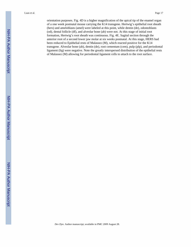
orientation purposes. Fig. 4D is a higher magnification of the apical tip of the enamel organof a one week postnatal mouse carrying the K14 transgene. Hertwig’s epithelial root sheath(hers) and ameloblasts (amel) were labeled at this point, while dentin (de), odontoblasts(od), dental follicle (df), and alveolar bone (ab) were not. At this stage of initial rootformation, Hertwig’s root sheath was continuous. Fig. 4E. Sagital section through theanterior root of a second lower jaw molar at six weeks postnatal. At this stage, HERS hadbeen reduced to Epithelial rests of Malassez (M), which reacted positive for the K14transgene. Alveolar bone (ab), dentin (de), root cementum (cem), pulp (plp), and periodontalligament (lig) were negative. Note the greatly interspersed distribution of the epithelial restsof Malassez (M) allowing for periodontal ligament cells to attach to the root surface.
Luan et al. Page 17
Dev Dyn. Author manuscript; available in PMC 2009 August 28.
NIH
-PA Author Manuscript
NIH
-PA Author Manuscript
NIH
-PA Author Manuscript
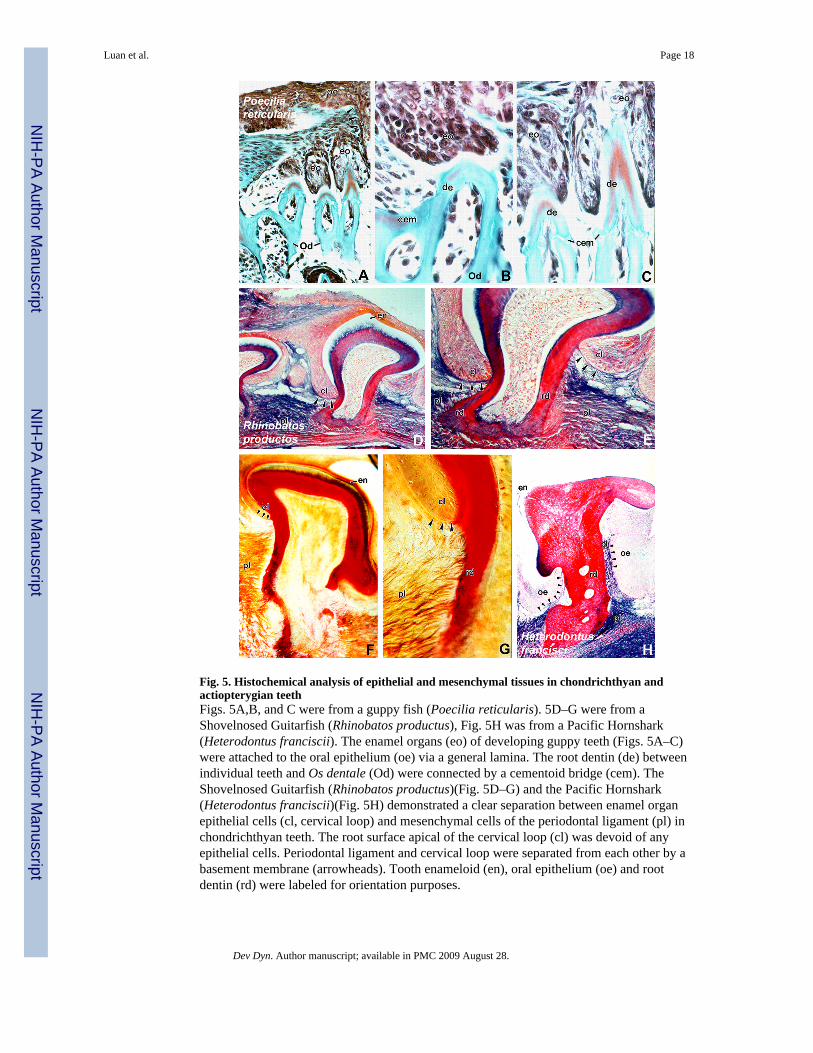
Fig. 5. Histochemical analysis of epithelial and mesenchymal tissues in chondrichthyan andactiopterygian teethFigs. 5A,B, and C were from a guppy fish (Poecilia reticularis). 5D–G were from aShovelnosed Guitarfish (Rhinobatos productus), Fig. 5H was from a Pacific Hornshark(Heterodontus franciscii). The enamel organs (eo) of developing guppy teeth (Figs. 5A–C)were attached to the oral epithelium (oe) via a general lamina. The root dentin (de) betweenindividual teeth and Os dentale (Od) were connected by a cementoid bridge (cem). TheShovelnosed Guitarfish (Rhinobatos productus)(Fig. 5D–G) and the Pacific Hornshark(Heterodontus franciscii)(Fig. 5H) demonstrated a clear separation between enamel organepithelial cells (cl, cervical loop) and mesenchymal cells of the periodontal ligament (pl) inchondrichthyan teeth. The root surface apical of the cervical loop (cl) was devoid of anyepithelial cells. Periodontal ligament and cervical loop were separated from each other by abasement membrane (arrowheads). Tooth enameloid (en), oral epithelium (oe) and rootdentin (rd) were labeled for orientation purposes.
Luan et al. Page 18
Dev Dyn. Author manuscript; available in PMC 2009 August 28.
NIH
-PA Author Manuscript
NIH
-PA Author Manuscript
NIH
-PA Author Manuscript

Fig. 6. Immunohistochemical staining of epithelial tissues in amphibian teeth using anti-keratinwide-spectrum screening antibodiesFigs. 6A and B were from a Leopard Frog (Rana pipiens), Figs. 6C–E were from a MexicanAxolotl (Ambystoma mexicanum). In the frog jaw (Figs. 6A and B) Hertwig’s epithelial rootsheath (hers) measured less than a quarter in length compared to the entire length of thetooth root. The remaining root surface was covered with cementoid of attachment betweenadjacent tooth roots and mandibular bone. Borders between root dentin (de), cementoid ofattachment (cem), and Os dentale (bone) were difficult to distinguish. The anti-keratinantibody labeled Hertwig’s epithelial root sheaths (hers) as well as oral epithelium (oe). Theaxolotl jaw (Figs. 6C–E) was similar to the frog jaw in that a cementoid tissue (cem)connected teeth amongst each other as well as individual teeth with the Os dentale (Od). Incomparison to the frog, the cementoid tissue (cem) of the axolotl was less prominent.Hertwig’s epithelial root sheath (hers) covered between one half and two thirds of the rootdentin (rd) surface. Note the presence of islands of epithelial cells (isl) in the interdentalregion and distant from HERS. The apical portion of the root dentin surface was occupiedby mesenchymal cells. The anti-keratin antibody recognized oral epithelium (oe), epithelialislands (isl), and Hertwig’s epithelial root sheath (hers).
Luan et al. Page 19
Dev Dyn. Author manuscript; available in PMC 2009 August 28.
NIH
-PA Author Manuscript
NIH
-PA Author Manuscript
NIH
-PA Author Manuscript

Fig. 7. Anti-keratin immunohistochemical staining of epithelial tissues in mammalian andreptilian teethFigs. 7A and F are micrographs of a developing tooth organ of a Texas Banded Gecko(Coleonyx brevis) immunostained with anti-keratin antibodies. Hertwig’s root sheath(HERS), ameloblasts (amel), and general lamina (general lamina) were labeled by the anti-keratin antibody. A typical characteristic of reptilian jaws was the position of bone ofattachment (cem) connecting adjacent teeth among each other. The body of the jaw bone(Od, Os dentale) was in distinct distance to the cementum-derived tooth-carrying bone. Notethat the epithelial cell layer was limited to the tooth crown allowing for cementoid tissue andbone of attachment (cem) forming between adjacent teeth. Figs. 7B, C, and D are anti-keratin immunoreactions in the epithelial tissues of erupting iguana teeth. Fig. 7C is anoverview; Figs. 7B and 7D are higher magnifications. The anti-keratin antibody labeled oralepithelium (oe) and Hertwig’s epithelial root sheath (hers). The cementum layer (cem)covered the entire root area and facilitated an ankylotic attachment between tooth root and
Luan et al. Page 20
Dev Dyn. Author manuscript; available in PMC 2009 August 28.
NIH
-PA Author Manuscript
NIH
-PA Author Manuscript
NIH
-PA Author Manuscript

alveolar bone (alv). Adjacent teeth were attached to a basal Os dentale (Od) instead of beingdirectly connected with each other via a cementoid bridge as seen in the gecko and frog. InFig. 7F, the anti-keratin antibody was applied to sagital paraffin sections through a caimantooth organ (Caiman crocodilus). Here, the antibody discretely labeled the coronalameloblasts (am) and remnants of Hertwig’s root sheath (hers) interspersed along the rootsurface. Note the wide spaces between individual HERS cell rests allowing for access of thecaiman periodontal ligament (pdl) to attach to the root surface. Fig. 7G is a micrograph froma mouse first mandibular molar prior to eruption. Oral epithelium (oe), ameloblast cell layer(amel), and papillary layer (pap) were distinctly stained with anti-keratin antibodies. Whilethe entire tooth crown (crown) was surrounded by keratin-positive epithelial cells, the rootsurface (root) and the alveolar bone (alv) were not labeled. Only the most apical portion ofHERS (ap, arrow) was recognized by the anti-keratin antibody.
Luan et al. Page 21
Dev Dyn. Author manuscript; available in PMC 2009 August 28.
NIH
-PA Author Manuscript
NIH
-PA Author Manuscript
NIH
-PA Author Manuscript
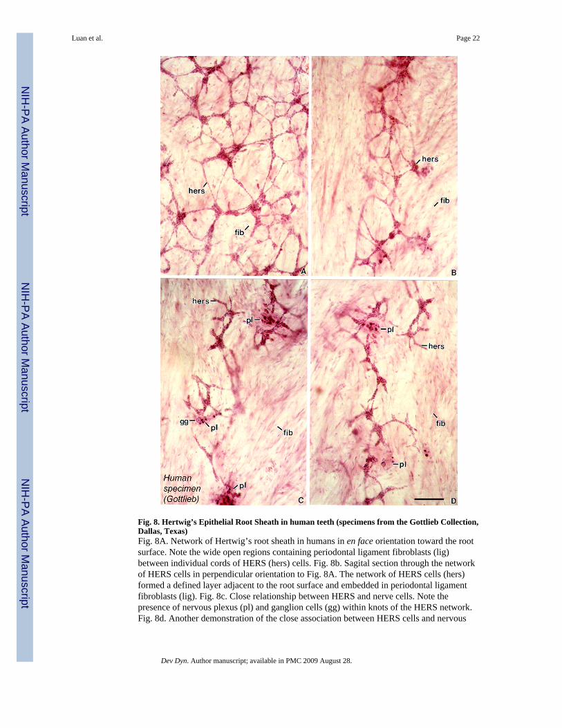
Fig. 8. Hertwig’s Epithelial Root Sheath in human teeth (specimens from the Gottlieb Collection,Dallas, Texas)Fig. 8A. Network of Hertwig’s root sheath in humans in en face orientation toward the rootsurface. Note the wide open regions containing periodontal ligament fibroblasts (lig)between individual cords of HERS (hers) cells. Fig. 8b. Sagital section through the networkof HERS cells in perpendicular orientation to Fig. 8A. The network of HERS cells (hers)formed a defined layer adjacent to the root surface and embedded in periodontal ligamentfibroblasts (lig). Fig. 8c. Close relationship between HERS and nerve cells. Note thepresence of nervous plexus (pl) and ganglion cells (gg) within knots of the HERS network.Fig. 8d. Another demonstration of the close association between HERS cells and nervous
Luan et al. Page 22
Dev Dyn. Author manuscript; available in PMC 2009 August 28.
NIH
-PA Author Manuscript
NIH
-PA Author Manuscript
NIH
-PA Author Manuscript

plexus within the fibroblast-rich (fib) periodontal ligament space. These preparations arefrom the historic Gottlieb collection at Baylor University. Bar = 100μm.
Luan et al. Page 23
Dev Dyn. Author manuscript; available in PMC 2009 August 28.
NIH
-PA Author Manuscript
NIH
-PA Author Manuscript
NIH
-PA Author Manuscript

Fig. 9. Demonstration of apoptotic cells in the periodontal ligament using the TUNEL techniqueFigs. 9A and 9C were DAPI labeled fluorographs of the same areas as in the TUNELstainings in Figs. 9B and 9D. The position of alveolar bone (ab), dental follicle (df),ameloblast nuclei (am), and odontoblast nuclei (od) was indicated for orientation purposes.Ameloblast mitochondria (am) provided orientation marks in the TUNEL stained Fig. 9B.Using the TUNEL technique, numerous apoptotic cell nuclei in the dental follicle werevisualized (arrows, Figs. 9B and D). Figs. 9E and 9F are double exposures of the same areausing both propidium iodide staining and ApopTag Red apoptosis labeling. Apoptotic nucleiwere marked by arrows. Note the two adjacent apoptotic nuclei in the proximity of the rootsurface (Fig. 9F). Alveolar bone (ab), dental follicle (df), root dentin (rd), and enamel (en)were labeled for orientation purposes.
Luan et al. Page 24
Dev Dyn. Author manuscript; available in PMC 2009 August 28.
NIH
-PA Author Manuscript
NIH
-PA Author Manuscript
NIH
-PA Author Manuscript

Fig. 10. Fate of Hertwig’s root sheath in six weeks old mice visualized in Keratin-14 transgenicmiceIn this study, the K14 transgene was detected using the lacZ reporter gene and stained withβ-galactosidase. Micrographs illustrate the incorporation of epithelial rests of Malassez (M)(Figs. 10A, B) as well as individual cells of Hertwig’s epithelial root sheath (hers) into themineralizing cementum layer (cem)(Fig. 10C). Tissues labeled for orientation areperiodontal ligament (pdl), alveolar bone (ab), and root dentin (den). Note the strongstaining intensity for the K14 transgene in Malassez’ rests of 6 weeks old mice. Figs. 10Aand 10B illustrate the incorporation of K14 Malassez’ rests into the cementum matrix of theroot shaft at multiple sites. Fig. 10C documents the encapsulation and incorporation ofindividual HERS cells in the mineralizing cementum mass of the root apex.
Luan et al. Page 25
Dev Dyn. Author manuscript; available in PMC 2009 August 28.
NIH
-PA Author Manuscript
NIH
-PA Author Manuscript
NIH
-PA Author Manuscript
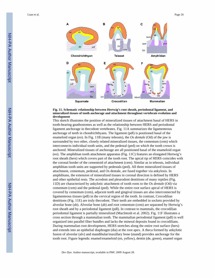
Fig. 11. Schematic relationship between Hertwig’s root sheath, periodontal ligament, andmineralized tissues of tooth anchorage and attachment throughout vertebrate evolution anddevelopmentThis sketch illustrates the position of mineralized tissues of attachment basal of HERS intooth-bearing gnathostomes as well as the relationship between HERS and periodontalligament anchorage in thecodont vertebrates. Fig. 11A summarizes the ligamenteousanchorage of teeth in chondrichthyans. The ligament (pdl) is positioned basal of theenameloid organ (eo). In Fig. 11B (many teleosts), the Os dentale (Od) of the jaw issurrounded by two other, closely related mineralized tissues, the cementum (cem) whichinterconnects individual tooth units, and the pedestal (ped) on which the tooth crown isanchored. Mineralized tissues of anchorage are all positioned basal of the enameloid organ(eo). The amphibian tooth attachment apparatus (Fig. 11C) features an elongated Hertwig’sroot sheath (hers) which covers part of the tooth root. The apical tip of HERS coincides withthe coronal border of the cementoid of attachment (cem). Similar as in teleosts, individualamphibian tooth units are supported by pedestals (ped). All three mineralized tissues ofattachment, cementum, pedestal, and Os dentale, are fused together via ankylosis. Inamphibians, the extension of mineralized tissues in coronal direction is defined by HERSand other epithelial rests. The acrodont and pleurodont dentitions of many reptiles (Fig.11D) are characterized by ankylotic attachment of tooth roots to the Os dentale (Od) viacementum (cem) and the pedestal (ped). While the entire root surface apical of HERS iscovered by cementum (cem), adjacent teeth and gingival tissues are also interconnected byligamenteous tissues (pdl) in the cervical region of the tooth. In contrast, Crocodiliandentitions (Fig. 11E) are truly thecodont. Their teeth are embedded in sockets provided byalveolar bone (ab). Alveolar bone (ab) and root cementum (cem) are separated by Hertwig’sroot sheath and by a periodontal ligament (pdl). In contrast to mammals, the crocodilianperiodontal ligament is partially mineralized (MacIntosh et al. 2002). Fig. 11F illustrates across section through a mammalian tooth. The mammalian periodontal ligament (pdl) is wellorganized into parallel fiber bundles and lacks the mineral deposits found in crocodilians.During mammalian root development, HERS stretches along the entire root surface (hers)and extends into an epithelial diaphragm (dia) at the root apex. A theca formed by ankyloticfusion of alveolar (alv) and mandibular/maxillary bone (mand) provides anchorage for thetooth root. Figure legends: enamel/enameloid (en, yellow), dentin (de, green), enamel organ
Luan et al. Page 26
Dev Dyn. Author manuscript; available in PMC 2009 August 28.
NIH
-PA Author Manuscript
NIH
-PA Author Manuscript
NIH
-PA Author Manuscript
(eo, red), cementum (cem, blue dots), pedestal (ped, mauve), Os dentale/mandible (Od, bluegraticule), periodontal ligament (pdl, blue interrupted lines), Hertwig’s Epithelial RootSheath (hers, red dots), Epithelial Diaphragm (dia, red oval). The orientation of illustrationpatterns in the periodontal ligament space does not follow physiological fiber orientation.
Luan et al. Page 27
Dev Dyn. Author manuscript; available in PMC 2009 August 28.
NIH
-PA Author Manuscript
NIH
-PA Author Manuscript
NIH
-PA Author Manuscript

Fig. 12. Schematic phylogeny of species involved in the current studyNote that the thecodont archosaurs are more distant from the thecodont mammals than thepleurodont/acrodont squamates (Lepidosauromorpha) suggesting convergent evolution ofmammals and crocodilians in respect to tooth morphogenesis.
Luan et al. Page 28
Dev Dyn. Author manuscript; available in PMC 2009 August 28.
NIH
-PA Author Manuscript
NIH
-PA Author Manuscript
NIH
-PA Author Manuscript
Copyright © 2022 FDOKUMEN