Odkritje portreta Antona Tomaža Linharta (uploaded 28. 1. 2015)
Upload
khangminh22Category
view
0download
0
For pe
rsona
l priv
ate us
e only
Not be
distr
ibuted
or up
loade
d to a
nyon
e or a
nywhe
re

Frontiers in Anti-Infective DrugDiscovery
(Volume 8)
Edited by Atta-ur-Rahman, FRS
Kings CollegeUniversity of Cambridge
CambridgeUK
M. Iqbal ChoudharyH.E.J. Research Institute of Chemistry,
International Center for Chemical and Biological Sciences,University of Karachi, Karachi,
Pakistan
&
For pe
rsona
l priv
ate us
e only
Not be
distr
ibuted
or up
loade
d to a
nyon
e or a
nywhe
re

Frontiers in Anti-Infective Drug Discovery
Volume # 8
Editors: Atta-ur-Rahman, FRS and M. Iqbal Choudhary
ISSN (Online): 1879-663X
ISSN (Print): 2451-9162
ISBN (Online): 978-981-14-1238-7
ISBN (Print): 978-981-14-1237-0
© 2020, Bentham Books imprint.
Published by Bentham Science Publishers Pte. Ltd. Singapore. All Rights Reserved.
ISBN (Paperback): 978-981-14-7005-9
For pe
rsona
l priv
ate us
e only
Not be
distr
ibuted
or up
loade
d to a
nyon
e or a
nywhe
re

BENTHAM SCIENCE PUBLISHERS LTD.End User License Agreement (for non-institutional, personal use)
This is an agreement between you and Bentham Science Publishers Ltd. Please read this License Agreementcarefully before using the ebook/echapter/ejournal (“Work”). Your use of the Work constitutes youragreement to the terms and conditions set forth in this License Agreement. If you do not agree to these termsand conditions then you should not use the Work.
Bentham Science Publishers agrees to grant you a non-exclusive, non-transferable limited license to use theWork subject to and in accordance with the following terms and conditions. This License Agreement is fornon-library, personal use only. For a library / institutional / multi user license in respect of the Work, pleasecontact: [email protected].
Usage Rules:All rights reserved: The Work is the subject of copyright and Bentham Science Publishers either owns the1.Work (and the copyright in it) or is licensed to distribute the Work. You shall not copy, reproduce, modify,remove, delete, augment, add to, publish, transmit, sell, resell, create derivative works from, or in any wayexploit the Work or make the Work available for others to do any of the same, in any form or by anymeans, in whole or in part, in each case without the prior written permission of Bentham SciencePublishers, unless stated otherwise in this License Agreement.You may download a copy of the Work on one occasion to one personal computer (including tablet,2.laptop, desktop, or other such devices). You may make one back-up copy of the Work to avoid losing it.The unauthorised use or distribution of copyrighted or other proprietary content is illegal and could subject3.you to liability for substantial money damages. You will be liable for any damage resulting from yourmisuse of the Work or any violation of this License Agreement, including any infringement by you ofcopyrights or proprietary rights.
Disclaimer:
Bentham Science Publishers does not guarantee that the information in the Work is error-free, or warrant thatit will meet your requirements or that access to the Work will be uninterrupted or error-free. The Work isprovided "as is" without warranty of any kind, either express or implied or statutory, including, withoutlimitation, implied warranties of merchantability and fitness for a particular purpose. The entire risk as to theresults and performance of the Work is assumed by you. No responsibility is assumed by Bentham SciencePublishers, its staff, editors and/or authors for any injury and/or damage to persons or property as a matter ofproducts liability, negligence or otherwise, or from any use or operation of any methods, products instruction,advertisements or ideas contained in the Work.
Limitation of Liability:
In no event will Bentham Science Publishers, its staff, editors and/or authors, be liable for any damages,including, without limitation, special, incidental and/or consequential damages and/or damages for lost dataand/or profits arising out of (whether directly or indirectly) the use or inability to use the Work. The entireliability of Bentham Science Publishers shall be limited to the amount actually paid by you for the Work.
General:Any dispute or claim arising out of or in connection with this License Agreement or the Work (including1.non-contractual disputes or claims) will be governed by and construed in accordance with the laws ofSingapore. Each party agrees that the courts of the state of Singapore shall have exclusive jurisdiction tosettle any dispute or claim arising out of or in connection with this License Agreement or the Work(including non-contractual disputes or claims).Your rights under this License Agreement will automatically terminate without notice and without the2.
For pe
rsona
l priv
ate us
e only
Not be
distr
ibuted
or up
loade
d to a
nyon
e or a
nywhe
re

need for a court order if at any point you breach any terms of this License Agreement. In no event will anydelay or failure by Bentham Science Publishers in enforcing your compliance with this License Agreementconstitute a waiver of any of its rights.You acknowledge that you have read this License Agreement, and agree to be bound by its terms and3.conditions. To the extent that any other terms and conditions presented on any website of Bentham SciencePublishers conflict with, or are inconsistent with, the terms and conditions set out in this LicenseAgreement, you acknowledge that the terms and conditions set out in this License Agreement shall prevail.
Bentham Science Publishers Pte. Ltd.80 Robinson Road #02-00Singapore 068898SingaporeEmail: [email protected]
For pe
rsona
l priv
ate us
e only
Not be
distr
ibuted
or up
loade
d to a
nyon
e or a
nywhe
re

CONTENTSPREFACE ................................................................................................................................................ i
LIST OF CONTRIBUTORS .................................................................................................................. iii
CHAPTER 1 ERADICATION OF HELICOBACTER PYLORI INFECTION WITH NON-BISMUTH QUADRUPLE CONCOMITANT THERAPY ................................................................. 1
Javier P. Gisbert and Adrian G. McNicholl INTRODUCTION .......................................................................................................................... 2OBJECTIVE ................................................................................................................................... 3BIBLIOGRAPHICAL SEARCHES ............................................................................................. 3EFFICACY OF THE CONCOMITANT REGIMEN ................................................................. 4COMPARISON BETWEEN CONCOMITANT AND STANDARD TRIPLE REGIMEN .... 14COMPARISON BETWEEN CONCOMITANT AND SEQUENTIAL REGIMENS ............. 16EFFECTS OF DIFFERENT VARIABLES ON THE EFFICACY OF CONCOMITANTTHERAPY ....................................................................................................................................... 20
Clarithromycin Resistance ...................................................................................................... 20Nitroimidazole Resistance ...................................................................................................... 21Dual Clarithromycin and Metronidazole Resistance .............................................................. 22Duration of Treatment ............................................................................................................. 23
HOW COULD WE EVEN INCREASE THE EFFICACY OF THE CONCOMITANTTREATMENT? ............................................................................................................................... 24WHAT ARE THE RESULTS WITH THE CONCOMITANT TREATMENT INCLINICAL PRACTICE? THE EXPERIENCE OF THE EUROPEAN REGISTRY ON H.PYLORI MANAGEMENT ............................................................................................................ 25CONCLUSIONS ............................................................................................................................. 26CONSENT FOR PUBLICATION ................................................................................................ 27CONFLICT OF INTEREST ......................................................................................................... 27ACKNOWLEDGEMENTS ........................................................................................................... 27REFERENCES ............................................................................................................................... 27
CHAPTER 2 DRUG DISCOVERY STRATEGIES AGAINST EMERGINGCORONAVIRUSES: A GLOBAL THREAT ....................................................................................... 35
Paulo Fernando da Silva Santos-J¼nior, Igor Jos® dos Santos Nascimento, ThiagoMendona de Aquino, Jo«o Xavier de Ara¼jo-J¼nior and Edeildo Ferreira da Silva-J¼niorINTRODUCTION .......................................................................................................................... 35BIOLOGICAL ASPECTS, SIGNS/SYMPTOMS AND DIAGNOSIS FOR MERS-COV,SARS-COV, AND SARS-COV-2 .................................................................................................. 37STRATEGIES USED FOR DISCOVERING NEW POTENTIAL DRUGS ............................ 38
Natural Source ........................................................................................................................ 38Classical Methods Applied for Discovering New Antiviral Agents ....................................... 46Virtual Screening and Computational Techniques for Designing New Antiviral Agents ...... 55Drug Repurposing ................................................................................................................... 64High-Throughput Screening (HTS) for Discovering New Antiviral Agents .......................... 74
CONCLUSION AND FUTURE OUTLOOK .............................................................................. 80CONSENT FOR PUBLICATION ................................................................................................ 80CONFLICT OF INTEREST ......................................................................................................... 80ACKNOWLEDGEMENTS ........................................................................................................... 80REFERENCES ............................................................................................................................... 81
For pe
rsona
l priv
ate us
e only
Not be
distr
ibuted
or up
loade
d to a
nyon
e or a
nywhe
re

CHAPTER 3 OPPORTUNITIES OFFERED BY FRAGMENT-BASED DRUG DESIGN INANTIBIOTIC DEVELOPMENT .......................................................................................................... 91
Sanjay Yapabandara, Lorna Wilkinson-White, Sandro Ataide and Ann H. KwanINTRODUCTION .......................................................................................................................... 91POTENTIAL ADVANTAGES OF FBDD FOR DEVELOPMENT OF ANTIBIOTICS ....... 93CASE STUDIES OF NEW ANTIBIOTIC DEVELOPMENT UTILISING FBDD ................. 97
Case Study 1: β-Lactamase ..................................................................................................... 97Case Study 2: Bacterial DNA Gyrase/Topoisomerase IV ...................................................... 99Case Study 3: Fatty Acid Synthesis (FAS) Pathways ............................................................. 102
Biotin Carboxylase ........................................................................................................ 102Ketoacyl Synthase ......................................................................................................... 103
Case Study 4: Mycobacterium Tuberculosis (Mtb) ................................................................ 103Pantothenate Synthetase (PanC) ................................................................................... 104Mtb Transcriptional Repressor (EthR) ......................................................................... 105Mycocyclosin Synthase (CYP121) ................................................................................. 106
Fructose 1,6-bisphosphate Aldolase (FBA) ............................................................................ 107RECENT ADVANCES IN FBDD ................................................................................................. 107CONCLUSION ............................................................................................................................... 109CONSENT FOR PUBLICATION ................................................................................................ 110CONFLICT OF INTEREST ......................................................................................................... 110ACKNOWLEDGEMENTS ........................................................................................................... 110REFERENCES ............................................................................................................................... 110
CHAPTER 4 PHAGE THERAPY AS A TOOL FOR CONTROL OF FOODBORNEDISEASES: ADVANTAGES AND LIMITATIONS ........................................................................... 114
S. Pacios-Michelena, R. Rodr²guez-Herrera, A. C. Flores-Gallegos, M.L Ch§vezGonz§lez, E.P. Segura-Ceniceros, R. Ramos-Gonz§lez and A. Ilyina INTRODUCTION .......................................................................................................................... 115BACTERIOPHAGE ....................................................................................................................... 115
History ..................................................................................................................................... 115General Phage Characteristics ................................................................................................ 116Replication Cycle .................................................................................................................... 118
Adsorption to the Bacterial Cell Wall ........................................................................... 118Replication .................................................................................................................... 120Release of Viral Progeny and Cell Lysis ....................................................................... 122Other Components in the Replication Cycle ................................................................. 122
BACTERIOPHAGE USE FOR PHAGE THERAPY ................................................................. 123Health Applications ................................................................................................................ 123Applications in the Food Industry ........................................................................................... 125
BACTERIOPHAGE PREPARATIONS ...................................................................................... 127Indications and Limitations ..................................................................................................... 128
Bacterial Resistance ...................................................................................................... 129SYNERGY BETWEEN BACTERIOPHAGES AND THE IMMUNE SYSTEM .................... 131CONCLUDING REMARKS ......................................................................................................... 133CONSENT FOR PUBLICATION ................................................................................................ 134CONFLICT OF INTEREST ......................................................................................................... 134ACKNOWLEDGEMENTS ........................................................................................................... 134REFERENCES ............................................................................................................................... 134
For pe
rsona
l priv
ate us
e only
Not be
distr
ibuted
or up
loade
d to a
nyon
e or a
nywhe
re

Fatima Shahid, Muhammad Shehroz, Tahreem Zaheer and Amjad Ali INTRODUCTION .......................................................................................................................... 145
Characteristics of an Ideal Drug Target .................................................................................. 145Significance of Drug Target Identification ............................................................................. 145Subtractive Genomics Based Efforts for Drug Target Identification ..................................... 146Approaches Used for Promising Drug Target Prediction ....................................................... 148DrugSol and its Utility ............................................................................................................ 148Non-host Homologues ............................................................................................................ 149Subcellular Localization ......................................................................................................... 150Essentiality .............................................................................................................................. 150Virulence ................................................................................................................................. 150Annotation ............................................................................................................................... 151Druggability Potential Evaluation ........................................................................................... 151Analyses of Metabolic Pathways ............................................................................................ 151
OUR CONTRIBUTION ................................................................................................................. 151Sequence Acquisition .............................................................................................................. 151Human Homology Filter ......................................................................................................... 151Subcellular Localization ......................................................................................................... 152Essentiality .............................................................................................................................. 152Virulence ................................................................................................................................. 152Annotated ................................................................................................................................ 152Druggability Potential Evaluation ........................................................................................... 152Analyses of Metabolic Pathways ............................................................................................ 153Physiochemical properties ...................................................................................................... 153Pros and Cons of in silico Drug Target Mining ...................................................................... 154Success Stories ........................................................................................................................ 154
CONSENT FOR PUBLICATION ................................................................................................ 155CONFLICT OF INTEREST ......................................................................................................... 155ACKNOWLEDGEMENTS ........................................................................................................... 155REFERENCES ............................................................................................................................... 155
CHAPTER 6 RECENT ADVANCES IN THE DISCOVERY OF ANTIMICROBIALSTHROUGH METAGENOMICS ........................................................................................................... 159
Daljeet Singh Dhanjal, Reena Singh and Chirag Chopra INTRODUCTION .......................................................................................................................... 160MAJOR MODULAR ENZYMES AND THEIR ASSOCIATED DOMAINS .......................... 161
Non-ribosomal Peptide Synthetases (NRPS) .......................................................................... 161Polyketide Synthases (PKS) ................................................................................................... 163
Type I PKSs ................................................................................................................... 164Type II PKSs .................................................................................................................. 164Type III PKSs ................................................................................................................ 164
Other Molecules ...................................................................................................................... 165EXPLORATION OF NEW HABITATS ...................................................................................... 165
Extreme Environment: A Niche for Novel Strains ................................................................. 166ROLE OF METAGENOMICS IN EXPLORATION OF UNCULTURED MICROBES ...... 167
The workflow of the Metagenomic Approach ........................................................................ 167Direct Metagenomic Screening based on Function ................................................................ 169Sequence-based Metagenomic Screening ............................................................................... 170Sequence Advancement: Potential of Single-Cell Genomics and High-ThroughputSequencing .............................................................................................................................. 172
MINING OF GENOMES: CURRENT SCENARIO AND FUTURE POTENTIAL ............... 172
CHAPTER 5 SUBTRACTIVE GENOMICS APPROACHES: TOWARDS ANTI-BACTERIALDRUG DISCOVERY .............................................................................................................................. 144
For pe
rsona
l priv
ate us
e only
Not be
distr
ibuted
or up
loade
d to a
nyon
e or a
nywhe
re

Prediction of Biosynthetic Gene Cluster (BGC) and their targeted activation ....................... 173Genetic Manipulations of the Biosynthetic Machinery for the Discovery of NewAntimicrobials ......................................................................................................................... 174Different Mechanisms of Activation of Silent BGCs ............................................................. 176
ANTIBIOTIC AND BIOACTIVE COMPOUNDS DISCOVERED THROUGHMETAGENOMIC APPROACH ................................................................................................... 177CONCLUSION AND FUTURE PERSPECTIVES ..................................................................... 179CONSENT FOR PUBLICATION ................................................................................................ 181CONFLICT OF INTEREST ......................................................................................................... 181ACKNOWLEDGEMENTS ........................................................................................................... 181REFERENCES ............................................................................................................................... 181
CHAPTER 7 PHYTO-NANO-ANTIMICROBIALS: SYNTHESIS, CHARACTER-IZATION,DISCOVERY, AND ADVANCES ......................................................................................................... 196
Pankaj Satapathy, Aishwarya S, Rashmi M Shetty, Akshaya Simha N, Dhanapal G,Aishwarya Shree R, Antara Biswas, Kounaina K, Anirudh G. Patil, Avinash MG,Aishwarya T Devi, Shubha Gopal, Nagendra Prasad MN, Veena SM, Hudeda SP,Muthuchelian K, Sunil S. More, Govindappa Melappa and Farhan Zameer INTRODUCTION .......................................................................................................................... 197SYNTHESIS OF NANOPARTICLES .......................................................................................... 198
Bacterial Mediated Synthesis .................................................................................................. 199Fungi Mediated Synthesis ....................................................................................................... 199Plant Extract Synthesis ........................................................................................................... 199Solvent for Synthesis .............................................................................................................. 200Mechanism of Synthesis ......................................................................................................... 200
CHARACTERIZATION OF PLANT MEDIATED NANOPARTICLES HAVINGMICROBIAL ACTIVITY ............................................................................................................. 203
General Characterization ........................................................................................................ 203UV- Visible Spectroscopy ...................................................................................................... 204X-ray Diffraction .................................................................................................................... 204Transmission Electron Microscopy ........................................................................................ 205Fourier Transformed Infrared ................................................................................................. 205Dynamic Light Scattering ....................................................................................................... 205Characterization of Silver Nanoparticle (AgNPs) .................................................................. 205
a). UV-Visible Spectrophotometer ................................................................................ 206b). FTIR Analysis .......................................................................................................... 206c). XRD Analysis ........................................................................................................... 206d). Thermogravimetric Analysis .................................................................................... 206e). TEM Analysis ........................................................................................................... 206f). FTIR Analysis ........................................................................................................... 207
Characterization of Gold Nanoparticles (AuNPs) .................................................................. 207a). UV- Vis Spectroscopy .............................................................................................. 207b). FTIR ......................................................................................................................... 207c). SEM .......................................................................................................................... 207d). X-ray Diffraction ...................................................................................................... 208
Characterization of Iron Oxide Nanoparticle .......................................................................... 208a). UV-Visible Spectroscopy ......................................................................................... 208b). ATR Spectroscopy .................................................................................................... 208c). FT-IR ........................................................................................................................ 209d). SEM Analysis ........................................................................................................... 209
For pe
rsona
l priv
ate us
e only
Not be
distr
ibuted
or up
loade
d to a
nyon
e or a
nywhe
re

e). TEM Analysis ........................................................................................................... 209f). X-ray Diffraction ...................................................................................................... 209
Zinc Oxide Nanoparticles ....................................................................................................... 209a). UV-Visible Spectroscopy ......................................................................................... 210b). XRD Analysis ........................................................................................................... 210c). FTIR Analysis ........................................................................................................... 210d). SEM .......................................................................................................................... 210e). TEM .......................................................................................................................... 211
APPLICATION OF NANOPARTICLES .................................................................................... 212Silver Nanoparticles ................................................................................................................ 212Gold Nanoparticles ................................................................................................................. 214Oxide Nanoparticles ................................................................................................................ 215Antibacterial Property ............................................................................................................. 216Gold Nanoparticles ................................................................................................................. 218Oxide Nanoparticles ................................................................................................................ 218
MEDICINAL APPLICATION ...................................................................................................... 219FUTURE PROSPECTS AND CONCLUSION ............................................................................ 220CONSENT FOR PUBLICATION ................................................................................................ 221CONFLICT OF INTEREST ......................................................................................................... 221ACKNOWLEDGEMENTS ........................................................................................................... 221REFERENCES ............................................................................................................................... 222
CHAPTER 8 APTAMERS AS ANTI-INFECTIVE AGENTS ......................................................... 232Muhammad Ali Syed, Nayab Ali, Bushra Jamil and Ammar Ahmed INTRODUCTION .......................................................................................................................... 232APTAMERS .................................................................................................................................... 233SYSTEMIC EVOLUTION OF LIGANDS BY EXPONENTIAL ENRICHMENT (SELEX) 234
Negative SELEX ..................................................................................................................... 236Counter SELEX ...................................................................................................................... 236Microfluidic SELEX ............................................................................................................... 237Capillary Electrophoresis SELEX .......................................................................................... 237Cell SELEX ............................................................................................................................ 237
APTAMERS VERSUS ANTIBODIES ......................................................................................... 238APTAMERS AS ANTI-INFECTIVE AGENTS AGAINST PATHOGENICMICROORGANISMS ................................................................................................................... 240APTAMERS AGAINST PATHOGENIC BACTERIA .............................................................. 241APTAMERS AGAINST VIRUSES .............................................................................................. 243APTAMERS AGAINST PROTOZOAN PARASITES .............................................................. 244FUTURE PERSPECTIVES ........................................................................................................... 245CONCLUSION ............................................................................................................................... 246CONSENT FOR PUBLICATION ................................................................................................ 246CONFLICT OF INTEREST ......................................................................................................... 246ACKNOWLEDGEMENTS ........................................................................................................... 246REFERENCES ............................................................................................................................... 246
SUBJECT INDEX .................................................................................................................................... 254
For pe
rsona
l priv
ate us
e only
Not be
distr
ibuted
or up
loade
d to a
nyon
e or a
nywhe
re

PREFACE
The recent COVID-19 pandemic has further highlighted our human incapacity to controlinfections. Pathogens of all forms and types are fast learners, and their mutations, spread andvirulence can overwhelm the entire health care system within weeks. The COVID-19pandemic has also exposed our inability to quickly come up with treatment and preventionregimes, despite the tremendous progress in pharmaceutical and biomedical sciences. In thepost COVID-19 world major attention to the surveillance, prevention, and treatment ofinfections of all kinds is expected. Research on infections and anti-infectious drug discoveryis already truly interdisciplinary in nature, and is published in journals of diverse disciplines,such as microbiology, molecular and structural biology, genomics, immunology,epidemiology, etc. It is imperative that the most exciting discoveries in this field are compiledas critically written reviews in frontier areas.
The aim of the book series “Frontiers in Anti-infectious Drug Discovery” is to focus onrecent important developments. Experts in various important aspects of anti-infectious drugdiscovery have therefore contributed review articles on the most recent advancements.Volume 8, like the previous volumes, of this well received book series, comprises eight (8)scholarly written review articles on certain key aspects. These include genomic basedidentification of new drug targets and metagenomics for antimicrobials; fragment-basedapproach for drug designing, and of various types of antimicrobials ranging from syntheticanalogs against coronaviruses, to bacterial phages against infections, nanoparticle basedagents, as well as aptamers.
The chapter contributed by Gisbert and McNicholl focuses on the key advantages ofconcomitant non-bismuth quadruple therapy for a range of infections caused by Helicobactorpylori. Silva-Junior et al have presented an interesting review on the discovery anddevelopment of bioactive drug leads against the recent pandemic caused by SARS-CoV-2,based on analogs developed during the past SARS and MERS epidemics. Advances andchallenges in fragment-based designing of new antibiotics is the key focus of the article byKwan et al, supported by numerous examples. Foodborne bacterial infections are widespread.Ilyina et al review the recent applications of phage therapies as alternatives to antimicrobialsfor the treatment of food borne bacterial infections. Amjad et al have contributed a chapter onthe applications of subtractive genomics to identify essential genes involved in crucialmetabolic pathways of pathogens, and validating their protein products as novel drug targets.Metagonomics has emerged as a key technique for the discovery of novel antibiotics from yetuncultured microbes. The tremendous pool of new antimicrobials in unexplored microbialflora is the focus of the review by Chopra et al. Zameer has contributed a chapter on the useof nanoparticles as drug careers of synthetic and natural antimicrobial agents. In the lastchapter, Syed et al have touched upon an important new field of the use of aptamers(oligonucleotides or peptide molecules) as novel diagnostic and anti-infective agents.
i
For pe
rsona
l priv
ate us
e only
Not be
distr
ibuted
or up
loade
d to a
nyon
e or a
nywhe
re

UK
Prof. Dr. M. Iqbal ChoudharyH.E.J. Research Institute of Chemistry
International Center for Chemical and Biological SciencesUniversity of Karachi
KarachiPakistan
ii
We would to express our sincere thanks eminent to all the authors for their excellentcontributions in this vibrant, and exciting field of biomedical and pharmaceutical research.The efforts of Ms. Fariya Zulfiqar (Manager Publications) and the excellent management ofMr. Mahmood Alam (Director Publications) are also gratefully acknowledged.
Prof. Dr. Atta-ur-Rahman, FRSHonorary Life FellowKings CollegeUniversity of Cambridge Cambridge
For pe
rsona
l priv
ate us
e only
Not be
distr
ibuted
or up
loade
d to a
nyon
e or a
nywhe
re

iii
LIST OF CONTRIBUTORSA. Ilyina Nanobioscience Research Group, University of Coahuila, Coahuila,
Mexico
Adrian G. McNicholl Department of Gastroenterology, Hospital Universitario de La Princesa,Instituto de Investigación Sanitaria Princesa (IIS-IP), UniversidadAutonoma de Madrid (UAM), Madrid, Spain
Ammar Ahmed Department of Medical Laboratory Sciences, University of Lahore,Islamabad, Pakistan
Amjad Ali Atta-ur-Rahman School of Applied Biosciences (ASAB), NationalUniversity of Sciences and Technology (NUST), Islamabad, Pakistan
Ann H. Kwan School of Life and Environmental Sciences, University of Sydney,Sydney, Australia
A.C. Flores-Gallegos Research Group in Molecular Biology, University of Coahuila,Coahuila, Mexico
Aishwarya T. Devi Department of Biotechnology, JSS Science and Technology University,Karnataka, India
Anirudh G. Patil Department of Biological Sciences, Dayananda Sagar University,Karnataka, India
Antara Biswas Department of Biological Sciences, Dayananda Sagar University,Karnataka, India
Azeddine Chaiba Department of industrial Engineering, University of Khenchela, Algeria
Bushra Jamil Department of Medical Laboratory Sciences, University of Lahore,Islamabad, Pakistan
Chirag Chopra School of Bioengineering and Biosciences, Lovely ProfessionalUniversity, Phagwara, India
Daljeet Singh Dhanjal School of Bioengineering and Biosciences, Lovely ProfessionalUniversity, Phagwara, India
Edeildo Ferreira da Silva-Júnior
Chemistry and Biotechnology Institute, Federal University of Alagoas,Maceió, BrazilLaboratory of Medicinal Chemistry, Pharmaceutical Sciences Institute,Federal University of Alagoas, Maceió, Brazil
E.P. Segura-Ceniceros Nanobioscience Research Group, University of Coahuila, Coahuila,Mexico
Fayssal Amrane LAS Research Laboratory Department of Electrical Engineering,University of Setif-1, Setif, Algeria
Fatima Shahid Atta-ur-Rahman School of Applied Biosciences (ASAB), NationalUniversity of Sciences and Technology (NUST), Islamabad, Pakistan
Farhan Zameer Department of Biological Sciences, Dayananda Sagar University,Karnataka, India
G. Dhanapal Department of Biological Sciences, Dayananda Sagar University,Karnataka, India
For pe
rsona
l priv
ate us
e only
Not be
distr
ibuted
or up
loade
d to a
nyon
e or a
nywhe
re

iv
Govindappa Melappa Department of Botany, Davangere University, Karnataka, India
N. Akshaya Simha Department of Biological Sciences, Dayananda Sagar University,Karnataka, India
Igor José dos SantosNascimento
Chemistry and Biotechnology Institute, Federal University of Alagoas,Maceió, Brazil
João Xavier de Araújo-Júnior Laboratory of Medicinal Chemistry, Pharmaceutical Sciences Institute,Federal University of Alagoas, Maceió, Brazil
Javier P. Gisbert Department of Gastroenterology, Hospital Universitario de La Princesa,Instituto de Investigación Sanitaria Princesa (IIS-IP), UniversidadAutonoma de Madrid (UAM), Madrid, Spain
K. Muthuchelian Department of Biological Sciences, Dayananda Sagar University,Karnataka, India
K. Kounaina Department of Dravyaguna, JSS Ayurvedic Medical College,Karnataka, India
Lorna Wilkinson-White Sydney Analytical Core Research Facility, University of Sydney,Sydney, Australia
M.G. Avinash Department of Studies in Microbiology, University of Mysore,Karnataka, India
M.L Chávez González Nanobioscience Research Group, University of Coahuila, Coahuila,Mexico
Muhammad Ali Syed Department of Microbiology, The University of Haripur, Haripur,Pakistan
Muhammad Shehroz Atta-ur-Rahman School of Applied Biosciences (ASAB), NationalUniversity of Sciences and Technology (NUST), Islamabad, Pakistan
M.N. Nagendra Prasad Department of Biotechnology, JSS Science and Technology University,Karnataka, India
Nayab Ali Department of Microbiology, The University of Haripur, Haripur,Pakistan
Pankaj Satapathy Department of Biological Sciences, Dayananda Sagar University,Karnataka, India
Paulo Fernando da SilvaSantos-Júnior
Chemistry and Biotechnology Institute, Federal University of Alagoas,Maceió, Brazil
R. Aishwarya Shree Department of Biological Sciences, Dayananda Sagar University,Karnataka, India
R. Rodríguez-Herrera Research Group in Molecular Biology, University of Coahuila,Coahuila, Mexico
R. Ramos-González Faculty of Chemical Sciences of the Autonomous, University ofCoahuila, Coahuila, Mexico
Reena Singh School of Bioengineering and Biosciences, Lovely ProfessionalUniversity, Phagwara, India
Rashmi M. Shetty Department of Biological Sciences, Dayananda Sagar University,Karnataka, India
For pe
rsona
l priv
ate us
e only
Not be
distr
ibuted
or up
loade
d to a
nyon
e or a
nywhe
re

S. Aishwarya Department of Biological Sciences, Dayananda Sagar University,Karnataka, India
S. Pacios-Michelena Research Group in Molecular Biology, University of Coahuila,Coahuila, MexicoNanobioscience Research Group, University of Coahuila, Coahuila,Mexico
S.P. Hudeda Department of Dravyaguna, JSS Ayurvedic Medical College,Karnataka, India
Sanjay Yapabandara School of Life and Environmental Sciences, University of Sydney,Sydney, Australia
Sandro Ataide School of Life and Environmental Sciences, University of Sydney,Sydney, Australia
Shubha Gopal Department of Studies in Microbiology, University of Mysore,Karnataka, India
Sunil S. More Department of Biological Sciences, Dayananda Sagar University,Karnataka, India
Tahreem Zaheer Atta-ur-Rahman School of Applied Biosciences (ASAB), NationalUniversity of Sciences and Technology (NUST), Islamabad, Pakistan
S.M. Veena Department of Biotechnology, Sapthagiri Engineering College,Karnataka, India
Thiago Mendonça de Aquino Chemistry and Biotechnology Institute, Federal University of Alagoas,Maceió, Brazil
v
For pe
rsona
l priv
ate us
e only
Not be
distr
ibuted
or up
loade
d to a
nyon
e or a
nywhe
re

Frontiers in Anti-Infective Drug Discovery, 2020, Vol. 8, 1-34 1
CHAPTER 1
Eradication of Helicobacter pylori Infection withNon-Bismuth Quadruple Concomitant TherapyJavier P. Gisbert* and Adrian G. McNichollDepartment of Gastroenterology, Hospital Universitario de La Princesa, Instituto deInvestigación Sanitaria Princesa (IIS-IP), Universidad Autonoma de Madrid (UAM), and Centrode Investigación Biomédica en Red de Enfermedades Hepáticas y Digestivas (CIBEREHD),Madrid, Spain
Abstract: Background: The main recommended regimens to eradicate Helicobacterpylori infection fail in ≥20% of the cases. Several substitutes for triple therapies havebeen proposed, and non-bismuth quadruple therapy is one of the most widely used.
Aim: To systematically review the efficacy of non-bismuth quadruple regimen (protonpump inhibitor, clarithromycin, amoxicillin and a nitroimidazole) in the eradication ofH. pylori infection.
Methods: Bibliographical searches were performed in MEDLINE/EMBASE andrelevant congresses. We pooled studies evaluating the concomitant regimen, and of therandomized controlled trials comparing concomitant vs. standard triple therapy, andconcomitant vs. sequential therapy.
Results: Fifty-five studies were included (6,906 patients). The meta-analysis showedthat concomitant regimen offers an overall eradication rate of 87%. A sub-analysis ofstudies comparing one-to-one concomitant and triple therapies showed an odds ratio of2.14 (95% CI=1.51-3.04) towards higher efficacy with concomitant regimen. Thisfigure increased up to 2.41 (95% CI=1.80-3.24; 85% vs. 72%) when comparing armslasting the same number of days. We also sub-analyzed the comparative efficacybetween non-bismuth quadruple concomitant and sequential treatments, andconcomitant achieved an odds ratio of 1.49 (95% CI=1.21-1.85) towards highereradication results than sequential regimen.
Conclusions: Non-bismuth quadruple (concomitant) therapy achieves high efficacy inH. pylori eradication, superior to standard triple and sequential therapy. Concomitantmay be more appropriate than sequential therapy for patients with clarithromycinand/or metronidazole resistance. Higher acid suppression and/or longer duration areoptimizations that can increase even more its efficacy.
* Corresponding author Javier P. Gisbert: Department of Gastroenterology, Hospital Universitario deLa Princesa, Instituto de Investigación Sanitaria Princesa (IIS-IP), Universidad Autonoma de Madrid(UAM), Madrid, Spain; Tel.: 34-913093911; Fax: 34-915204013, E-mail: [email protected]
Atta-ur-Rahman and M. Iqbal Choudhary (Eds.)All rights reserved-© 2020 Bentham Science Publishers
For pe
rsona
l priv
ate us
e only
Not be
distr
ibuted
or up
loade
d to a
nyon
e or a
nywhe
re
2 Frontiers in Anti-Infective Drug Discovery, Vol. 8 Gisbert and McNicholl
Keywords: Amoxicillin, Clarithromycin, Concomitant therapy, Helicobacterpylori, Metronidazole, Non-bismuth quadruple, Proton pump inhibitor,Resistance, Sequential therapy, Treatment.
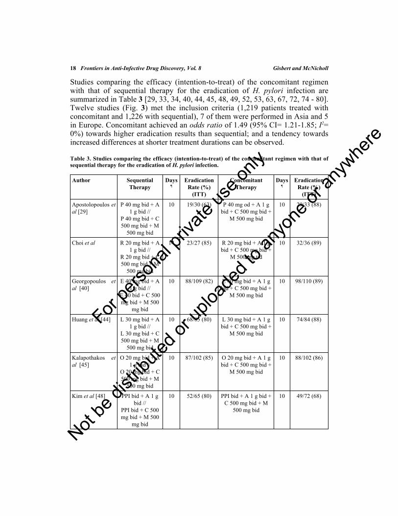
INTRODUCTION
Approximately fifty percent of the world population is infected by Helicobacterpylori, a bacterium linked to a broad range of upper gastrointestinal conditionssuch as gastritis, peptic ulcer disease, and gastric cancer [1]. The most commonlyused therapy for the eradication of H. pylori, traditionally recommended byinternational consensus, is the proton pump inhibitor (PPI)–based, standard tripletherapy, adding two antibiotics (clarithromycin plus amoxicillin or metronidazole)to a PPI [2 - 6]. However, the eradication rates with this regimen have fallenconsiderably [7, 8]. Previous meta-analyses (with more than 53,000 includedpatients) showed an efficacy below 80% [9, 10]. Therefore, recent debate hasbeen raised regarding how ethical it is to continue using standard triple therapy,and alternative approaches have been recommended [11]. Although, efforts toimprove eradication prolonging triple therapy’s duration have been tested, datahave not consistently provided significant benefits [12, 13]. Consequently, newcombinations to improve treatment of naïve patients remain as an urgent need.
Sequential treatment involving a dual regimen with a PPI plus amoxicillin for thefirst 5 days followed by a triple regimen including a PPI, clarithromycin, and anitroimidazole for the following 5 days, was proposed as an alternative [14].Several randomized clinical trials and meta-analyses have shown that thesequential regimen was more effective than the standard triple [15 - 19].Therefore, some consensus conferences suggested sequential regimen as asubstitute to standard triple for the first-line eradication of H. pylori [20].Nevertheless, results obtained by a meta-analysis by the Cochrane Collaboration[21] concluded that sequential regimen outcomes were heterogeneous, and thatmany of the latest manuscripts were unable to show any benefit from sequentialover standard triple therapy. The conclusions of the meta-analysis were clear eventhough the pooled eradication rate was 85%, and a potential trend towardsreduced efficacy was observed in the last years [21].
Sequential treatment faced another relevant issue, whether sequentialadministration was really necessary or if the 4 drugs could be given concurrently[14, 22, 23]. Questions were raised regarding the risk of failure to comply with thetreatment due to regimen complexity [11, 24] Moreover, the combination ofamoxicillin, clarithromycin and a nitroimidazole with a PPI has previously beenevaluated as a concomitant regimen in 1998: two research teams, one in Japan andthe other in Germany, recommended that this drug combination should be
For pe
rsona
l priv
ate us
e only
Not be
distr
ibuted
or up
loade
d to a
nyon
e or a
nywhe
re

Eradication of Helicobacter pylori Frontiers in Anti-Infective Drug Discovery, Vol. 8 3
prescribed as a concomitant 4-drug, 3-antibiotic, known as non–bismuthquadruple therapy [25, 26], providing high efficacy even in short durations (>90%by intention-to-treat in 5-day regimens).
This “non-bismuth quadruple concomitant” regimen has regained presence inrecent years [27]. It is easy to convert the standard triple therapy (PPI-clarithromycin-amoxicillin) to concomitant therapy by adding of 500 mg ofmetronidazole (or tinidazole) twice daily [28]. Beware that “concomitant” (takingall drugs all together) may cause confusion; this term is actually a misnomer, asall H. pylori treatments, except sequential therapy, could be called concomitanttherapies. Nonetheless, this will be the name used hereafter as it has been the mostcommon denomination in the literature.
OBJECTIVE
The aim of the present chapter is to perform a critical review of publishedevidence on the efficacy and safety of concomitant therapy in the eradication ofH. pylori infection. We will review the following aspects: 1) Efficacy of theconcomitant regimen; 2) Comparison between the concomitant regimen andstandard triple therapy; 3) Comparison between the concomitant and thesequential therapies; 4) Effects of different variables on the efficacy ofconcomitant therapy; 5) How could we increase the efficacy of the concomitanttreatment? and finally; 6) What are the results with the concomitant treatment inclinical practice? (the experience of the European registry on H. pylorimanagement).
BIBLIOGRAPHICAL SEARCHES
Bibliographical searches were performed in MEDLINE and ENDBASE using thefollowing keywords (all fields): ((concomitant OR quadruple OR concurrent OR((amoxicillin OR amoxycillin) AND (metronidazole OR tinidazole ORnitroimidazole) AND clarithromycin) AND (“Helicobacter pylori” OR “H.pylori”). No language restriction was applied. Bibliography from selectedmanuscripts and reviews were hand-searched to identify further relevant studies.Authors conducted a hand-search of communications from the AmericanDigestive Disease Week, the International Workshop of the EuropeanHelicobacter Study Group, and the United European Gastroenterology Week.Summaries of the manuscripts selected in the different searches were reviewed,and screened for exclusion and inclusion criteria. In cases of duplicate reportingof studies or evidently based on overlapping study population, the latest validreport was considered.
For pe
rsona
l priv
ate us
e only
Not be
distr
ibuted
or up
loade
d to a
nyon
e or a
nywhe
re

4 Frontiers in Anti-Infective Drug Discovery, Vol. 8 Gisbert and McNicholl
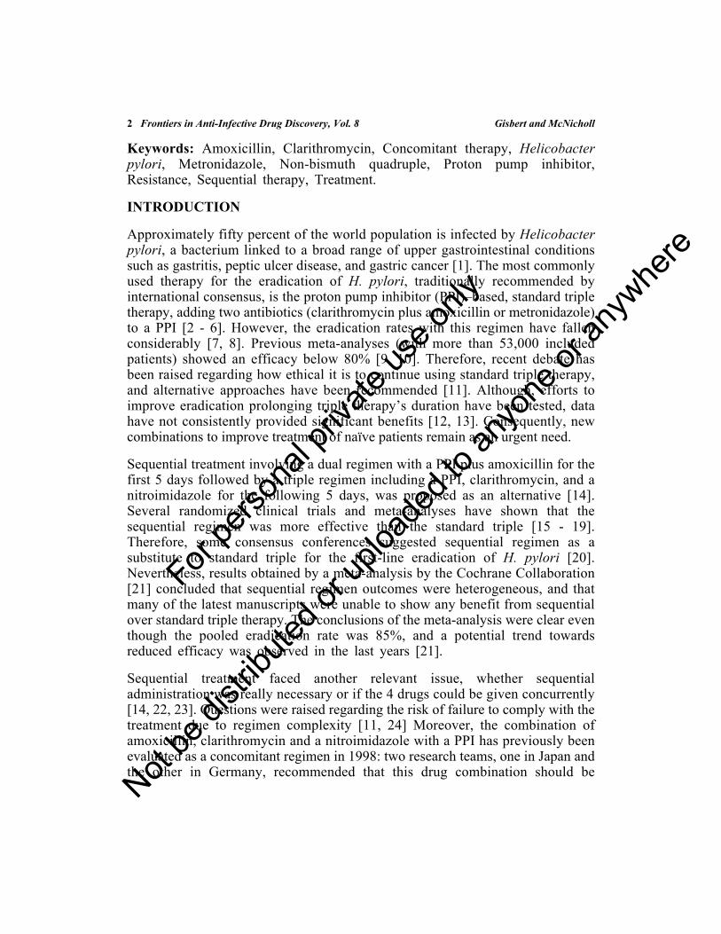
EFFICACY OF THE CONCOMITANT REGIMEN
A summary of studies evaluating concomitant regimen’s efficacy is shown inTable 1 [25, 26, 29 - 80]. Concomitant combinations were prescribedhomogenously, with only minimal alterations: the nitroimidazole (tinidazole ormetronidazole) and the PPI (omeprazole, lansoprazole, rabeprazole, oresomeprazole) and. However, there was a wide duration range between three andfourteen days. The analysis of the 55 studies (6,906 patients) showed a poolederadication percentage by intention-to-treat of 87%, with a 95% confidenceinterval (95% CI) ranging from 86 to 89% (Fig. 1). The data were pooled usingthe generic inverse variance method, which involves a weighted average of theeffect estimates from the included studies. The weight for each study equals onedivided by the square of the standard error (inverse of the variance) of the effectestimate. As population and regimens lengths were heterogeneous, a randomeffects model (DerSimonian and Laird) was applied to perform the meta-analysis(using Review Manager 5.0.25, developed by the Cochrane Collaboration).
Table 1. Studies evaluating the efficacy of non-bismuth quadruple (concomitant) regimen for thetreatment of Helicobacter pylori infection.
Author Country PublicationYear
StudyDesign
DiseaseType
TherapyRegimen
Days¶
No. ofPatients
EradicationRate (%)
(ITT)
EradicationRate (%)
(PP)
Ang et al. [80] Singapore 2015 RCT - O 20 mgbd + A 1g bid + C500 mgbid + M400 mg
bid
10 153 125/153(82)
125/131(95)
Apostolopouloset al. [29]
Greece 2013 RCT - P 40 mgod + A 1g bid + C500 mgbid + M500 mg
bid
10 33 29/33 (88) 29/30 (97)
Calvet et al.[30]
Spain 2000 NC PUD O 20 mgbid + A 1g bid + C500 mgbid + T500 mg
bid
4 56 49/56 (87) 49/54 (91)
For pe
rsona
l priv
ate us
e only
Not be
distr
ibuted
or up
loade
d to a
nyon
e or a
nywhe
re

Eradication of Helicobacter pylori Frontiers in Anti-Infective Drug Discovery, Vol. 8 5
Author Country PublicationYear
StudyDesign
DiseaseType
TherapyRegimen
Days¶
No. ofPatients
EradicationRate (%)
(ITT)
EradicationRate (%)
(PP)
Catalano et al.[31]
Italy 2000 RCT PUD O 40 mgod + A 1g bid + C500 mgbid + M500 mg
bid
3 56 50/56 (89) 50/54 (93)
Chan et al.[32]†
China 2001 NC PUD,NUD,others
O 20 mgbid + A
20 mg/kgtid + C
7.5mg/kgtid + M
7.5mg/kg 5times a
day
7 33 31/33 (94) 31/33 (94)
Choi et al. [34] Korea 2011 NC - R 20 mgbid + A 1g bid + C500 mgbid + M500 mg
tid
14 38 24/38 (63) 24/38 (63)
Choi et al. [79] Korea 2012 RCT - R 20 mgbid + A 1g bid + C500 mgbid + M500 mg
tid
14 36 32/36 (89) 31/35 (89)
De Francescoet al. (a) [36]
Italy 2014 RCT PUD,NUD
O 20 mgbid + A 1g bid + C500 mgbid + T500 mg
bid
5 110 86/110 (78) 86/101 (85)
De Francescoet al. (b) [36]
Italy 2014 RCT PUD,NUD
O 20 mgbid + A 1g bid + C500 mgbid + T500 mg
bid
14 110 95/110 (86) 95/100 (95)
(Table 1) cont.....
For pe
rsona
l priv
ate us
e only
Not be
distr
ibuted
or up
loade
d to a
nyon
e or a
nywhe
re

6 Frontiers in Anti-Infective Drug Discovery, Vol. 8 Gisbert and McNicholl
Author Country PublicationYear
StudyDesign
DiseaseType
TherapyRegimen
Days¶
No. ofPatients
EradicationRate (%)
(ITT)
EradicationRate (%)
(PP)
Georgopouloset al. [37]
Greece 2011 NC PUD,NUD
E 40 mgbid + A 1g bid + C500 mgbid + M500 mg
bid
10 131 120/131(92)
120/127(94)
Georgopouloset al. (a) [38]
Greece 2013 NC PUD,NUD
E 40 mgbid + A 1g bid + C500 mgbid + M500 mg
bid
10 165 151/165(91)
151/159(95)
Georgopouloset al. (b) [39]
Greece 2013 RCT PUD,NUD
E 40 mgbid + A 1g bid + C500 mgbid + M500 mg
bid
10 127 115/127(90)
111/119(93)
Georgopouloset al. [40]
Greece 2014 RCT - E 40 mgbid + A 1g bid + C500 mgbid + M500 mg
bid
10 110 98/110 (89) 98/105 (93)
Greenberg etal. [41]
LatinAmerica
2011 RCT PUD,NUD,others
L 30 mgbid + A 1g bid + C500 mgbid + M500 mg
bid
5 488 360/489(74)
348/442(79)
Heo et al. (a)[77]
Korea 2014 RCT - L 30 mgbid + A 1g bid + C500 mgbid + M500 mg
bid
10 169 137/174(79)
133/150(89)
Heo et al. (b)[43]
Korea 2014 RCT - E 20 mgbid + A 1g bid + C500 mgbid + M500 mg
bid
10 238 187/238(79)
176/196(90)
(Table 1) cont.....
For pe
rsona
l priv
ate us
e only
Not be
distr
ibuted
or up
loade
d to a
nyon
e or a
nywhe
re

Eradication of Helicobacter pylori Frontiers in Anti-Infective Drug Discovery, Vol. 8 7
Author Country PublicationYear
StudyDesign
DiseaseType
TherapyRegimen
Days¶
No. ofPatients
EradicationRate (%)
(ITT)
EradicationRate (%)
(PP)
Hsu et al. [76] Taiwan 2014 RCT P 40 mgbid + A 1g bid + C500 mgbid + M500 mg
bid
7 102 96/102 (94) 96/102 (94)
Huang et al.[44]
Taiwan 2012 RCT PUD,NUD
L 30 mgbid + A 1g bid + C500 mgbid + M500 mg
bid
10 84 74/84 (88) 70/74 (95)
Kalapothakoset al. [45]
Greece 2013 RCT - O 20 mgbid + A 1g bid + C500 mgbid + M500 mg
bid
10 95 88/102 (86) 88/95 (93)
Kao et al. [46] Taiwan 2012 NC PUD,NUD
P 40 mgbid + A 1g bid + C500 mgbid + M500 mg
bid
7 319 299/319(94)
297/308(96)
Kim et al. [47] Korea 2013 RCT PUD,NUD
L 30 mgbid + A 1g bid + C500 mgbid + M500 mg
bid
5 135 109/135(81)
106/116(91)
Kim et al. (a)[48]
Korea 2014 RCT - PPI 20mg bid +A 1 g bid+ C 500mg bid +M 500mg bid
10 65 52/65 (80) 50/52 (96)
Kim et al. (b)[78]
Korea 2014 NC - L 30 mgbid + A 1g bid + C500 mgbid + M500 mg
bid
10 68 118/125(94)
108/116(93)
(Table 1) cont.....
For pe
rsona
l priv
ate us
e only
Not be
distr
ibuted
or up
loade
d to a
nyon
e or a
nywhe
re

8 Frontiers in Anti-Infective Drug Discovery, Vol. 8 Gisbert and McNicholl
Author Country PublicationYear
StudyDesign
DiseaseType
TherapyRegimen
Days¶
No. ofPatients
EradicationRate (%)
(ITT)
EradicationRate (%)
(PP)
Kongchayanunet al. (a) [50]
Thailand 2012 RCT NUD R 20 mgbid + A 1g bid +
C¥ 1 g od+ M 500mg tid
5 55 49/55 (89) 49/55 (89)
Kongchayanunet al. (b) [50]
Thailand 2012 RCT NUD R 20 mgbid + A 1g bid +
C¥ 1 g od+ M 500mg tid
10 55 53/55 (96) 53/55 (96)
Kwon et al. (a)[51]
Korea 2011 RCT - L 30 mgbid + A 1g bid + C500 mgbid + M500 mg
bid
5 48 42/48 (87) 42/48 (87)
Kwon et al. (b)[51]
Korea 2011 RCT - L 30 mgbid + A 1g bid + C500 mgbid + M500 mg
bid
7 49 44/49 (90) 44/49 (90)
Lee et al. [75] Korea 2015 RCT PUD-NUD R 20 mgbid + A 1g bid + C500 mgbid + M500 mg
bid
7 170 135/170(79)
135/143(94)
Lim et al. [52] Korea 2013 RCT PUD,NUD
R 20 mgbid + A 1g bid + C500 mgbid + M500 mg
bid
14 78 63/78 (81) 61/75 (81)
McNicholl etal. (a) [53]
Spain 2014 RCT PUD,NUD
O 20 mgbid + A 1g bid + C500 mgbid + M500 mg
bid
10 168 146/168(87)
125/137(91)
(Table 1) cont.....
For pe
rsona
l priv
ate us
e only
Not be
distr
ibuted
or up
loade
d to a
nyon
e or a
nywhe
re
Eradication of Helicobacter pylori Frontiers in Anti-Infective Drug Discovery, Vol. 8 9
Author Country PublicationYear
StudyDesign
DiseaseType
TherapyRegimen
Days¶
No. ofPatients
EradicationRate (%)
(ITT)
EradicationRate (%)
(PP)
McNicholl etal. (b1) [54]
Spain 2014 NC PUD,NUD
E 40 mgbid + A 1g bid + C500 mgbid + M500 mg
bid
14 471 427/471(91)
401/432(93)
McNicholl etal. (b2) [54]
Spain 2014 NC PUD,NUD
O 20 mgbid + A 1g bid + C500 mgbid + M500 mg
bid
10 356 305/356(86)
282/329(86)
Molina-Infanteet al. [55]
Spain 2012 NC PUD,NUD
PPI bid +A 1 g bid+ C 500mg bid +M 500mg bid
10 182 182/209(87)
180/203(89)
Molina-Infanteet al. [56]
Spain 2013 RCT PUD,NUD
O 40 mgbid + A 1g bid + C500 mgbid + M500 mg
bid
14 170 156/170(92)
150/156(96)
Molina-Infante[57]
Spain 2014 NC PUD,NUD
E 40 mgbid + A 1g bid + C500 mgbid + M500 mg
bid
14 298 272/298(91)
272/290(94)
Moon et al.[58]
Korea 2011 RCT - PPI bid +A 1 g bid+ C 500mg bid +M 500mg bid
7 53 43/53 (81) 43/53 (81)
Moon et al.[59]
Korea 2014 NC - PPI bid +A 500
mg tid +C 500
mg bid +M 500mg tid
7 106 81/106 (76) 81/101 (80)
(Table 1) cont.....
For pe
rsona
l priv
ate us
e only
Not be
distr
ibuted
or up
loade
d to a
nyon
e or a
nywhe
re

10 Frontiers in Anti-Infective Drug Discovery, Vol. 8 Gisbert and McNicholl
Author Country PublicationYear
StudyDesign
DiseaseType
TherapyRegimen
Days¶
No. ofPatients
EradicationRate (%)
(ITT)
EradicationRate (%)
(PP)
Nagahara et al.[60]
Japan 2000 RCT PUD,NUD
R 10 mgbid + A750 mgbid + C200 mgbid + M250 mg
bid
5 55 52/55 (94) 52/53 (98)
Nagahara et al.[61]
Japan 2001 RCT PUD,NUD
R 20 mgbid + A750 mgbid + C200 mgbid + M250 mg
bid
5 80 74/80 (92) 74/79 (94)
Neville et al.[62]
UK 1999 RCT PUD,NUD,others
L 30 mgbid + A 1g bid + C250 mgbid + M400 mg
bid
5 56 49/56 (87) 49/54 (91)
Ntouli et al.[63]
Greece 2014 RCT - PPI bid +A 500
mg tid +C 500
mg bid +T 500mg bid
10 108 98/108 (91) 98/108 (91)
Okada et al.[26]
Japan 1998 RCT PUD,NUD,others
O 20 mgbid + A500 mgtid + R150 mgbid + M250 mg
tid
7 90 85/90 (94) 85/88 (97)
Okada et al.[64]
Japan 1999 RCT PUD,NUD,others
O 20 mgbid + A500 mgtid + R150 mgbid + M250 mg
tid
7 169 155/169(92)
155/163(95)
Seo et al. [65] Korea 2014 NC - R - + A -+ C - +
M -
7 210 194/210(92)
-
(Table 1) cont.....
For pe
rsona
l priv
ate us
e only
Not be
distr
ibuted
or up
loade
d to a
nyon
e or a
nywhe
re

Eradication of Helicobacter pylori Frontiers in Anti-Infective Drug Discovery, Vol. 8 11
Author Country PublicationYear
StudyDesign
DiseaseType
TherapyRegimen
Days¶
No. ofPatients
EradicationRate (%)
(ITT)
EradicationRate (%)
(PP)
Sharara et al(a)[66]
Lebanon 2014 RCT PUD,NUD
R 20 mgbid + A 1g bid + C500 mgbid + M500 mg
bid
7 100 78/100 (78) 78/95 (82)
Sharara et al(b)[66]
Lebanon 2014 RCT PUD,NUD
R 20 mgod + A 1g od + C500 mgod + M500 mg
od
7 100 78/100 (78) 78/93 (84)
Tepes [68] Slovenia 2014 RCT - E 20 mgbid + A 1g bid + C500 mgbid + M500 mg
bid
7 120 110/120(92)
-
Toros et al.[69]
Turkey 2011 NC NUD L 30 mgbid + A 1g bid + C500 mgbid + M500 mg
tid
14 84 63/84 (75) 63/84 (75)
Treiber et al (a)[70]
Germany 2002 RCT PUD,NUD,others
L 30 mgbid + A 1g bid + C250 mgbid + M400 mg
bid
3 80 65/80 (81) 65/76 (85)
Treiber et al(b) [70]
Germany 2002 RCT PUD,NUD,others
L 30 mgbid + A 1g bid + C250 mgbid + M400 mg
bid
5 83 74/83 (89) 74/79 (94)
Treiber et al.[25]
Germany 1998 RCT PUD,others
O 20 mgbid + A 1g bid + C250 mgbid + M400 mg
bid
5 46 42/46 (91) 42/44 (95)
(Table 1) cont.....
For pe
rsona
l priv
ate us
e only
Not be
distr
ibuted
or up
loade
d to a
nyon
e or a
nywhe
re

12 Frontiers in Anti-Infective Drug Discovery, Vol. 8 Gisbert and McNicholl
Author Country PublicationYear
StudyDesign
DiseaseType
TherapyRegimen
Days¶
No. ofPatients
EradicationRate (%)
(ITT)
EradicationRate (%)
(PP)
Wang et al.[71]
China 2014 RCT - E 20 mgbid + A 1g bid + C250 mgbid + T500 mg
bid
7 81 74/81 (91) 74/80 (92)
Wu et al. [72] Taiwan 2010 RCT PUD,NUD,others
E 40 mgbid + A 1g bid + C500 mgbid + M500 mg
bid
10 115 107/115(93)
107/115(93)
Yanai et al.[73]
Japan 2012 RCT PUD,NUD
L 30 mgbid + A750 mgbid + C200 mgbid + M250 mg
bid
7 59 56/59 (95) 56/57 (98)
Zullo et al. [74] Italy 2013 RCT NUD O 20 mgbid + A 1g bid + C500 mgbid + M500 mg
bid
5 90 77/90 (86) 77/84 (92)
ITT, intention-to-treat; PP, per-protocol.RCT, randomized controlled trial. NC, non-controlled.PUD, peptic ulcer disease; NUD, non-ulcer disease.PPI, proton pump inhibitor (at standard dose); O, omeprazole; L, lansoprazole; R, rabeprazole; E,esomeprazole; A: amoxicillin; C, clarithromycin; M, metronidazole; T, tinidazole; R, roxithromycin.od, once daily; bid: two times a day; tid: three times a day¶Days of antibiotic treatment; †Pediatric patients; C¥Sustained release clarithromycin.
(Table 1) cont.....
For pe
rsona
l priv
ate us
e only
Not be
distr
ibuted
or up
loade
d to a
nyon
e or a
nywhe
re

Eradication of Helicobacter pylori Frontiers in Anti-Infective Drug Discovery, Vol. 8 13
Fig. (1). Meta-analysis of efficacy (intention-to-treat) of studies evaluating the concomitant regimen for thetreatment of H. pylori infection.
For pe
rsona
l priv
ate us
e only
Not be
distr
ibuted
or up
loade
d to a
nyon
e or a
nywhe
re

14 Frontiers in Anti-Infective Drug Discovery, Vol. 8 Gisbert and McNicholl
COMPARISON BETWEEN CONCOMITANT AND STANDARD TRIPLEREGIMEN
The superiority of concomitant therapy over standard triple therapy has beenconfirmed in several randomized trials. A recent meta-analysis [81] evaluated 9prospective studies treating H. pylori with a concomitant regimen for up to 7 days.Prescribed regimens generally lasted 5 days (ranging from 4 in one study to 7 inanother). Overall, concomitant therapy achieved 90% intention-to-treateradication (93% per-protocol). Moreover, the estimates of the meta-analysis ofthe 5 randomized controlled trials demonstrated the superiority of concomitantregimen over standard triple therapy (odds ratio of 2.86; 95% CI, 1.73-4.73).
For this chapter, we have updated these meta-analytical evaluations with morerecent studies and have updated it including the new trials that have comparedthese two treatments. Table 2 describes the studies comparing the H. pylorieradication rate of concomitant regimen with that of standard triple therapy byintention-to-treat [25, 31, 39, 41, 42, 47, 48, 58, 60 - 62, 67, 68, 71, 73].
Table 2. Studies comparing the efficacy (intention-to-treat) of the concomitant regimen with that ofstandard triple therapy for the eradication of H. pylori infection.
Author Standard TripleTherapy
Days¶
EradicationRate (%)
(ITT)
Concomitant Therapy Days¶
EradicationRate (%)
(ITT)
Catalano et al[31]
O 40 mg od + A1 g bid + C 500
mg bid
10 45/55 (82) O 40 mg od + A 1 g bid+ C 500 mg bid + M
500 mg bid
3 50/56 (89)
Georgopoulos etal [39]
E 40 mg bid + A1 g bid + C 500
mg bid
10 96/130 (74) E 40 mg bid + A 1 gbid + C 500 mg bid +
M 500 mg bid
10 115/127 (90)
Greenberg et al[41]
L 30 mg bid + A1 g bid + C 500
mg bid
14 401/488 (82) L 30 mg bid + A 1 gbid + C 500 mg bid +
M 500 mg bid
5 360/489 (74)
Hsu et al P 40 mg bid + A1 g bid + C 500
mg bid
7 84/102 (82) P 40 mg bid + A 1 gbid + C 500 mg bid +
M 500 mg bid
7 96/108 (94)
Heo et al L 30 mg bid + A1 g bid + C 500
mg bid
10 123/174 (71) L 30 mg bid + A 1 gbid + C 500 mg bid +
M 500 mg bid
10 137/174 (79)
Kim et al [47] L 30 mg bid + A1 g bid + C 500
mg bid
7 98/135 (72) L 30 mg bid + A 1 gbid + C 500 mg bid +
M 500 mg bid
5 109/135 (81)
For pe
rsona
l priv
ate us
e only
Not be
distr
ibuted
or up
loade
d to a
nyon
e or a
nywhe
re

Eradication of Helicobacter pylori Frontiers in Anti-Infective Drug Discovery, Vol. 8 15
Author Standard TripleTherapy
Days¶
EradicationRate (%)
(ITT)
Concomitant Therapy Days¶
EradicationRate (%)
(ITT)
Kim et al [48] PPI 20 mg bid +A 1 g bid + C500 mg bid
10 47/79 (59) PPI 20 mg bid + A 1 gbid + C 500 mg bid +
M 500 mg bid
10 52/65 (80)
Lee et al L 30 mg bid + A1 g bid + C 500
mg bid
7 109/170 (64) L 30 mg bid + A 1 gbid + C 500 mg bid +
M 500 mg bid
7 135/170 (79)
L 30 mg bid + A1 g bid + M 500
mg bid
7 117/170 (69)
Moon et al [58] PPI bid + A 500mg tid + C 500
mg bid
7 55/85 (65) PPI bid + A 500 mg tid+ C 500 mg bid + M
500 mg bid
7 43/53 (81)
Nagahara et al[60]
R 10 mg bid + A750 mg bid + C
200 mg bid
5 40/50 (80) R 10 mg bid + A 750mg bid + C 200 mg bid
+ M 250 mg bid
5 52/55 (94)
Nagahara et al[61]
R 20 mg bid + A750 mg bid + C
200 mg bid
7 65/80 (81) R 20 mg bid + A 750mg bid + C 200 mg bid
+ M 250 mg bid
5 74/80 (92)
Neville et al[62]
L 30 mg bid + A1 g bid + C 250
mg bid
5 33/56 (59) L 30 mg bid + A 1 gbid + C 250 mg bid +
M 400 mg bid
5 49/56 (87)
Ang et al [67] O 20mg bid + A1 g bid + C 500
mg bid
10 129/155 (83) O 20mg bid + A 500mg bid + C 500 mg bid
+ M 400 mg bid
10 125/153 (82)
Tepes [68] E 20 mg bid + A1 g bid + C 500
mg bid
7 97/116 (77) E 20 mg bid + A 1 gbid + C 500 mg bid +
M 500 mg bid
7 110/120 (92)
Treiber et al[25]
O 20 mg bid + C250 mg bid + M
400 mg bid
7 38/42 (90) O 20 mg bid + A 1 gbid + C 250 mg bid +
M 400 mg bid
5 42/46 (91)
Wang et al [71] E 20 mg bid + A1 g bid + C 250
mg bid
7 65/82 (79) E 20 mg bid + A 1 gbid + C 250 mg bid + T
500 mg bid
7 74/81 (91)
Yanai et al [73] L 30 mg bid + A750 mg bid + C
200 mg bid
7 41/60 (68) L 30 mg bid + A 750mg bid + C 200 mg bid
+ M 250 mg bid
7 56/59 (95)
PPI, proton pump inhibitor (at standard dose); O, omeprazole; L, lansoprazole; R, rabeprazole; E,esomeprazole; A: amoxicillin; C, clarithromycin; M, metronidazole; T, tinidazole.od, once daily; bid: two times a day; tid: three times a day¶Days of antibiotic treatment.
(Table 2) cont.....
For pe
rsona
l priv
ate us
e only
Not be
distr
ibuted
or up
loade
d to a
nyon
e or a
nywhe
re

16 Frontiers in Anti-Infective Drug Discovery, Vol. 8 Gisbert and McNicholl
As summarized in Fig. (2), 2, 059 patients received the concomitant regimen and2,268 the standard triple regimen. The former was more effective than the latter:81% vs. 74% in the intention-to-treat analysis. The odds ratio for this comparisonwas 2.14 (95% CI, 1.51-3.04) (Fig. 2A). A sub-analysis was performed excludingthose studies in which both treatments had different treatment durations betweenarms. This sub-analysis showed (Fig. 2B) that, when comparing both concomitantand triple therapies lasting the same number of days, concomitant achieved anodds ratio of 2.41 (95% CI= 1.80-3.24; 85% vs. 72%). If we subdivide by lengthof both arms the differences in efficacy between both treatments were, asexpected, smaller at longer regimens due to the rapid decrease in the efficacy ofstandard triple therapy at shorter regimens (Fig. 2B).
Regarding tolerance, in a previously published meta-analysis [81], no severe sideeffects were reported in any of the manuscripts, except anaphylactic reactions tostudy drugs [26, 64, 70]. However, these antibiotic treatments do show a high rateof moderate or mild adverse events, in Essa et al. meta-analysis 27-51% ofpatients in the concomitant group suffered some discomfort with treatment (vs.21-48% standard triple therapy group) [81], suggesting that a similar safetyprofile can be expected from concomitant and standard triple therapies.
COMPARISON BETWEEN CONCOMITANT AND SEQUENTIALREGIMENS
As previously discussed, sequential therapy faces a limitation due to itscomplexity, as switching drugs in the middle of treatment may compromisecompliance. In this situation, studies comparing sequential regiment with anotherregimen using the same combination of drugs but concomitantly were necessary.Such comparisons would determine whether the two phase administration ofsequential regiment is actually helpful [24]. A one-to-one comparison ofconcomitant and sequential therapies would also answer which of these 2candidates can eventually substitute triple therapies in first-line recommendations[82].
For pe
rsona
l priv
ate us
e only
Not be
distr
ibuted
or up
loade
d to a
nyon
e or a
nywhe
re

Eradication of Helicobacter pylori Frontiers in Anti-Infective Drug Discovery, Vol. 8 17
Fig. (2). Meta-analysis comparing the efficacy (intention-to-treat) of the concomitant regimen with that ofstandard triple therapy for the eradication of H. pylori infection: A) overall; B) both arms with the samelength of treatment.
A)
B)
For pe
rsona
l priv
ate us
e only
Not be
distr
ibuted
or up
loade
d to a
nyon
e or a
nywhe
re
18 Frontiers in Anti-Infective Drug Discovery, Vol. 8 Gisbert and McNicholl
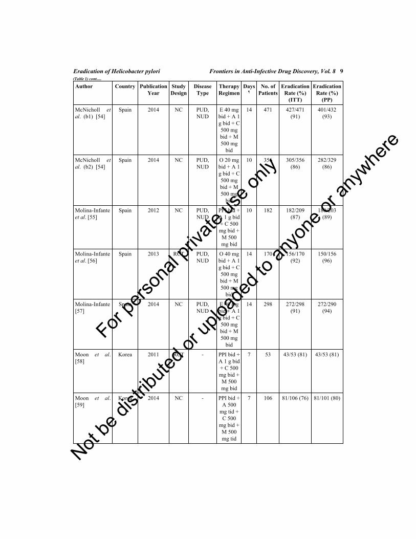
Studies comparing the efficacy (intention-to-treat) of the concomitant regimenwith that of sequential therapy for the eradication of H. pylori infection aresummarized in Table 3 [29, 33, 34, 40, 44, 45, 48, 49, 52, 53, 63, 67, 72, 74 - 80].Twelve studies (Fig. 3) met the inclusion criteria (1,219 patients treated withconcomitant and 1,226 with sequential), 7 of them were performed in Asia and 5in Europe. Concomitant achieved an odds ratio of 1.49 (95% CI= 1.21-1.85; I2=0%) towards higher eradication results than sequential; and a tendency towardsincreased differences at shorter treatment durations can be observed.
Table 3. Studies comparing the efficacy (intention-to-treat) of the concomitant regimen with that ofsequential therapy for the eradication of H. pylori infection.
Author SequentialTherapy
Days¶
EradicationRate (%)
(ITT)
ConcomitantTherapy
Days¶
EradicationRate (%)
(ITT)
Apostolopoulos etal [29]
P 40 mg bid + A1 g bid //
P 40 mg bid + C500 mg bid + M
500 mg bid
10 19/30 (63) P 40 mg od + A 1 gbid + C 500 mg bid +
M 500 mg bid
10 29/33 (88)
Choi et al R 20 mg bid + A1 g bid //
R 20 mg bid + C500 mg bid + M
500 mg bid
14 23/27 (85) R 20 mg bid + A 1 gbid + C 500 mg bid +
M 500 mg tid
10 32/36 (89)
Georgopoulos etal [40]
E 40 mg bid + A1 g bid //
E 40 bid + C 500mg bid + M 500
mg bid
10 88/109 (82) E 40 mg bid + A 1 gbid + C 500 mg bid +
M 500 mg bid
10 98/110 (89)
Huang et al [44] L 30 mg bid + A1 g bid //
L 30 mg bid + C500 mg bid + M
500 mg bid
10 68/85 (80) L 30 mg bid + A 1 gbid + C 500 mg bid +
M 500 mg bid
10 74/84 (88)
Kalapothakos etal [45]
O 20 mg bid + A1 g bid //
O 20 mg bid + C500 mg bid + M
500 mg bid
10 87/102 (85) O 20 mg bid + A 1 gbid + C 500 mg bid +
M 500 mg bid
10 88/102 (86)
Kim et al [48] PPI bid + A 1 gbid //
PPI bid + C 500mg bid + M 500
mg bid
10 52/65 (80) PPI bid + A 1 g bid +C 500 mg bid + M
500 mg bid
10 49/72 (68)
For pe
rsona
l priv
ate us
e only
Not be
distr
ibuted
or up
loade
d to a
nyon
e or a
nywhe
re

Eradication of Helicobacter pylori Frontiers in Anti-Infective Drug Discovery, Vol. 8 19
Author SequentialTherapy
Days¶
EradicationRate (%)
(ITT)
ConcomitantTherapy
Days¶
EradicationRate (%)
(ITT)
Lim et al [52] R 20 mg bid + A1 g bid //
R 20 mg bid + C500 mg bid + M
500 mg bid
14 65/86 (76) R 20 mg bid + A 1 gbid + C 500 mg bid +
M 500 mg bid
14 63/78 (81)
McNicholl et al(a)[53]
O 20 mg bid + A1 g bid //
O 20 mg bid + C500 mg bid + M
500 mg bid
10 138/170 (81) O 20 mg bid + A 1 gbid + C 500 mg bid +
M 500 mg bid
10 146/168 (87)
Ntouli et al [63] PPI bid + A 1 gbid //
PPI bid + C 500mg bid + M 500
mg bid
10 87/104 (84) PPI bid + A 500 mgtid + C 500 mg bid +
T 500 mg bid
10 98/108 (91)
Ang et al [67] O 20 mg bid + A1 g bid //
O 20 mg bid + C500 mg bid + M
400 mg bid
10 119/135 (88) PPI bid + A 1 g bid +C 500 mg bid + M
400 mg bid
10 125/153 (82)
Wu et al [72] O 20 mg bid + A1 g bid //
O 20 mg bid + C500 mg bid + M
500 mg bid
10 130/154 (84) E 40 mg bid + A 1 gbid + C 500 mg bid +
M 500 mg bid
10 107/115 (93)
PPI, proton pump inhibitor (at standard dose); O, omeprazole; L, lansoprazole; R, rabeprazole; E,esomeprazole; A: amoxicillin; C, clarithromycin; M, metronidazole; T, tinidazole.od, once daily; bid: two times a day; tid: three times a day¶Days of antibiotic treatment.
(Table 3) cont.....
For pe
rsona
l priv
ate us
e only
Not be
distr
ibuted
or up
loade
d to a
nyon
e or a
nywhe
re

20 Frontiers in Anti-Infective Drug Discovery, Vol. 8 Gisbert and McNicholl
Fig. (3). Meta-analysis comparing the efficacy (intention-to-treat) of the concomitant regimen with that ofsequential therapy for the eradication of H. pylori infection.
The alleged superiority of concomitant therapy –compared to sequential therapy –may be due to the longer period of time each antibiotic is prescribed (10 days inthe concomitant and 5 in the sequential treatments), or the expected lower impactof antibiotic resistance when the 3 antibiotics are administered concurrently,favoring synergistic effects.
EFFECTS OF DIFFERENT VARIABLES ON THE EFFICACY OFCONCOMITANT THERAPY
The eradication rate achieved with the concomitant treatment is affected bymultiple factors.
Clarithromycin Resistance
Antimicrobial resistance is largely responsible for the poor eradication rates withstandard triple therapy [83 - 85]. One meta-analysis identified an approximate60% reduction in efficacy with standard triple therapy if strains were
For pe
rsona
l priv
ate us
e only
Not be
distr
ibuted
or up
loade
d to a
nyon
e or a
nywhe
re

Eradication of Helicobacter pylori Frontiers in Anti-Infective Drug Discovery, Vol. 8 21
clarithromycin resistant [83, 86]. Taking this in consideration, the prescription ofstandard triple therapies may be acceptable only in regions where the rate ofresistance to clarithromycin is below 15-20% [2]. Clarithromycin resistanceimpacts the success rate of sequential regimen, although not as markedly as intriple therapy [14, 16 - 19, 87 - 89].
The potential benefits of sequential therapy regarding resistance seem to be alsoapplicable to concomitant regimen. The effect of H. pylori antibiotic resistance onthe efficacy of first-line treatments was determined in an early meta-analysis. Thedata clearly identified that quadruple combinations (especially those containingboth clarithromycin and metronidazole) could cure clarithromycin ormetronidazole resistance [85]. That meta-analysis showed that clarithromycinresistance’s effect on concomitant regimens was minor, with 95% cure-rate inpatients with clarithromycin-sensitive strains, and 96% in resistant [85].
Table 4 summarizes the studies that, up to now, have evaluated the efficacy ofclarithromycin resistance on the concomitant regimen [38, 44, 55, 56, 62, 64, 70,72]. From these data, a mean eradication rate of 83% was calculated in patientswith clarithromycin resistant strains. If only studies including in the same protocolboth non-bismuth quadruple regimens were considered [44, 55, 72], theeradication rate in patients with clarithromycin-resistant strains was 62% for thesequential treatment and 92% for the concomitant one.
Table 4. Studies evaluating the efficacy of clarithromycin resistance (in patients with metronidazole-susceptible strains) on the concomitant regimen.
Author Sequentialn/N (%)
Concomitantn/N (%)
Treatment Duration (days)
Huang et al [44] 3/5 (60%) 3/3 (100%) 10
Georgopoulos et al [38] - 13/15 (85%) 10
Molina-Infante et al [55] 3/4 (75%) 5/5 (100%) 10
Molina-Infante et al [56] - 5/5 (100%) 14
Neville et al [62] - 17/20 (85%) 5
Okada et al [64] - 25/27 (93%) 7
Treiber et al [70] - 8/16 (50%) 5
Wu et al [72] 4/7 (57%) 3/4 (75%) 10
n - patients cured, N – patients evaluated, % efficacy by ITT.
Nitroimidazole Resistance
Experience with concomitant therapy in patients with metronidazole-resistant
For pe
rsona
l priv
ate us
e only
Not be
distr
ibuted
or up
loade
d to a
nyon
e or a
nywhe
re

22 Frontiers in Anti-Infective Drug Discovery, Vol. 8 Gisbert and McNicholl
strains is still limited, but seems to be encouraging. Table 5 summarizes thestudies that, up to now, have evaluated the efficacy of metronidazole resistance onthe concomitant regimen [40, 44, 55, 56, 72]. From these data, a mean eradicationrate of 98% was calculated in patients with nitroimidazole resistant strains. If onlystudies including in the same protocol both non-bismuth quadruple regimens wereconsidered [40, 44, 72], the eradication rate in patients with metronidazole-resistant strains was 82% for the sequential treatment and 97% for theconcomitant one.
Table 5. Studies evaluating the efficacy of metronidazole resistance (in patients with clarithromycin-susceptible strains) on the concomitant regimen.
Author Sequentialn/N (%)
Concomitantn/N (%)
Treatment Duration (days)
Georgopoulos et al [38] - 25/25 (100%) 10
Georgopoulos et al [40] 21/28 (75%) 21/21 (100%) 10
Huang et al [44] 14/18 (78%) 16/16 (100%) 10
Molina-Infante et al [55] - 8/8 (100%) 10
Molina-Infante et al [56] - 8/8 (100%) 14
Wu et al [72] 27/30 (90%) 24/26 (92%) 10
n - patients cured, N – patients evaluated, % efficacy by ITT.
Dual Clarithromycin and Metronidazole Resistance
Table 6 summarizes the studies that, up to now, have evaluated the efficacy ofboth clarithromycin and metronidazole resistance on the concomitant regimen[38, 40, 44, 55, 56, 64, 70, 72]. From these data, a mean eradication rate of 79%was calculated in patients with dual clarithromycin and nitroimidazole resistantstrains. If only studies including in the same protocol both non-bismuth quadrupleregimens were considered [40, 44, 55, 72], the eradication rate in patients withboth clarithromycin and metronidazole-resistant strains was 47% for thesequential treatment and 79% for the concomitant one. Therefore, sequentialregimen does not qualify as a good therapeutic when the prevalence of dual–clarithromycin plus metronidazole– resistant strains is greater than 5% [90]. Thelower effect of antibiotic resistance on the eradication rate with concomitanttherapy than with sequential therapy may be due to the longer duration of therapywith one or all of the components of the concomitant therapy.
Based on aforementioned data, it may be concluded that the main limitation ofconcomitant therapy is this dual resistance to metronidazole–clarithromycin [90].Although isolated clarithromycin or metronidazole resistances by themselves did
For pe
rsona
l priv
ate us
e only
Not be
distr
ibuted
or up
loade
d to a
nyon
e or a
nywhe
re

Eradication of Helicobacter pylori Frontiers in Anti-Infective Drug Discovery, Vol. 8 23
not significantly impair concomitant therapy, it is expected that dual resistancerates over 15% may cause the efficacy to fall below 90% [91]. Therefore,concomitant therapy may be the recommended therapy of choice in areas andpatient groups with low risk of dual resistance, but it cannot be recommended as afirst-line empirical regimen in populations expected to have high dual resistancerates (i.e. after clarithromycin or metronidazole treatment failures) or in situationswhen metronidazole resistance reaches 60%, such as China, the Islamic Republicof Iran, India, or Central and South America [91]. As expected from theserecommendations, good/excellent reported outcomes have been published inSouthern Europe and some Asian countries with relatively low metronidazoleresistance regardless of the clarithromycin resistance rate (even at 40% resistanceprevalence); and poor results were obtained in areas with high prevalence ofclarithromycin or, specially, metronidazole resistances.
Table 6. Studies evaluating the efficacy of dual clarithromycin and metronidazole resistance on theconcomitant regimen.
Author Sequentialn/N (%)
Concomitantn/N (%)
Treatment Duration (days)
Georgopoulos et al [40] 2/5 (40%) 7/9 (78%) 10
Georgopoulos et al [38] - 7/10 (70%) 10
Huang et al [44] 2/4 (50%) 2/2 (100%) 10
Molina-Infante et al [55] 3/5 (60%) 3/4 (75%) 10
Molina-Infante et al [56] - 3/3 (100%) 14
Okada et al [64] - 3/4 (75%) 7
Treiber et al [70] - 2/2 (50%) 5
Wu et al [72] 1/3 (33%) 3/4 (75%) 10
n - patients cured, N – patients evaluated, % efficacy by ITT.
In summary, there is clear and abundant evidence pointing towards the superiorityof concomitant over sequential therapy, even more in populations expected tohave moderate to high dual resistances (>15%).
Duration of Treatment
The first meta-analysis including 9 manuscripts, Essa et al [81] identified thatconcomitant regimen achieved excellent results, even in very short treatmentdurations. The results of the studies included in Table 1 and in Fig. (1) have notbeen able to show a clear higher eradication results with longer treatments.However, several randomized controlled trials have compared, in the same studyand with the same protocol, two different durations of the concomitant therapy,
For pe
rsona
l priv
ate us
e only
Not be
distr
ibuted
or up
loade
d to a
nyon
e or a
nywhe
re

24 Frontiers in Anti-Infective Drug Discovery, Vol. 8 Gisbert and McNicholl
and have demonstrated that the longer duration is more effective, when comparing3 vs. 5 days (81% vs. 89% [70], 5 vs. 7 days (87% vs. 90%) [51], 5 vs. 10 days(89% vs. 96%) [50], or 5 vs. 14 days (78% vs. 86%) [36].
HOW COULD WE EVEN INCREASE THE EFFICACY OF THECONCOMITANT TREATMENT?
It has been pointed out that we should look for “good” or even “excellent”treatments, with eradication rates higher than 90% [93]. A recent study hascompared the efficacy and tolerability of the standard and the so called“optimized” concomitant regimen (using new generation PPIs at high doses andlonger treatment duration) [54]. Thus, in a prospective multicenter study, H.pylori-infected patients were consecutively treated with one of these twotreatments: in a first phase, 356 patients received a standard concomitant therapywith omeprazole 20 mg, amoxicillin 1 g, clarithromycin 500 mg andmetronidazole 500 mg for 10 days b.i.d.; in a second phase, 471 patients receivedthe same regimen but with esomeprazole 40 mg b.i.d. and lasting 14 days.Compliance with treatment was 94% and 95%, respectively (non-statisticallysignificant differences). Per-protocol eradication rates with standard concomitantand the optimized concomitant treatments were 86% and 93% (p<0.01).Respective intention-to-treat cure rates were 86% and 91% (p<0.01). Adverseeffects (mostly mild) were reported in 32% of patients in the standardconcomitant group and in 44% in optimized one (p<0.05), the most commonbeing metallic taste, diarrhea, nausea and abdominal pain. Therefore, the authorsconcluded that an optimized (fourteen-day and high-dose esomeprazole) non-bismuth quadruple concomitant regimen for the eradication of H. pylori is moreeffective than the standard concomitant one, and achieves over 90% cure rate (andalso concluded that although the incidence of adverse events is higher with theoptimized treatment, these are mostly mild, and do not negatively impact thecompliance). However, this study does not allow drawing conclusions regardingwhat percentage of that improvement is due to the longer regimen or to the highacid inhibition. Different meta-analyses have proved these beneficial effects(separately for each variable) on other anti-H. pylori treatments such as tripletherapies [12, 94 - 96]; however, only a head to head randomized trial with thedifferent concomitant regimens (e.g., 10 vs. 14 days; and standard dose vs. highdose PPI) would be able to answer this question.
In another multicenter study −the OPTRICON Study [97], the authors comparedthe effectiveness and safety of two “optimized” triple and concomitant therapies.This was a prospective study performed in 16 Spanish centers using triple therapyin clinical practice. In a 3-month two-phase fashion, the first 402 patients receivedan optimized triple therapy [esomeprazole (40 mg b.i.d.), amoxicillin (1 g b.i.d)
For pe
rsona
l priv
ate us
e only
Not be
distr
ibuted
or up
loade
d to a
nyon
e or a
nywhe
re

Eradication of Helicobacter pylori Frontiers in Anti-Infective Drug Discovery, Vol. 8 25
and clarithromycin (500 mg b.i.d) for 14 days] and the last 375 patients receivedan optimized concomitant treatment [optimized triple therapy plus metronidazole(500 mg b.i.d)]. The optimized concomitant therapy achieved significantly highereradication rates in the per protocol (82.3% vs. 93.8%; P<0.001) and intention-t--treat analysis (81.3% vs. 90.4%; P<0.001]. Adverse events (97.2%mild/moderate) were significantly more common with optimized concomitanttherapy (39% vs. 47%, P<0.05), but full compliance with therapy was similarbetween groups. In the multivariate analysis, optimized concomitant therapy wasthe only significant predictor of successful eradication. Therefore, the authorsconcluded that empiric optimized concomitant therapy achieves significantlyhigher cure rates (>90%) compared to optimized triple therapy; and that additionof metronidazole to optimized concomitant therapy increased eradication rates by10%, resulting in more mild adverse effects, but without impairing compliancewith therapy.
WHAT ARE THE RESULTS WITH THE CONCOMITANT TREATMENTIN CLINICAL PRACTICE? THE EXPERIENCE OF THE EUROPEANREGISTRY ON H. PYLORI MANAGEMENT
Aiming to evaluate the results from the clinical practice of Europeangastroenterologists regarding the management of H. pylori, the EuropeanHelicobacter Study Group organized a project to register the daily clinical practiceof 300 researchers from 30 European Countries: The European Registry onH. pylori Management (Hp-EuReg). This project has been ongoing for less thantwo years but it has already included 10.000 patients, and it is expected to last for10 years. The results from clinical practice presented during the InternationalWorkshop on Helicobacter (Rome, 2014) [98] correlate with the meta-analyticalestimation: for example the overall eradication rate estimated for concomitantregimen is 87% while the observed efficacy in the Registry for the standardcommonly used concomitant treatment is also 87%, although the coadjuvationwith esomeprazole and lengthening the regimen to 14 days is able to increase theefficacy up to 92%.
The Hp-EuReg also includes data on other treatment options such as standardtriple therapies, bismuth quadruple and sequential, all of them both in regular andoptimized regimens (longer and double dose PPIs). The registry shows a clearsuperiority of concomitant regimen over standard triple therapies (76%).However, so far it has not been able to obtain statistically significant differencesbetween concomitant, sequential and bismuth quadruple (at identical durations).The discordance with the meta-analytical estimation when comparingconcomitant and sequential in the registry may be explained to the fact that manyof the sequential prescriptions have used esomeprazole and probiotic
For pe
rsona
l priv
ate us
e only
Not be
distr
ibuted
or up
loade
d to a
nyon
e or a
nywhe
re

26 Frontiers in Anti-Infective Drug Discovery, Vol. 8 Gisbert and McNicholl
coadjuvation, which may have indirectly increased the efficacy of sequentialtreatment.
The preliminary results presented in Rome in 2014 concluded that the use ofstandard triple therapies (still the most frequently prescribed) offers sub-optimalresults in Europe, and that quadruple therapies achieved higher eradication rates,especially in longer treatment durations and/or with the coadjuvation ofesomeprazole.
CONCLUSIONS
Standard triple therapy is still the most widely used treatment in clinical practicein many countries. However, the prevalence of clarithromycin resistance hasincreased substantially in recent years, and there has been a correspondingdecrease in the eradication rate for H. pylori infection. Eradication rates are attheir lowest levels since a decade ago and are likely to fall further as antimicrobialresistance becomes more prevalent worldwide [92]. It is clear that alternativetreatment regimens are urgently needed, particularly for patients withclarithromycin-resistant strains of H. pylori [99].
In the present chapter, we have critically reviewed the evidence on the role ofnon-bismuth quadruple −concomitant− therapy in the treatment of H. pyloriinfection. Our meta-analysis of the 55 studies (including 6,906 patients) that up tonow have evaluated the efficacy of the concomitant treatment revealed a mean H.pylori cure rate (intention-to-treat) of approximately 90%. In addition, based onthe results of several randomized controlled trials comparing concomitant vs.standard triple therapy, we have performed a meta-analysis which demonstratesthat the first therapy is more effective than the second. Finally, our meta-analysisof randomized controlled trials comparing sequential and concomitant regimensconcluded that the concomitant regimen is more effective.
The efficacy of concomitant therapy was not significantly impaired by eitherclarithromycin or metronidazole isolated resistance, but it is expected to fallbelow 90% when the prevalence of dual clarithromycin–metronidazole-resistantstrains is greater than 15%.
In summary, non-bismuth quadruple concomitant therapy has shown to be aneffective, and well-tolerated substitute to standard triple therapy and being moreeffective and less complex than sequential therapy. Therefore, this regimen hasvalid use in situations where the efficacy of triple therapy is unacceptably low. Inaddition, concomitant therapy represents a valid alternative to bismuth quadrupleregimen when its implementation is troublesome. Concomitant therapy may bemore suitable than sequential therapy for patients with clarithromycin or
For pe
rsona
l priv
ate us
e only
Not be
distr
ibuted
or up
loade
d to a
nyon
e or a
nywhe
re

Eradication of Helicobacter pylori Frontiers in Anti-Infective Drug Discovery, Vol. 8 27
metronidazole resistance, and mainly with dual resistance. However, the mainlimitation of concomitant therapy is dual metronidazole–clarithromycinresistance.
CONSENT FOR PUBLICATION
Not applicable.
CONFLICT OF INTEREST
Dr. Gisbert has served as a speaker, a consultant and advisory member for or hasreceived research funding from MSD, Abbvie, Hospira, Pfizer, Kern Pharma,Biogen, Takeda, Janssen, Roche, Sandoz, Celgene, Ferring, Faes Farma, ShirePharmaceuticals, Dr. Falk Pharma, Tillotts Pharma, Chiesi, Casen Fleet, GebroPharma, Otsuka Pharmaceutical, Vifor Pharma, Casen Recordati, Mayoly,Allergan, Advia, Diasorin. Dr. McNicholl has received honoraria for educativeactions for Allergan, Takeda, and Mayoly, and is a member of an advisory boardfor Mayoly.
ACKNOWLEDGEMENTS
Declared none.
REFERENCES[1] McColl KE. Clinical practice. Helicobacter pylori infection. N Engl J Med 2010; 362(17): 1597-604.
[http://dx.doi.org/10.1056/NEJMcp1001110] [PMID: 20427808]
[2] Malfertheiner P, Megraud F, O’Morain C, et al. Current concepts in the management of Helicobacterpylori infection: the Maastricht III Consensus Report. Gut 2007; 56(6): 772-81.[http://dx.doi.org/10.1136/gut.2006.101634] [PMID: 17170018]
[3] Chey WD, Wong BC. American College of Gastroenterology guideline on the management ofHelicobacter pylori infection. Am J Gastroenterol 2007; 102(8): 1808-25.[http://dx.doi.org/10.1111/j.1572-0241.2007.01393.x] [PMID: 17608775]
[4] Lam SK, Talley NJ. Report of the 1997 Asia Pacific Consensus Conference on the management ofHelicobacter pylori infection. J Gastroenterol Hepatol 1998; 13(1): 1-12.[http://dx.doi.org/10.1111/j.1440-1746.1998.tb00537.x] [PMID: 9737564]
[5] Coelho LG, León-Barúa R, Quigley EM. Latin-American Consensus Conference on Helicobacterpylori infection. Latin-American National Gastroenterological Societies affiliated with the Inter-American Association of Gastroenterology (AIGE). Am J Gastroenterol 2000; 95(10): 2688-91.[http://dx.doi.org/10.1111/j.1572-0241.2000.03174.x] [PMID: 11051336]
[6] Gisbert JP, Calvet X, Gomollón F, Monés J. Eradication treatment of Helicobacter pylori.Recommendations of the II Spanish Consensus Conference. Med Clin (Barc) 2005; 125(8): 301-16.[http://dx.doi.org/10.1157/13078424] [PMID: 16159556]
[7] Graham DY, Lu H, Yamaoka Y. A report card to grade Helicobacter pylori therapy. Helicobacter2007; 12(4): 275-8.[http://dx.doi.org/10.1111/j.1523-5378.2007.00518.x] [PMID: 17669098]
For pe
rsona
l priv
ate us
e only
Not be
distr
ibuted
or up
loade
d to a
nyon
e or a
nywhe
re

28 Frontiers in Anti-Infective Drug Discovery, Vol. 8 Gisbert and McNicholl
[8] Sasaki M, Ogasawara N, Utsumi K, et al. Changes in 12-Year First-Line Eradication Rate ofHelicobacter pylori Based on Triple Therapy with Proton Pump Inhibitor, Amoxicillin andClarithromycin. J Clin Biochem Nutr 2010; 47(1): 53-8.[http://dx.doi.org/10.3164/jcbn.10-10] [PMID: 20664731]
[9] Janssen MJ, Van Oijen AH, Verbeek AL, Jansen JB, De Boer WA. A systematic comparison of tripletherapies for treatment of Helicobacter pylori infection with proton pump inhibitor/ ranitidine bismuthcitrate plus clarithromycin and either amoxicillin or a nitroimidazole. Aliment Pharmacol Ther 2001;15(5): 613-24.[http://dx.doi.org/10.1046/j.1365-2036.2001.00974.x] [PMID: 11328254]
[10] Laheij RJ, Rossum LG, Jansen JB, Straatman H, Verbeek AL. Evaluation of treatment regimens tocure Helicobacter pylori infection--a meta-analysis. Aliment Pharmacol Ther 1999; 13(7): 857-64.[http://dx.doi.org/10.1046/j.1365-2036.1999.00542.x] [PMID: 10383518]
[11] Graham DY, Lu H, Yamaoka Y. Therapy for Helicobacter pylori infection can be improved:sequential therapy and beyond. Drugs 2008; 68(6): 725-36.[http://dx.doi.org/10.2165/00003495-200868060-00001] [PMID: 18416582]
[12] Calvet X, García N, López T, Gisbert JP, Gené E, Roque M. A meta-analysis of short versus longtherapy with a proton pump inhibitor, clarithromycin and either metronidazole or amoxycillin fortreating Helicobacter pylori infection. Aliment Pharmacol Ther 2000; 14(5): 603-9.[http://dx.doi.org/10.1046/j.1365-2036.2000.00744.x] [PMID: 10792124]
[13] Fuccio L, Minardi ME, Zagari RM, Grilli D, Magrini N, Bazzoli F. Meta-analysis: duration of first-line proton-pump inhibitor based triple therapy for Helicobacter pylori eradication. Ann Intern Med2007; 147(8): 553-62.[http://dx.doi.org/10.7326/0003-4819-147-8-200710160-00008] [PMID: 17938394]
[14] Gisbert JP, Calvet X, O’Connor A, Mégraud F, O’Morain CA. Sequential therapy for Helicobacterpylori eradication: a critical review. J Clin Gastroenterol 2010; 44(5): 313-25.[http://dx.doi.org/10.1097/MCG.0b013e3181c8a1a3] [PMID: 20054285]
[15] Moayyedi P. Sequential regimens for Helicobacter pylori eradication. Lancet 2007; 370(9592): 1010-2.[http://dx.doi.org/10.1016/S0140-6736(07)61455-X] [PMID: 17889226]
[16] Zullo A, De Francesco V, Hassan C, Morini S, Vaira D. The sequential therapy regimen forHelicobacter pylori eradication: a pooled-data analysis. Gut 2007; 56(10): 1353-7.[http://dx.doi.org/10.1136/gut.2007.125658] [PMID: 17566020]
[17] Jafri NS, Hornung CA, Howden CW. Meta-analysis: sequential therapy appears superior to standardtherapy for Helicobacter pylori infection in patients naive to treatment. Ann Intern Med 2008;148(12): 923-31.[http://dx.doi.org/10.7326/0003-4819-148-12-200806170-00226] [PMID: 18490667]
[18] Tong JL, Ran ZH, Shen J, Xiao SD. Sequential therapy vs. standard triple therapies for Helicobacterpylori infection: a meta-analysis. J Clin Pharm Ther 2009; 34(1): 41-53.[http://dx.doi.org/10.1111/j.1365-2710.2008.00969.x] [PMID: 19125902]
[19] Gatta L, Vakil N, Leandro G, Di Mario F, Vaira D. Sequential therapy or triple therapy forHelicobacter pylori infection: systematic review and meta-analysis of randomized controlled trials inadults and children. Am J Gastroenterol 2009; 104(12): 3069-79.[http://dx.doi.org/10.1038/ajg.2009.555] [PMID: 19844205]
[20] Caselli M, Zullo A, Maconi G, et al. “Cervia II Working Group Report 2006”: guidelines on diagnosisand treatment of Helicobacter pylori infection in Italy. Dig Liver Dis 2007; 39(8): 782-9.[http://dx.doi.org/10.1016/j.dld.2007.05.016] [PMID: 17606419]
[21] Nyssen OP, McNicholl AG, Megraud F, et al. Meta-Analysis of Sequential vs. Standard TripleTherapy for Helicobacter pylori Eradication: Final Results of a Cochrane Systematic Review.
For pe
rsona
l priv
ate us
e only
Not be
distr
ibuted
or up
loade
d to a
nyon
e or a
nywhe
re

Eradication of Helicobacter pylori Frontiers in Anti-Infective Drug Discovery, Vol. 8 29
Gastroenterology 2014; 146 (Suppl. 5): S-393.[http://dx.doi.org/10.1016/S0016-5085(14)61413-X]
[22] Zullo A, De Francesco V, Hassan C. Sequential or concomitant therapy for Helicobacter pylorieradication? J Clin Gastroenterol 2010; 44(9): 658-9.[http://dx.doi.org/10.1097/MCG.0b013e3181d6b543] [PMID: 20308919]
[23] Vakil N. Helicobacter pylori treatment: is sequential or quadruple therapy the answer? RevGastroenterol Disord 2008; 8(2): 77-82.[PMID: 18641590]
[24] Graham DY, Lu H. Is there a role for sequential in sequential anti-H. pylori therapy? Gastroenterology2006; 130(6): 1930-1.[http://dx.doi.org/10.1053/j.gastro.2006.03.037] [PMID: 16697763]
[25] Treiber G, Ammon S, Schneider E, Klotz U. Amoxicillin/metronidazole/omeprazole/clarithromycin: anew, short quadruple therapy for Helicobacter pylori eradication. Helicobacter 1998; 3(1): 54-8.[http://dx.doi.org/10.1046/j.1523-5378.1998.08019.x] [PMID: 9546119]
[26] Okada M, Oki K, Shirotani T, et al. A new quadruple therapy for the eradication of Helicobacterpylori. Effect of pretreatment with omeprazole on the cure rate. J Gastroenterol 1998; 33(5): 640-5.[http://dx.doi.org/10.1007/s005350050150] [PMID: 9773927]
[27] Gisbert JP, Calvet X. Review article: non-bismuth quadruple (concomitant) therapy for eradication ofHelicobater pylori. Aliment Pharmacol Ther 2011; 34(6): 604-17.[http://dx.doi.org/10.1111/j.1365-2036.2011.04770.x] [PMID: 21745241]
[28] Graham DY, Shiotani A. New concepts of resistance in the treatment of Helicobacter pyloriinfections. Nat Clin Pract Gastroenterol Hepatol 2008; 5(6): 321-31.[http://dx.doi.org/10.1038/ncpgasthep1138] [PMID: 18446147]
[29] Apostolopoulos P, Koumoutsos I, Tsibouris P, et al. Sequential vs. concomitant therapy for the first-line Helicobacter pylori eradication treatment. UEG Journal 2013; 1(1S): A423.
[30] Calvet X, Titó L, Comet R, García N, Campo R, Brullet E. Four-day, twice daily, quadruple therapywith amoxicillin, clarithromycin, tinidazole and omeprazole to cure Helicobacter pylori infection: apilot study. Helicobacter 2000; 5(1): 52-6.[http://dx.doi.org/10.1046/j.1523-5378.2000.00007.x] [PMID: 10672052]
[31] Catalano F, Branciforte G, Catanzaro R, Cipolla R, Bentivegna C, Brogna A. Helicobacter pylori-positive duodenal ulcer: three-day antibiotic eradication regimen. Aliment Pharmacol Ther 2000;14(10): 1329-34.[http://dx.doi.org/10.1046/j.1365-2036.2000.00839.x] [PMID: 11012478]
[32] Chan KL, Zhou H, Ng DK, Tam PK. A prospective study of a one-week nonbismuth quadrupletherapy for childhood Helicobacter pylori infection. J Pediatr Surg 2001; 36(7): 1008-11.[http://dx.doi.org/10.1053/jpsu.2001.24726] [PMID: 11431766]
[33] Cheung D, Kim H, Seo J, et al. The comparison of Helicobacter eradication rates of three regimens:concomitant therapy vs. sequential therapy vs. standard triple therapy. Helicobacter 2014; 19(suppl. 1):P11.29.
[34] Choi C, Lee D, Chon I, et al. Concomitant therapy was more effective than ppi-base triple therapy inKorea: a preliminary report. Helicobacter 2011; 16(suppl. 1).
[35] Chung J, Chung J, Kim K, et al. Ten-days concomitant therapy is superior to sequential therapy forHelicobacter pylori eradication. Helicobacter 2014; 19(suppl. 1): P11.30.
[36] De Francesco V, Hassan C, Ridola L, Giorgio F, Ierardi E, Zullo A. Sequential, concomitant andhybrid first-line therapies for Helicobacter pylori eradication: a prospective randomized study. J MedMicrobiol 2014; 63(Pt 5): 748-52.[http://dx.doi.org/10.1099/jmm.0.072322-0] [PMID: 24586031]
For pe
rsona
l priv
ate us
e only
Not be
distr
ibuted
or up
loade
d to a
nyon
e or a
nywhe
re

30 Frontiers in Anti-Infective Drug Discovery, Vol. 8 Gisbert and McNicholl
[37] Georgopoulos S, Papastergiou V, Xirouchakis E, et al. Evaluation of a four-drug, three-antibiotic,nonbismuth-containing “concomitant” therapy as first-line Helicobacter pylori eradication regimen inGreece. Helicobacter 2012; 17(1): 49-53.[http://dx.doi.org/10.1111/j.1523-5378.2011.00911.x] [PMID: 22221616]
[38] Georgopoulos SD, Xirouchakis E, Martinez-Gonzalez B, et al. Clinical evaluation of a ten-dayregimen with esomeprazole, metronidazole, amoxicillin, and clarithromycin for the eradication ofHelicobacter pylori in a high clarithromycin resistance area. Helicobacter 2013; 18(6): 459-67.[http://dx.doi.org/10.1111/hel.12062] [PMID: 23714140]
[39] Georgopoulos S, Papastergiou V, Xirouchakis E, et al. Nonbismuth quadruple “concomitant” therapyversus standard triple therapy, both of the duration of 10 days, for first-line H. pylori eradication: arandomized trial. J Clin Gastroenterol 2013; 47(3): 228-32.[http://dx.doi.org/10.1097/MCG.0b013e31826015b0] [PMID: 22858517]
[40] Georgopoulos SD, Xirouchakis E, Zampeli E, et al. A randomised study comparing 10 daysconcomitant and sequential treatments for the eradication of Helicobacter pylori, in a highclarithromycin resistance area. Helicobacter 2014; 19(suppl.1): W2.4.[http://dx.doi.org/10.1016/S0016-5085(14)61432-3]
[41] Greenberg ER, Anderson GL, Morgan DR, et al. 14-day triple, 5-day concomitant, and 10-daysequential therapies for Helicobacter pylori infection in seven Latin American sites: a randomisedtrial. Lancet 2011; 378(9790): 507-14.[http://dx.doi.org/10.1016/S0140-6736(11)60825-8] [PMID: 21777974]
[42] Heo J, Jeon S, Cho C, et al. Randomised, Multicenter Clinical Trial: Comparison of 10-Day StandardTriple Therapy and Non-Bismuth-Containing Concomitant Therapy for Helicobacter pylori Infectionin Korea (Interim Result). Gastroenterology 2014; 146(supl.1).
[43] Heo J, Jeon S, Lee L, et al. Randomized clinical trial: Comparison of concomitant therapy with hybridtherapy for Helicobacter pylori eradication. Helicobacter 2014; 19(suppl. 1): P13.03.
[44] Huang YK, Wu MC, Wang SS, et al. Lansoprazole-based sequential and concomitant therapy for thefirst-line Helicobacter pylori eradication. J Dig Dis 2012; 13(4): 232-8.[http://dx.doi.org/10.1111/j.1751-2980.2012.00575.x] [PMID: 22435509]
[45] Kalapothakos P, Georgakila E, Georgantas P, Bizanias M, Spiliades C. Initial empirical treatment forHelicobacter pylori eradicaton in routine clinical practice. Non bismuth sequential or concomitantregimen? A preliminary prospective comparative study in Sparta Greece. UEG Journal 2013; 1(1S):A272.
[46] Kao SS, Chen WC, Hsu PI, et al. 7-Day Nonbismuth-Containing Concomitant Therapy Achieves aHigh Eradication Rate for Helicobacter pylori in Taiwan. Gastroenterol Res Pract 2012; 2012:463985.[http://dx.doi.org/10.1155/2012/463985] [PMID: 22888337]
[47] Kim SY, Lee SW, Hyun JJ, et al. Comparative study of Helicobacter pylori eradication rates with 5-day quadruple “concomitant” therapy and 7-day standard triple therapy. J Clin Gastroenterol 2013;47(1): 21-4.[http://dx.doi.org/10.1097/MCG.0b013e3182548ad4] [PMID: 22647826]
[48] Kim J, Kim J, Kim B, et al. Triple therapy, sequential therapy, and concomitant therapy forHelicobacter pylori infection in Korea: a multicenter, randomized controlled trial. Helicobacter 2014;19(suppl. 1): W2.6.
[49] Kim SY, Lee J, Chung SY, et al. Helicobacter pylori Eradication Rates with 10-day Non-bismuthQuadruple Therapy and 10-day Sequential Therapy in Korea. Helicobacter 2014; 19(suppl. 1): P11.10.
[50] Kongchayanun C, Vilaichone RK, Pornthisarn B, Amornsawadwattana S, Mahachai V. Pilot studies toidentify the optimum duration of concomitant Helicobacter pylori eradication therapy in Thailand.Helicobacter 2012; 17(4): 282-5.
For pe
rsona
l priv
ate us
e only
Not be
distr
ibuted
or up
loade
d to a
nyon
e or a
nywhe
re

Eradication of Helicobacter pylori Frontiers in Anti-Infective Drug Discovery, Vol. 8 31
[http://dx.doi.org/10.1111/j.1523-5378.2012.00953.x] [PMID: 22759328]
[51] Kwon BS, Park EB, Lee DH, et al. Effectiveness of 5-day and 7-day quadruple “concomitant” therapyregimen for Helicobacter pylori infection in Korea. Helicobacter 2011; 16 (Suppl. 1): 135.
[52] Lim JH, Lee DH, Choi C, et al. Clinical outcomes of two-week sequential and concomitant therapiesfor Helicobacter pylori eradication: a randomized pilot study. Helicobacter 2013; 18(3): 180-6.[http://dx.doi.org/10.1111/hel.12034] [PMID: 23305083]
[53] McNicholl AG, Marin AC, Molina-Infante J, et al. Randomised clinical trial comparing sequential andconcomitant therapies for Helicobacter pylori eradication in routine clinical practice. Gut 2014; 63(2):244-9.[http://dx.doi.org/10.1136/gutjnl-2013-304820] [PMID: 23665990]
[54] McNicholl A, Molina-Infante J, Bermejo F, et al. Non-bismuth quadruple concomitant therapies in theeradication of Helicobacter pylori: standard vs. optimized (14 days, high-dose PPI) regimens inclinical practice. Helicobacter 2014; 19(suppl. 1): P11.11.
[55] Molina-Infante J, Pazos-Pacheco C, Vinagre-Rodriguez G, et al. Nonbismuth quadruple (concomitant)therapy: empirical and tailored efficacy versus standard triple therapy for clarithromycin-susceptibleHelicobacter pylori and versus sequential therapy for clarithromycin-resistant strains. Helicobacter2012; 17(4): 269-76.[http://dx.doi.org/10.1111/j.1523-5378.2012.00947.x] [PMID: 22759326]
[56] Molina-Infante J, Romano M, Fernandez-Bermejo M, et al. Optimized nonbismuth quadrupletherapies cure most patients with Helicobacter pylori infection in populations with high rates ofantibiotic resistance. Gastroenterology 2013; 145: 121-128 e1.[http://dx.doi.org/10.1053/j.gastro.2013.03.050]
[57] Molina-Infante J, Lucendo AJ, Angueira T, et al. Optimized empiric triple and concomitant therapyfor Helicobacter pylori eradication in clinical practice: the OPTRICON study. Helicobacter 2014;19(suppl. 1): P11.13.
[58] Moon B, Lim H, Lee S, Han K, Chung J, Lee Y. Efficacy of concomitant nonbithmuth-basedquadruple therapy as first-line treatment for eradication of Helicobacter pylori. Helicobacter 2011; 16(Suppl. 1): 131.
[59] Moon BS. Comparison of the efficacy of 10 day-triple therapy-based, bismuth-containing quadrupletherapy with sequential therapy and concomitant therapy of Helicobacter pylori. Helicobacter 2014;19 (Suppl. 1): P11.17.
[60] Nagahara A, Miwa H, Ogawa K, et al. Addition of metronidazole to rabeprazole-amoxicilli--clarithromycin regimen for Helicobacter pylori infection provides an excellent cure rate with five-daytherapy. Helicobacter 2000; 5(2): 88-93.[http://dx.doi.org/10.1046/j.1523-5378.2000.00013.x] [PMID: 10849057]
[61] Nagahara A, Miwa H, Yamada T, Kurosawa A, Ohkura R, Sato N. Five-day proton pump inhibitor-based quadruple therapy regimen is more effective than 7-day triple therapy regimen for Helicobacterpylori infection. Aliment Pharmacol Ther 2001; 15(3): 417-21.[http://dx.doi.org/10.1046/j.1365-2036.2001.00929.x] [PMID: 11207518]
[62] Neville PM, Everett S, Langworthy H, et al. The optimal antibiotic combination in a 5-dayHelicobacter pylori eradication regimen. Aliment Pharmacol Ther 1999; 13(4): 497-501.[http://dx.doi.org/10.1046/j.1365-2036.1999.00493.x] [PMID: 10215734]
[63] Ntouli V, Vrakas S, Charalampopoulos S, et al. Sequential versus concomitant treatment againstH.Pylori study in a Greek Population. Helicobacter 2014; 19(suppl. 1): P11.22.
[64] Okada M, Nishimura H, Kawashima M, et al. A new quadruple therapy for Helicobacter pylori:influence of resistant strains on treatment outcome. Aliment Pharmacol Ther 1999; 13(6): 769-74.[http://dx.doi.org/10.1046/j.1365-2036.1999.00551.x] [PMID: 10383506]
[65] Seo J, Cheung D, Kim S, Kim J, Park S. Comparison of Helicobacter pylori eradication rate among
For pe
rsona
l priv
ate us
e only
Not be
distr
ibuted
or up
loade
d to a
nyon
e or a
nywhe
re

32 Frontiers in Anti-Infective Drug Discovery, Vol. 8 Gisbert and McNicholl
concomitant, tailored, and sequential therapy. Helicobacter 2014; 19(suppl. 1): P11.32.
[66] Sharara AI, Sarkis FS, El-Halabi MM, et al. Challenging the dogma: a randomized trial of standard vs.half-dose concomitant nonbismuth quadruple therapy for Helicobacter pylori infection. UnitedEuropean Gastroenterol J 2014; 2(3): 179-88.[http://dx.doi.org/10.1177/2050640614530919] [PMID: 25360301]
[67] Song M, Leong Ang T, Ming Fock K. An Update: A Randomized Controlled Trial of Triple TherapyVersus Sequential Therapy Versus Concomitant Therapy As First Line Treatment for H. pyloriInfection in Singapore. Gastroenterology 2014; 146 (Suppl. 1): S-104-5.[http://dx.doi.org/10.1016/S0016-5085(14)60376-0]
[68] Tepes B, Vujasinovič M, Šeruga M, Stefanovič M, Forte A, Jeverica S. Triple, sequential andconcomitant treatment of Helicobacter pylori infection -prospective randomized study. Helicobacter2014; 19(suppl. 1): P11.20.
[69] Toros AB, Ince AT, Kesici B, Saglam M, Polat Z, Uygun A. A new modified concomitant therapy forHelicobacter pylori eradication in Turkey. Helicobacter 2011; 16(3): 225-8.[http://dx.doi.org/10.1111/j.1523-5378.2011.00823.x] [PMID: 21585608]
[70] Treiber G, Wittig J, Ammon S, Walker S, van Doorn LJ, Klotz U. Clinical outcome and influencingfactors of a new short-term quadruple therapy for Helicobacter pylori eradication: a randomizedcontrolled trial (MACLOR study). Arch Intern Med 2002; 162(2): 153-60.[http://dx.doi.org/10.1001/archinte.162.2.153] [PMID: 11802748]
[71] Wang S, Wang W, Chu Y, Teng G, Hu F. Non-bismuth quadruple therapy versus standard tripletherapy for Helicobacter pylori eradication: a randomized controlled study. Zhonghua Yi Xue Za Zhi2014; 94(8): 576-9.[PMID: 24762684]
[72] Wu DC, Hsu PI, Wu JY, et al. Sequential and concomitant therapy with four drugs is equally effectivefor eradication of H pylori infection. Clin Gastroenterol Hepatol 2010; 8(1): 36-41.e1.[http://dx.doi.org/10.1016/j.cgh.2009.09.030] [PMID: 19804842]
[73] Yanai A, Sakamoto K, Akanuma M, Ogura K, Maeda S. Non-bismuth quadruple therapy for first-lineHelicobacter pylori eradication: A randomized study in Japan. World J Gastrointest Pharmacol Ther2012; 3(1): 1-6.[http://dx.doi.org/10.4292/wjgpt.v3.i1.1] [PMID: 22408744]
[74] Zullo A, Scaccianoce G, De Francesco V, et al. Concomitant, sequential, and hybrid therapy for H.pylori eradication: a pilot study. Clin Res Hepatol Gastroenterol 2013; 37(6): 647-50.[http://dx.doi.org/10.1016/j.clinre.2013.04.003] [PMID: 23747131]
[75] Lee HJ, Kim JI, Lee JS, et al. Concomitant therapy achieved the best eradication rate for Helicobacterpylori among various treatment strategies. World J Gastroenterol 2015; 21(1): 351-9.[http://dx.doi.org/10.3748/wjg.v21.i1.351] [PMID: 25574111]
[76] Hsu PI, Wu DC, Chen WC, et al. Randomized controlled trial comparing 7-day triple, 10-daysequential, and 7-day concomitant therapies for Helicobacter pylori infection. Antimicrob AgentsChemother 2014; 58(10): 5936-42.[http://dx.doi.org/10.1128/AAC.02922-14] [PMID: 25070099]
[77] Heo J, Jeon SW, Jung JT, et al. A randomised clinical trial of 10-day concomitant therapy andstandard triple therapy for Helicobacter pylori eradication. Dig Liver Dis 2014; 46(11): 980-4.[http://dx.doi.org/10.1016/j.dld.2014.07.018] [PMID: 25132282]
[78] Kim SY, Park DK, Kwon KA, Kim KO, Kim YJ, Chung JW. Ten day concomitant therapy is superiorto ten day sequential therapy for Helicobacter pylori eradication. Korean J Gastroenterol 2014; 64(5):260-7.[http://dx.doi.org/10.4166/kjg.2014.64.5.260] [PMID: 25420735]
[79] Choi C, Lee D, Chon I, Park H. The two weeks sequential therapy and the concomitant therapy for
For pe
rsona
l priv
ate us
e only
Not be
distr
ibuted
or up
loade
d to a
nyon
e or a
nywhe
re

Eradication of Helicobacter pylori Frontiers in Anti-Infective Drug Discovery, Vol. 8 33
Helicobacter pylori eradication were effective as a first line therapy in Korea: A preliminaryreport.Digestive Disease Week; 2012: Gastroenterology. 2012. p. S-740 AGa.[http://dx.doi.org/10.1016/S0016-5085(12)62872-8]
[80] Ang TL, Fock KM, Song M, et al. Ten-day triple therapy versus sequential therapy versusconcomitant therapy as first-line treatment for Helicobacter pylori infection. J Gastroenterol Hepatol2015; 30(7): 1134-9.[http://dx.doi.org/10.1111/jgh.12892] [PMID: 25639278]
[81] Essa AS, Kramer JR, Graham DY, Treiber G. Meta-analysis: four-drug, three-antibiotic, non-bismut--containing “concomitant therapy” versus triple therapy for Helicobacter pylori eradication.Helicobacter 2009; 14(2): 109-18.[http://dx.doi.org/10.1111/j.1523-5378.2009.00671.x] [PMID: 19298338]
[82] de Boer WA, Kuipers EJ, Kusters JG. Sequential therapy; a new treatment for Helicobacter pyloriinfection. But is it ready for general use? Dig Liver Dis 2004; 36(5): 311-4.[http://dx.doi.org/10.1016/j.dld.2004.01.016] [PMID: 15191198]
[83] Mégraud F. H pylori antibiotic resistance: prevalence, importance, and advances in testing. Gut 2004;53(9): 1374-84.[http://dx.doi.org/10.1136/gut.2003.022111] [PMID: 15306603]
[84] Megraud F. Helicobacter pylori and antibiotic resistance. Gut 2007; 56(11): 1502.[http://dx.doi.org/10.1136/gut.2007.132514] [PMID: 17938430]
[85] Fischbach L, Evans EL. Meta-analysis: the effect of antibiotic resistance status on the efficacy of tripleand quadruple first-line therapies for Helicobacter pylori. Aliment Pharmacol Ther 2007; 26(3): 343-57.[http://dx.doi.org/10.1111/j.1365-2036.2007.03386.x] [PMID: 17635369]
[86] Houben MH, van de Beek D, Hensen EF, de Craen AJ, Rauws EA, Tytgat GN. A systematic review ofHelicobacter pylori eradication therapy--the impact of antimicrobial resistance on eradication rates.Aliment Pharmacol Ther 1999; 13(8): 1047-55.[http://dx.doi.org/10.1046/j.1365-2036.1999.00555.x] [PMID: 10468680]
[87] Zullo A, Vaira D, Vakil N, et al. High eradication rates of Helicobacter pylori with a new sequentialtreatment. Aliment Pharmacol Ther 2003; 17(5): 719-26.[http://dx.doi.org/10.1046/j.1365-2036.2003.01461.x] [PMID: 12641522]
[88] Vaira D, Zullo A, Vakil N, et al. Sequential therapy versus standard triple-drug therapy forHelicobacter pylori eradication: a randomized trial. Ann Intern Med 2007; 146(8): 556-63.[http://dx.doi.org/10.7326/0003-4819-146-8-200704170-00006] [PMID: 17438314]
[89] Gatta L, Vakil N, Vaira D, Scarpignato C. Global eradication rates for Helicobacter pylori infection:systematic review and meta-analysis of sequential therapy. BMJ 2013; 347: f4587.[http://dx.doi.org/10.1136/bmj.f4587] [PMID: 23926315]
[90] Molina-Infante J, Gisbert JP. Optimizing clarithromycin-containing therapy for Helicobacter pylori inthe era of antibiotic resistance. World J Gastroenterol 2014; 20(30): 10338-47.[http://dx.doi.org/10.3748/wjg.v20.i30.10338] [PMID: 25132750]
[91] Graham DY, Lee YC, Wu MS. Rational Helicobacter pylori therapy: evidence-based medicine ratherthan medicine-based evidence. Clin Gastroenterol Hepatol 2014; 12: 17-86. e3; Discussion e12-3[http://dx.doi.org/10.1016/j.cgh.2013.05.028]
[92] Vakil N. H. pylori treatment: new wine in old bottles? Am J Gastroenterol 2009; 104(1): 26-30.[http://dx.doi.org/10.1038/ajg.2008.91] [PMID: 19098845]
[93] Graham DY. Efficient identification and evaluation of effective Helicobacter pylori therapies. ClinGastroenterol Hepatol 2009; 7(2): 145-8.[http://dx.doi.org/10.1016/j.cgh.2008.10.024] [PMID: 19026766]
[94] McNicholl AG, Linares PM, Nyssen OP, Calvet X, Gisbert JP. Meta-analysis: esomeprazole or
For pe
rsona
l priv
ate us
e only
Not be
distr
ibuted
or up
loade
d to a
nyon
e or a
nywhe
re

34 Frontiers in Anti-Infective Drug Discovery, Vol. 8 Gisbert and McNicholl
rabeprazole vs. first-generation pump inhibitors in the treatment of Helicobacter pylori infection.Aliment Pharmacol Ther 2012; 36(5): 414-25.[http://dx.doi.org/10.1111/j.1365-2036.2012.05211.x] [PMID: 22803691]
[95] Villoria A, Garcia P, Calvet X, Gisbert JP, Vergara M. Meta-analysis: high-dose proton pumpinhibitors vs. standard dose in triple therapy for Helicobacter pylori eradication. Aliment PharmacolTher 2008; 28(7): 868-77.[PMID: 18644011]
[96] Hsu PI, Wu DC, Wu JY, Graham DY. Is there a benefit to extending the duration of Helicobacterpylori sequential therapy to 14 days? Helicobacter 2011; 16(2): 146-52.[http://dx.doi.org/10.1111/j.1523-5378.2011.00829.x] [PMID: 21435093]
[97] Molina-Infante J, Lucendo AJ, Angueira T, et al. Optimized empiric triple and concomitant therapyfor Helicobacter pylori eradication in clinical practice: the OPTRICON study. Aliment PharmacolTher 2015; 41(6): 581-9.
[98] McNicholl AG, Gasbarrini A, Tepes B, et al. Pan-European Registry on H. pylori Management (Hp-EuReg): Interim Analysis of 5,792 Patients. Helicobacter. 2014; p. 69.
[99] Gisbert JP. Rescue therapy after Helicobacter pylori eradication failure. Gastroenterol Hepatol 2011;34(2): 89-99.[http://dx.doi.org/10.1016/j.gastrohep.2010.10.013] [PMID: 21371619]
For pe
rsona
l priv
ate us
e only
Not be
distr
ibuted
or up
loade
d to a
nyon
e or a
nywhe
re

Frontiers in Anti-Infective Drug Discovery, 2020, Vol. 8, 35-90 35
CHAPTER 2
Drug Discovery Strategies Against EmergingCoronaviruses: A Global ThreatPaulo Fernando da Silva Santos-Júnior1, Igor José dos Santos Nascimento1,Thiago Mendonça de Aquino1, João Xavier de Araújo-Júnior2 and EdeildoFerreira da Silva-Júnior1,2,*
1 Chemistry and Biotechnology Institute, Federal University of Alagoas, Maceió, Brazil2 Laboratory of Medicinal Chemistry, Pharmaceutical Sciences Institute, Federal University ofAlagoas, Maceió, Brazil
Abstract: After the discovery of the infectious bronchitis virus (IBV) in 1932,Coronaviridae emerged as a family of viruses constituted of a positive-sense single-stranded RNA ((+)ssRNA) genome. Recently, the Coronavirus disease-2019 (COVID-19), which is caused by a new virus called SARS-CoV-2 (provisionally titled 2019-nCoV), was declared pandemic since it reached global levels of infection. Incomparison, this disease spread globally more quickly than previously reported SARS-and MERS-CoV outbreaks. The impacts on global health systems (as well as the worldeconomy, estimated to cost US$ 1 trillion) highlighted the urgent need to search forefficient pharmacotherapy targeting potential macromolecules from SARS-CoV-2since there are no licensed vaccines or approved drugs until today. In this chapter, wewill demonstrate all strategies that have been used to discover and design bioactivemolecules against this viral infection, compiling from classical to computer-aided drugdesign, including also the drug repurposing. This last, it is based on analogs producedfor past outbreaks related to SARS- and MERS-CoV. Finally, we aim to providevaluable information that could be applied for designing new safe, low cost, andselective lead-compounds against these emerging viruses.
Keywords: Coronaviruses, Drug Design, MERS-CoV, SARS-CoV, SARS-CoV-2, HCoV.
INTRODUCTION
Coronaviridae term refers to the viruses family known as Coronavirus (CoV),which is potentially contagious to humans and causes severe infection in the res-
Atta-ur-Rahman and M. Iqbal Choudhary (Eds.)All rights reserved-© 2020 Bentham Science Publishers
* Corresponding author Edeildo Ferreira da Silva-Júnior: Chemistry and Biotechnology Institute, FederalUniversity of Alagoas, Maceió, Brazil; Tel: (+55)-87-9-9610-8311; E-mail: [email protected]
For pe
rsona
l priv
ate us
e only
Not be
distr
ibuted
or up
loade
d to a
nyon
e or a
nywhe
re

36 Frontiers in Anti-Infective Drug Discovery, Vol. 8 da Silva Santos-Júnior et al.
piratory tract [1, 2]. Previously, CoV was responsible for the MERS-CoVoutbreak in the Arabian Peninsula, with 2,123 cases and 740 deaths, leading to ahigh fatality rate of 35%. Furthermore, between the 2002-2003 SARS-CoVoutbreaks in Guangdong province (China) infected 8,500 people, causing 800deaths [3, 4].
On March 11th, 2020, WHO declared the so-called new Coronavirus (SARS-CoV-2) as a pandemic virus [5], in which it was first reported on December 8th, 2019, inWuhan, Hubei, China. SARS-CoV-2 is an emerging and severe respiratoryinfection that causes severe pneumonia. Nowadays, more than 1,8 millionindividuals have been affected by 215 territories, leading to more than 116,000deaths. Among these, 3,341 deaths were reported only in China [6 - 9].
The economic impact has reached global and catastrophic proportions,considering that China's production represents about 17% of the world. Bycomparison, in the past SARS 2003 outbreak, Chinese production represented 4%of the world [10]. Besides, the fact that China is the largest manufacturer andimporter of crude oil, has led economists to reduce the annual expectation ofglobal growth [11, 12].
Recently, several works have compiled targets and drugs with activity againstHuman-Coronaviruses (HCoV) to provide promising alternative treatments, suchas potential vaccines, peptides, monoclonal antibodies, and small-molecules [13 -15]. Also, the publication of the first crystal structure of the main protease (3CLpro,also named as 3C or Mpro) from SARS-CoV-2 obtained by X-ray crystallography(PDB ID: 6LU7, ref [16].) will potentially contribute for developing selectiveinhibitors against this viral target.
Despite considerable recent advances, there are no approved treatments orselective antiviral agents against HCoV, even after the first global 2002 SARSoutbreak [17, 18]. Thus, current therapy includes supplemental oxygen andmaintenance of body fluids. Moreover, hygienic precautions and mask utilizationcan reduce the risks of virus transmission [19 - 21].
Considering that a new therapy may take months or even years to becomeavailable, this chapter summarizes the methods and strategies used for discoveringactive molecules targeting SARS-CoV, MERS-CoV, and SARS-CoV-2. In thiscontext, we aim to demonstrate the targets studied and the most activecompounds, whether from synthetic or natural sources, to contribute todeveloping novel antiviral drugs that could be more selective and effective,reducing costs and time in the drug-race against this emerging global threat.
For pe
rsona
l priv
ate us
e only
Not be
distr
ibuted
or up
loade
d to a
nyon
e or a
nywhe
re

Emerging Coronaviruses Frontiers in Anti-Infective Drug Discovery, Vol. 8 37
BIOLOGICAL ASPECTS, SIGNS/SYMPTOMS AND DIAGNOSIS FORMERS-COV, SARS-COV, AND SARS-COV-2
Coronaviruses are known to cause severe respiratory, enteric, as well as systemicinfection, which can affect humans, swine, camels, horses, cats, rodents, dogs,bats, among several other hosts, facilitating the global spread [22, 23].
Similarly, SARS-CoV, MERS-CoV, and SARS-CoV-2 encode structural proteins,such as spike glycoproteins (S protein), membrane (M protein), nucleocapsid (Nprotein), and envelope (E protein) proteins. Additionally, non-structural proteins,such as RNA-dependent RNA polymerase (RdRp), 3-chymotrypsin-like protease(3CLpro), helicase (Hel), and papain-like protease (PLpro) are promising targets forbioactive molecules that could lead to the development of an unprecedented drugagainst these severe infections (Fig. 1) [24, 25].
Fig. (1). Potential drug targets from Coronavirus.
For pe
rsona
l priv
ate us
e only
Not be
distr
ibuted
or up
loade
d to a
nyon
e or a
nywhe
re

38 Frontiers in Anti-Infective Drug Discovery, Vol. 8 da Silva Santos-Júnior et al.
Normally, in infected individuals with CoV are observed fever, dry cough, andtiredness, as clinical symptoms. In some cases, body pain, diarrhea or sore throat,runny nose, and nasal congestion can also be observed in patients [26 - 28].
However, it is important to realize that the clinical fever aspect can bedifferentiated between HCoV infections, being mild to moderate for SARS-Co--2, high for SARS-CoV and finally higher for MERS-CoV infections [29, 30].Furthermore, infected patients by HCoV show similar incubation periods, such as3-6 days for SARS-CoV-2, and 5 days for SARS-CoV and MERS-CoV [12, 31].
In general, bats act as natural hosts for CoV, in which the SARS-CoV comes frombats belonging to the Hipposideridae family [32]. Among infected humans, thetransmission occurs through contact with droplets by sneezing or coughing, evenin patients who are asymptomatic [27, 33]. In this context, contact with hands,mouth, and nose of infected individuals, as well as the habit of do not frequentlywash hands facilitates HCoV dissemination, characterizing a crucial point forpreventing these emerging infections [34, 35].
The diagnosis of SARS-CoV-2 is based on the patient's history, identifyingprevious HCoV and clinical infections on it, to verify if the patient presentspersistent fever and cough, as well as if this patient resides or has been in endemiclocations. Laboratory tests will be required, these include CT-scan, C-reactiveprotein, lactate dehydrogenase, ELISA, and creatinine have been also employed toconfirm infection cases [12, 36]. However, nucleic acid detection using real-timequantitative polymerase chain reaction (RT-qPCR) and high-throughputsequencing of the viral genome have proved to be very effective, direct and low-cost methods for HCoV detection [37, 38]. Finally, hemoculture (although thevirus isolation has not been recommended by the CDC, ref [39].) and high-throughput sequencing have been considered as official methods for HCoVdiagnosis today [40, 41].
STRATEGIES USED FOR DISCOVERING NEW POTENTIAL DRUGS
Natural Source
Traditional Chinese herbs have been used for over 2000 years for various types ofdiseases, especially against viral infections such as HIV, Ebola virus (EBOV), andCoronaviruses [42 - 44], demonstrating recognized structural diversity andeffectiveness in discovering active natural products [45]. From this perspective,one of the first studies associated with the activity of natural products againstCoronaviruses was reported by Cinati and coworkers [46], shortly after the 2002-2003 SARS-CoV outbreak, where the authors demonstrated that the isolated
For pe
rsona
l priv
ate us
e only
Not be
distr
ibuted
or up
loade
d to a
nyon
e or a
nywhe
re

Emerging Coronaviruses Frontiers in Anti-Infective Drug Discovery, Vol. 8 39
natural product Glycyrrhizin (1), a triterpenoid saponin glycoside fromGlycyrrhiza uralensis (Liquorice) exhibited a promising antiviral activity (Fig. 2).The authors analyzed its anti-SARS-CoV activity against a strain isolated frompatients of the Clinical Center at the Frankfurt University (Germany), obtainingan EC50 value of 300 mg/L. Also, the compound (1) also presented good safety,exhibiting cellular toxicity over 20000 mg/L concentration. Finally, the authorswere not able to completely elucidate its mechanism of action, although it wasverified that the compound (1) induces nitrous oxide synthase (NOSs), inhibitingthe viral replication in host cells.
Fig. (2). Glycyrrhizin extracted from Glycyrrhiza urolensis (Liquorice).
Posteriorly, Chen and colleagues (2004) that the isolated the natural productBaicalin (2) (Fig. 3) obtained from a traditional Chinese medicinal herb,Scutellaria baicalensis (Huang Qin) [47]. This flavone glycoside represents apotential anti-SARS-CoV agent, with EC50 and CC50 values of 12.5 and > 100µg/mL, respectively. Baicalin is normally used as 1500 mg tablets, in which thesehave an approximated half-life of 3 hours, in humans. However, the authorsrecommended a dose of 600 mg.
Performing a prospective study for an antiviral natural-derived from Chineseherbs, Li and collaborators (2005) selected different 200 extracts with reportedSARS-CoV activity, using the virus-induced cytopathic effect (CPE) assay [48].Thus, only ethanol extracts from Lycoris radiata, Artemisia annua, Linderaaggregate, and the chloroform extract from Pyrrosia lingua showed antiviralactivity, with EC50 values ranging from 2.4 to 88.2 µg/mL (Table 1). Besides,these extracts only exhibited cytotoxic effects in high concentrations toward hostcells, ranging from 886.6 to 1374 µg/mL.
COOH
O
O
OHO
HO
COOH
OO
OH
OH
HO
HOOC
(1)EC50 = 300 mg/L
CC50 > 20000 mg/L
For pe
rsona
l priv
ate us
e only
Not be
distr
ibuted
or up
loade
d to a
nyon
e or a
nywhe
re

40 Frontiers in Anti-Infective Drug Discovery, Vol. 8 da Silva Santos-Júnior et al.
Fig. (3). Baicalin extracted from Scutellaria baicalensis (Huang Qin).
Table 1. Extracts screened against SARS-CoV by Li et al., 2005.
Plant Extract Family Part Used CC50 ± SDa EC50 –(BJ-001 Viral Strain)±SDa
SIb
Lycoris radiata Amaryllis Stem cortex 886.6 ± 35 2.4 ± 0.2 370
Artemisia annua Compositae Whole plant 1053.0 ± 92.8 34.5 ± 2.6 31
Pyrrosia lingua Polypodiaceae Leaves 2378.0 ± 87.3 43.2 ± 14.1 55
Lindera aggregate Lauraceae Root 1374.0 ± 39 88.2 ± 7.7 16
Interferon alpha >100,000 ± 710.1 660.3 ± 119.1 >151a: values in µg/mL ± standard deviation. b: selectivity index.
Subsequently, the authors investigated the compound responsible for the antiviralactivity observed in the extract from Lycoris radiata. Then, the alkaloid lycorine(3) (Fig. 4) was isolated by RP-HPLC, using a MeOH:H2O mixture (5:95).Finally, this alkaloid displayed EC50, CC50, and SI values of 15.7 ± 1.2 nM,14980.0 ± 912.0 µg/mL, and 954, respectively. Finally, the authors suggested thatthe compound (3) represents a highly promising natural derivative to supportother studies targeting CoV inhibitors.
(2)EC50 = 12.5 µg/mLCC50 > 100 µg/mL
O
OOH
HO
O
O
OHHO
O OH
For pe
rsona
l priv
ate us
e only
Not be
distr
ibuted
or up
loade
d to a
nyon
e or a
nywhe
re

Emerging Coronaviruses Frontiers in Anti-Infective Drug Discovery, Vol. 8 41
Fig. (4). Lycorine alkaloid isolated from Lycoris radiata.
Lectins correspond to proteins with great structural diversity that can reversiblybind with carbohydrates, where they have no enzymatic activity [49, 50]. Theseproteins are related to viruses and can inhibit the viral replication of theseorganisms since these interact with glycoproteins from the viral envelope (Eprotein). Also, these proteins have demonstrated activity against Chikungunya(CHIKV), HIV, and other viruses [51, 52].
Plant lectins as anti-viral agents were first described by Keyaerts andcollaborators (2007) [53], constituting an innovative study related to naturalproducts against SARS-CoV, presenting promising results, as shown in Table 2.
Table 2. Plant lectins with anti-SARS-CoV activity described by Keyaerts et al., 2007.
Plant Lectin EC50 ± SDa CC50a SIb
Mannose-specific agglutinins
HHA 3.2 ± 2.8 >100 >31.3
GNA 6.2 ± 0.6 >100 >16.1
NPA 5.7 ± 4.4 >100 >17.5
APA 0.45 ± 0.08 >100 >222.2
CA 4.9 ± 0.8 >100 >20
LOA 2.2 ± 1.3 >100 >45.5
EHA 1.8 ± 0.3 >100 >55.5
Morniga M II 1.6 ± 0.5 >100 >62.5
GlcNAc-specific agglutinins
Nictaba 1.7 ± 0.3 >100 >58.8
(GlcNAc)n-specific agglutinins
(3)EC50 = 15.7 nM
CC50 = 14980.0 µg/mLSI = 954
O
O
N
HO OH
For pe
rsona
l priv
ate us
e only
Not be
distr
ibuted
or up
loade
d to a
nyon
e or a
nywhe
re

42 Frontiers in Anti-Infective Drug Discovery, Vol. 8 da Silva Santos-Júnior et al.
Plant Lectin EC50 ± SDa CC50a SIb
UDA 1.3 ± 0.1 >100 >76.9
Gal/GalNAc specific agglutinins
ML II 0.0015 ± 0.003 <0.16 n.a.
GalNAcα(1,3)Gal>GalNAc>Gal-specific agglutinins
IRA 2.2 ± 0.9 50 22.7
IRA b 4.4 ± 3.1 36 8.2
IRA r 3.4 ± 2.0 55 16.2GlcNAc: N-acetyl glucosamine, GalNAc: N-acetyl galactosamine, Gal: galactose, n.a.: no activity. a: values inµg/mL ± standard deviation. b: selectivity index.
According to Table 2, it is observed that the lectins mannose-specific agglutininshave a greater spectrum of action against SARS-CoV, with values of EC50 rangingfrom 0.45 to 6.2 µg/mL. Moreover, all of these plant lectins demonstrated lowcytotoxicity values, over 100 µg/mL concentration. The most promising antivirallectin was Gal/GalNAc specific agglutinins (UDA), which displayed a potentialactivity of 0.0015 µg/mL against SARS-CoV. However, cytotoxicity was alsoquite high, presenting a lower CC50 value than 0.16 µg/mL concentration, whichcontributes to low security in the administration of this product. Finally, theauthors suggest that the mechanisms of action of these lectins are related to theearly stages of the viral replication.
In 2008, Kim and collaborators developed a study involving methanolic extractsnot previously evaluated for their anti-CoV activities. In total, 22 plants fromtraditional Oriental medicine were screened against SARS-CoV MHV-A59 strain,as shown in Table 3 [54].
Table 3. Plant extracts screened against SARS-CoV MHV-A59 strain by Kim et al., 2008.
Plant Extract EC50a CC50
a SIb
Cimicifuga rhizoma 19.4 ± 7.0 239.0 ± 44.4 12.3
Meliae cortex 13.0 ± 1.4 334.3 ± 7.0 25.6
Coptidis rhizoma 2.0 ± 0.5 71.3 ± 7.2 34.9
Phellodendron cortex 10.4 ± 2.2 139.5 ± 81.3 13.4
Sophora subprostrata radix 27.5 ± 1.1 307.3 ± 6.6 11.1
Moutan cortex radicis 61.9 ± 6.1 598.7 ± 12.5 9.7a: values in µg/mL ± standard deviation. b: selectivity index.
Thus, Table 3 summarizes the results of the most active methanolic extractsagainst SARS-CoV MHV-A59. Thus, it is observed that the extract from Coptidis
(Table 2) cont.....
For pe
rsona
l priv
ate us
e only
Not be
distr
ibuted
or up
loade
d to a
nyon
e or a
nywhe
re

Emerging Coronaviruses Frontiers in Anti-Infective Drug Discovery, Vol. 8 43
rhizoma proved to be more promising among those studied since it presented anEC50 value of 2.0 µg/mL. However, it was also the most cytotoxic, presenting aCC50 value of 71.3 µg/mL, where the others proved to be safer, with CC50 valuesranging from 139.5 to 598.7 µg/mL concentrations.
Among the studies involving natural products against CoV described so far, oneof the first studies involving computational methods was performed by Ryu andcollaborators (2010), where 12 biflavonoid isolated from ethanolic extract ofleaves from Torreya nucifera were used in such study [55]. Besides, they werealso the first to report an isolated product targeting a specific CoV macromolecule- in this case, inhibition of 3CLpro from SARS-CoV. It is extremely important forthe infection and viral replication processes. This macromolecule contains acatalytic dyad constituted by a cysteine (Cys141) and histidine (His163) residues,which generate a nucleophile (Cys(S-)/His(H+) ion-pair) by an acid-basemechanism [56, 57]. This study culminated in the biflavonoid amentoflavone (4),which proved to be the most promising compound among the isolated molecules,with an IC50 value of 8.3 µM upon SARS-CoV 3CLpro, being a non-competitiveinhibitor (Ki = 13.8 µM). By using molecular docking studies, it was possible tosuggest that compound (4) fits into the S1 pocket, interacting through hydrogen-bonding interactions with the nitrogen atom at the His163 amino acid residue (at adistance of 3.15 Å) and with the hydroxyl group at the Leu141 amino acid residue(at a distance of 2.96 Å), besides S2 pocket, in a similar mode, hydrogen-bondinginteractions with Gln189 (at a distance of 3,03 Å), exhibiting affinity energy of -11.42 kcal/mol (Fig. 5).
Park and coworkers (2012) reported tanshinones (Fig. 6) from Salvia miltiorrhizaas potential inhibitors against 3CLpro and PLpro enzymes from SARS-CoV [58].Also, this was the first study to correlate natural products with deubiquitinating(DUB) enzyme inhibitory activity. In this context, the compounds (5–10) wereisolated from the ethanol extract by using Sephadex® LH-20 and octadecyl-functionalized silica gel. These compounds were properly characterized astanshinone IIA (5), tanshinone IIB (6), methyl tanshinonate (7), cryptotanshinone(8), tanshinone I (9) and dihydrotanshinone I (10) (Fig. 6). Finally, these werefound to be non-competitive inhibitors for both 3CLpro and PLpro enzymes.
For pe
rsona
l priv
ate us
e only
Not be
distr
ibuted
or up
loade
d to a
nyon
e or a
nywhe
re

44 Frontiers in Anti-Infective Drug Discovery, Vol. 8 da Silva Santos-Júnior et al.
Fig. (5). Interactions of amentoflavone with 3CLpro from SARS-CoV.
Fig. (6). Tanshinone compounds extracted from Salvia miltiorrhiza with activity against 3CLpro and PLpro
enzymes from SARS-CoV.
O
O
HO
O
OHOHO
OH O
OH
(4)IC50= 8.3 µM
His163 Leu141
S1 pocket
Gln189
S2 pocket
O
O
O
O
O
O
O
O
O
O
O
O
O
O
O
O
O
O
HO MeO
O(5) (6) (7)
(8) (9) (10)
For pe
rsona
l priv
ate us
e only
Not be
distr
ibuted
or up
loade
d to a
nyon
e or a
nywhe
re

Emerging Coronaviruses Frontiers in Anti-Infective Drug Discovery, Vol. 8 45
These compounds were evaluated for their inhibitory activity against both SARS-CoV proteases, where it was verified that the compound (10) demonstrated thebest value of IC50 towards 3CLpro, corresponding to 14.4 µM. Concerning thePLpro, the compound (8) presented an IC50 value of 0.8 µM, being the mostpromising inhibitor among the described compounds. Besides, the compound (5)showed very promising inhibition (IC50 value of 1.6 µM) upon the PLpro enzyme.Finally, compounds (9) and (10) demonstrated to be the best compounds for theinhibition of deubiquitinating (DUB) enzyme, in which their IC50 values werefound to be 0.7 and 1.2 µM (Table 4).
Table 4. Evaluation of the enzymatic inhibitory activity of tashinones by Young Park et al., 2012.
IC50 ±SDa
Compound 3CLpro PLpro DUB
(5) 89.1 ± 5.2 1.6 ± 0.5 n.a.
(6) 24.8 ± 0.8 10.7 ± 1.7 52.0 ± 3.2
(7) 21.1 ± 0.8 9.2 ± 2.8 n.a.
(8) 226.7 ± 6.2 0.8 ± 0.2 87.6 ± 6.3
(9) 38.7 ± 8.2 8.8 ± 0.4 0.7 ± 0.2
(10) 14.4 ± 0.7 4.9 ± 1.2 1.2 ± 2.0n.a. = not active at 200 µM concentration. a: values in µM ± standard deviation.
Following innovative studies involving inhibitors of 3CLpro from CoV, a studyreported three anti-viral phlorotannins (11-13) (Fig. 7) from edible brown algaeEcklonia cava [59].
Fig. (7). Natural inhibitors of 3CLpro from SARS-CoV isolated from Ecklonia cava.
(13)(12)(11)
O
O
OH
OH
HO
OH O
HO OH
O
OH
O
O OH
OH
O OH
HO
O
O
OH
OH
HO
OH O
OH
OH
O
O
OH
O
HO
OH O
HO
OH
HO OH
OH
IC50 = 2.7 µMKi = 2.4 µM
IC50 = 8.8 µMKi = 8.2 µM
IC50 = 13.3 µMKi = 24.0 µM
For pe
rsona
l priv
ate us
e only
Not be
distr
ibuted
or up
loade
d to a
nyon
e or a
nywhe
re

46 Frontiers in Anti-Infective Drug Discovery, Vol. 8 da Silva Santos-Júnior et al.
Among these, compounds (11-13) demonstrated to be the most active upon the3CLpro enzyme, with IC50 values of 2.7, 8.8, and 13.3 µM, respectively. All ofthese natural products displayed safely upon host cells, with CC50 values higherthan 200 µM concentration. However, unlike the previous study, all isolatedcompounds were found to be competitive inhibitors, with Ki values of 2.4, 8.2,and 24.0 µM, respectively (see Fig. 7). Moreover, the most promising compound,dieckol (11), it is the unique compound among those studied that possess twoeckol units bonded by a phenyl diether. Also, from In silico studies, it exhibitedhydrogen bonding interactions with the catalytic dyad from 3CLpro in S1 pocket,resulting in the lowest affinity energy among the isolated molecules (11.51kcal/mol).
The latest study involving natural products active against HCoV-OC43 wasreported by Kim and coworkers (2019) [60], also being the first to report alkaloidswith such activity. The authors isolated the natural bis-benzylisoquinolinealkaloids tetrandrine (TET, 14), fangchinoline (FAN, 15), and cepharanthine(CEP, 16) (Fig. 8) from Stephania tetrandra. The Stephania genus(Menispermaceae) is native from Southeast Asia and is recognized for its greatmedicinal importance for providing natural products with several properties, suchas antiviral, antibacterial, anti-inflammatory, antiparasitic, among others [61 - 63].
As exhibited in Fig. (8), bis-benzylisoquinoline alkaloids TET (14), FAN (15),and CEP (16) were potentially active against the HCoV-OC43 strain, where thecompound (14) proved to be the most promising. Finally, the authors reported thatthe aforementioned compounds are active in the initial stage of infection,inhibiting the replication of the HCoV-OC43 lineage and viral protein expression.
Fig. (8). Bis-benzylisoquinoline alkaloids active against HCoV-OC43.
Classical Methods Applied for Discovering New Antiviral Agents
The first study involving the design of inhibitors of viral RNA synthesis from
(16)(15)(14)IC50 = 0.33 µM
CC50 = 13.41 µMSI = 40.19
IC50 = 1.01 µMCC50 = 11.54 µM
SI = 11.46
IC50 = 0.83 µMCC50 = 11.26 µM
SI = 13.63
N
O
OH
O
O
O
N
H
O
N
O
OHH
O
O
O
N
H
O
N
O
O
O
O
N
H
O
O
HFor pe
rsona
l priv
ate us
e only
Not be
distr
ibuted
or up
loade
d to a
nyon
e or a
nywhe
re

Emerging Coronaviruses Frontiers in Anti-Infective Drug Discovery, Vol. 8 47
CoV was described by Keck and colleagues (1989) [64], in which the authorsdesigned and screened five new 1-amino hydroxyguanidine tosylate derivatives(Fig. 9) since guanidine had already been described as an inhibitor in a previouslypublished study [65]. In this context, the authors added five aldehydes to thehydroxyguanidine tosylate, producing Schiff base-like structures.
Fig. (9). 1-Amino hydroxyguanidine tosylate described as potential inhibitors of the RNA synthesis fromCoV.
All the compounds (17-21) were evaluated as inhibitors and showed great results,with inhibition ranging from 86 to 95%, where all these derivatives were effectiveto inhibit the viral RNA synthesis (including negative-stranded RNA synthesis,mRNA transcription, and genome RNA replication). Besides, the compound (20)was found to be the most promising molecule from this series, showing inhibitionof 95% of the viral RNA synthesis. However, the same derivative demonstrated68% inhibition of human cellular RNA, conferring considerable toxicity. Evenafter this innovative study, over 15 years later, the design and synthesis ofbioactive molecules against the HCoV were continued. Thus, Martina andcollaborators (2005) [66] reported one of the first studies involvingpeptidomimetic analogs inhibiting the main SARS-CoV protease, 3CLpro. Theauthors used the HPLC- and FRET-substrate-based screening techniques forscreening 40 new electrophilic aziridinyl peptide analogs toward 3CLpro (Table 5).As a result, the first screening used the VS VNSTLQ|SGLRKMA amino acids
R N
HN
HN
NH
OH
HSO3
R
Cl
R
OH
R
ClO
O
R
OH
ClCl
R
HO OH
OH
(17) (18)
(19)
(20) (21)
For pe
rsona
l priv
ate us
e only
Not be
distr
ibuted
or up
loade
d to a
nyon
e or a
nywhe
re

48 Frontiers in Anti-Infective Drug Discovery, Vol. 8 da Silva Santos-Júnior et al.
sequence as a substrate and it was analyzed by HPLC. Then, the second screeningwas performed by using a fluorimetric assay, where FRET-pair labeled was usedas the substrate for this technique.
Table 5. Aziridinyl electrophilic peptides screened by Martina et al., 2005.
Compound Configurationof the aziridine ring
R1 R2 X Inhibition of SARS-CoVMpro at 100 µM
(22) trans (S,S) EtO2C Gly-Gly-OBn HN 54 ± 5a/75 ± 7b
(23)c (R + S) H OMe 39 ± 6a
(24)c (R + S) H OMe 48 ± 6a
a Percentage inhibition as obtained in the FRET-based assay, values are mean values of at least 2 independentassays. b Percentage inhibition as obtained in the HPLC assay, mean value of four independent assays. c Ratioof diastereomers.
It was observed that among 40 new analogs synthesized and screened, thecompounds (22-24) were found to be the most promising analogs, with emphasison the compound (22) which demonstrated inhibition of 54%, through FRET-based assay; while for HPLC assay, 75%. Thus, In silico studies revealed that theelectrophilic region of the aziridine ring from the compound (22) is located closeto the sulfur atom at the Cys145 amino acid residue in S1 pocket, constitutingvaluable information for understanding about the mechanism of action of thisanalog.
Similarly, Chen and coworkers (2005) [67] developed 39 new peptidomimeticisatin-derived analogs, a recognized prototype inhibitor of 3CLpro fromRhinovirus, where the active site of this protease is structurally similar to theSARS-CoV structure [68, 69]. Among all the compounds synthesized andscreened by HPLC- and FRET-substrate-based techniques, the analogs (25-28)demonstrated potential inhibitory activity of 3CLpro from SARS-CoV, with IC50values ranging from 0.98 to 4.82 µM (Fig. 11). Besides, it was observed thatamong the best analogs (in comparison with isatin), the compound (26) has abromine atom instead of a nitro group. In contrast, the compound (28) kept thenitro group conserved, however, there is a replacement of amide with iodine atom(Fig. 10).
For pe
rsona
l priv
ate us
e only
Not be
distr
ibuted
or up
loade
d to a
nyon
e or a
nywhe
re

Emerging Coronaviruses Frontiers in Anti-Infective Drug Discovery, Vol. 8 49
Fig. (10). Isatin analogs with inhibitory activity against 3CLpro from SARS-CoV.
One of the first studies associated with thiopurine analogs as inhibitors of PLpro
from SARS-CoV was developed by Chou and collaborators (2008) [70]. Thisenzyme represents an essential factor for viral replication, constituting anexcellent target for bioactive molecules [4]. Thus, the authors screened six newderivatives and categorized them as reversible, competitive, and selectiveinhibitors targeting PLpro from SARS-CoV. Among the screened molecules, the 6-mercaptopurine derivative (29) and its active metabolite 6-thioguanine (30) (Fig.11) were considered the most promising compounds, where the thiocarbonylgroup was found to be pharmacologically essential for this inhibitory action. Suchcompounds are used in current pharmacotherapy against lymphoblastic ormyeloblastic leukemia, especially in children [71].
As noted in Fig. (11), the 6-thioguanine (30) is a more potent inhibitor of PLpro
from SARS-CoV, with an IC50 value of 5.0 µM. Thus, the structure–activityrelationship (SAR) analysis and molecular docking simulations revealed that aprimary amine is added between the nitrogens in the first ring, via metabolism,making this metabolite more active than its precursor. Also, both compoundsinteract via hydrogen bonding interaction between the thiocarbonyl group andCys1651, which can block the essential sulfhydryl group from the enzyme,preventing its action and consequently inhibiting it. Finally, it was verified thatcompounds (29) and (30) presented affinity energies of 8.2 and 14.7 kcal/mol,respectively.
N
O
O
NO2
S N
O
O
BrS
N
O
O
F
S N
O
O
NO2
I
S
N
O
O
NO2
S
H2N
O
(25) (26)
(27) (28)
Isatin
IC50 = 2.00 µM IC50 = 0.98 µM
IC50 = 4.82 µM IC50 = 0.95 µM
For pe
rsona
l priv
ate us
e only
Not be
distr
ibuted
or up
loade
d to a
nyon
e or a
nywhe
re

50 Frontiers in Anti-Infective Drug Discovery, Vol. 8 da Silva Santos-Júnior et al.
Fig. (11). 6-Mercaptopurine (29) and its active metabolite 6-thioguanine (30) active against PLpro fromSARS-CoV.
After this study, Lee and coworkers (2009) [72] performed a study involving aryldiketoacids (Fig. 12) as selective inhibitors of helicase duplex DNA-unwindingfrom SARS-CoV, based on the fact of this chemical class to be active againstHIV-1 integrase and HCV RdRp enzymes. Also, the Hel is a potential target forbioactive drugs due to be fundamental for replication of the viral. Furthermore,this macromolecule has been targeted for other drugs against Herpes Simplexvirus (HSV) and Hepatitis C virus (HCV) infections [73]. Thus, among the activeanalogs, the compound (33) demonstrated to be the most promising, with an IC50value of 5.4 µM towards helicase duplex DNA-unwinding. It is observed that thisderivative has a benzylamine group inserted at position 3 from the aromatic ring.Surprisingly, when a chlorine atom is inserted at position 4 of the benzylaminederivative (34) its activity practically decreases in half, with an IC50 value of 11µM, indicating that electron-withdrawing groups at this position represent a notgood strategy.
One year later, a study involving semi-synthesis of bioactive compounds againstSARS-CoV was reported by Yang and collaborators (2009) [74]. From this, theauthors identified phenanthroindolizine and phenanthroquinolizidine derivativesfrom the methanolic extract from Tylophora indica and Tylophora ovata species.These traditional Asian medicines are recognized suppliers of active alkaloidswith antiviral activity [75, 76]. Thus, the compound tylophorine (35) was isolatedthrough silica-gel open-column chromatography and HPLC. Subsequently,tylophorine-based compounds were synthesized, characterized (36-39), andscreened toward infected cells by SARS-CoV (Fig. 13). All derivatives (36-37)were most promising than its natural precursor (35), where the compound (36)was designed from the insertion of a methoxyl group at position 4. The biologicalevaluation of it revealed that this analog has EC50, CC50, and SI values of < 0.005,0.5 µM, and > 100, respectively. In the analog (37), the methoxyl group wasremoved from position 2, followed by the insertion of the hydroxyl group atposition 14, resulting in an identical IC50 value (< 0.005 µM) observed for thecompound (36), but it is almost eight times less cytotoxic.
HN
N N
HN
S
H2N
HN
N N
HN
S
Active metabolite(29) (30)
IC50 = 21.6 µM IC50 = 5.0 µM
For pe
rsona
l priv
ate us
e only
Not be
distr
ibuted
or up
loade
d to a
nyon
e or a
nywhe
re
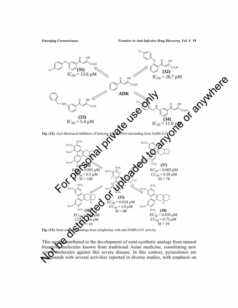
Emerging Coronaviruses Frontiers in Anti-Infective Drug Discovery, Vol. 8 51
Fig. (12). Aryl diketoacid inhibitors of helicase duplex DNA-unwinding from SARS-CoV.
Fig. (13). Semi-synthetic analogs from tylophorine with anti-SARS-CoV activity.
This work contributed to the development of semi-synthetic analogs from naturalbioactive molecules known from traditional Asian medicine, constituting newactive molecules against this severe disease. In this context, pyrazolones arecompounds with several activities reported in diverse studies, with emphasis on
(31) (32)
(33) (34)
IC50 = 13.6 µMIC50 = 28.7 µM
IC50 = 5.4 µM IC50 = 11.0 µM
O OH
CO2H
O OH
CO2HO OH
CO2H
O OH
CO2HO OH
CO2H
OO
Cl
Cl
HNHN
Cl
ADK
(36) (37)
(38) (39)
EC50 < 0.005 µMCC50 = 0.5 µM
SI > 100
EC50 < 0.005 µMCC50 = 0.39 µM
SI > 78
EC50 = 0.039 µMCC50 = 0.73 µM
SI = 19
N
OCH3
H3CO
H3CO
OCH3
(35)
N
OCH3
H3CO
H3CO
OCH3
N
H3CO
H3CO
OCH3
N
OCH3
H3CO
H3CO
OCH3
N
OCH3
H3CO
H3CO
OCH3
EC50 = 0.018 µMCC50 = 1.6 µM
SI = 88
H3CO
OH
O O
123
4
56
78
9 11
1314
12
EC50 = 0.34 µMCC50 = 3.4 µM
SI = 10
For pe
rsona
l priv
ate us
e only
Not be
distr
ibuted
or up
loade
d to a
nyon
e or a
nywhe
re

52 Frontiers in Anti-Infective Drug Discovery, Vol. 8 da Silva Santos-Júnior et al.
the edaravone which used in the treatment of brain and myocardial ischemia.Other pyrazolones have demonstrated antiviral activity against orthopoxvirus[77]. Based on these facts, Ramajayam and collaborators (2010) reported thesynthesis of pyrazolone derivatives with inhibitory activity of 3CLpro from SARS-CoV. This chemical class was identified by using a high-throughput screeningassay [78]. Thus, 21 new pyrazolone derivatives were synthesized and screened,resulting in the three most active analogs (40-42) (Fig. 14).
Fig. (14). Active pyrazolone analogs against SARS-CoV.
SAR analyses of these derivatives revealed that the compound (40) is the only onewith a cyano group at position 4 of the pyrazolone nitrogen-fused phenyl ring. Allthree analogs have electron-withdrawing groups in this same position. Besides,docking studies suggest that the benzylidene group is placed into the S3 pocketfrom the 3CLpro, and the carboxylic acid group interacts through a hydrogenbonding interaction with Gln192 amino acid residue.
Based on the study performed by Lee and coworkers in 2009 (as mentionedearlier in this chapter), Kim and colleagues (2011) [79] developed the other 10aryl diketoacids by using classic bioisosterism and screened as NTPase/helicaseinhibitors from SARS-CoV. As result, the compound (43) (Fig. 15) was found tobe a potent inhibitor of the NTPase and Hel enzymes from SARS-CoV, with anIC50 value of 4 µM and 11 µM, respectively. Besides, this compound was shownto be safe, with a CC50 value higher than 50 µM concentration. Finally, this study
IC50 = 5.5 µM
NN
O
R1
R2
NN
O
NN
O
NN
O
CN
HOOC
F
HOOC
HOOC
NO2
IC50 = 6.8 µM
IC50 = 8.4 µM
(40)
(41)
(42)
For pe
rsona
l priv
ate us
e only
Not be
distr
ibuted
or up
loade
d to a
nyon
e or a
nywhe
re

Emerging Coronaviruses Frontiers in Anti-Infective Drug Discovery, Vol. 8 53
contributes to the design of new bioactive enzyme inhibitors against HCoV,supporting the discovery of multi-target antiviral drugs.
Fig. (15). 2,6-Bis-arylmethyloxy-5-hydroxychromones with anti-SARS-CoV activity.
Another study involving inhibitors of 3CLpro from SARS-CoV was developed byChuck and coworkers (2013) [80], in which it was one of the first works using theauto-cleavage sequence of SARS-CoV 3CLpro (TSAVLQY) as a basis fordesigning peptidomimetic inhibitors. Besides, the study reports the insertion of anitrile warhead as essential for inhibitory activity, since this group remainscovalently bonded to the thiol group from the Cys145 amino acid residue from thecatalytic dyad of 3CLpro. Additionally, it was confirmed by crystal structures ofenzyme-inhibitor complexes [81, 82]. Based on this, the authors synthesized fourpeptidomimetic analogs, where the derivative (44) was found to be the mostpromising inhibitor upon the 3CLpro enzyme (Fig. 16), displaying an IC50 value of4.6 µM. Also, it was observed that the Cbz protective group at the N-terminusincreased the potency of this inhibitor about 10-times.
Fig. (16). Peptidomimetic with anti-SARS-CoV activity.
Considering the design of new inhibitors of 3CLpro based on peptidomimeticcompounds, Prior and colleagues (2013) evaluated a new class of analogs fromtripeptidyl transition-state inhibitors, where it is compatible with the naturalsubstrate of 3CLpro, to provide better recognition and, consequently high inhibition
O
OHO
OI
OCl
(43)
IC50 = 4.0 µM and 11.0 µM
(44)IC50 = 4.6 µM
OHN
O
NH
OHN
O
O
NH N
NH2O
Warhead group
For pe
rsona
l priv
ate us
e only
Not be
distr
ibuted
or up
loade
d to a
nyon
e or a
nywhe
re

54 Frontiers in Anti-Infective Drug Discovery, Vol. 8 da Silva Santos-Júnior et al.
[83]. Based on this information, the general structure contains a glutamine residue(P1), a hydrophobic amino acid residue (P2) - in this case, leucine; and anarylalanine (P3), to facilitate cell absorption. Finally, it was added aldehydes, α-ketoamides, bisulfite adducts or α-hydroxyl phosphonates as warhead groups [81,82]. Among these, the analogs (45 and 46) were the most promising derivativesidentified in this study, where the aldehyde-containing analog (45) exhibited goodactivity, with an IC50 value of 0.23 µM against 3CLpro. Also, the α-ketoamide-containing analog (46) was about 3-times less potent, with an IC50 value of 0.61µM. Besides, both peptidomimetics showed safe cytotoxic profiles, with CC50values of 87 and > 100 µM, respectively (Fig. 17).
Fig. (17). Peptidomimetics inhibitors of 3CLpro from SARS-CoV.
Fleximers process is a technique used for designing bioactive molecules fromrigid analogs, with advantages such as increased affinity energy for the active sitefrom enzymatic targets, as well as, ability to act on mutations [84]. Based on thisinformation, Peters and collaborators (2015) developed three fleximers analogsfrom acyclovir (Fig. 18), an antiviral drug nucleoside polymerase inhibitor used inpharmacotherapy against infections by Herpes Zoster virus (HZV) and HSV [85].The derivative (47) demonstrated promising activity against the HCoV-NL63strain, as well as MERS-CoV. It presented an IC50 value of 8.8 µM upon infectedcells with HCoV-NL63 and CC50 value of 120 µM. Also, it towards cells withMERS-CoV exhibited IC50 and CC50 values of 23 and 71 µM.
Fig. (18). Fleximer analog from Acyclovir with anti-HCoV-NL63 activity.
OHN
O
NH
O
NH
O
H
O
NHO
(45)IC50 = 0.23 µMCC50 = 87 µM
P1P2P3
Warhead group
Cap OHN
O
NH
O
NH
O
O
NH
O
(46)IC50 = 0.61 µMCC50 > 100 µM
P1P2P3
Warheadgroup
Cap HN
O
N
NHN
N
O
NH2O
HO
N
NO
HO
N
N NH2MeO
AcyclovirFleximer analogue (47)
For pe
rsona
l priv
ate us
e only
Not be
distr
ibuted
or up
loade
d to a
nyon
e or a
nywhe
re

Fig. (19). New pyrazolones analogs anti-3CLpro from SARS- and MERS-CoV.
Virtual Screening and Computational Techniques for Designing NewAntiviral Agents
The discovery of new drugs involves a long and complex process that includesdiverse steps such as selection and validation of a biological target; screening ofcompounds from different libraries; and evaluation of resulting compounds invitro and in vivo assays [87, 88]. In this context, computer modeling has gainedmore space in the field of medicinal chemistry, providing rapid screening in large
(48) (49)
(51)
IC50 SARS = 6.0 µMIC50 MERS = 7.3 µM N
N
OO
N
N
OO
Cl
HOOC
N
N
OO
HOOC
N
N
OO
Cl
HOOC
N
N
OO
COOH(50)
IC50 SARS = 5.8 µMIC50 MERS = 7.4 µM
IC50 SARS = 8.6 µMIC50 MERS = 7.7 µM
IC50 SARS = 6.4 µMIC50 MERS = 5.8 µM
Emerging Coronaviruses Frontiers in Anti-Infective Drug Discovery, Vol. 8 55
Considering the drugs oseltamivir and zanamivir, neuraminidase (NA) inhibitors,Kumar and coworkers (2016) published a work reporting the screening of 19analogs against 3CLpro enzymes from SARS- and MERS-CoV [86]. All thesynthesized compounds (48-50) exhibited promising activity against both SARS-and MERS-CoV 3CLpro protease (Fig. 19). Most of these compoundshydrophobically interact with Met49 and Gln189 amino acid residues, into S2pocket. However, the analog (51), which has a carboxylic acid group,preferentially interacts into pocket S1, through hydrogen bonding interactions.
For pe
rsona
l priv
ate us
e only
Not be
distr
ibuted
or up
loade
d to a
nyon
e or a
nywhe
re

56 Frontiers in Anti-Infective Drug Discovery, Vol. 8 da Silva Santos-Júnior et al.
compounds’ libraries, making the process more economically viable and easy [89,90]. Among the computational approaches in the search for new drugs, virtualscreening is a fast and effective technique for identifying new hits compounds, aswell as their optimized ones. Besides, it is divided into two categories: (i)structure-based drug design (SBDD), and (ii) ligand-based drug design (LBDD).In this context, the SBDD approach is described as the most effective, widelyused in research laboratories worldwide. Based on this, it will be discussed inmore detail in this chapter [90 - 92].
Computational modeling shows its importance not only in drug design but also inthe development of vaccines, increasing the probability of new therapeuticalternatives to be developed [20, 33, 93].
The most attractive targets in the development of antiviral compounds againstCoV are proteases, since they interfere in the viral replication and, consequentlyimplicates in the maturation of viral replicases [13, 94]. The proteases PLpro and3CLpro are critical for the pp1a and pp1b replicases [13]. Additionally, it isimportant to mention that the two proteins are cysteine proteases, and present areactive thiol group responsible for attacking carbonyl groups at the peptidebonds, leading to the formation of proteins essential for the survival of the virus[73]. Some studies show that the SARS-CoV-2 PLpro enzyme is highly homolog tothe SARS- and MERS-CoV PLpro. In this sense, it can be successfully used inmolecular modeling studies for discovering new drugs to potentially treat thecurrent outbreak [95, 96].
In this section, several compounds will also be shown (e.g., ribavirin (166),remdesivir (170), and others) that are nucleotide or nucleoside analog compounds,presenting purine, pyrimidine, modified sugar portion in their structures, whichalso show promising inhibition of replication against the CoV [13, 97].
One of the first studies using computational techniques for developing newcompounds against the CoV was carried out by Chou and collaborators (2003)[98]. The authors performed a molecular docking study of the compound KZ7088(53), an analog from AG7080 (52) developed by Pfizer, in which it is in clinicalphase trials against rhinovirus. As shown in Fig. (20), AG7080 (52) was modifiedby removing the methylene group from the side chain, generating KZ7080 (53).Through docking simulations, it was possible to propose that the SARS-CoV3CLpro binding pocket consists of 23 amino acid residues, and the maininteractions observed for the KZ7088 (53) are via hydrogen bonding interactionswith Thr25, Thr45, Leu50, Tyr54, and Asp187 amino acid residues. The authorshighlighted that this study has great importance for designing new drugs for thetreatment of the CoV.
For pe
rsona
l priv
ate us
e only
Not be
distr
ibuted
or up
loade
d to a
nyon
e or a
nywhe
re
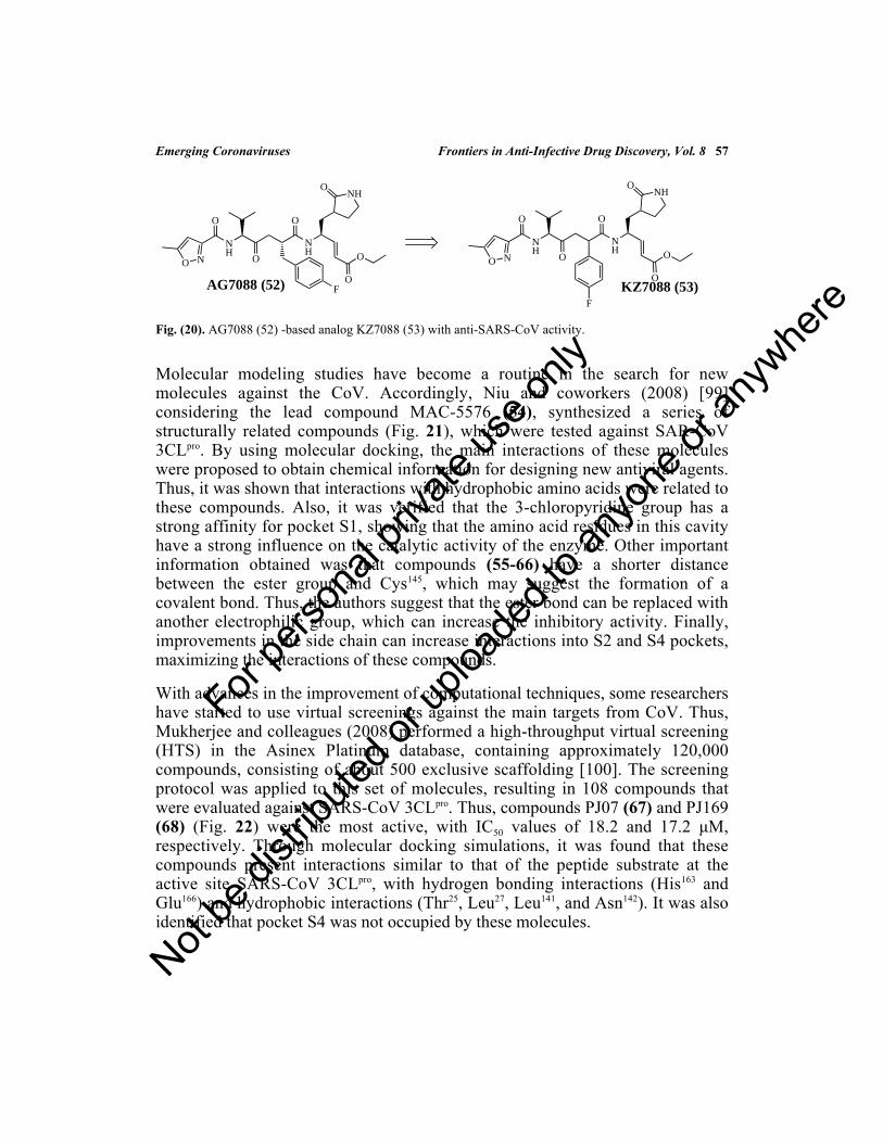
Emerging Coronaviruses Frontiers in Anti-Infective Drug Discovery, Vol. 8 57
Fig. (20). AG7088 (52) -based analog KZ7088 (53) with anti-SARS-CoV activity.
Molecular modeling studies have become a routine in the search for newmolecules against the CoV. Accordingly, Niu and coworkers (2008) [99]considering the lead compound MAC-5576 (54), synthesized a series ofstructurally related compounds (Fig. 21), which were tested against SAR-CoV3CLpro. By using molecular docking, the main interactions of these moleculeswere proposed to obtain chemical information for designing new antiviral agents.Thus, it was shown that interactions with hydrophobic amino acids were related tothese compounds. Also, it was verified that the 3-chloropyridine group has astrong affinity for pocket S1, showing that the amino acid residues in this cavityhave a strong influence on the catalytic activity of the enzyme. Other importantinformation obtained was that compounds (55-66) have a shorter distancebetween the ester group and Cys145, which may suggest the formation of acovalent bond. Thus, the authors suggest that the ester bond can be replaced withanother electrophilic group, which can increase the inhibitory activity. Finally,improvements in the side chain can increase interactions into S2 and S4 pockets,maximizing the interactions of these compounds.
With advances in the improvement of computational techniques, some researchershave started to use virtual screenings against the main targets from CoV. Thus,Mukherjee and colleagues (2008) performed a high-throughput virtual screening(HTS) in the Asinex Platinum database, containing approximately 120,000compounds, consisting of about 500 exclusive scaffolding [100]. The screeningprotocol was applied to this set of molecules, resulting in 108 compounds thatwere evaluated against SARS-CoV 3CLpro. Thus, compounds PJ07 (67) and PJ169(68) (Fig. 22) were the most active, with IC50 values of 18.2 and 17.2 μM,respectively. Through molecular docking simulations, it was found that thesecompounds present interactions similar to that of the peptide substrate at theactive site SARS-CoV 3CLpro, with hydrogen bonding interactions (His163 andGlu166) and hydrophobic interactions (Thr25, Leu27, Leu141, and Asn142). It was alsoidentified that pocket S4 was not occupied by these molecules.
O N
O
NH
O
NH
O
F
NH
O
O
O
AG7088 (52)
O N
O
NH
O
NH
O
NH
O
O
O
KZ7088 (53)F
For pe
rsona
l priv
ate us
e only
Not be
distr
ibuted
or up
loade
d to a
nyon
e or a
nywhe
re

58 Frontiers in Anti-Infective Drug Discovery, Vol. 8 da Silva Santos-Júnior et al.
Fig. (21). SARS-CoV 3CLpro inhibitors.
Fig. (22). SARS-CoV 3CLpro inhibitors identified from the Asinex Platinum database.
Nguyen and collaborators (2011) carried out a virtual study by HTS against 3CLpro
involving 308,307 compounds from the ChemBridge database [101]. In theirprocedure, the compounds were initially ranked, resulting in 1468 molecules withaffinity energy ranging from -14 to -17.09 kcal/mol. In total, 214 compounds wereselected based on their hydrogen bonding interactions with the main amino acidresidues. These compounds were categorized into 35 groups, where 53compounds were selected for biological assays against 3CLpro. In the sense, it wasobtained IC50 values ranging from 38.57 to 101.38 μM. After the docking studies,
ClO
O
O
N
Cl
(55)IC50 = 63 nM
O2NO
O
O
N
Cl
(56)IC50 = 60 nM
ClO
O
O
N
Cl
NO2
IC50 = 122 nM(57)
OO
O
N
Cl
NO2
IC50 = 208 nM(58)
OO
O
N
Cl
O2NIC50 = 500 nM
(59)
N
O
ON
Cl
(60)IC50 = 164 nM
N
O
ON
Cl
(61)IC50 = 697 nM
O
ON
Cl
Cl
(62)IC50 = 434 nM
O
ON
Cl
NO2
(63)IC50 = 333 nM
O
ON
Cl
(64)IC50 = 684 nM
Cl
O
ON
Cl
(65)IC50 = 124 nM
O
O
ON
Cl
(66)IC50 = 108 nM
O
S
O
O Cl
MAC - 5576 (54)IC50 = 0.5 ± 0.3 uM
NN
S
O
HN
O
PJ07 (67)IC50 = 18.2 uM
NH
O N
NH
O
PJ169 (68)IC50 = 17.2 uM
For pe
rsona
l priv
ate us
e only
Not be
distr
ibuted
or up
loade
d to a
nyon
e or a
nywhe
re

Emerging Coronaviruses Frontiers in Anti-Infective Drug Discovery, Vol. 8 59
it was shown that these compounds are stabilized by hydrogen bondinginteractions with catalytic residues and hydrophobic interactions in regionsopposite the active site. Besides, Ki assays for compounds (74) and (75) (Fig. 23)revealed that these molecules are competitive inhibitors, with values of 9.11 ±1.61 and 9.93 ± 0.44 μM, respectively. Finally, the authors state that thesecompounds can serve as new scaffolds for discovering new compounds againstthe CoV.
Fig. (23). SARS-CoV 3CLpro inhibitors identified from the ChemBridge database.
Zhu and coworkers (2011) carried out study kinetics of inhibition in thecrystalline structure of the complex of six different peptide aldehydes (Fig. 24)with the SARS-CoV 3CLpro [102]. The authors showed the binding modes by X-ray crystallography for the compounds at the active site, displaying that peptidehydrophilic residues (Ser and Asp) of the P2 position are placed into thehydrophobic S2 pocket, and hydrophobic residue (Phe) in the P1 position isinserted into the hydrophilic S1 pocket S1. Also, the study showed that thespecificity for these subsites can be canceled out by the presence of highlyelectrophilic chemical groups at P1 and P2 positions close to the aldehyde, whichjustifies the greater activity for the compound Cm-FF-H (80) (Ki = 2.24 ± 0.58
N
NN
SS
N NH2
(69)
O
CH3
H3C
H3C
O
NHO
O
N
CH3
CH3
O
OH(70)
O
N N
O OH
O
S N
CH3
CH3
O2N
(71)
NH
OHN
N
O
O
OH
O
CH3
CH3
(72)
O
N N S
NN
S
O
NH O
(73)
O2N OHN
O
H3C
O
HN
N CH3
H3C
(74)
OHN
O
H3C
O
HN
N CH3
H3C
(75)
O2N
IC50 = 58.35 ± 1.41 uM IC50 = 62.79 ± 3.19 uM IC50 = 101.38 ± 3.27 uM
IC50 = 77.09 ± 1.94 uM IC50 = 90.72 ± 5.54 uM
IC50 = 38.57 ± 2.41 uM IC50 = 41.39 ± 1.17 uMFor pe
rsona
l priv
ate us
e only
Not be
distr
ibuted
or up
loade
d to a
nyon
e or a
nywhe
re

60 Frontiers in Anti-Infective Drug Discovery, Vol. 8 da Silva Santos-Júnior et al.
μM). Through these results, the authors suggest that these interactions should beexplored for the discovery of new compounds against CoV.
Fig. (24). New SARS-CoV 3CLpro peptidomimetic inhibitors.
The study performed by Turlington and coworkers (2013) was developed basedon compound (81) (IC50 = 6.2 μM against 3CLpro enzyme), where the interactionsof the co-crystallized ligand with the enzyme were analyzed and used to proposestructural modifications (Fig. 25). However, the main interactions with the activesite were maintained (Gly163 and Glu166), leading to the synthesis of severalcompounds. From these, the compound ML300 (82) presented the best activity(IC50 = 4.11 ± 0.24 μM), showing that studies based on the structure of the targets(SBDD) could be extremely promising in the development of drugs against theCoronavirus [103].
HN
O
NH
O
O
H
Cm-FF-H (80)SARS-CoV Ki = 2.24 ± 0.58 uM
P1
P2
Ac-NSFSQ-H (79)
H2N
OHN
HOO
NH
HOO
HN
O
NH
HOO
NH O
H
H2N
O
H2N
OHN
HOO
NH
OHN
O
NH
HOO
NH O
H
H3C
O
O
HO
Ac-DSFDQ-H (78)
H2N
OHN
HOO
NH
OHN
O
NH
HOO
NH O
H
H3C
O
HO
HO
Ac-NSTSQ-H (77)
H2N
OHN
HOO
NH
OHN
O
NH
HOO
NH
H3C
O
HO
O
H
Ac-ESTLQ-H (76)
SARS-CoV Ki = 40.98 ± 2.63 uMSARS-CoV Ki = 8.27 ± 1.52 uM
SARS-CoV Ki = 41.24 ± 2.25 uM SARS-CoV Ki = 72.73 ± 3.60 uM
For pe
rsona
l priv
ate us
e only
Not be
distr
ibuted
or up
loade
d to a
nyon
e or a
nywhe
re

Emerging Coronaviruses Frontiers in Anti-Infective Drug Discovery, Vol. 8 61
Fig. (25). Benzotriazole inhibitors of SARS-CoV 3CLpro.
Interestingly, Lee and collaborators (2014) combined VS techniques withenzymatic HTS in the search for new inhibitors of SARS-CoV 3CLpro [104]. TheVS protocol was applied to the ZINC database, containing approximately 621,000compounds. In this context, only 68 were previously acquired and tested,obtaining eight compounds (83-90) with promising activity (Fig. 26), withinhibition of over 50% and IC50 values in the micromolar range. Based on the VSresults, the authors conducted an HTS campaign on three databases, adding up to40,000 compounds, where two compounds (91-92) (Fig. 26) were promising inbiological assays, with IC50 values less than 50 μM. Thus, the authors concludedthat the VS and HTS protocols could be considered as promising strategies forobtaining new scaffolding candidates for the optimization and design of newdrugs anti-SARS-CoV.
Molecular modeling studies were used in the study performed by Wang andcoworkers (2017) to determine the main interactions of the most activecompounds, as well as to develop a QSAR model for the unsymmetrical aromaticdisulfides derivatives synthesized and tested against the SARS-CoV 3CLpro [105].Then, the authors synthesized 40 very promising compounds (Fig. 27), with IC50values ranging from 0.516 to 5.95 μM. The docking study for compound (123)(most promising) was carried out, suggesting that its activity is mainly due tohydrophobic interactions with Phe140, Leu141, His163, Met165, Glu166, and His172, aswell as hydrogen bonding interactions with Asn142, Gly143, and Cys145. Finally, theQSAR study obtained values of q2= 0.681 and r2= 0.916, showing that the stericcontributions account with 43.6% for biological activity, and the electrostaticcontributions, with 56.4%. Finally, the authors highlighted that these results arepromising and could be used for developing compounds anti-SARS-CoV.
HN
O
N
O
NH
O
NN
N
N
(81)3CLpro IC50 = 6.2 uM
Structure-based design
S
N
ON
NN
NH
O
ML300 (82)3CLpro IC50 = 4.11 ± 0.24 uM
His163
Glu166
For pe
rsona
l priv
ate us
e only
Not be
distr
ibuted
or up
loade
d to a
nyon
e or a
nywhe
re

62 Frontiers in Anti-Infective Drug Discovery, Vol. 8 da Silva Santos-Júnior et al.
Fig. (26). SARS-Cov 3CLpro inhibitors identified from the ZINC database.
Fig. (27). SARS-CoV inhibitors synthesized by Wang et al. 2017.
O
O
N
NH
HNHO
OHN
O
NS
OOO
(83) (84)
NN
O
OH
(85)
NN
O
OH
OS
NH
OO
NH
O
O
(86)
O
SNH
OO
O
O
(87)
H2N
F
SNH
OO
CF3
(88)
S
Cl
N
OO HN
O
(89)
Virtual Screening
Cl
HN
O N
N
(90)
N
N
N
N
NH2
CF3
(91)IC50 = 18.2 ± 4.9 uM IC50 = 13.9 ± 2.2 uM
High-throughput screening
(92)
S
NS
S Cl
(93) IC50 = 1.871
± 0.071 uM
S
NS
S CH3
(94) IC50 = 2.803
± 0.052 uM
NH
NS
S
(95) IC50 = 3.675
± 0.193 uM
O
O N
N
NS
S
(96) IC50 = 3.130
± 0.052 uM
O2N
O
S
NS
S
(97) IC50 = 1.506
± 0.184 uM
NH
NH
O O
N
N
NS
S
(98) IC50 = 4.344
± 0.538 uM
O
N
N
NS
S
(99) IC50 = 4.100
± 0.832 uM
O
O N
N
NS
S
(100) IC50 = 1.762
± 0.044 uM
O
N
O2N
N
N
NS
S
(101) IC50 = 5.654
± 0.259 uM
O
N
OO
N
N
NS
S
(102) IC50 = 4.511
± 0.105 uM
O
N
OO
N
N
NS
S
(103) IC50 = 4.511
± 0.105 uM
O
N
O
S
NS
S
(104) IC50 = 5.794
± 0.050 uM
NH
O
S
NS
S Br
(105) IC50 = 1.651
± 0.048 uM
NH
O
NH
NS
S
(106) IC50 = 2.075
± 0.016 uM
O2N
NH
N
SS
(107) IC50 = 5.954
± 0.363 uM
O
O
OO
O O
NH
N
SS
(108) IC50 = 3.957
± 0.190 uM
OO O O
NH
NS
S
(109) IC50 = 4.126
± 0.094 uM
O
O Cl
S
NS
S F
(110) IC50 = 2.565
± 0.075 uM
NH
O
S
NS
S
(111) IC50 = 1.947
± 0.508 uM
NH
O
O2N
S
NS
S
(112) IC50 = 2.029
± 0.488 uM
O2N
S
NS
S
(113) IC50 = 1.250
± 0.023 uM
S
NS
S
(114) IC50 = 2.211
± 0.152 uM
F
S
NS
S
(115) IC50 = 3.321
± 0.068 uM
Br
S
NS
S
(116) IC50 = 2.555
± 0.270 uM
O2N
S
NS
S
(117) IC50 = 2.452
± 0.126 uM
OO
N
O
NS
S
(118) IC50 = 1.679
± 0.042 uM
OO
N
O
NS
S
(119) IC50 = 1.557
± 0.116 uM
OO
N
O
NS
S
(120) IC50 = 1.713
± 0.052 uM
O2N
N
O
NS
S
(121) IC50 = 1.118
± 0.132 uM
OO
O
NS
S
(122) IC50 = 1.264
± 0.033 uM
OO
N
O
NS
S
(123) IC50 = 0.516
± 0.060 uM
Cl SS
NO2
N
N
(124) IC50 = 0.921
± 0.060 uM
SS
N
N
(125) IC50 = 1.437
± 0.053 uM
Cl
SS
N
N
(126) IC50 = 1.121
± 0.060 uM
Br
SS
N
N
(127) IC50 = 1.991
± 0.086 uM
SS
N
N
(128) IC50 = 1.495
± 0.055 uM
SS
N
N
(129) IC50 = 1.495
± 0.055 uM
NO2
SS
N
N
(130) IC50 = 0.684
± 0.012 uM
Cl
SS
N
N
(131) IC50 = 0.697
± 0.053 uM
Br
SS
N
N
(132) IC50 = 1.522
± 0.214 uM
For pe
rsona
l priv
ate us
e only
Not be
distr
ibuted
or up
loade
d to a
nyon
e or a
nywhe
re
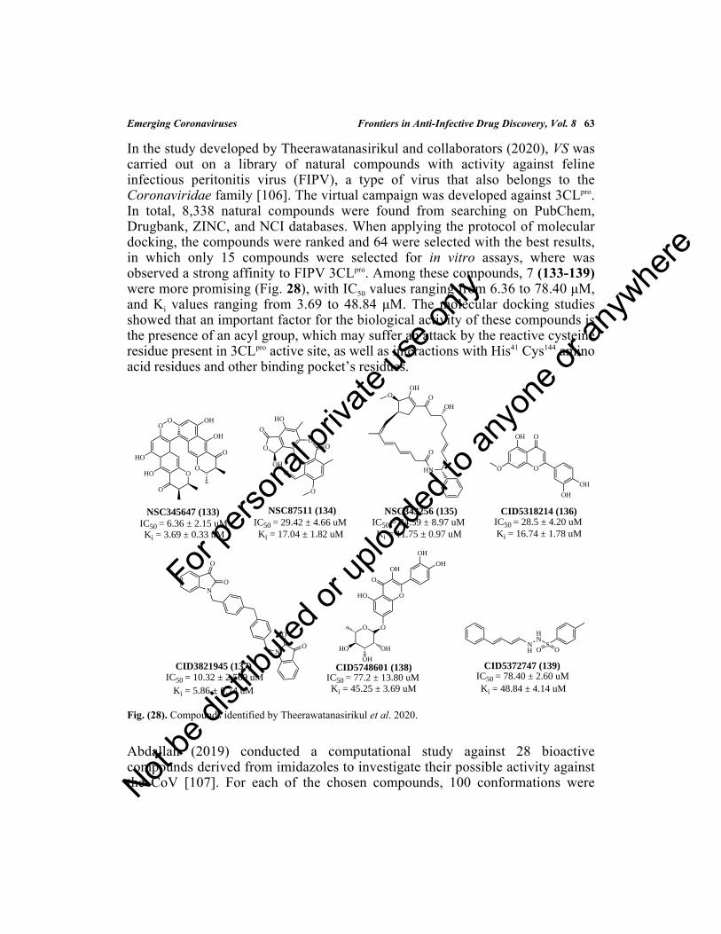
Emerging Coronaviruses Frontiers in Anti-Infective Drug Discovery, Vol. 8 63
In the study developed by Theerawatanasirikul and collaborators (2020), VS wascarried out on a library of natural compounds with activity against felineinfectious peritonitis virus (FIPV), a type of virus that also belongs to theCoronaviridae family [106]. The virtual campaign was developed against 3CLpro.In total, 8,338 natural compounds were found from searching on PubChem,Drugbank, ZINC, and NCI databases. When applying the protocol of moleculardocking, the compounds were ranked and 64 were selected with the best results,in which only 15 compounds were selected for in vitro assays, where wasobserved a strong affinity to FIPV 3CLpro. Among these compounds, 7 (133-139)were more promising (Fig. 28), with IC50 values ranging from 6.36 to 78.40 μM,and Ki values ranging from 3.69 to 48.84 μM. The molecular docking studiesshowed that an important factor for the biological activity of these compounds isthe presence of an acyl group, which may suffer an attack by the reactive cysteineresidue present in 3CLpro active site, as well as interactions with His41 Cys144 aminoacid residues and other binding pocket’s residues.
Fig. (28). Compounds identified by Theerawatanasirikul et al. 2020.
Abdallah (2019) conducted a computational study against 28 bioactivecompounds derived from imidazoles to investigate their possible activity againstthe CoV [107]. For each of the chosen compounds, 100 conformations were
O
OO
O
HO
HO
O
OH
OH
O
NSC345647 (133)IC50 = 6.36 ± 2.15 uMKi = 3.69 ± 0.33 uM
O
O
OH
HO
O
OO
O
O
NSC87511 (134)IC50 = 29.42 ± 4.66 uMKi = 17.04 ± 1.82 uM
OOH
OOH
HN
O
NSC343256 (135)IC50 = 24.59 ± 8.97 uMKi = 11.75 ± 0.97 uM
O
OOH
O
OH
OH
CID5318214 (136)IC50 = 28.5 ± 4.20 uMKi = 16.74 ± 1.78 uM
N
O
O
N
O
O
CID3821945 (137)IC50 = 10.32 ± 2.569 uM
Ki = 5.86 ± 0.74 uM
OHO
O
OH
OO
OH
OH
HO
OH
OH
CID5748601 (138)IC50 = 77.2 ± 13.80 uMKi = 45.25 ± 3.69 uM
NH
HN
SOO
CID5372747 (139)IC50 = 78.40 ± 2.60 uMKi = 48.84 ± 4.14 uM
For pe
rsona
l priv
ate us
e only
Not be
distr
ibuted
or up
loade
d to a
nyon
e or a
nywhe
re

64 Frontiers in Anti-Infective Drug Discovery, Vol. 8 da Silva Santos-Júnior et al.
generated and analyzed, where their affinity energies were calculated and ranked.In this sense, three of these showed the best results (140-142) (Fig. 29), in whichthe main interactions presented by the compound (140) were hydrogen bondinginteractions (Arg188, Leu141, and Ser144) and π-π interactions, similar to interactionsof compounds (141) and (142). The authors concluded that the high-affinityenergy of the compounds is due to the strong interactions with the amino acidresidues.
Fig. (29). Imidazoles and benzothiazole identified as promising 3CLpro inhibitors.
Another computational study was carried out by Gao and coworkers (2020), basedon a virtual screening in the ChEMBL database [14]. Then, 115 compounds wereselected as promising molecules against SARS-CoV. Also, 4,463 molecules wereselected from the PDB database. Then, the authors proposed that the 15 bestcompounds (143-157) can be promising against CoV because they have highaffinity, mainly related to the greater possibility of hydrogen bonding interactionsformation at the active site (Fig. 30).
Drug Repurposing
A very interesting strategy that is gaining more attention from the scientificcommunity is drug repurposing. This strategy is based on finding new therapeuticuses for known, commercialized, discontinued, or even drug candidates [108 -110]. The advantage of repurposing a drug lies in the fact that they are compoundsthat already have proven safety and adequate pharmacokinetic, thus avoidingproblems related to toxicity and stability, and being more economical viable [108,111]. This strategy is mainly associated with bioinformatics, which performing anIn silico screening, it is possible to propose a new activity for different previouslyapproved drugs [109, 110]. Thus, several drugs have been reused in the search fornew treatments against viral infections, such as ZIKV, EBOV, DENV, and morerecently against the SARS-CoV-2 [112]. Later in this chapter, it will be shown thediscovery of anti-CoV activity of various drugs on the market, such as ribavirin(166), hexachlorophene (222), and nitazoxanide (187) [113].
N
NN
HNN
N
OH
Cl
CH3(140)
O
Cl
Cl
N
N
S Cl
(141)N
NH
N
F
S
CH3
O
(142)
For pe
rsona
l priv
ate us
e only
Not be
distr
ibuted
or up
loade
d to a
nyon
e or a
nywhe
re

Emerging Coronaviruses Frontiers in Anti-Infective Drug Discovery, Vol. 8 65
Fig. (30). 3CLpro inhibitors identified from the ChEMBL database.
Based on this strategy, in 2003, it was discovered that the compounds lopinavir(158) and ritonavir (159) had activity against SARS-CoV, as well as niclosamide(160) (anti-parasite) and promazine (161) (anti-psychotic) were promising as well(Fig. 31). In this sense, Zhang and collaborators (2004) conducted an In silicostudy with these drugs, as well as two other compounds, PNU (162) and UC2(163) (Fig. 31), which are structurally related to niclosamide and promazine,respectively [114]. These compounds were promising as HIV-1 reversetranscriptase inhibitors. Furthermore, these compounds were found to be activeupon the 3CLpro SARS-CoV. Through docking studies and based on the bindingmodes presented by Chou and coworkers (2003), it was verified that part of thelopinavir structure does not fit into the catalytic site, as well as the side chain ofbenzene present in ritonavir structure, it is also difficult to fit at the active site.These results demonstrate why these compounds have low effectiveness againstthe SARS-CoV. However, the authors mention that when combined with ribavirin
O
Cl
Cl S
Cl
O
O O
O
Cl
Cl
SCl
O
O
-10.56 kcal/mol
ClCl
O
O
O
O
O
Cl
Cl
O
O
-9.71 kcal/molCl
O
O
OO
Cl
S OO
Cl
ClCl
-9.55 kcal/mol
N
HN
S
F3CO
N
-9.55 kcal/mol
S
O
O
O
ClO
Cl
O
OS O
O
Cl
Cl
Cl
-9.45 kcal/mol
Cl
Cl
S
Cl
OO
O
Cl
O
O ClCl
Cl
O
-9.35 kcal/mol
O
O
O
O
F
-9.31 kcal/mol
S
Cl
OO
SOO
Cl
S
NHO
O
Cl
Cl
Cl
-8.36 kcal/mol
N
Cl
Cl
N
O O
-8.87 kcal/mol
Cl
Cl
O
O
SO
OO
NH
ClCl
OCl
-8.87 kcal/mol
Cl
SO O
O
O Cl
Cl
S
ON
-8.86 kcal/mol
Cl
Cl
Cl
O
O
ClO
O
-8.85 kcal/mol
Cl
S
SOO
SOO
O
Cl
Cl
Cl
Cl
OCl
-8.55 kcal/mol
OS
OO
Cl
Cl
ClCl
SOO
Cl S
O
NO
- 8.85 kcal/mol
Cl
ClO
SOO
O
O
O
Cl Cl
Cl
-8.85 kcal/mol
(143) (144) (145)
(146) (147) (148)
(149) (150) (151)
(152) (153) (154)
(155) (156) (157)
For pe
rsona
l priv
ate us
e only
Not be
distr
ibuted
or up
loade
d to a
nyon
e or a
nywhe
re

66 Frontiers in Anti-Infective Drug Discovery, Vol. 8 da Silva Santos-Júnior et al.
(166) and corticosteroids, they can reduce the incubation and mortality rates of thevirus. As for niclosamide (160), promethazine (161), and PNU (162), similarly tothe others, they did not fit well in the active site mainly because they have a verylong side chain. Finally, UC2 (163) connects in a region far from the active site.
Fig. (31). Potential drugs with activity against SARS-CoV 3CLpro.
A computational approach that produces effective results is pharmacophoremodeling, based on the superposition of a set of molecules in which their commoncharacteristics are observed, looking the interactions of a ligand in a binding site,resulting in a three-dimensional (3D) arrangement used to search for compoundsthat have both structure and similar interactions [115, 116]. This approach hasbeen constantly used in medical chemistry laboratories, especially when it comesto virtual screenings, as well as lead optimization applied in the discovery of newdrugs [116].
In this context, using the compounds from previous work and the CMK peptide(Asn-Ser-Thr-Leu-Gln), Zhang and collaborators (2005) [117] developed a 3D-pharmacophore model against SAR-CoV to provide more solid information fordesigning new drugs. Then, the results were evaluated and applied to the anti-HIVand anti-opportunistic infection chemical compound database, containingapproximately 100,000 compounds that have anti-HIV activity and againstopportunistic infections. As results, 30 drugs were selected and, only 6 of thempresented experimental results against SARS-CoV, such as azauridine (164),pyrazofurin (165), ribavirin (166), 2,3-dideoxycytidine (167), dideoxyganosine(168), and 5-bromo-2-deoxycytidine (169), shown in Fig. (32).
HN N
OHN
O OH
NH
O
O
Lopinavir (158)
SN
N
CH3
O
NH
O
HN
OH
NH
O
O
SN
Ritonavir (159)
Cl
Cl
O
NH
Cl
NO2
Niclosamide (160)
S
N
N
Prometazine (161)
N
OS
CH3N
NCl
NH2
PNU (162)
HN
Cl
NO
S
OH3C
UC2 (163)
For pe
rsona
l priv
ate us
e only
Not be
distr
ibuted
or up
loade
d to a
nyon
e or a
nywhe
re

Emerging Coronaviruses Frontiers in Anti-Infective Drug Discovery, Vol. 8 67
Fig. (32). Compounds identified from the anti-HIV and anti-opportunistic infection chemical compounddatabase.
Other studies have revealed that the ribavirin (166) is a promising anti-CoV agent,as well as its combination with interferon-α2b. Thus, Al-Tawfiq and colleagues(2014) conducted a clinical study with the combination of these drugs in fivepatients infected with MERS-CoV [118]. In three cases, some adverse effects ofthis combination were observed, such as elevation of pancreatic enzymes andhemolysis, as well as laboratory abnormalities. Despite these effects, and due tothe urgent need to find a treatment for this disease, the authors concluded that thiscombination can be promising if used with caution and with monitoring ofadverse effects.
The promising potential of remdesivir (170) (Fig. 33) was investigated bySheahan and collaborators (2020) against MERS-CoV through in vitro and in vivoassays [119]. Thus, this drug presented an EC50 value of 0.09 μM, with noapparent toxicity at 10 μM, and SI >100. In mice, prophylactic and therapeutic usereduced viral load, leading to improved lung function. The results indicate thatthis drug may be useful in the treatment of infected patients with MERS-CoV,observing a decrease in pathogenesis, as well as a reduction of viral load, showingto be a potential drug that can be added to the therapy of the disease. Anothersuggestion by the authors was that the combination of lopinavir (158) and
N
O OH
OHHO
N
N
O
H2N
O OH
OHHO
N
NH
HO
O
H2N
N
O OH
OHHO
NHN
O
O
N
O
HO
N
NH2
O
O
HO
N
NHN
NH
H2N
O
N
O
HO
N
NH2
O
OH
Br
Azauridine
(164)
Pyrazofurin
(165)
Ribavirin
(166)
2,3-dideoxycytidine
(167)
Dideoxyguanosine
(168)
5-bromo-2-deoxycytidine
(169)
For pe
rsona
l priv
ate us
e only
Not be
distr
ibuted
or up
loade
d to a
nyon
e or a
nywhe
re

68 Frontiers in Anti-Infective Drug Discovery, Vol. 8 da Silva Santos-Júnior et al.
ritonavir (159) may be promising in the treatment. With this promising potential,remdesivir (170) was intravenously used, leading the patient recovery. Also, Liand colleagues (2020) conducted to phase III clinical trials in patients with SARS-CoV-2 and then confirmed the promising potential of these drugs [120].
Fig. (33). Chemical structure of remdesivir.
As VS campaigns are commonly applied to drug repurposing, Xu and coworkers(2020) conducted a molecular docking study on 1,903 compounds approved asdrugs obtained from the DrugBank database, as well as by homology studies [95].All compounds were virtually screened toward SARS-CoV-2 3CLpro. The authorsidentified that this enzyme is 96% similar to existing SARS-CoV. Thus, ahomology model was carried out, as well as the screening of approved drugs.Then, only 15 drugs were selected, where four molecules were performedscreened. Finally, nelfinavir (171) was identified as a potential drug for thetreatment of SARS-CoV-2, so that pitavastatin (172), perampanel (173) andpraziquantel (174), also showing moderate activity (Fig. 34).
N
NN
NH2
O
N
OH
OH
O
PO
O
NH
OO
Remdesivir
(170)
For pe
rsona
l priv
ate us
e only
Not be
distr
ibuted
or up
loade
d to a
nyon
e or a
nywhe
re

Emerging Coronaviruses Frontiers in Anti-Infective Drug Discovery, Vol. 8 69
Fig. (34). Chemical structures of pitavastatin, perampanel, and praziquantel drugs.
An interesting methodology developed by Zhou and colleagues (2020) was basedon the systematic identification of drugs, as well as combinations of drugs thatmay be useful in repurposing against HCoV, integrating drug-target networks,induction of HCoV transcriptome in human cells, interactions between HCoV andhost, and protein-protein interactions (PPI) [121]. Thus, the authors proposed 12drugs (175-186), as well as three combinations (Sirolimus + dactinomycin;Toremifene + emodin; Melatonin + mercaptopurine) as a potential treatment (Fig.35). The authors stated that although the compounds have the possibility of use intreatment, preclinical validation is necessary before being used by patients.Despite this, the study offers a powerful methodology based on a rapid drugidentification network that can be repositioned for the treatment of HCoV.
N
F
OOH
HO
HO
Pitavastatin
(172)
N
N
O
N
Perampanel
(173)
N
N
O
OH
Praziquantel
(174)
S
HN
N
OH
H
H
O
OH
O
NHNelfinavir
(171)
For pe
rsona
l priv
ate us
e only
Not be
distr
ibuted
or up
loade
d to a
nyon
e or a
nywhe
re

70 Frontiers in Anti-Infective Drug Discovery, Vol. 8 da Silva Santos-Júnior et al.
Fig. (35). Compounds identified by Zhou et al. 2020.
Instigated to discover new treatments more quickly against CoV, Wang andcoworkers (2020) evaluated the effectiveness of seven drugs on the market (Fig.37), such as ribavirin (166), nitazoxanide (187), penciclovir (188), nafamostat(190) and chloroquine (191), as well as two others with broad-spectrum antiviralactivity remdesivir (170) and favipiravir (189) against a clinical isolate of SARS-CoV-2 [122]. Thus, after evaluating their cytotoxicity in Vero E6 cells (ATCC-1586), the cells were infected with SARS-CoV-2 for the evaluation of the drugs.The results (Fig. 36) showed that the most effective compounds in controllingSARS-CoV-2 infection were remdesivir (170) and chloroquine (191), suggestingthat they can be promising in treatment and should be evaluated in infectedpatients by CoV. From this perspective, Keyaerts and colleagues (2004) alsoproposed chloroquine (191) as a promising drug against SARS-CoV, presentingan IC50 value of 8.8 μM [123]. In addition to this study, others point tonitazoxanide (187) as promising in the treatment of some types of viral infections,such as the CoV. Thus, Rossignol and collaborators (2016) through a literaturereview, showing in vitro potential against MERS-CoV for nitazoxanide (187), aswell as its pro-inflammatory cytokine and IL-6 inhibitions, and reduction ofinfluenza virus symptoms in humans [124]. So, the authors suggested as potentdrug candidates in the treatment of this disease.
NH2
OH
HO
OMezalazine
(175)
Cl
ON
Toremifene
(176)
O
O
O
O
O
H
H
H
O
Eplerenone
(177)
O
O
O
HN
F
H
H
Paroxetine
(178)
N
NH
S
N
HN
Mercaptopurine
(181)
NH
HNO
O
Melatonin
(179)
O
O
OHOH
OHH3C
Emodin
(182)
HO
O
H H
H
Equilin
(183)
OCH3
Camphor
(184)
HN
H
O
O
O
OO
O
Colchicine
(180)
NCl
O
HNN
Quinacrine
(185)
HO
O
OH
H
H
H H
Oxymetholone
(186)
For pe
rsona
l priv
ate us
e only
Not be
distr
ibuted
or up
loade
d to a
nyon
e or a
nywhe
re

Emerging Coronaviruses Frontiers in Anti-Infective Drug Discovery, Vol. 8 71
Fig. (36). Compounds identified as promisors against SARS-CoV-2 by Wang et al. 2020.
A molecular modeling study by Chen and collaborators (2020) proposed a 3CLpro
molecular model for SARS-CoV-2, followed by a virtual screening of drugs usedin clinical practice [125]. The compounds used in this screening were obtainedfrom the Drug-lib database, containing approximately 7,173 compounds. Amongthese 11 compounds (192-202) are commercially available drugs (Fig. 37), andwhich can be promising in the treatment of CoV.
In the area of drug discovery, an approach that has been gaining prominence isDeep Learning (DL). This approach is a subfield of machine learning (ML),which uses various algorithms and neural networks of artificial intelligence (AI)for analyzing high levels of data [126, 127]. The implementation of methodsbased in AI, especially DP, can be important in drug discovery, generating a moreeffective identification in HTS protocols, improving lead optimization by moreaccurate ADMET and QSAR models, and reducing efforts to discover newcompounds [93].
N
NN
O
NH2
OHO
HO OH
Ribavirin (166)EC50 = 109.50 uMCC50 > 400 uM
SI > 3.65
EC50 = 2.12 uMCC50 > 35.53 uM
SI > 16.76
S
N
O2N
NH
OO
O
Nitazoxanide (187)
HN
N N
N
O
H2N
OH
HO
Penciclovir (188)EC50 = 95.96 uM
CC50 > 400 uMSI > 4.17
N
NF
OH
NH2
O
Favipiravir (189)
EC50 = 61.88 uMCC50 > 400 uM
SI > 6.46
H2N
HNO
O
NNH2
H2NNafamostat (190)EC50 = 22.50 uMCC50 > 100 uM
SI > 4.44
N
NN
NH2
O
N
OH
OH
O
PO
O
NH
OO
Remdesivir (170)EC50 = 0.77 uMCC50 > 100 uM
SI > 129.87
NCl
HNN
Chloroquine (191)EC50 = 1.13 uMCC50 > 100 uM
SI > 88.50
For pe
rsona
l priv
ate us
e only
Not be
distr
ibuted
or up
loade
d to a
nyon
e or a
nywhe
re

72 Frontiers in Anti-Infective Drug Discovery, Vol. 8 da Silva Santos-Júnior et al.
Fig. (37). Compounds identified from the Drug-lib database.
Zhang and coworkers (2020) used an approach based on DL for searching for newactive compounds against SARS-CoV-2 [128]. The authors screened severaldatabases of small organic molecules containing FDA-approved drugs, as well asnatural compounds that have potential biological activity. From these, ninepotential compounds (203-211) (Fig. 38) were identified and they could bepromising in the treatment of SARS-CoV-2.
O OO O O
O
OH
OOH
OH
OHHO
HO OH
OH
Diosmin (192)
O
O
O
O
O
OH
O
HO
O
HO
HO
OH
HO
HO
OHHesperidin (193)
OHN
NO2
S
HN
OOO
O N
N
Cl
N
HN
Venetoclax (194)
NH
N
O HN
N
O
O N
O
OH
H
H
H
Dihydroergocristine (195)
O O
O
OO
O
O
O
O
OH
H
H
H
HHO
HO
Etoposide (196)
OO
O
OH
O
O
OO
O
O
OH
O
HOS
Teniposide (197)
N
N
O
O
N
NO
O
OOH
Irinotecan (198)
N
OHO
HN
O O
O F
FLumacaftor (199)
NHO
O
N
O
O
NH
N
O
N
HN
NO
NH
OO
Velpatasvir (201)
O
H2N NH2
O
N
NHN
O
OH
OEluxadoline (200)
N
NHN
O
HNO
O
NH
N
NO
HNO
O
FF
Ledipasvir (202)
For pe
rsona
l priv
ate us
e only
Not be
distr
ibuted
or up
loade
d to a
nyon
e or a
nywhe
re

Emerging Coronaviruses Frontiers in Anti-Infective Drug Discovery, Vol. 8 73
Fig. (38). Compounds identified by using an approach based on DL.
Li and collaborators (2020) carried out a high-throughput VS based on a library of8,000 drugs. This study identified nelfinavir (171), prulifloxacin (212), tegobuvir(213), and bictegravir (214) as potentials agents for treating CoV (Fig. 39) [20].Additionally, modeling studies suggested that these compounds may have theability to stop the formation of dimers of the viral protein, as well as blocking theactive sites. Finally, the authors suggested that the clinical potential of thesemolecules can be explored for the treatment of CoV.
Fig. (39). Chemical structures of nelfinavir (171), prulifloxacin (212), tegobuvir (213), and bictegravir (214).
Wilde and coworkers (2017) investigated the inhibitory potential of the alisporivir(215) (Fig. 40) against the replication of MERS- and SARS-CoV in cell cultures,showing that it could be a potent and broad-spectrum inhibitor, with EC50 valuesranging from 1.5 to 8.3 μM in different cell lines, and acting in the inhibition ofalpha- and beta-CoV [129]. However, the authors suggested that further studies
N
NN
N
NH2
O
OH
HO
HO
Vidabirine
(203)
N
NN
N
NH2
O
OH
HO
HO
Adenosine
(204)
HO
OH
OH
OH
OH
OH
Mannitol
(206)
HO
OH
OH
OH
OH
OH
Dulcitol
(207)
HO
OH
OH
OH
OH
OH
D-Sorbitol
(208)
HO
OH
OH
OH
OH
OH
Allitol
(209)
HO
OH
OH
OH
O
OH
Sodium gluconate
(211)
O
Na
HN
OH
OH
OH
OH
OH
Meglumine
(210)
N
HN
O
H2N
N
N
O
HO
HO
Ganciclovir
(205)
OO
O
NN
F
N
O
OH
O
S
Plulifloxacin
(212)
CF3F3C
NN
N N
N
F
Tegobuvir
(213)
O
N
N
H
H H
O OH
O
O
HN
FF
F
Bictegravir
(214)
S
HN
N
OH
H
H
O
OH
O
NHNelfinavir
(171)For pe
rsona
l priv
ate us
e only
Not be
distr
ibuted
or up
loade
d to a
nyon
e or a
nywhe
re

74 Frontiers in Anti-Infective Drug Discovery, Vol. 8 da Silva Santos-Júnior et al.
should be carried out to elucidate this mechanism of action. Another drug that hadits activity against MERS-CoV and SARS-CoV PLpro enzymes is disulfiram (216)(Fig. 40). In a study conducted by Lin and colleagues (2018) was demonstratedthat this compound acts as a non-competitive and competitive (or mixed) inhibitorfor MERS- and SARS-CoV, respectively [130]. Additionally, the authorsproposed that the inhibition of SARS-CoV PLpro is a covalent interaction with theCys112 residue.
Fig. (40). Chemical structures of alisporivir and disulfiram.
High-Throughput Screening (HTS) for Discovering New Antiviral Agents
The HTS has been evolving since the 90s, constituting a powerful tool in theidentification of new hits compounds [131 - 133]. This approach is based onbiochemical assays against large compound libraries, with the possibility oftesting approximately 20,000 compounds per week against enzyme targets,providing fast and effective results [134, 135]. It has great importance for theidentification of new compounds, given that the synthesis and combinatorialchemistry leads to the production of several compounds, being necessary the HTSassays for the quick evaluation of the molecules, and can also reduce costs relatedto the discovery of new drugs [136].
Using this approach, Blanchard and coworkers (2004) described a HTS procedurein a library of approximately 50,000 compounds [137]. Initially, 572 compoundswere identified on the primary screening, being categorized into 126 groups basedon their molecular similarity. The most active compound from each group wasselected for a second screening. For the remaining compounds, a dose-response
O
O
O
OHO
OO
O
O
O
O
O
NN
N
N
NH
N
N
HN
N
HN
HN
Alisporivir
(215)
N
S
SS
S
N
Dissulfiram
(216)
For pe
rsona
l priv
ate us
e only
Not be
distr
ibuted
or up
loade
d to a
nyon
e or a
nywhe
re

Emerging Coronaviruses Frontiers in Anti-Infective Drug Discovery, Vol. 8 75
curve was performed at 100 μM of the substrate and 1 μM 3CLpro, using 90– 00μM of the compounds, in which 72 molecules presented good results and 54 didnot present satisfactory results. From these 72 compounds, a test was carried outin the presence of BSA to eliminate non-specific inhibitors that may show“promiscuous” activity. In this sense, three molecules were discarded, resulting in69 potential drug candidates. Thus, five compounds (217-221) were identified(Fig. 41) with activity against SARS-CoV 3CLpro, with IC50 values ranging from0.5 to 7 μM.
Fig. (41). Thiophene, thiazolidine, and furan derivatives as promising anti-SARS-CoV 3CLpro agents.
The study conducted by Liu and collaborators (2005) was an HTS campaign in alibrary of 1000 commercially available compounds [138]. After this procedure, itwas detected that the compounds nelfinavir (171), hexachlorophene (222), andtriclosan (223) (Fig. 42) showed potential activity against 3CLpro.Hexachlorophene (222) showed to be the most promising, with an IC50 value of 5μM. The other compounds had lower potency, with IC50 values of 75 and 46 μM,respectively. Given these results, a docking study was carried out, showing thatthe main interactions of hexachlorophene (222) are hydrogen bonding interactionswith Glu166, His163, Cys145, Ser144, and Asn142. Additionally, the authors modified thehexachlorophene (222), obtaining nine new compounds (224-232), with IC50values ranging from 7.6 to 84.5 μM (Fig. 43). Thus, compounds HL-5 and HL-6were the most promising (IC50= 9.2 and 7.6 μM, respectively), generating new
N
Cl O
O
S
MAC - 5576
(217)IC50 = 0.5 ± 0.3 uM
SO
S
NNH2
O
OH
IC50 = 4.3 ± 0.5 uM
MAC - 8120
(218)
O N CF3
HO
O
O
IC50 = 7.0 ± 2.0 uM
MAC - 13985
(219)
NN
S S
NNH2H2N
MAC - 22272
(220)IC50 = 2.6 ± 0.4 uM
Cl
SO
O
Cl
Cl
O
MAC - 30731
(221)
IC50 = 7.0 ± 3.0 uM
For pe
rsona
l priv
ate us
e only
Not be
distr
ibuted
or up
loade
d to a
nyon
e or a
nywhe
re

76 Frontiers in Anti-Infective Drug Discovery, Vol. 8 da Silva Santos-Júnior et al.
scaffolding for the design of new 3CLpro inhibitors.
Fig. (42). Compounds identified by Liu et al. 2005.
Ge and coworkers (2008) used HTS assay under the replicon (BHK cell line) andSARS-CoV cell line to identify new antiviral compounds that could be promisingin the treatment of CoV [139]. The assays were performed against a library of7,035 compounds, where seven compounds (233-239) (Fig. 43) were found to bepromising, with IC50 values ranging from 1.4 to 5.8 μM against BHK, and 3.2-11.3 μM against SARS-CoV (Vero cells).
Cl
Cl
OH
Cl Cl
OH
Cl
Cl
Hexachlorophene (222)IC50 = 5 uM
O
Cl
Cl
Cl
OH
Triclosan (223)IC50 = 75 uM
N
O NH
OH
HN
S
O
CH3
OH
Nelfinavir (171)IC50 = 46 uM
OH
Cl
HO
Cl
HL - 1 (224)
Br
OH
ClHL - 2 (225)
OH
Cl
Br SCl
OH
Cl
HO
Cl
ClHL - 3 (226)
SBr
OH
Cl
Br
ClHL - 4 (227)
OH
SO2N
OH
Cl
NO2
Cl
HL - 5 (231)
OH
SCl
O
Cl
Cl
ClHL - 6 (230)
ClCl
O
O
O
SO O
OH OH
Cl
Cl
Cl
Cl
Cl
ClHL - 7 (228)
SO O
OH
Cl
Cl
Cl
ClHL - 8 (229)
S
OH
Cl
Cl
Cl
Cl
HL - 9 (232)
O
HOHO
IC50 = 9.2 uMIC50 = 7.6 uM
For pe
rsona
l priv
ate us
e only
Not be
distr
ibuted
or up
loade
d to a
nyon
e or a
nywhe
re

Emerging Coronaviruses Frontiers in Anti-Infective Drug Discovery, Vol. 8 77
Fig. (43). BHK compounds identified as promising anti-SARS-CoV agents.
The strategy used by Jacobs and collaborators (2012) was to carry out an HTScampaign followed by the structural optimization of the hit compound (Fig. 44)for the discovery of new compounds against CoV [140]. The HTS assays werecarried out against 3CLpro in a library containing approximately 293,000compounds, where from the 44 most active molecules, the compound (240) wasidentified as the most promising candidate. Structural modifications wereproposed, generating a new series of 80 compounds, where the compounds (241-242) were found to be the most active. Through the crystallographic analysis, itwas determined that the main interactions of compound (241) occurs with theresidues of His163 and Gly143 amino acid residues, being proposed a new series of
H2N
OS
OO
Cl
O
HN
OH
O
NH
OHN
O
NH2
O
O
NH
NO2O
O
O
N
O
HNHO
O
NH
SNO
Cl
SH
HN
N
NH2 N
S
O
HO
Replicon (BHK-233) IC50 = 3.1 ± 0.19 uMSARS-CoV (Vero) IC50 = 7.3 ± 2.09 uM
Replicon (BHK-234) IC50 = 5.8 ± 0.31 uMSARS-CoV (Vero) IC50 = 4.5 ± 1.31 uM
Replicon (BHK-235) IC50 = 3.2 ± 0.37 uMSARS-CoV (Vero) IC50 = 11.3 ± 2.56 uM
Replicon (BHK-236) IC50 = 2.0 ± 0.45 uMSARS-CoV (Vero) IC50 = 8.9 ± 1.17 uM
Replicon (BHK-237) IC50 = 2.1 ± 0.09 uMSARS-CoV (Vero) IC50 = 7.6 ± 1.21 uM
Replicon (BHK-238) IC50 = 1.4 ± 0.11 uMSARS-CoV (Vero) IC50 = 3.2 ± 1.97 uM
Replicon (BHK-239) IC50 = 2.1 ± 0.16 uMSARS-CoV (Vero) IC50 = 6.8 ± 1.87 uM
O
For pe
rsona
l priv
ate us
e only
Not be
distr
ibuted
or up
loade
d to a
nyon
e or a
nywhe
re

78 Frontiers in Anti-Infective Drug Discovery, Vol. 8 da Silva Santos-Júnior et al.
10 compounds. In contrast, they did not show satisfactory activity. Thus, it wasproposed to separate the compound (241) enantiomers, showing that the (R)-enantiomer (244) shows activity and it is a modest non-covalent inhibitor of3CLpro.
Fig. (44). Discovery of compound (244) (R)-enantiomer.
Based on an HTS study, Lee and collaborators (2019) applied this procedure toPLpro in different databases (FDA-approved Prestwick, Maybridge, andChembridge libraries) containing approximately 30,000 compounds, selectingonly those approved by the FDA [141]. Of these compounds, 48 molecules thatshowed inhibitory activity against MERS-CoV PLpro above 35% at 50 μMconcentration were selected for the second analysis, based on the visual inspection
High-throughput
O
O
N
O
HN
N
Hit (240)3CLpro IC50 = 2.2uMPLpro IC50 = 34 uM
OptimizationNR1
O
R2
R3
NH
O
80 compounds
O
O
N
O
HN
N
(241) IC50 = 4.8 ± uM
(242) IC50 = 28.1 ± 6.3 uM
screening assay
O
O
N
O
HN
NMost actives
Separation of enantiomers
of (241)
O
O
N
O
NH
N
O
O
N
O
NH
N
(243)(S): Inactive
(244)(R): 3CLpro
IC50 = 1.5 ± 0.3 uMCLpro = Inactive
Optimization of (241)
O
O
N
R
O
HN
10 compoundless powerful
For pe
rsona
l priv
ate us
e only
Not be
distr
ibuted
or up
loade
d to a
nyon
e or a
nywhe
re
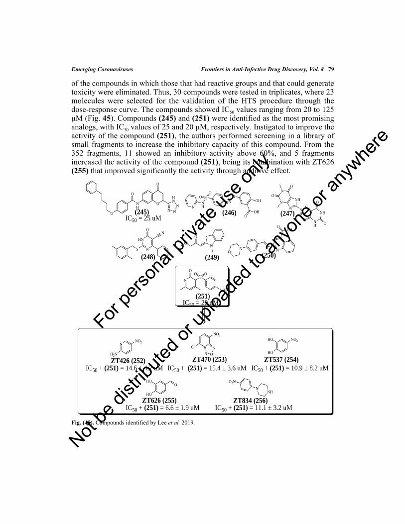
Emerging Coronaviruses Frontiers in Anti-Infective Drug Discovery, Vol. 8 79
of the compounds in which those that had reactive groups and that could generatetoxicity were eliminated. Thus, 30 compounds were tested in triplicates, where 23molecules were selected for the validation of the HTS procedure through thedose-response curve. The compounds showed IC50 values ranging from 20 to 125μM (Fig. 45). Compounds (245) and (251) were identified as the most promisinganalogs, with IC50 values of 25 and 20 μM, respectively. Instigated to improve theactivity of the compound (251), the authors performed screening in a library ofsmall fragments to increase the inhibitory capacity of this compound. From the352 fragments, 11 showed an inhibitory activity above 60%, and 5 fragmentsincreased the activity of the compound (251), being its combination with ZT626(255) that improved significantly the activity through additive effect.
Fig. (45). Compounds identified by Lee et al. 2019.
O
NH
O
O
O
N NN
HN
(245)N
NH
SO
ON
NOH
OOH(246)
N
N
N
NH
NH
O
O
N
NH
NH
O
OO
(247)
S N
HN
ON
(248)
O
O
N
S
(249)
NS
OO
NH2
O
(251)
ON N
OOH
(250)
IC50 = 25 uM
IC50 = 20 uM
N
H2N
NO2
IC50 + (251) = 14.6 ± 4.2 uMZT426 (252)
N O
NCl
NO2
ZT470 (253)IC50 + (251) = 15.4 ± 3.6 uM
NO2HO
HO
ZT537 (254)IC50 + (251) = 10.9 ± 8.2 uM
HO
HO
O
ZT626 (255)IC50 + (251) = 6.6 ± 1.9 uM
N
O2N
NH
ZT834 (256)IC50 + (251) = 11.1 ± 3.2 uM
For pe
rsona
l priv
ate us
e only
Not be
distr
ibuted
or up
loade
d to a
nyon
e or a
nywhe
re

Newly declared SARS-CoV-2 pandemic represents a global health emergency,which is having severe consequences for the lives of infected individuals, as wellas an economic recession of worldwide proportions since China is a strategiccountry for the productive architecture of all continents. This worldwide problemis aggravated by the fact that currently there is no pharmacological treatmentavailable on the market, as well as licensed antiviral vaccines. However, severalstudies demonstrated in this chapter, whether related to natural, synthetic orcomputational chemistry, present promising molecules, with low toxicity andaddressed to enzymatic targets, which represent a trend in selectivity.
The emerging pandemic generates an urgent need for new treatments, and it isevident that the drug repurposing strategy is the most used by research groupsworldwide, as it generates satisfactory results, at a lower financial cost, given thatmost studies use virtual screening among libraries containing drugs known inclinical practice that are safe for use in humans, providing faster results andimmediate application.
Among the repurposed drugs, hopes for a cure against the SARS-CoV-2 aredeposited in drugs of class antivirals and antimalarials, such as remdesivir,nelfinavir, hydroxychloroquine, chloroquine, favipiravir, and others, representingdrugs undergoing clinical trials, in which the hopes of health agencies around theworld are placed. In this way, in the medium and long term, the SARS-CoV-2outbreak, as well as SARS and MERS demonstrate that it is necessary to reachbroad-spectrum antiviral agents as soon as possible, as well as the development ofvaccines for the prophylaxis of these infections. Finally, we believe that thisreview will be essentially important for designing and developing new antiviralmolecules, especially enzyme inhibitors, that could be more effective, safe,selective, and low-costs of production.
CONSENT FOR PUBLICATION
Not applicable.
CONFLICT OF INTEREST
The authors confirm that this chapter content has no conflict of interest.
ACKNOWLEDGEMENTS
The authors thank to Coordenação de Aperfeiçoamento Pessoal de Nível Superior(CAPES), National Council for Scientific and Technological Development(CNPq), Fundação de Amparo à Pesquisa do Estado de Alagoas (FAPEAL), and
80 Frontiers in Anti-Infective Drug Discovery, Vol. 8 da Silva Santos-Júnior et al.
CONCLUSION AND FUTURE OUTLOOK
For pe
rsona
l priv
ate us
e only
Not be
distr
ibuted
or up
loade
d to a
nyon
e or a
nywhe
re

Emerging Coronaviruses Frontiers in Anti-Infective Drug Discovery, Vol. 8 81
Financier of Studies and Projects (FINEP) for their support to the Post-GraduateProgram in Chemistry and Biotechnology (PPGQB).
REFERENCES[1] Azhar EI, Hui DSC, Memish ZA, Drosten C, Zumla A. The Middle East Respiratory Syndrome
(MERS). Infect Dis Clin North Am 2019; 33(4): 891-905.[http://dx.doi.org/10.1016/j.idc.2019.08.001] [PMID: 31668197]
[2] Khan G, Sheek-Hussein M. Chapter 8 - The Middle East Respiratory Syndrome Coronavirus: AnEmerging Virus of Global Threat. In: Ennaji MM, Ed. Emerging and Reemerging Viral Pathogens.Cambridge: Academic Press 2020; pp 151-67
[3] Chapter 24-Coronaviridae. Fenner's Veterinary Virology Fifth Edition., MacLachlan NJ, Dubovi EJ,Eds. E Publisher: Academic Press 2017; 435-61.[http://dx.doi.org/10.1016/B978-0-12-800946-8.00024-6]
[4] Báez-Santos YM, St John SE, Mesecar AD. The SARS-coronavirus papain-like protease: structure,function and inhibition by designed antiviral compounds. Antiviral Res 2015; 115: 21-38.[http://dx.doi.org/10.1016/j.antiviral.2014.12.015] [PMID: 25554382]
[5] WHO Director-General’s opening remarks at the media briefing on COVID-19 - 11 March 2020
[6] Chen N, Zhou M, Dong X, et al. Epidemiological and clinical characteristics of 99 cases of 2019 novelcoronavirus pneumonia in Wuhan, China: a descriptive study. Lancet 2020; 395(10223): 507-13.[http://dx.doi.org/10.1016/S0140-6736(20)30211-7] [PMID: 32007143]
[7] Kasmi Y, Khataby K, Souiri A, Ennaji MM. Coronaviridae: 100,000 years of emergence andreemergence. Emerg Reemerging Viral Pathog 2020; pp. 127-49.
[8] WHO. https://www.who.int/neglected_diseases/diseases/en/
[9] Li YH, Hu CY, Wu NP, Yao HP, Li LJ. Molecular characteristics, functions, and related pathogenicityof MERS-CoV proteins. Engineering (Beijing) 2019; 5(5): 940-7.[http://dx.doi.org/10.1016/j.eng.2018.11.035] [PMID: 32288963]
[10] The coronavirus is more dangerous for the economy than SARS - Bloomberg nd 2019.
[11] China’s Xi Jinping knew of coronavirus earlier than first thought | Financial Times nd 2019.
[12] Sohrabi C, Alsafi Z, O’Neill N, et al. World health organization declares global emergency: A reviewof the 2019 novel coronavirus (COVID-19). Int J Surg 2020; 76:71-6.https://doi.org/https://doi.org/10.1016/j.ijsu.2020.02.034[http://dx.doi.org/10.1016/j.ijsu.2020.02.034] [PMID: 32112977]
[13] Pillaiyar T, Meenakshisundaram S, Manickam M. Recent discovery and development of inhibitorstargeting coronaviruses. Drug Discov Today 2020; 25(4): 668-88.[http://dx.doi.org/10.1016/j.drudis.2020.01.015] [PMID: 32006468]
[14] Gao K, Nguyen DD, Wang R, Wei G-W. Machine intelligence design of 2019-nCoV drugs 2020;2507: 1-9.
[15] Lu H. Drug treatment options for the 2019-new coronavirus (2019-nCoV). Biosci Trends 2020; 14(1):69-71.[http://dx.doi.org/10.5582/bst.2020.01020] [PMID: 31996494]
[16] RCSB PDB - 6LU7: The crystal structure of COVID-19 main protease in complex with an inhibitorN3 nd 2019.
[17] Malik YS, Sircar S, Bhat S, et al. Emerging novel coronavirus (2019-nCoV)-current scenario,evolutionary perspective based on genome analysis and recent developments. Vet Q 2020; 40(1): 68-76.[http://dx.doi.org/10.1080/01652176.2020.1727993] [PMID: 32036774]
For pe
rsona
l priv
ate us
e only
Not be
distr
ibuted
or up
loade
d to a
nyon
e or a
nywhe
re

82 Frontiers in Anti-Infective Drug Discovery, Vol. 8 da Silva Santos-Júnior et al.
[18] Beigel JH, Nam HH, Adams PL, et al. Advances in respiratory virus therapeutics - A meeting reportfrom the 6th isirv Antiviral Group conference. Antiviral Res 2019; 167: 45-67.[http://dx.doi.org/10.1016/j.antiviral.2019.04.006] [PMID: 30974127]
[19] Huang C, Wang Y, Li X, et al. Clinical features of patients infected with 2019 novel coronavirus inWuhan, China. Lancet 2020; 395(10223): 497-506.[http://dx.doi.org/10.1016/S0140-6736(20)30183-5] [PMID: 31986264]
[20] Li Y, Zhang J, Wang N, et al. Therapeutic drugs targeting 2019-ncov main protease by high-throughput screening. BioRxiv 2020; 922922.
[21] Kruse RL. Therapeutic strategies in an outbreak scenario to treat the novel coronavirus originating inWuhan, China. F1000 Res 2020; 9: 72.[http://dx.doi.org/10.12688/f1000research.22211.2] [PMID: 32117569]
[22] Momattin H, Al-Ali AY, Al-Tawfiq JA. A systematic review of therapeutic agents for the treatment ofthe middle east respiratory syndrome coronavirus (MERS-CoV). Travel Med Infect Dis 2019; 30: 9-18.[http://dx.doi.org/10.1016/j.tmaid.2019.06.012] [PMID: 31252170]
[23] Hu CS, Tkebuchava T. SARS and its treatment strategies. Asian Pac J Trop Med 2019; 12: 95-7.[http://dx.doi.org/10.4103/1995-7645.254934]
[24] Veljkovic V, Vergara-Alert J, Segalés J, Paessler S. Use of the informational spectrum methodologyfor rapid biological analysis of the novel coronavirus 2019-nCoV: prediction of potential receptor,natural reservoir, tropism and therapeutic/vaccine target. F1000 Res 2020; 9: 52.[http://dx.doi.org/10.12688/f1000research.22149.3] [PMID: 32419926]
[25] Jin Y-H, Cai L, Cheng Z-S, et al. A rapid advice guideline for the diagnosis and treatment of 2019novel coronavirus (2019-nCoV) infected pneumonia (standard version). Mil Med Res 2020; 7(1): 4.[http://dx.doi.org/10.1186/s40779-020-0233-6] [PMID: 32029004]
[26] Chang L, Yan Y, Wang L. Coronavirus disease 2019: Coronaviruses and blood safety. Transfus MedRev 2020; 34(2): 75-80.https://doi.org/https://doi.org/10.1016/j.tmrv.2020.02.003[http://dx.doi.org/10.1016/j.tmrv.2020.02.003] [PMID: 32107119]
[27] Lai CC, Liu YH, Wang CY, et al. Asymptomatic carrier state, acute respiratory disease, andpneumonia due to severe acute respiratory syndrome coronavirus 2 (SARS-CoV-2): Facts and myths. JMicrobiol Immunol Infect 2020; 53(3): 404-12.[http://dx.doi.org/10.1016/j.jmii.2020.02.012] [PMID: 32173241]
[28] Rothan HA, Byrareddy SN. The epidemiology and pathogenesis of coronavirus disease (COVID-19)outbreak. J Autoimmun 2020; 109: 102433. https://doi.org/https://doi.org/10.1016/j.jaut.2020.102433[http://dx.doi.org/10.1016/j.jaut.2020.102433] [PMID: 32113704]
[29] Lupia T, Scabini S, Mornese Pinna S, Di Perri G, De Rosa FG, Corcione S. 2019 novel coronavirus(2019-nCoV) outbreak: A new challenge. J Glob Antimicrob Resist 2020; 21: 22-7.[http://dx.doi.org/10.1016/j.jgar.2020.02.021] [PMID: 32156648]
[30] Lai C-C, Shih T-P, Ko W-C, Tang H-J, Hsueh P-R. Severe acute respiratory syndrome coronavirus 2(SARS-CoV-2) and coronavirus disease-2019 (COVID-19): The epidemic and the challenges. Int JAntimicrob Agents 2020; 55(3): 105924.[http://dx.doi.org/10.1016/j.ijantimicag.2020.105924] [PMID: 32081636]
[31] Farooq HZ, Davies E, Ahmad S, et al. Middle east respiratory syndrome coronavirus (MERS-CoV) -Surveillance and testing in North England from 2012 to 2019. Int J Infect Dis 2020; 93: 237-44.[http://dx.doi.org/10.1016/j.ijid.2020.01.043] [PMID: 32004690]
[32] Tang B, Bragazzi NL, Li Q, Tang S, Xiao Y, Wu J. An updated estimation of the risk of transmissionof the novel coronavirus (2019-nCov). Infect Dis Model 2020; 5: 248-55.[http://dx.doi.org/10.1016/j.idm.2020.02.001] [PMID: 32099934]
For pe
rsona
l priv
ate us
e only
Not be
distr
ibuted
or up
loade
d to a
nyon
e or a
nywhe
re

Emerging Coronaviruses Frontiers in Anti-Infective Drug Discovery, Vol. 8 83
[33] Kumar S. Drug and vaccine design against novel coronavirus (2019-nCoV) spike protein throughcomputational approach. Preprints 2020; 2020020071.
[34] Yang Y, Peng F, Wang R, et al. The deadly coronaviruses: The 2003 SARS pandemic and the 2020novel coronavirus epidemic in China. J Autoimmun 2020; 109: 102434.[http://dx.doi.org/10.1016/j.jaut.2020.102434] [PMID: 32143990]
[35] Mizumoto K, Chowell G. Transmission potential of the New Corona (COVID-19) onboard thePrincess Cruises Ship, 2020. MedRxiv 2020; 5: 20027649.[http://dx.doi.org/10.1101/2020.02.24.20027649]
[36] Li X, Geng M, Peng Y, Meng L, Lu S. Molecular immune pathogenesis and diagnosis of COVID-19. JPharm Anal 2020; 10(2): 102-8.[http://dx.doi.org/10.1016/j.jpha.2020.03.001] [PMID: 32282863]
[37] Shen M, Zhou Y, Ye J, et al. Recent advances and perspectives of nucleic acid detection forcoronavirus. J Pharm Anal 2020; 10(2): 97-101.[http://dx.doi.org/10.1016/j.jpha.2020.02.010] [PMID: 32292623]
[38] Xie C, Jiang L, Huang G, et al. Comparison of different samples for 2019 novel coronavirus detectionby nucleic acid amplification tests. Int J Infect Dis 2020; 93: 264-7.[http://dx.doi.org/10.1016/j.ijid.2020.02.050] [PMID: 32114193]
[39] Phan T. Novel coronavirus: From discovery to clinical diagnostics. Infect Genet Evol 2020; 79:104211.[http://dx.doi.org/10.1016/j.meegid.2020.104211] [PMID: 32007627]
[40] Al-Tawfiq JA, Memish ZA. Diagnosis of SARS-CoV-2 infection based on CT scan vs RT-PCR:reflecting on experience from MERS-CoV. J Hosp Infect 2020; 105(2): 154-5.[http://dx.doi.org/10.1016/j.jhin.2020.03.001] [PMID: 32147407]
[41] Cordes AK, Heim A. Rapid random access detection of the novel SARS-coronavirus-2 (SARS-CoV-2,previously 2019-nCoV) using an open access protocol for the Panther Fusion. J Clin Virol 2020; 125:104305.[http://dx.doi.org/10.1016/j.jcv.2020.104305] [PMID: 32143123]
[42] Rebensburg S, Helfer M, Schneider M, et al. Potent in vitro antiviral activity of Cistus incanus extractagainst HIV and Filoviruses targets viral envelope proteins. Sci Rep 2016; 6: 20394.[http://dx.doi.org/10.1038/srep20394] [PMID: 26833261]
[43] Shaikh F, Zhao Y, Alvarez L, et al. Structure-Based In silico screening identifies a potent ebolavirusinhibitor from a traditional chinese medicine library. J Med Chem 2019; 62(6): 2928-37.[http://dx.doi.org/10.1021/acs.jmedchem.8b01328] [PMID: 30785281]
[44] Ling CQ. Traditional chinese medicine is a resource for drug discovery against 2019 novelcoronavirus (SARS-CoV-2). J Integr Med 2020; 18(2): 87-8.[http://dx.doi.org/10.1016/j.joim.2020.02.004] [PMID: 32122812]
[45] Zhang DH, Wu KL, Zhang X, Deng SQ, Peng B. In silico screening of Chinese herbal medicines withthe potential to directly inhibit 2019 novel coronavirus. J Integr Med 2020; 18(2): 152-8.[http://dx.doi.org/10.1016/j.joim.2020.02.005] [PMID: 32113846]
[46] Cinatl J, Morgenstern B, Bauer G, Chandra P, Rabenau H, Doerr HW. Glycyrrhizin, an activecomponent of liquorice roots, and replication of SARS-associated coronavirus. Lancet 2003;361(9374): 2045-6.[http://dx.doi.org/10.1016/S0140-6736(03)13615-X] [PMID: 12814717]
[47] Chen F, Chan KH, Jiang Y, et al. In vitro susceptibility of 10 clinical isolates of SARS coronavirus toselected antiviral compounds. J Clin Virol 2004; 31(1): 69-75.[http://dx.doi.org/10.1016/j.jcv.2004.03.003] [PMID: 15288617]
[48] Li SY, Chen C, Zhang HQ, et al. Identification of natural compounds with antiviral activities against
For pe
rsona
l priv
ate us
e only
Not be
distr
ibuted
or up
loade
d to a
nyon
e or a
nywhe
re

84 Frontiers in Anti-Infective Drug Discovery, Vol. 8 da Silva Santos-Júnior et al.
SARS-associated coronavirus. Antiviral Res 2005; 67(1): 18-23.[http://dx.doi.org/10.1016/j.antiviral.2005.02.007] [PMID: 15885816]
[49] Mishra A, Behura A, Mawatwal S, et al. Structure-function and application of plant lectins in diseasebiology and immunity. Food Chem Toxicol 2019; 134: 110827.[http://dx.doi.org/10.1016/j.fct.2019.110827] [PMID: 31542433]
[50] Ribeiro AC, Ferreira R, Freitas R. Chapter 1-Plant Lectins: Bioactivities and Bioapplications. Studiesin Natural Products Chemistry. 1st ed. Elsevier B.V. 2018; pp. 1-42.
[51] Mitchell CA, Ramessar K, O’Keefe BR. Antiviral lectins: Selective inhibitors of viral entry. AntiviralRes 2017; 142: 37-54.[http://dx.doi.org/10.1016/j.antiviral.2017.03.007] [PMID: 28322922]
[52] Kaur R, Neetu , Mudgal R, Jose J, Kumar P, Tomar S. Glycan-dependent chikungunya viral infectiondivulged by antiviral activity of NAG specific chi-like lectin. Virology 2019; 526: 91-8.[http://dx.doi.org/10.1016/j.virol.2018.10.009] [PMID: 30388630]
[53] Keyaerts E, Vijgen L, Pannecouque C, et al. Plant lectins are potent inhibitors of coronaviruses byinterfering with two targets in the viral replication cycle. Antiviral Res 2007; 75(3): 179-87.[http://dx.doi.org/10.1016/j.antiviral.2007.03.003] [PMID: 17428553]
[54] Kim HY, Shin HS, Park H, et al. In vitro inhibition of coronavirus replications by the traditionallyused medicinal herbal extracts, Cimicifuga rhizoma, Meliae cortex, Coptidis rhizoma, andPhellodendron cortex. J Clin Virol 2008; 41(2): 122-8.[http://dx.doi.org/10.1016/j.jcv.2007.10.011] [PMID: 18036887]
[55] Ryu YB, Jeong HJ, Kim JH, et al. Biflavonoids from Torreya nucifera displaying SARS-CoV3CL(pro) inhibition. Bioorg Med Chem 2010; 18(22): 7940-7.[http://dx.doi.org/10.1016/j.bmc.2010.09.035] [PMID: 20934345]
[56] Galasiti Kankanamalage AC, Kim Y, Damalanka VC, et al. Structure-guided design of potent andpermeable inhibitors of MERS coronavirus 3CL protease that utilize a piperidine moiety as a noveldesign element. Eur J Med Chem 2018; 150: 334-46.[http://dx.doi.org/10.1016/j.ejmech.2018.03.004] [PMID: 29544147]
[57] Lim L, Gupta G, Roy A, et al. Structurally- and dynamically-driven allostery of the chymotrypsin-likeproteases of SARS, Dengue and Zika viruses. Prog Biophys Mol Biol 2019; 143: 52-66.[http://dx.doi.org/10.1016/j.pbiomolbio.2018.08.009] [PMID: 30217495]
[58] Park JY, Kim JH, Kim YM, et al. Tanshinones as selective and slow-binding inhibitors for SARS-CoV cysteine proteases. Bioorg Med Chem 2012; 20(19): 5928-35.[http://dx.doi.org/10.1016/j.bmc.2012.07.038] [PMID: 22884354]
[59] Park JY, Kim JH, Kwon JM, et al. Dieckol, a SARS-CoV 3CL(pro) inhibitor, isolated from the ediblebrown algae Ecklonia cava. Bioorg Med Chem 2013; 21(13): 3730-7.[http://dx.doi.org/10.1016/j.bmc.2013.04.026] [PMID: 23647823]
[60] Kim DE, Min JS, Jang MS, et al. Natural bis-benzylisoquinoline alkaloids-tetrandrine, fangchinoline,and cepharanthine, inhibit human coronavirus oc43 infection of mrc-5 human lung cells. Biomolecules2019; 9(11): E696.[http://dx.doi.org/10.3390/biom9110696] [PMID: 31690059]
[61] Semwal DK, Badoni R, Semwal R, Kothiyal SK, Singh GJP, Rawat U. The genus stephania(Menispermaceae): Chemical and pharmacological perspectives. J Ethnopharmacol 2010; 132(2): 369-83.[http://dx.doi.org/10.1016/j.jep.2010.08.047] [PMID: 20801207]
[62] Wang ST, Qian WQ, He P, Feng MQ, Kang Y, Wang YQ, et al. Two new glycoalkaloids fromStephania succifera. Phytochem Lett 2019; 34: 99-102.[http://dx.doi.org/10.1016/j.phytol.2019.10.001]
[63] Wang R, Liu Y, Shi G, et al. Bioactive bisbenzylisoquinoline alkaloids from the roots of Stephania
For pe
rsona
l priv
ate us
e only
Not be
distr
ibuted
or up
loade
d to a
nyon
e or a
nywhe
re

Emerging Coronaviruses Frontiers in Anti-Infective Drug Discovery, Vol. 8 85
tetrandra 2020; 98: 103697.[http://dx.doi.org/10.1016/j.bioorg.2020.103697]
[64] Keck JG, Wang PH, Lien EJ, Lai MMC. Inhibition of murine coronavirus RNA synthesis byhydroxyguanidine derivatives. Virus Res 1989; 14(1): 57-63.[http://dx.doi.org/10.1016/0168-1702(89)90069-5] [PMID: 2554614]
[65] Caliguiri LA, Tamm I. Guanidine. Oxford: Pergamon Press 1972; 1: pp. 181-230.
[66] Martina E, Stiefl N, Degel B, et al. Screening of electrophilic compounds yields an aziridinyl peptideas new active-site directed SARS-CoV main protease inhibitor. Bioorg Med Chem Lett 2005; 15(24):5365-9.[http://dx.doi.org/10.1016/j.bmcl.2005.09.012] [PMID: 16216498]
[67] Chen LR, Wang YC, Lin YW, et al. Synthesis and evaluation of isatin derivatives as effective SARScoronavirus 3CL protease inhibitors. Bioorg Med Chem Lett 2005; 15(12): 3058-62.[http://dx.doi.org/10.1016/j.bmcl.2005.04.027] [PMID: 15896959]
[68] Shie JJ, Fang JM, Kuo TH, et al. Inhibition of the severe acute respiratory syndrome 3CL protease bypeptidomimetic α,β-unsaturated esters. Bioorg Med Chem 2005; 13(17): 5240-52.[http://dx.doi.org/10.1016/j.bmc.2005.05.065] [PMID: 15994085]
[69] Xu Z, Zhao SJ, Lv ZS, et al. Fluoroquinolone-isatin hybrids and their biological activities. Eur J MedChem 2019; 162: 396-406.[http://dx.doi.org/10.1016/j.ejmech.2018.11.032] [PMID: 30453247]
[70] Chou CY, Chien CH, Han YS, et al. Thiopurine analogues inhibit papain-like protease of severe acuterespiratory syndrome coronavirus. Biochem Pharmacol 2008; 75(8): 1601-9.[http://dx.doi.org/10.1016/j.bcp.2008.01.005] [PMID: 18313035]
[71] Parmet S. Childhood Leukemia. The Journal of the American Medical Association 2004; 291(4): 514.[http://dx.doi.org/10.1001/jama.291.4.514]
[72] Lee C, Lee JM, Lee NR, et al. Aryl diketoacids (ADK) selectively inhibit duplex DNA-unwindingactivity of SARS coronavirus NTPase/helicase. Bioorg Med Chem Lett 2009; 19(6): 1636-8.[http://dx.doi.org/10.1016/j.bmcl.2009.02.010] [PMID: 19233643]
[73] Keum YS, Jeong YJ. Development of chemical inhibitors of the SARS coronavirus: viral helicase as apotential target. Biochem Pharmacol 2012; 84(10): 1351-8.[http://dx.doi.org/10.1016/j.bcp.2012.08.012] [PMID: 22935448]
[74] Yang CW, Lee YZ, Kang IJ, et al. Identification of phenanthroindolizines andphenanthroquinolizidines as novel potent anti-coronaviral agents for porcine enteropathogeniccoronavirus transmissible gastroenteritis virus and human severe acute respiratory syndromecoronavirus. Antiviral Res 2010; 88(2): 160-8.[http://dx.doi.org/10.1016/j.antiviral.2010.08.009] [PMID: 20727913]
[75] Sen S, Chakraborty R. Revival, modernization and integration of Indian traditional herbal medicine inclinical practice: Importance, challenges and future. J Tradit Complement Med 2016; 7(2): 234-44.[http://dx.doi.org/10.1016/j.jtcme.2016.05.006] [PMID: 28417092]
[76] Balasubramanian G, Sarathi M, Kumar SR, Hameed ASS. Screening the antiviral activity of Indianmedicinal plants against white spot syndrome virus in shrimp. Aquaculture 2007; 263: 15-9.[http://dx.doi.org/10.1016/j.aquaculture.2006.09.037]
[77] Zhao Z, Dai X, Li C, et al. Pyrazolone structural motif in medicinal chemistry: Retrospect andprospect. Eur J Med Chem 2020; 186: 111893.[http://dx.doi.org/10.1016/j.ejmech.2019.111893] [PMID: 31761383]
[78] Ramajayam R, Tan KP, Liu HG, Liang PH. Synthesis and evaluation of pyrazolone compounds asSARS-coronavirus 3C-like protease inhibitors. Bioorg Med Chem 2010; 18(22): 7849-54.[http://dx.doi.org/10.1016/j.bmc.2010.09.050] [PMID: 20947359]
For pe
rsona
l priv
ate us
e only
Not be
distr
ibuted
or up
loade
d to a
nyon
e or a
nywhe
re

86 Frontiers in Anti-Infective Drug Discovery, Vol. 8 da Silva Santos-Júnior et al.
[79] Kim MK, Yu MS, Park HR, et al. 2,6-Bis-arylmethyloxy-5-hydroxychromones with antiviral activityagainst both hepatitis C virus (HCV) and SARS-associated coronavirus (SCV). Eur J Med Chem 2011;46(11): 5698-704.[http://dx.doi.org/10.1016/j.ejmech.2011.09.005] [PMID: 21925774]
[80] Chuck CP, Chen C, Ke Z, Wan DC, Chow HF, Wong KB. Design, synthesis and crystallographicanalysis of nitrile-based broad-spectrum peptidomimetic inhibitors for coronavirus 3C-like proteases.Eur J Med Chem 2013; 59: 1-6.[http://dx.doi.org/10.1016/j.ejmech.2012.10.053] [PMID: 23202846]
[81] Ábrányi-Balogh P, Petri L, Imre T, et al. A road map for prioritizing warheads for cysteine targetingcovalent inhibitors. Eur J Med Chem 2018; 160: 94-107.[http://dx.doi.org/10.1016/j.ejmech.2018.10.010] [PMID: 30321804]
[82] Silva DG, Ribeiro JFR, De Vita D, et al. A comparative study of warheads for design of cysteineprotease inhibitors. Bioorg Med Chem Lett 2017; 27(22): 5031-5.[http://dx.doi.org/10.1016/j.bmcl.2017.10.002] [PMID: 29054358]
[83] Prior AM, Kim Y, Weerasekara S, et al. Design, synthesis, and bioevaluation of viral 3C and 3C-likeprotease inhibitors. Bioorg Med Chem Lett 2013; 23(23): 6317-20.[http://dx.doi.org/10.1016/j.bmcl.2013.09.070] [PMID: 24125888]
[84] Ku T, Lopresti N, Shirley M, et al. Synthesis of distal and proximal fleximer base analogues andevaluation in the nucleocapsid protein of HIV-1. Bioorg Med Chem 2019; 27(13): 2883-92.[http://dx.doi.org/10.1016/j.bmc.2019.05.019] [PMID: 31126822]
[85] Peters HL, Jochmans D, de Wilde AH, et al. Design, synthesis and evaluation of a series of acyclicfleximer nucleoside analogues with anti-coronavirus activity. Bioorg Med Chem Lett 2015; 25(15):2923-6.[http://dx.doi.org/10.1016/j.bmcl.2015.05.039] [PMID: 26048809]
[86] Kumar V, Tan KP, Wang YM, Lin SW, Liang PH. Identification, synthesis and evaluation of SARS-CoV and MERS-CoV 3C-like protease inhibitors. Bioorg Med Chem 2016; 24(13): 3035-42.[http://dx.doi.org/10.1016/j.bmc.2016.05.013] [PMID: 27240464]
[87] Chan HCS, Shan H, Dahoun T, Vogel H, Yuan S. Advancing drug discovery via artificial intelligence.Trends Pharmacol Sci 2019; 40(8): 592-604.[http://dx.doi.org/10.1016/j.tips.2019.06.004] [PMID: 31320117]
[88] Cerqueira NMFSA, Gesto D, Oliveira EF, et al. Receptor-based virtual screening protocol for drugdiscovery. Arch Biochem Biophys 2015; 582: 56-67.[http://dx.doi.org/10.1016/j.abb.2015.05.011] [PMID: 26045247]
[89] Beck KR, Kaserer T, Schuster D, Odermatt A. Virtual screening applications in short-chaindehydrogenase/reductase research. J Steroid Biochem Mol Biol 2017; 171: 157-77.[http://dx.doi.org/10.1016/j.jsbmb.2017.03.008] [PMID: 28286207]
[90] Murgueitio MS, Bermudez M, Mortier J, Wolber G. In silico virtual screening approaches for anti-viral drug discovery. Drug Discov Today Technol 2012; 9(3): e219-25.[http://dx.doi.org/10.1016/j.ddtec.2012.07.009] [PMID: 24990575]
[91] Danishuddin M, Khan AU. Structure based virtual screening to discover putative drug candidates:necessary considerations and successful case studies. Methods 2015; 71: 135-45.[http://dx.doi.org/10.1016/j.ymeth.2014.10.019] [PMID: 25448480]
[92] Slater O, Kontoyianni M. The compromise of virtual screening and its impact on drug discovery.Expert Opin Drug Discov 2019; 14(7): 619-37.[http://dx.doi.org/10.1080/17460441.2019.1604677] [PMID: 31025886]
[93] Panteleev J, Gao H, Jia L. Recent applications of machine learning in medicinal chemistry. BioorgMed Chem Lett 2018; 28(17): 2807-15.[http://dx.doi.org/10.1016/j.bmcl.2018.06.046] [PMID: 30122222]
For pe
rsona
l priv
ate us
e only
Not be
distr
ibuted
or up
loade
d to a
nyon
e or a
nywhe
re

Emerging Coronaviruses Frontiers in Anti-Infective Drug Discovery, Vol. 8 87
[94] Ghosh AK, Xi K, Johnson ME, Baker SC, Mesecar AD. Progress in Anti-SARS CoronavirusChemistry, Biology and Chemotherapy. Annu Rep Med Chem 2007; 41: 183-96.[http://dx.doi.org/10.1016/S0065-7743(06)41011-3] [PMID: 19649165]
[95] Xu Z, Peng C, Shi Y, et al. Nelfinavir was predicted to be a potential inhibitor of 2019 nCov mainprotease by an integrative approach combining homology modelling, molecular docking and bindingfree energy calculation. bioRxiv 2020.[http://dx.doi.org/10.1101/2020.01.27.921627]
[96] Stoermer M. Homology Models of the Papain-Like Protease PLpro from Coronavirus 2019. nCoV2020.[http://dx.doi.org/10.26434/chemrxiv.11799705.v1.]
[97] Pruijssers AJ, Denison MR. Nucleoside analogues for the treatment of coronavirus infections. CurrOpin Virol 2019; 35: 57-62.[http://dx.doi.org/10.1016/j.coviro.2019.04.002] [PMID: 31125806]
[98] Chou K-C, Wei D-Q, Zhong W-Z. Binding mechanism of coronavirus main proteinase with ligandsand its implication to drug design against SARS. Biochem Biophys Res Commun 2003; 308(1): 148-51.[http://dx.doi.org/10.1016/S0006-291X(03)01342-1] [PMID: 12890493]
[99] Niu C, Yin J, Zhang J, Vederas JC, James MNG. Molecular docking identifies the binding of 3-chloropyridine moieties specifically to the S1 pocket of SARS-CoV Mpro. Bioorg Med Chem 2008;16(1): 293-302.[http://dx.doi.org/10.1016/j.bmc.2007.09.034] [PMID: 17931870]
[100] Mukherjee P, Desai P, Ross L, White EL, Avery MA. Structure-based virtual screening against SARS-3CL(pro) to identify novel non-peptidic hits. Bioorg Med Chem 2008; 16(7): 4138-49.[http://dx.doi.org/10.1016/j.bmc.2008.01.011] [PMID: 18343121]
[101] Nguyen TT, Ryu HJ, Lee SH, et al. Virtual screening identification of novel severe acute respiratorysyndrome 3C-like protease inhibitors and in vitro confirmation. Bioorg Med Chem Lett 2011; 21(10):3088-91.[http://dx.doi.org/10.1016/j.bmcl.2011.03.034] [PMID: 21470860]
[102] Zhu L, George S, Schmidt MF, Al-Gharabli SI, Rademann J, Hilgenfeld R. Peptide aldehyde inhibitorschallenge the substrate specificity of the SARS-coronavirus main protease. Antiviral Res 2011; 92(2):204-12.[http://dx.doi.org/10.1016/j.antiviral.2011.08.001] [PMID: 21854807]
[103] Turlington M, Chun A, Tomar S, et al. Discovery of N-(benzo[1,2,3]triazol-1-y-)-N-(benzyl)acetamido)phenyl) carboxamides as severe acute respiratory syndrome coronavirus(SARS-CoV) 3CLpro inhibitors: identification of ML300 and noncovalent nanomolar inhibitors withan induced-fit binding. Bioorg Med Chem Lett 2013; 23(22): 6172-7.[http://dx.doi.org/10.1016/j.bmcl.2013.08.112] [PMID: 24080461]
[104] Lee H, Mittal A, Patel K, et al. Identification of novel drug scaffolds for inhibition of SARS-CoV 3-Chymotrypsin-like protease using virtual and high-throughput screenings. Bioorg Med Chem 2014;22(1): 167-77.[http://dx.doi.org/10.1016/j.bmc.2013.11.041] [PMID: 24332657]
[105] Wang L, Bao BB, Song GQ, et al. Discovery of unsymmetrical aromatic disulfides as novel inhibitorsof SARS-CoV main protease: Chemical synthesis, biological evaluation, molecular docking and 3D-QSAR study. Eur J Med Chem 2017; 137: 450-61.[http://dx.doi.org/10.1016/j.ejmech.2017.05.045] [PMID: 28624700]
[106] Theerawatanasirikul S, Kuo CJ, Phetcharat N, Lekcharoensuk P. In silico and in vitro analysis of smallmolecules and natural compounds targeting the 3CL protease of feline infectious peritonitis virus.Antiviral Res 2020; 174: 104697.[http://dx.doi.org/10.1016/j.antiviral.2019.104697] [PMID: 31863793]
For pe
rsona
l priv
ate us
e only
Not be
distr
ibuted
or up
loade
d to a
nyon
e or a
nywhe
re

88 Frontiers in Anti-Infective Drug Discovery, Vol. 8 da Silva Santos-Júnior et al.
[107] Abdallah HH. Theoretical study for the inhibition ability of some bioactive imidazole derivativesagainst the Middle-East respiratory syndrome corona virus (MERS-Co). Zanco J Pure Appl Sci 2019;31[http://dx.doi.org/10.21271/zjpas.31.2.10]
[108] Mercorelli B, Palù G, Loregian A. Drug Repurposing for Viral Infectious Diseases: How Far Are We?Trends Microbiol 2018; 26(10): 865-76.[http://dx.doi.org/10.1016/j.tim.2018.04.004] [PMID: 29759926]
[109] Bellera CL, Balcazar DE, Vanrell MC, et al. Computer-guided drug repurposing: identification oftrypanocidal activity of clofazimine, benidipine and saquinavir. Eur J Med Chem 2015; 93: 338-48.[http://dx.doi.org/10.1016/j.ejmech.2015.01.065] [PMID: 25707014]
[110] Bellera CL, Balcazar DE, Alberca L, Labriola CA, Talevi A, Carrillo C. Application of computer-aided drug repurposing in the search of new cruzipain inhibitors: discovery of amiodarone andbromocriptine inhibitory effects. J Chem Inf Model 2013; 53(9): 2402-8.[http://dx.doi.org/10.1021/ci400284v] [PMID: 23906322]
[111] Kumar R, Harilal S, Gupta SV, et al. Exploring the new horizons of drug repurposing: A vital tool forturning hard work into smart work. Eur J Med Chem 2019; 182: 111602.[http://dx.doi.org/10.1016/j.ejmech.2019.111602] [PMID: 31421629]
[112] Zhavoronkov A, Aladinskiy V, Zhebrak A, Zagribelnyy B, Terentiev V, Bezrukov DS, et al. Potential2019-nCoV 3C-like Protease Inhibitors Designed Using Generative Deep Learning Approaches 2019.[http://dx.doi.org/10.26434/CHEMRXIV.11829102.V1]
[113] Mustafa S, Balkhy H, Gabere MN. Current treatment options and the role of peptides as potentialtherapeutic components for Middle East Respiratory Syndrome (MERS): A review. J Infect PublicHealth 2018; 11(1): 9-17.[http://dx.doi.org/10.1016/j.jiph.2017.08.009] [PMID: 28864360]
[114] Zhang XW, Yap YL. Old drugs as lead compounds for a new disease? Binding analysis of SARScoronavirus main proteinase with HIV, psychotic and parasite drugs. Bioorg Med Chem 2004; 12(10):2517-21.[http://dx.doi.org/10.1016/j.bmc.2004.03.035] [PMID: 15110833]
[115] Wolber G, Seidel T, Bendix F, Langer T. Molecule-pharmacophore superpositioning and patternmatching in computational drug design. Drug Discov Today 2008; 13(1-2): 23-9.[http://dx.doi.org/10.1016/j.drudis.2007.09.007] [PMID: 18190860]
[116] Yang SY. Pharmacophore modeling and applications in drug discovery: challenges and recentadvances. Drug Discov Today 2010; 15(11-12): 444-50.[http://dx.doi.org/10.1016/j.drudis.2010.03.013] [PMID: 20362693]
[117] Zhang XW, Yap YL, Altmeyer RM. Generation of predictive pharmacophore model for SARS-coronavirus main proteinase. Eur J Med Chem 2005; 40(1): 57-62.[http://dx.doi.org/10.1016/j.ejmech.2004.09.013] [PMID: 15642409]
[118] Al-Tawfiq JA, Momattin H, Dib J, Memish ZA. Ribavirin and interferon therapy in patients infectedwith the Middle East respiratory syndrome coronavirus: an observational study. Int J Infect Dis 2014;20: 42-6.[http://dx.doi.org/10.1016/j.ijid.2013.12.003] [PMID: 24406736]
[119] Sheahan TP, Sims AC, Leist SR, et al. Comparative therapeutic efficacy of remdesivir andcombination lopinavir, ritonavir, and interferon beta against MERS-CoV. Nat Commun 2020; 11(1):222.[http://dx.doi.org/10.1038/s41467-019-13940-6] [PMID: 31924756]
[120] Li G, De Clercq E. Therapeutic options for the 2019 novel coronavirus (2019-nCoV). Nat Rev DrugDiscov 2020; 19(3): 149-50.[http://dx.doi.org/10.1038/d41573-020-00016-0] [PMID: 32127666]
For pe
rsona
l priv
ate us
e only
Not be
distr
ibuted
or up
loade
d to a
nyon
e or a
nywhe
re

Emerging Coronaviruses Frontiers in Anti-Infective Drug Discovery, Vol. 8 89
[121] Zhou Y, Hou Y, Shen J, Huang Y, Martin W, Cheng F. Network-based Drug Repurposing for HumanCoronavirus 2020.[http://dx.doi.org/10.1101/2020.02.03.20020263]
[122] Wang M, Cao R, Zhang L, et al. Remdesivir and chloroquine effectively inhibit the recently emergednovel coronavirus (2019-nCoV) in vitro. Cell Res 2020; 30(3): 269-71.[http://dx.doi.org/10.1038/s41422-020-0282-0] [PMID: 32020029]
[123] Keyaerts E, Vijgen L, Maes P, Neyts J, Van Ranst M. In vitro inhibition of severe acute respiratorysyndrome coronavirus by chloroquine. Biochem Biophys Res Commun 2004; 323(1): 264-8.[http://dx.doi.org/10.1016/j.bbrc.2004.08.085] [PMID: 15351731]
[124] Rossignol JF. Nitazoxanide, a new drug candidate for the treatment of Middle East respiratorysyndrome coronavirus. J Infect Public Health 2016; 9(3): 227-30.[http://dx.doi.org/10.1016/j.jiph.2016.04.001] [PMID: 27095301]
[125] Chen YW, Yiu CB, Wong K. Prediction of the 2019-nCoV 3C-like protease ( 3CL pro ) structure :virtual screening reveals velpatasvir , ledipasvir , and other drug repurposing candidates 2019; 1-15.
[126] Chen H, Engkvist O, Wang Y, Olivecrona M, Blaschke T. The rise of deep learning in drug discovery.Drug Discov Today 2018; 23(6): 1241-50.[http://dx.doi.org/10.1016/j.drudis.2018.01.039] [PMID: 29366762]
[127] Lavecchia A. Deep learning in drug discovery: opportunities, challenges and future prospects. DrugDiscov Today 2019; 24(10): 2017-32.[http://dx.doi.org/10.1016/j.drudis.2019.07.006] [PMID: 31377227]
[128] Zhang H, Saravanan KM, Yang Y, Hossain T. Deep learning based drug screening for novelcoronavirus 2019-nCov 2020; 19: 1-17.
[129] de Wilde AH, Falzarano D, Zevenhoven-Dobbe JC, et al. Alisporivir inhibits MERS- and SARS-coronavirus replication in cell culture, but not SARS-coronavirus infection in a mouse model. VirusRes 2017; 228: 7-13.[http://dx.doi.org/10.1016/j.virusres.2016.11.011] [PMID: 27840112]
[130] Lin MH, Moses DC, Hsieh CH, et al. Disulfiram can inhibit MERS and SARS coronavirus papain-likeproteases via different modes. Antiviral Res 2018; 150: 155-63.[http://dx.doi.org/10.1016/j.antiviral.2017.12.015] [PMID: 29289665]
[131] Yang ZY, He JH, Lu AP, Hou TJ, Cao DS. Frequent hitters: nuisance artifacts in high-throughputscreening. Drug Discov Today 2020; 25(4): 657-67.[http://dx.doi.org/10.1016/j.drudis.2020.01.014] [PMID: 31987936]
[132] Volochnyuk DM, Ryabukhin SV, Moroz YS, et al. Evolution of commercially available compoundsfor HTS. Drug Discov Today 2019; 24(2): 390-402.[http://dx.doi.org/10.1016/j.drudis.2018.10.016] [PMID: 30399443]
[133] Cronk D. High-throughput screening. Drug Discov Dev Technol Transit 2013; pp. 95-117.
[134] Rizk MA, El-Sayed SAE, Nassif M, Mosqueda J, Xuan X, Igarashi I. Assay methods for in vitro andin vivo anti-Babesia drug efficacy testing: Current progress, outlook, and challenges. Vet Parasitol2020; 279: 109013.[http://dx.doi.org/10.1016/j.vetpar.2019.109013] [PMID: 32070899]
[135] Deng H, Lei Q, Wu Y, He Y, Li W. Activity-based protein profiling: Recent advances in medicinalchemistry. Eur J Med Chem 2020; 191: 112151.[http://dx.doi.org/10.1016/j.ejmech.2020.112151] [PMID: 32109778]
[136] Szymański P, Markowicz M, Mikiciuk-Olasik E. Adaptation of high-throughput screening in drugdiscovery-toxicological screening tests. Int J Mol Sci 2012; 13(1): 427-52.[http://dx.doi.org/10.3390/ijms13010427] [PMID: 22312262]
[137] Blanchard JE, Elowe NH, Huitema C, et al. High-throughput screening identifies inhibitors of the
For pe
rsona
l priv
ate us
e only
Not be
distr
ibuted
or up
loade
d to a
nyon
e or a
nywhe
re

90 Frontiers in Anti-Infective Drug Discovery, Vol. 8 da Silva Santos-Júnior et al.
SARS coronavirus main proteinase. Chem Biol 2004; 11(10): 1445-53.[http://dx.doi.org/10.1016/j.chembiol.2004.08.011] [PMID: 15489171]
[138] Liu Y-C, Huang V, Chao T-C, et al. Screening of drugs by FRET analysis identifies inhibitors ofSARS-CoV 3CL protease. Biochem Biophys Res Commun 2005; 333(1): 194-9.[http://dx.doi.org/10.1016/j.bbrc.2005.05.095] [PMID: 15950190]
[139] Ge F, Xiong S, Lin FS, Zhang ZP, Zhang XE. High-throughput assay using a GFP-expressing repliconfor SARS-CoV drug discovery. Antiviral Res 2008; 80(2): 107-13.[http://dx.doi.org/10.1016/j.antiviral.2008.05.005] [PMID: 18584889]
[140] Jacobs J, Grum-Tokars V, Zhou Y, et al. Discovery, synthesis, and structure-based optimization of aseries of N-(tert-butyl)-2-(N-arylamido)-2-(pyridin-3-yl) acetamides (ML188) as potent noncovalentsmall molecule inhibitors of the severe acute respiratory syndrome coronavirus (SARS-CoV) 3CLprotease. J Med Chem 2013; 56(2): 534-46.[http://dx.doi.org/10.1021/jm301580n] [PMID: 23231439]
[141] Lee H, Ren J, Pesavento RP, et al. Identification and design of novel small molecule inhibitors againstMERS-CoV papain-like protease via high-throughput screening and molecular modeling. Bioorg MedChem 2019; 27(10): 1981-9.[http://dx.doi.org/10.1016/j.bmc.2019.03.050] [PMID: 30940566]
For pe
rsona
l priv
ate us
e only
Not be
distr
ibuted
or up
loade
d to a
nyon
e or a
nywhe
re

Frontiers in Anti-Infective Drug Discovery, 2020, Vol. 8, 91-113 91
CHAPTER 3
Opportunities Offered by Fragment-Based DrugDesign in Antibiotic DevelopmentSanjay Yapabandara1, Lorna Wilkinson-White2, Sandro Ataide1 and Ann H.Kwan1,*
1 School of Life and Environmental Sciences, University of Sydney, NSW 2006, Australia2 Sydney Analytical Core Research Facility, University of Sydney, NSW 2006, Australia
Abstract: In recent years, the discovery of new and effective antibiotics has sloweddramatically due to the rapid and widespread development of bacterial drug resistanceand many pharmaceutical companies exiting the field. Reliance on conventional drugdiscovery methods, while effective in the past, has led to significant present-daychallenges that are becoming increasingly difficult to overcome. A fundamentalchallenge to the development of new antibiotics against multi-drug resistant bacteria isthe high cost of development relative to expected revenues. Fragment-based drugdesign (FBDD), which involves screening low molecular weight ligands, can help todrastically reduce the cost of finding initial hits compared with traditional high-throughput screening (HTS). In addition, a knowledge-driven and multi-prongedapproach to the subsequent expansion of amenable fragments into high-affinityinhibitors may assist with overcoming hurdles in the hit-to-lead (H2L) optimisationprocess. Favourable pharmacological and physicochemical properties, as well asstrategies against the development of resistance, can be incorporated as part of thefragment expansion process. This chapter discusses the features of the FBDD approachthat are relevant and beneficial for antibiotic development. Successful examples andbarriers to progress from hit discovery to H2L development, as well as patents, arepresented. Finally, the outlook for FBDD in the field of antibiotic development,including the latest FBDD advances and challenges, is discussed.
Keywords: Antibiotic, Antibacterial, Antimicrobial, Drug design, Drugdiscovery, Drug lead, FBDD, FBLD, Fragment, Hit-to-lead, H2L, Resistance.
INTRODUCTION
The discovery of antibiotics was one of the most significant medical achievementsof the 20th century and has revolutionised modern medicine. However, theirwidespread overuse and misuse have resulted in the rise of antibacterial resistance
* Corresponding author Ann H. Kwan: School of Life and Environmental Sciences, University of Sydney, NSW2006, Australia; Tel: +612 93513911; Email: [email protected]
Atta-ur-Rahman and M. Iqbal Choudhary (Eds.)All rights reserved-© 2020 Bentham Science Publishers
For pe
rsona
l priv
ate us
e only
Not be
distr
ibuted
or up
loade
d to a
nyon
e or a
nywhe
re

92 Frontiers in Anti-Infective Drug Discovery, Vol. 8 Yapabandara et al.
across all drug classes. The emergence of multi-drug resistance in bacterialpathogens is of particular concern, specifically in Gram-negative bacteria whichalready have limited treatment options. International agencies such as the WorldHealth Organization (WHO) have warned that unless urgent action is taken, theworld may be about to enter a ‘post-antibiotic’ era, where once easily treatableinfections will cause significant mortality or morbidity. It has been estimated thatas many as 5–20 novel drugs would need to enter clinical development to keeppace with the high attrition rates of our current drug discovery model [1].
Despite these dire predictions, the last three decades have seen a distinct lack ofdiscoveries of novel antibiotics. This can be partly attributed to the abandonmentof antibiotic discovery methods by major pharmaceutical companies, for reasonsthat include the lack of revenue generated from competitive pricing, short-termuse, the rapid rise of drug resistance and very high development costs. However,scientific challenges remain a fundamental cause of stagnation in the field [1].
High throughput screening (HTS) is a well-established and commonly used small-molecule drug discovery method employed by pharmaceutical companies. It hasyielded the majority of the drugs in clinical use or undergoing clinical trials todate [2]. The fundamental requirement of HTS is the successful identificationfrom the screening library of at least one compound with high potency (sub μMaffinity; a “hit”) that can progress through the hit-to-lead (H2L) developmentphase. HTS libraries typically need to contain > 105 drug-like compounds(~300–500 Da) to cover enough structural scaffolds and functional chemical spaceto provide a reasonable chance of finding hits. As such, HTS is often a veryexpensive process and rarely affordable for academic researchers and smallerbiotechnology companies. Despite the cost, a number of large pharmaceuticalcompanies have attempted HTS for antibacterials but it has not been effective inbringing new antibiotics into the drug discovery pipeline. For example, between1995 and 2001, GlaxoSmithKline (GSK) led a concerted HTS campaign duringwhich 300 bacterial target genes were independently screened against over half amillion compounds. However, only five potential leads were identified with allfailing to progress to clinical trials due to a lack of broad-spectrum activity anddrug-like properties [3]. This is by no means an exception and similar experienceshave resulted in only a handful of large companies remaining engaged withantibiotic development today [4].
The successful revitalisation of the development of new antibiotics requires aconcerted effort. To drive scientific breakthroughs, new research strategies mustbe explored, innovative government policies to change funding schemesimplemented, and market subsidies to promote investment in the sector provided.An increasing number of policy statements and publications (such as reviewed by
For pe
rsona
l priv
ate us
e only
Not be
distr
ibuted
or up
loade
d to a
nyon
e or a
nywhe
re

Fragment-Based Drug Design Frontiers in Anti-Infective Drug Discovery, Vol. 8 93
Shlaes and Bradford [5]) have supported “fixing the broken antibiotic market” andsome progress has been made. For example, there have been discussions in theUnited States about the possibility of funding antibiotic innovations with vouchers[6], as well as significant buy-ins from non-government charitable agencies. Withthis backdrop, it is ever more important that researchers, both from industry andacademia, re-join the research and development effort to produce new antibioticswhile utilising more time- and cost-efficient methodologies and tools to overcomethe problems that have plagued the field. In this chapter, we first outline thepotential merits of the fragment-based drug design (FBDD) approach forantibiotic development. Four case studies, including relevant patents, are thenpresented to showcase how FBDD has assisted to resolve some of the keyscientific challenges in the antibiotic discovery pipeline. Finally, we discussrecent technical advances that can be incorporated into a FBDD researchworkflow to assist future antibiotic development efforts.
POTENTIAL ADVANTAGES OF FBDD FOR DEVELOPMENT OFANTIBIOTICS
Over the last two decades, FBDD has been established as a mainstream approachand a success story for developing new small molecule drugs for treating a rangeof conditions [7]. Despite this relatively short timeframe, FBDD has already led to40+ lead molecules in clinical trials and four drugs on the market (Table 1, seehttps://practicalfragments.blogspot.com/2020/3/fragments-in-clinic-2020- edition.html for the latest update) [8]. In some cases, FBDD has yielded lead moleculeswhere HTS has failed. This has given hope that FBDD may provide a system forthe development of drugs against otherwise “impossible” targets and establishpathways for finding new antibiotics in the face of rising drug resistance.
A number of excellent reviews have recently described FBDD methodology andits advantages and caveats [9 - 11], from library design [12] to new and integratedscreening methodologies [13], including a focus on specific protein classes suchas kinases [14, 15]. Therefore, herein only a very brief summary of the techniqueis provided in order to introduce terms and abbreviations frequently used in theliterature and in this chapter. Very briefly, an FBDD campaign begins withscreening a library of low molecular weight compounds (termed fragments) whichare only a fraction of the size of more typical drug-like molecules utilised in HTS.The small size of FBDD library compounds means only a few thousand fragmentscan effectively sample the chemical and structural space required to yield hitsagainst a target. Compared to HTS, this makes FBDD more cost-effective andaccessible to researchers. However, because of the small size of library fragments,the affinity of binding fragments for the target is typically very weak (hundreds ofµM or even low mM ). As such, FBDD relies on a plethora of techniques
For pe
rsona
l priv
ate us
e only
Not be
distr
ibuted
or up
loade
d to a
nyon
e or a
nywhe
re

94 Frontiers in Anti-Infective Drug Discovery, Vol. 8 Yapabandara et al.
(e.g. surface plasmon resonance, SPR; Nuclear magnetic resonance spectroscopy,NMR; X-ray crystallography; microscale thermophoresis, MST) that canaccurately measure these molecular interactions and/or provide detailed molecularinformation about the binding site. This allows the evolution and expansion of thefragment hits into high-affinity binders, in a similar fashion to the H2L processused in HTS.
Table 1. A brief history of the development of the first four drugs identified by FBDD and taken tomarket.
Drug Target (s);Indications
Original Fragment(Target: IC50)
Final IC50 Project Started;Drug Approved
Vemurafenib Pim-1/B-Raf;Metastatic melanoma
B-Raf: 0.031 µM 2005; 2011
VenclextaTM Bcl-xL/Bcl-2;Chronic lymphocyticleukemia
Bcl-xL: 0.048µMBcl-2: < 0.0001µM
1996; 2016
Pexidartinib CSF1R/KIT;Symptomatic tenosynovialgiant cell tumor
CSF1R: 0.013µMKIT: 0.027 µM
2013; 2019
Erdafitinib FGFR3/VEGRF2;Urothelial carcinomas
FGFR3: 0.003µMVEGRF2: 0.037µM
2006; 2019
(Pim-1: >200 µM)
(Bcl-xL: 300 µM)
(Bcl-xL: 6 mM)
(CSF1R: >100 µM)
(FGFR3: 120 µM)
(VEGRF2: 140 µM)
As shown in Fig. (1), following library screening and validation, the developmentof fragments typically utilises the approach of linking, merging or growing theinitial hits into larger more drug-like molecules [16]. To guide this process,structural information at the atomic level is often invaluable. For example, if twofragments are found to have bound in adjacent binding sites, a linking approachmay be employed to chemically join these fragments, with the aim of creating asingle molecule that can fulfil interactions at both sites simultaneously (Fig. 1A).One challenge in this approach is the identification of linkers that can maintain thedesired spacing and relative orientations of the initial fragments in their respectivebound states. Similarly, fragment merging can be employed when fragments are
For pe
rsona
l priv
ate us
e only
Not be
distr
ibuted
or up
loade
d to a
nyon
e or a
nywhe
re

Fragment-Based Drug Design Frontiers in Anti-Infective Drug Discovery, Vol. 8 95
found to have bound in overlapping regions within a binding pocket (Fig. 1B). Inthis case, a new series of larger compounds can be designed with the aim of bettercomplementing the overall shape of the binding pocket by incorporating desirablestructural and chemical features from contributing fragments. Lastly, if onlysingle fragments are identified at the targeted region, or little structuralinformation is available, then a growing approach is the most likely path forward(Fig. 1C). A series of “growing” compounds with the addition of a broad array offunctional groups based on the original fragment is generated. As for linking andmerging fragments, structural information can inform on the type and position offunctional groups to be added to the parent fragment and fast track the pathway tobetter binders.
Fig. (1). Schematic diagram showing the key steps of the FBDD pathway and common methodologies usedin the H2L development process. Figure based on http://luckprepopp.com/pharma-biotech/fbdd/attachment/190222_fbdd_fragment-development/.
For pe
rsona
l priv
ate us
e only
Not be
distr
ibuted
or up
loade
d to a
nyon
e or a
nywhe
re

96 Frontiers in Anti-Infective Drug Discovery, Vol. 8 Yapabandara et al.
In all cases, the new molecules that are generated will have their binding and/oractivity assessed using a combination of in vitro and in vivo assay techniques andthis data, along with new structural information, will be used to inform furthercycles of fragment development. Typically, the dissociation constant (KD) is usedas the primary measure to rank the binding affinity of evolving compounds, whileligand efficiency (LE) is used as a secondary measure. LE is defined as the ratiobetween the ligand’s total binding energy relative to the number of heavy atomsand can, therefore, provide a good indication of increasing favourable interactionsas fragments increase in size.
During the lead development phase, desirable drug-like properties are alsoconsidered and introduced where appropriate, along with information fromfunctional screens, such as toxicity and efficacy screens, ADME (absorption,distribution, metabolism, and excretion) profiling and optimisation. In short,FBDD is a multi-pronged approach with the aim of accurately determiningstructure-activity relationships (SARs) that ultimately result in the development ofdrugs from initial fragment hits to drug leads.
One key advantage of FBDD is that smaller fragments better complement theirtarget of interest. Hann’s framework of molecular complexity suggests that as amolecule increases in size and structural complexity, it has more interactions withthe target that can be favourable or unfavourable. In the case of a perfectlycomplementary interaction between a small molecule and protein, a singlemisplaced functional group can disrupt strong binding [17]. Thus, the screening ofmore complex, structurally diverse molecules in HTS will inevitably lead to lowerhit rates. While there has been moderate success in designing target-biased HTSlibraries to favour known interactions, this strategy cannot be applied toantibiotics, generally due to a lack of consensus on the specific chemicalproperties that define antibacterial activity [18]. In addition, typical selectioncriteria such as Lipinski’s ‘rule of five’ are not readily applicable to antibacterialdrugs, as other criteria such as bacterial membrane permeability and efflux ratemay be far more important. Therefore, smaller fragments are advantageous asthey can more easily fulfil perfect complementary interactions while theirprogressive expansion offers the opportunity to combine favourable interactionsas well as accommodate other selection criteria such as aqueous solubility andother desirable pharmacokinetic properties. Overall, fragment-derived drugs canbe smaller and more efficient than HTS-derived molecules in their mechanism ofaction, without compromising drug safety and efficacy which are particularlystrict requirements for antibiotics.
Another significant FBDD advantage is that the use of fragments greatly reducesthe complexity of complementing the targeted protein surface, requiring at most
For pe
rsona
l priv
ate us
e only
Not be
distr
ibuted
or up
loade
d to a
nyon
e or a
nywhe
re

Fragment-Based Drug Design Frontiers in Anti-Infective Drug Discovery, Vol. 8 97
one to two bonding interactions for binding due to the small size of the fragments.This enables a relatively unconstrained exploration of the protein surface and aidsin the identification of specific ‘hot spots’ or novel pockets that can be eventuallytargeted simultaneously to produce antibiotic drugs with high affinity, selectivity,and, most importantly, a low propensity for development of bacterial resistance[19]. In some cases, it may also be possible to divide the protein’s moleculararchitecture into simpler binding motifs during the screening process to enabletargeting of secondary binding sites. Such strategies are highly advantageous, asresistance to compounds that bind to multiple targets is unlikely to developrapidly due to a low probability of simultaneous mutagenesis in distinct bindingpockets [20].
Given these advantages, it is perhaps not surprising that over the last decadeFBDD has been increasingly applied in antibiotic development projects and thishas generated several promising H2L examples as discussed below. It is worthnoting that in many cases the targets have been unsuccessfully pursued at lengthwith alternative drug discovery methods (e.g. HTS). Indeed, several targets areenzymes involved in key cellular processes or antibiotic resistance mechanismswhich have been deemed to possess “non-druggable” binding pockets due to theirdepth, narrowness and hydrophobic nature [21]. The following sections outlinefour case studies to illustrate how FBDD has enabled significant breakthroughs inthe antibiotic development arena and has produced molecules against “non-druggable” pockets, as well as incorporated features that can counteract resistancedevelopment. The first two case studies focus on the bacterial DNA gyrase andbiotin carboxylase which also feature in a recent review by Lamoree and Hubbard[22]. The last two case studies showcase how FBDD can be used to targetmultiple enzymes in one pathway (Fatty Acid Synthesis) and against a multidrugresistant organism (Mycobacterium tuberculosis). In all cases, the capacity ofFBDD to translate complex molecular architecture into simpler motifs provides adistinct benefit.
CASE STUDIES OF NEW ANTIBIOTIC DEVELOPMENT UTILISINGFBDD
The following sections explore the success of applying FBDD approaches to fourselected targets, with relevant scientific publications and patents presented.
Case Study 1: β-Lactamase
β-Lactams are an extensively used class of antibiotics that target bacterial cellwall synthesis via inhibition of penicillin-binding proteins. Resistance to theseantibiotics has been primarily mediated via a family of hydrolases known as β-lactamases, which catalyse β-lactam inactivation. β-Lactamases can be classified
For pe
rsona
l priv
ate us
e only
Not be
distr
ibuted
or up
loade
d to a
nyon
e or a
nywhe
re

98 Frontiers in Anti-Infective Drug Discovery, Vol. 8 Yapabandara et al.
into two subgroups: serine β-lactamases which include Classes A, C and Dhydrolases and utilise a serine to catalyse hydrolysis, and Class B lactamaseswhich are metallo enzymes [23]. Novel β-lactam drugs, such as cephalosporinsand carbapenems, have been designed to either evade or inhibit β-lactamaseactivity, yet their efficacy is short-lived due to an invariable occurrence ofmutations in the bacteria or generation of enzymes that enable the recognition andsubsequent inactivation of these late generation antibiotics [24].
The combined use of a β-lactam inhibitor and additional antibiotic has been theprimary strategy utilised to tackle resistance. Classical β-lactamase inhibitors arelargely ineffective in modern multi-drug resistant (MDR) environments due toboth their lack of broad-spectrum activity against β-lactamases and their readilytargetable β-lactam ring structure for which resistance already exists. This hasnecessitated the search for novel β-lactamase inhibitor chemotypes with uniquemechanisms of action. HTS campaigns against serine and metallo β-lactamaseshave produced mixed results. There has been relative success in targeting themetalloenzymes largely due to their shallow and easily accessible active sites,which lower the probability of steric clashes for tested inhibitors [25]. In contrast,serine-β-lactamases lack one significant binding feature within their active sitesand that inhibits matching of the binding affinities of metal-ligand interactionsobserved for Class B enzymes. Given that multiple binding interactions need to besatisfied, the probability of identifying HTS hits of adequate affinity is consideredvery low.
Fortunately, the presence of numerous binding sub-sites makes these enzymeclasses well suited to FBDD. Indeed, FBDD has proven to be a promisingplatform for targeting both serine and metallo β-lactamases, specifically in thecontext of serine hydrolases, as reviewed in [26]. The so-called CTX-M β-lactamases, which can be further divided into five subgroups, are the mostcommonly encountered β-lactamases and they effectively hydrolyse thirdgeneration cephalosporins such as cefotaxime and ceftazidime. The CTX-Mfamily has been the primary focus of FBDD efforts to date with a series offragment screens and optimisation conducted for subgroup CTX-M-9. This workincorporated high-resolution X-ray crystallography of protein:fragmentcomplexes to drive molecule development by providing suitable templates forcomputational modelling and the rapid identification of notable features withinbinding sites. This has resulted in new compounds with sub micromolar affinities,including an 89-nM inhibitor that potentiated the effects of cefotaxime byreducing its MIC between 32-64-fold in E. coli isolates expressing CTX-M-9[27]. More recently, mechanistic considerations taking into account the reactioncoordinate and time spent in different chemical states have also been used tocomplement high-resolution X-ray structures of the β-lactamase:inhibitor
For pe
rsona
l priv
ate us
e only
Not be
distr
ibuted
or up
loade
d to a
nyon
e or a
nywhe
re

Fragment-Based Drug Design Frontiers in Anti-Infective Drug Discovery, Vol. 8 99
complexes and a “fragmented” approach to improve inhibitor binding. Thisapproach has contributed to five new inhibitors entering the market [28].
Case Study 2: Bacterial DNA Gyrase/Topoisomerase IV
Bacterial topoisomerases are essential modulators of DNA topology and are wellestablished antibacterial drug targets due to their vital roles in DNA replicationand repair. Two classes of antibiotics, quinolones and aminocoumarins, have beendevised [29]. However, only quinolone-based drugs are still in use, albeit withdecreasing efficacy due to emerging resistant strains. To prevent promotion ofcross-resistance, non-quinolone-based compounds which act via novel bindingsites found within two particular enzymes, DNA gyrase and its paraloguetopoisomerase IV, have been suggested [29]. DNA gyrase is comprised of twoGyrA subunits and two GyrB subunits (A2B2), while topoisomerase IV containstwo ParC subunits and two ParE subunits (C2E2), which are homologous to GyrAand GyrB, respectively [29]. Fragment-based targeting of these enzymes has beenconducted due to the potential for dual target inhibition, which would drasticallyreduce the chance of resistance development [30].
Fig. (2). Crystal structure of the 24-kDa N-terminal ATP-binding domain of Staphylococcus aureus GyrB incomplex with a pyrrole fragment derivative overlaid with native substrate ATP. The carbon atoms of thepyrrole derivative are coloured cyan while those from ATP are coloured grey. Other heavy atoms are shownin standard colouring. The GyrB protein is presented as a gold ribbon with key residues shown as sticks.Hydrogen bonds are indicated as yellow dotted lines between the adenine component of ATP, amino acidresidue, Asp81, and a water molecule (red sphere). Figure is based on [30] and prepared using PDB code3U2K.
For pe
rsona
l priv
ate us
e only
Not be
distr
ibuted
or up
loade
d to a
nyon
e or a
nywhe
re

100 Frontiers in Anti-Infective Drug Discovery, Vol. 8 Yapabandara et al.
Virtual screening and fragment-based methods have been utilised to identifyseveral classes of either independent or dual GyrB/ParE inhibitors. A pertinentexample was the development of DNA gyrase pyrrolamide inhibitors whichtargeted the ATP binding site of GyrB to prevent functional coupling of DNAsupercoiling to ATP hydrolysis [30]. Approximately 1000 chemically diversecompounds, including fragments deconstructed from known GyrB inhibitors,were screened against GyrB ATPase domain using NMR methods. Of thefragment hits identified, a low-affinity pyrrole carboxylate demonstrated abinding mode amenable to further expansion within the adenine pocket (Fig. 2).
Despite the pyrrole carboxylate (termed Fragment 7) only displaying initialaffinities in the millimolar range, the conserved engagement with aspartate andwater motifs upon expansion of the fragment (Fig. 3) indicated the formation of ahigh-quality interaction that was thermodynamically advantageous, and wasfavourable enough to offset the loss of rotational and translational freedomassociated with the rigid bonding. This points to another advantage of FBDD, asfragments that register as hits have already accounted for this entropic debt and itwas postulated that additions made to the fragments would directly strengthenbinding affinity to the targets.
In the same screen, another fragment (Fragment 8), while considered a weakbinder and an inefficient ligand, bound to a distinct distal aminocoumarin-bindingsite, providing motivation for further expansion. Subsequent design of a novelfragment library by elaboration of the quinoline structure led to pyrrole-containing compounds being screened for inhibition of ATPase activity in aspecific E. coli DNA gyrase assay. This eventuated in the identification ofpyrrolamides (Fragments 9 and 10), a chemotype that displayed broad-spectrumpotency against Gram-positive pathogens, including resistant strains. A follow-upstudy focused on optimising Fragment 10, which displayed both bacteriostatic andbactericidal activity against Staphylococcus aureus and efflux pump deficient E.coli. Stereochemical adjustments were made to the piperidine ring, a keycontributor to the binding orientations of the pyrrole and thiazole rings whichsubsequently influence ligand binding affinity. The final product with optimisedphysicochemical properties was compound 11 (AZD5099). AZD5099 alsoshowed notable efficacy in a mouse neutropenic S. aureus infection model.Development of resistance was low and there were few reports of clinicalresistance due to alteration of the target.
AZD5099 progressed through a Phase I, single-centre, double-blind, randomized,placebo-controlled, parallel-group trial but did not progress further. However, twonew DNA gyrase inhibitors from AstraZeneca developed from whole cellscreening assays are currently undergoing clinical trials. One such drug,
For pe
rsona
l priv
ate us
e only
Not be
distr
ibuted
or up
loade
d to a
nyon
e or a
nywhe
re

Fragment-Based Drug Design Frontiers in Anti-Infective Drug Discovery, Vol. 8 101
Zoliflodacin (AZD0914), completed Phase II against gonorrhoea in 2017, withPhase III recruiting as of December 2019.
Fig. (3). Optimisation of GyrB inhibitors, with progression of pyrrole hit 7 into clinical candidate AZD509911. IC50 = Half maximal inhibitory concentration. MIC90 = Minimum inhibitory concentration required toinhibit 90% of tested strains. LE= Ligand efficiency. Figure adapted from [22].
While AZD5099 has not progressed to Phase II trials to date, FBDD developmentof compounds that target similar processes is ongoing. For example, 2016 sawprovisional patenting (US20190248836A1) of compounds involved in targeting ofboth bacterial primase and gyrase enzymes. This patent, for which a combinedNMR-based FBDD and virtual screen was used to identify scaffolds that initiallybind T7 DNA primase, remained under examination in 2019. The scaffolds werethen used as a filter to select larger drug-like compounds from the ZINC databasebefore virtual screening. Afterwards, selected compounds were assessed for theirability to inhibit T7 DNA replication as well as Mycobacterium tuberculosisDnaG primase, with some compounds demonstrating complete primase inhibition.Because DNA primase shares active site motifs with GyrB, it was proposed thatsome of the designed molecules could potentially target bacterial gyrase and lead
For pe
rsona
l priv
ate us
e only
Not be
distr
ibuted
or up
loade
d to a
nyon
e or a
nywhe
re
102 Frontiers in Anti-Infective Drug Discovery, Vol. 8 Yapabandara et al.
to GyrB antibiotics; indeed, effective inhibition of DNA Gyrase was thenconfirmed. Furthermore, in a more recent patent (CN108504647A filed in 2018),a thermal shift assay-based FBDD approach was used to target DNA gyrase –specifically GyrB. Follow up work from the initial screen identified compoundsthat were also able to show inhibitory activity in an enzyme assay. It thereforeseems promising for more FBDD-derived inhibitors of DNA Gyrase to proceed toclinical trials in the near future.
Case Study 3: Fatty Acid Synthesis (FAS) Pathways
Fatty acids are integral structural components of bacterial cell walls thatcontribute to membrane permeability and transport processes; as a result, theirbiosynthesis has been targeted extensively for generation of novel antibiotics [31].Biosynthetic pathways differ significantly between humans and bacteria:mammalian cells utilise a Type I system which involves the action of a single,multifunctional fatty acid synthase; in contrast, bacterial cells utilise a Type IIsystem which incorporates a separate enzyme for each reaction. In vitro and invivo data from organisms and natural whole-cell screened inhibitors validated thissystem’s suitability for drug targeting [32]. The production of malonyl-CoA fromacetyl-CoA (Ac-CoA) is the initial step in the fatty acid synthesis (FAS) pathway.In bacteria this reaction is primarily driven by the acetyl-CoA carboxylasecomplex (ACC). Bacterial ACCs are multi-unit biotin-dependent complexes,composed of three separate proteins: a homodimeric biotin carboxylase (BC)subunit, BC carrier protein (BCCP), and carboxyltransferase (CT). Two halfreactions ensue: the first involves an ATP-dependent carboxyl group transfer frombicarbonate to BCCP; the second involves transfer of the carboxyl group fromcarboxy-biotinylated BCCP to Ac-CoA, producing malonyl-CoA. The resultingmalonyl-CoA is then utilised as a template substrate in which the developing fattyacid is elongated via an iterative series of reduction, dehydration, reduction, andcondensation reactions catalysed by fatty acid biosynthesis enzymes [31].
Biotin Carboxylase
BC was selected for a FBDD-centric campaign by Mochalkin and co-workers [33]due to its principal role in the first step of fatty acid synthesis, its structuraltractability for X-ray crystallography and its amenability to sensitive high-throughput enzymatic assays. This campaign was carried out via twocomplementary fragment-based approaches conducted in parallel. The firstconsisted of a virtual, 3D-shape similarity-based screening of ligands followed bya validation of hits via a high-concentration acetyl-CoA carboxylase assay. Thisapproach selectively sampled the chemical space for a novel chemotype byutilising an HTS-based pyridopyrimidine BC inhibitor as the starting point for
For pe
rsona
l priv
ate us
e only
Not be
distr
ibuted
or up
loade
d to a
nyon
e or a
nywhe
re

Fragment-Based Drug Design Frontiers in Anti-Infective Drug Discovery, Vol. 8 103
screening of structural modifications. The second approach involved the use ofthe same assay against an established fragment library, coupled with an NMRapproach to further characterise the binding epitope of fragment hits in complexwith BC. This parallel approach eventuated in the identification of severalfragment hits with distinct BC inhibitor chemotypes. Similar binding modes wereconfirmed through co-crystallisation and subsequent fragment optimisation togenerate several leads, with some demonstrating bactericidal activity.
Despite sharing similar pharmacophoric features to the HTS-identified hit,fragment-derived leads demonstrated substantially reduced efflux pumpsusceptibility and markedly enhanced cell penetration and antibacterial activity,possibly because of their higher LE. Despite the ATP binding site in BC beinghomologous to that in many human kinases, the lead inhibitors in this study, theamino-oxazoles, selectively inhibited BC when screened against 40 humanprotein kinases [33].
Ketoacyl Synthase
Ketoacyl synthase (KAS) enzymes in the FAS-II pathway, have also beentargeted in drug discovery projects. Patents US9056851B2 and US20140113941(published in 2014 and 2015) and WO2012135027A2/WO2012135027 (published2012 and 2013) all describe targeting of this pathway with a focus on utilisingFBDD to develop new analogues based on an existing molecule – thiolactomycin(TLM). TLM is a natural product known to be a selective and reversible inhibitorof KAS. Kinetic and structural data indicated TLM acted specifically as acompetitive inhibitor of malonyl-ACP. It binds preferentially with slow onsetkinetics to the covalently modified reaction intermediate, preventing the catalysedreaction of malonyl-ACP to 3-ketoacyl-ACP and thus blocking a key step in theFAS pathway.
An NMR-based FBDD approach was used to initially identify small moleculesthat bound adjacent to the TLM binding site. To guide further development, NMRspectroscopy was then used to specifically determine the interactions between thefragments and the protein. This was coupled with X-ray crystallographic analysisof the protein complexed to both TLM and binding fragments at adjacent sites.This led to production of a series of analogues containing a pantetheine fragmentcoupled with the TLM molecule, from which further expansions to the TLMmoiety were undertaken to improve contacts between the ligand and the enzyme.This approach has resulted in new high affinity molecules as drug leads.
Case Study 4: Mycobacterium Tuberculosis (Mtb)
Tuberculosis (TB) is the leading cause of death from a single infectious disease
For pe
rsona
l priv
ate us
e only
Not be
distr
ibuted
or up
loade
d to a
nyon
e or a
nywhe
re

104 Frontiers in Anti-Infective Drug Discovery, Vol. 8 Yapabandara et al.
agent and is caused by the bacterial pathogen, M. tuberculosis (Mtb), a speciesthat has evolved several strains which are either MDR, extensively drug resistant(XDR), or totally drug resistant (TDR). Current therapies rely on dated drugcombinations that are no longer able to treat these new strains; as a result, therehas been a decade-long search for effective TB drugs that might circumvent theresistance mechanisms that have developed [34]. Despite the identification ofhighly potent anti-TB compounds in vitro from HTS and phenotypic screeningcampaigns conducted during this period, these substances were ineffective in vivo.This has been attributed to poorly constructed chemical libraries [35] and theprioritisation of larger drug-like molecules over those that were smaller and moreefficacious. High attrition rates during clinical trials due to the complex nature ofTB also affected successful outcomes. More specifically, within a patient theremay be multiple replication states of Mtb and a variety of lesions with diverselocal environments that prevent sufficient penetration and accumulation ofadequate drug concentrations [36, 37]. Consequently, there is a need to utilisenovel drug discovery approaches to find effective TB drugs, and the recentapplication of FBDD, which enables a greater sampling of chemical space, hasshown promise across several Mtb targets which are discussed below.
Pantothenate Synthetase (PanC)
Coenzyme A is a ubiquitous cofactor required for an array of metabolic pathwaysincluding fatty acid biosynthesis and metabolism. It is generated via two separatepathways, the first of which is involved in producing pantothenic acid (VitaminB5), a critical intermediate [34]. Enzymes involved in this pathway are essentialfor Mtb survival and display low sequence identity to those in humans, makingthem ideal targets for the development of broad-spectrum antibacterial agents[37]. In recent years several of these enzymes have been investigated with themost prominent being pantothenate synthetase (PanC), an enzyme that synthesisesthe CoA precursor molecule, Vitamin B5. In addition to the host of genetic studiesthat have validated its status as an Mtb target [38], the vast array of X-ray crystalstructures, whether in apo form or in complex with substrates, have providedsufficient structural information to facilitate structure-guided drug designfollowing the identification of fragment hits [39]. As a result, PanC has proven tobe a suitable FBDD target. A screen of a 1250 compound fragment library wascarried out using thermal shift assays, with validation of hits carried out usingligand-based NMR techniques. Dissociation constants (KD) of binding fragmentsin the low millimolar range were subsequently obtained from isothermal titrationcalorimetry (ITC) [13] (Fig. 4).
For pe
rsona
l priv
ate us
e only
Not be
distr
ibuted
or up
loade
d to a
nyon
e or a
nywhe
re

Fragment-Based Drug Design Frontiers in Anti-Infective Drug Discovery, Vol. 8 105
Fig. (4). (A) Optimisation of PanC inihibitors, with progression of a benzofuran-based and methoxyindolefragments (1 and 2) through growing and linking to become drug lead 6. IC50 = Half maximal inhibitoryconcentration. LE = Ligand efficiency. KD = dissociation constant. (B) and (C) Crystal structures of PanC incomplex with fragments 1 and 2, as well as 6, respectively. The carbon atoms of fragments 1, 2 and 6 arecoloured cyan, grey and green, respectively. Other heavy atoms are shown in standard colouring. PanC ispresented as a gold ribbon. Figure is based on [34] and prepared using PDB codes 3IMG and 4MUK.
Furthermore, X-ray crystallography enabled identification of three bindingpockets within PanC’s active site, one of which was targeted using a fragment-growing approach. Subsequent optimisation yielded products that retained thebinding mode of the initial fragment hits and eventuated in a compound withnanomolar affinity [30].
Mtb Transcriptional Repressor (EthR)
A defining aspect of Mtb is that their cell wall incorporates a variety of lipid-richelements to form a covalently bound peptidoglycan-arabinogalactan-mycoliccomplex. The formation of this complex is well characterised, and many essentialsteps have been countered by drugs such as ethionamide, which inhibits the
For pe
rsona
l priv
ate us
e only
Not be
distr
ibuted
or up
loade
d to a
nyon
e or a
nywhe
re

106 Frontiers in Anti-Infective Drug Discovery, Vol. 8 Yapabandara et al.
synthesis of mycolic acid, a component that heavily contributes to Mtbpathogenicity [40]. Ethionamide is a second-line anti-TB prodrug that requiresactivation by mycobacterial mono-oxygenase EthA. However, the drug’s efficacyis hindered by the activity of a TetR-type transcriptional repressor, EthR, whichlowers EthA expression levels [41]. Current treatment of MDR-TB withethionamide necessitates a high dosage that is frequently associated with livertoxicity. Fragment-based inhibition of EthR has been investigated as a means ofelevating EthA expression levels to potentiate ethionamide’s efficacy and preventadministration of toxic dosages [22].
Given the assumed lack of possible H-bonding interactions to potential ligands,developing drugs that target the long and hydrophobic binding pocket of EthR,which routinely accommodates fatty acids, had proven to be a considerablechallenge [42]. However, a fragment-based screen identified novel H-bondingopportunities within the hydrophobic cavity that were previously unexplored bynatural ligands [43]. A follow-up study employed an optimisation process tofragment hits but merged and linked fragments generally demonstrated lowerpotencies [44]. This was attributed to either lowered solubility as molecularcomplexity increased, or that the focus to maintain ideal interactions whenmerging/linking fragment scaffolds led to limited functionality of connectinggroups [44]. Fortunately, several research efforts have generated lead inhibitorswhich penetrated the mycobacterial cell wall and subsequently produced anethionamide-boosting effect in vitro and within macrophage infection models[43].
Mycocyclosin Synthase (CYP121)
The disproportionately large number of cytochrome P450 enzymes encodedwithin the 4.4-Mb mycobacterial genome when compared to the substantiallylarger 3-Gb human genome (i.e. 20 vs 57 respectively), underlines the centralityof these enzymes to the organism’s virulence and survival. These cytosolic mono-oxygenase enzymes utilise a heme cofactor and separate electron donor system tooxidise substrates; as a result, they represent robust targets for selective inhibitors[34]. CYP121, an enzyme exclusive to Mtb, has been established as a promisingtarget for fragment-based drug development. This susceptibility is due to itsunique yet highly constrained binding site that has been ascertained from a highresolution apo X-ray crystal structure [45]. A fragment-based screening campaignutilising differential scanning fluorimetry (DSF), NMR and ITC identified severalhits that bound to the active site via two distinct binding modes, with the higheraffinity mode involving heme coordination. Subsequent fragment mergingstrategies were employed and resulted in the generation of compounds with lowmicromolar affinity that retained parent fragment binding modes but displayed
For pe
rsona
l priv
ate us
e only
Not be
distr
ibuted
or up
loade
d to a
nyon
e or a
nywhe
re

Fragment-Based Drug Design Frontiers in Anti-Infective Drug Discovery, Vol. 8 107
low ligand efficiency (LE) [46]. This LE deficiency highlights a limitation offragment development methods, as certain expansions may provide greaterpotency overall but at the expense of other essential drug parameters such as LEor solubility. Novel retro-fragmentation strategies were employed to circumventthis issue by analysing and sequentially rebuilding component fragments toachieve a higher overall LE [47]. This culminated in the design of a highly potentand selective inhibitor with low nanomolar affinity. However, this lead failed todemonstrate antimycobacterial activity which was hypothesised to be associatedwith an inability to combat drug-efflux mechanisms and achieve sufficientpermeability [47]. This highlights the importance of screening of antimy-cobacterial activity in the early stages of the drug development process.
Fructose 1,6-bisphosphate Aldolase (FBA)
The targeting of Mtb, specifically the Class II enzyme fructose 1,6-bisphosphatealdolase (FBA) is part of patents WO2014182954A1/US9394254B2. As of 2015,the WO patent had entered national phase and the US patent had been fullygranted. The patents focus on the targeting of antibiotics and anti-parasitic agentsto inhibit Class II fructose 1,6-bisphosphate aldolase (FBA). They aim to produceminimal off-target effects in humans by targeting Class II FBA and not Class IFBAs. To identify new antibiotics targeting Mtb FBA (mtFBA), an FBDDapproach was used with chemical fragments screened for their ability to block theenzymatic reaction – specifically targeting MtFBA's active site Zn(II), whilelacking groups that might interfere with pharmaceutical development. 8-Hydroxyquinoline-2-carboxylic acid (HCA) and several derivatives wereidentified as having inhibitory activity. These were of particular interest as theybind through an induced fit, with the drug essentially creating its own bindingpocket.
RECENT ADVANCES IN FBDD
There is no doubt that FBDD has become a powerful method for identification ofnew biologically active fragments and can aid in the development of newantibiotics and pharmacophores. Due to its modular nature, many new advances inscreening, structure determination, chemical synthesis and other methodologiesare readily applicable to FBDD. Advances in FBDD methodologies range fromdevelopment of new heterocyclic spirocycles [48] and multimeric fragments(US20150087043), the use of membrane associated scaffolds [49], in situ clickchemistry (WO2016210239A1), combined with in silico screening [50],differential scanning fluorimetry (DSF) screening [51], to cryogenic electronmicroscopy (cyro-EM) structural characterisation [52]. Some of these are brieflydiscussed below.
For pe
rsona
l priv
ate us
e only
Not be
distr
ibuted
or up
loade
d to a
nyon
e or a
nywhe
re

108 Frontiers in Anti-Infective Drug Discovery, Vol. 8 Yapabandara et al.
Library selection is a crucial starting point in the success of FBDD sincecompounds in the H2L pipeline are built on fragment scaffolds. In a search fornew scaffolds targeting membranes, Li and colleagues [49] used a xanthone-basedscaffold to develop new fragments and search for membrane-active microbials.By exploring the hydrophobic chemical environment of membranes, FBDD cannow be used to target the cell membrane of Gram-negative bacteria to overcomedrug resistance. Libraries containing scaffolds with structural constraints areimportant for a successful FBDD approach and the expansion of the scaffoldchemical space will improve the identification of new binding sites and fragmentsthat could not be identified previously due to a lack of structural constraints.Using structurally divergent heterocycles such as spirocyclic scaffolds, King et al.describe a new chemical approach to generating spirocyclic fragments [48]. Thesefragments contain synthetic handles that can be modified to cover more chemicalspace. Another new FBDD library development (patent US20150087043) is theuse of fragments that can oligomerize in solution, transitioning from monomers todimers, dimers to tetramers, etc. This technique uses a ligand moiety known tobind to a macromolecule fused to a connector followed by a linker. The linker canoligomerize and present one or more types of ligand that represent a combinationof known binders with new fragments. This approach helps to identify newbinding sites and can lead to a re-evaluation of weaker binders to increase theirpotency, potentially providing a new avenue for developing antibiotics after re-evaluation of lead compounds that previously failed to progress in thedevelopment pipeline.
Another idea to better fuse fragments that target nearby binding sites to improvepotency is utilisation of click chemistry [53]. As described in a recent patent(WO2016210239A1), this method uses a library of fragments capable ofundergoing click chemistry in situ when in proximity, such as to nearby bindingsites of macromolecules. The product is then identified using chromatography andmass spectrometry. The use of a library ready for click chemistry is a promisingnew avenue to speed up the chemical synthesis of biologically active antibiotics.
In terms of in silico screening methodology, the small size of fragments combinedwith low binding affinities and possible multiple binding sites impose toughconstraints for accurate scoring during FBDD. Bian [50] describes a combinationof multiple in silico approaches to identify new binders against the targetedmacromolecule. Casu [51] describes the use of DSF to efficiently identifyfragments that bind to TraE protein in a high-throughput manner by using RT-PCR equipment readily available in most biochemistry and molecular biologylaboratories. On the other hand, Saur [52] describes the use of cryo-EM to identifythe interactions of fragments with large macromolecules. Recent advances inquality, resolution and reproducibility of cryo-EM structures have opened the
For pe
rsona
l priv
ate us
e only
Not be
distr
ibuted
or up
loade
d to a
nyon
e or a
nywhe
re

Fragment-Based Drug Design Frontiers in Anti-Infective Drug Discovery, Vol. 8 109
door to the possibility of obtaining structural information of large and evenrelatively dynamic macromolecules bound to fragments and analogues that cannotbe studied with NMR spectroscopy and X-ray crystallography. In particular, byfacilitating in silico screens and using more cost-effective techniques such asDSF, FBDD will become more accessible which should in turn help advance thedevelopment of new antibiotics. Lastly, FBDD and HTS are not mutuallyexclusive techniques and there has been a suggestion that for difficult targets theirintegration would increase the chance of success (https://www.ddw-online.com/screening/p92852-integrating-hts-and-fragment-base--drug-discovery.html).
CONCLUSION
In summary, FBDD is a multi-pronged approach towards accurate determinationof SARs to culminate in the rapid development of drug leads from initial fragmenthits with weak affinities for their targets. As such and as discussed in this chapter,FBDD has overcome many of the specific challenges in new antibioticdevelopment to combat rising antibiotic resistance. Depending on the target orsystem of interest, the exact FBDD pathway may be quite different and the exacttechnique or protocols chosen from library design, selection, screening to SARdevelopment may differ. Ultimately, it is this adaptability that underlies thesuccess of the technique and allows the development of new inhibitors of targetsthat were previously considered undruggable.
However, despite the capacity of FBDD to resolve key aspects of drug potencyand selectivity, there are significant issues that have hampered the progression ofseveral promising drug leads. The primary concern is that there is no consistentcorrelation between drug potency and antibacterial activity. This can be attributedto the lack of sufficient drug accumulation within bacterial cells, due to either adeficiency in drug permeation across bacterial membranes or substantial efflux ofthese molecules via drug-resistant pumps. A secondary concern is that theefficacy of single molecular target inhibitors may be affected by point mutationsin the drug binding site. New developments in FBDD have been aimed atovercoming these hurdles, for example by coupling biophysical screens withwhole-cell assays earlier in the process, as well as expanding the range of targetsby utilising new methodologies that can provide structural and mechanisticinformation on multiple states while screening in more native-like environmentssuch as inside membranes. In addition, there have been advances in FBDDdetection methods as well as new strategies to help the fragment H2L process,from fragment expansion that samples larger chemical space to re-evaluation ofdrugs that previously failed to reach the market. All these efforts towardsfacilitating new drug discovery will hopefully improve success rates in antibiotic
For pe
rsona
l priv
ate us
e only
Not be
distr
ibuted
or up
loade
d to a
nyon
e or a
nywhe
re

110 Frontiers in Anti-Infective Drug Discovery, Vol. 8 Yapabandara et al.
development and may finally lead to breaking of the new antibiotic drought.
CONSENT FOR PUBLICATION
Not applicable.
CONFLICT OF INTEREST
The authors confirm that this chapter content has no conflict of interest.
ACKNOWLEDGEMENTS
We thank the SPARK Oceania program and its mentors for helpful advice anddiscussions about the antibiotic development process and possiblecommercialisation pathways. We thank Bill Bubb for careful proofreading of thechapter.
REFERENCES[1] Jackson N, Czaplewski L, Piddock LJV. Discovery and development of new antibacterial drugs:
learning from experience? J Antimicrob Chemother 2018; 73(6): 1452-9.[http://dx.doi.org/10.1093/jac/dky019] [PMID: 29438542]
[2] Macarron R, Banks MN, Bojanic D, et al. Impact of high-throughput screening in biomedical research.Nat Rev Drug Discov 2011; 10(3): 188-95.[http://dx.doi.org/10.1038/nrd3368] [PMID: 21358738]
[3] Payne DJ, Gwynn MN, Holmes DJ, Pompliano DL. Drugs for bad bugs: confronting the challenges ofantibacterial discovery. Nat Rev Drug Discov 2007; 6(1): 29-40.[http://dx.doi.org/10.1038/nrd2201] [PMID: 17159923]
[4] Nass SJ, Madhavan G, Augustine NR, Madhavan G, Nass SJ, Eds. National Academies of Sciences,Engineering, and Medicine Making Medicines Affordable: A National Imperative. Washington, DC:National Academies Press 2017.[http://dx.doi.org/10.17226/24946]
[5] Shlaes DM, Bradford PA. Antibiotics-From There to Where?: How the antibiotic miracle is threatenedby resistance and a broken market and what we can do about it. Pathog Immun 2018; 3(1): 19-43.[http://dx.doi.org/10.20411/pai.v3i1.231] [PMID: 30993248]
[6] Rome BN, Kesselheim AS. Transferrable Market Exclusivity Extensions to Promote AntibioticDevelopment: An Economic Analysis. Clin Infect Dis 2019; ciz1039.[http://dx.doi.org/10.1093/cid/ciz1039] [PMID: 31630159]
[7] Erlanson DA, Fesik SW, Hubbard RE, Jahnke W, Jhoti H. Twenty years on: the impact of fragmentson drug discovery. Nat Rev Drug Discov 2016; 15(9): 605-19.[http://dx.doi.org/10.1038/nrd.2016.109] [PMID: 27417849]
[8] Jacquemard C, Kellenberger E. A bright future for fragment-based drug discovery: what does it hold?Expert Opin Drug Discov 2019; 14(5): 413-6.[http://dx.doi.org/10.1080/17460441.2019.1583643] [PMID: 30793989]
[9] Kashyap A, Singh PK, Silakari O. Counting on Fragment Based Drug Design Approach for DrugDiscovery. Curr Top Med Chem 2018; 18(27): 2284-93.[http://dx.doi.org/10.2174/1568026619666181130134250] [PMID: 30499406]
For pe
rsona
l priv
ate us
e only
Not be
distr
ibuted
or up
loade
d to a
nyon
e or a
nywhe
re

Fragment-Based Drug Design Frontiers in Anti-Infective Drug Discovery, Vol. 8 111
[10] Kirsch P, Hartman AM, Hirsch AKH, Empting M. Concepts and Core Principles of Fragment-BasedDrug Design. Molecules 2019; 24(23): E4309.[http://dx.doi.org/10.3390/molecules24234309] [PMID: 31779114]
[11] Osborne J, Panova S, Rapti M, Urushima T, Jhoti H. Fragments: where are we now? Biochem SocTrans 2020; 48(1): 271-80.[http://dx.doi.org/10.1042/BST20190694] [PMID: 31985743]
[12] Taylor A, Doak BC, Scanlon MJ. Design of a Fragment-Screening Library. Methods Enzymol 2018;610: 97-115.[http://dx.doi.org/10.1016/bs.mie.2018.09.018] [PMID: 30390807]
[13] Silvestre HL, Blundell TL, Abell C, Ciulli A. Integrated biophysical approach to fragment screeningand validation for fragment-based lead discovery. Proc Natl Acad Sci USA 2013; 110(32): 12984-9.[http://dx.doi.org/10.1073/pnas.1304045110] [PMID: 23872845]
[14] Panicker RC, Chattopadhaya S, Coyne AG, Srinivasan R. Allosteric Small-Molecule Serine/ThreonineKinase Inhibitors. Adv Exp Med Biol 2019; 1163: 253-78.[http://dx.doi.org/10.1007/978-981-13-8719-7_11] [PMID: 31707707]
[15] Wang PF, Qiu HY, Zhu HL. A patent review of BRAF inhibitors: 2013-2018. Expert Opin Ther Pat29; 1163(8): 595-603.
[16] de Souza Neto LR, Moreira-Filho JT, Neves BJ, et al. In silico Strategies to Support Fragment-to-LeadOptimization in Drug Discovery. Front Chem 2020; 8: 93.[http://dx.doi.org/10.3389/fchem.2020.00093] [PMID: 32133344]
[17] Hann MM, Leach AR, Harper G. Molecular complexity and its impact on the probability of findingleads for drug discovery. J Chem Inf Comput Sci 2001; 41(3): 856-64.[http://dx.doi.org/10.1021/ci000403i] [PMID: 11410068]
[18] McDowell LL, Quinn CL, Leeds JA, Silverman JA, Silver LL. Perspective on Antibacterial LeadIdentification Challenges and the Role of Hypothesis-Driven Strategies. SLAS Discov 2019; 24(4):440-56.[http://dx.doi.org/10.1177/2472555218818786] [PMID: 30890054]
[19] Konaklieva MI. Addressing Antimicrobial Resistance through New Medicinal and SyntheticChemistry Strategies. SLAS Discov 2019; 24(4): 419-39.[http://dx.doi.org/10.1177/2472555218812657] [PMID: 30523713]
[20] Chilingaryan Z, Headey SJ, Lo ATY, et al. Fragment-Based Discovery of Inhibitors of the BacterialDnaG-SSB Interaction. Antibiotics (Basel) 2018; 7(1): E14.[http://dx.doi.org/10.3390/antibiotics7010014] [PMID: 29470422]
[21] Thangavelu B, Bhansali P, Viola RE. Elaboration of a fragment library hit produces potent andselective aspartate semialdehyde dehydrogenase inhibitors. Bioorg Med Chem 2015; 23(20): 6622-31.[http://dx.doi.org/10.1016/j.bmc.2015.09.017] [PMID: 26404410]
[22] Lamoree B, Hubbard RE. Using Fragment-Based Approaches to Discover New Antibiotics. SLASDiscov 2018; 23(6): 495-510.[http://dx.doi.org/10.1177/2472555218773034] [PMID: 29923463]
[23] Bush K, Jacoby GA, Medeiros AA. A functional classification scheme for beta-lactamases and itscorrelation with molecular structure. Antimicrob Agents Chemother 1995; 39(6): 1211-33.[http://dx.doi.org/10.1128/AAC.39.6.1211] [PMID: 7574506]
[24] Fisher JF, Meroueh SO, Mobashery S. Bacterial resistance to beta-lactam antibiotics: compellingopportunism, compelling opportunity. Chem Rev 2005; 105(2): 395-424.[http://dx.doi.org/10.1021/cr030102i] [PMID: 15700950]
[25] King DT, Worrall LJ, Gruninger R, Strynadka NC. New Delhi metallo-β-lactamase: structural insightsinto β-lactam recognition and inhibition. J Am Chem Soc 2012; 134(28): 11362-5.
For pe
rsona
l priv
ate us
e only
Not be
distr
ibuted
or up
loade
d to a
nyon
e or a
nywhe
re

112 Frontiers in Anti-Infective Drug Discovery, Vol. 8 Yapabandara et al.
[http://dx.doi.org/10.1021/ja303579d] [PMID: 22713171]
[26] Nichols DA, Renslo AR, Chen Y. Fragment-based inhibitor discovery against β-lactamase. FutureMed Chem 2014; 6(4): 413-27.[http://dx.doi.org/10.4155/fmc.14.10] [PMID: 24635522]
[27] Nichols DA, Jaishankar P, Larson W, et al. Structure-based design of potent and ligand-efficientinhibitors of CTX-M class A β-lactamase. J Med Chem 2012; 55(5): 2163-72.[http://dx.doi.org/10.1021/jm2014138] [PMID: 22296601]
[28] van den Akker F, Bonomo RA. Exploring Additional Dimensions of Complexity in Inhibitor Designfor Serine β-Lactamases: Mechanistic and Intra- and Inter-molecular Chemistry Approaches. FrontMicrobiol 2018; 9: 622.[http://dx.doi.org/10.3389/fmicb.2018.00622] [PMID: 29675000]
[29] Tomašić T, Mašič LP. Prospects for developing new antibacterials targeting bacterial type IIAtopoisomerases. Curr Top Med Chem 2014; 14(1): 130-51.[http://dx.doi.org/10.2174/1568026613666131113153251] [PMID: 24236722]
[30] Eakin AE, Green O, Hales N, et al. Pyrrolamide DNA gyrase inhibitors: fragment-based nuclearmagnetic resonance screening to identify antibacterial agents. Antimicrob Agents Chemother 2012;56(3): 1240-6.[http://dx.doi.org/10.1128/AAC.05485-11] [PMID: 22183167]
[31] Parsons JB, Rock CO. Is bacterial fatty acid synthesis a valid target for antibacterial drug discovery?Curr Opin Microbiol 2011; 14(5): 544-9.[http://dx.doi.org/10.1016/j.mib.2011.07.029] [PMID: 21862391]
[32] Miller JR, Dunham S, Mochalkin I, et al. A class of selective antibacterials derived from a proteinkinase inhibitor pharmacophore. Proc Natl Acad Sci USA 2009; 106(6): 1737-42.[http://dx.doi.org/10.1073/pnas.0811275106] [PMID: 19164768]
[33] Mochalkin I, Miller JR, Narasimhan L, et al. Discovery of antibacterial biotin carboxylase inhibitorsby virtual screening and fragment-based approaches. ACS Chem Biol 2009; 4(6): 473-83.[http://dx.doi.org/10.1021/cb9000102] [PMID: 19413326]
[34] Marchetti C, Chan DSH, Coyne AG, Abell C. Fragment-based approaches to TB drugs. Parasitology2018; 145(2): 184-95.[http://dx.doi.org/10.1017/S0031182016001876] [PMID: 27804891]
[35] Koul A, Arnoult E, Lounis N, Guillemont J, Andries K. The challenge of new drug discovery fortuberculosis. Nature 2011; 469(7331): 483-90.[http://dx.doi.org/10.1038/nature09657] [PMID: 21270886]
[36] Prideaux B, Via LE, Zimmerman MD, et al. The association between sterilizing activity and drugdistribution into tuberculosis lesions. Nat Med 2015; 21(10): 1223-7.[http://dx.doi.org/10.1038/nm.3937] [PMID: 26343800]
[37] Zumla A, Nahid P, Cole ST. Advances in the development of new tuberculosis drugs and treatmentregimens. Nat Rev Drug Discov 2013; 12(5): 388-404.[http://dx.doi.org/10.1038/nrd4001] [PMID: 23629506]
[38] Sambandamurthy VK, Wang X, Chen B, et al. A pantothenate auxotroph of Mycobacteriumtuberculosis is highly attenuated and protects mice against tuberculosis. Nat Med 2002; 8(10): 1171-4.[http://dx.doi.org/10.1038/nm765] [PMID: 12219086]
[39] Wang S, Eisenberg D. Crystal structures of a pantothenate synthetase from M. tuberculosis and itscomplexes with substrates and a reaction intermediate. Protein Sci 2003; 12(5): 1097-108.[http://dx.doi.org/10.1110/ps.0241803] [PMID: 12717031]
[40] Mendes V, Blundell TL. Targeting tuberculosis using structure-guided fragment-based drug design.Drug Discov Today 2017; 22(3): 546-54.[http://dx.doi.org/10.1016/j.drudis.2016.10.003] [PMID: 27742535]
For pe
rsona
l priv
ate us
e only
Not be
distr
ibuted
or up
loade
d to a
nyon
e or a
nywhe
re

Fragment-Based Drug Design Frontiers in Anti-Infective Drug Discovery, Vol. 8 113
[41] Engohang-Ndong J, Baillat D, Aumercier M, et al. EthR, a repressor of the TetR/CamR familyimplicated in ethionamide resistance in mycobacteria, octamerizes cooperatively on its operator. MolMicrobiol 2004; 51(1): 175-88.[http://dx.doi.org/10.1046/j.1365-2958.2003.03809.x] [PMID: 14651620]
[42] Frénois F, Engohang-Ndong J, Locht C, Baulard AR, Villeret V. Structure of EthR in a ligand boundconformation reveals therapeutic perspectives against tuberculosis. Mol Cell 2004; 16(2): 301-7.[http://dx.doi.org/10.1016/j.molcel.2004.09.020] [PMID: 15494316]
[43] Surade S, Ty N, Hengrung N, et al. A structure-guided fragment-based approach for the discovery ofallosteric inhibitors targeting the lipophilic binding site of transcription factor EthR. Biochem J 2014;458(2): 387-94.[http://dx.doi.org/10.1042/BJ20131127] [PMID: 24313835]
[44] Villemagne B, Flipo M, Blondiaux N, et al. Ligand efficiency driven design of new inhibitors ofMycobacterium tuberculosis transcriptional repressor EthR using fragment growing, merging, andlinking approaches. J Med Chem 2014; 57(11): 4876-88.[http://dx.doi.org/10.1021/jm500422b] [PMID: 24818704]
[45] McLean KJ, Carroll P, Lewis DG, et al. Characterization of active site structure in CYP121. Acytochrome P450 essential for viability of Mycobacterium tuberculosis H37Rv. J Biol Chem 2008;283(48): 33406-16.[http://dx.doi.org/10.1074/jbc.M802115200] [PMID: 18818197]
[46] Hudson SA, McLean KJ, Surade S, et al. Application of fragment screening and merging to thediscovery of inhibitors of the Mycobacterium tuberculosis cytochrome P450 CYP121. Angew ChemInt Ed Engl 2012; 51(37): 9311-6.[http://dx.doi.org/10.1002/anie.201202544] [PMID: 22890978]
[47] Kavanagh ME, Coyne AG, McLean KJ, et al. Fragment-Based Approaches to the Development ofMycobacterium tuberculosis CYP121 Inhibitors. J Med Chem 2016; 59(7): 3272-302.[http://dx.doi.org/10.1021/acs.jmedchem.6b00007] [PMID: 27002486]
[48] King TA, Stewart HL, Mortensen KT, North AJP, Sore HF, Spring DR. Cycloaddition Strategies forthe Synthesis of Diverse Heterocyclic Spirocycles for Fragment-Based Drug Discovery. Eur J OrgChem 2019; 2019(31-32): 5219-29.[http://dx.doi.org/10.1002/ejoc.201900847] [PMID: 31598091]
[49] Li J, Liu S, Koh JJ, et al. A novel fragment based strategy for membrane active antimicrobials againstMRSA. Biochim Biophys Acta 2015; 1848(4): 1023-31.[http://dx.doi.org/10.1016/j.bbamem.2015.01.001] [PMID: 25582665]
[50] Bian Y, Xie XS. Computational Fragment-Based Drug Design: Current Trends, Strategies, andApplications. AAPS J 2018; 20(3): 59.[http://dx.doi.org/10.1208/s12248-018-0216-7] [PMID: 29633051]
[51] Casu B, Arya T, Bessette B, Baron C. Fragment-based screening identifies novel targets for inhibitorsof conjugative transfer of antimicrobial resistance by plasmid pKM101. Sci Rep 2017; 7(1): 14907.[http://dx.doi.org/10.1038/s41598-017-14953-1] [PMID: 29097752]
[52] Saur M, Hartshorn MJ, Dong J, et al. Fragment-based drug discovery using cryo-EM. Drug DiscovToday 2020; 25(3): 485-90.[PMID: 31877353]
[53] Kolb HC, Finn MG, Sharpless KB. Click Chemistry: Diverse Chemical Function from a Few GoodReactions. Angew Chem Int Ed Engl 2001; 40(11): 2004-21.[http://dx.doi.org/10.1002/1521-3773(20010601)40:11<2004::AID-ANIE2004>3.0.CO;2-5] [PMID:11433435]
For pe
rsona
l priv
ate us
e only
Not be
distr
ibuted
or up
loade
d to a
nyon
e or a
nywhe
re

114 Frontiers in Anti-Infective Drug Discovery, 2020, Vol. 8, 114-143
CHAPTER 4
Phage Therapy as a Tool for Control of FoodborneDiseases: Advantages and LimitationsS. Pacios-Michelena1,2, R. Rodríguez-Herrera1, A. C. Flores-Gallegos1, M.LChávez González2, E.P. Segura-Ceniceros2, R. Ramos-González3 and A.Ilyina2,*
1 Research Group in Molecular Biology. Postgraduate Program in Food Science and Technology.Faculty of Chemical Sciences of the Autonomous University of Coahuila. Blvd. V. Carranza e Ing.José Cárdenas V., Col. República, Saltillo, CP 25280, Coahuila, Mexico2 Nanobioscience Research Group. Postgraduate Program in Food Science and Technology.Faculty of Chemical Sciences of the Autonomous University of Coahuila. Blvd. V. Carranza e Ing.José Cárdenas V., Col. República, Saltillo, CP 25280, Coahuila, Mexico3 CONACYT- Autonomous University of Coahuila. Postgraduate Program in Food Science andTechnology. Faculty of Chemical Sciences of the Autonomous University of Coahuila. Blvd. V.Carranza e Ing. José Cárdenas V., Col. República, Saltillo, CP 25280, Coahuila, Mexico
Abstract: It is estimated that only in USA, foodborne pathogens cause 48 millionillnesses, with 128,000 hospitalizations and 3,000 deaths each year. The growing globalemergence of multi-drug-resistant infections raises the need to find alternative methodsfor the effective treatment of infectious illnesses. Phages possess properties that makethem interesting but challenging candidates for different applications, including phagetherapy against foodborne bacteria. The results of different clinical studies confirm thesafety and efficiency of the use of bacteriophages for this purpose. Bacteriophageapplications include water and food safety, agriculture and animal health. There arealready several products available in the market. Studies indicate that phages havepotent immunomodulatory and anti-inflammatory properties, and are recognized as animportant part of the immune system. The use of bacteriophages for the control offoodborne infections should lead to promising alternative therapy. This review focuseson the application of bacteriophages as an antimicrobial alternative for therapiesagainst antibiotic-resistant bacterial infections.
Keywords: Antibiotic-resistant bacteria, Antimicrobial, Bacteria, Bacteriophage,Biological control, Food, Foodborne pathogens, Foodborne diseases, Phagetherapy, Safety food.
* Corresponding author A. Ilyina: Nanobioscience Research Group. Postgraduate Program in Food Science andTechnology. Faculty of Chemical Sciences of the Autonomous University of Coahuila. Blvd. V. Carranza e Ing. JoséCárdenas V., Col. República, Saltillo, CP 25280, Coahuila, Mexico; Tel: +528444159534; E-mail:[email protected]
Atta-ur-Rahman and M. Iqbal Choudhary (Eds.)All rights reserved-© 2020 Bentham Science Publishers
For pe
rsona
l priv
ate us
e only
Not be
distr
ibuted
or up
loade
d to a
nyon
e or a
nywhe
re

Foodborne Diseases Frontiers in Anti-Infective Drug Discovery, Vol. 8 115
INTRODUCTION
Foodborne pathogens cause 48 million illnesses with 128,000 hospitalizations and3000 deaths each year in the USA; 9.4 million of them are caused by 31 knownpathogens of bacterial, viral, and parasitic origin. Around 800 annual outbreaksare reported, and the most frequent bacterial pathogens include Campylobactersp., Salmonella enterica non-typhoid, Clostridium botulinum, Escherichia coli,and Listeria monocytogenes [1, 2].
Recently, some strategies have been explored to reduce foodborne bacterialdisease incidence. The uncontrolled use of antibiotics in antimicrobial therapiesbrings a negative impact on human health. Improper use of antibiotics haspromoted bacterial resistance that reduces the effectiveness of antibiotictreatments [3, 4]. The worldwide increase in multi-drug resistant bacteria isalarming. Bacteria that form biofilms are particularly resistant to antibiotics [5].Thus, substantial efforts are made to produce antibiotics and non-antibioticderivatives, such as vaccines, immunostimulants, adjuvants, and probiotics.However, the production of drugs is not enough to cover their global demand [6].Finding alternative ways to treat infectious diseases is necessary. These factorshave aroused interest in phage therapy around the world with clinical studiesconfirming the safe use of bacteriophages. This review focuses on the applicationof bacteriophages as an antimicrobial alternative for therapies against antibiotic-resistant bacterial infections.
BACTERIOPHAGE
History
The term bacteriophage was coined by the Canadian bacteriologist Felix d'Herellein 1917, to describe both: the phenomenon of a spontaneous decrease in bacterialculture’s turbidity and the presence of a hypothetical agent behind this process.d´Herelle believed in the presence of a virus or a small microbe that could passthrough the best bacteriological filters [7]. The English bacteriologist FrederickTwort had described a similar phenomenon in 1915, observing the bactericidaleffect of these viruses [8].
Moreover, in the 1920s, d'Herelle published extensive work on phage biologyafter performing numerous studies on the isolation, characterization, and clinicalevaluation of them. d'Herelle proposed the existence of viruses capable ofinfecting bacteria. This work allowed him to start as an assistant at theInternational Institute of Bacteriophages in Georgia. During his studies, he madefiltrates without bacteria from fecal specimens from patients suffering dysenteryand mixed them and incubated with Shigella strains isolated from patients. In an
For pe
rsona
l priv
ate us
e only
Not be
distr
ibuted
or up
loade
d to a
nyon
e or a
nywhe
re

116 Frontiers in Anti-Infective Drug Discovery, Vol. 8 Pacios-Michelena et al.
agar culture, d'Herelle observed the appearance of small and precise areas thatcontained no bacteria, which were later called plaques [7].
Shortly after the discovery, d'Herelle used phages to treat dysentery. Probably itwas the first description of using bacteriophage as a therapeutic agent. The studieswere performed in the Hôpital des enfants-malades in Paris in 1919 under theclinical supervision of Professor Victor-Henri Hutinel, pediatrics head of thehospital. d'Herelle, Hutinel and several hospital workers ingested the phagepreparation to confirm its safety before administering it the next day to a 12-yea--old child with severe dysentery. The symptoms of the patient ceased after asingle administration of the phage, and the child recovered entirely in a few days[9]. The laboratory of d'Herelle in Paris produced at least five phage preparationsagainst various bacterial infections that were marketed. Later it became the well-known French company L'Oréal [10].
Therapeutic phages were also produced in the United States. In 1940, Eli LillyCompany (Indianapolis, Indiana) produced seven phage products for human use,including targeted preparations against Staphylococcus, Streptococcus,Escherichia coli, and other bacterial pathogens. Initially, the bacteriophagesseemed to offer great potential as first-line therapies against infectious diseases inthe pre-antibiotic era. Until World War II, many countries used phage therapy[11]. In almost the entire western world, the production of phages as therapiesceased due to the arrival of antibiotics. However, in Eastern Europe and theformer Soviet Union, phages continued to be therapeutically used altogether withor instead of other therapeutic medicines [11].
General Phage Characteristics
Phages are the most abundant biological entities on Earth, estimated to be a totalof 1030 to 1032 viral particles throughout the planet and play a decisive role in thebalance of bacterial ecosystems [12]. It is thought that phages play a keyecological role in the modulation of ecological equilibria, in preventing bacterialpopulation overgrowth in nature, and helps in the recycling of nutrients via lysisof bacterial cells in these environmental niches [13]. Additionally, prokaryoticevolution is mediated by phages through gene transfer processes, such astransduction and lysogenic conversion. Phages are present in soil, oceans andsediments, river, underground water, pants, animal biomass, waste streams fromagricultural and food industries, households, human waste, hospitals, schools,airports and municipal sewage [14].
Bacteriophages are viruses that lack genomic attributes to generate ATP. Hence,they are obligate intracellular parasites, do not have a cytoplasmic membrane andare quite complex macromolecules of nucleic acids and proteins. They are inert in
For pe
rsona
l priv
ate us
e only
Not be
distr
ibuted
or up
loade
d to a
nyon
e or a
nywhe
re

Foodborne Diseases Frontiers in Anti-Infective Drug Discovery, Vol. 8 117
their extracellular form, replicating only after being recognized and infecting asuitable bacterial host cell [14].
The International Committee on Taxonomy of Viruses (ICTV) reported thatalmost 90% of the studied phages possess a tail and an icosahedral head thatsurrounds a double-stranded DNA molecule (Table 1). The order Caudoviralescontain most phages [15]. Three families form this order: Myoviridae,Siphoviridae, and Podoviridae [16]. The other families are not grouped in highertaxonomic categories, and they are Microviridae, Corticoviridae, Tectiviridae,Leviviridae, Cystoviridae, Inoviridae, Lipothrixiviridae, Rudiviridae,Plasmavirus, Fuselloviridae, and Guttaviridae [4].
Table 1. Molecular and morphological characteristics of phages.
Family Nucleic Acid Morphology Characteristics Phage Host Reference
Myoviridae Double-strandedDNA
Rigid, long and contractile tail,no envelope, icosahedral capsid.
T4- like Entericbacteria
[17]
Siphoviridae Double-strandedDNA
Non-contractile, long andflexible tail
phageΦCD6356
Clostridiumdifficile
[17]
Podoviridae Double-strandedDNA
Non-contractile and short tail Phage φKMV
Salmonella sp. [17]
Corticoviridae Double-strandedcircular DNA
Icosahedral, internal-membran--containing virions, singlecapsid protein P2, and a singlespike protein P1
PhagePM2
Pseudomonassp.
[18]
Microviridae, Single-strandedcircular DNA
Icosahedral capsid, size ~27 nm. Phage φX174
Escherichiacoli
[17]
Tectiviridae Double-strandedDNA
Icosahedral capsid, inner lipidvesicle, pseudo- tail, size ~60nm.
PhagePRD1
E. coliS. entericP. aeruginosa
[17]
Guttaviridae Double-StrandedRNA
Ovoid, enveloped viruses, adiameter of 55–80 nm and alength of 75–130 nm
PhageAPOV1
Aeropyrumpernix
[19]
Fuselloviridae Double-StrandedRNA
Pleomorphic PhageSSV1
Sulfolobusshibatae
[17]
Leviviridae Single-StrandedRNA
Icosahedral symmetric proteinlayers, size ~23 nm.
PhageMS2
E. coli [17]
Cystoviridae Double-StrandedRNA
Icosahedral symmetric proteinlayers. Enveloped virions.Spikes protrude from the virionsurface. Size between 70-80 nm.
Phage φ6 Pseudomonassp.
[20]
Plasmavirus Double-StrandedRNA
Pleomorphic, lipidic envelope,no capsid, 80 nm
PhageMVL2
Acholeplasmalaidlawii
[21]
For pe
rsona
l priv
ate us
e only
Not be
distr
ibuted
or up
loade
d to a
nyon
e or a
nywhe
re

118 Frontiers in Anti-Infective Drug Discovery, Vol. 8 Pacios-Michelena et al.
Replication Cycle
Adsorption to the Bacterial Cell Wall
A main characteristic of phages is the presence of receptor-binding proteins(RBP) in the tail. This protein determines the phage´s specificity in recognizingthe host bacterial cell. The structural differences of RBP between phages are dueto their morphology and guarantee a specific binding mechanism of the viralparticle to the bacterium. In some cases, these proteins act with Ca2+ for anecessary conformational change to perform their function. In other cases, thepresence of Ca2+ is not essential for their activation [22, 23]. Their nature andlocalization also vary between genera: Myovirus contain the RBP in their long orshort tails or are associated with the viral baseplate (Fig. 1A) [24, 25]. For theSiphovirus family such as Lactococcus lactis phage TP901-1, they were found onthe surface of the tails base. In contrast, Bacillus subtilis phage SPP1 has its RBPassociated with its tail fibers (Fig. 1B) [25]. Phages belonging to the Podoviridaefamily such as P22 phage specific to Salmonella enterica serotype Typhimurium,bind to the cellular receptors via homotrimeric tail fibers (Fig. 1C). Forinfectivity, at least three to six tail fibers are required [26].
Fig. (1). Schematic representation of phage structures of the different families: Myoviridae (A), Syphoviridae(B), and Podoviridae (C). (The RBP are marked with black circles).
The nature and localization of the phage-like host bacteria receptor also vary.Bacteriophage bind to molecules located on the walls of Gram-positive [27] or
For pe
rsona
l priv
ate us
e only
Not be
distr
ibuted
or up
loade
d to a
nyon
e or a
nywhe
re

Foodborne Diseases Frontiers in Anti-Infective Drug Discovery, Vol. 8 119
Gram-negative [28] bacteria and to various appendages (e.g., pili [29] and flagella[30]). This diversity of receptors and associated structures is evidence of thepresence of mechanisms developed by phage and host bacteria as evolutionarystrategies of adoption to changes [31]. In some cases, the phage may have RBPfor reversible and irreversible junctions in the bacterial cell wall. The phageproteins and host receptors involved in reversible adsorption are not always thesame as those involved in irreversible binding. For example, in the case of theabsorption of phage T5 to the Gram-negative bacterium E. coli, reversibleadsorption occurs through the union of the L-shaped fibers to the polymannoseportion of the host cell’s lipopolysaccharide (LPS) O antigen. The irreversiblebinding is achieved by the connection of the pb5 protein of the phage tail with theouter membrane protein receptor FhuA [32]. In the case of phage SPP1 thatinfects the Gram-positive bacterium Bacillus subtilis, the host’s glucosylated cellwall teichoic acid (WTA) residues are targeted for reversible binding. In contrast,the interaction between the phage’s gp21 protein and the cell membrane proteinYueB leads to the irreversible adsorption [33].
If the receptors become inaccessible or non-complementary to the phage receptor-binding protein (RBP), a phage loses the ability to infect its host effectively.Consequently, receptors play a crucial role in the emergence of bacterialresistance to phage attack. Bacteria have developed strategies to avoid phageattacks. For example, they can block phage receptors with the production ofcapsule or exopolysaccharides that obstruct phage access to the surface of the hostcell or with the presence of competitive inhibitors like ferrichrome compounds[31]. But modifications of the receptor structure or even complete loss of thephage receptor leads to adsorption resistance [34].
Some studies of the phage genome of the Siphoviridae family show that thesephages infect Gram-positive bacteria and encode for certain endolysins. Thesegenes are among the tmp genes encoding for Tmp proteins that determine the taillength [35]. dit is a highly conserved gene within the family. This gene encodesfor the distal tail protein (Dit) responsible for the viral particle basal plaqueformation. The tal gene, which encodes for an endolysin (lysin) associated withthe tail (tal), is usually located after dit. Tal has murolithical activity associatedwith the virion and facilitates the phage penetration of the cell by the degradationof the cell wall´s peptidoglycan [23, 35, 36]. Structural differences between phagehave been found in the same family [25]. These genes are essential for phageadsorption and virulence for certain bacterial infections [37].
The diversity of cell wall components between Gram-positive and Gram-negativebacteria causes structural differences between phages and leads to their diversity.In the case of Gram-positive bacteria, the peptidoglycan, or murein and teichoic
For pe
rsona
l priv
ate us
e only
Not be
distr
ibuted
or up
loade
d to a
nyon
e or a
nywhe
re

120 Frontiers in Anti-Infective Drug Discovery, Vol. 8 Pacios-Michelena et al.
acid moieties, are essential components of the bacterial cell wall and are ofteninvolved in bacteriophage adsorption. However, in the case of Gram-negativebacteria, it was challenging to identify phage receptors in their complex cell walls,because the cell wall is composed of several different moieties that can contributeto or interfere with phage adsorption [38]. A lipid bilayer with proteins,polysaccharides, comprise the outer membrane (cell wall) of Gram-negativebacteria. The last two molecules form the lipopolysaccharides (LPS) layer. LPSconsists of three parts: lipid A, the central polysaccharide domain, and a repetitiveglycosylated polymer referred to as the O antigen or O polysaccharide. Somestudies showed that coliphage and Salmonella phage used proteins, sugarmoieties, or both types of receptors. On the other hand, some Pseudomonasphages surveyed adsorb onto polysaccharide receptors [39].
Other bacterial structures that serve as a phage receptor are flagella and Fimbriae.Flagella are long thin helical structures that confer motility to cells. Flagellarfilaments are composed of many subunits of a single protein called flagellin. Theadhesion of phage to the filament structure is generally reversible. The helicalmovement of the flagellum favors the approach of the phage to the cell surfaceuntil reaching the receptors located on the bacterial surface near the base of theflagellum [29].
Fimbriae or pili are filamentous appendages used for bacterial conjugation (sexpilus) and colonization onto surfaces (adherence factors). The sex pili or type IVsecretion systems extend from the donor cell to receptors in the wall of therecipient cell. Binding proteins on their surfaces achieved the depolymerization ofthe pilus, which then causes its retraction, bringing both cells closer fortransference of the genetic material on a plasmid. Phage adsorption through pili isnot well understood, and most phages that use this system belong to the familiesCystoviridae and Inoviridae [32]. Adsorption of phages to capsules or slimelayers is mediated by enzymatic cleavage of the exopolysaccharides that composethese structures [31].
Replication
After phage adsorption takes place through reversible or irreversible interactions,enzymatic degradation of the bacterial cell wall occurs before the insertion of itsgenome in the host cell [15]. Once internalized, the genome is methylated by anadenine-methylase where the methylase enzyme introduces clumps into thecarbon 5 of the DNA pyrimidine ring to protect it from the restrictionendonucleases of the host cell cytoplasm [40, 41].
The second step is replication. The bacterial cell provides the molecules andenzymes needed to replicate the genetic material of the phage to produce progeny.
For pe
rsona
l priv
ate us
e only
Not be
distr
ibuted
or up
loade
d to a
nyon
e or a
nywhe
re

Foodborne Diseases Frontiers in Anti-Infective Drug Discovery, Vol. 8 121
Four different infection cycles have been observed, depending on the phage type.The first is the lysogenic cycle (or temperate). It is favored when thebacteriophage is higher concentration that host cell numbers. It is known as themultiplicity of infection (MOI) [4]. In the lysogenic cycle, the viral genomeintegrates into the bacterial genome, where it is replicated during each cell cyclefor a specified period. The virus remains latent. When the host cell is under somestressful growth situation, induced by bacterial SOS responses like antibiotictreatment, oxidative stress, or DNA damage, the integrated phage induces its lyticcycle. In this cycle, replication of the viral genome is initiated through thesynthesis of its proteins, maturation of progeny occurs, and virion genomes arepassed down to all daughter cells. The viral genome uses part or all of thebiosynthetic machinery of the host cell for its replication. Recently, themechanism by which bacteriophage elect when to perform each step of the cyclewas described. Molecular communication between phage particles was found.This communication involves small molecules that are released into the mediumby the bacteriophage after entering a lysogenic cycle. These molecules informother viral particles to perform a lysogenic cycle instead of a lytic cycle. Thiscommunication system was termed “arbitrary” by Erez et al. [42]. It allowscommunication between virions from one generation to another, estimates thenumber of recent infections, and decides whether to use the lytic or lysogeniccycle [42, 43].
Lytic phages (virulent) infect bacterial cells causing inhibition of the host'smetabolism and directing it exclusively to produce progeny. The result is the lysisof the bacterium accompanied by the release of multiple phage particles. Newphage can then infect and propagate in other host cells. The time for the wholecycle to take is usually 1 to 2 h, and the number of phages produced depends onthe type of phage [44].
In the continuous development cycle, phages are formed uninterruptedly withinthe host cell without causing lysis and are released when the maturation of thevirion is completed [4].
The pseudolysogenic replication cycle has been studied to a lesser extent.Therefore, its molecular and physiological basis is not well understood. Theconstant production of phages in the presence of a great abundance of host cellsallows the co-existence of both. Miller and Day [45] proposed that thepseudolysogenic process occurs when host cells are in limited nutrient conditions.Consequently, there is no energy available for the phage to produce a lysogenic orlytic cycle response. With the right conditions of nutrients for the cell and optimallevels of metabolic energy, the phage can use this energy for its gene expressionand produce lysogenic or lytic phage. In many hostile environments, virions
For pe
rsona
l priv
ate us
e only
Not be
distr
ibuted
or up
loade
d to a
nyon
e or a
nywhe
re
122 Frontiers in Anti-Infective Drug Discovery, Vol. 8 Pacios-Michelena et al.
inactivate the cycle, resulting in pseudo-lysogenicity, which ultimately serves toprotect and extend the useful life of the viral genome under stressful host growthconditions [45].
Release of Viral Progeny and Cell Lysis
The first step in the release of viral particles is caused by smoothing of the hostcell wall due to the activity of holins, enzymes encoded by phages which producepores in the host cell membranes, and by lysins, which digest the bacterialpeptidoglycan. The function of the holins is to pierce the cytoplasmic membraneof the host cell, thus allowing the endolysins to access the peptidoglycan layer ofthe cell wall [46]. The holins determine the time of bacterial lysis and control theaccess of phage endolysins to the cell wall murein. Therefore, they synchronizethe activity of the holin-lysin system with late-phase events of the phagereplication cycle [47]. The primary structure of the holins is not well preserved inevolution. However, differences in amino acid sequences between holins do nothave a significant impact on their function [46]. S105 is the product of theexpression of ƛ phage S gene and causes lethal lesions in lipid bilayer [47].
Phage endolysins are the enzymes responsible for the degradation of the cell wall.These enzymes are amidase, glycosidases, transglycosidases, and can hydrolyzecell wall peptidoglycans [48]. There are structural differences between lysins thatact against Gram-positive and Gram-negative bacteria. Differences were observedbetween substrates and degradation sites of the enzymes. The cell wall structureof Gram-negative bacteria limits accesses to the cell wall. Endolysins targeted toGram-negative bacteria are small globular proteins composed of a single domain,called the Enzymatically Active Domain (EAD) [49]. Endolysins directed againstGram-positive organisms have a cell wall binding domain (CBD) and thereforeremain immobilized on the surface of peptidoglycan [50, 51]. CBD participates inthe hydrolytic effects of endolysin by synergy with EAD that performs a catalyticfunction of the enzyme protein. During this process, the endolysin remains tightlybound to a site associated with the peptidoglycan. The time of bacterial lysisdepends on the ratio between holin and its antagonist, antiholins. The holin-antiholins rate is strictly regulated by controlling its expression at the translationlevel. The loss of the plasma membrane integrity, followed by the elevation of theholin-antiholins rate, allows the endolysin to reach the periplasm and begin todegrade the peptidoglycan of the host [52]. The progeny is then released, ready toinfect other cells and start the cycle again [4, 25].
Other Components in the Replication Cycle
During the synthesis of the viral enzymes within the replication cycle, encodedendolysins and phage accumulate within the bacterial cytoplasm until the
For pe
rsona
l priv
ate us
e only
Not be
distr
ibuted
or up
loade
d to a
nyon
e or a
nywhe
re

Foodborne Diseases Frontiers in Anti-Infective Drug Discovery, Vol. 8 123
assembly of the viral particles is complete. Bacteria secrete endolysins, whichremain inactive, anchored to the membrane until the membrane potential collapses[53]. This process is triggered by holins, a small membrane protein that controlsendolysin function. Holin molecules accumulate in the host cytoplasmic and formsmall pores that dissipate the potential of the membrane [54]. Even though thenatural mode of action of endolysins is internal, the endolysins can degrade thepeptidoglycan layer of Gram-positive bacteria when applied exogenously. Thismechanism has sparked interest in its use as an alternative to antimicrobials [55].
Three enzyme groups exist which are implicated in the viral replication cycle:
The glycosidase group of enzymes includes N-acetyl-β-D-glucosaminidases,1.which hydrolyzed the β-1,4 linkage between GlcNAc and MurNAc subunits ofthe bacterial peptidoglycan. N-acetyl-β-D-muramidase hydrolyzes the β-1,4link between MurNAc and GlcNAc; and transglycosylases that are very similarto muramidase. The difference in their mechanism of action is that they breakthe β-1,4 bond between the MurNAc and GlcNAc, and they form a 1,6-anhydrous residue of MurNAc [56].Amidases: The N-acetylmuramic-L-alanine amidase activity catalyzes the2.hydrolysis of the amide bond between MurNAc and the first amino acid of thestem peptide [57].Endopeptidases: The endopeptidases hydrolyze the bond between two amino3.acids, thus cleaving the peptides within the peptidoglycan [57].
BACTERIOPHAGE USE FOR PHAGE THERAPY
Phages possess properties that make them interesting but challenging candidatesfor different applications. The medical and veterinary fields and the hygiene,sanitation, food, agriculture, and environment sectors could potentially applyphages. Advances in the field of phage biology have found multiple applicationsin different science areas, especially in biotechnology, including vaccinedevelopment, bacterial detection systems, nanotechnology, antimicrobialsproduction, and foodborne pathogens control [58 - 60].
Health Applications
The use of antimicrobials is the most affordable alternative for controlling andeliminating pathogenic bacteria. These can act by inhibiting the biosynthesis ofthe nucleic acids or molecules linked to the cell wall structure, causing damage tothe integrity of the membranes or interfering with the wide variety of essentialmetabolic processes [61]. The rapid increase in multi-drug-resistant bacteria hasled to an interest in phage therapy as a possible alternative to antibiotics or, at
For pe
rsona
l priv
ate us
e only
Not be
distr
ibuted
or up
loade
d to a
nyon
e or a
nywhe
re

124 Frontiers in Anti-Infective Drug Discovery, Vol. 8 Pacios-Michelena et al.
least, a complementary approach for the treatment of some bacterial infections [62- 64]. The ease of administration is one of the essential reasons for using phage asan antimicrobial agent. They can be administered in multiple ways: topically,orally, by inhalation, directly into body tissues or intravenously. Besides, patientsthat are allergic to antibiotics can be treated with phages, usually without adverseeffects [65]. However, the most significant concern during therapy is thedevelopment of resistance by bacteria. If phage-resistant bacteria develop duringtreatment, a round of detection of new phages, active against the resistant strain,can be easily and quickly configured [66].
The incorporation of phages in the combined therapy against infectiouscomplications gave improved positive results in 81.5% of the cases, while theantibiotics alone were effective in 60.6% of the cases [67]. These authors suggestthat treatment of mono-bacterial infections is efficient only with bacteriophages,but its efficiency decreases when it comes to mixed infections. However, the useof both phages and antibiotics is recommended for complicated cases [67]. Incases of patients with burns, phage therapy has shown to provide faster healing,temperature normalization, wound purification, and less mortality [68]. In the caseof the treatment of suppurative foot wounds in diabetic individuals withantibiotics and phage therapy, the depuration time is shorter, and granulation andepithelization of lesions compared with the traditional treatment were alsoobserved [69].
Studies were performed with patients with suppurative wounds to investigate theeffect of phage therapy on serum levels of tumor necrosis factor-alpha (TNF-alpha) and interleukin-6 (IL-6), as well as the ability of blood cells to producethese cytokines. The release of TNF was modified according to the initial capacityto produce this cytokine: the production of TNF was reduced in high-responsepatients and was increased in low-response patients. These results correspond tothe first study that demonstrated that phage therapy could normalize serum TNF-alpha levels and the production of IL-6 [70].
Perepanova et al. [71] studied the clinical and bacteriological efficacy oftreatment with bacteriophage preparations in urinary tract infections. This studydescribed the sensitivity of infectious agents to phage. The phage preparationswere adapted to increase the sensitivity of the infectious agents isolated frompatients with urinary tract infections. The effectiveness of the therapy amountedto 84%. So, the authors infer that phage therapy is an effective and safetherapeutic modality in the treatment of urinary tract infection in monotherapyand combination with antibiotics [71].
For pe
rsona
l priv
ate us
e only
Not be
distr
ibuted
or up
loade
d to a
nyon
e or a
nywhe
re

Foodborne Diseases Frontiers in Anti-Infective Drug Discovery, Vol. 8 125
Moreover, studies were performed in mice to control localized and systemicinfections caused by methicillin-resistant Staphylococcus aureus. Phages wereadministered in three schemes: monotherapy, multiple-dose phage cocktails, andsingle-dose phage cocktail. The phage cocktail and therapy with varying doseswere effective in preventing and controlling localized infections. However, thesingle-dose phage cocktail did not control infection, and phage therapy was noteffective in systemic infections [72].
Different studies have been carried out to treat health problems as dangerous asobesity, which is correlated to the composition of the intestinal microbiota. Somestudies demonstrated the relationship between Enterobacter cloacae B29 and theintestinal microbiota in obese patients. In this sense, Gongze and Bridgewater [73]studied the lytic activity of some phages against this bacterium, suggesting phagetherapy as a possible control treatment for this disease.
Currently, countries like France, Belgium, Switzerland (Phagoburn project) andthe United States have included phage therapy in their health programs [74]. TheCenter for Phage Therapy of the Hirszfeld Institute of Immunology andExperimental Therapy in Wroclaw offers its patients phage therapies as analternate treatment for bacterial diseases caused by Acinetobacter, Burkholderia,Citrobacter, Enterobacter, Enterococcus, Escherichia, Klebsiella, Morganella,Proteus, Pseudomonas, Shigella, Salmonella, Serratia, Staphylococcus, andStenotrophomonas [46]. Eliava Phage Therapy Center in Tbilisi used phage forthe treatment of infections caused by Enterococcus faecalis, E. coli (O11, O18,O20, O25, O26, O44, O55, O113, O125, O128), Proteus vulgaris, P. mirabilis,Pseudomonas aeruginosa, Salmonella, Shigella flexneri (serovars 1, 2, 3, 4), Sh.Sonnei (serovar 6), Sh. newcastle, S. aureus, S. epidermidis, S. saprophyticus, andStreptococcus pyogenes, St. sanguis, and St. salivarius.
Many studies have been carried out in the last decade to apply phage lysins tocontrol pathogens topically and systemically on mucosal surfaces and biofilms[75]. Lytic phage enzymes were first tested in 2001 to prevent and treatcolonization of the upper airways in mice infected with group Streptococci [76].After two hours of administration of 500 U of PlyC lysin to colonized animals, theremaining bacteria were not detected.
Applications in the Food Industry
Many authors discussed the control of foodborne pathogens using phages at all ofthe stages of the food industry production chain. Examples of the use of phage infood production are shown in Table 2.
For pe
rsona
l priv
ate us
e only
Not be
distr
ibuted
or up
loade
d to a
nyon
e or a
nywhe
re

126 Frontiers in Anti-Infective Drug Discovery, Vol. 8 Pacios-Michelena et al.
Table 2. Some cases of phage therapy against foodborne bacteria used in the food industry.
Phage Bacteria Food Product Reference
Salmonella phage SJ2 Salmonella Cheddar cheese [77]
Salmonella phage cocktails Salmonella Fruits [78]
Salmonella phage Felix-O1 Salmonella enterica serotypeTyphimurium
Chicken [79]
Lytic bacteriophages Salmonella enterica serotype Enteritidis Chicken skin [80]
Lytic bacteriophages Campylobacter jejuni Chicken skin [80]
Phage cocktail Listeria monocytogenes LCDC 81-861 Honeydew MelonTissue
[81]
Phage cocktail E. coli O157:H7 Drinking water [82]
Phage KHIPhage SH1
E. coli O157:H7 Meat [83]
S. aureus phage K Staphylococcus aureus Milk [84]
Lytic phages Staphylococcus aureus Curd [85]
E. sakazakii phages Enterobacter sakazakii Infant formulaMilk
[86]
Listeria bacteriophages LMP1LMP7
L. monocytogenes ATCC 7644, 15313,19114, and 19115
Milk [87]
Phages were applied as biosanitizers to decontaminate fruit and vegetables as wellas surfaces of food contact equipment (Table 2). Moreover, they were useful asbiopreservatives to extend the shelf life of manufactured foods [59, 88 - 91].
The Food and Drug Administration (FDA) approved the use of phages as apreservative in food, and the interest in these bioproducts has increased in thefood safety field to allow proper starter culture performance in fermented productsand to keep the natural microbiota undisturbed [54, 92].
Due to their specificity, bacteriophages do not affect food’s properties, do notinfect other bacterial cultures used in its manufacture, as the case of probiotics ordairy, and are harmless for eukaryotic cells. These characteristics make thempotentially natural food additives for biocontrol of foodborne pathogenic bacteria.There are already studies of the application of phages used to control L.monocytogenes [88, 93], E. coli [89], and S. enterica subspecies enterica serotypeEnteritidis [91].
According to the WHO in 2015 [3], the most frequent causes of foodbornediseases are diarrheal agents, mainly Campylobacter spp., which causes about
For pe
rsona
l priv
ate us
e only
Not be
distr
ibuted
or up
loade
d to a
nyon
e or a
nywhe
re

Foodborne Diseases Frontiers in Anti-Infective Drug Discovery, Vol. 8 127
230000 deaths per year, and non-typhoidal Salmonella enterica, which causesdiarrhea, gastroenteritis, and invasive infections.
Some authors have focused on the search and application of phage therapy for thecontrol of these bacteria in farm animals before and after being processed forhuman consumption [79, 94 - 98]. Atterbury et al. [95] showed thatbacteriophages could be used to significantly reduce colonization of Salmonellaenterica serotype Enteritidis and Typhimurium in chicken destined for humanconsumption. A considerable reduction of Colony Forming Units (CFU) wasobserved after 24 h for both bacteria compared with controls. However, in thecase of S. enterica serotype Hadar treatment with phages was ineffective. Theyconcluded that the selection of the appropriate bacteriophages and optimization oftime and phage administration method are critical factors in the successful controlof pathogens mediated by phages.
Wagenaar et al. [94] controlled disease-causing agents in chicken withbacteriophages specific for Campylobacter jejuni using CFU and plaqueformation units (PFU). As the CFU decreased, the PFU increased, indicatingproductive phage replication and host cell lysis. They concluded that treatmentwith phages is a promising alternative to reduce colonization by C. jejuni inchicken. In other studies, the activity of phage proteins with endoglycosidase orhydrolase activity has been evaluated with promising results. Waseh et al. [99]tested Tsps P22. Tsps is a well-characterized phage protein of the Podoviridaefamily that recognizes the lipopolysaccharides of Salmonella enterica serotypeTyphimurium. These authors demonstrated the decrease in bacterial motility,significantly reducing the colonization of these bacteria after use.
E. coli O157:H7 is one of the critical pathogens foodborne and a focus on humanhealth because its ingestion at concentrations as low as 10-100 cells can result inpotent toxin exposure. The principal reservoir for this organism is ruminants. Thecontamination of animal products occurs during the slaughtering process. Toreduce pathogen contamination phage CEV1 was orally delivered to sheep, and intwo days, a decrease of 2 logs in the intestine of E. coli O157:H7 was observed[100]. Some authors tested other combinations of phages in other food such asbeef [47] and could not find viable cells in most cases after storage at 37 ° C.
BACTERIOPHAGE PREPARATIONS
Several products have been developed and approved to control foodbornepathogens in water and food safety, agriculture, and animal health. EBI FoodSafety commercializes Listex P100 to control Listeria sp. in meat and cheeseproducts [88]. In August 2006, the U.S. Food and Drug Administration (FDA)approved the use of LMP 102 phage preparation (Intralytix, Inc.) targeted to
For pe
rsona
l priv
ate us
e only
Not be
distr
ibuted
or up
loade
d to a
nyon
e or a
nywhe
re

128 Frontiers in Anti-Infective Drug Discovery, Vol. 8 Pacios-Michelena et al.
Listeria spp in ready-to-eat meat and poultry products [101]. Ecoshield is aproduct marketed by the company Intralytix. It is a mixture of three phages thatact against E. coli O157:H7, which was tested in foods with a risk ofcontamination by this pathogen, such as beef. The same company madeListShieldTM, which is a preparation that includes six phages against L.monocytogenes, and can be applied to foods and on surfaces that may havecontact with contaminated foods. SalmFresh is a concentrated combination of sixphages with positive results against S. enterica in red meat, chicken meat, fish,fruits, and vegetables. FDA approved all these cocktails [62].
An important aspect of phage therapy is the highest effectiveness whendeveloping a product and its application, whether through the usage of a specificbacteriophage or the use of a phage cocktail. The application of highly particularbacteriophages, adapted to bacterial strains isolated from individual patients by aprevious culture, was more effective than the treatment with a specific poly-phages cocktail [102]. The significant efficiency of this type of personalizedphage therapy can be explained by phage specificity and virulence against thehost strains. However, the preparation of adapted phages requires detailedcharacterization because they may also contain temperate phages [34].
Other ongoing experimental developments are the construction of recombinanttherapeutic phages that are highly lytic and engineered for different bacterial cellreceptor specificities, or that have extra genes coding for host lethal toxin proteinsto enhance their killing effects [103].
Indications and Limitations
Liquid preparations were initially proposed for phage therapy and have beenwidely used in local situations. Still, the route of administration in patients hasbeen extended to include other forms such as oral, subcutaneous, parental, andperitoneal preparations. It is necessary to consider the bacteriophage sensitivity toseveral chemical and physical conditions such as gastric acids and presence ofantiseptics. Preparation of bacteriophages may also contain bacterial parts thatcause some undesirable reactions such as fever and headache. So, elimination ofhost factors is significant in production of phage preparations for clinical use. Thetolerance of these preparations to the chemical and physical conditions during itsadministration is difficult to predict. Still, it is an indicator that should beconsidered when designing quality therapy [104].
Oral administration of bacteriophages is easy and should not have side effects. Itshould consider, however, some antagonistic factors that, in some cases, aredifficult to control. Gastric acids are a hostile barrier in oral bacteriophage intake,and the intestinal microbiota constitutes an interactive, competitive and complex
For pe
rsona
l priv
ate us
e only
Not be
distr
ibuted
or up
loade
d to a
nyon
e or a
nywhe
re

Foodborne Diseases Frontiers in Anti-Infective Drug Discovery, Vol. 8 129
environment that makes phage treatment to be uncertain. For this reason, phagetherapy is personalized and a particular treatment should be used for each specificinfectious event. This avoids effects on the intestinal microbiota [104].
The interaction between bacterial and viral particles increases in vitro by agitationconditions, while in vivo, it is less common. However, Loc Carrillo et al.demonstrated the dose-dependent decrease of more than 90% of the bacteria thatcolonize the intestine [105], which suggests that these bacteria were infected, butthe dose was not enough for the complete elimination of host bacteria [106].Occasionally, cells may also localize in inaccessible sites for the bacteriophageinfection such as intestinal crypts or villi, which hinders the recognition of targetcells by viral particles [106] and results in a physical means of protection of thehost bacterial cell.
Phage DNA used for therapeutic purposes should be sequenced to ensure that itdoes not include genes for toxins, pathogenicity, or unwanted bacterial host genes[107]. Azeredo and Sutherland [108] report that phage is sensitive to acidicenvironments. Oral administration in case of phage therapy works better with anantacid since it increases the number of phages that survive through the stomach.However, the effectiveness of the treatment depends on the environment to whichthe virus is exposed, the concentration of pathogenic bacteria at the site of theinfection and the composition of the dose applied [108].
Bacterial Resistance
Before testing bacteriophage preparations, a study should be carried out toevaluate host susceptibility to the phages before beginning treatment, whichcarries additional costs [34]. Bacteriophages do not always recognize all strainswithin the same bacterial species; by this reasoning, the spectrum of phageactivity is another critical point for the development and application of phagetherapies. As more phages are included in a cocktail, the possibility is greater thanthis formulation will cover future medical and commercial demands. However,this could have an impact on other bacteria of probiotic importance. The fact thatsome phage within the cocktail use bacteria with probiotic characteristics as hostinside the intestine and causes its lysis, could result in an imbalance in theintestinal microbiota [109]. The characterization of the phages that are applied forthis purpose is of vital importance.
In most cases, this impact is even lower than expected with typical commercialantibiotics [34]. Besides, it can promote bacterial resistance against specificphages inside the cocktail. It is important to evaluate that phages within thepreparation have a strictly lytic replication cycle to avoid the emergence ofbacterial resistance [106]. Modifying receptor structure through mutation and
For pe
rsona
l priv
ate us
e only
Not be
distr
ibuted
or up
loade
d to a
nyon
e or a
nywhe
re

130 Frontiers in Anti-Infective Drug Discovery, Vol. 8 Pacios-Michelena et al.
hiding receptors with additional physical barriers are some of the strategies usedby the host cell to prevent phage adsorption [107]. Some bacteria can developresistance mechanisms to phage. The phage receptor can be blocked by either thepresence of inhibitors or the synthesis of a protective biofilm. However, somestudies indicate that phages can overcome this hurdle by expressing lytic enzymesthat can penetrate through the bacterial exopolysaccharide matrix [108]. The K1capsule expressed by E. coli has shown to directly interfere with phage T7attachment to its LPS receptor [110]. The bacterial endonucleases recognize andcan destroy the injected foreign phage nucleic acid, although some phages canevade this mechanism [111]. Phage resistance mechanisms may cause phage-infected bacterial cells to die off before completing the lytic cycle [112]. In thegenome of most bacteria is a genetic locus with the function as a prokaryoticimmune system in conferring acquired immunity (CRISPR-Cas interference)towards exogenous genetic elements like plasmids and phages [113]. Somecompeting phages modulate the expression of receptors on the bacterial surface.The availability of the P. aeruginosa type IV pilus, which is essential inpathogenesis and biofilm formation, can be modulated by lysogenic conversion.Phage D3112 encodes a protein called Tip that binds to an ATPase from P.aeruginosa type IV pilus and prevents its localization, resulting in a loss ofsurface piliation and protection from other phages that depend on the P.aeruginosa type IV pilus for infection. The presence of outer membrane vesicles(OMVs) reduced Phage T4 levels, leading to the suggestion that shedding ofOMVs into the environment may act as a decoy to prevent phage adsorption thatwould otherwise lead to a productive infection [114].
If the phage can recognize the surface receptor, bacteria have a superinfectionexclusion (Sie) system that can act to block the phage DNA injection into hostcells. The Sie systems are membrane-anchored or membrane-associated proteins.The TP-J34 thermophilic phage of Streptococcus produces the lipoprotein locatedin the membrane LtpTP-J34, which is thought to interact with the tape measureprotein of other phages [115]. Some proteins in Siphoviridae are involved in thechannel formation for DNA passage, LtpTP-J34 blocks the injection process andthe incoming phage replication. The E. coli phage HK97 produces gp15, apredicted transmembrane protein that inhibits DNA entry of HK97 and the closelyrelated phage HK75 [116]. The Sie system protects the specific cell that facesphage superinfection and the surrounding population since the phage will not beable to infect the cell [107].
There are other innate intracellular defenses to prevent the replication and releaseof the phage in case it manages to adsorb and inject its DNA successfully. Therestriction-modification (R-M) systems are composed of a restrictionendonuclease (REase) and a cognate methyltransferase (MTase). This system can
For pe
rsona
l priv
ate us
e only
Not be
distr
ibuted
or up
loade
d to a
nyon
e or a
nywhe
re

Foodborne Diseases Frontiers in Anti-Infective Drug Discovery, Vol. 8 131
destroy invading DNA [117]. REases recognize unmodified DNA and cleave itinto harmless fragments, and the MTase normally methylates self-DNA at specificrecognition sites, while foreign DNA is not modified [107].
Abortive infection (Abi) systems lead to death of the infected cell as a sacrifice toprotect the surrounding population from viral infection. These systems can act atany stage of phage development to decrease or eliminate the production ofprogeny viruses [107, 118]. Chopin et al. [118] reported that the majority of Abisare on plasmids of Gram-positive lactococcal strains. Still, some Abi systems arein Gram-negative species, including E. coli, V. cholerae, and Sh. dysenteriae.From AbiA to AbiZ systems are located on L. lactis [118]. Fineran et al. [119]describe a highly effective 2-gene Abi system from the phytopathogen Erwiniacarotovora subspecies atroseptica, designated ToxIN. The ToxIN Abi system alsofunctions as a pair of toxin-antitoxin (TA). The ToxI RNA antitoxin repeated intandem counteracts toxicity, and ToxN inhibits bacterial growth [119]. AbiAinterferes with the DNA replication of small isometric phages [120]. AbiB is aconstitutive protein, and its expression does not increase following phageinfection. Overexpression of AbiB is toxic for both L. lactis and E. coli. It appearsthat a new phage product induces the synthesis or stimulates the activity of anRNase [118, 121]. AbiC reduces the synthesis of structural phage proteins [122].AbiD1 expression is induced by the presence of the phage bIL66 infection. AbiD1protein interacts with an isometric phage gene product (ORF1) to prevent thetranslation of the phage ORF3 RNA and interferes with a phage RuvC-likeendonuclease [118, 123, 125]. AbiP is a membrane-anchored protein able to bindsingle-stranded RNA or DNA in a sequence-independent manner and acts early inthe phage replication cycle to disrupt the phage DNA replication and the temporalswitch from early to late gene expression [118, 124]. AbiU delays phagetranscription, and AbiZ induces premature lysis of infected cells, aborting thecycle, ensuring that the viral assembly is incomplete and that infectious virionsare not released [125].
SYNERGY BETWEEN BACTERIOPHAGES AND THE IMMUNESYSTEM
Many studies demonstrated the antimicrobial activity of bacteriophages and theirapplication in patients with bacterial infections. However, its relationship with theimmune system has not been explored. Studies indicate that phage have potentimmunomodulatory properties like probiotics and is recognized as an importantpart of the mammalian immune system by maintaining their homeostasis [126].The immune response against bacteriophages depends on the localization ofbacterial infection and the injection site of therapeutic phages. During phagetherapy, an immune response is raised in which neutralizing antibodies produced
For pe
rsona
l priv
ate us
e only
Not be
distr
ibuted
or up
loade
d to a
nyon
e or a
nywhe
re

132 Frontiers in Anti-Infective Drug Discovery, Vol. 8 Pacios-Michelena et al.
against them can inhibit the effectiveness of phages from lysing the targetedbacteria in vivo [127, 128]. This is due to the fact that the neutralizing antibodiesbind to epitopes in the virion structure that are essential for infecting the host cell[129]. Anti-phage neutralizing antibodies are probably one of the most criticalfactors responsible for the efficacy limitation of phage therapy. However, somestudies suggest that the development of neutralizing antibodies does not representa real problem since the kinetics of the action of the phage are much faster thanthe production of neutralizing antibodies from the host [130].
Phage causes anti-inflammatory effects by reducing the levels of inflammationindicators such as C-reactive protein [131]. Van Belleghem et al. [132] studiedthe immune response of human peripheral blood mononuclear cells againstpurified phages. They evidenced that phage blocks the expression ofproinflammatory cytokines and inhibits T1 cells, NK cells, and macrophagesactivity; while induces a reduction of type 4 Toll-like receptor (TLR4)proinflammatory markers. Sun and Feng [133] obtained similar data. Theyshowed that phage reduced the inflammatory response and induced highexpression of interleukin (IL). Międzybrodzki et al. [134] showed that T4 phageacts by inhibiting the production of reactive oxygen species through endotoxinsand living bacteria, by inhibiting the NF-κB activity in epithelial cells stimulatedby the herpes virus type 1, as well as decreasing the cellular infiltration ofallograft skin allogeneic [134, 135].
Besides, T4 phage does not only act directly on the immune system response;microorganisms, such as coliforms or S. aureus, caused some allergies or asthmaillnesses. The application of phages could selectively eliminate these bacterialpathogens and thus alleviate or even prevent allergy symptoms [135]. In thetreatment with several preparations of anti-staphylococcal bacteriophage directedto patients with Netherton Syndrome (NS) with the recurrence of bacterialinfections, Zhvania et al. [136] reported significant improvement after seven daysand very substantial changes in their symptoms and life quality after treatment forsix months. One of the mechanisms recently studied showed that the activity ofphage and the immune system focused on the GP12 protein, an adhesin from thetail of phage T4 that mediates binding of the phage with the lipopolysaccharide(LPS) layer of the bacterial cell wall. LPSs are also known as endotoxins and canexert a significant impact on the immune response in animals and humans. TheLPS represents a Pathogen-associated Molecular Patterns (PAMP), which causesrapid activation of intracellular signaling pathways in a live system that is verysimilar to the signaling systems of IL-1 and IL-18 [137]. At the systemic level,this reaction means an acute inflammatory response and is a critical factor insepsis and septic shock [138]. GP12 protein can be considered as a potential toolfor modular and specifically to counteract the physiological effects related to LPS
For pe
rsona
l priv
ate us
e only
Not be
distr
ibuted
or up
loade
d to a
nyon
e or a
nywhe
re

Foodborne Diseases Frontiers in Anti-Infective Drug Discovery, Vol. 8 133
in vivo.
Phages contain PAMPs as genetic material, either RNA or DNA, sounderstanding the immune response against phage administration is necessary[139]. In addition to PAMPs, some phages may express proteins that mediateinteraction with host cells and promote immune responses [140], along withpossible differences in levels of endotoxin contamination that would explain thedifferences between levels of cytokine induction [139].
The adjuvant properties of phages are being exploited for phage-based approachesto vaccination and therapy of cancer but may also influence other types of phagetherapy. For example, the recruitment of tumor-associated macrophages andtumor regression was absent in MyD88-deficient mice treated with geneticallyengineered phages that target tumor cells in vivo [139]. The absence of MyD88 orTLR9 affected the phage-mediated recognition of Ag for dendritic cells and thesubsequent activation of T cells. The lack of MyD88 abolished vaccine responsesto phages displaying peptides and altered in the absence of TLR9. Neutrophilswere essential for successful treatment of pneumonia with anti–P. aeruginosaphages [139]. It showed that interactions between phage-derived ligands and hostcontribute to the efficacy of phage therapies.
Phages show an adaptive response to the immune system. Studies carried out withphage ФX174 showed a specific reaction of IgM and IgG in patients with intact Bcell activity [141]. In patients with absent or altered B cell activity, this responseis not present [142]. For the adaptive response to take place, the viral particlesmust undergo a degradation process for the presentation of the Ag to occur. Somestudies conducted in mice revealed that immunization using T4 phage inducedhigh titers of anti-Hoc neutralizing antibodies, and immunomodulation of viralcapsid glycoprotein expression [143]. This effect correlated with loss ofprotection mediated by T4 against E. coli [142]. The anti-phage antibodiesmediated elimination of phages from the intestine and circulation, with anincrease in the levels of phage-specific IgA in the feces, which correlated with thegradual absence of phages ingested orally [142].
The improvement of phage therapy could include the selection of phages withnatural resistance to phagocytosis degradation, with the rational design ofrecombinant phages to achieve this. In this case, the induction of phage-specificadaptive immune responses would be avoided or delayed, and possibly thepersistence of phages in immunocompetent individuals would be prolonged [139].
CONCLUDING REMARKS
The global increase in antibiotic-resistant pathogenic bacteria makes a priority to
For pe
rsona
l priv
ate us
e only
Not be
distr
ibuted
or up
loade
d to a
nyon
e or a
nywhe
re

134 Frontiers in Anti-Infective Drug Discovery, Vol. 8 Pacios-Michelena et al.
explore new strategies for solving this problem. The use of bacteriophages forcontrol of these infections shows a promising alternative therapy. A combinationof antibiotics can increase the efficiency of this therapy. Safe use ofbacteriophages in clinical treatments will require a detailed study of the biologicalcharacteristics of phage and the properties of the product, as well as a study of thebehavior of phages with target cells in vivo systems fundamentally.
CONSENT FOR PUBLICATION
Not applicable.
CONFLICT OF INTEREST
The authors confirm that this chapter content has no conflict of interest.
ACKNOWLEDGEMENTS
Declared none.
REFERENCES[1] Dewey-Mattia D, Manikonda D, Hall K, et al. Surveillance for Foodborne Disease Outbreaks - United
States, 2009-2015 Morbidity and mortality weekly report Surveil summ 2018; 67: 1-11.
[2] González T, Rojas R. Foodborne diseases and PCR: prevention and diagnosis. Mex Public Health2005; 47(5): 388-90.
[3] World Health Organization. WHO estimates of the global burden of foodborne diseases WHO 2015;13: 13-6.
[4] Prada-Peñaranda C, Holguín-Moreno AV, González-Barrios AF, Vives-Flórez MJ. Fagoterapia,alternativa para el control de las infecciones bacterianas. Perspectivas en Colombia. Univ Sci 2015;20(1): 43-59.[http://dx.doi.org/10.11144/Javeriana.SC20-1.faci]
[5] Balcázar JL, Subirats J, Borrego CM. The role of biofilms as environmental reservoirs of antibioticresistance. Front Microbiol 2015; 6(1216): 1216.[http://dx.doi.org/10.3389/fmicb.2015.01216] [PMID: 26583011]
[6] Tawil N. Immobilisation de plasma de bactériophages et ses applications WO PatentWO/2018/198051 2018.
[7] Sulakvelidze A, Alavidze Z, Glenn Morris JJr. Bacteriophage therapy. Antimicrob Agents Chemother2001; 45(3): 649-59.[http://dx.doi.org/10.1128/AAC.45.3.649-659.2001]
[8] Twort FW, Long LRCP. An investigation on the nature of ultra-microscopic viruses. Lancet 1915;186(4814): 1241-3.[http://dx.doi.org/10.1016/S0140-6736(01)20383-3]
[9] Stout BF. Bacteriophage therapy. Tex State J Med 1933; 29: 205-9.
[10] d’Herelle F, Summers WC. Felix d’Herelle and the origins of molecular biology. London: YaleUniversity Press 1999; p. 230.
[11] Fruciano DE, Bourne S. Phage as an antimicrobial agent: d’Herelle’s heretical theories and their role
For pe
rsona
l priv
ate us
e only
Not be
distr
ibuted
or up
loade
d to a
nyon
e or a
nywhe
re

Foodborne Diseases Frontiers in Anti-Infective Drug Discovery, Vol. 8 135
in the decline of phage prophylaxis in the West. Can J Infect Dis Med Microbiol 2007; 18(1): 19-26.[http://dx.doi.org/10.1155/2007/976850] [PMID: 18923687]
[12] Goyal SM. Viruses in foods. New York: Springer 2006; p. 345.[http://dx.doi.org/10.1007/0-387-29251-9]
[13] Appelt S, Fancello L, Le Bailly M, Raoult D, Drancourt M, Desnues C. Viruses in a 14th-centurycoprolite. Appl Environ Microbiol 2014; 80(9): 2648-55.[http://dx.doi.org/10.1128/AEM.03242-13] [PMID: 24509925]
[14] Ackermann HW. Bacteriophage observations and evolution. Res Microbiol 2003; 154(4): 245-51.[http://dx.doi.org/10.1016/S0923-2508(03)00067-6] [PMID: 12798228]
[15] Kutter E, Sulakvelidze A. Bacteriophages: Biology and Applications. United States of America: CRCPress 2005; p. 527.
[16] Monk AB, Rees CD, Barrow P, Hagens S, Harper DR. Bacteriophage applications: where are we now?Lett Appl Microbiol 2010; 51(4): 363-9.[http://dx.doi.org/10.1111/j.1472-765X.2010.02916.x] [PMID: 20796209]
[17] Ackermann HW. Bacteriophage taxonomy. Microbiol Aust 2011; 146: 90-4.
[18] Hanna M. Oksanen and ICTV Report Consortium. J Gen Virol 2017; 98(5): 888-9.[http://dx.doi.org/10.1099/jgv.0.000795] [PMID: 28581380]
[19] Prangishvili D, Mochizuki T, Krupovic M. Ictv Report Consortium. ICTV Virus Taxonomy Profile:Guttaviridae. J Gen Virol 2018; 99(3): 290-1.[http://dx.doi.org/10.1099/jgv.0.001027] [PMID: 29458561]
[20] Poranen MM, Mäntynen S. Ictv Report Consortium. ICTV Virus Taxonomy Profile: Cystoviridae. JGen Virol 2017; 98(10): 2423-4.[http://dx.doi.org/10.1099/jgv.0.001027] [PMID: 29458561]
[21] Mart Krupovic, Ictv Report Consortium. ICTV Virus Taxonomy Profile: Plasmaviridae. J Gen Virol.Microbiol Sci 2018; 99(5): 617-8.
[22] Veesler D, Spinelli S, Mahony J, et al. Structure of the phage TP901-1 1.8 MDa baseplate suggests analternative host adhesion mechanism. Proc Natl Acad Sci USA 2012; 109(23): 8954-8.[http://dx.doi.org/10.1073/pnas.1200966109] [PMID: 22611190]
[23] Spinelli S, Veesler D, Bebeacua C, Cambillau C. Structures and host-adhesion mechanisms oflactococcal siphophages. Front Microbiol 2014; 5(3): 3.[http://dx.doi.org/10.3389/fmicb.2014.00003] [PMID: 24474948]
[24] Habann M, Leiman PG, Vandersteegen K, et al. Listeria phage A511, a model for the contractile tailmachineries of SPO1-related bacteriophages. Mol Microbiol 2014; 92(1): 84-99.[http://dx.doi.org/10.1111/mmi.12539] [PMID: 24673724]
[25] Dowah ASA, Clokie MRJ. Review of the nature, diversity and structure of bacteriophage receptorbinding proteins that target Gram-positive bacteria. Biophys Rev 2018; 10(2): 535-42.[http://dx.doi.org/10.1007/s12551-017-0382-3] [PMID: 29299830]
[26] Cvirkaite-Krupovic V. Entry of the membrane-containing bacteriophages into their hosts. PhDdissertation, 2010.
[27] Xia G, Corrigan RM, Winstel V, Goerke C, Gründling A, Peschel A. Wall teichoic Acid-dependentadsorption of staphylococcal siphovirus and myovirus. J Bacteriol 2011; 193(15): 4006-9.[http://dx.doi.org/10.1128/JB.01412-10] [PMID: 21642458]
[28] Marti R, Zurfluh K, Hagens S, Pianezzi J, Klumpp J, Loessner MJ. Long tail fibres of the novel broad-host-range T-even bacteriophage S16 specifically recognize Salmonella OmpC. Mol Microbiol 2013;87(4): 818-34.[http://dx.doi.org/10.1111/mmi.12134] [PMID: 23289425]
For pe
rsona
l priv
ate us
e only
Not be
distr
ibuted
or up
loade
d to a
nyon
e or a
nywhe
re

136 Frontiers in Anti-Infective Drug Discovery, Vol. 8 Pacios-Michelena et al.
[29] Guerrero-Ferreira RC, Viollier PH, Ely B, et al. Alternative mechanism for bacteriophage adsorptionto the motile bacterium Caulobacter crescentus. Proc Natl Acad Sci USA 2011; 108(24): 9963-8.[http://dx.doi.org/10.1073/pnas.1012388108] [PMID: 21613567]
[30] Shin H, Lee J-H, Kim H, Choi Y, Heu S, Ryu S. Receptor diversity and host interaction ofbacteriophages infecting Salmonella enterica serovar Typhimurium. PLoS One 2012; 7(8): e43392.[http://dx.doi.org/10.1371/journal.pone.0043392] [PMID: 22927964]
[31] Bertozzi Silva J, Storms Z, Sauvageau D. Host receptors for bacteriophage adsorption. FEMSMicrobiol Lett 2016; 363(4): 1-11.[http://dx.doi.org/10.1093/femsle/fnw002] [PMID: 26755501]
[32] Frost L. Conjugative pili and pilus-specific phages. In: Clewell DB, Ed. Bacterial Conjugation. NewYork: Plenum Press 1993; pp. 189-222.[http://dx.doi.org/10.1007/978-1-4757-9357-4_7]
[33] Rakhuba DV, Kolomiets EI, Dey ES, Novik GI. Bacteriophage receptors, mechanisms of phageadsorption and penetration into host cell. Pol J Microbiol 2010; 59(3): 145-55.[http://dx.doi.org/10.33073/pjm-2010-023] [PMID: 21033576]
[34] Heller KJ. Identification of the phage gene for host receptor specificity by analyzing hybrid phages ofT5 and BF23. Virology 1984; 139(1): 11-21.[http://dx.doi.org/10.1016/0042-6822(84)90325-8] [PMID: 6093378]
[35] Vinga I, Baptista C, Auzat I, et al. Role of bacteriophage SPP1 tail spike protein gp21 on host cellreceptor binding and trigger of phage DNA ejection. Mol Microbiol 2012; 83(2): 289-303.[http://dx.doi.org/10.1111/j.1365-2958.2011.07931.x] [PMID: 22171743]
[36] Hyman P, Abedon ST. Bacteriophage host range and bacterial resistance.Adv Appl Microbiol. SanDiego: Elsevier Inc. 2010; Vol. 70: pp. 217-48.[http://dx.doi.org/10.1016/S0065-2164(10)70007-1]
[37] Li X, Koç C, Kühner P, et al. An essential role for the baseplate protein Gp45 in phage adsorption toStaphylococcus aureus. Sci Rep 2016; 6(26455): 26455.[http://dx.doi.org/10.1038/srep26455] [PMID: 27212064]
[38] Bielmann R, Habann M, Eugster MR, et al. Receptor binding proteins of Listeria monocytogenesbacteriophages A118 and P35 recognize serovar-specific teichoic acids. Virology 2015; 477: 110-8.[http://dx.doi.org/10.1016/j.virol.2014.12.035] [PMID: 25708539]
[39] Drulis-Kawa Z, Majkowska-Skrobek G, Maciejewska B. Bacteriophages and phage-derived proteins--application approaches. Curr Med Chem 2015; 22(14): 1757-73.[http://dx.doi.org/10.2174/0929867322666150209152851] [PMID: 25666799]
[40] Maura D, Debarbieux L. Bacteriophages as twenty-first century antibacterial tools for food andmedicine. Appl Microbiol Biotechnol 2011; 90(3): 851-9.[http://dx.doi.org/10.1007/s00253-011-3227-1] [PMID: 21491205]
[41] Wittebole X, de Roock S, Opal SM. A historical overview of bacteriophage therapy as an alternativeto antibiotics for the treatment of bacterial pathogens. Virulence 2013; 4(8): 1-10.[PMID: 23973944]
[42] Erez Z, Steinberger-Levy I, Shamir M, et al. Communication between viruses guides lysis-lysogenydecisions. Nature 2017; 541(7638): 488-93.[http://dx.doi.org/10.1038/nature21049] [PMID: 28099413]
[43] Gerritsen VB. Between you and me. Protein Spotlight 2017; 190: 1-2.
[44] Guttman B, Raya R, Kutter E. Basic phage biology.Bacteriophages: Biology and applications. CRCPress 2005; pp. 28-36.
[45] Miller R, Day M. Contribution of lysogeny, pseudolysogeny, and starvation to phage ecology. In:Stephen T Abedon, Ed. Bacteriophage ecology: population growth, evolution, and impact of bacterial
For pe
rsona
l priv
ate us
e only
Not be
distr
ibuted
or up
loade
d to a
nyon
e or a
nywhe
re

Foodborne Diseases Frontiers in Anti-Infective Drug Discovery, Vol. 8 137
viruses. Cambridge: Cambridge University Press 2008; pp. 114-44.[http://dx.doi.org/10.1017/CBO9780511541483.008]
[46] Cisek AA, Dąbrowska I, Gregorczyk KP, Wyżewski Z. Phage therapy in bacterial infectionstreatment: One hundred years after the discovery of bacteriophages. Curr Microbiol 2017; 74(2): 277-83.[http://dx.doi.org/10.1007/s00284-016-1166-x] [PMID: 27896482]
[47] Dewey JS, Savva CG, White RL, Vitha S, Holzenburg A, Young R. Micron-scale holes terminate thephage infection cycle. Proc Natl Acad Sci USA 2010; 107(5): 2219-23.[http://dx.doi.org/10.1073/pnas.0914030107] [PMID: 20080651]
[48] Linden SB, Zhang H, Heselpoth RD, et al. Biochemical and biophysical characterization of PlyGRCS,a bacteriophage endolysin active against methicillin-resistant Staphylococcus aureus. Appl MicrobiolBiotechnol 2015; 99(2): 741-52.[http://dx.doi.org/10.1007/s00253-014-5930-1] [PMID: 25038926]
[49] Schmelcher M, Donovan DM, Loessner MJ. Bacteriophage endolysins as novel antimicrobials. FutureMicrobiol 2012; 7(10): 1147-71.[http://dx.doi.org/10.2217/fmb.12.97] [PMID: 23030422]
[50] Borysowski J, Weber-Dabrowska B, Górski A. Bacteriophage endolysins as a novel class ofantibacterial agents. Exp Biol Med (Maywood) 2006; 231(4): 366-77.[http://dx.doi.org/10.1177/153537020623100402] [PMID: 16565432]
[51] Loessner MJ. Bacteriophage endolysins--current state of research and applications. Curr OpinMicrobiol 2005; 8(4): 480-7.[http://dx.doi.org/10.1016/j.mib.2005.06.002] [PMID: 15979390]
[52] Shi Y, Yan Y, Ji W, et al. Characterization and determination of holin protein of Streptococcus suisbacteriophage SMP in heterologous host. Virol J 2012; 9: 70.[http://dx.doi.org/10.1186/1743-422X-9-70] [PMID: 22436471]
[53] Wang IN, Smith DL, Young R. Holins: the protein clocks of bacteriophage infections. Annu RevMicrobiol 2000; 54: 799-825.[http://dx.doi.org/10.1146/annurev.micro.54.1.799] [PMID: 11018145]
[54] Park T, Struck DK, Dankenbring CA, Young R. The pinholin of lambdoid phage 21: control of lysisby membrane depolarization. J Bacteriol 2007; 189(24): 9135-9.[http://dx.doi.org/10.1128/JB.00847-07] [PMID: 17827300]
[55] Rodríguez-Rubio L, Gutiérrez D, Martínez B, Rodríguez A, García P. Lytic activity of LysH5endolysin secreted by Lactococcus lactis using the secretion signal sequence of bacteriocin Lcn972.Appl Environ Microbiol 2012; 78(9): 3469-72.[http://dx.doi.org/10.1128/AEM.00018-12] [PMID: 22344638]
[56] Höltje JV, Mirelman D, Sharon N, Schwarz U. Novel type of murein transglycosylase in Escherichiacoli. J Bacteriol 1975; 124(3): 1067-76.[http://dx.doi.org/10.1128/JB.124.3.1067-1076.1975] [PMID: 357]
[57] Rodríguez-Rubio L, Gutiérrez D, Donovan DM, Martínez B, Rodríguez A, García P. Phage lyticproteins: Biotechnological applications beyond clinical antimicrobials. Crit Rev Biotechnol 2016;36(3): 542-52.[PMID: 25603721]
[58] Vandamme EJ, Mortelmans K. A century of bacteriophage research and applications: impacts onbiotechnology, health, ecology and the economy. J Chem Technol Biotechnol 2019; 94(2): 323-42.[http://dx.doi.org/10.1002/jctb.5810]
[59] Sunderland KS, Yang M, Mao C. Phage-enabled nanomedicine: From probes to therapeutics inprecision medicine. Angew Chem Int Ed Engl 2017; 56(8): 1964-92.[http://dx.doi.org/10.1002/anie.201606181] [PMID: 27491926]
For pe
rsona
l priv
ate us
e only
Not be
distr
ibuted
or up
loade
d to a
nyon
e or a
nywhe
re

138 Frontiers in Anti-Infective Drug Discovery, Vol. 8 Pacios-Michelena et al.
[60] García P, Martínez B, Obeso JM, Rodríguez A. Bacteriophages and their application in food safety.Lett Appl Microbiol 2008; 47(6): 479-85.[http://dx.doi.org/10.1111/j.1472-765X.2008.02458.x] [PMID: 19120914]
[61] Rodríguez Sauceda EN. Natural antimicrobial agent used in the preservation of fruits and vegetables.Rev Soc Cult Desar Sustent 2011; 7(1): 153-70.
[62] Chan BK, Abedon ST, Loc-Carrillo C. Phage cocktails and the future of phage therapy. FutureMicrobiol 2013; 8(6): 769-83.[http://dx.doi.org/10.2217/fmb.13.47] [PMID: 23701332]
[63] Morozova VV, Vlassov VV, Tikunova NV. Applications of bacteriophages in the treatment oflocalized infections in humans. Front Microbiol 1696; 2018(9): 1-8.[PMID: 30116226]
[64] Gabisonia T, Loladze M, Chakhunashvili N, et al. New Bacteriophage Cocktail against AntibioticResistant Escherichia coli. Bull Georg Nati Acad Sci 2018; 12(3): 95-102.
[65] Rios AC, Vila MMDC, Lima R, et al. Structural and functional stabilization of bacteriophage particleswithin the aqueous core of a W/O/W multiple emulsion: a potential biotherapeutic system for theinhalation treatment of bacterial pneumonia. Process Biochem 2018; 64: 177-92.[http://dx.doi.org/10.1016/j.procbio.2017.09.022]
[66] Pirnay JP, De Vos D, Verbeken G, et al. The phage therapy paradigm: prêt-à-porter or sur-mesure?Pharm Res 2011; 28(4): 934-7.[http://dx.doi.org/10.1007/s11095-010-0313-5] [PMID: 21063753]
[67] Kochetkova VA, Mamontov AS, Moskovtseva RL, et al. [Phagotherapy of postoperative suppurative-inflammatory complications in patients with neoplasms]. Sov Med 1989; 6(6): 23-6.[PMID: 2799488]
[68] Lazareva EB, Smirnov SV, Khvatov VB, et al. [Efficacy of bacteriophages in complex treatment ofpatients with burn wounds]. Antibiot Khimioter 2001; 46(1): 10-4.[PMID: 11221078]
[69] Dzholdoshbekov J, Sidikov B. Limphostimulation and bacteriophages in treatment of suppurativecomplication of diabetic foot. Вестник КРСУ 2014; 14(10): 111-3.
[70] Weber-Dabrowska B, Zimecki M, Mulczyk M. Effective phage therapy is associated withnormalization of cytokine production by blood cell cultures. Arch Immunol Ther Exp (Warsz) 2000;48(1): 31-7.[PMID: 10722229]
[71] Perepanova TS, Darbeeva OS, Kotliarova GA, et al. The efficacy of bacteriophage preparations intreating inflammatory urologic diseases Urolog Nefrolog 1995; 5: 14-7.
[72] Tamariz JH, Lezameta L, Guerra H. [Phagotherapy faced with Staphylococcus aureus methicilinresistant infections in mice]. Rev Peru Med Exp Salud Publica 2014; 31(1): 69-77.[PMID: 24718529]
[73] Gongze Zhao J, Bridgewater LC. Combating obesity through gut microbiome targeted bacteriophagetherapy. Life Sci Undergrad Poster. Schol Arch 2017; 2: 2572-4479.
[74] Reardon S. Phage therapy gets revitalized. Nature 2014; 510(7503): 15-6.[http://dx.doi.org/10.1038/510015a] [PMID: 24899282]
[75] Pastagia M, Schuch R, Fischetti VA, Huang DB. Lysins: the arrival of pathogen-directed anti-infectives. J Med Microbiol 2013; 62(Pt_10): 1506-16.
[76] Nelson D, Loomis L, Fischetti VA. Prevention and elimination of upper respiratory colonization ofmice by group A streptococci by using a bacteriophage lytic enzyme. Proc Natl Acad Sci [Internet]2001; 98(7): 4107 LP-12.[http://dx.doi.org/10.1073/pnas.061038398]
For pe
rsona
l priv
ate us
e only
Not be
distr
ibuted
or up
loade
d to a
nyon
e or a
nywhe
re

Foodborne Diseases Frontiers in Anti-Infective Drug Discovery, Vol. 8 139
[77] Modi R, Hirvi Y, Hill A, Griffiths MW. Effect of phage on survival of Salmonella enteritidis duringmanufacture and storage of cheddar cheese made from raw and pasteurized milk. J Food Prot 2001;64(7): 927-33.[http://dx.doi.org/10.4315/0362-028X-64.7.927] [PMID: 11456198]
[78] Leverentz B, Conway WS, Alavidze Z, et al. Examination of bacteriophage as a biocontrol method forsalmonella on fresh-cut fruit: a model study. J Food Prot 2001; 64(8): 1116-21.[http://dx.doi.org/10.4315/0362-028X-64.8.1116] [PMID: 11510645]
[79] Whichard JM, Sriranganathan N, Pierson FW. Suppression of Salmonella growth by wild-type andlarge-plaque variants of bacteriophage Felix O1 in liquid culture and on chicken frankfurters. J FoodProt 2003; 66(2): 220-5.[http://dx.doi.org/10.4315/0362-028X-66.2.220] [PMID: 12597480]
[80] Goode D, Allen VM, Barrow PA. Reduction of experimental Salmonella and Campylobactercontamination of chicken skin by application of lytic bacteriophages. Appl Environ Microbiol 2003;69(8): 5032-6.[http://dx.doi.org/10.1128/AEM.69.8.5032-5036.2003] [PMID: 12902308]
[81] Leverentz B, Conway WS, Janisiewicz W, Camp MJ. Optimizing concentration and timing of a phagespray application to reduce Listeria monocytogenes on honeydew melon tissue. J Food Prot 2004;67(8): 1682-6.[http://dx.doi.org/10.4315/0362-028X-67.8.1682] [PMID: 15330534]
[82] O’Flynn G, Ross RP, Fitzgerald GF, Coffey A. Evaluation of a cocktail of three bacteriophages forbiocontrol of Escherichia coli O157:H7. Appl Environ Microbiol 2004; 70(6): 3417-24.[http://dx.doi.org/10.1128/AEM.70.6.3417-3424.2004] [PMID: 15184139]
[83] Sheng H, Knecht HJ, Kudva IT, Hovde CJ. Application of bacteriophages to control intestinalEscherichia coli O157:H7 levels in ruminants. Appl Environ Microbiol 2006; 72(8): 5359-66.[http://dx.doi.org/10.1128/AEM.00099-06] [PMID: 16885287]
[84] Gill JJ, Sabour PM, Leslie KE, Griffiths MW. Bovine whey proteins inhibit the interaction ofStaphylococcus aureus and bacteriophage K J Appl Microbiol 2006; 1101: 377-86.
[85] Garcia P, Madera C, Martınez B, Rodrıguez A. Biocontrol of Staphylococcus aureus in curdmanufacturing processes using bacteriophages. Int Dairy J 2007; 17: 1232-9.[http://dx.doi.org/10.1016/j.idairyj.2007.03.014]
[86] Kim KP, Klumpp J, Loessner MJ. Enterobacter sakazakii bacteriophages can prevent bacterial growthin reconstituted infant formula. Int J Food Microbiol 2007; 115(2): 195-203.[http://dx.doi.org/10.1016/j.ijfoodmicro.2006.10.029] [PMID: 17196280]
[87] Lee S, Kim MG, Lee HS, Heo S, Kwon M, Kim G. Isolation and characterization of Listeria phagesfor control of growth of Listeria monocytogenes in milk. Han-gug Chugsan Sigpum Hag-hoeji 2017;37(2): 320-8.[http://dx.doi.org/10.5851/kosfa.2017.37.2.320] [PMID: 28515656]
[88] Leverentz B, Conway WS, Camp MJ, et al. Biocontrol of Listeria monocytogenes on fresh-cutproduce by treatment with lytic bacteriophages and a bacteriocin. Appl Environ Microbiol 2003;69(8): 4519-26.[http://dx.doi.org/10.1128/AEM.69.8.4519-4526.2003] [PMID: 12902237]
[89] O’Flynn G, Ross RP, Fitzgerald GF, Coffey A. Evaluation of a cocktail of three bacteriophages forbiocontrol of Escherichia coli O157:H7. Appl Environ Microbiol 2004; 70(6): 3417-24.[http://dx.doi.org/10.1128/AEM.70.6.3417-3424.2004] [PMID: 15184139]
[90] Carlton RM, Noordman WH, Biswas B, de Meester ED, Loessner MJ. Bacteriophage P100 for controlof Listeria monocytogenes in foods: genome sequence, bioinformatic analyses, oral toxicity study, andapplication. Regul Toxicol Pharmacol 2005; 43(3): 301-12.[http://dx.doi.org/10.1016/j.yrtph.2005.08.005] [PMID: 16188359]
For pe
rsona
l priv
ate us
e only
Not be
distr
ibuted
or up
loade
d to a
nyon
e or a
nywhe
re

140 Frontiers in Anti-Infective Drug Discovery, Vol. 8 Pacios-Michelena et al.
[91] Galarce NE, Bravo JL, Robeson JP, Borie CF. Bacteriophage cocktail reduces Salmonella entericaserovar Enteritidis counts in raw and smoked salmon tissues. Rev Argent Microbiol 2014; 46(4): 333-7.[http://dx.doi.org/10.1016/S0325-7541(14)70092-6] [PMID: 25576418]
[92] Sulakvelidze A, Barrow P. Phage therapy in animals and agribusiness, in bacteriophages: Biology andApplications. In: Kutter E, Sulakvelidze A, Eds. Boca Raton, FL: CRC Press 2005; pp. 335-80.
[93] Chibeu A, Agius L, Gao A, Sabour PM, Kropinski AM, Balamurugan S. Efficacy of bacteriophageLISTEX™P100 combined with chemical antimicrobials in reducing Listeria monocytogenes incooked turkey and roast beef. Int J Food Microbiol 2013; 167(2): 208-14.[http://dx.doi.org/10.1016/j.ijfoodmicro.2013.08.018] [PMID: 24125778]
[94] Wagenaar JA, Van Bergen MA, Mueller MA, Wassenaar TM, Carlton RM. Phage therapy reducesCampylobacter jejuni colonization in broilers. Vet Microbiol 2005; 109(3-4): 275-83.[http://dx.doi.org/10.1016/j.vetmic.2005.06.002] [PMID: 16024187]
[95] Atterbury RJ, Van Bergen MAP, Ortiz F, et al. Bacteriophage therapy to reduce salmonellacolonization of broiler chickens. Appl Environ Microbiol 2007; 73(14): 4543-9.[http://dx.doi.org/10.1128/AEM.00049-07] [PMID: 17526794]
[96] Borie C, Albala I, Sánchez P, et al. Bacteriophage treatment reduces Salmonella colonization ofinfected chickens. Avian Dis 2008; 52(1): 64-7.[http://dx.doi.org/10.1637/8091-082007-Reg] [PMID: 18459298]
[97] Firlieyanti AS, Connerton PL, Connerton IF. Campylobacters and their bacteriophages from chickenliver: The prospect for phage biocontrol. Int J Food Microbiol 2016; 237: 121-7.[http://dx.doi.org/10.1016/j.ijfoodmicro.2016.08.026] [PMID: 27565524]
[98] Grant A, Parveen S, Schwarz J, Hashem F, Vimini B. Reduction of Salmonella in ground chickenusing a bacteriophage. Poult Sci 2017; 96(8): 2845-52.[http://dx.doi.org/10.3382/ps/pex062] [PMID: 28371846]
[99] Waseh S, Hanifi-Moghaddam P, Coleman R, et al. Orally administered P22 phage tailspike proteinreduces salmonella colonization in chickens: prospects of a novel therapy against bacterial infections.PLoS One 2010; 5(11): e13904.[http://dx.doi.org/10.1371/journal.pone.0013904] [PMID: 21124920]
[100] Raya RR, Varey P, Oot RA, et al. Isolation and characterization of a new T-even bacteriophage,CEV1, and determination of its potential to reduce Escherichia coli O157:H7 levels in sheep. ApplEnviron Microbiol 2006; 72(9): 6405-10.[http://dx.doi.org/10.1128/AEM.03011-05] [PMID: 16957272]
[101] García P, Martínez B, Obeso JM, Rodríguez A. Bacteriophages and their application in food safety.Lett Appl Microbiol 2008; 47(6): 479-85.[http://dx.doi.org/10.1111/j.1472-765X.2008.02458.x] [PMID: 19120914]
[102] Zhukov-Verezhnikov NN, Peremitina LD, Berillo EA, Komissarov VP, Bardymov VM. [Therapeuticeffect of bacteriophage preparations in the complex treatment of suppurative surgical diseases]. SovMed 1978; 12(12): 64-6.[PMID: 734488]
[103] Elbreki M, Ross RP, Hill C, O’Mahony J, McAuliffe O, Coffey A. Bacteriophages and theirderivatives as biotherapeutic agents in disease prevention and treatment. J Viruses 2014; 2014: 1-20.[http://dx.doi.org/10.1155/2014/382539]
[104] Dublanchet A, Patey O, Mazure H, Liddle M, Smithyman AM. Indications and limitations of phagetherapy in human medicine: personal experience and literature review. Preprints 2018; pp. 1-30.
[105] Loc Carrillo C, Atterbury RJ, el-Shibiny A, et al. Bacteriophage therapy to reduce Campylobacterjejuni colonization of broiler chickens. Appl Environ Microbiol 2005; 71(11): 6554-63.[http://dx.doi.org/10.1128/AEM.71.11.6554-6563.2005] [PMID: 16269681]
For pe
rsona
l priv
ate us
e only
Not be
distr
ibuted
or up
loade
d to a
nyon
e or a
nywhe
re

Foodborne Diseases Frontiers in Anti-Infective Drug Discovery, Vol. 8 141
[106] Butler SM, Camilli A. Going against the grain: chemotaxis and infection in Vibrio cholerae. Nat RevMicrobiol 2005; 3(8): 611-20.[http://dx.doi.org/10.1038/nrmicro1207] [PMID: 16012515]
[107] Seed KD. Battling phages: How bacteria defend against viral attack. PLoS Pathog 2015; 11(6):e1004847.[http://dx.doi.org/10.1371/journal.ppat.1004847] [PMID: 26066799]
[108] Azeredo J, Sutherland IW. The use of phages for the removal of infectious biofilms. Curr PharmBiotechnol 2008; 9(4): 261-6.[http://dx.doi.org/10.2174/138920108785161604] [PMID: 18691087]
[109] Ventura M, Sozzi T, Turroni F, Matteuzzi D, van Sinderen D. The impact of bacteriophages onprobiotic bacteria and gut microbiota diversity. Genes Nutr 2011; 6(3): 205-7.[http://dx.doi.org/10.1007/s12263-010-0188-4] [PMID: 21484155]
[110] Scholl D, Adhya S, Merril C. Escherichia coli K1's capsule is a barrier to bacteriophage T7. ApplEnviron Microbiol 2005; 71(8): 4872-4.[http://dx.doi.org/10.1128/AEM.71.8.4872-4874.2005] [PMID: 16085886]
[111] Levin BR, Bull JJ. Population and evolutionary dynamics of phage therapy. Nat Rev Microbiol 2004;2(2): 166-73.[http://dx.doi.org/10.1038/nrmicro822] [PMID: 15040264]
[112] Coffey A, Ross RP. Bacteriophage-resistance systems in dairy starter strains: molecular analysis toapplication. Antonie van Leeuwenhoek 2002; 82(1-4): 303-21.[http://dx.doi.org/10.1023/A:1020639717181] [PMID: 12369198]
[113] Terns MP, Terns RM. CRISPR-based adaptive immune systems. Curr Opin Microbiol 2011; 14(3):321-7.[http://dx.doi.org/10.1016/j.mib.2011.03.005] [PMID: 21531607]
[114] Manning AJ, Kuehn MJ. Contribution of bacterial outer membrane vesicles to innate bacterial defense.BMC Microbiol 2011; 11: 258.[http://dx.doi.org/10.1186/1471-2180-11-258] [PMID: 22133164]
[115] Cumby N, Edwards AM, Davidson AR, Maxwell KL. The bacteriophage HK97 gp15 moron elementencodes a novel superinfection exclusion protein. J Bacteriol 2012; 194(18): 5012-9.[http://dx.doi.org/10.1128/JB.00843-12] [PMID: 22797755]
[116] Tock MR, Dryden DT. The biology of restriction and anti-restriction. Curr Opin Microbiol 2005; 8(4):466-72.[http://dx.doi.org/10.1016/j.mib.2005.06.003] [PMID: 15979932]
[117] Chopin MC, Chopin A, Bidnenko E. Phage abortive infection in lactococci: Variations on a theme.Curr Opin Microbiol 2005; 8(4): 473-9.[http://dx.doi.org/10.1016/j.mib.2005.06.006] [PMID: 15979388]
[118] Fineran PC, Blower TR, Foulds IJ, Humphreys DP, Lilley KS, Salmond GPC. The phage abortiveinfection system, ToxIN, functions as a protein-RNA toxin-antitoxin pair. Proc Natl Acad Sci USA2009; 106(3): 894-9.[http://dx.doi.org/10.1073/pnas.0808832106] [PMID: 19124776]
[119] Dinsmore PK, Klaenhammer TR. Phenotypic consequences of altering the copy number of abiA, agene responsible for aborting bacteriophage infections in Lactococcus lactis. Appl Environ Microbiol1994; 60(4): 1129-36.[http://dx.doi.org/10.1128/AEM.60.4.1129-1136.1994] [PMID: 16349225]
[120] Parreira R, Ehrlich SD, Chopin MC. Dramatic decay of phage transcripts in lactococcal cells carryingthe abortive infection determinant AbiB. Mol Microbiol 1996; 19(2): 221-30.[http://dx.doi.org/10.1046/j.1365-2958.1996.371896.x] [PMID: 8825768]
For pe
rsona
l priv
ate us
e only
Not be
distr
ibuted
or up
loade
d to a
nyon
e or a
nywhe
re

142 Frontiers in Anti-Infective Drug Discovery, Vol. 8 Pacios-Michelena et al.
[121] Durmaz E, Higgins DL, Klaenhammer TR. Molecular characterization of a second abortive phageresistance gene present in Lactococcus lactis subsp. lactis ME2. J Bacteriol 1992; 174(22): 7463-9.[http://dx.doi.org/10.1128/JB.174.22.7463-7469.1992] [PMID: 1429469]
[122] Bidnenko E, Ehrlich D, Chopin MC. Phage operon involved in sensitivity to the Lactococcus lactisabortive infection mechanism AbiD1. J Bacteriol 1995; 177(13): 3824-9.[http://dx.doi.org/10.1128/JB.177.13.3824-3829.1995] [PMID: 7601849]
[123] Domingues S, Chopin A, Ehrlich SD, Chopin MC. The Lactococcal abortive phage infection systemAbiP prevents both phage DNA replication and temporal transcription switch. J Bacteriol 2004;186(3): 713-21.[http://dx.doi.org/10.1128/JB.186.3.713-721.2004] [PMID: 14729697]
[124] Durmaz E, Klaenhammer TR. Abortive phage resistance mechanism AbiZ speeds the lysis clock tocause premature lysis of phage-infected Lactococcus lactis. J Bacteriol 2007; 189(4): 1417-25.[http://dx.doi.org/10.1128/JB.00904-06] [PMID: 17012400]
[125] Górski A, Międzybrodzki R, Borysowski J, et al. Phage as a modulator of immune responses: practicalimplications for phage therapy. Adv Virus Res 2012; 83: 41-71.[http://dx.doi.org/10.1016/B978-0-12-394438-2.00002-5] [PMID: 22748808]
[126] Górski A, Jończyk-Matysiak E, Łusiak-Szelachowska M, Międzybrodzki R, Weber-Dąbrowska B,Borysowski J. Phage therapy in allergic disorders? Exp Biol Med (Maywood) 2018; 243(6): 534-7.[http://dx.doi.org/10.1177/1535370218755658] [PMID: 29359577]
[127] Ly-Chatain MH. The factors affecting effectiveness of treatment in phages therapy. Front Microbiol2014;18(5):51; Sulakvelidze A. Alavidze Z. Morris JG. Jr. Bacteriophage therapy. Antimicrob AgentsChemother 2001; 45: 649-59.
[128] Forthal DN, Moog C. Fc receptor-mediated antiviral antibodies. Curr Opin HIV AIDS 2009; 4(5):388-93.[http://dx.doi.org/10.1097/COH.0b013e32832f0a89] [PMID: 20048702]
[129] Sulakvelidze A, Alavidze Z, Morris JG Jr. Bacteriophage therapy. Antimicrob Agents Chemother2001; 45(3): 649-59.[http://dx.doi.org/10.1128/AAC.45.3.649-659.2001] [PMID: 11181338]
[130] Miedzybrodzki R, Fortuna W, Weber-Da˛browska B, Górski A. A retrospective analysis of changes ininflammatory markers in patients treated with bacterial viruses. Clin Exp Med 2009; 9(4): 303-12.[http://dx.doi.org/10.1007/s10238-009-0044-2] [PMID: 19350363]
[131] Van Belleghem JD, Clement F, Merabishvili M, Lavigne R, Vaneechoutte M. Pro- and anti-inflammatory responses of peripheral blood mononuclear cells induced by Staphylococcus aureus andPseudomonas aeruginosa phages. Sci Rep 2017; 7(1): 8004.[http://dx.doi.org/10.1038/s41598-017-08336-9] [PMID: 28808331]
[132] Sun Y, Feng B. Inflammation response of phage-based films on titanium surface in vitro Faseb J 2017;31 (Suppl 1): 657.15.
[133] Międzybrodzki R, Świtala-Jeleń K, Fortuna W, et al. Bacteriophage preparation inhibition of reactiveoxygen species generation by endotoxin-stimulated polymorphonuclear leukocytes. Virus Res 2008;131(2): 233-42.[http://dx.doi.org/10.1016/j.virusres.2007.09.013] [PMID: 17996972]
[134] Górski A, Kniotek M, Perkowska-Ptasińska A, et al. Bacteriophages and transplantation tolerance.Transplant Proc 2006; 38(1): 331-3.[http://dx.doi.org/10.1016/j.transproceed.2005.12.073] [PMID: 16504739]
[135] Zhvania P, Hoyle NS, Nadareishvili L, Nizharadze D, Kutateladze M. Phage Therapy in a 16-Yea--Old Boy with Netherton Syndrome. Front Med (Lausanne) 2017; 4(94): 94.[http://dx.doi.org/10.3389/fmed.2017.00094] [PMID: 28717637]
For pe
rsona
l priv
ate us
e only
Not be
distr
ibuted
or up
loade
d to a
nyon
e or a
nywhe
re

Foodborne Diseases Frontiers in Anti-Infective Drug Discovery, Vol. 8 143
[136] Alexander C, Rietschel ET. Bacterial lipopolysaccharides and innate immunity. J Endotoxin Res 2001;7(3): 167-202.[PMID: 11581570]
[137] Miernikiewicz P, Kłopot A, Soluch R, et al. T4 phage tail adhesin Gp12 counteracts LPS-inducedinflammation in vivo. Front Microbiol 2016; 7(1112): 1112.[http://dx.doi.org/10.3389/fmicb.2016.01112] [PMID: 27471503]
[138] Krut O, Bekeredjian-Ding I. Contribution of the immune response to phage therapy. J Immunol 2018;200(9): 3037-44.[http://dx.doi.org/10.4049/jimmunol.1701745] [PMID: 29685950]
[139] Dąbrowska K, Miernikiewicz P, Piotrowicz A, et al. Immunogenicity studies of proteins forming theT4 phage head surface. J Virol 2014; 88(21): 12551-7.[http://dx.doi.org/10.1128/JVI.02043-14] [PMID: 25142581]
[140] Pyun KH, Ochs HD, Wedgwood RJ, Yang XQ, Heller SR, Reimer CB. Human antibody responses tobacteriophage phi X 174: sequential induction of IgM and IgG subclass antibody. Clin ImmunolImmunopathol 1989; 51(2): 252-63.[http://dx.doi.org/10.1016/0090-1229(89)90024-X] [PMID: 2522846]
[141] Rubinstein A, Mizrachi Y, Bernstein L, et al. Progressive specific immune attrition after primary,secondary and tertiary immunizations with bacteriophage phi X174 in asymptomatic HIV-1 infectedpatients. AIDS 2000; 14(4): F55-62.[http://dx.doi.org/10.1097/00002030-200003100-00004] [PMID: 10770533]
[142] Majewska J, Beta W, Lecion D, et al. Oral application of T4 phage induces weak antibody productionin the gut and in the blood. Viruses 2015; 7(8): 4783-99.[http://dx.doi.org/10.3390/v7082845] [PMID: 26308042]
[143] Robertson K, Furukawa Y, Underwood A, Black L, Liu JL. Deletion of the Hoc and Soc capsidproteins affects the surface and cellular uptake properties of bacteriophage T4 derived nanoparticles.Biochem Biophys Res Commun 2012; 418(3): 537-40.[http://dx.doi.org/10.1016/j.bbrc.2012.01.061] [PMID: 22285187]
For pe
rsona
l priv
ate us
e only
Not be
distr
ibuted
or up
loade
d to a
nyon
e or a
nywhe
re

144 Frontiers in Anti-Infective Drug Discovery, 2020, Vol. 8, 144-158
CHAPTER 5
Subtractive Genomics Approaches: Towards Anti-Bacterial Drug DiscoveryFatima Shahid, Muhammad Shehroz, Tahreem Zaheer and Amjad Ali*
Atta-ur-Rahman School of Applied Biosciences (ASAB), National University of Sciences andTechnology (NUST), Islamabad, Pakistan
Abstract: Pathogenic bacteria are evolving at a much faster rate and have the ability toacquire new antibacterial resistance patterns. The most common pathogenic bacteriaare now becoming increasingly resistant to available antibiotics. The CDC hassuggested to find alternative therapeutics to combat the growing antimicrobialresistance. Thanks to technological development in sequencing platforms andsophisticated bioinformatics pipelines, it now easier to analyze large-scale genomicdata and propose alternative and novel treatment options. Subtractive genomics is onesuch approach that mines whole genomic DNA for identification of potential drugtarget(s). This strategy employs various computational filters using databases andonline servers to screen and prioritize certain candidate proteins. Each filter analyzesthe whole proteome of bacteria under study in a step-wise manner. Initially, strain-specific paralogous and host-specific homologous sequences are subtracted from thebacterial proteome to remove duplicates and prevent cytotoxicity and autoimmunityrelated challenges. The sorted proteome is further refined to identify essential genesinvolved in crucial metabolic pathways of the pathogen and thus can be used as targetsfor treatment interventions. Functional annotation is carried out to elucidate theinvolvement of these proteins in important cellular processes, metabolic pathway, andsubcellular location analyses are carried out for finding the probable cellular location ofthe candidate proteins in the cell. Proteins with certain physicochemical properties likefavorable molecular weight, hydrophobicity, and pI are rendered fine drug targets, thusfilter. Importantly, the scrutinized proteins are screened against FDA approvedDrugBank to identify their druggability potential. Finally, molecular docking analysesof the novel druggable targets with already present drugs are carried out. Only then, theprioritized candidate proteins can prove to be promising candidates for novel drugdesign and development.
Keywords: Cytoplasmic proteins, Drug targets, Membrane proteins, Metabolicpathways, Proteome, Subtractive proteomics.
* Corresponding author Amjad Ali: Atta-ur-Rahman School of Applied Biosciences (ASAB), National University ofSciences and Technology (NUST), Islamabad, Pakistan; Tel: +92 51 90856138; E-mail: [email protected]
Atta-ur-Rahman and M. Iqbal Choudhary (Eds.)All rights reserved-© 2020 Bentham Science Publishers
For pe
rsona
l priv
ate us
e only
Not be
distr
ibuted
or up
loade
d to a
nyon
e or a
nywhe
re

Subtractive Genomics Approaches Frontiers in Anti-Infective Drug Discovery, Vol. 8 145
INTRODUCTION
Comparative and subtractive genomics approach, together with metabolic-pathway analysis provide a potent regime to categorize the set of proteins that areessential for the pathogen’s existence but are not present in the host [1].Subtracting host genome from the set of pathogen essential genes assists infinding non-human homologous protein candidates which guarantee nointeraction of drugs with host proteins [2]. Subtraction in its literal terms meansremoved from bottom, taking a smaller yet important chunk from a larger dataset.Conversely, comparative genomics approach focuses on choosing proteins thatare highly conserved among a number of species and interpreting them aspromising targets. Subtractive genomics is an approach where the genomic datasetis analyzed and a smaller chunk under observation is subtracted and analyzed forits likely results. Using innovative bioinformatics tools that have been integratedwith genomics, proteomics, and metabolomics may support the identification ofputative drug targets against pathogens causing deadly infectious diseases [3].After the target(s) have been predicted, in silico virtual screening of differentchemical databases could generate fresh opportunities to choose and design thefinest inhibitors [4].
Characteristics of an Ideal Drug Target
A candidate that fulfils the following criteria can be referred to as an ideal drugtarget.
It must be involved in the pathogenesis of the organism under study ori.significant for its survival.It should be structurally druggable; possess the power to bind small moleculesii.(inhibitors).It should be functionally and structurally characterized to assist the study ofiii.small molecular inhibition via already established assaysIt should be different from already known drug targets against which FDAiv.have approved drugs, in order to escape the toxicity of cross-resistance [5].
Both wet lab and dry lab studies can assist drug target prediction, however,computational biology aided methods are preferred as they cut down cost, savetime and resources.
Significance of Drug Target Identification
For the past few years, Drug target discovery has been the fundamental emphasisin both the research and development sector as well as in the pharmaceutical
For pe
rsona
l priv
ate us
e only
Not be
distr
ibuted
or up
loade
d to a
nyon
e or a
nywhe
re

146 Frontiers in Anti-Infective Drug Discovery, Vol. 8 Shahid et al.
industry. Choosing the precise drug targets is required for the fruitfuldevelopment, testing, and marketing of drugs [6].
Literature indicates that therapeutic drug targets belong to approximately 130different protein families but mainly confine to G-protein-coupled receptors,enzymes, nuclear hormone receptors, certain transporters, as well as ion channels[7].
Prediction of putative targets in pathogens can significantly aid in developinginnovative drug candidates against known and new targets or determining noveltargets for existing drugs. Nevertheless, the experimental methods for this arevery costly, laborious and challenging.
Late phase drug approval-failures are contributing to the increasing economicstrain. This has pushed scientists to optimize their drug target identificationregime to accelerate success rates.
Although it is essential to adjust drugs according to efficacy, pharmacokinetics,and toxicity, however, it should be ensured that they modulate the appropriatetarget, to begin with.
Unfortunately, several drugs fail to give the promising results either due of theirpoor efficacy or toxicity issues, while dealing with large populations.Undoubtedly, minor changes can lead to significant enhancements to alreadypracticed schemes. However, drug candidate optimization cannot help with theseproblems if the prevalence of the target is low among the focused patientpopulation. Alongside, the improved success rates of drug approval by regulatoryauthorities significantly rely upon picking the most promising target timely.
The best targets are the one strongly associated with the disease under study,having a well-established function in pathology plus they can be detected in highnumbers/amount among the concerned patient population. Selecting the optimumtarget effectively curbs the anticipated disease pathway while leaving all otherrelated pathways unaffected to produce minimal or no side effects.
Subtractive Genomics Based Efforts for Drug Target Identification
Drug target prediction efforts are initiated at the end of the previous century,probably started with drug targets prediction in pathogens utilizing the 10 publiclyavailable complete genome sequences [2]. To date, drug target identificationusing in silico regime has been quite successful as it has yielded a large number oftargets against several pathogens. This process mostly involves comparativegenomics and DEG is a key component of the entire scheme [8]. Nevertheless,
For pe
rsona
l priv
ate us
e only
Not be
distr
ibuted
or up
loade
d to a
nyon
e or a
nywhe
re

Subtractive Genomics Approaches Frontiers in Anti-Infective Drug Discovery, Vol. 8 147
certain strain-specific, as well as species-specific and novel targets, have alsobeen predicted [9]. While the major focus of this framework is metabolicpathways linked to cytoplasmic and membrane-associated proteins, some studieshave only focused on cytoplasmic entities and used membrane related proteinsalong with secretome for vaccine target identification [10]. Normally, onlypathogen unique pathways are selected but some studies have described otherwise[9]. It has been reported that targets belonging to pathogen unique pathways arearound 60-70% common in all genotypes while 30-40% are strain or genus-specific. Yet, to attack a pathogen after the entire screening process, non-hosthomologous proteins are preferred targets [8]. Extensive computational studieshave been carried out regarding various human and animal infecting pathogens.Some of them that showed significant results are briefly discussed here.
Recently, 16 strains of the zoonotic pathogen Leptospira were screened by Guptaet al. using target identification tool (TiD) followed by comparative genomicapproach and metabolic pathway analysis to identify 8 broad spectrum novel drugtargets [11]. The Target-pathogen database was developed by Sosa et al. in 2018and used to prioritize drug targets in Leishmania major and nine other humanpathogens based on essentiality, druggablity, structural and functionalsignificance [12]. Spore forming Clostridium botulinum has become a majorthreat to the human race. Using drugs against this deadly pathogen has detrimentaleffects like bursting of cells and leading to worse neurological symptoms. Hence astudy was conducted by Ahmed et al. to predict bacterial essential genes thatprove to be bactericidal in case of C. botulinum. For mitigating botulism acomputational framework was exploited to screen out 131 antibacterial targets onthe basis of their evolutionary status, essentiality and low percentage homologywith human host and gut flora. These can be further explored for theireffectiveness in wet lab C. botulinum 131 targets [13]. Likewise, putative drugtargets have been predicted against Helicobacter pylori, Leptospira interrogans,Acinetobacter baumannii, Klebsiella pneumonia, Providencia stuartii, Trypano-soma cruzi, Shigella flexneri, Schistosoma manosoni, Leishmania major andmany others [10, 14 - 21].
Beside human hosts, drug targets have also been predicted against animalpathogens using comparative genomics frame works. Corynebacteriumpseudotuberculosis (Cp) responsible for goat and sheep disease, the Caseouslymphadenitis. The pathogenesis of this disease was not widely explored andneeded to study for development proper drugs against these infections. In order totackle this veterinary pathogen, in silico subtractive genomic approach wasapplied by using four strains. Resultantly, 6 targets were identified and subjectedto virtual screening [8]. Later, an integrated structure based in silico approach wasimplemented to evaluate the druggability and physiochemical nature of pathogen
For pe
rsona
l priv
ate us
e only
Not be
distr
ibuted
or up
loade
d to a
nyon
e or a
nywhe
re

148 Frontiers in Anti-Infective Drug Discovery, Vol. 8 Shahid et al.
proteins that resulted in prediction of 31 putative drug targets for targetingbacterium [22].
In 2016, a total of 13 drug targets were identified in Dichelobacter nodosus usingsubtractive genomics approach [23], while recently in 2019 subtractive genomicswas employed against Clostridium botulinum, Mycobacterium tuberculosis,Brucella abortus strain 2308, and Salmonella typhi [24 - 27].
Approaches Used for Promising Drug Target Prediction
Subtractive genomics involves various reduction/screening steps to predictputative therapeutic targets. These steps need to be amalgamated in a frameworkthat can offer the entire antibacterial regime on the same platform. Hence in 1998,Bruccoleri et al. developed a user-friendly approach that enabled users to specifythe input genomes and worked on the principle of essentiality and genomeconservation to facilitate efficient in silico target discovery [28]. Later, toautomate the entire process, FindTarget was introduced. It operated by usingBLASTp to compare proteomes however it has challenges in performing thewhole procedure [29]. As an improvement T-iDT tool was developed to predictbacterial essential genes that were not homologues to human hosts. Nevertheless,this tool was devoid of pathway analysis filter [30]. This is followed by,mGenomeSubtractor which is an integrated tool that offered multiple filters likeessentiality, virulence, and species-specific target identification [31]. We believe,there is room for improvement in the existing pipelines and efforts are required toform and comprehensive in silico pipeline for target prediction, neverthelesstarget validation will yet to be done through appropriate experimental studies. Theschematic workflow of drug target prediction is presented in Fig. (1), highlightingthe important steps and stages in drug designing and development process.
DrugSol and its Utility
After thorough literature survey, we have come up with the idea of integrating thebelow-mentioned filters to design a framework called DrugSol (providing a smartsolution for alternative drug targets prediction). It will automate the in silico drugtarget prediction process exploiting the genomes (proteomes) of challengingbacterial pathogens. The pipeline will take protein multi-FASTA (amino acid)files as input that will be processed through a series of established tools, databasesand algorithms. The individual steps are discussed here in brief.
For pe
rsona
l priv
ate us
e only
Not be
distr
ibuted
or up
loade
d to a
nyon
e or a
nywhe
re

Subtractive Genomics Approaches Frontiers in Anti-Infective Drug Discovery, Vol. 8 149
Fig. (1). The scheme for prediction and validation of potential drug targets A) DrugSol, our in-house pipeline(unpublished) is employed here which integrating all the filters elaborated in this chapter for predict noveltargets for the FDA approved drugs B) steps in experimental validation of the prioritized drug targets till theircommercialization, the whole process may take about 12-15 years.
Non-host Homologues
The accessibility of the human genome sequence has simplified the drugdiscovery process. Readily available human genome can be searched for
For pe
rsona
l priv
ate us
e only
Not be
distr
ibuted
or up
loade
d to a
nyon
e or a
nywhe
re

150 Frontiers in Anti-Infective Drug Discovery, Vol. 8 Shahid et al.
similarities with the pathogen’s shortlisted proteins by employ BLAST or othersuitable algorithms. A bacterial gene/protein exhibiting minimal resemblance withthe human host is regarded as a good target to be dealt with drug [32]. In the eraof personalized medicine, it is however recommended to use the best referencegenome for a given population.
Subcellular Localization
It is usually believed that protein function is linked to its subcellular localizationbecause the surrounding environment significantly contribute to and influence theprotein functions. Subcellular localization of pathogens’ proteins is a vital elementfor the identification of suitable drug targets in biological studies. Bothcytoplasmic and membrane proteins have been pronounced as the fine drugtargets as suggested by experimental reported and the literature [33]. In thiscontext, CELLO2GO has emerged as a readily available, online system to screensubcellular localization of the query protein [34].
Essentiality
The idea of non-host homologous proteomic target identification emphases onbacteria essential proteins. These are the proteins that play a vital role in growth,development, adaptability and viability of the pathogen. Targeting such proteinsensures extermination of the pathogen. The database of essential genes (DEG) is acomprehensive repository of genes manifesting essential functions across a vastmajority of organisms. Comparative genomics and subtractive proteomicsapproaches employ DEG as a key component of target identification scheme tofilter essential genes and proteins [35].
Virulence
In the modern era, pathogenic bacterial strains are a major cause of global diseaseoutbreaks. Over the last few years, bacterial pathogenesis has been intenselystudied to obtain better insights into disease mechanisms at a cellular level.Reportedly, many pathogens produce virulence factors that influence the hostimmune system. They can lead to host cell lysis and stimulate tissue destruction,while in some cases they might inhibit the process of complement activation,inhibit neutrophil function and hinder phagocytosis. Several studies elucidatingthe mechanisms of action, structural and functional properties of virulence factorshave paved the way to get better insights into anti-virulence strategies. VirulenceFactor Databae (VFDB), a database that has collectively stored virulence factorsfrom pathogens, has emerged as a comprehensive repository for all virulencefactors from medically challenging pathogens [36].
For pe
rsona
l priv
ate us
e only
Not be
distr
ibuted
or up
loade
d to a
nyon
e or a
nywhe
re

Subtractive Genomics Approaches Frontiers in Anti-Infective Drug Discovery, Vol. 8 151
Annotation
Uniprot (universal protein resource) is a comprehensive repository that is freelyavailable and provides thorough information about protein sequence and function.Integrated information from his database can be readily used to annotate theprotein under study [37].
Druggability Potential Evaluation
Proteins that exhibit around 80% or more matched percentage when searchedagainst targets for to FDA approved drugs present in DrugBank are termed asdruggable targets and their ability to modulate a target is termed as druggability[38]. DrugBank is a highly annotated, free, online resource that groupscomprehensive drug data with wide-ranging drug targets and detailed knowledgeof mechanisms of drug action. It has extensively facilitated in silico drug targetdiscovery, paved ways towards drug design, simplified drug and ligand docking,enabled easy drug metabolism and outcome prediction, and enlightened scientistsabout possible drug interactions and their consequences [39].
Analyses of Metabolic Pathways
It is necessary to discover and evaluate the biological processes in which theputative drug targets participate. This enables us to identify choke pointsreactions. Supposedly, blocking these reactions can trigger an increase in certaintoxic metabolite concentration inside the cell or lead to deficiency of some vitalcompound, rendering genes and proteins associated with these reactionspharmacokinetically significant [22]. KEGG Automatic Annotation Server(KAAS) offers high performance functional annotation of genes obtained throughBLAST evaluations against KEGG database. As a result of this analysis, KEGGOrthology (KO) and KEGG pathways are obtained [40].
OUR CONTRIBUTION
Sequence Acquisition
The protein sequences of Neisseria gonorrhoeae strain FA 1090 (reference strain)were retrieved from NCBI GenBank. The proteome is comprised of 1886 proteinsthat were subjected to subtractive processing with certain established rules andparameters.
Human Homology Filter
The initial step was executed by finding non-human homologs. This step wasmandatory to elute proteins that were similar to host proteins in order to
For pe
rsona
l priv
ate us
e only
Not be
distr
ibuted
or up
loade
d to a
nyon
e or a
nywhe
re

152 Frontiers in Anti-Infective Drug Discovery, Vol. 8 Shahid et al.
circumvent chances of autoimmunity. A total of 1886 proteins present in thereference sequence were subjected to BLASTp against RefSeq using percentageidentity cutoff >35%, a Bit Score >100 and E-Value < 0000.1. Resultantly, 1668proteins were found to be non- human homologs. Strict criteria of BLASTp filtersnon homologous proteins which are considered safe targets (as explained in Nonhost homologues section). Non-human homology bearing proteins also preventthe chances of autoimmunity and adverse reactions.
Subcellular Localization
The hence filtered 1668 proteins were subjected to further protein screening byusing CELLO2GO. As a result, 687 proteins were found to be a part of cytoplasmwhile 338 were membrane proteins. Protein prioritization according to specificsubcellular localizations substantially assisted in drug target identification aseluting inapt pathogenic proteins for targeting reduces the overall process cost andtime.
Essentiality
Next, 1025 proteins in the selected subcellular locations were checked foressentiality using DEG. BLASTp was performed by using E-value <0.0001 andBit score >100 to rule out non-essential proteins. Among them, 543 proteins werefound to be involved in essential metabolic pathways of the pathogen and wereattractive entities to be targeted by drugs. Essential proteins required for survivalof pathogens are generally conserved. Hence, they are considered as promisingdrug targets.
Virulence
In this next step, the filtered proteins (543) were checked through VFDB forvirulence potential analysis. Only 146 proteins fulfilled all the above-mentionedcriteria were additionally found to be virulent BlastP considering an E-value<0.0001.
Annotated
Among 146 proteins, only 126 candidate virulent proteins were functionallyannotated and then subjected to further analysis, such as druggability analysis.
Druggability Potential Evaluation
Druggability potential of the screened proteins was accessed and only 21 proteinsthat showed high matched frequencies with targets of FDA approved drugs, theyare found to be drugable and further explored.
For pe
rsona
l priv
ate us
e only
Not be
distr
ibuted
or up
loade
d to a
nyon
e or a
nywhe
re

Subtractive Genomics Approaches Frontiers in Anti-Infective Drug Discovery, Vol. 8 153
Analyses of Metabolic Pathways
The concluding step in the drug target prediction was to check the involvement ofshortlisted proteins in certain essential metabolic processes. Proteins thatparticipated in human, as well as pathogenic pathways, were discarded and finallythe 11 pathogen associated unique proteins were filtered.
Physiochemical properties
The 11 proteins were further examined for physiochemical characteristics usingExpasy ProtParam to identify their amino acid composition, instability index,theoretical pI, estimated half-life, GRAVY index (grand average ofhydropathicity). These 11 proteins were: acyl-ACP--UDP-N-acetylglucosamineO-acyltransferase, GlnA, tryptophan synthase subunit alpha, RNA polymerasesigma factor RpoH, thioredoxin-disulfide reductase, RNA polymerase sigmafactor RpoD, anthranilate phosphoribosyltransferase, dihydropteroate synthase,multidrug efflux RND transporter periplasmic adaptor subunit MtrC, penicillin-binding protein 2, and LysR family transcriptional regulator OxyR. Among theseproteins only 5 proteins were found to be stable and had favourable GRAVyindex. The detailed information of these proteins can be seen in Table (1).
Table 1. The detailed information about 11 pathogen-associated unique proteins.
Sr. No. Protein Name KEGGOrthology Pathways pI Mol
Weight GRAVY
1551|sp|B4RR10|LPXA_NEIG2YP_208836.1 acyl-ACP--UDP-N-acetylglucosamineO-acyltransferase
ko00540 Lipopolysaccharidebiosynthesis
6.13 28170.9 -0.096ko01100 Metabolic
pathways
ko01503
Cationicantimicrobial
peptide (CAMP)resistance
234|sp|Q5F9Y6|TRPA_NEIG1YP_207413.1 tryptophan synthase subunit alpha[Neisseria gonorrhoeae]
K01695
5.05 27992.2 0.113
ko04727 GABAergicsynapse
ko00260Glycine, serine and
threoninemetabolism
ko00400
Phenylalanine,tyrosine andtryptophan
biosynthesis
ko01100 Metabolicpathways
ko01110Biosynthesis of
secondarymetabolites
530|sp|P80892|TRXB_ALIFSYP_207723.1 thioredoxin-disulfide reductase K00384 ko00450 Selenocompoundmetabolism 5.15 33654.1 -0.106
880|sp|P52325|RPOD_NEIGOYP_208094.1 RNA polymerase sigma factor RpoD[Neisseria gonorrhoeae] K03086 None 4.88 73680.3 -0.602
For pe
rsona
l priv
ate us
e only
Not be
distr
ibuted
or up
loade
d to a
nyon
e or a
nywhe
re

154 Frontiers in Anti-Infective Drug Discovery, Vol. 8 Shahid et al.
Sr. No. Protein Name KEGGOrthology Pathways pI Mol
Weight GRAVY
1159|sp|Q51161|DHPS_NEIMBYP_208404.1 dihydropteroate synthase K00796 ko00790 Folate biosynthesis 5.44 30379.8 -0.037
1175|sp|P43505|MTRC_NEIGOYP_208425.1multidrug efflux RND transporterperiplasmic adaptor subunit MtrC
[Neisseria gonorrhoeae]
ko01100 Metabolicpathways
8.75 42757.6 -0.062ko01501 beta-Lactam
resistance
ko01503
Cationicantimicrobial
peptide (CAMP)resistance
Pros and Cons of in silico Drug Target Mining
Subtractive genomics-based framework has the potential to the entire bacterialpathogen proteome promptly and predicts putative drug target proteins. This canhelp saving in the total project costs and time, as it competently reduces the false-positive protein hits. It can also reduce the total reagent and animal cost of thestudy. The final results are independent of the universal set of rules and dependonly on the applied filters and the input [41]. The filter may be made stringent andenhanced as per the researcher desire and requirement, this may affect theprediction in either way. The complete pathogen and host genomes/proteome arerequired for the implementation of subtractive genomic approach. If one of themis not attainable the process becomes less effective or impracticable. Moreover,the flexibility of proteins under study and their molecular conformation mayhamper precise predictions. Even though the drug targets are ought to effective forhuman hosts, nevertheless animal models are required for prior testing andapplications. A major species barrier exists between humans and model animalsthat need to be addressed for better results [42].
Success Stories
Choice of an appropriate drug target leads to fruitful results in clinical testing andformulation of effective new drugs. Several studies have been carried out toapproximate the number of drug targets and some databases, namely DrugBankand TTD (therapeutic drug target database), have been developed for this purposeas well. In 2009, a study reported 156 putative drug targets that were devoid of adrug at that time [43]. After a thorough examination of their structure, sequencephysiochemical properties and comparison with human host profiles, the 41putative drug targets were ought to show clinically promising results. Out of these41 targets, 16 reached Phase 3 clinical trials. The subsequent evaluation led toshortlisting of 10 candidates that have produced Food and Drug Administrationapproved drugs formulation. Additionally, neutral endopeptidase (one of theputative target) when joined with an already existing approved target, angiotensinII receptor, led to the formulation of a renowned FDA approved combination drug
(Table 1) cont.....
For pe
rsona
l priv
ate us
e only
Not be
distr
ibuted
or up
loade
d to a
nyon
e or a
nywhe
re

Subtractive Genomics Approaches Frontiers in Anti-Infective Drug Discovery, Vol. 8 155
sacubitril/valsartan [44].
CONSENT FOR PUBLICATION
Not applicable.
CONFLICT OF INTEREST
The author declares that there is no conflict of interest in this chapter.
ACKNOWLEDGEMENTS
Declared none.
REFERENCES[1] Mondal SI, Ferdous S, Jewel NA, et al. Identification of potential drug targets by subtractive genome
analysis of Escherichia coli O157:H7: an in silico approach. Adv Appl Bioinform Chem 2015; 8: 49-63.[http://dx.doi.org/10.2147/AABC.S88522] [PMID: 26677339]
[2] Galperin MY, Koonin EV. Searching for drug targets in microbial genomes. Curr Opin Biotechnol1999; 10(6): 571-8.[http://dx.doi.org/10.1016/S0958-1669(99)00035-X] [PMID: 10600691]
[3] Koonin EV, Tatusov RL, Galperin MY. Beyond complete genomes: from sequence to structure andfunction. Curr Opin Struct Biol 1998; 8(3): 355-63.[http://dx.doi.org/10.1016/S0959-440X(98)80070-5] [PMID: 9666332]
[4] Lavecchia A, Di Giovanni C. Virtual screening strategies in drug discovery: a critical review. CurrMed Chem 2013; 20(23): 2839-60.[http://dx.doi.org/10.2174/09298673113209990001] [PMID: 23651302]
[5] Holman AG, Davis PJ, Foster JM, Carlow CK, Kumar S. Computational prediction of essential genesin an unculturable endosymbiotic bacterium, Wolbachia of Brugia malayi. BMC Microbiol 2009; 9(1):243.[http://dx.doi.org/10.1186/1471-2180-9-243] [PMID: 19943957]
[6] Drews J. Drug discovery: a historical perspective science 2000; 287(5460): 1960-4.
[7] Imming P, Sinning C, Meyer A. Drugs, their targets and the nature and number of drug targets. NatRev Drug Discov 2006; 5(10): 821-34.[http://dx.doi.org/10.1038/nrd2132] [PMID: 17016423]
[8] Barh D, Jain N, Tiwari S, et al. A novel comparative genomics analysis for common drug and vaccinetargets in Corynebacterium pseudotuberculosis and other CMN group of human pathogens. Chem BiolDrug Des 2011; 78(1): 73-84.[http://dx.doi.org/10.1111/j.1747-0285.2011.01118.x] [PMID: 21443692]
[9] Barh D, Kumar A. In silico identification of candidate drug and vaccine targets from various pathwaysin Neisseria gonorrhoeae. In silico Biol (Gedrukt) 2009; 9(4): 225-31.[http://dx.doi.org/10.3233/ISB-2009-0399] [PMID: 20109152]
[10] Dutta A, Singh SK, Ghosh P, Mukherjee R, Mitter S, Bandyopadhyay D. In silico identification ofpotential therapeutic targets in the human pathogen Helicobacter pylori. In silico biology 2006; 6(1,2):43-7.
[11] Gupta R, Pradhan D, Jain AK, Rai CS. TiD: Standalone software for mining putative drug targets from
For pe
rsona
l priv
ate us
e only
Not be
distr
ibuted
or up
loade
d to a
nyon
e or a
nywhe
re

156 Frontiers in Anti-Infective Drug Discovery, Vol. 8 Shahid et al.
bacterial proteome. Genomics 2017; 109(1): 51-7.[http://dx.doi.org/10.1016/j.ygeno.2016.11.005] [PMID: 27856224]
[12] Sosa EJ, Burguener G, Lanzarotti E, et al. Target-Pathogen: a structural bioinformatic approach toprioritize drug targets in pathogens. Nucleic Acids Res 2018; 46(D1): D413-8.[http://dx.doi.org/10.1093/nar/gkx1015] [PMID: 29106651]
[13] Muhammad SA, Ahmed S, Ali A, et al. Prioritizing drug targets in Clostridium botulinum with acomputational systems biology approach. Genomics 2014; 104(1): 24-35.[http://dx.doi.org/10.1016/j.ygeno.2014.05.002] [PMID: 24837790]
[14] Kim HU, Kim TY, Lee SY. Genome-scale metabolic network analysis and drug targeting of multi-drug resistant pathogen Acinetobacter baumannii AYE. Mol Biosyst 2010; 6(2): 339-48.[http://dx.doi.org/10.1039/B916446D] [PMID: 20094653]
[15] Raman K, Yeturu K, Chandra N. targetTB: a target identification pipeline for Mycobacteriumtuberculosis through an interactome, reactome and genome-scale structural analysis. BMC Syst Biol2008; 2(1): 109.[http://dx.doi.org/10.1186/1752-0509-2-109] [PMID: 19099550]
[16] Amineni U, Pradhan D, Marisetty H. In silico identification of common putative drug targets inLeptospira interrogans. J Chem Biol 2010; 3(4): 165-73.[http://dx.doi.org/10.1007/s12154-010-0039-1] [PMID: 21572503]
[17] Georrge JJ, Umrania V. In silico identification of putative drug targets in Klebsiella pneumoniaMGH78578. Indian Journal of Biotechnology 2011; 10(4): 432-9.
[18] Reguera RM, Calvo-Álvarez E, Álvarez-Velilla R, Balaña-Fouce R. Target-based vs. phenotypicscreenings in Leishmania drug discovery: A marriage of convenience or a dialogue of the deaf? Int JParasitol Drugs Drug Resist 2014; 4(3): 355-7.[http://dx.doi.org/10.1016/j.ijpddr.2014.05.001] [PMID: 25516847]
[19] Caffrey CR, Rohwer A, Oellien F, et al. A comparative chemogenomics strategy to predict potentialdrug targets in the metazoan pathogen, Schistosoma mansoni. PLoS One 2009; 4(2): e4413.[http://dx.doi.org/10.1371/journal.pone.0004413] [PMID: 19198654]
[20] Arora N, Banerjee AK, Murty US. In silico characterization of Shikimate Kinase of Shigella flexneri:a potential drug target. Interdiscip Sci 2010; 2(3): 280-90.[http://dx.doi.org/10.1007/s12539-010-0012-2] [PMID: 20658341]
[21] Alves-Ferreira M, Guimarães ACR, Capriles PV, Dardenne LE, Degrave WM. A new approach forpotential drug target discovery through in silico metabolic pathway analysis using Trypanosoma cruzigenome information. Mem Inst Oswaldo Cruz 2009; 104(8): 1100-10.[http://dx.doi.org/10.1590/S0074-02762009000800006] [PMID: 20140370]
[22] Radusky LG, Hassan S, Lanzarotti E, et al. An integrated structural proteomics approach along thedruggable genome of Corynebacterium pseudotuberculosis species for putative druggable targets.BMC Genomics 2015; 16(5) (Suppl. 5): S9.[http://dx.doi.org/10.1186/1471-2164-16-S5-S9] [PMID: 26041381]
[23] Acharya A, Garg LC. Drug target identification and prioritization for treatment of ovine foot rot: an Insilico approach. Int J Genomics 2016; 2016: 7361361.
[24] Sudha R, Katiyar A, Katiyar P, Singh H, Prasad P. Identification of potential drug targets and vaccinecandidates in Clostridium botulinum using subtractive genomics approach. Bioinformation 2019;15(1): 18-25.[http://dx.doi.org/10.6026/97320630015018] [PMID: 31359994]
[25] Saravanan S, Shylaja G. Genome subtraction to identify the novel therapeutic targets inMycobacterium tuberculosis. Drug Invention Today 2019; 12(8): 1620-4.
[26] Mahmud A, Khan MT, Iqbal A. Identification of novel drug targets for humans and potential vaccinetargets for cattle by subtractive genomic analysis of Brucella abortus strain 2308. Microb Pathog
For pe
rsona
l priv
ate us
e only
Not be
distr
ibuted
or up
loade
d to a
nyon
e or a
nywhe
re
Subtractive Genomics Approaches Frontiers in Anti-Infective Drug Discovery, Vol. 8 157
2019; 137: 103731.[http://dx.doi.org/10.1016/j.micpath.2019.103731] [PMID: 31509762]
[27] Mukherjee S, Gangopadhay K, Mukherjee SB. Identification of potential new vaccine candidates inSalmonella typhi using reverse vaccinology and subtractive genomics-based approach. bioRxiv 2019;521518.
[28] Bruccoleri RE, Dougherty TJ, Davison DB. Concordance analysis of microbial genomes. NucleicAcids Res 1998; 26(19): 4482-6.[http://dx.doi.org/10.1093/nar/26.19.4482] [PMID: 9742253]
[29] Chetouani F, Glaser P, Kunst F. FindTarget: software for subtractive genome analysis. Microbiology2001; 147(Pt 10): 2643-9.[http://dx.doi.org/10.1099/00221287-147-10-2643] [PMID: 11577143]
[30] Singh NK, Selvam SM, Chakravarthy P. T-iDT : tool for identification of drug target in bacteria andvalidation by Mycobacterium tuberculosis. In silico Biol (Gedrukt) 2006; 6(6): 485-93.[PMID: 17518759]
[31] Shao Y, He X, Harrison EM, Tai C, Ou H-Y, Rajakumar K, et al. mGenomeSubtractor: a web-basedtool for parallel in silico subtractive hybridization analysis of multiple bacterial genomes Nucleic acidsresearch 2010; 38(suppl_2): .reviews2002.1- reviews2002.10.
[32] Pertsemlidis A, Fondon JW. Having a BLAST with bioinformatics (and avoiding BLASTphemy).Genome biology 2001; 2(10): reviews2002.1- reviews2002.10.
[33] Hasan MA, Khan MA, Sharmin T, Hasan Mazumder MH, Chowdhury AS. Identification of putativedrug targets in Vancomycin-resistant Staphylococcus aureus (VRSA) using computer aided proteindata analysis. Gene 2016; 575(1): 132-43.[http://dx.doi.org/10.1016/j.gene.2015.08.044] [PMID: 26319513]
[34] Yu C-S, Cheng C-W, Su W-C, et al. CELLO2GO: a web server for protein subCELlular LOcalizationprediction with functional gene ontology annotation. PLoS One 2014; 9(6): e99368.[http://dx.doi.org/10.1371/journal.pone.0099368] [PMID: 24911789]
[35] Zhang R, Lin Y. DEG 5.0, a database of essential genes in both prokaryotes and eukaryotes. Nucleicacids research 2008; 37 (1): D455-8.
[36] Chen L, Yang J, Yu J, Yao Z, Sun L, Shen Y, et al. VFDB: a reference database for bacterial virulencefactors. Nucleic acids research 2005; 33(1): D325-D8.
[37] Bairoch A, Apweiler R, Wu CH, Barker WC, Boeckmann B, Ferro S, et al. The universal proteinresource (UniProt) Nucleic acids research 2005; 33 (1): D154-D9.
[38] Hossain T, Kamruzzaman M, Choudhury TZ, Mahmood HN, Nabi A, Hosen M. Application of thesubtractive genomics and molecular docking analysis for the identification of novel putative drugtargets against Salmonella enterica subsp. enterica serovar Poona BioMed research international2017; 2017
[39] Wishart DS, Knox C, Guo AC, et al. DrugBank: A comprehensive resource for in silico drugdiscovery and exploration. Nucleic acids research 2006; 34 (1): D668-D72.[http://dx.doi.org/10.1093/nar/gkj067]
[40] Aoki KF, Kanehisa M. Using the KEGG database resource. Cur proto bioinform 2005; 11(1): 12. 1-1..54.[http://dx.doi.org/10.1002/0471250953.bi0112s11]
[41] Ekins S, Mestres J, Testa B. In silico pharmacology for drug discovery: applications to targets andbeyond. Br J Pharmacol 2007; 152(1): 21-37.[http://dx.doi.org/10.1038/sj.bjp.0707306] [PMID: 17549046]
[42] Barh D, Tiwari S, Jain N, Ali A, Santos AR, Misra AN, et al. In silico subtractive genomics for targetidentification in human bacterial pathogens. Drug Dev Res 2011; 72(2): 162-77.
For pe
rsona
l priv
ate us
e only
Not be
distr
ibuted
or up
loade
d to a
nyon
e or a
nywhe
re

158 Frontiers in Anti-Infective Drug Discovery, Vol. 8 Shahid et al.
[http://dx.doi.org/10.1002/ddr.20413]
[43] Zhu F, Han L, Zheng C, et al. What are next generation innovative therapeutic targets? Clues fromgenetic, structural, physicochemical, and systems profiles of successful targets. J Pharmacol Exp Ther2009; 330(1): 304-15.[http://dx.doi.org/10.1124/jpet.108.149955] [PMID: 19357322]
[44] Zhu F, Li XX, Yang SY, Chen YZ. Clinical success of drug targets prospectively predicted by in silicostudy. Trends Pharmacol Sci 2018; 39(3): 229-31.[http://dx.doi.org/10.1016/j.tips.2017.12.002] [PMID: 29295742]
For pe
rsona
l priv
ate us
e only
Not be
distr
ibuted
or up
loade
d to a
nyon
e or a
nywhe
re

Frontiers in Anti-Infective Drug Discovery, 2020, Vol. 8, 159-195 159
CHAPTER 6
Recent Advances in the Discovery ofAntimicrobials through MetagenomicsDaljeet Singh Dhanjal, Reena Singh* and Chirag Chopra*
School of Bioengineering and Biosciences, Lovely Professional University, Phagwara, Punjab,India
Abstract: Natural products obtained from the microbes have been reported assubstitutes to contemporary drugs obtained from plants. With the increasing need fornew therapies, new natural products are being explored using the traditional methods.As only a small fraction of microbes can be cultured in the laboratory, many microbescontinue to remain unexplored for their ability to synthesize secondary metabolites. Inthe past few decades, the reduced cost of DNA sequencing and developments incomputational tools have made the Metagenomic Approach effective and popular.Uncultured microbes can be studied through bioprospecting of the unexploredgeographical niches. Moreover, Bioinformatics tools have enabled us to find the geneclusters that, in metagenomics, imply the real potential of finding novel open readingframes (ORFs). Screening of genomes for secondary metabolite-genes like non-ribosomal peptide synthases (NRPS) and polyketide synthases (PKS), has resulted inthe discovery of new or previously known metabolites. Technological advancementand innovations in the culture-independent approach have allowed us to explore novelchemistries from environmental samples to identify the molecules of therapeutic value.This chapter will discuss the methods for identifying secondary metabolite genes fromthe genome, and the new approaches for functional metagenomic screening toward thediscovery of antimicrobials. Moreover, insights into this approach will be provided togenerate opportunities to explore natural products for combating the global demand fornovel antibiotics.
Keywords: Antimicrobial, Bioinformatics, Bioprospecting, Metagenomics,Mining, Multi-Drug Resistance, Non-Ribosomal Peptide Synthases, PolyketideSynthases, Secondary Metabolite-Regulated Expression, Substrate-Induced GeneExpression.
* Corresponding authors Reena Singh & Chirag Chopra: School of Bioengineering and Biosciences, LovelyProfessional University, Phagwara, Punjab, India; Tel: +91 96220 22616; E-mails: [email protected];[email protected]
Atta-ur-Rahman and M. Iqbal Choudhary (Eds.)All rights reserved-© 2020 Bentham Science Publishers
For pe
rsona
l priv
ate us
e only
Not be
distr
ibuted
or up
loade
d to a
nyon
e or a
nywhe
re

160 Frontiers in Anti-Infective Drug Discovery, Vol. 8 Dhanjal et al.
INTRODUCTION
Infectious diseases are the primary cause of morbidity and mortality globally.Human intervention and massive usage of antimicrobial compounds havecontributed to the progression of drug-resistance in microorganism [1, 2]. As aresult, microorganisms are becoming resistant to multiple drugs, making thetreatment difficult and expensive and are becoming a worldwide threat [3]. Themulti-drug resistant (MDR) microbial species include Acinetobacter, Escherichiacoli, Klebsiella pneumoniae, Salmonella spp., Shigella spp., Staphylococcusaureus, Streptococcus pneumoniae, and Neisseria gonorrhoeae [4]. Some reportshave stated that N. gonorrhoeae is evolving and becoming resistant to broad-spectrum drugs cephalosporin and fluoroquinolones. It has also been classified asthe priority pathogen by the world health organization (WHO) [5]. On the otherhand, various synthetic medicines like aspirin, diclofenac, and Ibuprofen arereadily available in the market and widely used for treating different diseases.However, their association with minor side-effects like headaches and back painand severe side-effects like toxicity, breathlessness and haemorrhage are gravematters of concern. These challenges have caused a shift towards the explorationof natural products [6, 7].
Nature provides a generous niche for a large variety of medicinal plants, marineand terrestrial organisms and microbes, from which new antimicrobial agents canbe obtained [8]. Different microbes produce a diverse variety of structurallydifferent compounds. Such compounds obtained from microbes as penicillin,gentamicin, omegamycin, and streptomycin, have encouraged the discovery ofnewer and better compounds, that can act as sedatives, pain killers, heartstimulators, and show anti-cancer activity [9]. A definitive characteristic of anymedicine, whether man-made or natural is that it should be effective, non-toxic,target-specific, non-mutagenic, non-irritant and stable [10]. These variations inchemical structures of compounds help us in developing new compounds as wellas scaffolds, which can help us to meet the demand and need of new drugs fortreating critical human diseases [11].
Culture-dependent approaches have enabled the discovery of new bioactivemolecules but are limited by the fact that many microbes continue to remainunexplored and uncultivable [12]. As a result, a large variety of microbes isexpected to stay elusive if we rely only on the traditional culture-dependentapproach [13]. Thus, to have better insights into the microbes, metagenomics hasemerged as a valuable tool to unearth most unexplored microbes. We can nowunderstand the diverse biochemical pathways of uncultured microbes and surpassthe limitations of culture-dependent approaches [14]. The approaches used inbioprospecting the metagenomes for useful genes or ORFs have bene summarized
For pe
rsona
l priv
ate us
e only
Not be
distr
ibuted
or up
loade
d to a
nyon
e or a
nywhe
re

Discovery of Antimicrobials Frontiers in Anti-Infective Drug Discovery, Vol. 8 161
in Fig. (1). Proximally-located genes generally encode the enzymes involved inthe biosynthesis of metabolites. These genes together form a cluster called thebiosynthetic gene cluster (BGC). Metagenomics entails the construction ofmetagenomic libraries, their screening, and recognising these biosynthetic geneclusters [15]. The researchers using this approach, thereby, highlight theimportance of new habitats in the exploration of untapped microbes that produceuseful antimicrobial compounds having lesser side-effects [16, 17]. Mining of themetagenomes for BGCs has given us non-ribosomal peptide synthetases (NRPSs),polyketide synthetases (PKSs) and NRPS-PKS complexes through robustscreening of the potent antimicrobial producers producing diverse antimicrobialcompounds [18].
Fig. (1). Diagrammatic illustration of Approaches for Microbiome Mining.
MAJOR MODULAR ENZYMES AND THEIR ASSOCIATED DOMAINS
Bacteria are the primary producers of secondary metabolites which exhibitantimicrobial activities. These compounds are synthesised by modular enzymeslike NRPS and PKS [19]. The diversity of PKS and NRPS explains the diversityof secondary metabolites [20].
Non-ribosomal Peptide Synthetases (NRPS)
Generally, the length of a non-ribosomal peptide is 2-45 amino acids, and theirdiversity is due to different combinations formed by amino acids, including N-
For pe
rsona
l priv
ate us
e only
Not be
distr
ibuted
or up
loade
d to a
nyon
e or a
nywhe
re

162 Frontiers in Anti-Infective Drug Discovery, Vol. 8 Dhanjal et al.
methylated amino acids [21]. These diverse NRPSs have a modular organizationand are composed of three functional domains:
Adenylation domain, which performs the function of selection and adenylationa.of amino acids;Peptidyl carrier protein domain (PCP), which catalyses the formation ofb.covalent bonds between the amino acids; andCondensation domain, that performs the catalytic function for elongation of thec.peptide [22].
Further, different domains may also be present, which work like tailoring-enzymes. These domains include cyclases, glycosyltransferases,methyltransferases, oxygenases, and racemases, for modifying the structure ofsecondary metabolites [23]. The list of different domains and their function in thesynthesis of secondary metabolites is summarised in Table 1.
Table 1. List of Functional Domains involved in Secondary Metabolites.
Domain ContainingProtein
Function Reference(s)
Acyltransferase PKS • Selection of acyl-CoA substrate• Activation of acyl-CoA via acylation• Binding to the acyl carrier protein
[24]
Acyl carrier protein(Thiolation)
PKS • Binding to acyl-CoA substrate [24]
Adenylation NRPS • Selection of amino acid• Binding of amino acid to PCP
[25]
Acyl carrier protein synthase PKS • Catalyze the conversion reaction of apo-ACP (inactive) to holo-ACP (active)
[26]
Condensation domain NRPS • Helps in peptide bond formation [25]
Cyclization Domain NRPS • Helps in peptide bond formation• Catalyzes the cyclisation reaction
[25]
Coenzyme A ligase NRPS • Catalyzes the ligation reaction of acylgroups with CoA
[27]
Dehydratase PKS • Helps in dehydration reaction of β-hydroxycarbonyl to a C=C bond
[24]
Enoylreductase PKS • Reduces the double bond [24]
Epimerization domain NRPS • Helps in flipping stereo-chemistry [28]
Enoyl CoA hydratase NRPS • Helps in hydration of double bond on acyl-CoA
[29]
For pe
rsona
l priv
ate us
e only
Not be
distr
ibuted
or up
loade
d to a
nyon
e or a
nywhe
re

Discovery of Antimicrobials Frontiers in Anti-Infective Drug Discovery, Vol. 8 163
Domain ContainingProtein
Function Reference(s)
FkbH – like domain PKS • Incorporation of “scarce” extender units• Transferring of glyceryl moiety to an ACP
[30]
Ketosynthase PKS • Helps in catalyzing the condensationreaction
[24]
Ketoreductase PKS • Reduces the β-keto group to an OH group [24]
O/N/C methyltransferase NRPS • Addition of methyl group to carbon,nitrogen, and oxygen
[25]
Peptidyl carrier protein(thiolation)
NRPS • Helps in the binding of amino acid [25]
Thioesterase NRPS • Helps in releasing the full-length chain• Catalyzes the macro-cyclisation reaction
[31]
Terminal reductase domain NRPS • Catalyzes the process of reductive releasefrom NRPS or PKS assembly lines
[32]
X-domain NRPS • Recruiting the targeted P450 oxygenase• Binding of P450 oxygenase to NRPSbound peptide for cross-linking of the sidechain in glycopeptides
[33]
Polyketide Synthases (PKS)
Various microbial secondary metabolites have been classified as polyketides.Polyketides are compounds that are synthesized by a diverse class of multi-domain enzymes called polyketide synthases (PKSs) [34]. The compounds areknown to display several bioactivities. PKSs are large proteins and work asassembly lines, and the different component subunits of this assembly line arereferred to as “modules”. Generally, PKS modules are made of three functionaldomains, namely:
Acyl-transferase domain (AT), which performs the function in activating,a.binding of specific substrates (e.g. CoA-activated acyl group);Acyl-carrier protein domain (ACP), andb.Ketosynthase domain (KS), which catalyzes the condensation and de-c.carboxylation reaction between acyl-CoA (substrate) and the elongatingpolyketide chain [35].
Further, functional group modifications are done via tailoring-domains likedehydratase (DH), enoyl reductase (ER), and keto-reductase (KR) as illustrated inTable 1. A thioesterase domain (TE) performs the role in the termination of theassembly line. Moreover, an alternative mechanism involves trans-AT domain
(Table 1) cont.....
For pe
rsona
l priv
ate us
e only
Not be
distr
ibuted
or up
loade
d to a
nyon
e or a
nywhe
re

164 Frontiers in Anti-Infective Drug Discovery, Vol. 8 Dhanjal et al.
performing a similar function of TE. Here, the genes encoding AT domains arenot present within the module but somewhere else within the gene cluster [36].
The sheer diversity of the products and the several industrial applications of theseproducts make PKSs potential biotechnological targets. PKSs are versatileproteins whose structure shows remarkable diversity and yet share commonfunctionality enough to belong to the same class [37]. The PKSs are furtherclassified into three different categories based on differences in the arrangementof synthetic modules.
Type I PKSs
This category of PKSs is composed of diverse, non-iteratively acting modulespresent on one or more proteins that successively assemble to produce the finalcompound. Moreover, each module executes only one elongation step. The acyl-carrier protein domain is used for substrate activation [38]. The type I PKSs arefurther divided into two sub-classes viz. the iterative type I PKSs and the modulartype I PKSs. The iterative type I PKSs repeatedly use their modules in a cyclicmanner to generate the final product. An example of the iterative type I PKS is thefatty acid synthase complex of humans. The iterative type I PKSs have beenfurther classified into three subtypes known as highly-reducing (HR-PKS),partially reducing (PR-PKS) and non- reducing (NR-PKS).
The modular type I PKSs are diverse, and except for their conserved AT domain(responsible for chain-extension), they do not use their domain in a cyclinmanner. The most studied example of the modular type I PKS is the erythromycinsynthase complex. Since these are modular PKSs, their further classification isalso logical, depending on the combination of subunits present in the complex(AT+KS+ACP or AT+KS+ACP+KR and many more) [39].
Type II PKSs
These clusters of PKS are composed of the same core structure domains likeACP, AT and KS, but irrespective of the modular structure of these clusters, eachgene encodes a mono-functioning catalytic domain. These PKSs synthesizeoxidized aromatic compounds [40].
Type III PKSs
They work as homodimeric enzymes and catalyze the condensation reaction. Thiscategory differs from type II PKSs in that the type III PKS modules directly act on
For pe
rsona
l priv
ate us
e only
Not be
distr
ibuted
or up
loade
d to a
nyon
e or a
nywhe
re

Discovery of Antimicrobials Frontiers in Anti-Infective Drug Discovery, Vol. 8 165
the Acyl-CoA (substrate) iteratively and lack the substrate-binding domain ACP[41].
Some researchers suggest that the PKSs can form hybrid clusters with FASs(Fatty acid synthases) and NRPSs (Non-ribosomal Peptide Synthases).Remarkably, these biosynthetic pathways share homology in their basicchemistry, which allows these modules to cross-talk and synthesize a hybridmolecule [42]. Moreover, the synthesized hybrid molecules can incorporateunusual amino acids apart from the 20 usual amino acids [43].
Other Molecules
Other bacterial secondary metabolites that work as antibiotics are Ribosomally-synthesized and Post-translationally-modified Peptides (RiPPs). Although thefinal products show high variation in size and structure, all RiPPs are synthesizedvia the same biosynthesis mechanism. These synthesized peptides are precursorsas they contain the leader as well as the core peptide [44]. Later, the processing ofRiPPs takes place via modifying enzymes like cyclases, dehydratases, andmethylases. In the end, the leader peptide is proteolytically digested for releasingthe core peptide. A few essential classes of RiPPs are linear azo-containingpeptides (LAPs), lanti-peptides, lasso-peptides, and thiopeptides [45]. Further-more, bacterial terpenes often referred to as terpenoids with antibiotic activity,have also been discovered [46].
EXPLORATION OF NEW HABITATS
Nature beholds untold secrets, for thousands of years it has been providing uswith bioactive compounds of pharmacological importance and continues to do so.Microbes are the biological factories that synthesize useful molecules and inhabitdiverse ecosystems like soil, sediments, marine, hot springs, the artic. They canlive in symbiosis with animals and plants (example, endophytic microorganisms)[20, 47, 48]. In 1940, the discovery of antibiotics like penicillin, actinomycin,chloramphenicol, and streptomycin highlighted the potential of soil-bornemicrobes in pharmaceutical industries [49]. These discoveries also marked thebeginning of research on antimicrobials derived from microorganisms. Thesources of these antimicrobials range from bacteria and fungi to highly specializedgenera of Archaea and Myxobacteria. Although the science of metagenomicscame decades later, the research on microbial antimicrobials had already providednumerous antibiotics. As it turns out, there is an excellent potential for theidentification of antibiotic-production genes from metagenomes. The productionof the secondary metabolites varies with the geographical location of themetagenomic sample. Research done by Reddy et al. showed that the biosynthetic
For pe
rsona
l priv
ate us
e only
Not be
distr
ibuted
or up
loade
d to a
nyon
e or a
nywhe
re

166 Frontiers in Anti-Infective Drug Discovery, Vol. 8 Dhanjal et al.
gene sequences show diversity in samples from different locations. This factindicates a huge number of such genes in the biosphere [50].
Moreover, recent studies have provided evidence about the association ofenvironmental factors with variations in PKSs or NRPSs of microbes isolatedfrom different geographical niches and have established a relationship betweenbiosynthetic domain-composition with changes in latitude/longitude [51].However, with the use of conventional approaches, we are only able to isolate ahandful of microbes. More than 90% of the microbes remain hidden as they areunculturable through standard practices [10]. The advent of the metagenomicapproach revealed that the uncultured microbes contain many unknown BGCs andmoved the focus from actinobacteria to other microbes like cyanobacteria, marinearchaea, symbiotic fungi and proteobacteria [52, 53]. For example,Asenjonamides A–C, belonging to the β-diketone polyketides, were synthesizedby Streptomyces asenjonii KNN 42.f isolated from the hyper-arid soil of AtacamaDesert [54]. In 1992, Gold stated that “the microbes exist in all locations wherethey can survive”. He also advocated further that there is no place on earth, whichis free from microbes [55]. Deducing from these studies, the assessment ofmicrobial diversity and exploration of new habitats for discovering the novelbioactive molecules is needed. It is not only sustainable for human healthcare butalso provides better insights into the rich microbial world.
Extreme Environment: A Niche for Novel Strains
Every microbial species has its self-chemotype, and to discover newantimicrobials, we must have a better understanding of the genome as well as themetabolome of the new species [56]. The chances of discovering a novelmicrobial species are better if the samples are collected from extremeenvironments having extreme temperature, extreme pressure, high radiation, highsalinity, limited nutrients and having unusually high/low pH [57]. The microbesresiding in such environments are adapted to survive and reproduce under the saidharsh environmental conditions. They can synthesize small molecules possessinguseful biological activities, such as melanin for protection from UV radiation andextreme temperatures, polyol-compounds as well as sugar molecules to bearosmotic stress and maintain turgor pressure and membrane structure [58]. Sincemicrobes from extreme environments are exposed to harsh conditions which arenot experienced by mesophilic microbes, it is likely that the compounds theyproduce are explicitly for survival and reproducing in such conditions [59].Microbes in the extreme environments seem to be promising candidates forisolating new bioactive molecules, as evident from increasing reports about thediscovery of new compounds from extreme environments [60, 61].Marthiapeptide A, a cyclic peptide compound, was obtained from
For pe
rsona
l priv
ate us
e only
Not be
distr
ibuted
or up
loade
d to a
nyon
e or a
nywhe
re

Discovery of Antimicrobials Frontiers in Anti-Infective Drug Discovery, Vol. 8 167
Marinactinospora thermotolerans SCSIO 00652, which was isolated from deep-sea sediments of the South China Sea [62]. Abyssomicin C, member of thespirotetronate class I was isolated from Verrucosispora strain AB 18-032, whichwas isolated from Japanese sea sediments [63]. Halocin C8, an antimicrobialcompound was obtained from the halophilic archaea isolated from chotts, dry saltlakes, salt flat and sebkha of Algerian Sahara (hypersaline environment) [64].Three rhamnolipids having antimicrobial activity were isolated fromPseudomonas strain BNT1, isolated from the different microbial strain-isolates ofRosa Sea, Antarctica [65]. Gene for palmitoylputrescine, an antibiotic wasscreened from the metagenomic DNA isolated from a Bromeliad tank watercollected from Costa Rica [66]. In contrast, the gene for an isocyanide-containingantibiotic was also screened from the soil metagenome [67]. These studieshighlight and present these extreme environments as a promising resource forextensive research on the production of biologically active products like anti-bacterial, anti-fungal, anti-tumor, and antioxidant compounds.
ROLE OF METAGENOMICS IN EXPLORATION OF UNCULTUREDMICROBES
Till date, about 90% of microbes have remained uncultured under in-vitroconditions as per the 16S rRNA (ribosomal RNA) data. Therefore, novelapproaches are required to tap into the vast diversity of microbes producingvarious natural products and surpass the limitation of conventional culture-dependent approach [68]. Metagenomics is an approach that involves theextraction and sequencing of environmental DNA from different environmentalsamples. Most importantly, this field has grown exponentially in the last fewyears, and its techniques are now routinely used [69]. This approach, combinedwith traditional culture-dependent approaches serves to unravel the unknownmicrobial population from different habitats [70]. The gene clusters of NRPSs andPKSs of interest are used for designing probes for high-throughput screening ofthe genes for natural products which include various antibiotics, antifungal andimmunosuppressants [71]. This part focusses on the recent developments in themetagenomics approach and procedures used to explore genes producing newsecondary metabolites.
1. The workflow of the Metagenomic Approach
Generally, there are two main strategies used in the metagenomic approach. Thefirst one is based on the functional screening of environmental DNA library [72].The clones in the library are screened based on information like visuallynoticeable reporter activity, bioassays, enzyme-assays or advanced screeningmethods using biosensors [17]. Environmental samples are collected from diverse
For pe
rsona
l priv
ate us
e only
Not be
distr
ibuted
or up
loade
d to a
nyon
e or a
nywhe
re

168 Frontiers in Anti-Infective Drug Discovery, Vol. 8 Dhanjal et al.
ecological and geographical regions, natural habitats or extreme environment [73,74].
Further, the environmental DNA (eDNA) is extracted either by direct or indirectextraction methods. The direct methods are those in which the cell lysis isperformed directly on the environmental sample, without any intermittent step toseparate the cells. The lysis solution is directly mixed with the environmentalsample, followed by purification of the DNA. The indirect methods first separatethe cells from the rest of the sample, and then the cell lysis step follows. Theindirect method involves blending and ion-exchange matrices. The extractedeDNA is then cloned and ligated into a shuttle vector and transferred into aheterologous host for creating the metagenomic library [10, 75]. In function-basedscreening, the metagenomic library is assessed for observable phenotypicchanges, heterologous complementation, METREX (Metabolite-RegulatedEXpression) or SIGEX (Substrate-Induced Gene EXpression) [76]. The pigment,indirubin having an antibacterial activity was isolated from the metagenomiclibrary of forest soil of Jindong Valley, Korea by using function-based screening[77]. Sequence-based screening involves screening during polymerase chainreaction in which specific BGC sequences are used for amplification followed bysequencing. Sequence tags of DNA are then organised through phylogenetic trees,and their biosynthetic origin is determined by comparing it with referencedatabase entries [78]. Based on the sequence similarity with the known BGCs;potential BGCs could be identified from the environmental sample.
Moreover, in-silico determination of targeted BGCs and successive heterologousgene expression can be used to identify novel antimicrobial compounds [73].Further, BGCs can be reassembled in-silico. The metagenomic library constructedvia this approach harbours BGCs resembling the in-silico constructed BGC ofinterest, and the sequence data is analysed for specific sequences of interest. Bothapproaches follow similar steps-
assembling and amending of novel BGCs for heterologous expression in thea.targeted host,production of natural product in the targeted host andb.structure elucidation of the natural product by high-throughput techniquesc.[79].
Per the recent studies conducted on polyketide BGCs cloned directly from thesoil, two antibiotics, i.e. Fasamycin A and B were discovered which inhibits theFabF activity in FASII (Fatty Acid Biosynthesis Type II) pathway [80]. Thediscovery of antibiotics from the culture-independent approach has repeatedlyproven that this method has the potential to provide access to various novel
For pe
rsona
l priv
ate us
e only
Not be
distr
ibuted
or up
loade
d to a
nyon
e or a
nywhe
re

Discovery of Antimicrobials Frontiers in Anti-Infective Drug Discovery, Vol. 8 169
antimicrobial compounds with different modes of action compared to theantimicrobials currently in use. The two methods, i.e. classical functional-basedscreening and targeted sequence-based screening, which are employed inmetagenomic studies, are summarised in a workflow as illustrated in Fig. (2).
2. Direct Metagenomic Screening based on Function
Function-based metagenomic screening involves DNA isolation from themicrobial population to explore the function of encoded proteins. This processinvolves the cloning of fragmented eDNA, expression of genes in the surrogatehost, and assessment of enzyme activities. This approach enables us to discovernew enzymes whose functions cannot be predicted based on sequence only [81].The information gathered from the function-based approach is further used toannotate genes and genomes obtained by metagenomics approach, which is basedon sequence [82]. This approach is not exclusively dependent on structuralchanges such as altered chromatographic patterns, which attributes to identifyingnew bioactive molecules [83]. Another approach is the top agar overlay method,which assesses the growth inhibition of pathogenic microbes [84].
Further, advanced approaches involve biosensor/reporter-based screens likeMETREX and SIGEX. In METREX approach, we screen the quorum sensinginhibitors which bind at transcriptional activator regions such as LuxR, whichinduces the expression of the targeted gene and initiates the accumulation of GFP
ENVIRONMENTAL SAMPLESolid (biofilms. Fecal, sediments and soil) or Liquid (hydrothermal plume, water)
ISOLATION OF METAGENOMIC DNA
FU
NC
TIO
NA
L S
CR
EE
NIN
G
Extraction of Metagenomic DNA
Vectors containing ligated DNA fragments
Generation of Metagenomic Library
Functional Screening (Dependent on heterologous expression)
Metabolite Identification
Gene Cluster Identification
Phenotypic Detection
Modulated Detection
Substrate Induction
• Colony Morphology• Pigmentation• Colorimetric
• Heterologous Complementation
• Reporters
• SIGEX• PIGEX• METREX
HTS• 454-Pyrosequencing• AB SOLiD• Heliscope• Illumina• Ion Torrent• Nanopore• PacBio
PCR Hybridization• Probes• High-Density Array• Microarray
SE
QU
EN
CE
BA
SE
D S
CR
EE
NIN
G
Targeted Sequence Screening, Amplification and Sequencing
Bioinformatic Analysis and Data Mining
Gene Cluster Identification
DNA Preparation
Fig. (2). Systemic workflow of metagenomic approach.
For pe
rsona
l priv
ate us
e only
Not be
distr
ibuted
or up
loade
d to a
nyon
e or a
nywhe
re

170 Frontiers in Anti-Infective Drug Discovery, Vol. 8 Dhanjal et al.
(Green Fluorescent Protein). The accumulated GFP serves as the reporting signalfor targeted clones, thus showing communication [85]. On the other hand, SIGEXshows the expression of a catabolic gene in the presence of the substrate [86].These two methods help us to explore new genes or gene products which remainunknown via the sequence-based approach as no prior knowledge is available forthat gene or gene product. However, to date, very few antimicrobial agents havebeen discovered by function-based metagenomics like Indigo, Indirubin, N-acyltyrosines, turbomycin A, and violacein [77, 78, 87, 88].
3. Sequence-based Metagenomic Screening
The main feature which differentiates the sequence-based and function-basedscreening are that we do not need a heterologous expression for identifying theclone of interest in the sequence-based approach. Instead, it requires a targetedgene cluster of interest via sequence analysis [89]. Thus, genomic information iscollected without culturing the microbes in in-vitro conditions [90]. High-throughput sequencing (HTS) has helped us in identifying BGCs in somegenomes and small endobiont metagenomes. Thus, this approach can be used todetermine the degree of diversity of the microbial species present in a sample[91]. The large size and complexity of soil metagenomes have restricted its usagefor exploration. If we sequence the tagged amplicons from these soilmetagenomes and gather information, there is a potential of bioprospecting largemetagenomes containing an estimated over 20,000 unique species [92]. Thesesequences may further contain novel BGCs sequence tags. This makes sense, afterall, the sequence tags that may be used for screening would have someconservation because a majority of these tags would identify NRPSs and PKSs[92, 93]. Moreover, the assessment of microbial diversity is convenient as noassemblage of the genome is required, and we can directly infer the informationrelated to the ecology of microbes from the existing sample [94]. Therefore, mostdiscoveries made using this approach have used degenerate PCR primers. Thedegenerate primers amplify the conserved BGCs of interest. Thus, the complexmixture of amplicons having various conserved fragments from potent BGCsobtained from environmental samples defines the term NPSTs (Natural ProductSequence Tags) [95].
In contrast to high-throughput sequencing (HTS), the available data involvessmall stretches of DNA and thus obstructs the identification of novel BGCs. Suchsmaller genomic reads are assembled using clustering algorithms to form themetagenome-assembled clusters. The protein sequences are taken as the inputalong with the metagenomic reads. The input protein sequences are mapped withmetagenomic reads to generate contiguous stretches of DNA. These sequences are
For pe
rsona
l priv
ate us
e only
Not be
distr
ibuted
or up
loade
d to a
nyon
e or a
nywhe
re

Discovery of Antimicrobials Frontiers in Anti-Infective Drug Discovery, Vol. 8 171
then assembled by aligning the contigs and eliminating common sequences [96].Also, tools like MetaCAA allow generation of metagenome-assembled genomes(MAGs). MetaCAA uses a sequential clustering algorithm, in which themetagenome reads are clustered into smaller groups, followed by alignment andassembly of the clusters to form the contiguous sequences. The contigs and theunassembled reads are then combined to generate the larger “assembled” genome[97]. Tools like anti-SMASH (Antibiotics and Secondary Metabolite AnalysisShell; https://antismash.secondary metabolites.org/#!/start) utilise suchmetagenome assemblies as input for the prediction of gene clusters, non-ribosomal peptides and RiPP BGCs [98]. On the other hand, tools based on thephylogenetic relationships of sequence tags like eSNAPD (Surveyor of NAturalProduct Diversity) and NaPDoS (Natural Product Domain Seeker) aid us insearching both close and distantly related species from big datasets [99, 100].eSNAPD is curated by the Rockefeller University and is a web-server providingthe tool for analysis of the metagenomic libraries and bacterial culture collectionsfor BGCs. For this analysis, the server requires two inputs. The first input is theamplicon sequence data or library barcodes of the Illumina or 454 amplicons. Thesecond input file is a simple text file containing the illumine tag sequence,sequencing primers and the library code. The input sequence is refined byidentification of sequences like the BGCs for characterized known molecules. Forrefinement, the input environmental DNA sequences are aligned with thedatabases such as Genbank using the BLAST (Basic Local Alignment SearchTool) program. For the similarity search, eSNAPD matches the input sequenceswith fourteen different domains of NRPSs and PKSs. The best results are definedby the BLAST e-value (expectation value).
On the other hand, NaPDoS is a tool that identifies the sequences of the C and KSdomains from the input sequences. NaPDoS accepts both protein and nucleotidesas input. Within the nucleotide sequences, the user can input the amplicon data,assembled contigs or the whole genome sequence. From the input sequences,NaPDoS trims-out the C and KS domain sequences, followed by a BLAST searchas well construction of the phylogenetic. The final output gives the domainidentification along with a prediction of the natural compounds that may beproduced by the members of the environmental sample. Recent advancementshave enabled the use of hosts of different phylogenetic backgrounds forheterologous gene expression such as myxobacteria, for the biosynthesis ofnatural products [101, 102]. However, sequence-based metagenomic screeninghas enabled us to identify antimicrobials like arimetamycin A, clarepoxcins A-E,landepoxcins A-B, malacidins A-B, and tetarimycin A [79, 103 - 105].
For pe
rsona
l priv
ate us
e only
Not be
distr
ibuted
or up
loade
d to a
nyon
e or a
nywhe
re

172 Frontiers in Anti-Infective Drug Discovery, Vol. 8 Dhanjal et al.
4. Sequence Advancement: Potential of Single-Cell Genomics and High-Throughput Sequencing
Advancement in high-throughput sequencing has made the cost of whole-genomesequencing affordable. Therefore, development of techniques for isolation ofDNA from a single cell, transcriptome, and whole genome amplification, andgenome sequencing of a single cell can potentially allow high-resolution analysisand open new prospects for genetic analysis [106]. In metagenomics, sequencingof single-cell microbe enables identification of new phyla and allows us to gaininsights into the genetics of non-culturable microbes [107]. Another advantage ofsingle-cell genomics is that this approach reduces the complexity of genomicsignals. In this process, three WGA (Whole Genome Amplification) methods arethe most commonly used viz. DOP-PCR (Degenerate-Oligonucleotide-PrimedPCR – a purely PCR dependent approach), MDA (Multiple DisplacementAmplification- isothermal based approach) and combination of both likeMALBAC (Multiple Annealing and Looping Based Amplification Cycles) [108].These approaches have increased the coverage of identification of novel BGCsexhibiting antibacterial, antifungal and cytotoxic activity. The Apratoxin A hasbeen identified based on MDA and sequence-based screening of the metagenomiclibrary [109]. High-throughput techniques like DNA-microarray, SNP array andHTS are commonly used for obtaining single-cell WGA. Among these, HTS hasnumerous advantages such as evaluation of each amplified nucleotide from thecell and identification of several mutations, the precision of results, andidentification and differentiation of structural variations within a genome throughread-pair mapping [110].
MINING OF GENOMES: CURRENT SCENARIO AND FUTUREPOTENTIAL
In 2002, the whole-genome sequence of Streptomyces coelicolor A3, a modelmicrobe was published, which provided new information about this well-characterized strain in the context of secondary metabolites [111]. Previously, itwas known to produce actinorhodin, a calcium-dependent antibiotic,methylenomycin, and undecylprodigiosin [112 - 115]. However, later in 2011, thedevelopment of antiSMASH allowed us to identify novel BGCs, which wasevident from its potential in exploring the unknown metabolites [116]. Thus, wefocus on the envision of genome mining that how BGCs can drive natural productdiscovery.
For pe
rsona
l priv
ate us
e only
Not be
distr
ibuted
or up
loade
d to a
nyon
e or a
nywhe
re

Discovery of Antimicrobials Frontiers in Anti-Infective Drug Discovery, Vol. 8 173
1. Prediction of Biosynthetic Gene Cluster (BGC) and their targetedactivation
Due to advances in sequencing technology, we can unravel the hiddeninformation about the microbes. More than 19,000 complete sequences ofmicrobes have been published, and more than 200,000 permanent drafts are nowavailable in the Genomes Online Database (https://gold.jgi.doe.gov/) [117].Further, analysis of the available data revealed that there is far more informationabout BGCs embedded in the genome of microbes than the numbers of isolatednatural products, providing us with the essence that a large number of naturalproducts have remained unexplored till date [118, 119]. Nowadays, differentexperiments that are conducted for analyzing the BGCs are mainly dependent onthe in-silico analysis of the microbial genome [120]. Both NRPS-precursors andPKS-assembly lines can now be predicted using various computational tools likeanti-SMASH, ARTS (Antibiotic Resistance Target Seeker), NRPSpredictor2,PRISM4, SEARCHPKS and more as described in Table 2 [121]. The predictionsbased on computational analysis of the unexplored natural products, are based onbiosynthetic genes and serve as powerful tools for subsequent analyses likegenomic-metabolomic correlation. Also, structure elucidation solely based on in-silico analysis is quite reliable [122].
On the other hand, knocking out or targeted activation of BGCs also needscomputational analysis for the selection of the BGCs [123]. Numerous onlinetools have been developed to categorize the new compounds in specific familiesby correlating BGCs of NRPSs and RiPPs with the obtained data [124]. Now,BGCs are also accessed for resistance genes present near the biosynthetic genes,which provide the information about their mode of action via tools like ARTS andPRISM3 [125, 126]. Anti-SMASH is a tool for identification of metabolite gene-clusters and well as a comparison of gene clusters in different genomic sequences.The input for anti-SMASH is the assembled metagenome sequence in standardformats, based on which it gives the prediction analysis of NRPSs as well asPKSs. The ARTS server is a multi-utility server that provides the users with anautomated server for carrying out anti-SMASH analysis, prediction of antibiotictargets as well as resistance genes. The NRPSpredictor2 is an online web-serverdesigned for predicting the specificity of the adenylation domain of NRPSs. Itsutility comes from the fact that there is a huge diversity of the adenylation domainspecificities, which determines the amino acid residue to be incorporated on thenon-ribosomally synthesized peptide. In the NRPSpredictor2, an input query of anadenylation domain is matched with a trained set of adenylation domainsequences to yield the potential substrate specificities. PRISM is an online toolused for prediction of BGCs using the microbial genome sequence as an input.The output also provides the structures of the proteins that could be biosynthetic
For pe
rsona
l priv
ate us
e only
Not be
distr
ibuted
or up
loade
d to a
nyon
e or a
nywhe
re

174 Frontiers in Anti-Infective Drug Discovery, Vol. 8 Dhanjal et al.
enzymes. One of the resistance genes associated with BGCs has been reported inwhich the spectinomycin synthesis gene has similar domains as in DNAmethylase (N-6 adenine-specific DNA methylases and N-4 cytosine-specific DNAmethylases) that confer the aminoglycoside-resistance to Streptomyces spectabilis[127]. Hence, the selection of BGCs in the proximity of resistance genes found inthe gene clusters, confirms the potential for the identification of new antibiotics.
Table 2. List of Computational Prediction Tools and Database for mining Secondary MetabolitesBGCs.
Tools Web Address Function References
BGCA BGCDb
BGCBP DA GM M NRPDb
RBMP RDS RiPPA sgRNA
antiSMASH 4 http://antismash.secondarymetabolites.org √ √ √ [98]
antiSMASHdatabase
http://antismash-db.secondarymetabolites.org √ [128]
ARTS http://arts.ziemertlab.com √ [126]
BAGEL 3 http://bagel.molgenrug.nl/ √ [129]
CASSIS https://sbi.hki-jena.de/cassis/cassis.php √ [130]
CRISPy-web http://crispy.secondarymetabolites.org √ [131]
eSNaPD v2 http://esnapd2.rockefeller.edu √ [100]
FunGeneClusterS https://fungiminions.shinyapps.io/FunGeneClusterS √ [132]
fungiSMASH http://fungismash.secondarymetabolites.org √ √ √ [98]
GNP http://magarveylab.ca/gnp √ [133]
GRAPE/GARLIC https://magarveylab.ca/gast/ √ [134, 135]
MIBiG http://mibig.secondarymetabolites.org √ √ [136]
NaPDoS http://napdos.ucsd.edu √ [99]
NORINE http://bioinfo.lifl.fr/NRP √ [137]
NP.searcher http://dna.sherman.lsi.umich.edu/ √ √ [138]
NRPSpredictor http://nrps.informatik.uni-tuebingen.de √ [139]
plantiSMASH http://plantismash.secondarymetabolites.org √ √ [140]
PRISM 3 http://magarveylab.ca/prism √ √ √ [125]
RODEO http://www.ripprodeo.org √ √ [141]
SBSPKSv2/SEARCHPKS
http://202.54.226.228/~pksdb/sbspks_updated/master.html √ √ [142]
Smile2Monomers http://bioinfo.lifl.fr/norine/smiles2monomers.jsp √ [143]
SMURF http://www.jcvi.org/smurf √ [144]
BGCA – BGC Analysis; BGC Db – BGC Database; BGCBP – BGC Boundary Prediction; DA – DomainAnalysis; GM – Genome Mining; M – Metabolomics; NRP Db – Non-ribosomal Peptide Database; RBMP –Retro-biosynthetic Monomer Prediction; RDS - Reference Data Set; RiPPA - RiPP Analysis; sgRNA –sgRNA design
2. Genetic Manipulations of the Biosynthetic Machinery for the Discovery ofNew Antimicrobials
Genetic manipulation of antimicrobial-producing strains is challenging, as theycontain various BGCs which on modification may alter other metabolic pathways[145]. The available literature has a few cases involving modification of one ofthe biosynthetic genes, producing a known compound in the host via geneknockdown. The need for such a knock-down is justified because differentmetabolites are expressed in different concentrations. As a result, the more
For pe
rsona
l priv
ate us
e only
Not be
distr
ibuted
or up
loade
d to a
nyon
e or a
nywhe
re

Discovery of Antimicrobials Frontiers in Anti-Infective Drug Discovery, Vol. 8 175
abundant metabolites can mast the bioactivities of the rarer ones [146]. There areinstances of research that prove the efficacy of selective knockdown indiscovering novel metabolites. As an example, the new amexanthomycins A–Jwere also discovered by knocking out of rifA gene encoding the synthesis ofrifampicin in Amycolatopsis mediterranei strain S699 [147].
Further, transcription regulators present near the BGCs of known antimicrobialshave significant potential on the expression of secondary metabolite [148]. Someof these regulators act as activators or repressors, but, replacing the naturalpromoter with a conditional one can lead to activation or inhibition of priorityBGCs [149]. For example, rpoB mutation in Bacillus subtilis resulted in theoverexpression of 3,3′-neotrehalosediamine, an amino-sugar antibiotic [150]. Incontrast, the two novel anguicyclinone metabolites UVM6 and rabelomycin wereisolated through the disruption of tetR repressor surrounded by two silent BGCsin Streptomyces sp [151]. Discovery of the antibiotic thienamycin fromStreptomyces cattleya was achieved from ThnI regulator gene of the LTTR(LysR-type transcriptional regulator) family, working as an activator ofthienamycin (β-lactam antibiotic) biosynthesis [152]. On the other hand, swappingthe native promoter with an inducible one upregulates the expression of firstBGCs of the operon, which can thus be stringently controlled by regulating theactivation of the promoter [153]. For example, the introduction of ermE promoterin PKS-NRPS type I (hybrid) operon upregulated the production of polycyclictetramate macrolactam 6-epi-alteramide A [154]. In the knock-out mutant,without any stimulation, no metabolite was produced, but the same could beachieved upon addition of inducible promoter for overexpression of metabolites[155]. Considering the strategies as mentioned above, we need to have betterknowledge about the operon structure and the metabolic profile reported byprecise detection system like high-resolution MS (Mass Spectrometry) to detectthe minute concentration of the targeted mass [156].
The native, modified and synthetic promoters are required to increase the yield ofnatural products. Now, research groups are focusing on increasing the number ofnew promoters in promoter libraries. These findings favor and unlock the realpotential of this approach for better regulation of genes in the near future [157].For an organism with larger genome size, the heterologous gene expression islesser. Per the observations made in one report, strains with small genomes facethe challenge of heterologous expression for studying gene-function andmetabolic changes in-detail and correlating it with targeted manipulation withinthe strain [158]. Thus, to overcome these obstructions, secondary metabolite geneclusters and insertion sequences (unnecessary genomic segments) are removed toimprove the genetic stability and lessen the burden on metabolic machinery [159].For example, deletion of four endogenous BGCs from S. coelicolor strain M145
For pe
rsona
l priv
ate us
e only
Not be
distr
ibuted
or up
loade
d to a
nyon
e or a
nywhe
re

176 Frontiers in Anti-Infective Drug Discovery, Vol. 8 Dhanjal et al.
and the addition of point mutations into rpoB and rpsL genes increased theexpression of biosynthetic genes. The introduction of actinorhodin (rpoB and rpsLgenes) gene cluster drastically increased the antibiotic production when comparedto the parental strain [160]. After the success of this approach with smallgenomes, it is now possible to use these genomes for studying the heterologousgene expression of BGCs discovered by metagenomic approach and silent BGCsexpression obtained from different strains [161].
3. Different Mechanisms of Activation of Silent BGCs
Several BGCs that synthesize antimicrobials remain unnoticed because of veryless concentration of the product and are designated as cryptic or silent BGCs.Currently, two strategies are used to access these silent BGCs [148]. The first oneinvolves the optimization of growth conditions, different small-molecule inducers,trace metals, and epigenetic modifications, and the second approach involvestargeted genome sequencing, notably for regulatory genes. Systematic variation ingrowth conditions like the addition of chemical elicitors, co-cultivation,temperature, and pH has been reported to upregulate expression of BGCs [162].Variation in the chemical composition of media and culturing time promotes theproduction of secondary metabolites. For example, limiting concentration of ironelicits the production of antibiotic lugdunin from Staphylococcus lugdunensis,which was previously reported as silent under in-vitro conditions. During the co-culturing of S. griseus and S. tanashiensis, the siderophore desferrioxamine Esynthesized by S. griseus promotes the growth of S. tanashiensis [163, 164]. Rareearth metals have been reported to elicit the cryptic metabolite production inStreptomyces spp [165]. However, innovative strategies like iChip (isolation chip)allow recovery of about 50% of the microbes residing in an environmental sample[166]. iChip provides a multicellular scaffold containing minute diffusionchambers. Each diffusion chamber is inoculated with a dilution of theenvironmental sample that s mixed with an agar solution. After seeding of thechamber, the chip is returned to the original environment from which the dilutionsare prepared (burying in the soil or suspension in water stream). This allows thegrowth of the microorganisms in the chip. The use of iChip led to the discovery oflassomycin, an antibiotic having anti-TB activity and teixobactin, an antibacterialcompound [167]. The production of novel metabolites at the industrial scale isstill under evaluation.
Certain studies have provided evidence that antibiotics like gentamicin,streptomycin, and rifamycin upregulate the expression of BGCs in which theribosomal proteins and RNA polymerase have been mutated [168]. Mutation inspecific repressors causes activation of cryptic or silent BGCs, because of which
For pe
rsona
l priv
ate us
e only
Not be
distr
ibuted
or up
loade
d to a
nyon
e or a
nywhe
re

Discovery of Antimicrobials Frontiers in Anti-Infective Drug Discovery, Vol. 8 177
two new aminoglycosides were isolated from Streptomyces sp. strain PGA64[169]. In recent years, chromatin remodeling used for regulating the geneexpression of eukaryotes has now been used on the microbial genomessuccessfully [170]. Silent BGCs can also be easily located within the tightlypacked heterochromatin regions via epigenetic modification (DNA/histonemethylation and acetylation) and chromatin remodeling, as they induce theexpression of silent BGCs [171]. Till date, no natural products have beendiscovered by using this approach. However, variation in expression of thebiosynthetic gene was observed although in most of the cases expression was verylow.
ANTIBIOTIC AND BIOACTIVE COMPOUNDS DISCOVEREDTHROUGH METAGENOMIC APPROACH
Isolation of Turbomycin A and B, Indirubin, and N-acyltyrosine antibiotics fromthe metagenomic library has proven the prospect of metagenomic approach inexploring new antimicrobial compounds [77, 88, 172]. In 2013, the researchprovided evidence of Bor gene cluster belonging to a rare family ofindolotryptoline compounds and was isolated from soil. The Bor gene productshave an anti-cancerous property and show prominent effects on cancer cell lines[173]. In addition to these studies, the metagenomic approach has shed some lighton the microbial biodiversity and prompted research for exploring the bioactivecompounds of medical importance [10]. Both homology-based and function-basedapproaches are used for screening and isolating the novel bioactive compoundsthrough heterologous gene expression. However, these approaches havelimitations, that the expression vectors might not express specific gene clusters[174]. Thus, the development of better expression systems is required to enhancethe selectivity, sensitivity, specificity, and efficacy of gene expression, whichwould fundamentally increase the value of metagenomics. METREX (metabolite-regulated EXpression), a uniquely designed system which involves GFPexpression in the host under a conditional promoter, generates fluorescenceleading to the identification of the target gene on interaction with the metaboliteof interest. Even, small segments of compounds have been recognised by thisapproach [16].
Moreover, continuous developments in sequencing techniques and bioinformaticstools have made the sequence-based metagenomic approach more targeted anduseful for the discovery of antimicrobial and bioactive compounds [175]. Thedecreasing cost of sequencing genomic or metagenomic DNA has enabledscientists to investigate the microbial community at a large scale [176]. Borchet etal. (2016) chose the subunits of NRPS and PKS domains for investigating themicrobial diversity of different sponge species [177]. The results revealed the
For pe
rsona
l priv
ate us
e only
Not be
distr
ibuted
or up
loade
d to a
nyon
e or a
nywhe
re

178 Frontiers in Anti-Infective Drug Discovery, Vol. 8 Dhanjal et al.
presence of NRPSs or PKSs of microbial origin, implying a vast repository ofbioactive molecules. Further, better knowledge of these antimicrobial biosyntheticpathways has led to the development of various bioinformatics tools likeantiSMASH, eSNAPD, and PRISM, that can predict the presence of the gene ofinterest [175]. Integration of these tools with new heterologous expressionsystems and pathway engineering methods will pave a new way to produce novelantimicrobials. List of information regarding the antimicrobial compoundsidentified by metagenomic approach is provided in Table 3.
Table 3. Systematic summary of antimicrobials discovered by the metagenomic approach.
Antimicrobial Environment Country Method References
Argolaphos A and B Soil USA SBS [178]
Arimetamycin A Soil USA SBS [103]
Arixanthomycins A Soil USA SBS [179]
Ascidiacyclamide Marine Invertebrate Australia ABS [180]
Beta-lactamases Topsoil Alaska FBS [181]
Bisucaberin Deep-Sea Sediment East China ABS [182]
Borregomycin A Dessert Soil USA SBS [173]
Bryostatins Marine Invertebrate USA SBS [183]
Calixanthomycin A Desert soil USA SBS [184]
Calyculin A Marine Sponge Japan SBS [185]
Clarepoxin D Soil USA SBS [79]
Deoxyviolacein Soil USA FBS [78]
Difluostatin A Sea Sediment South China SBS [186]
Enterocin Marine Sediment Fiji FBS [187]
Enterobactin Soil USA FBS [188]
Erdacin Soil USA FBS [189]
Erdasporine A Soil USA SBS [190]
Fasamycins A and B Topsoil USA ABS [80]
Fluostatin F-H Soil USA ABS [191]
Hydroxysporine Soil USA SBS [192]
Indirubin Topsoil Korea FBS [77]
Isocyanide -containing antibiotic Soil USA FBS [67]
Lactocillin human gut microbiome USA SBS [193]
Landepoxin A Soil USA SBS [79]
Landomycin E Soil USA SBS [194]
For pe
rsona
l priv
ate us
e only
Not be
distr
ibuted
or up
loade
d to a
nyon
e or a
nywhe
re

Discovery of Antimicrobials Frontiers in Anti-Infective Drug Discovery, Vol. 8 179
Antimicrobial Environment Country Method References
Lazarimide A Soil USA SBS [195]
Long-chain N-acyltyrosine antibiotics Soil and Water The USA and theRepublic of CostaRica
ABS [172]
The long-chain fatty acid enol estercompound
Soil USA FBS [196]
Long-chain N-acyl tryptophan Soil USA FBS [197]
Long-chain N-acyl arginine Soil USA FBS [197]
Malacidin A and B Dessert Soil USA ABS [105]
Minimide Marine Invertebrate USA ABS [198]
Onnamide A Marine Sponge Indonesia SBS [199]
Palmitoylputrescine Water Republic of CostaRica
ABS [66]
Patellamide D Marine Invertebrate Australia ABS [180]
Pederin Terrestrial lichen Europe SBS [200]
Reductasporine Soil USA SBS [192]
Sulfo-glycopeptide compounds Desert Soil USA SBS [201]
Taromycin A Marine Sediment USA SBS [202]
Tetarimycin A Soil USA SBS [104]
Terragine Topsoil Canada ABS [203]
Trisulfo-teicoplanin Soil USA SBS [204]
Turbomycin A and B Topsoil USA ABS [88]
Ulithiacyclamide Marine invertebrate Fiji, Palau,Solomon island
SBS [205]
Utahmycins A and B Soil USA SBS [206]
Vibrioferrin Tidal-flat Sediment Japan FBS [207]
Violacein Soil Australia FBS [208]
Zn-coproporphyrin III Marine Sponge Japan FBS [209]*SBS - Sequence-Based Screening; ABS – Activity-Based Screening, FBS – Functional-Based Screening
CONCLUSION AND FUTURE PERSPECTIVES
Metagenomics is a prolific approach for unearthing the natural products showingvaluable bioactivities and unique chemical characteristics. As the majority ofmicrobes producing secondary metabolites remain unexplored because of beingnon-cultivable, characterizing BGCs and their machinery is important, and therole of analytical chemistry (for identifying and characterization of new
(Table 3) cont.....
For pe
rsona
l priv
ate us
e only
Not be
distr
ibuted
or up
loade
d to a
nyon
e or a
nywhe
re

180 Frontiers in Anti-Infective Drug Discovery, Vol. 8 Dhanjal et al.
compounds) is indispensable.
In the past few decades, the focus on the isolation of natural products fromculturable microbes has moved from extensively studied locations to relatively-unexplored habitats with the varied environment. This shift has had a massiveinfluence on the discovery of novel secondary metabolites with sufficientbioactivity even though a large number of unexplored habitats await exploration.Moreover, developments in techniques of genomics provided extensive insightsinto BGCs in the genome of microbes in comparison to the number of secondarymetabolites obtained under standard in-vitro conditions. These findingsdemonstrate that culturing novel strains for lesser compounds devalues their realpotential.
Technologies like iChip have provided the platform for robust and high-throughput isolation of unculturable microbes, as discussed above. Suchtechnology-driven culture-dependent approaches are recommendable for naturalproduct discovery. In the past decade, the field of genomics has tremendouslydeveloped through innovative methods for exploring diversity as well as thedistribution of BGCs encoding for novel antimicrobial at the gene level. Thus, toovercome these obstructions of a traditional isolation procedure and to explore theuncultured microbes, different metagenomics approaches seem to be an amicablesolution. Deep insights into the organization of BGCs combined with theaffordability of whole genome sequencing and development of prediction toolsfor elucidating the structure of natural products have together strengthened thisresearch field to explore novel compounds. BGCs obtained from cultured andnon-cultured microbes can further be analyzed through heterologous geneexpression by cloning the BGCs and restructuring them. Therefore, by combiningdifferent methods of recombinant DNA technology, we can expand the diversityof natural chemicals via the production of newer and better metabolites andincreasing the expression of downregulated BGCs. Regardless of the developmentin the field of genomics, we must not forget that sequence-based approaches needthe support of analytical chemistry and microbial technology for isolation,identification, and production of compounds. For the field of analytical chemistry,different bioinformatics tools have been developed for easy and robust analysis.Nowadays, natural products research involving the field of applied analyticalchemistry is focused on the retro-biosynthetic logic to link secondary metaboliteswith its respective BGCs. Therefore, we need to further develop the microbialculturing techniques to fully utilize the real potential of these techniques todiscover new antimicrobials. Even improvement in any one of the threedisciplines will cause a synergistic effect when effectively used in combinationwith other techniques. The exchange of information and a combination ofdifferent techniques of analytical chemistry, microbiology, and molecular biology
For pe
rsona
l priv
ate us
e only
Not be
distr
ibuted
or up
loade
d to a
nyon
e or a
nywhe
re

Discovery of Antimicrobials Frontiers in Anti-Infective Drug Discovery, Vol. 8 181
will help us to get the deep insight into unexplored natural product reservoir andexploit it to full potential.
Advancements in the field of molecular and synthetic biology have drasticallyaltered the approaches to discover natural products. Exponentially increasinggenomic information of culturable microbes has led to the development oftargeted approaches to access silent BGCs. Although, the therapeutic productsdiscovered from culturable microbes represent only the tip of the iceberg ofmicrobiomes. Whole Genome Sequencing has provided the means for by-passingthe traditional barriers involved in drug discovery and allow us to access theBGCs of the environmental microbiome. Single-cell sequencing has also reducedthe problems associated with sequence assembly, as this approach provides thecomplete sequence of the genome of one cell. The success of the metagenomicapproach could be increased multiple folds if we overcame two significantobstacles: high-throughput functional screening and efficient heterologousexpression. In the recent years, considerable advances have been made on bothobstructions, but further progress is needed to make the screening, andheterologous expression approaches more effective, cheaper and less labor-intensive, which can reduce the overall time for gene discovery and annotation.Such progress will be a great achievement in the expedition for natural productsof therapeutic importance.
CONSENT FOR PUBLICATION
Not applicable.
CONFLICT OF INTEREST
The authors confirm that this chapter content has no conflict of interest.
ACKNOWLEDGEMENTS
Declared none.
REFERENCES[1] Franco BE, Altagracia Martínez M, Sánchez Rodríguez MA, Wertheimer AI. The determinants of the
antibiotic resistance process. Infect Drug Resist 2009; 2: 1-11.[http://dx.doi.org/10.2147/idr.s4899] [PMID: 21694883]
[2] Prestinaci F, Pezzotti P, Pantosti A. Antimicrobial resistance: a global multifaceted phenomenon.Pathog Glob Health 2015; 109(7): 309-18.[http://dx.doi.org/10.1179/2047773215Y.0000000030] [PMID: 26343252]
[3] Hayashi MA, Bizerra FC, Da Silva PI Jr. Antimicrobial compounds from natural sources. FrontMicrobiol 2013; 4: 195.[http://dx.doi.org/10.3389/fmicb.2013.00195] [PMID: 23874329]
For pe
rsona
l priv
ate us
e only
Not be
distr
ibuted
or up
loade
d to a
nyon
e or a
nywhe
re

182 Frontiers in Anti-Infective Drug Discovery, Vol. 8 Dhanjal et al.
[4] Dahal RH, Chaudhary DK. Microbial Infections and Antimicrobial Resistance in Nepal: CurrentTrends and Recommendations. Open Microbiol J 2018; 12: 230-42.[http://dx.doi.org/10.2174/1874285801812010230] [PMID: 30197696]
[5] Suay-García B, Pérez-Gracia MT. Future prospects for neisseria gonorrhoeae treatment. Antibiotics(Basel) 2018; 7(2): E49.[http://dx.doi.org/10.3390/antibiotics7020049] [PMID: 29914071]
[6] Cooper ZD. Adverse Effects of Synthetic Cannabinoids: Management of Acute Toxicity andWithdrawal. Curr Psychiatry Rep 2016; 18(5): 52.[http://dx.doi.org/10.1007/s11920-016-0694-1] [PMID: 27074934]
[7] Chatterjee A, Bandyopadhyay SK. Herbal Remedy: An Alternate Therapy of Nonsteroidal Anti-Inflammatory Drug Induced Gastric Ulcer Healing. Ulcers 2014; 2014: 1-13.[http://dx.doi.org/10.1155/2014/361586]
[8] Cragg GM, Newman DJ. Natural product drug discovery in the next millennium. Pharm Biol 2001; 39(Suppl. 1): 8-17.[http://dx.doi.org/10.1076/phbi.39.s1.8.0009] [PMID: 21554167]
[9] Clardy J, Fischbach MA, Currie CR. The natural history of antibiotics. Curr Biol 2009; 19(11): R437-41.[http://dx.doi.org/10.1016/j.cub.2009.04.001] [PMID: 19515346]
[10] Dhanjal DS, Sharma D. Microbial metagenomics for industrial and environmental bioprospecting: Theunknown envoy. In: Singh J, Sharma D, Kumar G, Sharma NR, Eds. Microbial Bioprospecting forSustainable Development. Singapore: Springer Singapore 2018; pp. 327-52.
[11] Yuan H, Ma Q, Ye L, Piao G. The traditional medicine and modern medicine from natural products.Molecules 2016; 21(5): E559.[http://dx.doi.org/10.3390/molecules21050559] [PMID: 27136524]
[12] Miao V, Davies J. Metagenomics and Antibiotic Discovery from Uncultivated Bacteria. In: Epstein S,Ed. Uncultivated Microorg. 1st ed. Berlin, Heidelberg: Springer 2008; 10: pp. 217-36.[http://dx.doi.org/10.1007/978-3-540-85465-4_8]
[13] Dhanjal DS, Chopra C, Anand P, Chopra RS. Accessing the microbial diversity of sugarcane fieldsfrom Gujjarwal village, Ludhiana and their molecular identification. Res J Pharm Technol 2017; 10:3439-42.[http://dx.doi.org/10.5958/0974-360X.2017.00612.6]
[14] Miller IJ, Chevrette MG, Kwan JC. Interpreting microbial biosynthesis in the genomic age: Biologicaland practical considerations. Mar Drugs 2017; 15(6): 165.[http://dx.doi.org/10.3390/md15060165] [PMID: 28587290]
[15] Kimura N. Metagenomic approaches to understanding phylogenetic diversity in quorum sensing.Virulence 2014; 5(3): 433-42.[http://dx.doi.org/10.4161/viru.27850] [PMID: 24429899]
[16] Bashir Y, Pradeep Singh S, Kumar Konwar B. Metagenomics: An Application Based Perspective.Zhongguo Shengwuzhipinxue Zazhi 2014; 2014: 1-7.[http://dx.doi.org/10.1155/2014/146030]
[17] Neelakanta G, Sultana H. The Use of Metagenomic Approaches to Analyze Changes in MicrobialCommunities. Microbiol Insights 2013; 6: MBI.S10819.[http://dx.doi.org/10.4137/MBI.S10819]
[18] Müller CA, Oberauner-Wappis L, Peyman A, Amos GCA, Wellington EMH, Berg G. Mining fornonribosomal peptide synthetase and polyketide synthase genes revealed a high level of diversity inthe Sphagnum bog metagenome. Appl Environ Microbiol 2015; 81(15): 5064-72.[http://dx.doi.org/10.1128/AEM.00631-15] [PMID: 26002894]
For pe
rsona
l priv
ate us
e only
Not be
distr
ibuted
or up
loade
d to a
nyon
e or a
nywhe
re

Discovery of Antimicrobials Frontiers in Anti-Infective Drug Discovery, Vol. 8 183
[19] Crits-Christoph A, Diamond S, Butterfield CN, Thomas BC, Banfield JF. Novel soil bacteria possessdiverse genes for secondary metabolite biosynthesis. Nature 2018; 558(7710): 440-4.[http://dx.doi.org/10.1038/s41586-018-0207-y] [PMID: 29899444]
[20] Loureiro C, Medema MH, van der Oost J, Sipkema D. Exploration and exploitation of theenvironment for novel specialized metabolites. Curr Opin Biotechnol 2018; 50: 206-13.[http://dx.doi.org/10.1016/j.copbio.2018.01.017] [PMID: 29454184]
[21] Du L, Lou L. PKS and NRPS release mechanisms. Nat Prod Rep 2010; 27(2): 255-78.[http://dx.doi.org/10.1039/B912037H] [PMID: 20111804]
[22] Drake EJ, Miller BR, Shi C, et al. Structures of two distinct conformations of holo-non-ribosomalpeptide synthetases. Nature 2016; 529(7585): 235-8.[http://dx.doi.org/10.1038/nature16163] [PMID: 26762461]
[23] Walsh CT, Chen H, Keating TA, et al. Tailoring enzymes that modify nonribosomal peptides duringand after chain elongation on NRPS assembly lines. Curr Opin Chem Biol 2001; 5(5): 525-34.[http://dx.doi.org/10.1016/S1367-5931(00)00235-0] [PMID: 11578925]
[24] Gokhale RS, Sankaranarayanan R, Mohanty D. Versatility of polyketide synthases in generatingmetabolic diversity. Curr Opin Struct Biol 2007; 17(6): 736-43.[http://dx.doi.org/10.1016/j.sbi.2007.08.021] [PMID: 17935970]
[25] Hur GH, Vickery CR, Burkart MD. Explorations of catalytic domains in non-ribosomal peptidesynthetase enzymology. Nat Prod Rep 2012; 29(10): 1074-98.[http://dx.doi.org/10.1039/c2np20025b] [PMID: 22802156]
[26] Chan DI, Vogel HJ. Current understanding of fatty acid biosynthesis and the acyl carrier protein.Biochem J 2010; 430(1): 1-19.[http://dx.doi.org/10.1042/BJ20100462] [PMID: 20662770]
[27] Koetsier MJ, Jekel PA, Wijma HJ, Bovenberg RAL, Janssen DB. Aminoacyl-coenzyme A synthesiscatalyzed by a CoA ligase from Penicillium chrysogenum. FEBS Lett 2011; 585(6): 893-8.[http://dx.doi.org/10.1016/j.febslet.2011.02.018] [PMID: 21334330]
[28] Stein DB, Linne U, Hahn M, Marahiel MA. Impact of epimerization domains on the intermodulartransfer of enzyme-bound intermediates in nonribosomal peptide synthesis. ChemBioChem 2006;7(11): 1807-14.[http://dx.doi.org/10.1002/cbic.200600192] [PMID: 16952189]
[29] Agnihotri G, Liu HW. Enoyl-CoA hydratase. reaction, mechanism, and inhibition. Bioorg Med Chem2003; 11(1): 9-20.[http://dx.doi.org/10.1016/S0968-0896(02)00333-4] [PMID: 12467702]
[30] Chan YA, Podevels AM, Kevany BM, Thomas MG. Biosynthesis of polyketide synthase extenderunits. Nat Prod Rep 2009; 26(1): 90-114.[http://dx.doi.org/10.1039/B801658P] [PMID: 19374124]
[31] Weissman KJ. Genetic engineering of modular PKSs: from combinatorial biosynthesis to syntheticbiology. Nat Prod Rep 2016; 33(2): 203-30.[http://dx.doi.org/10.1039/C5NP00109A] [PMID: 26555805]
[32] Barajas JF, Phelan RM, Schaub AJ, et al. Comprehensive structural and biochemical analysis of theterminal myxalamid reductase domain for the engineered production of primary alcohols. Chem Biol2015; 22(8): 1018-29.[http://dx.doi.org/10.1016/j.chembiol.2015.06.022] [PMID: 26235055]
[33] Haslinger K, Peschke M, Brieke C, Maximowitsch E, Cryle MJ. X-domain of peptide synthetasesrecruits oxygenases crucial for glycopeptide biosynthesis. Nature 2015; 521(7550): 105-9.[http://dx.doi.org/10.1038/nature14141] [PMID: 25686610]
[34] Helfrich EJN, Piel J. Biosynthesis of polyketides by trans-AT polyketide synthases. Nat Prod Rep
For pe
rsona
l priv
ate us
e only
Not be
distr
ibuted
or up
loade
d to a
nyon
e or a
nywhe
re

184 Frontiers in Anti-Infective Drug Discovery, Vol. 8 Dhanjal et al.
2016; 33(2): 231-316.[http://dx.doi.org/10.1039/C5NP00125K] [PMID: 26689670]
[35] Robbins T, Liu YC, Cane DE, Khosla C. Structure and mechanism of assembly line polyketidesynthases. Curr Opin Struct Biol 2016; 41: 10-8.[http://dx.doi.org/10.1016/j.sbi.2016.05.009] [PMID: 27266330]
[36] Piel J. Biosynthesis of polyketides by trans-AT polyketide synthases. Nat Prod Rep 2010; 27(7): 996-1047.[http://dx.doi.org/10.1039/b816430b] [PMID: 20464003]
[37] Shen B, Thorson JS. Expanding nature’s chemical repertoire through metabolic engineering andbiocatalysis. Curr Opin Chem Biol 2012; 16(1-2): 99-100.[http://dx.doi.org/10.1016/j.cbpa.2012.03.006] [PMID: 22464247]
[38] Cheng YQ, Coughlin JM, Lim SK, Shen B. Type I polyketide synthases that require discreteacyltransferases. Methods Enzymol 2009; 459: 165-86.[http://dx.doi.org/10.1016/S0076-6879(09)04608-4] [PMID: 19362640]
[39] Keatinge-Clay AT. The structures of type I polyketide synthases. Nat Prod Rep 2012; 29(10): 1050-73.[http://dx.doi.org/10.1039/c2np20019h] [PMID: 22858605]
[40] Hertweck C, Luzhetskyy A, Rebets Y, Bechthold A. Type II polyketide synthases: gaining a deeperinsight into enzymatic teamwork. Nat Prod Rep 2007; 24(1): 162-90.[http://dx.doi.org/10.1039/B507395M] [PMID: 17268612]
[41] Yu D, Xu F, Zeng J, Zhan J. Type III polyketide synthases in natural product biosynthesis. IUBMBLife 2012; 64(4): 285-95.[http://dx.doi.org/10.1002/iub.1005] [PMID: 22362498]
[42] Masschelein J, Mattheus W, Gao LJ, et al. A PKS/NRPS/FAS hybrid gene cluster from Serratiaplymuthica RVH1 encoding the biosynthesis of three broad spectrum, zeamine-related antibiotics.PLoS One 2013; 8(1): e54143.[http://dx.doi.org/10.1371/journal.pone.0054143] [PMID: 23349809]
[43] Du L, Sánchez C, Shen B. Hybrid peptide-polyketide natural products: biosynthesis and prospectstoward engineering novel molecules. Metab Eng 2001; 3(1): 78-95.[http://dx.doi.org/10.1006/mben.2000.0171] [PMID: 11162234]
[44] Arnison PG, Bibb MJ, Bierbaum G, et al. Ribosomally synthesized and post-translationally modifiedpeptide natural products: overview and recommendations for a universal nomenclature. Nat Prod Rep2013; 30(1): 108-60.[http://dx.doi.org/10.1039/C2NP20085F] [PMID: 23165928]
[45] Ortega MA, van der Donk WA. New insights into the biosynthetic logic of ribosomally synthesizedand post-translationally modified peptide natural products. Cell Chem Biol 2016; 23(1): 31-44.[http://dx.doi.org/10.1016/j.chembiol.2015.11.012] [PMID: 26933734]
[46] Cane DE, Ikeda H. Exploration and mining of the bacterial terpenome. Acc Chem Res 2012; 45(3):463-72.[http://dx.doi.org/10.1021/ar200198d] [PMID: 22039990]
[47] Monciardini P, Iorio M, Maffioli S, Sosio M, Donadio S. Discovering new bioactive molecules frommicrobial sources. Microb Biotechnol 2014; 7(3): 209-20.[http://dx.doi.org/10.1111/1751-7915.12123] [PMID: 24661414]
[48] Penesyan A, Kjelleberg S, Egan S. Development of novel drugs from marine surface associatedmicroorganisms. Mar Drugs 2010; 8(3): 438-59.[http://dx.doi.org/10.3390/md8030438] [PMID: 20411108]
[49] Bérdy J. Bioactive microbial metabolites. J Antibiot (Tokyo) 2005; 58(1): 1-26.[http://dx.doi.org/10.1038/ja.2005.1] [PMID: 15813176]
For pe
rsona
l priv
ate us
e only
Not be
distr
ibuted
or up
loade
d to a
nyon
e or a
nywhe
re

Discovery of Antimicrobials Frontiers in Anti-Infective Drug Discovery, Vol. 8 185
[50] Reddy BVB, Kallifidas D, Kim JH, Charlop-Powers Z, Feng Z, Brady SF. Natural productbiosynthetic gene diversity in geographically distinct soil microbiomes. Appl Environ Microbiol 2012;78(10): 3744-52.[http://dx.doi.org/10.1128/AEM.00102-12] [PMID: 22427492]
[51] Lemetre C, Maniko J, Charlop-Powers Z, Sparrow B, Lowe AJ, Brady SF. Bacterial natural productbiosynthetic domain composition in soil correlates with changes in latitude on a continent-wide scale.Proc Natl Acad Sci USA 2017; 114(44): 11615-20.[http://dx.doi.org/10.1073/pnas.1710262114] [PMID: 29078342]
[52] Handelsman J. Metagenomics: Application of genomics to uncultured microorganisms. Microbiol MolBiol Rev 2004; 68(4): 669-85.[http://dx.doi.org/10.1128/MMBR.68.4.669-685.2004] [PMID: 15590779]
[53] Schloss PD, Handelsman J. Metagenomics for studying unculturable microorganisms: Cutting theGordian knot. Genome Biol 2005; 6(8): 229.[http://dx.doi.org/10.1186/gb-2005-6-8-229] [PMID: 16086859]
[54] Abdelkader MSA, Philippon T, Asenjo JA, et al. Asenjonamides A-C, antibacterial metabolitesisolated from Streptomyces asenjonii strain KNN 42.f from an extreme-hyper arid Atacama Desertsoil. J Antibiot (Tokyo) 2018; 71(4): 425-31.[http://dx.doi.org/10.1038/s41429-017-0012-0] [PMID: 29362461]
[55] Gold T. The deep, hot biosphere. Proc Natl Acad Sci USA 1992; 89(13): 6045-9.[http://dx.doi.org/10.1073/pnas.89.13.6045] [PMID: 1631089]
[56] Fields FR, Lee SW, McConnell MJ. Using bacterial genomes and essential genes for the developmentof new antibiotics. Biochem Pharmacol 2017; 134: 74-86.[http://dx.doi.org/10.1016/j.bcp.2016.12.002] [PMID: 27940263]
[57] Poli A, Finore I, Romano I, Gioiello A, Lama L, Nicolaus B. Microbial diversity in extreme marinehabitats and their biomolecules. Microorganisms 2017; 5(2): 25.[http://dx.doi.org/10.3390/microorganisms5020025] [PMID: 28509857]
[58] Robinson CH. Cold adaptation in Arctic and Antarctic fungi. New Phytol 2001; 151: 341-53.[http://dx.doi.org/10.1046/j.1469-8137.2001.00177.x]
[59] Rampelotto PH. Extremophiles and extreme environments. Life (Basel) 2013; 3(3): 482-5.[http://dx.doi.org/10.3390/life3030482] [PMID: 25369817]
[60] Lewin A, Wentzel A, Valla S. Metagenomics of microbial life in extreme temperature environments.Curr Opin Biotechnol 2013; 24(3): 516-25.[http://dx.doi.org/10.1016/j.copbio.2012.10.012] [PMID: 23146837]
[61] Di Donato P, Buono A, Poli A, et al. Exploring marine environments for the identification ofextremophiles and their enzymes for sustainable and green bioprocesses. Sustain 2018; 11(1): 149.[http://dx.doi.org/10.3390/su11010149]
[62] Zhou X, Huang H, Chen Y, et al. Marthiapeptide A, an anti-infective and cytotoxic polythiazolecyclopeptide from a 60 L scale fermentation of the deep sea-derived Marinactinospora thermotoleransSCSIO 00652. J Nat Prod 2012; 75(12): 2251-5.[http://dx.doi.org/10.1021/np300554f] [PMID: 23215246]
[63] Bister B, Bischoff D, Ströbele M, et al. Abyssomicin C-A polycyclic antibiotic from a marineVerrucosispora strain as an inhibitor of the p-aminobenzoic acid/tetrahydrofolate biosynthesispathway. Angew Chem Int Ed Engl 2004; 43(19): 2574-6.[http://dx.doi.org/10.1002/anie.200353160] [PMID: 15127456]
[64] Quadri I, Hassani II, l’Haridon S, Chalopin M, Hacène H, Jebbar M. Characterization andantimicrobial potential of extremely halophilic archaea isolated from hypersaline environments of theAlgerian Sahara. Microbiol Res 2016; 186-187: 119-31.[http://dx.doi.org/10.1016/j.micres.2016.04.003] [PMID: 27242149]
For pe
rsona
l priv
ate us
e only
Not be
distr
ibuted
or up
loade
d to a
nyon
e or a
nywhe
re
186 Frontiers in Anti-Infective Drug Discovery, Vol. 8 Dhanjal et al.
[65] Tedesco P, Maida I, Palma Esposito F, et al. Antimicrobial activity of monoramnholipids produced bybacterial strains isolated from the Ross Sea (Antarctica). Mar Drugs 2016; 14(5): E83.[http://dx.doi.org/10.3390/md14050083] [PMID: 27128927]
[66] Brady SF, Clardy J. Palmitoylputrescine, an antibiotic isolated from the heterologous expression ofDNA extracted from bromeliad tank water. J Nat Prod 2004; 67(8): 1283-6.[http://dx.doi.org/10.1021/np0499766] [PMID: 15332842]
[67] Brady SF, Clardy J. Cloning and heterologous expression of isocyanide biosynthetic genes fromenvironmental DNA. Angew Chem Int Ed Engl 2005; 44(43): 7063-5.[http://dx.doi.org/10.1002/anie.200501941] [PMID: 16206308]
[68] Streit WR, Schmitz RA. Metagenomics--the key to the uncultured microbes. Curr Opin Microbiol2004; 7(5): 492-8.[http://dx.doi.org/10.1016/j.mib.2004.08.002] [PMID: 15451504]
[69] Tringe SG, Rubin EM. Metagenomics: DNA sequencing of environmental samples. Nat Rev Genet2005; 6(11): 805-14.[http://dx.doi.org/10.1038/nrg1709] [PMID: 16304596]
[70] Wang Y, Chen Y, Zhou Q, et al. A culture-independent approach to unravel uncultured bacteria andfunctional genes in a complex microbial community. PLoS One 2012; 7(10): e47530.[http://dx.doi.org/10.1371/journal.pone.0047530] [PMID: 23082176]
[71] Liu X, Ashforth E, Ren B, et al. Bioprospecting microbial natural product libraries from the marineenvironment for drug discovery. J Antibiot (Tokyo) 2010; 63(8): 415-22.[http://dx.doi.org/10.1038/ja.2010.56] [PMID: 20606699]
[72] Brady SF. Construction of soil environmental DNA cosmid libraries and screening for clones thatproduce biologically active small molecules. Nat Protoc 2007; 2(5): 1297-305.[http://dx.doi.org/10.1038/nprot.2007.195] [PMID: 17546026]
[73] Lok C. Mining the microbial dark matter. Nature 2015; 522(7556): 270-3.[http://dx.doi.org/10.1038/522270a] [PMID: 26085253]
[74] Liu J, Hua ZS, Chen LX, et al. Correlating microbial diversity patterns with geochemistry in anextreme and heterogeneous environment of mine tailings. Appl Environ Microbiol 2014; 80(12):3677-86.[http://dx.doi.org/10.1128/AEM.00294-14] [PMID: 24727268]
[75] Batra N, Bhatia S, Behal A, Singh J, Joshi A. Metagenomic research: Methods and ecologicalapplications. In: Nelson KE, Ed. Encyclopedia of Metagenomics. Boston, MA: Springer 2013; pp. 1-11.
[76] Simon C, Daniel R. Metagenomic analyses: past and future trends. Appl Environ Microbiol 2011;77(4): 1153-61.[http://dx.doi.org/10.1128/AEM.02345-10] [PMID: 21169428]
[77] Lim HK, Chung EJ, Kim JC, et al. Characterization of a forest soil metagenome clone that confersindirubin and indigo production on Escherichia coli. Appl Environ Microbiol 2005; 71(12): 7768-77.[http://dx.doi.org/10.1128/AEM.71.12.7768-7777.2005] [PMID: 16332749]
[78] Brady SF, Chao CJ, Handelsman J, Clardy J. Cloning and heterologous expression of a natural productbiosynthetic gene cluster from eDNA. Org Lett 2001; 3(13): 1981-4.[http://dx.doi.org/10.1021/ol015949k] [PMID: 11418029]
[79] Owen JG, Charlop-Powers Z, Smith AG, et al. Multiplexed metagenome mining using short DNAsequence tags facilitates targeted discovery of epoxyketone proteasome inhibitors. Proc Natl Acad SciUSA 2015; 112(14): 4221-6.[http://dx.doi.org/10.1073/pnas.1501124112] [PMID: 25831524]
[80] Feng Z, Chakraborty D, Dewell SB, Reddy BVB, Brady SF. Environmental DNA-encoded antibiotics
For pe
rsona
l priv
ate us
e only
Not be
distr
ibuted
or up
loade
d to a
nyon
e or a
nywhe
re

Discovery of Antimicrobials Frontiers in Anti-Infective Drug Discovery, Vol. 8 187
fasamycins A and B inhibit FabF in type II fatty acid biosynthesis. J Am Chem Soc 2012; 134(6):2981-7.[http://dx.doi.org/10.1021/ja207662w] [PMID: 22224500]
[81] Uchiyama T, Miyazaki K. Functional metagenomics for enzyme discovery: challenges to efficientscreening. Curr Opin Biotechnol 2009; 20(6): 616-22.[http://dx.doi.org/10.1016/j.copbio.2009.09.010] [PMID: 19850467]
[82] Lam KN, Cheng J, Engel K, Neufeld JD, Charles TC. Current and future resources for functionalmetagenomics. Front Microbiol 2015; 6: 1196.[http://dx.doi.org/10.3389/fmicb.2015.01196] [PMID: 26579102]
[83] Craig JW, Chang FY, Kim JH, Obiajulu SC, Brady SF. Expanding small-molecule functionalmetagenomics through parallel screening of broad-host-range cosmid environmental DNA libraries indiverse proteobacteria. Appl Environ Microbiol 2010; 76(5): 1633-41.[http://dx.doi.org/10.1128/AEM.02169-09] [PMID: 20081001]
[84] Perron GG, Whyte L, Turnbaugh PJ, et al. Functional characterization of bacteria isolated fromancient arctic soil exposes diverse resistance mechanisms to modern antibiotics. PLoS One 2015;10(3): e0069533.[http://dx.doi.org/10.1371/journal.pone.0069533] [PMID: 25807523]
[85] Williamson LL, Borlee BR, Schloss PD, Guan C, Allen HK, Handelsman J. Intracellular screen toidentify metagenomic clones that induce or inhibit a quorum-sensing biosensor. Appl EnvironMicrobiol 2005; 71(10): 6335-44.[http://dx.doi.org/10.1128/AEM.71.10.6335-6344.2005] [PMID: 16204555]
[86] Ferrer M, Beloqui A, Timmis KN, Golyshin PN. Metagenomics for mining new genetic resources ofmicrobial communities. J Mol Microbiol Biotechnol 2009; 16(1-2): 109-23.[http://dx.doi.org/10.1159/000142898] [PMID: 18957866]
[87] Brady SF, Chao CJ, Clardy J. New natural product families from an environmental DNA (eDNA) genecluster. J Am Chem Soc 2002; 124(34): 9968-9.[http://dx.doi.org/10.1021/ja0268985] [PMID: 12188643]
[88] Gillespie DE, Brady SF, Bettermann AD, et al. Isolation of antibiotics turbomycin a and B from ametagenomic library of soil microbial DNA. Appl Environ Microbiol 2002; 68(9): 4301-6.[http://dx.doi.org/10.1128/AEM.68.9.4301-4306.2002] [PMID: 12200279]
[89] Culligan EP, Sleator RD, Marchesi JR, Hill C. Metagenomics and novel gene discovery: promise andpotential for novel therapeutics. Virulence 2014; 5(3): 399-412.[http://dx.doi.org/10.4161/viru.27208] [PMID: 24317337]
[90] Handelsman J. Metagenomics and Microbial Communities. Encycl Life Sci. Chichester, UK: JohnWiley & Sons, Ltd 2007.
[91] Shokralla S, Spall JL, Gibson JF, Hajibabaei M. Next-generation sequencing technologies forenvironmental DNA research. Mol Ecol 2012; 21(8): 1794-805.[http://dx.doi.org/10.1111/j.1365-294X.2012.05538.x] [PMID: 22486820]
[92] Frisli T, Haverkamp THA, Jakobsen KS, Stenseth NC, Rudi K. Estimation of metagenome size andstructure in an experimental soil microbiota from low coverage next-generation sequence data. J ApplMicrobiol 2013; 114(1): 141-51.[http://dx.doi.org/10.1111/jam.12035] [PMID: 23039191]
[93] Giddings L-A, Newman DJ. Bioactive Compounds from Extremophiles. Cham: Springer 2015; pp. 1-47.
[94] Hiraoka S, Yang CC, Iwasaki W. Metagenomics and bioinformatics in microbial ecology: Currentstatus and beyond. Microbes Environ 2016; 31(3): 204-12.[http://dx.doi.org/10.1264/jsme2.ME16024] [PMID: 27383682]
[95] Hug JJ, Bader CD, Remškar M, Cirnski K, Müller R. Concepts and methods to access novel
For pe
rsona
l priv
ate us
e only
Not be
distr
ibuted
or up
loade
d to a
nyon
e or a
nywhe
re

188 Frontiers in Anti-Infective Drug Discovery, Vol. 8 Dhanjal et al.
antibiotics from actinomycetes. Antibiotics (Basel) 2018; 7(2): E44.[http://dx.doi.org/10.3390/antibiotics7020044] [PMID: 29789481]
[96] Sim M, Kim J. Metagenome assembly through clustering of next-generation sequencing data usingprotein sequences. J Microbiol Methods 2015; 109: 180-7.[http://dx.doi.org/10.1016/j.mimet.2015.01.002] [PMID: 25572018]
[97] Reddy RM, Mohammed MH, Mande SS. MetaCAA: A clustering-aided methodology for efficientassembly of metagenomic datasets. Genomics 2014; 103(2-3): 161-8.[http://dx.doi.org/10.1016/j.ygeno.2014.02.007] [PMID: 24607570]
[98] Blin K, Wolf T, Chevrette MG, et al. antiSMASH 4.0-improvements in chemistry prediction and genecluster boundary identification. Nucleic Acids Res 2017; 45(W1): W36-41.[http://dx.doi.org/10.1093/nar/gkx319] [PMID: 28460038]
[99] Ziemert N, Podell S, Penn K, Badger JH, Allen E, Jensen PR. The natural product domain seekerNaPDoS: a phylogeny based bioinformatic tool to classify secondary metabolite gene diversity. PLoSOne 2012; 7(3): e34064.[http://dx.doi.org/10.1371/journal.pone.0034064] [PMID: 22479523]
[100] Reddy BV, Milshteyn A, Charlop-Powers Z, Brady SF. eSNaPD: a versatile, web-basedbioinformatics platform for surveying and mining natural product biosynthetic diversity frommetagenomes. Chem Biol 2014; 21(8): 1023-33.[http://dx.doi.org/10.1016/j.chembiol.2014.06.007] [PMID: 25065533]
[101] Thakur P, Chopra C, Anand P, Dhanjal DS, Chopra RS. Myxobacteria: Unraveling the potential of aunique microbiome niche. In: Singh J, Sharma D, Kumar G, Sharma NR, Eds. MicrobialBioprospecting for Sustainable Development. Springer Singapore 2018; pp. 137-63.
[102] Ongley SE, Bian X, Neilan BA, Müller R. Recent advances in the heterologous expression ofmicrobial natural product biosynthetic pathways. Nat Prod Rep 2013; 30(8): 1121-38.[http://dx.doi.org/10.1039/c3np70034h] [PMID: 23832108]
[103] Kang HS, Brady SF, Arimetamycin A. Arimetamycin A: Improving clinically relevant families ofnatural products through sequence-guided screening of soil metagenomes. Angew Chem Int Ed Engl2013; 52(42): 11063-7.[http://dx.doi.org/10.1002/anie.201305109] [PMID: 24038656]
[104] Kallifidas D, Kang HS, Brady SF. Tetarimycin A, an MRSA-active antibiotic identified throughinduced expression of environmental DNA gene clusters. J Am Chem Soc 2012; 134(48): 19552-5.[http://dx.doi.org/10.1021/ja3093828] [PMID: 23157252]
[105] Hover BM, Kim SH, Katz M, et al. Culture-independent discovery of the malacidins as calcium-dependent antibiotics with activity against multidrug-resistant Gram-positive pathogens. Nat Microbiol2018; 3(4): 415-22.[http://dx.doi.org/10.1038/s41564-018-0110-1] [PMID: 29434326]
[106] Zhang J, Chiodini R, Badr A, Zhang G. The impact of next-generation sequencing on genomics. JGenet Genomics 2011; 38(3): 95-109.[http://dx.doi.org/10.1016/j.jgg.2011.02.003] [PMID: 21477781]
[107] de Fátima Alves L, Westmann CA, Lovate GL, et al. metagenomic approaches for understanding newconcepts in microbial science. Int J Genomics 2018; 2018: 2312987.
[108] Wang Y, Navin NE. Advances and applications of single-cell sequencing technologies. Mol Cell 2015;58(4): 598-609.[http://dx.doi.org/10.1016/j.molcel.2015.05.005] [PMID: 26000845]
[109] Grindberg RV, Ishoey T, Brinza D, et al. Single cell genome amplification accelerates identification ofthe apratoxin biosynthetic pathway from a complex microbial assemblage. PLoS One 2011; 6(4):e18565.[http://dx.doi.org/10.1371/journal.pone.0018565] [PMID: 21533272]
For pe
rsona
l priv
ate us
e only
Not be
distr
ibuted
or up
loade
d to a
nyon
e or a
nywhe
re

Discovery of Antimicrobials Frontiers in Anti-Infective Drug Discovery, Vol. 8 189
[110] Macaulay IC, Voet T. Single cell genomics: advances and future perspectives. PLoS Genet 2014;10(1): e1004126.[http://dx.doi.org/10.1371/journal.pgen.1004126] [PMID: 24497842]
[111] Bentley SD, Chater KF, Cerdeño-Tárraga AM, et al. Complete genome sequence of the modelactinomycete Streptomyces coelicolor A3(2). Nature 2002; 417(6885): 141-7.[http://dx.doi.org/10.1038/417141a] [PMID: 12000953]
[112] Lakey JH, Lea EJA, Rudd BAM, Wright HM, Hopwood DA. A new channel-forming antibiotic fromStreptomyces coelicolor A3(2) which requires calcium for its activity. J Gen Microbiol 1983; 129(12):3565-73.[http://dx.doi.org/10.1099/00221287-129-12-3565] [PMID: 6321633]
[113] Rudd BAM, Hopwood DA. A pigmented mycelial antibiotic in Streptomyces coelicolor: control by achromosomal gene cluster. J Gen Microbiol 1980; 119(2): 333-40.[http://dx.doi.org/10.1099/00221287-119-2-333] [PMID: 7229612]
[114] Tsao SW, Rudd BAM, He XG, Chang CJ, Floss HG. Identification of a red pigment fromStreptomyces coelicolor A3(2) as a mixture of prodigiosin derivatives. J Antibiot (Tokyo) 1985; 38(1):128-31.[http://dx.doi.org/10.7164/antibiotics.38.128] [PMID: 3972724]
[115] Wright LF, Hopwood DA. Actinorhodin is a chromosomally-determined antibiotic in Streptomycescoelicolar A3(2). J Gen Microbiol 1976; 96(2): 289-97.[http://dx.doi.org/10.1099/00221287-96-2-289] [PMID: 993778]
[116] Medema MH, Blin K, Cimermancic P, et al. antiSMASH: rapid identification, annotation and analysisof secondary metabolite biosynthesis gene clusters in bacterial and fungal genome sequences. NucleicAcids Res 2011; 39(Web Server issue): W339-46.[http://dx.doi.org/10.1093/nar/gkr466] [PMID: 21672958]
[117] Li P, Guo Z, Tang W, Chen Y. Activation of three natural product biosynthetic gene clusters fromStreptomyces lavendulae CGMCC 4.1386 by a reporter-guided strategy. Synth Syst Biotechnol 2018;3(4): 254-60.[http://dx.doi.org/10.1016/j.synbio.2018.10.010] [PMID: 30417141]
[118] Harvey AL, Edrada-Ebel R, Quinn RJ. The re-emergence of natural products for drug discovery in thegenomics era. Nat Rev Drug Discov 2015; 14(2): 111-29.[http://dx.doi.org/10.1038/nrd4510] [PMID: 25614221]
[119] Medema MH, Fischbach MA. Computational approaches to natural product discovery. Nat Chem Biol2015; 11(9): 639-48.[http://dx.doi.org/10.1038/nchembio.1884] [PMID: 26284671]
[120] Vallenet D, Calteau A, Cruveiller S, et al. MicroScope in 2017: an expanding and evolving integratedresource for community expertise of microbial genomes. Nucleic Acids Res 2017; 45(D1): D517-28.[http://dx.doi.org/10.1093/nar/gkw1101] [PMID: 27899624]
[121] Blin K, Kim HU, Medema MH, Weber T. Recent development of antiSMASH and othercomputational approaches to mine secondary metabolite biosynthetic gene clusters. Brief Bioinform2019; 20(4): 1103-13.[http://dx.doi.org/10.1093/bib/bbx146] [PMID: 29112695]
[122] Covington BC, McLean JA, Bachmann BO. Comparative mass spectrometry-based metabolomicsstrategies for the investigation of microbial secondary metabolites. Nat Prod Rep 2017; 34(1): 6-24.[http://dx.doi.org/10.1039/C6NP00048G] [PMID: 27604382]
[123] Reen FJ, Romano S, Dobson ADW, O’Gara F. The sound of silence: Activating silent biosyntheticgene clusters in marine microorganisms. Mar Drugs 2015; 13(8): 4754-83.[http://dx.doi.org/10.3390/md13084754] [PMID: 26264003]
[124] Weber T, Kim HU. The secondary metabolite bioinformatics portal: Computational tools to facilitate
For pe
rsona
l priv
ate us
e only
Not be
distr
ibuted
or up
loade
d to a
nyon
e or a
nywhe
re

190 Frontiers in Anti-Infective Drug Discovery, Vol. 8 Dhanjal et al.
synthetic biology of secondary metabolite production. Synth Syst Biotechnol 2016; 1(2): 69-79.[http://dx.doi.org/10.1016/j.synbio.2015.12.002] [PMID: 29062930]
[125] Skinnider MA, Merwin NJ, Johnston CW, Magarvey NA. PRISM 3: expanded prediction of naturalproduct chemical structures from microbial genomes. Nucleic Acids Res 2017; 45(W1): W49-54.[http://dx.doi.org/10.1093/nar/gkx320] [PMID: 28460067]
[126] Alanjary M, Kronmiller B, Adamek M, et al. The Antibiotic Resistant Target Seeker (ARTS), anexploration engine for antibiotic cluster prioritization and novel drug target discovery. Nucleic AcidsRes 2017; 45(W1): W42-8.[http://dx.doi.org/10.1093/nar/gkx360] [PMID: 28472505]
[127] Kim KR, Kim TJ, Suh JW. The gene cluster for spectinomycin biosynthesis and the aminoglycoside-resistance function of spcM in Streptomyces spectabilis. Curr Microbiol 2008; 57(4): 371-4.[http://dx.doi.org/10.1007/s00284-008-9204-y] [PMID: 18663525]
[128] Blin K, Medema MH, Kottmann R, Lee SY, Weber T. The antiSMASH database, a comprehensivedatabase of microbial secondary metabolite biosynthetic gene clusters. Nucleic Acids Res 2017;45(D1): D555-9.[http://dx.doi.org/10.1093/nar/gkw960] [PMID: 27924032]
[129] van Heel AJ, de Jong A, Montalbán-López M, Kok J, Kuipers OP. BAGEL3: Automated identificationof genes encoding bacteriocins and (non-)bactericidal posttranslationally modified peptides. NucleicAcids Res 2013; 41(W1): W448-53.[http://dx.doi.org/10.1093/nar/gkt391] [PMID: 23677608]
[130] Wolf T, Shelest V, Nath N, Shelest E. CASSIS and SMIPS: promoter-based prediction of secondarymetabolite gene clusters in eukaryotic genomes. Bioinformatics 2016; 32(8): 1138-43.[http://dx.doi.org/10.1093/bioinformatics/btv713] [PMID: 26656005]
[131] Blin K, Pedersen LE, Weber T, Lee SY. CRISPy-web: An online resource to design sgRNAs forCRISPR applications. Synth Syst Biotechnol 2016; 1(2): 118-21.[http://dx.doi.org/10.1016/j.synbio.2016.01.003] [PMID: 29062934]
[132] Vesth TC, Brandl J, Andersen MR. FunGeneClusterS: Predicting fungal gene clusters from genomeand transcriptome data. Synth Syst Biotechnol 2016; 1(2): 122-9.[http://dx.doi.org/10.1016/j.synbio.2016.01.002] [PMID: 29062935]
[133] Johnston CW, Skinnider MA, Wyatt MA, et al. An automated Genomes-to-Natural Products platform(GNP) for the discovery of modular natural products. Nat Commun 2015; 6: 8421.[http://dx.doi.org/10.1038/ncomms9421] [PMID: 26412281]
[134] Johnston CW, Skinnider MA, Dejong CA, et al. Assembly and clustering of natural antibiotics guidestarget identification. Nat Chem Biol 2016; 12(4): 233-9.[http://dx.doi.org/10.1038/nchembio.2018] [PMID: 26829473]
[135] Dejong CA, Chen GM, Li H, et al. Polyketide and nonribosomal peptide retro-biosynthesis and globalgene cluster matching. Nat Chem Biol 2016; 12(12): 1007-14.[http://dx.doi.org/10.1038/nchembio.2188] [PMID: 27694801]
[136] Medema MH, Kottmann R, Yilmaz P, et al. Minimum Information about a Biosynthetic Gene cluster.Nat Chem Biol 2015; 11(9): 625-31.[http://dx.doi.org/10.1038/nchembio.1890] [PMID: 26284661]
[137] Pupin M, Esmaeel Q, Flissi A, Dufresne Y, Jacques P, Leclère V. Norine: A powerful resource fornovel nonribosomal peptide discovery. Synth Syst Biotechnol 2016; 1(2): 89-94.[http://dx.doi.org/10.1016/j.synbio.2015.11.001] [PMID: 29082924]
[138] Li MHT, Ung PMU, Zajkowski J, Garneau-Tsodikova S, Sherman DH. Automated genome mining fornatural products. BMC Bioinformatics 2009; 10: 185.[http://dx.doi.org/10.1186/1471-2105-10-185] [PMID: 19531248]
[139] Röttig M, Medema MH, Blin K, Weber T, Rausch C, Kohlbacher O. NRPSpredictor2--a web server
For pe
rsona
l priv
ate us
e only
Not be
distr
ibuted
or up
loade
d to a
nyon
e or a
nywhe
re

Discovery of Antimicrobials Frontiers in Anti-Infective Drug Discovery, Vol. 8 191
for predicting NRPS adenylation domain specificity. Nucleic Acids Res 2011; 39(2): W362-7.[http://dx.doi.org/10.1093/nar/gkr323] [PMID: 21558170]
[140] Kautsar SA, Suarez Duran HG, Blin K, Osbourn A, Medema MH. plantiSMASH: automatedidentification, annotation and expression analysis of plant biosynthetic gene clusters. Nucleic AcidsRes 2017; 45(W1): W55-63.[http://dx.doi.org/10.1093/nar/gkx305] [PMID: 28453650]
[141] Tietz JI, Schwalen CJ, Patel PS, et al. A new genome-mining tool redefines the lasso peptidebiosynthetic landscape. Nat Chem Biol 2017; 13(5): 470-8.[http://dx.doi.org/10.1038/nchembio.2319] [PMID: 28244986]
[142] Khater S, Gupta M, Agrawal P, et al. SBSPKSv2: structure-based sequence analysis of polyketidesynthases and non-ribosomal peptide synthetases. Nucleic Acids Res 2017; 45(W1): W72-9.[http://dx.doi.org/10.1093/nar/gkx344] [PMID: 28460065]
[143] Dufresne Y, Noé L, Leclère V, Pupin M. Smiles2Monomers: a link between chemical and biologicalstructures for polymers. J Cheminform 2015; 7: 62.[http://dx.doi.org/10.1186/s13321-015-0111-5] [PMID: 26715946]
[144] Khaldi N, Seifuddin FT, Turner G, et al. SMURF: Genomic mapping of fungal secondary metaboliteclusters. Fungal Genet Biol 2010; 47(9): 736-41.[http://dx.doi.org/10.1016/j.fgb.2010.06.003] [PMID: 20554054]
[145] Genilloud O. Mining actinomycetes for novel antibiotics in the omics era: Are we ready to exploit thisnew paradigm? Antibiotics (Basel) 2018; 7(4): E85.[http://dx.doi.org/10.3390/antibiotics7040085] [PMID: 30257490]
[146] Romano S, Jackson SA, Patry S, Dobson ADW. Extending the “one strain many compounds”(OSMAC) principle to marine microorganisms. Mar Drugs 2018; 16(7): E244.[http://dx.doi.org/10.3390/md16070244] [PMID: 30041461]
[147] Li X, Wu X, Zhu J, Shen Y. Amexanthomycins A-J, pentangular polyphenols produced byAmycolatopsis mediterranei S699∆rifA. Appl Microbiol Biotechnol 2018; 102(2): 689-702.[http://dx.doi.org/10.1007/s00253-017-8648-z] [PMID: 29181568]
[148] Baral B, Akhgari A, Metsä-Ketelä M. Activation of microbial secondary metabolic pathways:Avenues and challenges. Synth Syst Biotechnol 2018; 3(3): 163-78.[http://dx.doi.org/10.1016/j.synbio.2018.09.001] [PMID: 30345402]
[149] Zhang MM, Qiao Y, Ang EL, Zhao H. Using natural products for drug discovery: the impact of thegenomics era. Expert Opin Drug Discov 2017; 12(5): 475-87.[http://dx.doi.org/10.1080/17460441.2017.1303478] [PMID: 28277838]
[150] Inaoka T, Takahashi K, Yada H, Yoshida M, Ochi K. RNA polymerase mutation activates theproduction of a dormant antibiotic 3,3′-neotrehalosadiamine via an autoinduction mechanism inBacillus subtilis. J Biol Chem 2004; 279(5): 3885-92.[http://dx.doi.org/10.1074/jbc.M309925200] [PMID: 14612444]
[151] Metsä-Ketelä M, Ylihonko K, Mäntsälä P. Partial activation of a silent angucycline-type gene clusterfrom a rubromycin β producing Streptomyces sp. PGA64. J Antibiot (Tokyo) 2004; 57(8): 502-10.[http://dx.doi.org/10.7164/antibiotics.57.502] [PMID: 15515887]
[152] Rodríguez M, Méndez C, Salas JA, Blanco G. Transcriptional organization of ThnI-regulatedthienamycin biosynthetic genes in Streptomyces cattleya. J Antibiot (Tokyo) 2010; 63(3): 135-8.[http://dx.doi.org/10.1038/ja.2009.133] [PMID: 20094070]
[153] Wang W, Yang T, Li Y, et al. Development of a Synthetic Oxytetracycline-Inducible ExpressionSystem for Streptomycetes Using de Novo Characterized Genetic Parts. ACS Synth Biol 2016; 5(7):765-73.[http://dx.doi.org/10.1021/acssynbio.6b00087] [PMID: 27100123]
[154] Myronovskyi M, Luzhetskyy A. Native and engineered promoters in natural product discovery. Nat
For pe
rsona
l priv
ate us
e only
Not be
distr
ibuted
or up
loade
d to a
nyon
e or a
nywhe
re

192 Frontiers in Anti-Infective Drug Discovery, Vol. 8 Dhanjal et al.
Prod Rep 2016; 33(8): 1006-19.[http://dx.doi.org/10.1039/C6NP00002A] [PMID: 27438486]
[155] Newman DJ, Cragg GM. Natural Products as Sources of New Drugs from 1981 to 2014. J Nat Prod2016; 79(3): 629-61.[http://dx.doi.org/10.1021/acs.jnatprod.5b01055] [PMID: 26852623]
[156] Bachmann BO, Van Lanen SG, Baltz RH. Microbial genome mining for accelerated natural productsdiscovery: is a renaissance in the making? J Ind Microbiol Biotechnol 2014; 41(2): 175-84.[http://dx.doi.org/10.1007/s10295-013-1389-9] [PMID: 24342967]
[157] Gilman J, Love J. Synthetic promoter design for new microbial chassis. Biochem Soc Trans 2016;44(3): 731-7.[http://dx.doi.org/10.1042/BST20160042] [PMID: 27284035]
[158] Lieder S, Nikel PI, de Lorenzo V, Takors R. Genome reduction boosts heterologous gene expressionin Pseudomonas putida. Microb Cell Fact 2015; 14: 23.[http://dx.doi.org/10.1186/s12934-015-0207-7] [PMID: 25890048]
[159] Wang B, Lv Y, Li X, Lin Y, Deng H, Pan L. Profiling of secondary metabolite gene clusters regulatedby LaeA in Aspergillus niger FGSC A1279 based on genome sequencing and transcriptome analysis.Res Microbiol 2018; 169(2): 67-77.[http://dx.doi.org/10.1016/j.resmic.2017.10.002] [PMID: 29054463]
[160] Gomez-Escribano JP, Bibb MJ. Engineering Streptomyces coelicolor for heterologous expression ofsecondary metabolite gene clusters. Microb Biotechnol 2011; 4(2): 207-15.[http://dx.doi.org/10.1111/j.1751-7915.2010.00219.x] [PMID: 21342466]
[161] Rigali S, Anderssen S, Naômé A, van Wezel GP. Cracking the regulatory code of biosynthetic geneclusters as a strategy for natural product discovery. Biochem Pharmacol 2018; 153: 24-34.[http://dx.doi.org/10.1016/j.bcp.2018.01.007] [PMID: 29309762]
[162] Okada BK, Seyedsayamdost MR. Antibiotic dialogues: induction of silent biosynthetic gene clustersby exogenous small molecules. FEMS Microbiol Rev 2017; 41(1): 19-33.[http://dx.doi.org/10.1093/femsre/fuw035] [PMID: 27576366]
[163] Yamanaka K, Oikawa H, Ogawa HO, et al. Desferrioxamine E produced by Streptomyces griseusstimulates growth and development of Streptomyces tanashiensis. Microbiology 2005; 151(Pt 9):2899-905.[http://dx.doi.org/10.1099/mic.0.28139-0] [PMID: 16151202]
[164] Missineo A, Di Poto A, Geoghegan JA, et al. IsdC from Staphylococcus lugdunensis induces biofilmformation under low-iron growth conditions. Infect Immun 2014; 82(6): 2448-59.[http://dx.doi.org/10.1128/IAI.01542-14] [PMID: 24686057]
[165] Tanaka Y, Hosaka T, Ochi K. Rare earth elements activate the secondary metabolite-biosynthetic geneclusters in Streptomyces coelicolor A3(2). J Antibiot (Tokyo) 2010; 63(8): 477-81.[http://dx.doi.org/10.1038/ja.2010.53] [PMID: 20551989]
[166] Sherpa RT, Reese CJ, Montazeri Aliabadi H. Application of iChip to grow “uncultivable”microorganisms and its impact on antibiotic discovery. J Pharm Pharm Sci 2015; 18(3): 303-15.[http://dx.doi.org/10.18433/J30894] [PMID: 26517134]
[167] Piddock LJV. Teixobactin, the first of a new class of antibiotics discovered by iChip technology? JAntimicrob Chemother 2015; 70(10): 2679-80.[http://dx.doi.org/10.1093/jac/dkv175] [PMID: 26089440]
[168] Hosaka T, Ohnishi-Kameyama M, Muramatsu H, et al. Antibacterial discovery in actinomycetesstrains with mutations in RNA polymerase or ribosomal protein S12. Nat Biotechnol 2009; 27(5): 462-4.[http://dx.doi.org/10.1038/nbt.1538] [PMID: 19396160]
[169] Guo F, Xiang S, Li L, et al. Targeted activation of silent natural product biosynthesis pathways by
For pe
rsona
l priv
ate us
e only
Not be
distr
ibuted
or up
loade
d to a
nyon
e or a
nywhe
re

Discovery of Antimicrobials Frontiers in Anti-Infective Drug Discovery, Vol. 8 193
reporter-guided mutant selection. Metab Eng 2015; 28: 134-42.[http://dx.doi.org/10.1016/j.ymben.2014.12.006] [PMID: 25554073]
[170] Riechmann JL. Transcriptional regulation: a genomic overview. Arabidopsis Book 2002; 1: e0085.[http://dx.doi.org/10.1199/tab.0085] [PMID: 22303220]
[171] Javaid N, Choi S. Acetylation- and methylation-related epigenetic proteins in the context of theirtargets. Genes (Basel) 2017; 8(8): E196.[http://dx.doi.org/10.3390/genes8080196] [PMID: 28783137]
[172] Brady SF, Chao CJ, Clardy J. Long-chain N-acyltyrosine synthases from environmental DNA. ApplEnviron Microbiol 2004; 70(11): 6865-70.[http://dx.doi.org/10.1128/AEM.70.11.6865-6870.2004] [PMID: 15528554]
[173] Chang FY, Brady SF. Discovery of indolotryptoline antiproliferative agents by homology-guidedmetagenomic screening. Proc Natl Acad Sci USA 2013; 110(7): 2478-83.[http://dx.doi.org/10.1073/pnas.1218073110] [PMID: 23302687]
[174] Banik JJ, Brady SF. Recent application of metagenomic approaches toward the discovery ofantimicrobials and other bioactive small molecules. Curr Opin Microbiol 2010; 13(5): 603-9.[http://dx.doi.org/10.1016/j.mib.2010.08.012] [PMID: 20884282]
[175] Tortorella E, Tedesco P, Palma Esposito F, et al. Antibiotics from deep-sea microorganisms: Currentdiscoveries and perspectives. Mar Drugs 2018; 16(10): E355.[http://dx.doi.org/10.3390/md16100355] [PMID: 30274274]
[176] Forbes JD, Knox NC, Ronholm J, Pagotto F, Reimer A. Metagenomics: The next culture-independentgame changer. Front Microbiol 2017; 8: 1069.[http://dx.doi.org/10.3389/fmicb.2017.01069] [PMID: 28725217]
[177] Borchert E, Jackson SA, O’Gara F, Dobson ADW. Diversity of natural product biosynthetic genes inthe microbiome of the deep sea sponges Inflatella pellicula, Poecillastra compressa, and Stellettanormani. Front Microbiol 2016; 7: 1027.[http://dx.doi.org/10.3389/fmicb.2016.01027] [PMID: 27446062]
[178] Ju KS, Gao J, Doroghazi JR, et al. Discovery of phosphonic acid natural products by mining thegenomes of 10,000 actinomycetes. Proc Natl Acad Sci USA 2015; 112(39): 12175-80.[http://dx.doi.org/10.1073/pnas.1500873112] [PMID: 26324907]
[179] Kang HS, Brady SF. Arixanthomycins A-C: Phylogeny-guided discovery of biologically activeeDNA-derived pentangular polyphenols. ACS Chem Biol 2014; 9(6): 1267-72.[http://dx.doi.org/10.1021/cb500141b] [PMID: 24730509]
[180] Long PF, Dunlap WC, Battershill CN, Jaspars M. Shotgun cloning and heterologous expression of thepatellamide gene cluster as a strategy to achieving sustained metabolite production. ChemBioChem2005; 6(10): 1760-5.[http://dx.doi.org/10.1002/cbic.200500210] [PMID: 15988766]
[181] Allen HK, Moe LA, Rodbumrer J, Gaarder A, Handelsman J. Functional metagenomics revealsdiverse beta-lactamases in a remote Alaskan soil. ISME J 2009; 3(2): 243-51.[http://dx.doi.org/10.1038/ismej.2008.86] [PMID: 18843302]
[182] Fujita MJ, Kimura N, Yokose H, Otsuka M. Heterologous production of bisucaberin using abiosynthetic gene cluster cloned from a deep sea metagenome. Mol Biosyst 2012; 8(2): 482-5.[http://dx.doi.org/10.1039/C1MB05431G] [PMID: 22051782]
[183] Sudek S, Lopanik NB, Waggoner LE, et al. Identification of the putative bryostatin polyketidesynthase gene cluster from “Candidatus Endobugula sertula”, the uncultivated microbial symbiont ofthe marine bryozoan Bugula neritina. J Nat Prod 2007; 70(1): 67-74.[http://dx.doi.org/10.1021/np060361d] [PMID: 17253852]
[184] Kang HS, Brady SF. Mining soil metagenomes to better understand the evolution of natural productstructural diversity: pentangular polyphenols as a case study. J Am Chem Soc 2014; 136(52): 18111-9.
For pe
rsona
l priv
ate us
e only
Not be
distr
ibuted
or up
loade
d to a
nyon
e or a
nywhe
re

194 Frontiers in Anti-Infective Drug Discovery, Vol. 8 Dhanjal et al.
[http://dx.doi.org/10.1021/ja510606j] [PMID: 25521786]
[185] Wakimoto T, Egami Y, Nakashima Y, et al. Calyculin biogenesis from a pyrophosphate protoxinproduced by a sponge symbiont. Nat Chem Biol 2014; 10(8): 648-55.[http://dx.doi.org/10.1038/nchembio.1573] [PMID: 24974231]
[186] Yang C, Huang C, Zhang W, Zhu Y, Zhang C. Heterologous Expression of Fluostatin Gene ClusterLeads to a Bioactive Heterodimer. Org Lett 2015; 17(21): 5324-7.[http://dx.doi.org/10.1021/acs.orglett.5b02683] [PMID: 26465097]
[187] Bonet B, Teufel R, Crüsemann M, Ziemert N, Moore BS. Direct capture and heterologous expressionof Salinispora natural product genes for the biosynthesis of enterocin. J Nat Prod 2015; 78(3): 539-42.[http://dx.doi.org/10.1021/np500664q] [PMID: 25382643]
[188] Rusnak F, Sakaitani M, Drueckhammer D, Reichert J, Walsh CT. Biosynthesis of the Escherichia colisiderophore enterobactin: sequence of the entF gene, expression and purification of EntF, and analysisof covalent phosphopantetheine. Biochemistry 1991; 30(11): 2916-27.[http://dx.doi.org/10.1021/bi00225a027] [PMID: 1826089]
[189] King RW, Bauer JD, Brady SF. An environmental DNA-derived type II polyketide biosyntheticpathway encodes the biosynthesis of the pentacyclic polyketide erdacin. Angew Chem Int Ed Engl2009; 48(34): 6257-61.[http://dx.doi.org/10.1002/anie.200901209] [PMID: 19621341]
[190] Chang FY, Ternei MA, Calle PY, Brady SF. Discovery and synthetic refactoring of tryptophan dimergene clusters from the environment. J Am Chem Soc 2013; 135(47): 17906-12.[http://dx.doi.org/10.1021/ja408683p] [PMID: 24171465]
[191] Feng Z, Kim JH, Brady SF. Fluostatins produced by the heterologous expression of a TARreassembled environmental DNA derived type II PKS gene cluster. J Am Chem Soc 2010; 132(34):11902-3.[http://dx.doi.org/10.1021/ja104550p] [PMID: 20690632]
[192] Chang FY, Ternei MA, Calle PY, Brady SF. Targeted metagenomics: finding rare tryptophan dimernatural products in the environment. J Am Chem Soc 2015; 137(18): 6044-52.[http://dx.doi.org/10.1021/jacs.5b01968] [PMID: 25872030]
[193] Donia MS, Cimermancic P, Schulze CJ, et al. A systematic analysis of biosynthetic gene clusters inthe human microbiome reveals a common family of antibiotics. Cell 2014; 158(6): 1402-14.[http://dx.doi.org/10.1016/j.cell.2014.08.032] [PMID: 25215495]
[194] Matseliukh BP, Tymoshenko SH, Bambura OI, Kopeĭko OP. [Screening and characteristics ofregulators of landomycin E biosynthesis in Streptomyces globisporus]. Mikrobiol Z 2011; 73(5): 16-20. [Screening and characteristics of regulators of landomycin E biosynthesis in Streptomycesglobisporus].[PMID: 22164695]
[195] Montiel D, Kang HS, Chang FY, Charlop-Powers Z, Brady SF. Yeast homologous recombination-based promoter engineering for the activation of silent natural product biosynthetic gene clusters. ProcNatl Acad Sci USA 2015; 112(29): 8953-8.[http://dx.doi.org/10.1073/pnas.1507606112] [PMID: 26150486]
[196] Brady SF, Clardy J. Synthesis of long-chain fatty acid enol esters isolated from an environmentalDNA clone. Org Lett 2003; 5(2): 121-4.[http://dx.doi.org/10.1021/ol0267681] [PMID: 12529120]
[197] Brady SF, Clardy J. N-acyl derivatives of arginine and tryptophan isolated from environmental DNAexpressed in Escherichia coli. Org Lett 2005; 7(17): 3613-6.[http://dx.doi.org/10.1021/ol0509585] [PMID: 16092832]
[198] Donia MS, Ruffner DE, Cao S, Schmidt EW. Accessing the hidden majority of marine naturalproducts through metagenomics. ChemBioChem 2011; 12(8): 1230-6.
For pe
rsona
l priv
ate us
e only
Not be
distr
ibuted
or up
loade
d to a
nyon
e or a
nywhe
re

Discovery of Antimicrobials Frontiers in Anti-Infective Drug Discovery, Vol. 8 195
[http://dx.doi.org/10.1002/cbic.201000780] [PMID: 21542088]
[199] Kurnia NM, Uria AR, Kusnadi Y, Dinawati L, Zilda DS, Hadi TA, et al. Metagenomic Survey ofPotential Symbiotic Bacteria and Polyketide Synthase Genes in an Indonesian Marine Sponge. HayatiJ Biosci 2017; 24: 6-15.[http://dx.doi.org/10.1016/j.hjb.2017.04.004]
[200] Kampa A, Gagunashvili AN, Gulder TAM, et al. Metagenomic natural product discovery in lichenprovides evidence for a family of biosynthetic pathways in diverse symbioses. Proc Natl Acad SciUSA 2013; 110(33): E3129-37.[http://dx.doi.org/10.1073/pnas.1305867110] [PMID: 23898213]
[201] Owen JG, Reddy BVB, Ternei MA, et al. Mapping gene clusters within arrayed metagenomic librariesto expand the structural diversity of biomedically relevant natural products. Proc Natl Acad Sci USA2013; 110(29): 11797-802.[http://dx.doi.org/10.1073/pnas.1222159110] [PMID: 23824289]
[202] Yamanaka K, Reynolds KA, Kersten RD, et al. Direct cloning and refactoring of a silent lipopeptidebiosynthetic gene cluster yields the antibiotic taromycin A. Proc Natl Acad Sci USA 2014; 111(5):1957-62.[http://dx.doi.org/10.1073/pnas.1319584111] [PMID: 24449899]
[203] Wang GYS, Graziani E, Waters B, et al. Novel natural products from soil DNA libraries in astreptomycete host. Org Lett 2000; 2(16): 2401-4.[http://dx.doi.org/10.1021/ol005860z] [PMID: 10956506]
[204] Banik JJ, Brady SF. Cloning and characterization of new glycopeptide gene clusters found in anenvironmental DNA megalibrary. Proc Natl Acad Sci USA 2008; 105(45): 17273-7.[http://dx.doi.org/10.1073/pnas.0807564105] [PMID: 18987322]
[205] Donia MS, Fricke WF, Ravel J, Schmidt EW. Variation in tropical reef symbiont metagenomesdefined by secondary metabolism. PLoS One 2011; 6(3): e17897.[http://dx.doi.org/10.1371/journal.pone.0017897] [PMID: 21445351]
[206] Bauer JD, King RW, Brady SF. Utahmycins a and B, azaquinones produced by an environmentalDNA clone. J Nat Prod 2010; 73(5): 976-9.[http://dx.doi.org/10.1021/np900786s] [PMID: 20387794]
[207] Fujita MJ, Kimura N, Sakai A, Ichikawa Y, Hanyu T, Otsuka M. Cloning and heterologous expressionof the vibrioferrin biosynthetic gene cluster from a marine metagenomic library. Biosci BiotechnolBiochem 2011; 75(12): 2283-7.[http://dx.doi.org/10.1271/bbb.110379] [PMID: 22146715]
[208] Pemberton JM, Vincent KM, Penfold RJ. Cloning and heterologous expression of the violaceinbiosynthesis gene cluster from Chromobacterium violaceum. Curr Microbiol 1991; 22: 355-8.[http://dx.doi.org/10.1007/BF02092154]
[209] He R, Wakimoto T, Takeshige Y, et al. Porphyrins from a metagenomic library of the marine spongeDiscodermia calyx. Mol Biosyst 2012; 8(9): 2334-8.[http://dx.doi.org/10.1039/c2mb25169h] [PMID: 22735778]
For pe
rsona
l priv
ate us
e only
Not be
distr
ibuted
or up
loade
d to a
nyon
e or a
nywhe
re

196 Frontiers in Anti-Infective Drug Discovery, 2020, Vol. 8, 196-231
CHAPTER 7
Phyto-Nano-Antimicrobials: Synthesis, Character-ization, Discovery, and AdvancesPankaj Satapathy1, S. Aishwarya1, M. Rashmi Shetty1, N. Akshaya Simha1, G.Dhanapal1, R. Aishwarya Shree1, Antara Biswas1, K. Kounaina2, Anirudh G.Patil1, M.G. Avinash3, Aishwarya T. Devi4, Shubha Gopal3, M.N. NagendraPrasad4, S.M. Veena5, S.P. Hudeda2, K. Muthuchelian1, Sunil S. More1,Govindappa Melappa6,* and Farhan Zameer1,*
1 School of Basic and Applied Sciences, Department of Biological Sciences, Dayananda SagarUniversity, Shavige Malleshwara Hills, Kumaraswamy Layout, Bengaluru - 560 111, Karnataka,India2 Department of Dravyaguna, JSS Ayurvedic Medical College, Lalithadripura, Mysuru - 570 028,Karnataka, India3 Department of Studies in Microbiology, University of Mysore, Manasagangotri, Mysuru - 560006, Karnataka, India4 Department of Biotechnology, JSS Science and Technology University, JSS ResearchFoundation, SJCE Campus, Manasagangotri, Mysore - 560 006, Karnataka, India5 Department of Biotechnology, Sapthagiri Engineering College, Bengaluru - 560 057, Karnataka,India6 Department of Botany, Davangere University,Shivagangothri, Davangere - 577 007, Karnataka,India
Abstract: Nanotechnology has brought a revolution to the world of science andmedicine. With time, the dependency on nanotechnological advancement is increasing.Synthesis of nano-scale modulators is a significant domain of focus that employs crudeformulations, retro-synthesized, and pure chemicals, mostly from herbal sources withlesser side effects. However, all these methods suffer from drawbacks and limitations.For an eco-friendly nanoparticle synthesis, green chemistry has evolved with atangential approach for the synthesis of metals (Au, Ag) and metal oxides (ZnO, CuO,TiO). Green synthesis uses plant extracts (leaves, stem, shoot) and microbes (bacteria,fungi, yeast) as reducing intermediate for the production of nanoparticles.
* Corresponding author Farhan Zameer: Assistant Professor in Biochemistry, School of Basic andApplied Sciences, Department of Biological Sciences, Dayananda Sagar University, ShavigeMalleshwaraHills, Kumaraswamy Layout, Bengaluru-560 111, Karnataka, India; Tel: 0091-9844576378;E-mail: [email protected]* Co-Corresponding author Govindappa Melappa: Department of Botany, Davangere University,Shivagangothri, Davangere-577 007, Karnataka, India; Tel: 0091-7338601980;E-mail: [email protected]
Atta-ur-Rahman and M. Iqbal Choudhary (Eds.)All rights reserved-© 2020 Bentham Science Publishers
For pe
rsona
l priv
ate us
e only
Not be
distr
ibuted
or up
loade
d to a
nyon
e or a
nywhe
re

Phyto-Nano-Antimicrobials Frontiers in Anti-Infective Drug Discovery, Vol. 8 197
The advantage of these extracts lies within the phenolic constitutes of aldehydes,ketones, proteins, and other biomolecules that implicate the reduction of thenanoparticles.
These green synthesized nanoparticles have high efficacy ranging from anti-bacterial,anti-fungal, and wide applications in medicine. In this chapter, we discuss the methodsof green synthesis, their applications, and prospects. The current chapter will pave theway for future applications and better means for the synthesis of nanoparticles leadinginto a newer direction with varied recognition in nano-life sciences.
Keywords: Antibacterial, Antifungal, Gold, Green Synthesis, Metal oxides,Metal, Nanomaterials, Nanoparticles, Particle diameter, Particle size, Quantumdots, Silver.
INTRODUCTION
Nanotechnology has changed the world with revolutionary thinking and methodsthat were not imaginable. This revolution has brought much new technology tothe industry like CNT (Carbon nanotubes), QDs (Quantum dots), Graphene, andtheir composites. These inventions have a variety of uses and are involved in theday to day life. Nanotechnology has become an integral part of life, which isinseparable from us. To obtain nanomaterial of the desired shape, size, andfunctions, various fundamental synthesis approaches like top-down and bottom-upare used. To achieve this, methods like etching, milling, and sputtering are used.The use of the bottom-up approach has gained popularity in which nanomaterialsare derived from simpler molecules. The methods used are chemical vapordeposition, Sol-gel processes, spray pyrolysis, laser pyrolysis, andatomic/molecular condensation. Chemical methods have a lot of limitations ingetting the desired shape and size. These include environmental stability, lack offundamental understanding of the mechanism, toxicity, extensive analysis, skilledoperators, devices involved, and reduce/recycle/reuse. With the advancement oftechnology and the need for an eco-friendly approach, green synthesis ofnanoparticles has revolutionized the world. This has caught attention in modernscience and technology for nanomaterial synthesis. Green synthesis has an answerto many fundamental questions like minimization of waste, pollution reduction,and the use of safer solvent for synthesis. Green synthesis reduces the use ofunwanted solvent, being sustainable and reliable. To achieve these objectives, asuitable solvent and a natural source are necessary. Green synthesis uses bothplant extracts as well as microorganisms (fungi, bacteria, algae) for the productionof metal/metal oxide nanoparticles. Green synthesis methods are dependent onparameters like pH, temperature, pressure, and solvent. For all these to beadequate, there is a need for plant extracts that can provide a holistic environment.Plant extracts are a rich source of aldehydes, ketones, flavanones, amines,
For pe
rsona
l priv
ate us
e only
Not be
distr
ibuted
or up
loade
d to a
nyon
e or a
nywhe
re

198 Frontiers in Anti-Infective Drug Discovery, Vol. 8 Satapathy et al.
terpenoids, carboxylic acids, ascorbic acids, and phenols. These components helpin the reduction of metals to metal oxide nanoparticles. This has a variety of usesranging from antimicrobial to diagnostics. In this book chapter, an overview oftypes of green synthesis followed by their characterization technique is wellexplored. Further, the insights on nano-application with prospects have beenelaborated exploiting the new avenues in nano-life sciences.
SYNTHESIS OF NANOPARTICLES
Nanoparticles are mostly metal derived; the size ranges from 1-100 nanometers.Many novel approaches have been used for the synthesis of nanoparticles. Earliersynthetic approaches were common to derive nanoparticles but had pros and cons.Hence the green synthesis approach has given a new dimension to various studies.Nanoparticles can be produced using chemical synthesis, which requires highradiation, highly toxic reductants, and stabilizing agents, further they might harmboth humans and the ecosystem. Nanoparticles have various processes andapplications; for example, nanostructured powders by a flame pyrolysis processare used as commercial products, nanoparticle fabrication by precipitation, andsurface controlling agents (SCAs), which have a role in size control andagglomeration avoidance. Standard nanoparticles that are chemically synthesizedare SiO2, TiO2, FeOx, mainly used in in vitro tumor cell penetration andhyperthermal treatment [1]. Endosymbionts share a commensal relationship withthe host organism. They have an application in nanoparticle synthesis; one amongthem is silver nanoparticles. A unique property of silver is that it is integrated intoantimicrobial applications, biosensor materials, composite fibers, cryogenicsuper-conducting materials, cosmetic products, and electronic components. Thesilver nanoparticle was synthesized using endosymbionts Pseudomonasfluorescens CA 417 inhabiting Coffea arabica L. further characterized usingspectroscopic techniques like UV-Vis spectroscopy at maximum absorption of425nm. The average particle size was determined by dynamic light scattering(DLS) method that revealed the size to be 20.66 nm. These synthesized silvernanoparticles showed antibacterial activity on test pathogens. This also led to thefeasibility study on both hydrophilic and hydrophobic substances [2].
To overcome all these problems, green synthesis of nanoparticles using plants andmicroorganisms makes it eco-friendly, cost-effective, with reduced use of organicsolvent, averting the use of waste, and leading to reduction of pollution. “Greensynthesis” aims to achieve the goal of eliminating hazardous substances if notentirely at least to the extent that it does not cause any harm to the environment byusing ideal solvent systems and making the most use of natural resources. Thesynthesis of metallic nanoparticles through the biological precursor methoddepends on several factors such as pressure, temperature, solvent, and pH
For pe
rsona
l priv
ate us
e only
Not be
distr
ibuted
or up
loade
d to a
nyon
e or a
nywhe
re

Phyto-Nano-Antimicrobials Frontiers in Anti-Infective Drug Discovery, Vol. 8 199
conditions. The synthesis of nanoparticles can be through bacteria, fungi, yeast,plants, and solvent-based systems.
Bacterial Mediated Synthesis
Bacterial species are mainly used in industrial and biotechnological applicationssuch as bioleaching, genetic engineering, and bioremediation [3]. Bacteria canreduce metal ions and play a significant role in nanoparticle preparation [4]. Forthe synthesis of metal/metal oxide nanoparticles, several different bacterialspecies are utilized; the most used microorganisms are prokaryotes andactinomycetes. Bacterial synthesis of nanoparticles is preferred because of theease of manipulating the bacteria [5]. Some bacterial strains that are enormouslyused for the synthesis of silver nanoparticles that are bio-reduced with differentshape/size include Escherichia coli, Enterobacter cloacae, Lactobacillus casei,Bacillus cereus, Aeromonas sp. SH10 Phaeocystis antarctica, Shewanellaoneidensis, Bacillus amyloliquefaciens, Bacillus indicus, Geobacter spp.,Arthrobacter gangotriensis, Corynebacterium sp. SH09, Pseudomonasproteolytica, and Bacillus cecembensis. Some bacteria that are used for thesynthesis of gold nanoparticles include Plectonema boryanum UTEX 485,Desulfovibrio desulfuricans, Bacillus subtilis 168, Shewanella alga,Rhodopseudomonas capsulate, E. coli DH5a, and Bacillus megaterium D01.
Fungi Mediated Synthesis
Fungi mediated synthesis of metal/metal oxide nanoparticles is effective in gettingthe desired shape and size. This is possible due to various intracellular enzymespresent in fungi [6]. Fungi are found to synthesize an enormous amount ofnanoparticles when compared to bacteria [7]. As compared to other organismsfungi have better activity due to the presence of proteins, reducing components,and enzymes on their cell surfaces [8]. The metallic nanoparticles are synthesizedby enzymatic reduction (reductase) that takes place in the cell wall/cell of thefungus. Silver-tolerant yeast strain Saccharomyces cerevisiae broth is reported tobiosynthesize silver and gold nanoparticles.
Plant Extract Synthesis
Plant's ability to accumulate heavy metal in their parts makes them a bettercandidate for nanoparticle synthesis. Biosynthesis of nanoparticles using plantextracts is advised for its simplicity, feasibility, and effectiveness. A variety ofplants can be used to synthesize nanoparticles through the “one-pot” process andcan be used to stabilize and reduce the metallic nanoparticles. Components of theplant include biomolecules like coenzymes, carbohydrates, and proteins with theability to reduce metal salt into nanoparticles. Plant-assisted synthesis of
For pe
rsona
l priv
ate us
e only
Not be
distr
ibuted
or up
loade
d to a
nyon
e or a
nywhe
re

200 Frontiers in Anti-Infective Drug Discovery, Vol. 8 Satapathy et al.
nanoparticles was first investigated on gold and silver metal nanoparticles.Several plants were used to synthesize gold and silver metallic nanoparticles,including Aloe barbadensis Miller (aloe vera), Coriandrum sativum (coriander),Azadirachta indica (neem), Cymbopogon flexuosus (lemongrass), Osimumsanctum (tulsi) and Citrus limon (lemon). Ex vivo synthesis of nanoparticles isintensively researched, whereas the metallic nanoparticles are synthesizedmediated through plants (in vivo) by reducing the metal salt ions absorbed as salts[9]. Plant leaf extracts like Coriandrum sativum (coriander) [10], Acalypha indica(copper leaf) [11], and Aloe barbadensis Miller (Aloe leaf broth extract) [12] wereused to synthesize zinc oxide nanoparticles.
Solvent for Synthesis
Solvent system-based green synthesis is one of the fundamental components insynthesis processes. Water is considered as one of the best suitable ideal solventsystems, as it is cheapest and universal. According to Sheldon, “The best solventis no solvent, and if a solvent is desirable, then water is ideal” [13]. Partialoxidation of the synthesized gold nanoparticles takes place due to oxygen presentin the water that increases the chemical reactivity and plays a major role in itsgrowth [14]. Green synthesis consists of two major routes: a natural extract/sourceis used as the main component for synthesis, and water is used as the solventsystem. Ionic liquids are used for the synthesis of nanoparticles; they act as bothprotective and as a reductant. Due to the nature of the cations and anions, ionicliquids can either be hydrophilic or hydrophobic. The advantages of using ionicliquids than other solvents include: (a) polar organic compounds, gases, and metalcatalysts can easily dissolve in ionic liquids to support biocatalysts, (b) Ionicliquids have a broad range of thermal stability; therefore, they decompose above300-400°C and melt below room temperature. Ionic liquids are not accepted forthe synthesis process due to the problems with its biodegradability. To overcomethe problems associated with biodegradability, benign ionic liquids have beenestablished with maximum biodegradation efficiency [15]. Many typical solventscan be used by changing the temperature and pressure above the critical point.The most available supercritical, inert solvent that is non-hazardous, is found tobe carbon-di-oxide [16]. Water has a critical temperature of 646K and a pressureof 22.1MPa; therefore, water can serve as a suitable solvent system for manyreactions [17].
Mechanism of Synthesis
Green synthesis of metallic nanoparticles through a microorganism based processinvolves the capturing of the metal ions into the microbial cells; these capturedions are then reduced to metallic nanoparticles through the involvement of
For pe
rsona
l priv
ate us
e only
Not be
distr
ibuted
or up
loade
d to a
nyon
e or a
nywhe
re

Phyto-Nano-Antimicrobials Frontiers in Anti-Infective Drug Discovery, Vol. 8 201
enzymes [18]. Silver and gold nanoparticles were synthesized using Verticilliumsp. and algal biomass by trapping the silver and gold nanoparticles onto the fungalcells through electrostatic interactions. These silver and gold ions were convertedinto silver and gold nuclei that later grew. The primary requirement for thesynthesis of these nanoparticles is NADH and NADH-dependent nitratereductase. The mechanism of bioreduction results in metal salt ions, and in turn,metallic nanoparticles remain unexplored [19]. Green synthesis of metallicnanoparticles through plant leaf extracts involves metal precursor solution mixedwith the leaf extract at different conditions [20]. The conditions include thephytochemical concentration, the type of phytochemicals, pH, the concentrationof the metal salt, and temperature. These conditions enhance the yield, stability,and control the rate of formation of nanoparticles [21]. Compared to bacteria andfungi, the phytomolecules present in the plants are found to reduce metal ionswith less time and without any incubation period [22]; due to this property, plantextract is considered to be the best source for metal and metal oxidesnanoparticles. Plant extracts act as both stabilizing and as reducing agents in thesynthesis of nanoparticles and enhance the synthesis process [23]. The keycomponents present in the plant that are involved in the bioreduction processinclude terpenoids, aldehydes, ketones, flavones, sugars, amides, carboxylic acids[24]. The functional groups present in flavonoids enhance the capability to reducemetal ions; enol-form is converted into keto-form by releasing the hydrogen atom,this phenomenon is called as tautomerism. Plant extracts consist of sugars likeglucose and fructose that are involved in the formation of metallic nanoparticles.The major role of glucose in the synthesis of nanoparticles is involved withdifferent sizes and shapes. The fructose-mediated synthesis of gold and silvernanoparticles is mono-disperse in nature [25]. Synthesized nanoparticles that aremediated through plant extract were found to be associated with proteins whenconfirmed through FTIR analysis [26]; amino acids also have differentmechanisms of reducing the metal ions. Arginine, lysine, methionine, andcysteine are dexterous in binding with silver ions [27]. Over the last decade, greensynthesis of metal and metal oxide nanoparticles has become the most attractivearea of research. Various kinds of natural extracts, including bacteria, fungi,yeasts, and plant extracts have been used as a proficient resource for theproduction of nanoparticles [28]. The mechanism of synthesis of thenanoparticles, the role of the solvents, and the existing problems associated withthe synthesis of nanoparticles have been explained in detail, referring to theavailable literature (Fig. 1) and various species-mediated conversion Table 1.
For pe
rsona
l priv
ate us
e only
Not be
distr
ibuted
or up
loade
d to a
nyon
e or a
nywhe
re

202 Frontiers in Anti-Infective Drug Discovery, Vol. 8 Satapathy et al.
Fig. (1). Procedure for the synthesis of metal and metal oxide nanoparticles using plant and microbe extract.
Table 1. Green synthesis of nanoparticles from microbe and plant extracts.
Sl. No Species Nanoparticle Size (nm) References
Bacteria
1 Bacillus cereus Silver 20-40 [29]
2 Pseudomonas proteolytica, Bacillus cecembensis Silver 6-13 [30]
3 Bacillus megaterium D01 Gold <2.5 [31]
4 Plectonema boryanum UTEX 485 Gold <10-25 [32]
5 Magnetospirillum magnetotacticum Iron oxide 47 [33]
6 Aquaspirillum magnetotacticum Iron oxide 40-50 [34]
7 Klebsiella aerogenes Cadmium sulfide 20-200 [35]
8 E. coli Cadmium sulfide 2-5 [36]
Fungus
9 Rhizopus nigricans Silver 35-40 [37]
10 Verticillium Silver 21-25 [38]
11 Thermophilic filamentous fungi Gold 6-40 [39]
12 Trichothecium sp. Gold 10-25 [40]
13 Aspergillus terreus Zinc oxide 8 [41]
14 Aspergillus flavus TFR7 Titanium dioxide 12-15 [42]
Yeast
For pe
rsona
l priv
ate us
e only
Not be
distr
ibuted
or up
loade
d to a
nyon
e or a
nywhe
re

Phyto-Nano-Antimicrobials Frontiers in Anti-Infective Drug Discovery, Vol. 8 203
Sl. No Species Nanoparticle Size (nm) References
15 MKY3 Silver 2-5 [43]
16 Saccharomyces cerevisiae broth Gold 4-15 [44]
Plant Origin
17 Brassica juncea (mustard) Silver 2-35 [45]
18 Carica papaya (papaya) Silver 60-80 [46]
19 Citrus limon (lemon) Silver <50 [47]
20 Cycas sp. (cycas) Silver 2-6 [48]
21 Eucalyptus citriodora (neelagiri) Silver 20 [49]
22 Avena sativa (oat) Gold 5-20 [50]
23 Coriandrum sativum (coriander) Gold 7-58 [51]
24 Cymbopogon flexuosus (lemongrass) Gold 200-500 [52]
25 Syzygium aromaticum (clove buds) Gold 5-100 [53]
26 Medicago sativa (alfalfa) Gold 2-40 [54]
27 Sedum alfredii Hance Zinc oxide 53.7 [55]
28 Gardenia jasminoides Ellis (gardenia) Palladium 3-5 [56]
CHARACTERIZATION OF PLANT MEDIATED NANOPARTICLESHAVING MICROBIAL ACTIVITY
General Characterization
Nanoparticles exist in a nanometer-scale possessing unique physical and chemicalproperties. They are majorly aerosols, suspensions, or emulsions. Thecharacterization of nanoparticles is broadly described as nanometrology, whichdeals with the characterization of the physical and chemical properties ofnanoparticles. This contributes to studying nanotoxicology, health, and safetyhazards. A wide range of instruments characterize the synthesized nanoparticles,which involve UV-visible spectroscopy, X-ray diffraction, transmission electronmicroscopy, scanning electron microscopy, Fourier transformed infrared, anddynamic light scattering (Fig. 2).
(Table 1) cont.....
For pe
rsona
l priv
ate us
e only
Not be
distr
ibuted
or up
loade
d to a
nyon
e or a
nywhe
re

204 Frontiers in Anti-Infective Drug Discovery, Vol. 8 Satapathy et al.
Fig. (2). Account of instruments and their applications in nanotechnology.
UV- Visible Spectroscopy
UV- visible spectroscopy is a widely used method to determine the concentrationof an analyte in solution (e.g.: organic compounds). A ray of light from a visibleor UV light source is separated into its component wavelengths by a prism ordiffraction grating. Each monochromatic (single wavelength) beam is split intotwo equal intensity beams by a half-mirrored device. The sample beam passes thesolution in the sample. The other beam called the reference passes the solvent.The intensities of these light beams are then measured by electronic detectors andare compared. The ultraviolet (UV) region scanned usually is from 200 to 400 nm,and the visible portion is from 400 to 800 nm. Visible light and ultraviolet lightare used to analyze the chemical structure of a substance at 200-400 nm [57].
X-ray Diffraction
X-ray diffraction is a non-destructive technique for characterizing crystallinematerial. X-ray diffraction is the adaptable scattering of x-ray photons by atoms ina periodic lattice. It provides information on structures, preferred crystalorientations, and other parameters, such as average grain size, crystallinity, strain,and crystal defects. X-ray diffraction peaks are formed by a monochromatic beamof X-rays scattered through constructive interference in a sample at specificangles from each set of lattice planes. The peak intensities are found by the atomic
For pe
rsona
l priv
ate us
e only
Not be
distr
ibuted
or up
loade
d to a
nyon
e or a
nywhe
re

Phyto-Nano-Antimicrobials Frontiers in Anti-Infective Drug Discovery, Vol. 8 205
positions within the lattice planes [57].
Transmission Electron Microscopy
TEM is one of the most frequently used tools for the atomic-scale characterizationof atomic and electron structure. TEM forms an image from the interaction of theelectrons with the sample as the beam is transmitted through the specimen, andelectrons are scattered. The advanced system of electromagnetic lenses focuses onthe distributed electrons into a diffraction pattern or image depending on the modeof operation [58].
Fourier Transformed Infrared
FTIR is a tool used to obtain an infrared spectrum of absorption or emission of thesample (solid, liquid, or gas). It is used to characterize organic, polymeric, andinorganic materials. It scans the test sample and collects the data of its chemicalproperties. Fourier transformed infrared spectroscopy (FTIR) uses discrete energylevels to study functional groups based on their vibrations on the surface ofmaterials. The functional groups in the material absorb light that is transmittedthrough the sample with specific intensity. This happens when the frequency ofvibrations and the frequency of incoming light correspond to bonds between theatoms. Chemical environment, mass, and the type of vibrations are the factors thatinfluence vibration energy. The functional groups that are present on the materialcan be determined by scanning over a range of wavelengths (400-4000 cm-1) andrecording the amount of transmitted light for each wavelength [59].
Dynamic Light Scattering
Dynamic light scattering measures the intensity of laser light when dispersedthrough the molecules in a sample solution. This helps in characterizing theaggregation phenomena in the protein sample. The light source from laser lightirradiates the sample in the cell. The dispersed light signal is collected with one oftwo detectors, either at a 90 degree (right angle) or 173 degrees (back angle)scattering angle. The specification of both detectors allows more flexibility inchoosing measurement conditions. Particles get dispersed in a variety of liquids.Only liquid refractive index and viscosity need to be known for interpreting themeasurement results. The obtained optical signal shows random changes due tothe randomly changing relative position of the particles, and the graphs aregenerated [59].
CHARACTERIZATION OF SILVER NANOPARTICLE (AGNPS)
Synthesized AgNPs using the flower extract of C. epicheirema were characterized
For pe
rsona
l priv
ate us
e only
Not be
distr
ibuted
or up
loade
d to a
nyon
e or a
nywhe
re
206 Frontiers in Anti-Infective Drug Discovery, Vol. 8 Satapathy et al.
using diverse spectral analysis
a). UV-Visible Spectrophotometer
The initial synthesis of AgNPs was confirmed using a UV-Vis absorptionspectrophotometer. The solution absorbed in 350-400 range operated at 10 nminterval results in a reduction of Ag+ ions. The influence of various parameterssuch as boiling time, concentration, incubating reaction mixture was observed[60].
b). FTIR Analysis
The functional group involved in the stabilization was studied by FTIR. The FTIRspectrum recorded in the range of 400-4000 cm-1 gives modes of vibration, whichare identified and utilized in determining the functional groups [60].
c). XRD Analysis
XRD helps in analyzing the structure and composition of synthesized AgNPsoperating at a voltage of 40 kV and a current of 30 mA with Cu-Ka radiation in h-2h configurations. The structure of the crystallite sphere is intended from thewidth of the XRD peaks [60].
d). Thermogravimetric Analysis
DTG-60H detects the thermal stability and surface weight loss of AgNPs byevaluating the sample under a nitrogen gas flow of 100.0 ml/min and heating rateof 108°C/min to 1000.8°C [60].
e). TEM Analysis
TEM analysis is performed to assess the diameter of biologically synthesizedAgNPs and to visualize the shape. Double distilled water is used to disperse thesample. A drop of thin dispersion is sited on a staining mat. Carbon coated coppergrid is implanted into the drop with the coated side upwards. After about 10 min,the grid is detached, air-dried, and screened in a transmission Electron microscope[60].
For pe
rsona
l priv
ate us
e only
Not be
distr
ibuted
or up
loade
d to a
nyon
e or a
nywhe
re

Phyto-Nano-Antimicrobials Frontiers in Anti-Infective Drug Discovery, Vol. 8 207
f). FTIR Analysis
The decline in the silver ions to silver nanoparticles is due to the functional grouppresent in it, which is exposed by FTIR analysis. The FTIR spectrum is recordedin the range of 500–4000 cm [60].
Characterization of Gold Nanoparticles (AuNPs)
a). UV- Vis Spectroscopy
Preliminary synthesis of AuNPs was confirmed by the visual observation offorming nanoparticle in the range between 300–800 nm. UV-Vis absorption wascarried out, and position of the maxima of localized surface plasmon resonance(LSPR) band was considered, and the peak was observed at 528 nm that ischaracteristic to AuNPs and which clearly indicated the formation of goldnanoparticles by the color change of the reaction mixture from brown to ruby redand SPR band centered at 525 nm which confirmed the formation of Aunanoparticles. The LSPR absorption band of AuNPs exhibited characteristicbands maximum between 520-580 nm. A wide plasmonic band was observed inthe samples and is indicative of spherical shaped AuNPs [61].
b). FTIR
The functional groups and other biomolecules concerned in the formation of goldnanoparticles via the reduction of chloroauric acid were illuminated by the FTIRanalysis. The FTIR illustrated a broad peak at 3409 cm-1 corresponding to theoverlapping of stretching vibrations of -OH and -NH2 group, assigned to waterand M. indica seed extract molecules. The stretching vibration of the aromaticring appears at 1449 cm-1, and O-H scissoring deformation of an aromatic ring isobserved at 1326 cm-1. The band at 1360 cm-1 is attributed to C-H stretching. Thecharacteristic bands of C=O aromatic stretching and C-O-C asymmetric stretchingare observed at 1207 cm−1 and 1180 cm−1, respectively. O=C=C stretching isobserved by a characteristic band at 1043 cm−1. The vibrational bands areobserved at 838 cm−1 that represent the C-O stretching of alcohol. These observedpeaks are mainly contributed by the flavanoids, terpenoids, and tannins present inthe seed extract that aids in establishing the functional group involved innanoparticles [61].
c). SEM
To establish the morphology of the synthesized gold nanoparticles, they were
For pe
rsona
l priv
ate us
e only
Not be
distr
ibuted
or up
loade
d to a
nyon
e or a
nywhe
re

208 Frontiers in Anti-Infective Drug Discovery, Vol. 8 Satapathy et al.
subjected to FE-SEM analysis and TEM that demonstrate a spherical morphologywith the formation of aggregates ~50nm, and TEM investigation confirms the sizeof the nanoparticles to be ~50nm. It was observed that the AuNPs have a sphericaland pseudospherical shape with a narrow size distribution. It is possible that shortreaction times have a potential role in the formation of spherical shapednanoparticles [61].
d). X-ray Diffraction
X-ray spectroscopy (EDX) is carried out to investigate the elementalconfiguration of synthesized AuNPs and encircling. EDX spectrum assesses astrong signal, peaks assigned for metallic gold. The presence of other elementalcomposition of the extracts was not seen in the EDX spectrum as the sampleswere thoroughly washed to remove unreacted reactants and plant extracts. EDXspectra confirm the purity of the synthesized gold nanoparticles, similar to areported study [61].
Characterization of Iron Oxide Nanoparticle
a). UV-Visible Spectroscopy
The formation of iron oxide nanoparticles is mainly due to the presence of twomain components: iron salt and phyto-extract. The immediate reaction betweeniron chloride and plant extract is accountable for the change in color, and ironoxide nanoparticles that have been created. The UV- Visible absorption spectraexhibit a peak around 204nm. These findings display that biomolecules arecapping on the surface of iron oxide nanoparticles with the absence of surfacePlasmon resonance [61].
b). ATR Spectroscopy
The ATR spectra of Agrewia optiva provide that the peak at 1219 cm-1 is due toC-F stretching vibrations while the peak at 1363 cm-1 may be ascribed to C-Cstretching of phenyl groups. The C-O stretching vibrations of amide appear at1637 cm-1. The CH2 bending and symmetric -COO stretching frequency appear at1416 and 1485 cm-1, respectively, due to C-H stretching of alkanes while the peakat 1634 cm-1 is because of C=O stretching of acid anhydrides. The absorptionpeaks lying in the range 3277˗3779 cm-1 represents the hydroxyl group present inthe plant species extract [62].
For pe
rsona
l priv
ate us
e only
Not be
distr
ibuted
or up
loade
d to a
nyon
e or a
nywhe
re

Phyto-Nano-Antimicrobials Frontiers in Anti-Infective Drug Discovery, Vol. 8 209
c). FT-IR
FT-IR analysis is very cardinal in ascertaining the functional group of thebiomolecules responsible for capping and stabilizing during the synthesis ofnanoparticles. The FT-IR spectrum of iron oxide nanoparticles using Agrewiaoptiva represents peak appearing at 3313˗3209 cm-1 due to O-H stretching ofphenol and also gives a broad range of 1628-1652 cm-1 C=O stretching vibrationswhile the band at 1036 cm-1 is due to C-O stretching, whereas the band at 1377cm-1 is due to C-N stretching vibrations of aromatic amines and 846 cm-1 is due toC-H bending of alkenes. The multiple peaks appearing at 484 and 538 cm-1 aredue to Fe-O in conformity [62].
d). SEM Analysis
The morphological analysis of iron-based nanoparticles derived from Agrewiaoptiva by SEM observed them to be in irregular clusters with rough surfaces,agglomerated, quasi-spherical, and their size ranging from 15-60 nm [62].
e). TEM Analysis
Iron oxide nanoparticles’ size and morphology were confirmed by TEM. TheTEM micrograph of iron oxide nanoparticles using Agrewia optiva exhibits someirregular particles. The micrograph shows that the nanoparticles are agglomerateddue to the capping of biomolecules on the surface of nanoparticles. Thenanoparticles are small in size, and the average size is 14 and 17 nm [62].
f). X-ray Diffraction
The synthesized iron oxide nanoparticles using Agrewia optiva were characterizedby powdered X-ray diffraction. The main diffraction peaks of Agrewia optivamediated iron oxide nanoparticles appear at different degrees Celsius. Thesepeaks match with the standard data JCPDS number-75-033. The diffraction peaksrepresent the cubic spinel structure of (Fe3O4). The obtained results showed lowerintensity because the biomaterials from the leaf extract were capped on the surfaceof nanoparticles. Iron oxide nanoparticle's average crystallite size is measured bythe Debye-Scherrer equation: D=Kλ/(β.cosθ) [62].
Zinc Oxide Nanoparticles
The green synthesized zinc oxide nanoparticle from the methanolic leaf extractwas characterized using the following methods:
For pe
rsona
l priv
ate us
e only
Not be
distr
ibuted
or up
loade
d to a
nyon
e or a
nywhe
re

210 Frontiers in Anti-Infective Drug Discovery, Vol. 8 Satapathy et al.
a). UV-Visible Spectroscopy
This method was used to inspect the confirmation of the synthesized particles.The zinc oxide particles exhibit an absorbance in the range between 300-400 nm.The ZnO nanoparticles synthesized from the leaf extract of G. pentaphyllaexhibited a strong absorption peak at 351 nm [63].
b). XRD Analysis
The X-ray diffraction pattern was used to evaluate the structural properties ofparticles and to confirm the crystalline nature and phase purity of ZnOnanoparticles. XRD Spectrum of ZnO nanoparticles displayed several strongdiffraction peaks corresponding to 100, 002, 101, 102, 110, 103, 200, 112, 201and 004 diffraction lines of hexagonal wurtzite structure of ZnO nanoparticlesmatched with the JCPDS card number 008, 79–2205 and 05–0664. The crystallitesize of the narrow and sharp diffraction peak (101) was 30 nm calculated usingDebye Scherrer's formula. The narrow peak was used to confirm for thecrystallization of synthesized ZnO nanoparticles [63].
c). FTIR Analysis
FTIR analysis was performed to categorize the possible functional groupsinvolved in the synthesis of ZnO nanoparticles. FT-IR spectra of ZnOnanoparticles were in the range of 500–4000 cm-1. The broad peak at 3442 cm-1
indicated the -OH stretching vibrations. The sharp peak present in the range of2943 cm-1 indicates the Alkane-CH stretching while the peak at 2848 cm-1 is dueto the presence of stretching vibrations of the C-H bond. The band at 1741 cm-1 isdue to the presence of C=O stretching. The peak at 1455 cm-1 is the characteristicabsorption of C-H bending, the peak at 1125 cm-1 can be ascribed to the typicalabsorption of C-O stretching, and finally, weak band at 876 cm-1 is the result ofC=C bending [63].
d). SEM
SEM describes the spherical shape and the diameter range of 32-40 nm of ZnOnanoparticles with images [63].
For pe
rsona
l priv
ate us
e only
Not be
distr
ibuted
or up
loade
d to a
nyon
e or a
nywhe
re

Phyto-Nano-Antimicrobials Frontiers in Anti-Infective Drug Discovery, Vol. 8 211
e). TEM
The TEM image of ZnO nanoparticle recorded at 50 nm resolutions is SAEDpattern. The SAED pattern reveals that the prepared ZnO material is ofpolycrystalline material, and the average particle size of the ZnO nanoparticle wascalculated using histogram analysis. The average value of particle size is around36 nm, which is correlated with crystallite size calculated from XRD analysis[63]. Table 2 determines the plant-mediated synthesis of nanoparticles.
Table 2. Characterization of nanoparticle from various plant-mediated extracts.
Type ofNanoparticle
Plant Plants Part Size (nm) Morphology ofNanoparticle
References
Silver Datura metel Leaves 16-40 Quasi-linearsuperstructures
[64]
Alternanthera dentate Leaves 50-100 Spherical [65]
Acorus calamus Rhizome 31.83 Spherical [65]
Boerhaavia diffusa Whole plant 25 Spherical [66]
Tribulus Terrestris Leaves 16-28 Spherical [67]
Eclipta prostrate Leaves 30-65 Triangles pentagonshexagons
[68]
Gold Mangifera indica Seed extract 46.8 Spherical [69]
Gymnocladus assamicus Leaf 4-22 Hexagonal [70]
Nerium oleander 2-10 Spherical [71]
Solanum nigrum 50 Spherical [72]
Galaxaura elongata 3.85-77.13 Spherical [73]
Sesbania grandiflora 7-43 Spherical [74]
Iron oxide Agrewia optiva Leaf 15-70 Spherical [62]
Zinc oxide Glycosmis pentaphylla Leaf 32-36 Spherical [63]
Titaniumdioxide
Annona squamosal Peel 23 Spherical [75]
Bauhinia variegate Leaf 6-20 Irregular [76]
Calotropis gigantea Flower 10.52 Spherical [77]
Catharanthus roseus Leaf 25-110 Irregular [78]
Euphorbia heteradenaJaub
Leaf 20 Irregular [79]
For pe
rsona
l priv
ate us
e only
Not be
distr
ibuted
or up
loade
d to a
nyon
e or a
nywhe
re
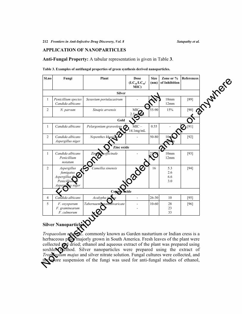
212 Frontiers in Anti-Infective Drug Discovery, Vol. 8 Satapathy et al.
APPLICATION OF NANOPARTICLES
Anti-Fungal Property: A tabular representation is given in Table 3.
Table 3. Examples of antifungal properties of green synthesis derived nanoparticles.
Sl.no Fungi Plant Dose(LC50/LC90/
MIC)
Size(nm)
Zone or %of Inhibition
References
Silver
1 Penicillium speciesCandida albicans
Sesuvium portulacastrum - 5-20 18mm12mm
[89]
2 N. parvum Sinapis arvensis MIC –2.5µg/mL
55-90 15% [90]
Gold
1 Candida albicans Pelargonium graveolens MIC –14.1mg/mL
0.55 - [91]
2 Candida albicansAspergillus niger
Nepenthes khasiana - 50-80 10mm16mm
[92]
Zinc oxide
1 Candida albicansPenicillium
notatum
Zingiber officinale - 23-25 10mm12mm
[93]
2 Aspergillusfumigatus
Aspergillus flavusPenicillium sp
Aspergillus niger
Camellia sinensis - 16 5.32.66.63.0
[94]
Copper oxide
4 Candida albicans Acalypha indica - 26-30 10 [95]
5 F. oxysporumF. graminearum
F. culmorum
Tabernaemontanadivaricate --
10-60 282333
[96]
Silver Nanoparticles
Tropaeolum majus L. commonly known as Garden nasturtium or Indian cress is aherbaceous plant majorly grown in South America. Fresh leaves of the plant werecollected and dried; ethanol and aqueous extract of the plant was prepared usingsoxhlet method. Silver nanoparticles were prepared using the extract ofTropaeolum majus and silver nitrate solution. Fungal cultures were collected, andthe spore suspension of the fungi was used for anti-fungal studies of ethanol,
For pe
rsona
l priv
ate us
e only
Not be
distr
ibuted
or up
loade
d to a
nyon
e or a
nywhe
re

Phyto-Nano-Antimicrobials Frontiers in Anti-Infective Drug Discovery, Vol. 8 213
aqueous extracts, and silver nanoparticles. Antifungal activity was tested againstA. niger, C. albicans, P. notatum, T. viridae, and Mucor spp. Among all the testorganisms, P. notatum and Mucor spp. were inhibited more by AgNPs, followedby the remaining fungi. The MIC of P. notatum was found to be 31.2 µg/mL. Wecan conclude that silver nanoparticles have a productive antifungal activity thatcan be used in the preparation of medicine [80].
Aloe vera is a type of luscious plant that is grown in the Southwest ArabianPeninsula. Antifungal activity was performed by adding different combinations of100 µL of AgNPs, leaf extract, and salt solution. Antifungal activity wasperformed by disc diffusion method wherein the zone of inhibition was observed.Antifungal activity was highest against Aspergillus spp. and Rhizopus spp. Apartfrom the zone of inhibition because of nanoparticles, the nanoparticles also causeddamage to conidial germination, fungal hyphae, inhibiting normal budding anddamaging the structure of the cell membrane. The MIC of AgNPs was found to be21.8ng/mL, which infers that the AgNPs are effective antifungal agents than theleaf extract [81]. Banana is an herbaceous flowering plant, botanically a berry thatis an edible fruit grown all over the world. Pathogenic fungal strains of C.albicans, A. niger, and Alternaria alternata were obtained for the antifungalactivity. Antifungal activity was performed using the standard Kirby-Bauer discdiffusion method. Different concentrations of AgNPs - 25, 50, 75, and 100mg/mL were added to PDA plates containing the fungal strains. Antifungalactivity was indicated by the zone of inhibition. Antifungal activity was more forA. niger with the zone of inhibition of 13.3 mm. With a low concentration ofAgNPs, there was no antifungal activity. Antifungal activity is because of theformation of insoluble compounds by inactivation of sulfhydryl groups in the cellwall of fungus and disruption of membrane-bound enzymes that causes cell lysis.By this, we can infer that the AgNPs were effective against A. niger and can beused as an efficient antifungal agent against A. niger [82].
Gelidiella acerosa is a type of seaweed that is grown in tropical and subtropicalregions. Antifungal activity was performed by well diffusion method. The fungalstrains used were - a) Humicola insolens, b) Fusarium dimerum, c) Mucorindicus, and d) Trichoderma reesei. The plates were added with 50µL of thenanoparticles along with the control containing 5 mg/mL of the standard drug i.e.,clotrimazole. Antifungal activity was indicated by the zone of inhibition.Biosynthesized AgNPs were effective in inhibiting the growth of the fungalstrains with the zone of inhibition in mm as follows – a) 19, b) 12, c) 14, and d)12, respectively that indicated the nanoparticles were potent antifungal agents incomparison with the standard drug Clotrimazole. Hence the AgNPs can be used inthe preparation of medicines [83].
For pe
rsona
l priv
ate us
e only
Not be
distr
ibuted
or up
loade
d to a
nyon
e or a
nywhe
re

214 Frontiers in Anti-Infective Drug Discovery, Vol. 8 Satapathy et al.
Ocimum sanctum is commonly known as Tulasi, holy basil because of itsreligious importance. It is grown in India and the Indian subcontinent. Tulasileaves were collected, dried, and powdered by grinding. 10g of the dried powderwas dissolved in 90% methanol. After 24 hrs, it was filtered and centrifuged. Thesupernatant was collected by evaporating the solvent. The crude extract wasdiluted with 5% DMSO and used for further studies. The AgNPs were preparedby adding 1 mM of Silver nitrate and plant extract. Antifungal activity wasperformed by well diffusion assay method. It was performed against differentfungal strains- a) C. albicans, b) C. kefyr, c) A. niger, d) C. tropicalis, e) A. flavus,and f) A.fumigatus. Antifungal activity was indicated by the zone of inhibition inthe plate. Antifungal activity was performed with the nanoparticles and incomparison with the standard drugs ketoconazole and itraconazole. The highestantifungal activity was against the strains b) a) and c) with the zone of inhibition15mm, 13mm, and 12mm, respectively. The intermediate activity was against d)e) and f) with the zone of inhibition 11mm, 6mm, and 7mm, respectively. Theseresults indicate that the synthesized AgNPs were a potent antifungal agent [84].
Gold Nanoparticles
Abelmoschus esculentus L. commonly known as okra or lady finger is a floweringplant majorly grown in tropical, sub-tropical and warm temperate regions of theworld. The seeds were collected, washed with distilled water, dried for 15 days,and the dried seeds were powdered. The prepared seed powder was mixed withion free water and boiled for 30mins, cooled, filtered, and was used fresh toprepare AuNPs. Gold nanoparticles were prepared using the aqueous extract ofthe seeds (40mL) and aqueous chloroauric acid. Fungal cultures were isolatedfrom the soil and used for the antifungal study. Antifungal activity was doneagainst Puccinia graminis, Aspergillus flavus, Aspergillus niger, and Candidaalbicans using the standard well diffusion method. AuNPs were very effective ininhibiting the growth of Puccinia graminis with a zone of inhibition of 17mm,Candida albicans with a zone of inhibition of 18mm, Aspergillus flavus with thezone of inhibition of 16mm and A. niger with the zone of inhibition of 15mm.From these results, we can infer that the biosynthesized AuNPs is an effectiveantifungal agent, and these AuNPs can be used in the preparation of medicine.AuNPs can be used as an antimicrobial agent, and this can be used in the foodindustry as preservatives [85].
Terminalia chebula, commonly known as black or Chebulic myrobalan, is grownin India, Srilanka, China, and other South Asian countries. W. bancrofti is thecausal agent for filariasis (elephantiasis) characterized by swelling of leg(elephant leg). The synthesized nanoparticles were subjected to antifilarial assayagainst W. bancrofti and S. cervi by the method of MIT assay. LC50 was
For pe
rsona
l priv
ate us
e only
Not be
distr
ibuted
or up
loade
d to a
nyon
e or a
nywhe
re

Phyto-Nano-Antimicrobials Frontiers in Anti-Infective Drug Discovery, Vol. 8 215
determined by the assay. Percentage inhibitions at different doses were found tobe 9.1%, 32.8%, 76.04%, 88.3% and 93.7% for 5µg/mL, 10µg/mL, 20µg/mL,30µg/mL and 40µg/mL, respectively. LC50 of microfilaria of S.cervi and adultfilariids were found to be 8.27%, 31.3%, 74.8%, 87.2%, and 91.03%,respectively. A slight increase in microfilaricidal activity was observedagainst W. bancrofti. Viability was reduced, and this was found to be 9.47%,32.8%, 76.38%, 89%, and 93.47%, respectively. LC50 and LC90 values weredetermined and are found to be 14.3, and 38 µg/mL, LC50 and LC90 of microfilariaand adult S. cervi were determined to be 13.97 and 35.71µg/mL, respectively. Inthe case of microfilaria of W. bancrofti, a decrease in LC50 and LC90 was observedas 13.9 and 35.57µg/mL. By the results of MIT assay, it was inferred that thebiosynthesized AuNPs is a potent antifilarial agent against both human and bovinefilariids [86].
Oxide Nanoparticles
Cissus quadrangularis is commonly known as veldt grape, devil’s backbone, andadamant creeper is a perennial plant of the grape family that is grown in India,Srilanka, Bangladesh, and African countries. The leaf powder was obtained fromVellore local market. 10g of leaf powder was added to 300mL of distilled waterand boiled for 1hour in a boiling water bath; after the required time, it wasfiltered, concentrated, and was refrigerated. The CuO nanoparticles weresynthesized using 20 mg/mL of the plant extract and 1mM of Copper acetate. Theantifungal activity was performed against A. niger and A. flavus. Antifungalactivity was done using the Clinical and Laboratory Standards Institute (CLSI)method. 1 mg/mL of strain and 1 mg/mL of the biosynthesized CuO NPs wereadded in two concentrations (500 and 1000ppm) to the media (Potato DextroseBroth), stirred for 1 week; biomass was obtained by filtration and was calculatedfor the antifungal activity of the two species in comparison with the standardantifungal drug Carbendazim. From the CLSI antifungal study, the CuO NPs hadefficient antifungal property in comparison with the standard antifungalCarbendazim. CuO NPs damaged the cytoplasm and led to the apoptosis of thefungus. 500ppm of CuO NPs inhibited the growth of 82% of A. niger, 1000ppmof CuO NPs inhibited 88% of A. niger, and the standard antifungal inhibited 40%and 43%, respectively. 500ppm of CuO NPs inhibited 82% of the growth of A.flavus, and 1000ppm of CuO NPs inhibited 88% of A. flavus. These results inferthat the green synthesized CuO NPs were potent antifungal agents [87].
Trianthema portulacastrum is commonly known as black pigweed, giant pigweed,and desert horse purslane. It is a weed that is grown in Africa, North America, andSouth America. Plant biomass was collected from the field, washed with distilledwater to separate particulate matters which are adhered to the biomass. Biomass
For pe
rsona
l priv
ate us
e only
Not be
distr
ibuted
or up
loade
d to a
nyon
e or a
nywhe
re

216 Frontiers in Anti-Infective Drug Discovery, Vol. 8 Satapathy et al.
was dried; the powder was prepared by grinding the biomass. The plant extractwas prepared by mixing 40g of powder in 100mL distilled water with constantstirring for 3 hours. ZnO NPs were prepared by mixing 10mL of the preparedextract and ZnSO4. Antifungal activity was performed against Aspergillus niger,A. flavus, and A. fumigatus. Antifungal activity was performed by adding themixture 100 µg/L of nanoparticles and DMSO to the tubes containing Sabaroud’sDextrose Agar with fungi, and the tubes were incubated for 7 days. Inhibition ofthe mycelial growth with the area of fungal growth was observed and expressed interms of percentage inhibition. Antifungal activity studies were carried out incomparison with the standard drug Terbinafine. ZnO NPs were efficient in theinhibition of mycelial growth. 45% of the linear growth of Aspergillus niger, 41%of linear growth of A. flavus, and 51% of linear growth of A. fumigatus wereinhibited by ZnO NPs. From these results, ZnO NPs have been proven to beeffective antifungal agents [88].
Antibacterial Property
Silver nanoparticles: Since ancient times, silver is enormously used as atherapeutic to treat various diseases [97]. Even before antibiotics were used, silverwas used to treat burns and wounds [98]. Gram-positive and gram-negativebacteria exhibit antibacterial activity against silver nanoparticles differently; theycompete with one another [99]. It is found that gram-negative bacterial strains aremore sensitive to AgNPs when compared to gram-positive bacteria or vice-versa[100]. Silver nanoparticles are positively charged, and the bacterial cellularmembrane is negatively charged; AgNPs accumulate on the negatively chargedbacterial membrane and cause conformational changes in the membrane. Thisconformational change causes more permeability to the bacterial cell membrane.Transportation of the nanoparticles in and out of the cellular membrane leads tocell death [101]. Silver nanoparticles block transcription and translation byentering the bacterial cell and by binding to its genetic material [102].Antibacterial activity of nanoparticles is of two types: biocidal action andinhibitory action [103]. The antibacterial activity of nanoparticles depends onseveral parameters, including temperature, pH, silver nitrate concentration, andtypes of bacteria Table 4 [104, 105].
Table 4. Illustrations of nanoparticles for their anti-bacterial efficacy.
Bacteria Nanoparticle Plant Dose Time Inference References
E. coli,S.aureus, and B.
subtilis
Silvernanoparticle
Selaginellabryopteris
1.0 mL 48-72hrs
The best antibacterialeffect was against E.
coli
[113]
For pe
rsona
l priv
ate us
e only
Not be
distr
ibuted
or up
loade
d to a
nyon
e or a
nywhe
re

Phyto-Nano-Antimicrobials Frontiers in Anti-Infective Drug Discovery, Vol. 8 217
Bacteria Nanoparticle Plant Dose Time Inference References
E. coli, S.aureus, and K.
pneumoniae
Ferric oxidenanoparticle
Ruelliatuberosa
75 µL 24 hrs The best antibacterialeffect was against E.
coli and K.pneumoniae
[114]
S. aureus, B.cereus, S.
thyphimuriumand E. coli.
Silvernanoparticle
Glutamicumcorniculatum
(L.) Curtis
30 µL 16 hrs E. coli, S.thyphimurium, S.
thyphimurium, and B.cereus was the
increasing order
[115]
E. coli, B.subtilis and S.
aureus.
Cupric oxidenanoparticle
Madhukalongifolia
100 µL 24hrs Zone of inhibitionwas
15.67±0.68mmfor E.coli
[116]
B. cereus, S.aureus, E. coli,
S. typhi, K.pneumoniae, S.aureus, and B.
cereus
Zinc oxidenanoparticle
Albizialebbeck
10 µL 48hrs The best antibacterialactivity was seen
against E. coli and S.typhi
[117]
E. coli, S. typhi,S. enterica, and
B. subtilis
Silvernanoparticle
Tithoniadiversifolia
0.2 mL 24hrs The best antibacterialactivity was seenagainst B. subtilis
[118]
E. coli, andB. subtilis
Silvernanoparticle
Rhazya stricta 50 µL 24hrs-48hrs
The best antibacterialactivity was against
E. coli than B. subtilis
[119]
S. epidermidisand E. coli
Goldnanoparticle
Coleusaromaticus
0.5MacFarland
24 hrs The best antibacterialactivity was against
E. coli than S.epidermidis
[120]
A. hydrophila Platinumnanoparticle
Orange peelextract
0.5 µL 24 hrs Antibacterial activitywas observed
[121]
B. subtilis, S.aureus, P.
mirabilis, S.typhi
Zinc oxidenanoparticle
PedaliumMurex
100 µL 24 hrs Antibacterial activitywas seen against P.
mirabilis and S. typhi
[122]
E. coli, P.aeruginosa B.subtilis and S.
aureus.
Goldnanoparticle
thyme 60 µL 24 hrs Antibacterial activitywas seen against all
the bacteria
[123]
(Table 4) cont.....
For pe
rsona
l priv
ate us
e only
Not be
distr
ibuted
or up
loade
d to a
nyon
e or a
nywhe
re

218 Frontiers in Anti-Infective Drug Discovery, Vol. 8 Satapathy et al.
Bacteria Nanoparticle Plant Dose Time Inference References
S. aureus and E.coli
Goldnanoparticle
Mangiferaindica
2.5 µL 24 hrs Significant growthinhibition was seen
for both E. coli and S.aureus
[61]
Stem bark extract from Ficus krishnae was used for the synthesis of silvernanoparticles. The extract was tested against various organisms, includingSalmonella typhimurium (MTCC98), Escherichia coli (MTCC 45), andStaphylococcus aureus (ATTC 29122). These silver nanoparticles penetrate thecell membrane and interact with proteins that contain sulfur and with DNA. Theytarget the respiratory chain, and finally, cell division leading to cell death. Thesenanoparticles release silver ions that enhance the bactericidal activity [106, 107].The best activity was exhibited by Staphylococcus aureus with a zone ofinhibition of 12mm with 6mm of the extract followed by Salmonella typhimuriumand Escherichia coli with a zone of inhibition of 13mm and 18mm, respectively.
Gold Nanoparticles
Gold nanoparticles are one of the nano-scaled metallic nanoparticles that havebeen widely studied because of their tunable surface plasmon resonance (SPR),electronic properties, and optical properties. They are widely used in biomedicalsciences that include tumor imaging, photothermal therapy, drug delivery, andbio-labeling [68]. The antibacterial activity of biosynthesized Nigella arvensisgold nanoparticles was tested against gram-positive bacterial strains consisting ofStaphylococcus epidermidis (ATCC 12228), Pseudomonas aeruginosa(ATCC27253), Escherichia coli (ATCC 25922), Staphylococcus aureus (ATCC43300), Serratia marcescens (ATCC 13880), and Bacillus subtilis (ATCC 6633).The zone of inhibition was calculated through a well diffusion method along withminimum inhibitory concentration. The best activity was exhibited byStaphylococcus epidermidis with a full zone of inhibition at 250 µg/mL of theextract. The mechanism of action of the gold nanoparticles involves binding to thecell membrane and causing conformational changes disrupting the cell membraneleading to degradation and finally cell death. Inside the bacterial cell, they bind tothe genetic material of the bacteria and inhibit transcription and translation,leading to growth inhibition and cellular damage [108, 109].
Oxide Nanoparticles
Even ZnO have shown antibacterial properties against E. coli, P. aeruginosa andS. aureus; the results obtained were impressive. The particle size ranged from100-800nm among all three bacteria; better results were obtained against S.
(Table 4) cont.....
For pe
rsona
l priv
ate us
e only
Not be
distr
ibuted
or up
loade
d to a
nyon
e or a
nywhe
re

Phyto-Nano-Antimicrobials Frontiers in Anti-Infective Drug Discovery, Vol. 8 219
aureus. This paves the way for the use of ZnO nanoparticle against skinpathogens. The optimum concentration of 6 mM showed better MIC value againstbacteria [110].
The antimicrobial properties of CuO NPs were studied using Escherichia coliATCC 25922 and Staphylococcus aureus ATCC 43300 as multidrug-resistant(MDR) bacteria. The anti-bacterial activities of CuO nanoparticles wereconcluded based on the diameter of the inhibition zone in disk diffusion tests ofNPs. The antibacterial effect on E.coli 33±0.57 and 6±2 mm, as well as S. aureus,shows no growth signs [111].
The antibacterial activity of TiO2 was studied against Escherichia coli (E. coli) inan actual food packaging application test under various conditions, includingtypes of light (fluorescent and ultraviolet (UV)) and the length of time the filmwas exposed to light. The antibacterial activity of TiO2 was evaluated through twotypes of experiments under UV irradiation: (I) in a slurry with physiological water(stirred suspension); and (II) in a drop deposited on a glass plate, the significanteffect of the nature of the suspension on the photocatalytic disinfection ability washighlighted. The results exhibited significant antibacterial activities after 2 h and 4h. It is suggested that improving the formulation would increase its efficiency[112].
MEDICINAL APPLICATION
In the preview of human welfare, nanotechnology has a huge role to contribute; afew are illustrated in Table 5 below.
Table 5. Medicinal applications of metal and metal oxide nanoparticles.
Nanoparticles Plant Activity Dose References
Silver Tropaeolum majus Anticancer 156 µg/mL-IC50
[80]
Zinc Oxide Ziziphus nummularia In vitro cytotoxic 200 µg/mL [124]
Zinc Oxide Trianthema Portulacastrum Anti-oxidant 500 µg/mL [88]
Zinc Oxide Caulerpa peltate Anti-biofilm - [125]
Gold Terminaliachebula Anti-filarial LC50 – 38µg/mL
[86]
Copper Oxide Cissus quadrangularis Antifungal 500 ppm [126]
Silver Alternanthera sessilis Antioxidant IC50 – 300.6µg/mL
[127]
Carbon - Enhances susceptibility toseizures
- [128]
For pe
rsona
l priv
ate us
e only
Not be
distr
ibuted
or up
loade
d to a
nyon
e or a
nywhe
re

220 Frontiers in Anti-Infective Drug Discovery, Vol. 8 Satapathy et al.
Nanoparticles Plant Activity Dose References
Gold (fluorescent) - Growth factor expressionstudy in colorectal cancer
- [129]
Bimetallic – SilverPlatinum nanoparticles
- Osteopromotiveanti-microbial
- [130]
FUTURE PROSPECTS AND CONCLUSION
Nanotechnology has influenced the world with its uniqueness and applicabilitybut there is still a long way to go to achieve the goal of its applicability [131,132]. The future of nanotechnology and green synthesis is not very much evident.There are many questions to be answered; the problems need to be assessed andsolved at every step of the process. Questions on synthesis majorly lie in theproduction of metallic nanoparticles (Fig. 3). There are a lot of plants andmicrobes from which nanoparticles are being extracted, but the effective ones arenot quantified. The stability of nanoparticles is to be explored more. There is aneed for understanding the amount of the extract required , what happens whenup-scaled, which is the best-suited solvent, which of the nanoparticle shows thebetter result, and what is the efficacy of the extract?
Fig. (3). Prospects of metal and metal oxide green synthesis derived nanoparticle.
(Table 5) cont.....
For pe
rsona
l priv
ate us
e only
Not be
distr
ibuted
or up
loade
d to a
nyon
e or a
nywhe
re

Phyto-Nano-Antimicrobials Frontiers in Anti-Infective Drug Discovery, Vol. 8 221
These questions pave the way for a better synthesis procedure. The next questionis about the characterization of the nanoparticles. The instrumentation involved istedious and time taking which adds on to the cost and labor. Hence there is a needfor better and faster characterization of the nanoparticle. This will reduce the costand will devote more time to the application of it. The next question is thenanoparticle, and its application; nanoparticles are extracted from various plantand microbial sources. This has been applied to various microbes and diseasesystems. But the effort should be made to bring out a combination ofnanoparticles for a better understanding of the system and as well as itseffectiveness. Rather than using various extractions, effort has to be made for asingle source and multiple applications.
CONSENT FOR PUBLICATION
Not applicable.
CONFLICT OF INTEREST
The authors declare that no conflict of interest.
ACKNOWLEDGEMENTS
All the authors are thankful to Dr. Shaukath Ara Khanum, Department ofChemistry, Yuvaraja College, University of Mysore, Mysore for the long-termcollaboration in understanding the biology of phytochemical molecules. Mr.Pankaj Satapathy would like to thank DST-KSTePS, GoK, for providing DSTPh.D. fellowship (LIF-09-2018-19). Ms. Aishwarya. T. Devi would like to thankthe Indian Council of Medical Research (ICMR) for the award of Senior ResearchFellow (SRF) Award Letter Number - File no.5/3/8/55/ITR-F/2018-ITR dated11.6.2018. All authors thank the Hon’ble Vice-Chancellor, JSS Science andTechnology University, Principal, SJCE, Mysore for his encouragement and JSSresearch foundation for their constant inspiration. Mr. Avinash MG would like tothank the Indian Council of Medical Research (ICMR) for the award of SeniorResearch Fellow (SRF) Award Letter Number - File no.AMR/Fellowship/17/2019 ECD-II dated 28.6.2019. FZ sincerely acknowledgesthe University Grants Commission (UGC), Govt. of India, New Delhi, forawarding Raman Post-Doctoral Fellowship 2014-15 to USA (Ref No. F.5-97/2014 (IC) FD dairy no: 6725). Further, we extend our gratitude towards themanagement and office bearers of Dayananda Sagar University, Bengaluru,Karnataka, India, for constant inspiration, motivation, and encouragement topursue scientific research.
For pe
rsona
l priv
ate us
e only
Not be
distr
ibuted
or up
loade
d to a
nyon
e or a
nywhe
re

222 Frontiers in Anti-Infective Drug Discovery, Vol. 8 Satapathy et al.
REFERENCES[1] Stober W, Fink A, Bohn EJ. Controlled growth of monodisperse silica spheres in the micron size
range. J Colloid Interface Sci 1968; 26(1): 62-9.
[2] Zameer F, Gopal S, Krohne G, Kreft J. Development of a biofilm model for Listeria monocytogenes.World J Microbiol Biotechnol 2009; 26(6): 1143-7.[http://dx.doi.org/10.1007/s11274-009-0271-4]
[3] Gericke M, Pinches A. Microbial production of gold nanoparticles. Gold Bull 2006; 39(1): 22-8.[http://dx.doi.org/10.1007/BF03215529]
[4] Iravani S. Bacteria in nanoparticle synthesis: current status and future prospects. Int Sch Res Notices2014; 2014: 359316.[http://dx.doi.org/10.1155/2014/359316] [PMID: 27355054]
[5] Thakkar KN, Mhatre SS, Parikh RY. Biological synthesis of metallic nanoparticles. Nanomedicine(Lond) 2010; 6(2): 257-62.[http://dx.doi.org/10.1016/j.nano.2009.07.002] [PMID: 19616126]
[6] Chen YL, Tuan HY, Tien CW, Lo WH, Liang HC, Hu YC. Augmented biosynthesis of cadmiumsulfide nanoparticles by genetically engineered Escherichia coli. Biotechnol Prog 2009; 25(5): 1260-6.[http://dx.doi.org/10.1002/btpr.199] [PMID: 19630084]
[7] Mohanpuria P, Rana NK, Yadav SK. Biosynthesis of nanoparticles: technological concepts and futureapplications. J Nanopart Res 2008; 10(3): 507-17.[http://dx.doi.org/10.1007/s11051-007-9275-x]
[8] Narayanan KB, Sakthivel N. Synthesis and characterization of nano-gold composite usingCylindrocladium floridanum and its heterogeneous catalysis in the degradation of 4-nitrophenol. JHazard Mater 2011; 189(1-2): 519-25.[http://dx.doi.org/10.1016/j.jhazmat.2011.02.069] [PMID: 21420237]
[9] Prasad Ashwini, Baker Syed. Phytogenic synthesis of silver nanobactericides for antibiofilm activityagainst human pathogen H pylori 2019; 1(4): 341-7.
[10] Anastas PT. JC Warner Green Chemistry. Theory Pract 1998.
[11] Gnanasangeetha D, Thambavani DS. Biogenic production of zinc oxide nanoparticles using Acalyphaindica. J Chem Biol Phys Sci 2013; 4(1): 238.
[12] Gunalan S, Sivaraj R, Rajendran V. Green synthesized ZnO nanoparticles against bacterial and fungalpathogens. Progress in Natural Science: Materials International 2012; 22(6): 693-700.[http://dx.doi.org/10.1016/j.pnsc.2012.11.015]
[13] Shanker U, Jassal V, Rani M, Kaith BS. Towards green synthesis of nanoparticles: from bio-assistedsources to benign solvents. A review. Int J Environ Anal Chem 2016; 96(9): 801-35.
[14] Sylvestre JP, Poulin S, Kabashin AV, Sacher E, Meunier M, Luong JH. Surface chemistry of goldnanoparticles produced by laser ablation in aqueous media. J Phys Chem B 2004; 108(43): 16864-9.[http://dx.doi.org/10.1021/jp047134+]
[15] Bouquillon S, Courant T, Dean D, et al. Biodegradable ionic liquids: selected synthetic applications.Aust J Chem 2007; 60(11): 843-7.[http://dx.doi.org/10.1071/CH07257]
[16] Wittmann K, Wisniewski W, Mynott R, et al. Supercritical carbon dioxide as solvent and temporaryprotecting group for rhodium-catalyzed hydroaminomethylation. Chemistry 2001; 7(21): 4584-9.[http://dx.doi.org/10.1002/1521-3765(20011105)7:21<4584::AID-CHEM4584>3.0.CO;2-P] [PMID:11757649]
[17] Pollet P, Eckert CA, Liotta CL. Solvents for sustainable chemical processes. WIT Trans Ecol Environ2011; 154: 21-31.[http://dx.doi.org/10.2495/CHEM110031]
For pe
rsona
l priv
ate us
e only
Not be
distr
ibuted
or up
loade
d to a
nyon
e or a
nywhe
re

Phyto-Nano-Antimicrobials Frontiers in Anti-Infective Drug Discovery, Vol. 8 223
[18] Sneha K. Sathish kumar M, Mao J, Kwak IS, Yun YS. Corynebacterium glutamicum-mediatedcrystallization of silver ions through sorption and reduction processes. Chem Eng J 2010; 162(3): 989-96.[http://dx.doi.org/10.1016/j.cej.2010.07.006]
[19] Zameer F, Kreft J, Gopal S. Interaction of the dual species biofilms of Listeria monocytogenes andStaphylococcus epidermidis. J Food Saf 2010; 30(4): 954-68.[http://dx.doi.org/10.1111/j.1745-4565.2010.00254.x]
[20] Mittal AK, Chisti Y, Banerjee UC. Synthesis of metallic nanoparticles using plant extracts. BiotechnolAdv 2013; 31(2): 346-56.[http://dx.doi.org/10.1016/j.biotechadv.2013.01.003] [PMID: 23318667]
[21] Dwivedi AD, Gopal K. Biosynthesis of silver and gold nanoparticles using Chenopodium album leafextract. Colloids Surf A Physicochem Eng Asp 2010; 369(1-3): 27-33.[http://dx.doi.org/10.1016/j.colsurfa.2010.07.020]
[22] Jha AK, Prasad K, Kumar V, Prasad K. Biosynthesis of silver nanoparticles using Eclipta leaf.Biotechnol Prog 2009; 25(5): 1476-9.[http://dx.doi.org/10.1002/btpr.233] [PMID: 19725113]
[23] Meghashri. S, Chauhan. JB, Syed. AA and Farhan Zameer. Effect of Ocimum tenuiflorum leaf extractagainst infective endocarditis. Int J Phytomed 2011; 3(4): 470-4.
[24] Prathna TC, Chandrasekaran N, Raichur AM, Mukherjee A. Biomimetic synthesis of silvernanoparticles by Citrus limon (lemon) aqueous extract and theoretical prediction of particle size.Colloids Surf B Biointerfaces 2011; 82(1): 152-9.[http://dx.doi.org/10.1016/j.colsurfb.2010.08.036] [PMID: 20833002]
[25] Panigrahi S, Kundu S, Ghosh S, Nath S, Pal T. General method of synthesis for metal nanoparticles. JNanopart Res 2004; 6(4): 411-4.[http://dx.doi.org/10.1007/s11051-004-6575-2]
[26] Ashwini P, Sumana MN, Shilpa U, et al. Farhan Zameer, Nagendra Prasad M.N. A review onHelicobacter pylori: its biology, complications and management. Int J Pharm Pharm Sci 2014; 7(1):14-20.
[27] Tan YN, Lee JY, Wang DI. Uncovering the design rules for peptide synthesis of metal nanoparticles. JAm Chem Soc 2010; 132(16): 5677-86.[http://dx.doi.org/10.1021/ja907454f] [PMID: 20355728]
[28] Singh J, Dutta T, Kim K-H, Rawat M, Samddar P, Kumar P. ‘Green’ synthesis of metals and theiroxide nanoparticles: applications for environmental remediation. J Nanobiotechnology 2018; 16(1):84.[http://dx.doi.org/10.1186/s12951-018-0408-4] [PMID: 30373622]
[29] Zameer F, Rukmangada MS, Chauhan JB, et al. Evaluation of adhesive and anti-adhesive properties ofPseudomonas aeruginosa biofilms and its inhibition by herbal plants. Iran J Microbiol 2016; 8(2):108-19.[PMID: 27307976]
[30] Shivaji S, Madhu S, Singh S. Extracellular synthesis of antibacterial silver nanoparticles usingpsychrophilic bacteria. Process Biochem 2011; 46: 1800-7.[http://dx.doi.org/10.1016/j.procbio.2011.06.008]
[31] Wen L, Lin Z, Gu P, et al. Extracellular biosynthesis of monodispersed gold nanoparticles by a SAMcapping route. J Nanopart Res 2009; 11(2): 279-88.[http://dx.doi.org/10.1007/s11051-008-9378-z]
[32] Lengke MF, Fleet ME, Southam G. Morphology of gold nanoparticles synthesized by filamentouscyanobacteria from gold(I)-thiosulfate and gold(III)--chloride complexes. Langmuir 2006; 22(6):2780-7.
For pe
rsona
l priv
ate us
e only
Not be
distr
ibuted
or up
loade
d to a
nyon
e or a
nywhe
re

224 Frontiers in Anti-Infective Drug Discovery, Vol. 8 Satapathy et al.
[http://dx.doi.org/10.1021/la052652c] [PMID: 16519482]
[33] Philipse AP, Maas D. Magnetic colloids from magnetotactic bacteria: chain formation and colloidalstability. Langmuir 2002; 18(25): 9977-84.[http://dx.doi.org/10.1021/la0205811]
[34] Mann S. Structure, morphology, and crystal growth of bacterial magnetite.Magnetite biomineralizationand magnetoreception in organisms. Boston, MA: Springer 1985; pp. 311-32.[http://dx.doi.org/10.1007/978-1-4613-0313-8_15]
[35] Holmes JD, Smith PR, Evans-Gowing R, Richardson DJ, Russell DA, Sodeau JR. Energy-dispersiveX-ray analysis of the extracellular cadmium sulfide crystallites of Klebsiella aerogenes. ArchMicrobiol 1995; 163(2): 143-7.[http://dx.doi.org/10.1007/BF00381789] [PMID: 7710328]
[36] Beulah KC, Aishwarya T. Devi, Waseem K, Meghashri S, Hedgekatte R, Nagendra Prasad MN,Dhananjaya BL, Zameer F. Phyto-Antiquorumones: An Herbal Approach for Blocking BacterialTrafficking and Pathogenesis. Int J Pharm Pharm Sci 2015; 7(1): 29-34.
[37] Ravindra BK, Rajasab AH. A comparative study on biosynthesis of silver nanoparticles using fourdifferent fungal species. Int J Pharm 2014; 6(1): 372-6.
[38] Soni N, Prakash S. Fungal-mediated nano silver: an effective adulticide against mosquito. ParasitolRes 2012; 111(5): 2091-8.[http://dx.doi.org/10.1007/s00436-012-3056-x] [PMID: 22864863]
[39] Molnár Z, Bódai V, Szakacs G, et al. Green synthesis of gold nanoparticles by thermophilicfilamentous fungi. Sci Rep 2018; 8(1): 3943.[http://dx.doi.org/10.1038/s41598-018-22112-3] [PMID: 29500365]
[40] Ahmad A, Senapati S, Khan MI, Kumar R, Sastry M. Extra-/intracellular biosynthesis of goldnanoparticles by an alkalotolerant fungus, Trichothecium sp. J Biomed Nanotechnol 2005; 1(1): 47-53.[http://dx.doi.org/10.1166/jbn.2005.012]
[41] Raliya R, Tarafdar JC. Biosynthesis and characterization of zinc, magnesium and titaniumnanoparticles: an eco-friendly approach. Int Nano Lett 2014; 4(1): 93.[http://dx.doi.org/10.1007/s40089-014-0093-8]
[42] Raliya R, Biswas P, Tarafdar JC. TiO2 nanoparticle biosynthesis and its physiological effect on mungbean (Vigna radiata L.). Biotechnol Rep (Amst) 2014; 5: 22-6.[http://dx.doi.org/10.1016/j.btre.2014.10.009] [PMID: 28626678]
[43] Kowshik M, Ashtaputre S, Kharrazi S, et al. Extracellular synthesis of silver nanoparticles by a silver-tolerant yeast strain MKY3. Nanotechnology 2002; 14(1): 95.[http://dx.doi.org/10.1088/0957-4484/14/1/321]
[44] Mourato A, Gadanho M, Lino AR, Tenreiro R. Biosynthesis of crystalline silver and goldnanoparticles by extremophilic yeasts. Bioinorg Chem Appl 2011; 2011: 546074.[http://dx.doi.org/10.1155/2011/546074] [PMID: 21912532]
[45] Haverkamp RG, Marshall AT. The mechanism of metal nanoparticle formation in plants: limits onaccumulation. J Nanopart Res 2009; 11(6): 1453-63.[http://dx.doi.org/10.1007/s11051-008-9533-6]
[46] Mude N, Ingle A, Gade A, Rai M. Synthesis of silver nanoparticles using callus extract of Caricapapaya-a first report. J Plant Biochem Biotechnol 2009; 18(1): 83-6.[http://dx.doi.org/10.1007/BF03263300]
[47] Najimu Nisha S, Aysha OS, Syed Nasar Rahaman J, et al. Lemon peels mediated synthesis of silvernanoparticles and its antidermatophytic activity. Spectrochim Acta A Mol Biomol Spectrosc 2014;124: 194-8.[http://dx.doi.org/10.1016/j.saa.2013.12.019] [PMID: 24486863]
For pe
rsona
l priv
ate us
e only
Not be
distr
ibuted
or up
loade
d to a
nyon
e or a
nywhe
re

Phyto-Nano-Antimicrobials Frontiers in Anti-Infective Drug Discovery, Vol. 8 225
[48] Jha AK, Prasad K. Green synthesis of silver nanoparticles using Cycas leaf. International Journal ofGreen Nanotechnology: Physics and Chemistry 2010; 1(2): 110-7.[http://dx.doi.org/10.1080/19430871003684572]
[49] Ravindra S, Mohan YM, Reddy NN, Raju KM. Fabrication of antibacterial cotton fibres loaded withsilver nanoparticles via “Green Approach”. Colloids Surf A Physicochem Eng Asp 2010; 367(1-3):31-40.[http://dx.doi.org/10.1016/j.colsurfa.2010.06.013]
[50] Armendariz V, Herrera I, Jose-yacaman M, Troiani H, Santiago P, Gardea-Torresdey JL. Sizecontrolled gold nanoparticle formation by Avena sativa biomass: use of plants in nanobiotechnology. JNanopart Res 2004; 6(4): 377-82.[http://dx.doi.org/10.1007/s11051-004-0741-4]
[51] Narayanan KB, Sakthivel N. Coriander leaf mediated biosynthesis of gold nanoparticles. Mater Lett2008; 62(30): 4588-90.[http://dx.doi.org/10.1016/j.matlet.2008.08.044]
[52] Shankar SS, Rai A, Ahmad A, Sastry M. Controlling the optical properties of lemongrass extractsynthesized gold nanotriangles and potential application in infrared-absorbing optical coatings. ChemMater 2005; 17(3): 566-72.[http://dx.doi.org/10.1021/cm048292g]
[53] Raghunandan D, Bedre MD, Basavaraja S, Sawle B, Manjunath SY, Venkataraman A. Rapidbiosynthesis of irregular shaped gold nanoparticles from macerated aqueous extracellular dried clovebuds (Syzygium aromaticum) solution. Colloids Surf B Biointerfaces 2010; 79(1): 235-40.[http://dx.doi.org/10.1016/j.colsurfb.2010.04.003] [PMID: 20451362]
[54] Gardea-Torresdey JL, Gomez E, Peralta-Videa JR, Parsons JG, Troiani H, Jose-Yacaman M. Alfalfasprouts: a natural source for the synthesis of silver nanoparticles. Langmuir 2003; 19(4): 1357-61.[http://dx.doi.org/10.1021/la020835i]
[55] Qu J, Luo C, Hou J. Synthesis of ZnO nanoparticles from Zn-hyperaccumulator (Sedum alfrediiHance) plants. Micro & Nano Lett 2011; 6(3): 174-6.[http://dx.doi.org/10.1049/mnl.2011.0004]
[56] Jia L, Zhang Q, Li Q, Song H. The biosynthesis of palladium nanoparticles by antioxidants inGardenia jasminoides Ellis: long lifetime nanocatalysts for p-nitrotoluene hydrogenation.Nanotechnology 2009; 20(38): 385601.[http://dx.doi.org/10.1088/0957-4484/20/38/385601] [PMID: 19713585]
[57] Nagendraswamy G, Lakshmi RV, Bushra BA, et al. Extraction and evaluation of antimicrobial andantiviral efficacy of Terminalia Bellirica fruits. Indo Am j pharm 2013; 3(2): 4262-8.
[58] Williams DB, Carter CB. The transmission electron microscope.Transmission electron microscopy.Boston, MA: Springer 1996; pp. 3-17.[http://dx.doi.org/10.1007/978-1-4757-2519-3_1]
[59] Sulthana R, Taqui SN, Zameer F, Syed UT, Syed AA. Adsorption of ethidium bromide from aqueoussolution onto nutraceutical industrial fennel seed spent: Kinetics and thermodynamics modelingstudies. Int J Phytoremediation 2018; 20(11): 1075-86.[http://dx.doi.org/10.1080/15226514.2017.1365331] [PMID: 30156921]
[60] Prasad Ashwini, Devi Aishwarya Tripurasundari. Phyto-Antibiofilm elicitors as potential inhibitors ofHelicobacter pylori 3Biotech 2019; 9(53): 1-9.
[61] Vimalraj S, Ashokkumar T, Saravanan S. Biogenic gold nanoparticles synthesis mediated byMangifera indica seed aqueous extracts exhibits antibacterial, anticancer and anti-angiogenicproperties. Biomed Pharmacother 2018; 105: 440-8.[http://dx.doi.org/10.1016/j.biopha.2018.05.151] [PMID: 29879628]
[62] Mirza AU, Kareem A, Nami SAA, et al. Biogenic synthesis of iron oxide nanoparticles using Agrewia
For pe
rsona
l priv
ate us
e only
Not be
distr
ibuted
or up
loade
d to a
nyon
e or a
nywhe
re

226 Frontiers in Anti-Infective Drug Discovery, Vol. 8 Satapathy et al.
optiva and Prunus persica phyto species: Characterization, antibacterial and antioxidant activity. JPhotochem Photobiol B 2018; 185: 262-74.[http://dx.doi.org/10.1016/j.jphotobiol.2018.06.009] [PMID: 29981488]
[63] Vijayakumar S, Krishnakumar C, Arulmozhi P, Mahadevan S, Parameswari N. Biosynthesis,characterization and antimicrobial activities of zinc oxide nanoparticles from leaf extract of Glycosmispentaphylla (Retz.) DC. Microb Pathog 2018; 116: 44-8.[http://dx.doi.org/10.1016/j.micpath.2018.01.003] [PMID: 29330059]
[64] Kesharwani J, Yoon KY, Hwang J, Rai M. Phytofabrication of silver nanoparticles by leaf extract ofDatura metel: hypothetical mechanism involved in synthesis. J Bionanosci 2009; 3: 39-44.[http://dx.doi.org/10.1166/jbns.2009.1008]
[65] Nakkala JR, Mata R, Gupta AK, Sadras SR. Biological activities of green silver nanoparticlessynthesized with Acorous calamus rhizome extract. Eur J Med Chem 2014; 85: 784-94.[http://dx.doi.org/10.1016/j.ejmech.2014.08.024] [PMID: 25147142]
[66] Suna Q, Cai X, Li J, Zheng M, Chenb Z, Yu CP. Green synthesis of silver nanoparticles using tea leafextract and evaluation of their stability and antibacterial activity. Colloids Surf A Physicochem EngAsp 2014; 444: 226-31.[http://dx.doi.org/10.1016/j.colsurfa.2013.12.065]
[67] Mariselvam R, Ranjitsingh AJA, Usha Raja Nanthini A, Kalirajan K, Padmalatha C, MosaeSelvakumar P. Green synthesis of silver nanoparticles from the extract of the inflorescence of Cocosnucifera (Family: Arecaceae) for enhanced antibacterial activity. Spectrochim Acta A Mol BiomolSpectrosc 2014; 129: 537-41.[http://dx.doi.org/10.1016/j.saa.2014.03.066] [PMID: 24762541]
[68] Rajakumar G, Abdul Rahuman A. Larvicidal activity of synthesized silver nanoparticles using Ecliptaprostrata leaf extract against filariasis and malaria vectors. Acta Trop 2011; 118(3): 196-203.
[69] Yang N. WeiHong L, Hao L. Biosynthesis of Au nanoparticles using agricultural waste mango peelextract and its in vitro cytotoxic effect on two normal cells. Mater Lett 2014; 134: 67-70.[http://dx.doi.org/10.1016/j.matlet.2014.07.025]
[70] Tamuly C, Hazarika M, Bordoloi M. Biosynthesis of Au nanoparticles by Gymnocladus assamicus andits catalytic activity. Mater Lett 2013; 108: 276-9.[http://dx.doi.org/10.1016/j.matlet.2013.07.020]
[71] Tahir K, Nazir S, Li B, et al. Nerium oleander leaves extract mediated synthesis of gold nanoparticlesand its antioxidant activity. Mater Lett 2015; 156: 198-201.[http://dx.doi.org/10.1016/j.matlet.2015.05.062]
[72] Muthuvel A, Adavallan K, Balamurugan K, Krishnakumar N. Biosynthesis of gold nanoparticles usingSolanum nigrum leaf extract and screening their free radical scavenging and antibacterial properties.Biomedicine & Preventive Nutrition 2014; 4(2): 325-32.[http://dx.doi.org/10.1016/j.bionut.2014.03.004]
[73] Abdel-Raouf N, Al-Enazi NM, Ibraheem IB. Green biosynthesis of gold nanoparticles usingGalaxaura elongata and characterization of their antibacterial activity. Arab J Chem 2017; 10: S3029-39.[http://dx.doi.org/10.1016/j.arabjc.2013.11.044]
[74] Das J, Velusamy P. Catalytic reduction of methylene blue using biogenic gold nanoparticles fromSesbania grandiflora L. Journal of the Taiwan Institute of Chemical Engineers 2014; 45(5): 2280-5.[http://dx.doi.org/10.1016/j.jtice.2014.04.005]
[75] Madhumitha G, Rajakumar G, Roopan SM, et al. Acaricidal, insecticidal, and larvicidal efficacy offruit peel aqueous extract of Annona squamosa and its compounds against blood-feeding parasites.Parasitol Res 2012; 111(5): 2189-99.[http://dx.doi.org/10.1007/s00436-011-2671-2] [PMID: 22006187]
For pe
rsona
l priv
ate us
e only
Not be
distr
ibuted
or up
loade
d to a
nyon
e or a
nywhe
re

Phyto-Nano-Antimicrobials Frontiers in Anti-Infective Drug Discovery, Vol. 8 227
[76] Zhu X, Pathakoti K, Hwang HM. Green synthesis of titanium dioxide and zinc oxide nanoparticles andtheir usage for antimicrobial applications and environmental remediation. In: Shukla AK, Iravani S,Eds. Green Synthesis, Characterization and Applications of Nanoparticles. Elsevier 2019; pp. 223-63.[http://dx.doi.org/10.1016/B978-0-08-102579-6.00010-1]
[77] Marimuthu S, Rahuman AA, Jayaseelan C, et al. Acaricidal activity of synthesized titanium dioxidenanoparticles using Calotropis gigantea against Rhipicephalus microplus and Haemaphysalisbispinosa. Asian Pac J Trop Med 2013; 6(9): 682-8.[http://dx.doi.org/10.1016/S1995-7645(13)60118-2] [PMID: 23827143]
[78] Velayutham K, Rahuman AA, Rajakumar G, et al. Evaluation of Catharanthus roseus leaf extract-mediated biosynthesis of titanium dioxide nanoparticles against Hippobosca maculata and Bovicolaovis. Parasitol Res 2012; 111(6): 2329-37.[http://dx.doi.org/10.1007/s00436-011-2676-x] [PMID: 21987105]
[79] Nasrollahzadeh M, Atarod M, Sajjadi M, Sajadi SM, Issaabadi Z. Plant-mediated green synthesis ofnanostructures: mechanisms, characterization, and applications. Interface Science and Technology.Elsevier 2019; 28: pp. 199-322.
[80] Valsalam S, Agastian P, Arasu MV, et al. Rapid biosynthesis and characterization of silvernanoparticles from the leaf extract of Tropaeolum majus L. and its enhanced in-vitro antibacterial,antifungal, antioxidant and anticancer properties. J Photochem Photobiol B 2019; 191: 65-74.[http://dx.doi.org/10.1016/j.jphotobiol.2018.12.010] [PMID: 30594044]
[81] Medda S, Hajra A, Dey U, Bose P, Mondal NK. Biosynthesis of silver nanoparticles from Aloe veraleaf extract and antifungal activity against Rhizopus sp. and Aspergillus sp. Appl Nanosci 2015; 5(7):875-80.[http://dx.doi.org/10.1007/s13204-014-0387-1]
[82] Das J, Velusamy P. Biogenic synthesis of antifungal silver nanoparticles using aqueous stem extract ofbanana. Nano Biomed Eng 2013; 5(1): 34-8.[http://dx.doi.org/10.5101/nbe.v5i1.p34-38]
[83] Vivek M, Kumar PS, Steffi S, Sudha S. Biogenic silver nanoparticles by Gelidiella acerosa extractand their antifungal effects. Avicenna J Med Biotechnol 2011; 3(3): 143-8.[PMID: 23408653]
[84] Rout Y, Behera S, Ojha AK, Nayak PL. Green synthesis of silver nanoparticles using Ocimumsanctum (Tulashi) and study of their antibacterial and antifungal activities. J Microbiol Antimicrob2012; 4(6): 103-9.[http://dx.doi.org/10.5897/JMA11.060]
[85] Jayaseelan C, Ramkumar R, Rahuman AA, Perumal P. Green synthesis of gold nanoparticles usingseed aqueous extract of Abelmoschus esculentus and its antifungal activity. Ind Crops Prod 2013; 45:423-9.[http://dx.doi.org/10.1016/j.indcrop.2012.12.019]
[86] Roy P, Saha SK, Gayen P, Chowdhury P, Santi P. Sinha Babu. Exploration of ant filarial activity ofgold nanoparticle against human and bovine filarial parasites: A nanomedicinal mechanistic approach.Colloids Surf B Biointerfaces 2017; 7765(17): 30707-5.
[87] Devipriya D, Roopan SM. Cissus quadrangularis mediated ecofriendly synthesis of copper oxidenanoparticles and its antifungal studies against Aspergillus niger, Aspergillus flavus. Mater Sci Eng C2017; 80: 38-44.[http://dx.doi.org/10.1016/j.msec.2017.05.130] [PMID: 28866178]
[88] Khan ZUH, Sadiq HM, Shah NS, et al. Greener synthesis of zinc oxide nanoparticles usingTrianthema portulacastrum extract and evaluation of its photocatalytic and biological applications. JPhotochem Photobiol B 2019; 192: 147-57.[http://dx.doi.org/10.1016/j.jphotobiol.2019.01.013] [PMID: 30738346]
For pe
rsona
l priv
ate us
e only
Not be
distr
ibuted
or up
loade
d to a
nyon
e or a
nywhe
re

228 Frontiers in Anti-Infective Drug Discovery, Vol. 8 Satapathy et al.
[89] Nabikhan A, Kandasamy K, Raj A, Alikunhi NM. Synthesis of antimicrobial silver nanoparticles bycallus and leaf extracts from saltmarsh plant, Sesuvium portulacastrum L. Colloids Surf BBiointerfaces 2010; 79(2): 488-93.[http://dx.doi.org/10.1016/j.colsurfb.2010.05.018] [PMID: 20627485]
[90] Khatami M, Pourseyedi S, Khatami M, Hamidi H, Zaeifi M, Soltani L. Synthesis of silvernanoparticles using seed exudates of Sinapis arvensis as a novel bioresource, and evaluation of theirantifungal activity. Bioresour Bioprocess 2015; 2(1): 19.[http://dx.doi.org/10.1186/s40643-015-0043-y]
[91] Kumari A, Kumar V, Yadav SK. The Use of Syzygium cumini in Nanotechnology In the GenusSyzygium. CRC Press 2017; pp. 171-94.[http://dx.doi.org/10.1201/9781315118772-10]
[92] Bhau BS, Ghosh S, Puri S, Borah B, Sarmah DK, Khan R. Green synthesis of gold nanoparticles fromthe leaf extract of Nepenthes khasiana and antimicrobial assay. Adv Mater Lett 2015; 6(1): 55-8.[http://dx.doi.org/10.5185/amlett.2015.5609]
[93] Janaki AC, Sailatha E, Gunasekaran S. Synthesis, characteristics and antimicrobial activity of ZnOnanoparticles. Spectrochim Acta A Mol Biomol Spectrosc 2015; 144: 17-22.[http://dx.doi.org/10.1016/j.saa.2015.02.041] [PMID: 25748589]
[94] Senthil kumar SR, Sivakumar T. Green tea (Camellia sinensis) mediated synthesis of zinc oxide (ZnO)nanoparticles and studies on their antimicrobial activities. Int J Pharm Pharm Sci 2014; 6(6): 461-5.
[95] Sivaraj R, Rahman PK, Rajiv P, Narendhran S, Venckatesh R. Biosynthesis and characterization ofAcalypha indica mediated copper oxide nanoparticles and evaluation of its antimicrobial andanticancer activity. Spectrochim Acta A Mol Biomol Spectrosc 2014; 129: 255-8.[http://dx.doi.org/10.1016/j.saa.2014.03.027] [PMID: 24747845]
[96] Shende S, Ingle AP, Gade A, Rai M. Green synthesis of copper nanoparticles by Citrus medica Linn.(Idilimbu) juice and its antimicrobial activity. World J Microbiol Biotechnol 2015; 31(6): 865-73.[http://dx.doi.org/10.1007/s11274-015-1840-3] [PMID: 25761857]
[97] Cassir N, Rolain JM, Brouqui P. A new strategy to fight antimicrobial resistance: the revival of oldantibiotics. Front Microbiol 2014; 5: 551.[http://dx.doi.org/10.3389/fmicb.2014.00551] [PMID: 25368610]
[98] Rai M, Yadav A, Gade A. Silver nanoparticles as a new generation of antimicrobials. Biotechnol Adv2009; 27(1): 76-83.[http://dx.doi.org/10.1016/j.biotechadv.2008.09.002] [PMID: 18854209]
[99] Srikar SK, Giri DD, Pal DB, Mishra PK, Upadhyay SN. Green synthesis of silver nanoparticles: areview. Green and Sustainable Chemistry 2016; 6(01): 34.[http://dx.doi.org/10.4236/gsc.2016.61004]
[100] Dehnavi AS, Raisi A, Aroujalian A. Control size and stability of colloidal silver nanoparticles withantibacterial activity prepared by a green synthesis method. Synth React Inorg Met-Org Nano-MetChem 2013; 43(5): 543-51.[http://dx.doi.org/10.1080/15533174.2012.741182]
[101] Chanda S. Silver nanoparticles (medicinal plants mediated): a new generation of antimicrobials tocombat microbial pathogens–a review Microbial Pathogens and Strategies for Combating Them:Science Technology and Education. Badajoz, Spain: Formatex Research Center 2014; pp. 1314-23.
[102] Ahmed S, Ahmad M, Swami BL, Ikram S. A review on plants extract mediated synthesis of silvernanoparticles for antimicrobial applications: A green expertise. J Adv Res 2016; 7(1): 17-28.[http://dx.doi.org/10.1016/j.jare.2015.02.007] [PMID: 26843966]
[103] Perni S, Hakala V, Prokopovich P. Biogenic synthesis of antimicrobial silver nanoparticles cappedwith L-cysteine. Colloids Surf A Physicochem Eng Asp 2014; 460: 219-24.[http://dx.doi.org/10.1016/j.colsurfa.2013.09.034]
For pe
rsona
l priv
ate us
e only
Not be
distr
ibuted
or up
loade
d to a
nyon
e or a
nywhe
re

Phyto-Nano-Antimicrobials Frontiers in Anti-Infective Drug Discovery, Vol. 8 229
[104] Marambio-Jones C, Hoek EM. A review of the antibacterial effects of silver nanomaterials andpotential implications for human health and the environment. J Nanopart Res 2010; 12(5): 1531-51.[http://dx.doi.org/10.1007/s11051-010-9900-y]
[105] Rajesh kumar S, Bharath LV. Mechanism of plant-mediated synthesis of silver nanoparticles–a reviewon biomolecules involved, characterization and antibacterial activity. Chem Biol Interact 2017; 273:219-27.[http://dx.doi.org/10.1016/j.cbi.2017.06.019] [PMID: 28647323]
[106] Vasanth N, Melchias G, Kumaravel P. Ficus benghalensis mediates synthesis of silver nanoparticles:the green approach yields NPs that are its anti-bacterial and anti-oxidant. World J Pharm Sci 2016;4(7): 1-12.[PMID: 27810096]
[107] Amarvani P. Characterization of phyto-nanoparticles from Ficus krishnae for their antibacterial andanticancer activities. Drug Dev Ind Pharm 2018; 44(3): 377-84.[PMID: 29098876]
[108] Srivastava N, Mukhopadhyay M. Biosynthesis and characterization of gold nanoparticles usingzooglearamigera and assessment of its antibacterial property. J Cluster Sci 2015; 26(3): 675-92.[http://dx.doi.org/10.1007/s10876-014-0726-0]
[109] Chahardoli A, Karimi N, Sadeghi F, Fattahi A. Green approach for synthesis of gold nanoparticlesfrom Nigella arvensis leaf extract and evaluation of their antibacterial, antioxidant, cytotoxicity andcatalytic activities. Artif Cells Nanomed Biotechnol 2018; 46(3): 579-88.[PMID: 28541741]
[110] Sirelkhatim A, Mahmud S, Seeni A, et al. Review on zinc oxide nanoparticles: antibacterial activityand toxicity mechanism. Nano-Micro Lett 2015; 7(3): 219-42.[http://dx.doi.org/10.1007/s40820-015-0040-x] [PMID: 30464967]
[111] Taran M, Rad M, Alavi M. Antibacterial activity of copper oxide (CuO) nanoparticles biosynthesizedby Bacillus sp. FU4: Optimization of experiment design. Pharm Sci 2017; 23(3): 198-206.[http://dx.doi.org/10.15171/PS.2017.30]
[112] Transcriptome analysis of thiol disulfide redox metabolism genes in Listeria monocytogenes in biofilmand planktonic forms. International Journal of Pure and Applied Sciences 2010; 4(1): 21-7.
[113] S S D, M B M, M N SK, et al. Antimicrobial, anticoagulant and antiplatelet activities of greensynthesized silver nanoparticles using Selaginella (Sanjeevini) plant extract. Int J Biol Macromol2019; 131: 787-97.[http://dx.doi.org/10.1016/j.ijbiomac.2019.01.222] [PMID: 30876901]
[114] Vasantharaj S, Sathiyavimal S, Senthilkumar P, LewisOscar F, Pugazhendhi A. Biosynthesis of ironoxide nanoparticles using leaf extract of Ruellia tuberosa: Antimicrobial properties and theirapplications in photocatalytic degradation. J Photochem Photobiol B 2019; 192: 74-82.[http://dx.doi.org/10.1016/j.jphotobiol.2018.12.025] [PMID: 30685586]
[115] Allafchian AR, Jalali SA, Aghaei F, Farhang HR. Green synthesis of silver nanoparticles usingGlaucium corniculatum (L.) Curtis extract and evaluation of its antibacterial activity IETnanobiotechnology 122018; (5): 8-574.
[116] Das P, Ghosh S, Ghosh R, Dam S, Baskey M. Madhuca longifolia plant mediated green synthesis ofcupric oxide nanoparticles: A promising environmentally sustainable material for waste watertreatment and efficient antibacterial agent. J Photochem Photobiol B 2018; 189: 66-73.[http://dx.doi.org/10.1016/j.jphotobiol.2018.09.023] [PMID: 30312922]
[117] Umar H, Kavaz D, Rizaner N. Biosynthesis of zinc oxide nanoparticles using Albizia lebbeck stembark, and evaluation of its antimicrobial, antioxidant, and cytotoxic activities on human breast cancercell lines. Int J Nanomedicine 2018; 14: 87-100.[http://dx.doi.org/10.2147/IJN.S186888] [PMID: 30587987]
For pe
rsona
l priv
ate us
e only
Not be
distr
ibuted
or up
loade
d to a
nyon
e or a
nywhe
re

230 Frontiers in Anti-Infective Drug Discovery, Vol. 8 Satapathy et al.
[118] Dada AO, Inyinbor AA, Idu EI, et al. Effect of operational parameters, characterization andantibacterial studies of green synthesis of silver nanoparticles using Tithonia diversifolia. PeerJ 2018;6: e5865.[http://dx.doi.org/10.7717/peerj.5865] [PMID: 30397553]
[119] Shehzad A, Qureshi M, Jabeen S, et al. Synthesis, characterization and antibacterial activity of silvernanoparticles using Rhazya stricta. PeerJ 2018; 6: e6086.[http://dx.doi.org/10.7717/peerj.6086] [PMID: 30588401]
[120] Boomi P, Ganesan RM, Poorani G, Gurumallesh Prabu H, Ravikumar S, Jeyakanthan J. Biologicalsynergy of greener gold nanoparticles by using Coleus aromaticus leaf extract. Mater Sci Eng C 2019;99: 202-10.[http://dx.doi.org/10.1016/j.msec.2019.01.105] [PMID: 30889692]
[121] Castro L, Blazquez ML, Gonzalez F, Munoz JA, Ballester A. Biosynthesis of silver and platinumnanoparticles using orange peel extract: Characterization and applications. IET Nanobiotechnol 2015;9(5): 252-8.[http://dx.doi.org/10.1049/iet-nbt.2014.0063]
[122] Babitha N, Priya LS, Christy SR, et al. Enhanced Antibacterial Activity and Photo-Catalytic Propertiesof ZnO Nanoparticles: Pedalium Murex Plant Extract-Assisted Synthesis. J Nanosci Nanotechnol2019; 19(5): 2888-94.[http://dx.doi.org/10.1166/jnn.2019.16023] [PMID: 30501796]
[123] Hamelian M, Varmira K, Veisi H. Green synthesis and characterizations of gold nanoparticles usingThyme and survey cytotoxic effect, antibacterial and antioxidant potential. J Photochem Photobiol B2018; 184: 71-9.[http://dx.doi.org/10.1016/j.jphotobiol.2018.05.016] [PMID: 29842987]
[124] Padalia H, Chanda S. Characterization, antifungal and cytotoxic evaluation of green synthesized zincoxide nanoparticles using Ziziphus nummularia leaf extract. Artif Cells Nanomed Biotechnol 2017;45(8): 1751-61.[http://dx.doi.org/10.1080/21691401.2017.1282868] [PMID: 28140658]
[125] Nagarajan S, Arumugam Kuppusamy K. Extracellular synthesis of zinc oxide nanoparticle usingseaweeds of gulf of Mannar, India. J Nanobiotechnology 2013; 11(1): 39.[http://dx.doi.org/10.1186/1477-3155-11-39] [PMID: 24298944]
[126] Devipriya D, Roopan SM. Cissus quadrangularis mediated ecofriendly synthesis of copper oxidenanoparticles and its antifungal studies against Aspergillus niger, Aspergillus flavus. Mater Sci Eng C2017; 80: 38-44.[http://dx.doi.org/10.1016/j.msec.2017.05.130] [PMID: 28866178]
[127] Niraimathi KL, Sudha V, Lavanya R, Brindha P. Biosynthesis of silver nanoparticles usingAlternanthera sessilis (Linn.) extract and their antimicrobial, antioxidant activities. Colloids Surf BBiointerfaces 2013; 102: 288-91.[http://dx.doi.org/10.1016/j.colsurfb.2012.08.041] [PMID: 23006568]
[128] He M, Jiang X, Zou Z, et al. Exposure to carbon black nanoparticles increases seizure susceptibility inmale mice. Nanotoxicology 2020; 14(5): 595-611.[http://dx.doi.org/10.1080/17435390.2020.1728412] [PMID: 32091294]
[129] Alagaratnam S, Yang SY, Loizidou M, Fuller B, Ramesh B. Mechano-growth factor expression incolorectal cancer investigated with fluorescent gold nanoparticles. Anticancer Res 2019; 39(4): 1705-10.[http://dx.doi.org/10.21873/anticanres.13276] [PMID: 30952709]
[130] Breisch M, Grasmik V, Loza K, et al. Bimetallic silver-platinum nanoparticles with combined osteo-promotive and antimicrobial activity. Nanotechnology 2019; 30(30): 305101.[http://dx.doi.org/10.1088/1361-6528/ab172b] [PMID: 30959494]
For pe
rsona
l priv
ate us
e only
Not be
distr
ibuted
or up
loade
d to a
nyon
e or a
nywhe
re

Phyto-Nano-Antimicrobials Frontiers in Anti-Infective Drug Discovery, Vol. 8 231
[131] Kounaina K, Deci AT, Patil AG, et al. Synthetic gutomics: Deciphering the microbial code forfuturistic diagnosis and personalized medicine. In: Satapathy P, Ed. Methods in Microbiology.Elsevier 2018; 46: pp. 197-225.
[132] Chinnaswamy S, Zameer F, Muthuchelian K. Molecular and Biological Mechanisms of Apoptosis andits Detection Techniques. Journal of Oncological Sciences 2020; 6(1): 49-64.
For pe
rsona
l priv
ate us
e only
Not be
distr
ibuted
or up
loade
d to a
nyon
e or a
nywhe
re

232 Frontiers in Anti-Infective Drug Discovery, 2020, Vol. 8, 232-253
CHAPTER 8
Aptamers as Anti-Infective AgentsMuhammad Ali Syed1, Nayab Ali1, Bushra Jamil2 and Ammar Ahmed2
1 Department of Microbiology, The University of Haripur, Haripur, Pakistan2 Department of Medical Laboratory Sciences, University of Lahore, Islamabad campus,Islamabad, Pakistan
Abstract: Rapidly emerging drug resistance in all classes of pathogenicmicroorganisms has become a challenging task and a global health issue in recentyears. There are very limited alternative options available to cure infectious diseases, asthe rate of rise in drug resistance in infectious agents is higher than the arrival of newantimicrobial drugs. There is a dire need to look for new types of anti-infective agents,besides looking for new antibiotics. One of the promising types of antimicrobial agentsis aptamers, synthesized through systematic evolution of ligands by exponentialenrichment (SELEX) technique. Aptamers hold a significant promise for the treatmentof various infectious diseases in the future. In the recent past, a number of successfulattempts have been made to select and apply aptamers for the detection and binding ofinfectious agents and their products for therapeutic purposes. This chapter presents abasic introduction to aptamers and their application as anti-infective agents.
Keywords: Aptamers, Antibiotics, Drug Resistance, Infectious Diseases,Systemic Evolution of Ligands by Exponential Enrichment (SELEX).
INTRODUCTION
Infectious diseases have been a major human enemy on the earth, accounting formillions of illnesses and deaths annually [1, 2]. Throughout human history,infectious diseases have been threatening the existence of the human race fromextinction by global pandemics of major infectious diseases such as smallpox,typhoid, malaria, influenza, cholera and many others [3, 4]. The current globalCOVIC-19 pandemic has emerged as one of the most notable global health crisesacross the globe, where billions of humans are staying at home as a measure tocontrol the spread of disease and reduce morbidity and mortality rate [5].
* Corresponding author Muhammad Ali Syed: Department of Microbiology, The University of Haripur, HattarRoad, Haripur, Khyber Pakhtunkhwa, Pakistan;Tel: +92-995-615075; Email:[email protected]; [email protected]
Atta-ur-Rahman and M. Iqbal Choudhary (Eds.)All rights reserved-© 2020 Bentham Science Publishers
For pe
rsona
l priv
ate us
e only
Not be
distr
ibuted
or up
loade
d to a
nyon
e or a
nywhe
re

Aptamers as Anti-Infective Agents Frontiers in Anti-Infective Drug Discovery, Vol. 8 233
Combating infectious diseases has been a dream of scientists of all the time fromthe oldest civilization to modern age due to the seriousness of the issue [1]. Theadvent of antibiotics, vaccines, and several other classes of antimicrobial agentsagainst infections caused by different kinds of microorganisms such as bacteria,fungi, viruses, protozoans as well as microbial toxins has played a key role inreducing the global burden of infectious diseases [6]. The novel concept ofselective toxicity and magic bullets introduced by Paul Ehrlich in 1900 was foundhighly fascinating by the medical community and has been a milestone in thehistory of anti-infective drug discovery [7]. Soon, humans became capable ofcuring bacterial infectious diseases using a class of antimicrobial agents calledantibiotics. Penicillin was the first antibiotic introduced by Sir Alexander Flemingin 1928 followed by a range of natural, semi-synthetic and synthetic antibioticsthat made it possible to cure the diseases that humans seemed to be fightingthroughout the known history [6].
Drug resistance is a real challenge that humans are facing today, which is beingreported in all classes of microorganisms [7, 8]. For instance, antibiotic resistancein bacteria is one of the most serious issue healthcare providers are facing today.The emergence of antibiotic resistant strains of pathogenic bacteria cost millionsof lives across the globe annually. Multidrug resistant and extremely drugresistant clones of different bacterial species such as Staphylococcus aureus,Pseudomonas aeruginosa, Escherichia coli, Salmonella enterica serovar. Typhi,Mycobacterium tuberculosis, Acinetobacter baumannii appear to be resistant tothe majority of antibiotics. There are very few treatment options available in hand,as there are increasing reports of bacterial resistance to drugs of last resort such asvancomycin, colistin, carbapenems, etc. Similar situation is with other types ofmicroorganisms such as viruses, protozoans and fungi [8 - 10].
On one hand, drug discovery efforts are aimed at focusing on new antibiotics, onthe other hand, new options for the cure of infectious agents are being seriouslyconsidered. One of the alternative options is aptamers [9, 11]. In the last fewdecades, aptamers have attracted more attention due to their potential applicationsin therapeutics and diagnostics. This class of biomolecules is also beinginvestigated for potential applications in the cure of infectious diseases [12, 13].This chapter introduces aptamers, their unique features, methods of synthesis andtheir potential applications to target infectious agents.
APTAMERS
The word aptamer is derived from a Latin word aptus meaning “To Fix”.Aptamers are single stranded oligonucleotides (ssDNA or RNA), or peptidesequences that bind their targets very specifically. Target may be may be a
For pe
rsona
l priv
ate us
e only
Not be
distr
ibuted
or up
loade
d to a
nyon
e or a
nywhe
re

234 Frontiers in Anti-Infective Drug Discovery, Vol. 8 Syed et al.
biomolecule, toxin, cell, viral particle or even an inorganic substance [14]. Theseshort secondary structures of ssDNA or RNA bind their target with high affinityand specificity [12, 15] (Fig. 1). Since early experiments, a high number of effortshave been made to select aptamers against a range of targets including differentmicrobial species as well as their products. Attempts to study the binding of RNAsequences with proteins began in 1980s when scientists were studying interactionof human immunodeficiency syndrome virus (HIV) and adenovirus nucleic acidswith proteins. It was discovered that these viruses produce small structural RNAsequences that bind viral or host cell proteins with high affinity and specificity[16, 17]. In vitro selection of aptamers against specific targets using systematicevolution of ligands by exponential enrichment (SELEX) was introduced bySzostak`s and Gold`s groups in 1990 [18, 19].
Fig. (1). Schematic diagram of aptamers binding its target. Oligonucleotide sequences first form 3-Dstructure that binds the target with higher affinity and specificity (Reproduced with permission from [100].
As stated above, aptamers can bind a number of targets. Aptamer binding to theirtargets rely upon nature of their target as well as flexible nature of aptamers. Theshort oligonucleotide sequences of ssDNA or RNA aptamers can form a numberof three dimensional structures, such as hairpin, pseudoknots, bulges and G-quadruplexes. On the basis of these conformations,
aptamers bind their targets via electrostatic interactions, hydrogen bonding, Vander Waals forces and π-π stacking or combination some of these forces [20].
SYSTEMIC EVOLUTION OF LIGANDS BY EXPONENTIALENRICHMENT (SELEX)
RNA or DNA aptamers are selected randomly from a pool of oligonucleotide
For pe
rsona
l priv
ate us
e only
Not be
distr
ibuted
or up
loade
d to a
nyon
e or a
nywhe
re

Aptamers as Anti-Infective Agents Frontiers in Anti-Infective Drug Discovery, Vol. 8 235
sequences (~1015 random sequences) called a library by the process of SELEX[21]. The library is generated by using chemical methods of oligonucleotidesynthesis. The complexity of library is important and it may be calculatedmathematically.
Each oligonucleotide sequence or probe (ssDNA or RNA) in the library is a shortchain of 40-100 nucleotides having random sequence in the middle and fixedsequences in flanking regions. The central or middle region of theseoligonucleotides may bind their target in case their secondary structure has highaffinity for it. SELEX process comprises of several rounds of selection ofoligonucleotide sequences that bind their target. After each round of SELEX,lesser number of binding sequences with higher affinity is selected. Each round ofSELEX comprises of three main steps or stages, detail of that is given infollowing paragraphs (Fig. 2):
Fig. (2). An overview of different steps of Cell SELEX process. A target microbial cell is exposed tooligonucleotide library. DNA sequences bound to the cell are selected, while the unbound sequences areremoved. The oligonucleotide sequences bound to target cell are separated and amplified by PCR and usedfor next round of SELEX. After several such rounds, selected sequences are PCR amplified and sequenced(Figure reproduced with permission from reference No. 40, https://jbiomedsci.biomedcentral.com/ articles/10.1186/s12929-019-0611-0 under creative common licence http://creativecommons.org/ l icenses/ by/4.0/).
For pe
rsona
l priv
ate us
e only
Not be
distr
ibuted
or up
loade
d to a
nyon
e or a
nywhe
re

236 Frontiers in Anti-Infective Drug Discovery, Vol. 8 Syed et al.
I. Incubation of oligonucleotide sequences from a given library or pool (~10015
random sequences) with the target molecule or cells. Some of this large pool ofoligonucleotide sequences will bind the target with higher affinity. In this step,favorable conditions are provided that aid binding of the nucleic acid probes withtheir targets.
II. Separation of oligonucleotide-target complexes from unbound oligonucleotidesequences. The unbound sequences are removed and the oligonucleotidesequences bound to the target are selected for next round of SELEX.
III. The oligonucleotides that bound target are separated and amplified by usingpolymerase chain reaction (PCR). The flanking fixed oligonucleotide sequencesserve for binding of PCR primers [21].
IV. The PCR amplification of the selected sequences provides more quantity ofprobes for next cycle of SELEX [22].
After each round of SELEX, only the oligonucleotide sequences that bind thetarget with even higher affinity are selected. After several rounds of SELEX, thereare only few oligonucleotide sequences left (usually < 10). These oligonucleotidesequences may be sequenced by conventional DNA sequencing methods [22].
Since the development of earlier protocols of SELEX, researchers have madeamendments and improvements for better selection of aptamers. Nowadays, anumber of different versions of SELEX protocols are available; some of them arejust a strategy to improve a selectivity of SELEX or just an additional step. Detailis briefly described in the proceeding paragraphs.
Negative SELEX
In SELEX, the target is attached to immobilization matrix or solid support i.e.nitrocellulose membrane or agarose beads. The problem encountered in theprocess is non-specific selection of aptamers that bind this solid support, hencegiving false positive results. These oligonucleotide sequences affect the efficiencyof aptamer selection. The problem may be solved by using negative SELEX. Incase of negative SELEX, oligonucleotide library is first incubated with the solidsupport or matrix to which the target is attached. The oligonucleotide sequencesbinding the solid support are removed from each pool, so that only probes bindingthe target are selected in each cycle. This strategy has improved the SELEXperformance to about 50% [22, 24].
Counter SELEX
Counter SELEX is similar in approach to negative SELEX. The purpose of
For pe
rsona
l priv
ate us
e only
Not be
distr
ibuted
or up
loade
d to a
nyon
e or a
nywhe
re

Aptamers as Anti-Infective Agents Frontiers in Anti-Infective Drug Discovery, Vol. 8 237
counter SELEX is to increase the specificity of the selected aptamers. In counterSELEX, oligonucleotide library is incubated with the structures similar to thetarget. It is especially relevant for the isolation of aptamers against target in acomplex matrix, such as membrane proteins or receptors on the surface of the cell.Implementation of counter selection in SELEX enables discrimination betweenvery similar targets. The aptamers binding structurally similar targets areremoved, so that aptamers with higher specificity and affinity to the target areselected. This strategy is being applied in a number of SELEX protocols includingCELL SELEX [25, 26].
Microfluidic SELEX
Most of the separation protocols work at the interfaces at large scale. They sufferfrom non-specific binding. Miniaturization of the system enhances surface tovolume ratio that increases the stringency of the selection process as well aspossibilities of automation, reduces the volume required for the assay and timetaken for aptamer selection. In a typical microfluidic system, aptamers may besynthesized to a given target using small volume of targets as well as all otherSELEX reagents. Furthermore, these systems are easy to operate requiring lesserexpertise, as compared to conventional SELEX protocols. This approach may alsobe combined in capillary electrophoresis [27].
Capillary Electrophoresis SELEX
Conventional SELEX protocols are labor intensive and time consuming, requiringmore than 15 rounds of SELEX for aptamer selection. To overcome this problem,a new type of SELEX was introduced called capillary electrophoresis SELEX (CESELEX) [28]. It involves separation of bound oligonucleotide sequences fromunbound on the basis of difference in their mobility in electrophoresis. CESELEX is a very fast method of aptamer selection that is completed in few (1–4)rounds. It also aid in reducing non-specific binding and target immobilization isalso not required. However, it actually utilizes very expensive equipment and alsoit is not suitable for small molecules [29, 26].
Cell SELEX
Aptamers may also be selected against molecules on the surface of specific celltypes. For example, you may discriminate a particular type of bacteria frombacteria of other species. Aptamers may also be used to discriminate pathogenicand non-pathogenic, antibiotic resistant and susceptible strains of a singlebacterial species on the basis of molecular difference in their cell surface.Nevertheless, most of the efforts have been made to select aptamers thatselectively bind cancer cells. Counter SELEX may be used to improve the SELEX
For pe
rsona
l priv
ate us
e only
Not be
distr
ibuted
or up
loade
d to a
nyon
e or a
nywhe
re

238 Frontiers in Anti-Infective Drug Discovery, Vol. 8 Syed et al.
specificity to remove the aptamers that bind non-specifically (e.g. normal cells)[23, 11].
CELL SELEX may utilize a whole live cell as a target for aptamer selection. Forthis specific type of SELEX, prior knowledge of the target is not required.Aptamers are selected against molecules in their native state. Many potentialtargets are available on the cell surface. Likewise, protein purification is also notrequired for this technique. A number of successful attempts have been made toselect aptamers against different types of infectious agents such as bacteria,protozoans and yeasts [23, 30].
APTAMERS VERSUS ANTIBODIES
Aptamers, also called chemical antibodies, show similarity to antibodies due totheir high affinity to their target. Antibodies are classified as high molecularweight whereas the aptamers are classified as the middle molecular weighttherapeutic agents and both differ chemically too [31].
Antibodies attract great attention due to their unique features such as higherbinding affinity and specificity to their targets. Monoclonal antibodies are in usefor several decades for their significant role in diagnostics and therapeutics. Theymay be synthesized using hybridoma technology and used in a number ofapplications including research purpose. Larger size of antibodies prevents themfrom renal filtration. They are stable inside the human body, as they are notdegraded by nucleases. Antibodies are massively produced by a number ofcompanies comprising of multi billion global market. In spite of their higherbinding affinity, massive production, huge global market and a range ofdiagnostic and therapeutic as well as research applications, antibodies still sufferfrom some limitations. Antibody production requires an in vivo step, means theirproduction requires a host animal. Furthermore, they are easily denatured atelevated temperatures and cannot be renatured. Being biologically synthesized,their production is difficult to scale up without compromising the productcharacteristics. In some cases, antibodies may also be immunogenic. Similarly,their larger size limits their bioavailability in some biological compartments [32].
Aptamers may also bind their target with similar affinity and specificity. They canbe selected against a single biological or non-biological molecule or entire cellmay be used in SELEX for specific aptamer selection against surface molecules.Aptamers have been selected against different drugs, organic compounds as wellas different toxins [33]. Some groups have even selected aptamers to metal ions[101]. Furthermore, aptamers are stable molecules that are less vulnerable todegradation at elevated temperatures. Aptamers may re-attain their conformationafter denaturation at higher temperatures [33]; therefore, there is a possibility of
For pe
rsona
l priv
ate us
e only
Not be
distr
ibuted
or up
loade
d to a
nyon
e or a
nywhe
re

Aptamers as Anti-Infective Agents Frontiers in Anti-Infective Drug Discovery, Vol. 8 239
their reuse in diagnostic applications. Their preparation is facile and economicalwith very less to no variability [34]. The aptamer selected using SELEX may besequenced and this sequence information may be used to synthesize aptamerschemically to fulfill future needs. Aptamers offer hence solutions to the problemsassociated with monoclonal antibodies, i.e. reproducibility failures as well asbatch to batch variation in diagnostics and therapeutics. Once aptamer sequence ispublished, it is transferrable and any one can use this information to synthesizeaptamer against that particular target [35]. Furthermore, conjugation chemistriesof aptamers are orthogonal and functional groups as well as dyes may easily beintroduced during their synthesis [32].
Despite many efforts by different groups, aptamers still suffer from somelimitations. There are very few aptamers selected against small molecules. Intherapeutics and diagnostics, one often needs to target small molecules. Further,they also present a challenge that they are easily cleared by the body due tosmaller size or they are degraded by the nucleases inside the body, unless they arechemically modified. Although previous literature [36, 102] claimed low or noimmunogenicity of aptamers inside the body, recent work suggests that aptamersmay trigger immune response and they should be checked for immunogenicity[103, 104]. In some situations, selecting aptamers is still a challenge, as not everyattempt of aptamer selection brings about fruitful results [37]. Furthermore, theirpharmacokinetic properties are still unpredictable [32].
Aptamers can be synthesized chemically, making them amenable to all necessarymodifications. In order to make aptamers more stable with enhanced specificityand affinity, modifications can be introduced in their structure. For example, RNAaptamers may be made nuclease resistant by introducing some functional groupsin their structure such as 2`OH, ` 2NH2 or making their spiegelmers. Furthermore,some groups have attempted to develop multivalent or multidentate aptamers inwhich more than one aptamers are connected through a linker [37]. Researchersare working on streamlining the methods to cope with the challenges associatedwith aptamer use. For example, excretion of aptamers from body may beprevented by attaching some biocompatible polymers such as poly ethylene glycol(PEG). PEG conjugated aptamers are retained in the body for several weeks andthey are removed very slowly. As stated previously, chemical modifications ofaptamers with different functional groups may prevent degradation by nucleases.Aptamers may also be used against intracellular targets such has host cell proteinsinside the cells. In such cases, aptamer sequences may be expressed inside thehost cells to ensure their accumulation inside the cytoplasm or nucleus Table 1.Transfection of recombinant expression vector capable of expressing aptamersequence may produce aptamers inside the cell [39].
For pe
rsona
l priv
ate us
e only
Not be
distr
ibuted
or up
loade
d to a
nyon
e or a
nywhe
re

240 Frontiers in Anti-Infective Drug Discovery, Vol. 8 Syed et al.
APTAMERS AS ANTI-INFECTIVE AGENTS AGAINST PATHOGENICMICROORGANISMS
Aptamers may be used as therapeutic agents to target microbial cells as well astheir products. In a number of studies, aptamers have successfully inhibited orreduced microbial growth. This may be due to binding of aptamers with thebacterial cell surface proteins or cell wall [38].
Both DNA and RNA aptamers may bind bacteria or other microorganisms. RNAaptamers offer complex and stable secondary structures. However, as mentionedbefore, in case of RNA aptamers nucleotides need to be chemically modified toavoid degradation of RNA aptamers by nucleases. Therefore, RNA aptamers aremore expensive to produce than DNA aptamers [39]
Several studies have been carried out to understand aptamer binding, killing orinactivation of microbes. It is understandable that aptamers bind a single or justvery few targets on a large microbial cell. The target may vary in different classesof microorganisms as well as in different species or strains of same class ofmicrobes. For example, bacteria have different surface molecules and featuresthan fungi, whereas viruses are quite different than these two [39, 41, 42] (Fig. 3).
Fig. (3). Schematic representation of therapeutic use aptamers for infectious agents. (Figure reproduced withpermission from reference No. 40, https://jbiomedsci.biomedcentral.com/articles/10.1186/s12929-019-0611-0under creative common licence http://creativecommons.org/licenses/by/4.0/).
For pe
rsona
l priv
ate us
e only
Not be
distr
ibuted
or up
loade
d to a
nyon
e or a
nywhe
re

Aptamers as Anti-Infective Agents Frontiers in Anti-Infective Drug Discovery, Vol. 8 241
A number of studies have been conducted to exactly know the molecular target ofaptamers on bacterial surface and killing mechanism. For example, the bindingmechanism of DNA aptamers was studied on Escherichia coli (E. coli) O157H7[43]. They first selected DNA aptamers against these bacteria and later theytreated the bacteria with trypsin and proteinase K. This did not affect aptamerbinding on the bacterial surface. They later treated cell with EDTA, whichremoves bacterial lipopolysaccharide (LPS). It was found that the selectedaptamer bound bacterial LPS. Thus, blocking of bacterial surface molecules mayresult in cell growth inhibition or bacterial binding with host cells. Furthermore,aptamers may also antagonize bacterial surface proteins [42].
Aptamers may also be synthesized against a particular microbial cell or viralsurface molecule alone without using entire cell or viruses. Such aptamer may betreated with the microbial cell for killing or inactivation. For example, Choi et al.(2011) selected aptamers against haemagglutinin protein of H9N2 Avian flu virus[43]. They cloned the gene encoding H9 and expressed in E. coli and thenselected DNA aptamers though SELEX. The aptamers not only bound the H9N2with higher affinity, but they also inhibited viral entry into the host cell due tostrong binding to the viral particle. Similar approach has also been applied in thecase of bacteria. For example, Pan et al. (2005) [44] selected aptamers againsttype IV fimbriae of Salmonella enterica serovar. Typhi. The selected aptamerswere able to reduce bacterial invasion into the monocyte cells.
A number of successful attempts have also been made to select aptamers againstdifferent types of infectious agents such as bacteria, protozoans, fungi and viruses.It is found that aptamers alone or conjugated with other antimicrobial agents orsiRNA can bring about microbial death or inactivation. In in vivo studies,aptamers may also be used for targeted drug delivery into the infected cells [40,42]. Aptamer selection against different types of microorganisms has beendiscussed in the following sections:
APTAMERS AGAINST PATHOGENIC BACTERIA
Bacteria are the class of microbes that cause highest number of infections amonghumans and animals. There is a huge list of bacterial pathogens that cause fromsuperficial and opportunistic to serious and life threatening infections. A numberof groups have selected aptamers against major disease causing bacterialpathogens after dedicated efforts [40, 11, 13, 45, 46].
Worldwide, tuberculosis exists as a major health problem with increasingmorbidity and mortality rate. Annually, 2.0 Million people are infected with TBworldwide and it is one of the top 3 three infectious diseases after HIV andmalaria with high mortality rate. A study carried out by Chen et al. (2007)
For pe
rsona
l priv
ate us
e only
Not be
distr
ibuted
or up
loade
d to a
nyon
e or a
nywhe
re

242 Frontiers in Anti-Infective Drug Discovery, Vol. 8 Syed et al.
selected DNA aptamers against virulent strain H37Rv of Mycobacteriumtuberculosis with high binding affinity and specificity. The study showed that thesurvival rate of the mice challenged with the virulent strain H37Rv prolongedwith the single injection of NK2aptamer, suggesting NK2 as a successful anti-tuberculosis agent [47]. Similarly, a study conducted by Rotherham et al.suggested that diagnosis of TB in sputum sample is feasible by using culturefiltrate protein-10 and early secreted antigen target-6 DNA aptamer ( anti- CFP-10.ESAT-6 DNA aptamer) [48].
Treatment of intracellular pathogens like Salmonella spp. is a difficult task,because of the poor uptake of antibacterial agents by the infected host cells [49].Antimicrobial peptides are successful antibacterial compounds, but theirapplications for intracellular pathogens may be limited due to lower in vivostability of antimicrobial compounds. A study conducted by Yeom et al. (2016)showed successful treatment of Hela cell lines infected with Salmonella entericaserovar Typhimurium with gold nanoparticles conjugated with DNA aptamer[50].
Staphylococcus aureus (S. aureus) is a Gram positive, ubiquitous bacterial speciesassociated with a variety of diseases ranging from superficial dermal infections tolife threatening conditions such as endocarditis, pneumonia, toxic shock syndromeleading to multiple organ failure and septicemia [51 - 53]. This pathogen hasdeveloped different strategies for antibiotic resistance, making it resistant to anumber of antibiotics and therapeutic agents including methicillin. A studyconducted by Vivekananda et al. (2014) reported that the DNA aptamerssynthesized by them successfully neutralized S. aureus alpha toxins, a majorcause of S. aureus cytotoxicity. About 49 aptamers were demonstrated by in vitroneutralization assays. Out of these, 4 aptamers AT-27, AT-33, AT-36 and AT-49were found to inhibit alpha toxin mediated cell death significantly [54].
As stated previously, increasing antibiotic resistance is one of the major hurdles inthe treatment of bacterial infections. Bacteria employ various strategies toovercome antibiotic inhibitory effect such as production of beta lactamaseenzymes to inhibit the action of beta lactam drugs by Bacillus cereus, S. aureusand several other bacterial pathogens. To overcome this problem, several β-lactamases inhibitors have been found. But one class of beta lactamases existsagainst which no beta lactamase inhibitors can work. This class of β-lactamases isknown as metalloβ-lactamases [46]. A study conducted by Kim et al. (2009)selected ssDNA aptamer against metalo beta lactamase resistant Bacillus cereus.The metal ion dependence and inhibition pattern of these aptamers suggest thatthey can change the metal ion active site. Therefore, their inhibition is highlyspecific and efficient [55].
For pe
rsona
l priv
ate us
e only
Not be
distr
ibuted
or up
loade
d to a
nyon
e or a
nywhe
re

Aptamers as Anti-Infective Agents Frontiers in Anti-Infective Drug Discovery, Vol. 8 243
APTAMERS AGAINST VIRUSES
Viruses are much smaller in size than bacteria and may cause a variety ofinfections. The main difference between bacteria and viruses is that viruses areobligate intracellular pathogens and, therefore, they cannot survive without a hostcell. In contrast, bacteria can survive at their own. Examples of human viraldiseases include AIDS, hepatitis, influenza, polio, rabies, COVID-19 etc. [56].
Aptamers have also been reported as therapeutic agents for viral particles. Theseaptamers inhibit viral replication in their host cell by interfering with differentstages of viral replication. Several studies have reported potential application ofaptamers in the diagnosis and treatment of viral infections such as hepatitis Bvirus (HBV), hepatitis C virus (HCV), human immunodeficiency syndrome virus(HIV), herpes simplex virus types 2 (HSV-2) and Ebola etc. [57 - 61]. Infollowing paragraph different attempts made to select specific and inhibitoryaptamers against different types of viruses are discussed.
Influenza virus is one of the most important types of viruses. In the pathogenesisof influenza A virus, amino acid residues in the N terminus of the polymeraseacidic (PAN) protein plays a key role. Viral RNA promoter binding, proteinstability and endonuclease activity is associated with PA domain. Amongdifferent subtypes of H5N1 strains, PA domain is conserved making this asuitable target for several therapeutic agents [62]. A study conducted by Yuan etal. (2015) reported DNA aptamers against the intact PA protein or PAN domain ofthe PA protein. The study selected aptamers against PAN and 3 aptamers againstPA protein. No antiviral efficacy was exhibited by PA selected aptamers, whereasfour of the six PAN selected aptamers exhibited inhibition of both H5N1 virusinfection and endonuclease activity [63].
Attempts have also been made to select inhibitory aptamers against hepatitis Bvirus. Available treatment options against HBV infections have limited efficacy.Interferon alpha induces viral suppression in only about 30 – 40% of HBVinfected patients [64]. Recently introduced lamivudine, a nucleoside analogue cansignificantly suppress viral replication of treated patients. But, lamivudineresistant HBV mutants have been observed with the long term usage of the drug[58, 59]. A study carried out by Butz et al. reported C1-1 aptamer against HBVcapsid proteins and was found to be effective in inhibition of virus capsidformation. Therefore, this aptamer has the potential to be used as anti HBV agent[65]. A study conducted by Zhang et al. (2014) selected DNA aptamers againstHBV that could inhibit assembly of nucleocapsid [66]. More recent study byRashedi et al. (2018) have selected DNA aptamers that bind hepatitis B surfaceantigen [67].
For pe
rsona
l priv
ate us
e only
Not be
distr
ibuted
or up
loade
d to a
nyon
e or a
nywhe
re

244 Frontiers in Anti-Infective Drug Discovery, Vol. 8 Syed et al.
HIV causes one of the most significant viral diseases, accounting for hundreds ofthousands of deaths annually. Various antiviral drugs can significantly slow theprogression of HIV infection. Highly active antiretroviral therapy (HAART) hasgained significant success in combating the disease, but drug resistance andtoxicity are still a major issue concerned with HAART. To overcome theseproblems, development of alternate approaches is essential. One of the alternativeapproaches to treat HIV infection is the use of combination therapy of smallinterfering RNA (siRNA) and aptamers. Aptamers will help selective binding ofthe infected cells expressing viral antigens whereas; siRNA causes inhibition ofviral replication. This can successfully inhibit virus replication in the host cell andavoid the emergence of resistant HIV variants [24]. Mufhandi et al. (2012)selected an aptamer to target surface glycoprotein gp-120 of HIV virus, thusinterfering its entry into the host cell [68]. Similarly, Duclair et al. (2015) selectedRNA aptamers showing inhibitory action against HIV-1 protease [69]. Likewise,efforts for selecting aptamers to HIV-1 integrase have been made by Rose et al.(2019) [79] (Fig. 4).
Fig. (4). Different aptamers selected to HIV integrase enzyme (Reproduced with permission from 79,https://pubs.acs.org/doi/10.1021/acschembio.9b00237. Note: Further permissions related to the materialexcerpted should be directed to the ACS).
APTAMERS AGAINST PROTOZOAN PARASITES
Though most of the infectious diseases are caused by bacteria and viruses, but theunicellular parasites are also responsible for some diseases like malaria,toxoplasmosis, giardia, etc. [70]. A review by Ospina-Villa et al. suggestedseveral RNA and DNA aptamers to treat parasitic infections such asLeishmaniasis, Amoebiasis, and many other parasitic infections affecting the livesof hundreds of thousands of people in both developed as well as underdevelopedworld [71]. In following are discussed some studies that selected aptamers against
For pe
rsona
l priv
ate us
e only
Not be
distr
ibuted
or up
loade
d to a
nyon
e or a
nywhe
re

Aptamers as Anti-Infective Agents Frontiers in Anti-Infective Drug Discovery, Vol. 8 245
different protozoan species.
Trypanosomiasis, caused by Trypanosome species, is a medically significantprotozoan disease responsible for high morbidity and mortality in Sub-SaharanAfrica and Latin America. Trypanosome species possess surface proteins knownas variant surface glycoproteins (VSG) that help the parasite to escape from theimmune response of the infected cells. Antigenic variation of this proteincontributes in protection from the host immune response. Lorger et al. (2003)reported RNA aptamers that target the surface proteins of African Trypanosomes.Upon attachment to the target molecules, these aptamers direct antibodies to thesurface of trypanosomes thus promoting their elimination [72].
Plasmodium species have also been a point of focus due to the serious humanillness caused by them called malaria [73]. Of all plasmodium species,Plasmodium falciparum is associated with the severe form of disease in humans.Each year malaria results in about 200-600 million cases and 1 million deaths. Tocombat the disease, antimalarial agents are available, but drug resistance is amajor obstacle. A study conducted by Niles et al. (2009 reported use of DNAaptamers to heme binding protein with mode of action similar to chloroquine.Like chloroquine, these aptamers inhibit hemozoin formation in vitro [74].
Entamoeba histolyica (E. histolytica), causative agent of amoebiasis, isresponsible for one of three major causes of parasitic diseases worldwide.According to an estimate, 50 Million people are infected with E. histolyticaworldwide annually [75]. The cleavage factor Entamoeba histolytica cleavagefactor Im (EhCFIm25) plays a key role for survival and mobility of Entamoebahistolytica. In order to reduce parasite mobility and its capacity to initiateerythrophagocytosis, silencing of EhCFIm25 is needed [76]. Opsina-Villa et al.(2018) selected RNA aptamer via SELEX that specifically bound to EhCFIm25 ofEntamoeba histolytica, thus inhibiting its growth and leading to death [77].
FUTURE PERSPECTIVES
To be used as therapeutic agents and in target validation, aptamers serve aspromising molecules. As designer drugs, these compounds exhibit severalproperties like high affinity, specificity and amenability to the requiredmodifications. To facilitate the transition of aptamers from lab conditions topharmaceuticals, animal and human data is now necessary. The results of studiesconducted so far by different groups on use of aptamers as therapeutic agents haveshown promising agents. The aptamers may be able to be used to cure infectiousdiseases and help resolving the issue of microbial drug resistance. Furthermore,aptamers offer an opportunity of targeted drug delivery into the infected cells,since they may be combined with antimicrobial drugs, siRNA/miRNA or
For pe
rsona
l priv
ate us
e only
Not be
distr
ibuted
or up
loade
d to a
nyon
e or a
nywhe
re
246 Frontiers in Anti-Infective Drug Discovery, Vol. 8 Syed et al.
nanomaterials [40]. Even aptamers may be selected against living cells.Nevertheless, aptamers still remain largely far from reaching commercializationand market. The list of aptamers in clinical trials is increasing, but most of thosein pipeline are for non-infectious diseases [77, 78].
Over the last few years, a number of new methods and improvements in existingSELEX protocols have been introduced. Further, a number of groups are workingon improving the stability of aptamers as well as overcome the challengesassociated with their use [4]. Though aptamer research is in infancy and lacks setprotocols and infrastructure in many institutions, more researchers are beingattracted towards it. Moreover, collaboration between academic researchers andindustry is essential for development of therapeutic aptamers.
CONCLUSION
Results of the studies conducted so far by different groups on antimicrobialefficacy of DNA and RNA aptamers are promising. Aptamers provide a valuableplatform in pharmaceuticals to overcome the ever increasing challenge of drugresistance. Beside this, aptamers possess several advantages, such as lowmanufacturing cost, stability over a wide temperature range, high efficiency andspecificity. Despite so many advantages, very limited aptamers are available astherapeutic agents commercially. Most of the current research on aptamersinvolves animal models and cell culture. More research is needed in order toevaluate and develop aptamers as therapeutic agents to treat different infections.
CONSENT FOR PUBLICATION
Not applicable.
CONFLICT OF INTEREST
The authors confirm that this chapter content has no conflict of interest.
ACKNOWLEDGEMENTS
Declared none.
REFERENCES[1] Bloom DE, Cadarette D. Infectious disease threats in the twenty-first century: strengthening the global
response. Front Immunol 2019; 10: 549.[http://dx.doi.org/10.3389/fimmu.2019.00549] [PMID: 30984169]
[2] Brachman PS. Infectious diseases--past, present, and future. Int J Epidemiol 2003; 32(5): 684-6.[http://dx.doi.org/10.1093/ije/dyg282] [PMID: 14559728]
[3] Casanova JL, Abel L. The genetic theory of infectious diseases: a brief history and selected
For pe
rsona
l priv
ate us
e only
Not be
distr
ibuted
or up
loade
d to a
nyon
e or a
nywhe
re

Aptamers as Anti-Infective Agents Frontiers in Anti-Infective Drug Discovery, Vol. 8 247
illustrations. Annu Rev Genomics Hum Genet 2013; 14: 215-43.[http://dx.doi.org/10.1146/annurev-genom-091212-153448] [PMID: 23724903]
[4] Davydova A, Vorobjeva M, Pyshnyi D, Altman S, Vlassov V, Venyaminova A. Aptamers againstpathogenic microorganisms. Crit Rev Microbiol 2016; 42(6): 847-65.[http://dx.doi.org/10.3109/1040841X.2015.1070115] [PMID: 26258445]
[5] Chowell G, Mizumoto K. The COVID-19 pandemic in the USA: what might we expect? Lancet 2020;395(10230): 1093-4.[http://dx.doi.org/10.1016/S0140-6736(20)30743-1] [PMID: 32247381]
[6] Ventola CL. The antibiotic resistance crisis: part 1: causes and threats. P&T 2015; 40(4): 277-83.[PMID: 25859123]
[7] Tan SY, Grimes S. Paul Ehrlich (1854-1915): man with the magic bullet. Singapore Med J 2010;51(11): 842-3.[PMID: 21140107]
[8] Berman and Krysan. Drug resistance and tolerance in fungi. Nat Rev Microbiol 2020; 20: 1-13.
[9] Nikaido H. Multidrug resistance in bacteria. Annu Rev Biochem 2009; 78: 119-46.[http://dx.doi.org/10.1146/annurev.biochem.78.082907.145923] [PMID: 19231985]
[10] Strasfeld L, Chou S. Antiviral drug resistance: mechanisms and clinical implications. Infect Dis ClinNorth Am 2010; 24(3): 809-33.[http://dx.doi.org/10.1016/j.idc.2010.07.001] [PMID: 20674805]
[11] Syed MA, Jamil B. Aptamers and aptasensors as novel approach for microbial detection andidentification: An appraisal. Curr Drug Targets 2018; 19(13): 1560-72.[http://dx.doi.org/10.2174/1389450119666180105115429] [PMID: 29303077]
[12] Syed MA, Pervaiz S. Advances in aptamers. Oligonucleotides 2010; 20(5): 215-24.[http://dx.doi.org/10.1089/oli.2010.0234] [PMID: 20677985]
[13] Syed MA, Ali N. Nanomaterials for selective targeting of intracellular pathogens. Published inNanotheranostics: applications and limitations 2019; 1: 115-336.[http://dx.doi.org/10.1007/978-3-030-29768-8_6]
[14] Jing M, Bowser MT. Methods for measuring aptamer-protein equilibria: a review. Anal Chim Acta2011; 686(1-2): 9-18.[http://dx.doi.org/10.1016/j.aca.2010.10.032] [PMID: 21237304]
[15] Nimjee SM, White RR, Becker RC, Sullenger BA. Aptamers as therapeutics. Annu Rev PharmacolToxicol 2017; 57: 61-79.[http://dx.doi.org/10.1146/annurev-pharmtox-010716-104558] [PMID: 28061688]
[16] Dollins CM, Nair S, Sullenger BA. Aptamers in immunotherapy. Hum Gene Ther 2008; 19(5): 443-50.[http://dx.doi.org/10.1089/hum.2008.045] [PMID: 18473674]
[17] Song KM, Lee S, Ban C. Aptamers and their biological applications. Sensors (Basel) 2012; 12(1):612-31.[http://dx.doi.org/10.3390/s120100612] [PMID: 22368488]
[18] Tuerk C, Gold L. Systematic evolution of ligands by exponential enrichment: RNA ligands tobacteriophage T4 DNA polymerase. Science 1990; 249(4968): 505-10.[http://dx.doi.org/10.1126/science.2200121] [PMID: 2200121]
[19] Ellington AD, Szostak JW. In vitro selection of RNA molecules that bind specific ligands. Nature1990; 346(6287): 818-22.[http://dx.doi.org/10.1038/346818a0] [PMID: 1697402]
[20] Cai S, Yan J, Xiong H, Liu Y, Peng D, Liu Z. Investigations on the interface of nucleic acid aptamersand binding targets. Analyst (Lond) 2018; 143(22): 5317-38.
For pe
rsona
l priv
ate us
e only
Not be
distr
ibuted
or up
loade
d to a
nyon
e or a
nywhe
re

248 Frontiers in Anti-Infective Drug Discovery, Vol. 8 Syed et al.
[http://dx.doi.org/10.1039/C8AN01467A] [PMID: 30357118]
[21] Zhang Y, Lai BS, Juhas M. Recent advances in aptamer discovery and applications. Molecules 2019;24(5): 941.[http://dx.doi.org/10.3390/molecules24050941] [PMID: 30866536]
[22] Sampson T. Aptamers and SELEX: The technology. World Pat Inf 2003; 25(2): 123-9.[http://dx.doi.org/10.1016/S0172-2190(03)00035-8]
[23] Ohuchi S. Cell-SELEX technology. BioResearch Open Access 2012; 1(6): 265-75.[http://dx.doi.org/10.1089/biores.2012.0253]
[24] Zhou J, Li H, Li S, Zaia J, Rossi JJ. Novel dual inhibitory function aptamer-siRNA delivery system forHIV-1 therapy. Mol Ther 2008; 16(8): 1481-9.[http://dx.doi.org/10.1038/mt.2008.92] [PMID: 18461053]
[25] Mercier MC, Dontenwill M, Choulier L. Selection of nucleic acid aptamers targeting tumor cell-surface protein biomarkers. Cancers (Basel) 2017; 9(6): 1-33.[PMID: 28635657]
[26] Zhuo Z, Yu Y, Wang M, et al. Recent advances in SELEX technology and aptamer applications inbiomedicine. Int J Mol Sci 2017; 18(10): 2142.[http://dx.doi.org/10.3390/ijms18102142] [PMID: 29036890]
[27] Demobowski SK, Bowser MT. Microfluidic methods for aptamer selection and characterization.Analyst (Lond) 2018; 143(1): 21-32.[http://dx.doi.org/10.1039/C7AN01046J]
[28] Mendonsa SD, Bowser MT. In vitro evolution of functional DNA using capillary electrophoresis. JAm Chem Soc 2004; 126(1): 20-1.[http://dx.doi.org/10.1021/ja037832s] [PMID: 14709039]
[29] Zhu C, Yang G, Ghulam M, Li L, Qu F. Evolution of multi-functional capillary electrophoresis forhigh-efficiency selection of aptamers. Biotechnol Adv 2019; 37(8): 107432.[http://dx.doi.org/10.1016/j.biotechadv.2019.107432] [PMID: 31437572]
[30] Sefah K, Shangguan D, Xiong X, O’Donoghue MB, Tan W. Development of DNA aptamers usingCell-SELEX. Nat Protoc 2010; 5(6): 1169-85.[http://dx.doi.org/10.1038/nprot.2010.66] [PMID: 20539292]
[31] Nakamura Y. Aptamers as therapeutic middle molecules. Biochimie 2018; 145: 22-33.[http://dx.doi.org/10.1016/j.biochi.2017.10.006] [PMID: 29050945]
[32] Keefe AD, Pai S, Ellington A. Aptamers as therapeutics. Nat Rev Drug Discov 2010; 9(7): 537-50.[http://dx.doi.org/10.1038/nrd3141] [PMID: 20592747]
[33] Mascini M. Aptamers and their applications. Anal Bioanal Chem 2008; 390(4): 987-8.[http://dx.doi.org/10.1007/s00216-007-1769-y] [PMID: 18193207]
[34] Dhiman A, Kalra P, Bansal V, Bruno JG, Sharma TK. Aptamer-based point-of-care diagnosticplatforms. Sens Actuators B Chem 2017; 246: 535-53.[http://dx.doi.org/10.1016/j.snb.2017.02.060]
[35] Bauer M, Strom M, Hammond DS, Shigdar S. Anything You Can Do, I Can Do Better: Can AptamersReplace Antibodies in Clinical Diagnostic Applications? Molecules 2019; 24(23): 4377.[http://dx.doi.org/10.3390/molecules24234377] [PMID: 31801185]
[36] Ireson CR, Kelland LR. Discovery and development of anticancer aptamers. Mol Cancer Ther 2006;5(12): 2957-62.[http://dx.doi.org/10.1158/1535-7163.MCT-06-0172] [PMID: 17172400]
[37] Bruno JG. Predicting the uncertain future of aptamer-based diagnostics and therapeutics. Molecules2015; 20(4): 6866-87.[http://dx.doi.org/10.3390/molecules20046866] [PMID: 25913927]
For pe
rsona
l priv
ate us
e only
Not be
distr
ibuted
or up
loade
d to a
nyon
e or a
nywhe
re

Aptamers as Anti-Infective Agents Frontiers in Anti-Infective Drug Discovery, Vol. 8 249
[38] Ali MH, Elsherbiny ME, Emara M. Updates on aptamer research. Int J Mol Sci 2019; 20(10): 2511.[http://dx.doi.org/10.3390/ijms20102511] [PMID: 31117311]
[39] Lakhin AV, Tarantul VZ, Gening LV. Aptamers: problems, solutions and prospects. Acta Naturae2013; 5(4): 34-43.[http://dx.doi.org/10.32607/20758251-2013-5-4-34-43] [PMID: 24455181]
[40] Afrasiabi S, Pourhajibagher M, Raoofian R, Tabarzad M, Bahador A. Therapeutic applications ofnucleic acid aptamers in microbial infections. J Biomed Sci 2020; 27(1): 6.[http://dx.doi.org/10.1186/s12929-019-0611-0] [PMID: 31900238]
[41] Zou Y, Duan N, Wu S, Shen M, Wang Z. Selection, identification, and binding mechanism studies ofan ssDNA aptamer targeted to different stages of E. coli O157: H7. J Agric Food Chem 2018; 66(22):5677-82.[http://dx.doi.org/10.1021/acs.jafc.8b01006] [PMID: 29756774]
[42] Özalp VC, Bilecen K, Kavruk M, Öktem HA. Antimicrobial aptamers for detection and inhibition ofmicrobial pathogen growth. Future Microbiol 2013; 8(3): 387-401.[http://dx.doi.org/10.2217/fmb.12.149] [PMID: 23464374]
[43] Choi SK, Lee C, Lee KS, et al. DNA aptamers against the receptor binding region of hemagglutininprevent avian influenza viral infection. Mol Cells 2011; 32(6): 527-33.[http://dx.doi.org/10.1007/s10059-011-0156-x] [PMID: 22058017]
[44] Pan Q, Zhang XL, Wu HY, et al. Aptamers that preferentially bind type IVB pili and inhibit humanmonocytic-cell invasion by Salmonella enterica serovar typhi. Antimicrob Agents Chemother 2005;49(10): 4052-60.[http://dx.doi.org/10.1128/AAC.49.10.4052-4060.2005] [PMID: 16189080]
[45] Hamula CLA, Zhang H, Li F, Wang Z, Chris Le X, Li XF. Selection and analytical applications ofaptamers binding microbial pathogens. Trends Analyt Chem 2011; 30(10): 1587-97.[http://dx.doi.org/10.1016/j.trac.2011.08.006] [PMID: 32287535]
[46] Schlesinger SR, Lahousse MJ, Foster TO, Kim SK. Metallo-β-lactamases and aptamer-basedinhibition. J Pharm (Cairo) 2011; 4(2): 419-28.
[47] Chen F, Zhou J, Luo F, Mohammed AB, Zhang XL. Aptamer from whole-bacterium SELEX as newtherapeutic reagent against virulent Mycobacterium tuberculosis. Biochem Biophys Res Commun2007; 357(3): 743-8.[http://dx.doi.org/10.1016/j.bbrc.2007.04.007] [PMID: 17442275]
[48] Rotherham LS, Maserumule C, Dheda K, Theron J, Khati M. Selection and application of ssDNAaptamers to detect active TB from sputum samples. PLoS One 2012; 7(10): e46862.[http://dx.doi.org/10.1371/journal.pone.0046862] [PMID: 23056492]
[49] Prokesch RC, Hand WL. Antibiotic entry into human polymorphonuclear leukocytes. AntimicrobAgents Chemother 1982; 21(3): 373-80.[http://dx.doi.org/10.1128/AAC.21.3.373] [PMID: 7103442]
[50] Yeom JH, Lee B, Kim D, et al. Gold nanoparticle-DNA aptamer conjugate-assisted delivery ofantimicrobial peptide effectively eliminates intracellular Salmonella enterica serovar Typhimurium.Biomaterials 2016; 104: 43-51.[http://dx.doi.org/10.1016/j.biomaterials.2016.07.009] [PMID: 27424215]
[51] Zetola N, Francis JS, Nuermberger EL, Bishai WR. Community-acquired meticillin-resistantStaphylococcus aureus: an emerging threat. Lancet Infect Dis 2005; 5(5): 275-86.[http://dx.doi.org/10.1016/S1473-3099(05)70112-2] [PMID: 15854883]
[52] Lowy FD. Antimicrobial resistance: the example of Staphylococcus aureus. J Clin Invest 2003;111(9): 1265-73.[http://dx.doi.org/10.1172/JCI18535] [PMID: 12727914]
For pe
rsona
l priv
ate us
e only
Not be
distr
ibuted
or up
loade
d to a
nyon
e or a
nywhe
re

250 Frontiers in Anti-Infective Drug Discovery, Vol. 8 Syed et al.
[53] Bhakdi S, Tranum-Jensen J. Alpha-toxin of Staphylococcus aureus. Microbiol Rev 1991; 55(4): 733-51.[http://dx.doi.org/10.1128/MMBR.55.4.733-751.1991] [PMID: 1779933]
[54] Vivekananda J, Salgado C, Millenbaugh NJ. DNA aptamers as a novel approach to neutralizeStaphylococcus aureus α-toxin. Biochem Biophys Res Commun 2014; 444(3): 433-8.[http://dx.doi.org/10.1016/j.bbrc.2014.01.076] [PMID: 24472539]
[55] Kim SK, Sims CL, Wozniak SE, Drude SH, Whitson D, Shaw RW. Antibiotic resistance in bacteria:novel metalloenzyme inhibitors. Chem Biol Drug Des 2009; 74(4): 343-8.[http://dx.doi.org/10.1111/j.1747-0285.2009.00879.x] [PMID: 19751419]
[56] Forterre P. To be or not to be alive: How recent discoveries challenge the traditional definitions ofviruses and life. Stud Hist Philos Biol Biomed Sci 2016; 59: 100-8.[http://dx.doi.org/10.1016/j.shpsc.2016.02.013] [PMID: 26996409]
[57] Moore MD, Bunka DHJ, Forzan M, et al. Generation of neutralizing aptamers against herpes simplexvirus type 2: potential components of multivalent microbicides. J Gen Virol 2011; 92(Pt 7): 1493-9.[http://dx.doi.org/10.1099/vir.0.030601-0] [PMID: 21471320]
[58] Wandtke T, Woźniak J, Kopiński P. Aptamers in diagnostics and treatment of viral infections. Viruses2015; 7(2): 751-80.[http://dx.doi.org/10.3390/v7020751] [PMID: 25690797]
[59] Zou X, Wu J, Gu J, Shen L, Mao L. Application of Aptamers in virus detection and antiviral therapy.Front Microbiol 2019; 10: 1462.[http://dx.doi.org/10.3389/fmicb.2019.01462] [PMID: 31333603]
[60] Lai CL, Chien RN, Leung NW, et al. Asia Hepatitis Lamivudine Study Group. A one-year trial oflamivudine for chronic hepatitis B. N Engl J Med 1998; 339(2): 61-8.[http://dx.doi.org/10.1056/NEJM199807093390201] [PMID: 9654535]
[61] Dienstag JL, Schiff ER, Wright TL, et al. Lamivudine as initial treatment for chronic hepatitis B in theUnited States. N Engl J Med 1999; 341(17): 1256-63.[http://dx.doi.org/10.1056/NEJM199910213411702] [PMID: 10528035]
[62] Dias A, Bouvier D, Crépin T, et al. The cap-snatching endonuclease of influenza virus polymeraseresides in the PA subunit. Nature 2009; 458(7240): 914-8.[http://dx.doi.org/10.1038/nature07745] [PMID: 19194459]
[63] Yuan S, Zhang N, Singh K, et al. Cross-protection of influenza A virus infection by a DNA aptamertargeting the PA endonuclease domain. Antimicrob Agents Chemother 2015; 59(7): 4082-93.[http://dx.doi.org/10.1128/AAC.00306-15] [PMID: 25918143]
[64] Wright TL, Lau JY. Clinical aspects of hepatitis B virus infection. Lancet 1993; 342(8883): 1340-4.[http://dx.doi.org/10.1016/0140-6736(93)92250-W] [PMID: 7694023]
[65] Butz K, Denk C, Fitscher B, et al. Peptide aptamers targeting the hepatitis B virus core protein: a newclass of molecules with antiviral activity. Oncogene 2001; 20(45): 6579-86.[http://dx.doi.org/10.1038/sj.onc.1204805] [PMID: 11641783]
[66] Zhang Z, Zhang J, Pei X, et al. An aptamer targets HBV core protein and suppresses HBV replicationin HepG2.2.15 cells. Int J Mol Med 2014; 34(5): 1423-9.[http://dx.doi.org/10.3892/ijmm.2014.1908] [PMID: 25174447]
[67] Rashedi H, Arjmand S, Ranaei Siadat SO, Pouryaqubi M. Detection of DNA Aptamer with HighAffinity against Hepatitis B Surface Antigen by Systematic Evolution of Ligands by ExponentialEnrichment. Modares J Biotechnol 2018; 9(3): 317-23.
[68] Mufhandu HT, Gray ES, Madiga MC, et al. UCLA1, a synthetic derivative of a gp120 RNA aptamer,inhibits entry of human immunodeficiency virus type 1 subtype C. J Virol 2012; 86(9): 4989-99.[http://dx.doi.org/10.1128/JVI.06893-11] [PMID: 22379083]
For pe
rsona
l priv
ate us
e only
Not be
distr
ibuted
or up
loade
d to a
nyon
e or a
nywhe
re
Aptamers as Anti-Infective Agents Frontiers in Anti-Infective Drug Discovery, Vol. 8 251
[69] Duclair S, Gautam A, Ellington A, Prasad VR. High-affinity RNA aptamers against the HIV-1protease inhibit both in vitro protease activity and late events of viral replication. Mol Ther NucleicAcids 2015; 4: e228.[http://dx.doi.org/10.1038/mtna.2015.1] [PMID: 25689224]
[70] Solomons NW, Keusch GT. Nutritional implications of parasitic infections. Nutr Rev 1981; 39(4):149-61.[http://dx.doi.org/10.1111/j.1753-4887.1981.tb06762.x] [PMID: 7029357]
[71] Ospina-Villa JD, López-Camarillo C, Castañón-Sánchez CA, Soto-Sánchez J, Ramírez-Moreno E,Marchat LA. Advances on aptamers against protozoan parasites. Genes (Basel) 2018; 9(12): 584.[http://dx.doi.org/10.3390/genes9120584] [PMID: 30487456]
[72] Lorger M, Engstler M, Homann M, Göringer HU. Targeting the variable surface of Africantrypanosomes with variant surface glycoprotein-specific, serum-stable RNA aptamers. Eukaryot Cell2003; 2(1): 84-94.[http://dx.doi.org/10.1128/EC.2.1.84-94.2003] [PMID: 12582125]
[73] Cowman AF, Healer J, Marapana D, Marsh K. Malaria: Biology and Disease. Cell 2016; 167(3): 610-24.[http://dx.doi.org/10.1016/j.cell.2016.07.055] [PMID: 27768886]
[74] Niles JC, Derisi JL, Marletta MA. Inhibiting Plasmodium falciparum growth and heme detoxificationpathway using heme-binding DNA aptamers. Proc Natl Acad Sci USA 2009; 106(32): 13266-71.[http://dx.doi.org/10.1073/pnas.0906370106] [PMID: 19633187]
[75] Kantor M, Abrantes A, Estevez A, et al. Ochner C.Entamoeba histolytica: Updates in clinicalmanifestation, pathogenesis, and vaccine development. Can J Gastroenterol Hepatol 2018; 2018:4601420.[http://dx.doi.org/10.1155/2018/4601420] [PMID: 30631758]
[76] Yang Q, Gilmartin GM, Doublié S. Structural basis of UGUA recognition by the Nudix proteinCFI(m)25 and implications for a regulatory role in mRNA 3′ processing. Proc Natl Acad Sci USA2010; 107(22): 10062-7.[http://dx.doi.org/10.1073/pnas.1000848107] [PMID: 20479262]
[77] Ospina-Villa JD, Dufour A, Weber C, et al. Targeting the polyadenylation factor EhCFIm25 withRNA aptamers controls survival in Entamoeba histolytica. Sci Rep 2018; 8(1): 5720.[http://dx.doi.org/10.1038/s41598-018-23997-w] [PMID: 29632392]
[78] Ozalp VC, Eyidogan F, Oktem HA. Aptamer-gated nanoparticles for smart drug delivery. J Pharm(Cairo) 2011; 4(8): 1137-57.
[79] Rose KM, Alves Ferreira-Bravo I, Li M, et al. Selection of 2′-deoxy-2′-fluoroarabino nucleic acid(FANA) aptamers that bind HIV-1 integrase with picomolar affinity. ACS Chem Biol 2019; 14(10):2166-75. https://pubs.acs.org/doi/ 10.1021/ acschembio.9b00237[http://dx.doi.org/10.1021/acschembio.9b00237] [PMID: 31560515]
[80] Bayraç AT, Donmez SI. Selection of DNA aptamers to Streptococcus pneumonia and fabrication ofgraphene oxide based fluorescent assay. Anal Biochem 2018; 556: 91-8.[http://dx.doi.org/10.1016/j.ab.2018.06.024] [PMID: 29964028]
[81] Frohnmeyer E, Frisch F, Falke S, Betzel C, Fischer M. Highly affine and selective aptamers againstcholera toxin as capture elements in magnetic bead-based sandwich ELAA. J Biotechnol 2018; 269:35-42.[http://dx.doi.org/10.1016/j.jbiotec.2018.01.012] [PMID: 29408200]
[82] Kolovskaya OS, Savitskaya AG, Zamay TN, et al. Development of bacteriostatic DNA aptamers forsalmonella. J Med Chem 2013; 56(4): 1564-72.[http://dx.doi.org/10.1021/jm301856j] [PMID: 23387511]
[83] DeGrasse JA. A single-stranded DNA aptamer that selectively binds to Staphylococcus aureus
For pe
rsona
l priv
ate us
e only
Not be
distr
ibuted
or up
loade
d to a
nyon
e or a
nywhe
re

252 Frontiers in Anti-Infective Drug Discovery, Vol. 8 Syed et al.
enterotoxin B PLOSONE 2012; 7(3): e33410.
[84] Duan N, Ding X, He L, Wu S, Wei Y, Wang Z. Selection, identification and application of a DNAaptamer against Listeria monocytogenes. Food Control 2013; 33(1): 239-43.[http://dx.doi.org/10.1016/j.foodcont.2013.03.011]
[85] Lavu PSR, Mondal B, Ramlal S, Murali HS, Batra HV. Selection and characterization of aptamersusing a modified whole cell bacterium SELEX for the detection of Salmonella enterica serovarTyphimurium. ACS Comb Sci 2016; 18(6): 292-301.[http://dx.doi.org/10.1021/acscombsci.5b00123] [PMID: 27070414]
[86] Lijuan C, Xing Y, Minxi W, Wenkai L, Le D. Development of an aptamer-ampicillin conjugate fortreating biofilms. Biochem Biophys Res Commun 2017; 483(2): 847-54.[http://dx.doi.org/10.1016/j.bbrc.2017.01.016] [PMID: 28069377]
[87] Lahousse M, Park H-C, Lee S-C, et al. Inhibition of anthrax lethal factor by ssDNA aptamers. ArchBiochem Biophys 2018; 646: 16-23.[http://dx.doi.org/10.1016/j.abb.2018.03.028] [PMID: 29580944]
[88] Chang TW, Janardhanan P, Mello CM, Singh BR, Cai S. Selection of RNA aptamers againstbotulinum neurotoxin type A light chain through a non-radioactive approach. Appl BiochemBiotechnol 2016; 180(1): 10-25.[http://dx.doi.org/10.1007/s12010-016-2081-0] [PMID: 27085355]
[89] Bayramoglu G, Ozalp VC, Oztekin M, Arica MY. Rapid and label-free detection of Brucellamelitensis in milk and milk products using an aptasensor. Talanta 2019; 200: 263-71.[http://dx.doi.org/10.1016/j.talanta.2019.03.048] [PMID: 31036183]
[90] Yan W, Gu L, Ren W, et al. Recognition of Helicobacter pylori by protein-targeting aptamers 2019;24(3): e12577.
[91] Huang Y, Chen X, Duan N, et al. Selection and characterization of DNA aptamers againstStaphylococcus aureus enterotoxin C1. Food Chem 2015; 166: 623-9.[http://dx.doi.org/10.1016/j.foodchem.2014.06.039] [PMID: 25053102]
[92] Toscano-Garibay JD, Benítez-Hess ML, Alvarez-Salas LM. Isolation and characterization of an RNAaptamer for the HPV-16 E7 oncoprotein. Arch Med Res 2011; 42(2): 88-96.[http://dx.doi.org/10.1016/j.arcmed.2011.02.005] [PMID: 21565620]
[93] Escudero-Abarca BI, Suh SH, Moore MD, Dwivedi HP, Jaykus L-A. Selection, characterization andapplication of nucleic acid aptamers for the capture and detection of human norovirus strains. PLoSOne 2014; 9(9): e106805.[http://dx.doi.org/10.1371/journal.pone.0106805] [PMID: 25192421]
[94] Li W, Feng X, Yan X, Liu K, Deng L. A DNA Aptamer Against Influenza A Virus: An EffectiveInhibitor to the Hemagglutinin-Glycan Interactions. Nucleic Acid Ther 2016; 26(3): 166-72.[http://dx.doi.org/10.1089/nat.2015.0564] [PMID: 26904922]
[95] Joseph DF, Nakamoto JA, Garcia Ruiz OA, et al. DNA aptamers for the recognition of HMGB1 fromPlasmodium falciparum. PLoS One 2019; 14(4): e0211756.[http://dx.doi.org/10.1371/journal.pone.0211756] [PMID: 30964875]
[96] Vargas-Montes M, Cardona N, Moncada DM, Molina DA, Zhang Y, Gómez-Marín JE. Enzyme-linked aptamer assay (ELAA) for detection of toxoplasma ROP18 protein in human serum. Front CellInfect Microbiol 2019; 9: 386.[http://dx.doi.org/10.3389/fcimb.2019.00386] [PMID: 31799213]
[97] Guerra-Pérez N, Ramos E, García-Hernández M, et al. Molecular and Functional Characterization ofssDNA Aptamers that Specifically Bind Leishmania infantum PABP. PLoS One 2015; 10(10):e0140048.[http://dx.doi.org/10.1371/journal.pone.0140048] [PMID: 26457419]
[98] Bachtiar BM, Srisawat C, Bachtiar EW. RNA aptamers selected against yeast cells inhibit Candida
For pe
rsona
l priv
ate us
e only
Not be
distr
ibuted
or up
loade
d to a
nyon
e or a
nywhe
re

Aptamers as Anti-Infective Agents Frontiers in Anti-Infective Drug Discovery, Vol. 8 253
albicans biofilm formation in vitro. MicrobiologyOpen 2019; 8(8): e00812.[http://dx.doi.org/10.1002/mbo3.812] [PMID: 30779315]
[99] Malhotra S, Pandey AK, Rajput YS, Sharma R. Selection of aptamers for aflatoxin M1 and theircharacterization. J Mol Recognit 2014; 27(8): 493-500.[http://dx.doi.org/10.1002/jmr.2370] [PMID: 24984866]
[100] Yang S, Li H, Xu L, et al. Oligonucleotide aptamer-mediated precision therapy of hematologicalmalignancies. Mol Ther Nucleic Acids 2018; 13: 164-75.[http://dx.doi.org/10.1016/j.omtn.2018.08.023] [PMID: 30292138]
[101] Qu H, Csordas AT, Wang J, Oh SS, Eisenstein MS, Soh HT. Rapid and label-free strategy to isolateaptamers for metal ions. ACS Nano 2016; 10(8): 7558-65.[http://dx.doi.org/10.1021/acsnano.6b02558] [PMID: 27399153]
[102] Eyetech Study Group. Preclinical and phase 1A clinical evaluation of an anti-VEGF pegylated aptamer(EYE001) for the treatment of exudative age-related macular degeneration. Retina 2002; 22(2): 143-52.[http://dx.doi.org/10.1097/00006982-200204000-00002] [PMID: 11927845]
[103] Avci-Adali M, Steinle H, Michel T, Schlensak C, Wendel HP. Potential capacity of aptamers to triggerimmune activation in human blood. PLoS One 2013; 8(7): e68810.[http://dx.doi.org/10.1371/journal.pone.0068810] [PMID: 23935890]
[104] Avci-Adali M, Hann L, Michel T, et al. In vitro test system for evaluation of immune activationpotential of new single-stranded DNA-based therapeutics. Drug Test Anal 2015; 7(4): 300-8.[http://dx.doi.org/10.1002/dta.1670] [PMID: 24817283]
For pe
rsona
l priv
ate us
e only
Not be
distr
ibuted
or up
loade
d to a
nyon
e or a
nywhe
re

254 Frontiers in Anti-Infective Drug Discovery, 2020, Vol. 8, 254-261
Atta-ur-Rahman and M. Iqbal Choudhary (Eds.)
All rights reserved-© 2020 Bentham Science Publishers
SUBJECT INDEX
A
Acids 102, 106, 116, 123, 128, 130, 198, 201,
234
ascorbic 198
carboxylic 198, 201
fatty 102, 106
gastric 128
nucleic 116, 123, 130, 234
Actinomycetes 199
Actinomycin 165
Action, biocidal 216
Activity 36, 38, 41, 42, 44, 46, 50, 51, 57, 63,
65, 66, 74, 75, 78, 79, 107, 122, 127,
132, 160, 165, 172, 219, 243
anti-bacterial 219
antibiotic 165
anti-cancer 160
antimycobacterial 107
catalytic 57
cytotoxic 172
endonuclease 243
enzymatic 41
hydrolase 127
macrophages 132
Acyl carrier protein synthase 162
Acyl-CoA substrate 162
Affinity energies 43, 49, 64
Agents 75, 80, 104, 107, 126, 213, 214, 216
anti-parasitic 107
broad-spectrum antiviral 80
diarrheal 126
effective antifungal 213, 214, 216
American digestive disease 3
Amino acid residues, hydrophobic 54
Amino acids 57, 148, 161, 162, 163, 165, 201
hydrophobic 57
methylated 162
Analogs 35, 48, 49, 50, 52, 53, 54, 55, 56
fleximers 54
isatin 49
peptidomimetic 53
Analysis 144, 209, 211
histogram 211
molecular docking 144
morphological 209
Antibacterial 46, 91, 92, 96, 99, 103, 104, 109,
167, 168, 172, 176, 197, 216, 217, 218,
219
activity 96, 103, 109, 168, 216, 217, 218
agents, broad-spectrum 104
drug targets 99
effect 219
Antibiotic 175
thienamycin 175
Antifungal 167, 172, 197, 213, 214, 215, 216,
219
activity 213, 214, 215, 216
productive 213
Antimicrobial 20, 26, 124, 131, 161, 167, 170,
178, 214, 232, 233, 241, 242
activities 131, 161, 167
agents 124, 170, 214, 232, 233, 241
biosynthetic pathways 178
peptides 242
resistance 20, 26
Antimicrobials 91, 114, 115, 123, 159, 165,
167, 169, 171, 176, 178, 179, 180
microbial 165
novel 178, 180
synthesize 176
Aptamers, inhibitory 243
Arthrobacter gangotriensis 199
Aspergillus 202, 212, 214, 216
flavus 214
fumigatus 212
niger 212, 214, 216
terreus 202
ATP 99, 100, 102, 103, 116
binding site 100, 103
-dependent carboxyl group transfer 102
hydrolysis 100
native substrate 99
ATPase 100, 130
activity 100
ATR spectroscopy 208
For pe
rsona
l priv
ate us
e only
Not be
distr
ibuted
or up
loade
d to a
nyon
e or a
nywhe
re

Subject Index Frontiers in Anti-Infective Drug Discovery, Vol. 8 255
B
Bacillus 175, 199, 202, 218, 242
amyloliquefaciens 199
cecembensis 199
cereus 199, 202, 242
indicus 199
subtilis 175, 199, 218
Bacterial 99, 114, 115, 130, 241
endonucleases 130
infections, antibiotic-resistant 114, 115
invasion 241
topoisomerases 99
Bacteriophages 114, 115, 116, 118, 121, 124,
126, 127, 128, 129, 131, 132, 134
anti-staphylococcal 132
Binding pocket 56, 95, 97, 105, 107
distinct 97
non-druggable 97
Biocompatible polymers 239
Bioinformatic 145, 159, 169, 177, 178, 180
analysis 169
tools 159, 177, 178, 180
innovative 145
Biosynthesis 102, 104, 123, 153, 161, 171,
175
fatty acid 104
lipopolysaccharide 153
tryptophan 153
Biosynthetic gene cluster (BGCs) 161, 168,
171, 172, 173, 174, 176, 179, 180, 181
Biotin carboxylase (BC) 97, 102, 103
homodimeric 102
C
Campylobacter jejuni 126, 127
Cancer 2, 133, 220
colorectal 220
gastric 2
Candida albicans 212, 214
Candidate proteins 144
Catastrophic proportions 36
Cell 122, 133, 168, 213, 218
activity 133
division 218
lysis 122, 168, 213
Cellular infiltration 132
Cephalosporins 98
Chain 56, 57, 65, 66, 125, 163, 215, 218, 235
elongating polyketide 163
food industry production 125
respiratory 218
short 235
Characterization of 203, 205, 207, 208
gold nanoparticles 207
iron oxide nanoparticle 208
plant mediated nanoparticles 203
silver nanoparticle 205
Chicken skin 126
serotype enteritidis 126
Chikungunya 41
Chloroauric acid 207
Clarithromycin 1, 2, 3, 12, 15, 19, 20, 21, 22,
23, 24, 25, 26, 27
cure 21
isolated 22
resistance 20, 21, 26, 27
Clones 167, 170, 233
resistant 233
targeted 170
Clostridium 115, 117, 147, 148
botulinum 115, 147, 148
difficile 117
Combination therapy 244
Complexes 53, 99, 102, 236
biotin-dependent 102
enzyme-inhibitor 53
oligonucleotide-target 236
Computational 55, 56, 57, 145
biology 145
modeling 56
techniques 55, 56, 57
Concomitant regimen 1, 2, 3, 4, 13, 14, 16, 17,
18, 20, 21, 22, 23, 24, 25, 26
non-bismuth quadruple 24
optimized 24
Concomitant therapy 2, 3, 14, 15, 18, 19, 20,
21, 22, 23, 24, 25, 26, 27
efficacy of 3, 20, 26
limitation of 22, 27
optimized 25
standard 24
Coronaviruses 35, 37, 38
Cough 38
dry 38
Cryogenic electron microscopy 107
Cryptotanshinone 43
Cysteine proteases 56
For pe
rsona
l priv
ate us
e only
Not be
distr
ibuted
or up
loade
d to a
nyon
e or a
nywhe
re

256 Applications of NMR Spectroscopy, Vol. 8 Atta-ur-Rahman and M. Iqbal Choudhary
Cytochrome P450 enzymes 106
Cytokines 70, 124
pro-inflammatory 70
Cytoplasmic proteins 144
Cytosolic monooxygenase enzymes 106
D
Debye-Scherrer equation 209
Deep learning (DL) 71, 73
Degradation 119, 120, 122, 133, 218, 238,
239, 240
enzymatic 120
phagocytosis 133
process 133
Dehydratases 162, 163, 165
Dendritic cells 133
Differential scanning fluorimetry (DSF) 106,
107, 108, 109
Diffusion method, standard Kirby-Bauer disc
213
Dihydropteroate synthase 153
Diseases 2, 12, 35, 38, 51, 67, 70, 125, 146,
147, 232, 233, 244, 245, 246
bacterial 125
non-infectious 246
parasitic 245
peptic ulcer 2, 12
Disk diffusion tests 219
DNA 121, 130, 131, 133, 144, 167, 168, 169,
170, 172, 174, 218
damage 121
environmental 167, 168
foreign 131
genomic 144
invading 131
isolation 169, 172
methylases 174
DNA gyrase 97, 99, 100, 102
bacterial 97
pyrrolamide inhibitors 100
DNA primase 101
shares 101
DNA replication 99, 101, 131
of small isometric phages 131
DNA sequences 171, 235
environmental 171
DNA sequencing 159, 236
methods 236
Drug design 35, 56, 91, 104, 151
computer-aided 35
ligand-based 56
structure-based 56
structure-guided 104
Drug resistance 91, 92, 93, 108, 232, 233,
244, 245, 246
bacterial 91
emerging 232
microbial 245
Drugs 72, 96, 97, 104, 242
antibacterial 96
antibiotic 97
beta lactam 242
containing FDA-approved 72
effective TB 104
Drug targets 144, 145, 146, 147, 148, 149,
150, 151, 154
novel 147
prioritized 149
putative 145, 147, 148, 151, 154
therapeutic 146
Dynamic light scattering (DLS) 198, 203, 205
E
Ebola virus 38
Effects 39, 106, 122, 115
bactericidal 115
cytotoxic 39
ethionamide-boosting 106
hydrolytic 122
virus-induced cytopathic 39
Electrophoresis 237
capillary 237
Elephantiasis 214
Endocarditis 242
Endoglycosidase 127
Endolysins 119, 122, 123
Endonuclease 131
Endopeptidases 123
hydrolyze 123
Energy 64, 121, 205
high-affinity 64
influence vibration 205
Enzymatically active domain (EAD) 122
Enzymatic cleavage 120
Enzyme(s) 43, 45, 46, 49, 53, 55, 57, 60, 97,
98, 99, 101, 103, 104, 106, 122, 123,
164, 169
activities 169
For pe
rsona
l priv
ate us
e only
Not be
distr
ibuted
or up
loade
d to a
nyon
e or a
nywhe
re
Subject Index Frontiers in Anti-Infective Drug Discovery, Vol. 8 257
inhibitory activity 43
gyrase 101
homodimeric 164
Eradication rate 4, 5, 6, 7, 8, 9, 10, 11, 12, 14,
15, 18, 19, 20, 21, 22, 26
Erythromycin synthase 164
Erythrophagocytosis 245
Extracts 39, 40, 42, 43, 50, 197, 200, 201,
205, 208, 212, 213, 214, 218, 220
aqueous 212, 213, 214
chloroform 39
crude 214
ethanol 39, 43
ethanolic 43
flower 205
methanolic 50
plant leaf 200, 201
F
Fatty acid 97, 102, 164, 165
synthases 102, 164, 165
synthesis (FASs) 97, 102, 165
Feline infectious peritonitis virus (FIPV) 63
FE-SEM analysis 208
Fleximer(s) 54
analog 54
process 54
Flipping stereo-chemistry 162
Folate biosynthesis 154
Food 78, 126, 127, 128, 144, 145, 149, 151,
152
contaminated 128
and drug administration (FDA) 78, 126,
127, 128, 144, 145, 149, 151, 152
Foodborne 114, 115, 125, 126, 127
bacteria 114, 126
critical pathogens 127
infections 114
pathogens 114, 115, 125
Fragment-based 100, 106
drug development 106
methods 100
Fragments 107, 170
conserved 170
rebuilding component 107
FRET 47, 48
-based assay 48
-substrate-based screening techniques 47
-substrate-based techniques 48
FTIR Analysis 201, 206, 207, 210
G
GABAergic synapse 153
Genes 92, 119, 122, 129, 151, 159, 161, 164,
165, 166, 167, 169, 170, 173, 174, 175,
177, 178
annotate 169
antibiotic-production 165
bacterial host 129
bacterial target 92
biosynthetic 173, 174, 176, 177
catabolic 170
identifying secondary metabolite 159
spectinomycin synthesis 174
Genome sequencing 172, 180, 181
Genomes online database 173
GFP expression 177
Glycoproteins 37, 41, 245
spike 37
variant surface 245
Glycosidases 122
GRAVY index 153
Green fluorescent protein 170
Green synthesis 196, 197, 198, 200, 201, 212,
220
methods 197
of metallic nanoparticles 200, 201
Growth 131, 150, 176, 200, 213, 214, 215,
216, 240, 245
bacterial 131
fungal 216
mycelial 216
reduced microbial 240
H
Habitats 167, 168
natural 168
unexplored 180
HBV capsid proteins 243
HCoV 38, 69
diagnosis 38
dissemination 38
infections 38
transcriptome in human cells 69
Heme binding protein 245
Hepatitis 50, 243
B virus (HBV) 243
C virus (HCV) 50, 243
For pe
rsona
l priv
ate us
e only
Not be
distr
ibuted
or up
loade
d to a
nyon
e or a
nywhe
re

258 Applications of NMR Spectroscopy, Vol. 8 Atta-ur-Rahman and M. Iqbal Choudhary
Herpes 50, 54, 243
simplex virus (HSV) 50, 54, 243
zoster virus (HZV) 54
High-throughput 38, 57, 58, 74, 91, 92, 93, 94,
96, 97, 167, 169, 170, 172
screening (HTS) 57, 58, 74, 91, 92, 93, 94,
96, 97, 167, 169, 170, 172
sequencing 38, 170, 172
HIV 244
infection 244
integrase enzyme 244
variants, resistant 244
Homeostasis 131
Homologous 99, 103, 152
proteins 152
Host cell 120, 127, 150, 234, 239
cytoplasm 120
lysis 127, 150
proteins 234, 239
HTS 61, 98, 102, 104
and phenotypic screening campaigns 104
-based pyridopyrimidine BC inhibitor 102
enzymatic 61
identifying 98
Human 35, 36, 38, 46, 47, 49, 52, 53, 55, 56,
57, 58, 59, 61, 64, 69, 75
-coronaviruses (HCoV) 35, 36, 38, 47, 53,
69
gut microbiome 178
Hydrogen bonding interactions 46, 49, 52, 55,
56, 57, 58, 59, 61, 64, 75
Hydrolysis 98, 123
catalyse 98
Hydrolyze cell wall peptidoglycans 122
Hydropathicity 153
Hydrophobic 106, 108, 200
binding pocket 106
cavity 106
Hydrophobicity 144
I
Infections 35, 36, 37, 43, 46, 50, 54, 66, 67,
114, 121, 124, 125, 127, 129, 130, 241,
243, 242, 244, 246
anti-opportunistic 66, 67
dermal 242
invasive 127
mono-bacterial 124
multi-drug-resistant 114
opportunistic 66
parasitic 244
respiratory 36
urinary tract 124
Infectious bronchitis virus (IBV) 35
Infectious diseases 115, 116, 145, 160, 232,
233, 241, 244, 245
bacterial 233
deadly 145
Inflammatory response 132
Influenza 232, 243
Inhibitors 100, 242
beta lactamase 242
new DNA gyrase 100
Inhibitory activity, enzymatic 45
Innate intracellular defenses 130
International committee on taxonomy of
viruses (ICTV) 117
Intracellular pathogens 242, 243
Irinotecan 72
Iron oxide nanoparticles 208, 209
synthesized 209
nanoparticle’s average crystallite size 209
Isothermal titration calorimetry (ITC) 104,
106
K
Ketoacyl synthase 103
Klebsiella 147, 202
aerogenes 202
pneumonia 147
L
Lactamases 98
Lactate dehydrogenase 38
Lactobacillus casei 199
Leishmaniasis 244
Leptospira interrogans 147
Ligand-based drug design (LBDD) 56
Ligands, co-crystallized 60
Light 204, 205
transmitted 205
ultraviolet 204
Lipopolysaccharides 119, 120, 127, 132, 241
bacterial 241
host cell’s 119
Lipoprotein 130
For pe
rsona
l priv
ate us
e only
Not be
distr
ibuted
or up
loade
d to a
nyon
e or a
nywhe
re

Subject Index Frontiers in Anti-Infective Drug Discovery, Vol. 8 259
Liquid refractive index 205
Localized surface plasmon resonance (LSPR)
207
Lysis 116, 121, 122, 129
bacterial 122
Lytic 125, 129
phage enzymes 125
replication cycle 129
M
Mass spectrometry 108, 175
Medicine 42, 50, 51, 91, 160, 196, 197
revolutionised modern 91
synthetic 160
traditional asian 50, 51
traditional oriental 42
Meta-analysis 1, 2, 4, 13, 14, 16, 17, 20, 21,
24, 26
of efficacy 13
Metabolic pathways 104, 144, 147, 151, 152,
153, 154, 174
essential 152
Metagenome-assembled genomes (MAGs)
171
Metagenomic 161, 167, 168, 169, 171, 172,
177
DNA 167, 169, 177
library 161, 168, 169, 171, 172, 177
Microbes 166, 169, 173, 172, 180
genome of 173, 180
mesophilic 166
pathogenic 169
single-cell 172
Multi-drug resistant (MDR) 91, 98, 104, 115,
160, 219
Mycocyclosin synthase 106
Mycolic acid 106
N
NADH-dependent nitrate reductase 201
Nanoparticles 197, 198, 199, 200, 201, 202,
203, 207, 208, 209, 211, 212, 213, 216,
220, 221
antibacterial activity of 216
green synthesis of 197, 198, 202
iron-based 209
silver and gold 201
silver metal 200
synthesize 199
Nanotoxicology 203
Natural products 38, 41, 43, 46, 159, 160, 167,
168, 171, 173, 175, 177, 179, 180, 181
discovering active 38
isolated 173
Neisseria gonorrhoeae 153, 154, 160
Netherton syndrome (NS) 132
Neutral endopeptidase 154
Nitrous oxide synthase (NOSs) 39
NMR spectroscopy 103, 109
Non-ribosomal peptide synthetases (NRPSs)
159, 161, 162, 163, 165, 167, 170, 171,
173, 178
O
Open reading frames (ORFs) 159, 160
Osmotic stress 166
Oxidative stress 121
Oxygenases 162, 163
P
Pancreatic enzymes 67
Pantothenate synthetase 104
Pathogen-associated Molecular Patterns
(PAMPs) 132, 133
Pathogenic bacteria 123, 126, 129, 133, 144,
241
antibiotic-resistant 133
foodborne 126
Pathways 102, 132, 146, 153, 160
anticipated disease 146
biosynthetic 102
diverse biochemical 160
intracellular signaling 132
pathogenic 153
Penicillin-binding proteins 97, 153
Peptides 36, 123, 133, 161, 162, 165, 166, 173
cyclic 166
non-ribosomal 161
synthesized 165, 173
Peptidomimetics inhibitors 54, 60
Phage 119, 122, 125, 127, 128, 131
endolysins 122
genome 119
lysins 125
For pe
rsona
l priv
ate us
e only
Not be
distr
ibuted
or up
loade
d to a
nyon
e or a
nywhe
re
260 Applications of NMR Spectroscopy, Vol. 8 Atta-ur-Rahman and M. Iqbal Choudhary
proteins 119, 127
receptor-binding protein 119
replication cycle 122, 131
therapy, personalized 128
Plant 199, 203
extract synthesis 199
mediated nanoparticles 203
Plaque formation units (PFU) 127
Plasma membrane integrity 122
Plasmodium falciparum 245
Poly ethylene glycol (PEG) 239
Polyketide Synthases (PKSs) 159, 161, 162,
163, 164, 165, 166, 167, 170, 171, 173
Polymerase chain reaction (PCR) 168, 172,
235, 236
Process 91, 94, 97, 109, 115, 122, 123, 144,
146, 148, 149, 150, 151, 153, 154, 163,
169, 172, 198
biological 151
cellular 97, 144
drug discovery 149
essential metabolic 123, 153
flame pyrolysis 198
fragment expansion 91
silico drug target prediction 148
Proteins 37, 41, 56, 96, 103, 120, 122, 130,
144, 145, 150, 151, 152, 153, 176, 199,
234
enzyme 122
pathogenic 152
ribosomal 176
Proteus vulgaris 125
Proton pump inhibitor (PPI) 1, 2, 4, 7, 12, 15,
19, 69
Pseudomonas aeruginosa 125, 218, 233
R
Receptor-binding proteins (RBP) 118, 119
Replication 39, 41, 42, 49, 47, 56, 127, 130,
243, 244
genome RNA 47
incoming phage 130
productive phage 127
viral 39, 41, 42, 49, 56, 243, 244
Residues 59, 63
binding pocket’s 63
catalytic 59
Restriction endonucleases 120, 130
Retro-biosynthetic monomer prediction 174
RNA 37, 47, 133, 176, 233, 234, 235, 244
and DNA aptamers 244
dependent RNA polymerase 37
human cellular 47
polymerase 176
small interfering 244
RNA aptamers 234, 239, 240, 246
nucleotides 240
RNase 131
S
Salmonella 148, 218
typhi 148
typhimurium 218
Screening 47, 48, 55, 68, 71, 74, 91, 93, 96,
107, 109, 168, 170, 171
sequence-based metagenomic 170, 171
Secondary metabolite-regulated expression
159
Sequences 48, 144, 168, 169, 170, 171, 172,
173, 233, 235
adenylation domain 173
genomic 173
homologous 144
nucleotide 171
peptide 233
whole-genome 172
Silver nanoparticles 198, 199, 201, 205, 207,
212, 213, 216, 217, 218
block transcription and translation 216
penetrate 218
synthesis of 199, 218
synthesized 198
Solanum nigrum 211
Sol-gel processes 197
Solution 108, 201, 204, 206, 212, 239
metal precursor 201
silver nitrate 212
Solvent system-based green synthesis 200
Spectroscopy 204, 205
transformed infrared 205
Stability 64, 197, 200, 201, 206, 220, 246
environmental 197
genetic 175
thermal 200, 206
Staphylococcus 100, 125, 126, 160, 176, 218,
233, 242
aureus 100, 125, 126, 160, 218, 233, 242
methicillin-resistant 125
For pe
rsona
l priv
ate us
e only
Not be
distr
ibuted
or up
loade
d to a
nyon
e or a
nywhe
re

Subject Index Frontiers in Anti-Infective Drug Discovery, Vol. 8 261
epidermidis 218
lugdunensis 176
pneumoniae 160
pyogenes 125
Structure 35, 36, 49, 55, 56, 60, 80, 96, 109
activity relationships (SARs) 35, 36, 49,
55, 56, 80, 96, 109
based drug design (SBDD) 56, 60
Surface controlling agents (SCAs) 198
Surface plasmon resonance (SPR) 94, 207,
208, 218
localized 207
tunable 218
Synthesis 201, 211, 235
fructose-mediated 201
oligonucleotide 235
plant-mediated 211
T
Techniques 48, 54, 61, 93, 104, 108, 109, 167,
168, 172, 180, 198, 204, 232, 238
cost-effective 109
high-throughput 168, 172
ligand-based NMR 104
microbial culturing 180
non-destructive 204
spectroscopic 198
Therapy 1, 2, 3, 14, 15, 18, 19, 22, 25, 114,
115, 116, 124, 125, 133, 134, 244
active antiretroviral 244
bismuth quadruple 3
combined 124
Toxicity 39, 47, 64, 67, 79, 96, 145, 146, 160,
197, 244
cellular 39
Toxic 198, 242
reductants 198
shock syndrome 242
Toxoplasmosis 244
Transmission electron microscopy 203, 205
Trypanosoma cruzi 147
Trypanosomiasis 245
Tumor-associated macrophages 133
Turbomycin 170, 177, 179
U
UV light source 204
UV-VIS absorption 206, 207
spectrophotometer 206
spectroscopy 207
V
Variant surface glycoproteins (VSG) 245
Viral 43, 56, 241, 243
replicases 56
replication processes 43
RNA promoter binding 243
surface molecule 241
Virions 119, 121, 131
infectious 131
Virus 35, 56, 63, 66, 115, 121, 129, 234, 243
human immunodeficiency syndrome 234,
243
infectious bronchitis 35
influenza 243
W
Wall teichoic acid (WTA) 119
World health organization (WHO) 36, 92,
126, 160
X
X-ray crystallography 36, 59, 94, 98, 102,
105, 109
incorporated high-resolution 98
Z
Zinc oxide 209, 217, 219
nanoparticle 209, 217
green synthesized 209
trianthema portulacastrum anti-oxidant 219
For pe
rsona
l priv
ate us
e only
Not be
distr
ibuted
or up
loade
d to a
nyon
e or a
nywhe
re

254 Frontiers in Anti-Infective Drug Discovery, Vol. 8 Syed et al.
For pe
rsona
l priv
ate us
e only
Not be
distr
ibuted
or up
loade
d to a
nyon
e or a
nywhe
re
Copyright © 2022 FDOKUMEN


![08_08_12.html.ppt [Read-Only]](https://static.fdokumen.com/doc/165x107/633217ef5696ca4473030eca/080812htmlppt-read-only.jpg)


![Bles.ppt [Read-Only]](https://static.fdokumen.com/doc/165x107/633bffc7197a6737f10ceddf/blesppt-read-only.jpg)
![Dolenjska grupa [Text] (uploaded 10. 10. 2014)](https://static.fdokumen.com/doc/165x107/631e9ee1dc32ad07f307ab90/dolenjska-grupa-text-uploaded-10-10-2014.jpg)