Foot-and-mouth disease virus strains and examination of exposure factors associated with...
10
This article appeared in a journal published by Elsevier. The attached copy is furnished to the author for internal non-commercial research and education use, including for instruction at the authors institution and sharing with colleagues. Other uses, including reproduction and distribution, or selling or licensing copies, or posting to personal, institutional or third party websites are prohibited. In most cases authors are permitted to post their version of the article (e.g. in Word or Tex form) to their personal website or institutional repository. Authors requiring further information regarding Elsevier’s archiving and manuscript policies are encouraged to visit: http://www.elsevier.com/copyright
Transcript of Foot-and-mouth disease virus strains and examination of exposure factors associated with...
This article appeared in a journal published by Elsevier. The attachedcopy is furnished to the author for internal non-commercial researchand education use, including for instruction at the authors institution
and sharing with colleagues.
Other uses, including reproduction and distribution, or selling orlicensing copies, or posting to personal, institutional or third party
websites are prohibited.
In most cases authors are permitted to post their version of thearticle (e.g. in Word or Tex form) to their personal website orinstitutional repository. Authors requiring further information
regarding Elsevier’s archiving and manuscript policies areencouraged to visit:
http://www.elsevier.com/copyright

Author's personal copy
Preventive Veterinary Medicine 109 (2013) 334– 342
Contents lists available at SciVerse ScienceDirect
Preventive Veterinary Medicine
j our na l ho me p age: ww w.elsev ier .com/ locate /prevetmed
Short communication
Foot-and-mouth disease virus strains and examination of exposurefactors associated with seropositivity of cattle herds in Nigeria during2007–2009
Folorunso O. Fasinaa,∗, Dana R. Connellb, Oladele A. Talabi c, David D. Lazarusa,e,Gabriel A. Adeleked, Taiwo P. Olusanyad, Jorge A. Hernandezb
a Department of Production Animal Studies, Faculty of Veterinary Science, University of Pretoria, Onderstepoort 0110, South Africab Department of Large Animal Clinical Sciences, College of Veterinary Medicine, University of Florida, Gainesville, FL, USAc College of Veterinary Medicine, University of Agriculture, Abeokuta, Ogun State, Nigeriad Department of Animal Production, Olabisi Onabanjo University, Ogun State, Nigeriae National Veterinary Research Institute, Vom, Plateau State, Nigeria
a r t i c l e i n f o
Article history:Received 7 June 2012Received in revised form25 September 2012Accepted 9 October 2012
Keywords:FMDCase–controlNigeriaCattle
a b s t r a c t
New outbreaks of foot-and-mouth disease (FMD) occurred in cattle herds in Nigeria during2007–2009. The objectives of the study reported here were: (i) to identify current FMDvirus strains circulating in cattle herds and (ii) to identify exposure factors associated witha seropositive diagnosis of FMD in cattle herds. This study provides evidence that FMD virusserotypes O, A and SAT-2 were co-circulating in cattle herds in Nigeria during 2007–2009.Cattle herds in a neighborhood affected with FMD had higher odds of being classified asseropositive to FMD, compared to herds that were in a neighborhood not affected with FMD(OR = 16.27; 95% CI = 3.61, 18.74; P < 0.01). Cattle herds that share water points along thetrek routes with other cattle herds had higher odds of being classified as seropositive toFMD (adjusted OR = 4.15; 95% CI = 0.92, 18.74; P < 0.06). Results from this study can be usedby veterinary services in Nigeria and neighboring countries to evaluate current or futureFMD control and eradication programs.
© 2012 Elsevier B.V. All rights reserved.
1. Introduction
Foot and mouth disease virus (FMDV) is an RNA virusof the Picornaviridae family that naturally infects cattleand other livestock species, causing an acute illness char-acterized by lameness and vesicular lesions in the buccalcavity, interdigital space and teats. There are seven FMDVserotypes worldwide (O, A, C, Southern African Territories:SAT-1, SAT-2, and SAT-3, and Asia-1). The genome is over8 kb in length and encode four structural proteins (SPs, VP1,VP2, VP3 and VP4) that form an icosahedral capsid, and atotal of ten mature nonstructural proteins (NSPs) (L, 2A, 2B,
∗ Corresponding author. Tel.: +27 12 529 8069; fax: +27 12 529 8396.E-mail address: [email protected] (F.O. Fasina).
2C, 3A, 3B, 3C, 3D; or some complex, such as 3AB or 3ABC)(Mason et al., 2003; Ma et al., 2011).
Foot-and-mouth disease (FMD) is considered one of themost contagious diseases affecting economically impor-tant livestock species such as cattle, sheep, and pigs inthe 2007 Terrestrial Animal Health Code by the WorldOrganization for Animal Health (Office-International-des-Epizooties) (Orsel et al., 2009). Although FMD is reportedworldwide, it is particularly endemic in sub-Saharan Africa,with widespread outbreaks of clinical disease occurringalmost yearly (Sahle et al., 2004; Rweyemamu et al., 2008).In sub-Saharan Africa, two cycles of FMD occur: one wherethe virus circulates between wildlife and domestic animalsand the other where the virus spreads among domes-tic animals (Vosloo et al., 2004). In southern Africa andeastern Africa, the cycle between wildlife and domestic
0167-5877/$ – see front matter © 2012 Elsevier B.V. All rights reserved.http://dx.doi.org/10.1016/j.prevetmed.2012.10.004
Author's personal copy
F.O. Fasina et al. / Preventive Veterinary Medicine 109 (2013) 334– 342 335
animals occurs, while in West Africa, due to the lownumbers of wildlife, the disease is maintained mainly indomestic animals. Six serotypes (A, O, C, SAT-1, SAT-2, andSAT-3) have been identified in Africa and four in West Africa(A, O, SAT-1, and SAT-2) (Sangare et al., 2004). Diseasecontrol has become more complicated because of markedregional differences in the distribution and prevalence ofvarious serotypes and topotypes (Knowles and Samuel,2003; Vosloo et al., 2004; Sahle et al., 2004).
Foot-and-mouth disease was first reported in Nigeriain 1924 in sporadic outbreaks in cattle herds attributedto serotype O virus (Libeau, 1960). Subsequently, otherserotypes (A, SAT-1 and SAT-2) were identified, and eachof these introductions was associated with trade of cattleentering Nigeria from neighboring countries (Owolodun,1971; Nawathe and Goni, 1976; Durojaiye, 1981). Thedisease remains endemic because other transboundaryanimal diseases (rinderpest, African swine fever, highlypathogenic avian influenza) have been assigned higher pri-orities by Nigeria’s national veterinary services, movementof cattle is not controlled, and vaccination is not practicedexcept for a few established farms that have exotic animals.Control and eradication of FMD in Nigeria is importantto meet a growing population’s high demand for animalprotein and to access regional and international marketsof animals and animal products. During 2007–2009, newoutbreaks of FMD occurred in cattle herds in Nigeria. Theobjectives of the study reported here were: (i) to identifycurrent FMD virus strains circulating in cattle herds and (ii)to identify exposure factors associated with a seropositivediagnosis of FMD in cattle herds.
2. Materials and methods
2.1. Study site
Nigeria is a West African country that shares largelyuncontrolled land borders with the Republic of Benin inthe west, Chad and Cameroon in the east, and Niger in thenorth. Its coasts lie on the Gulf of Guinea in the south andit borders Lake Chad to the northeast. The country has acattle population of ∼16 million (FAO, 2012a,b) aside thecountless heads of cattle that cross the various borders intothe country daily. The country serves as a major meetingpoint for most of cattle arriving from certain West and Cen-tral African countries (Sumption et al., 2007) in view of theabundant feed resources, the enormous human population,geographic contiguity to many countries and the relativewealth/purchasing power of the country in the sub-region.
The majority of the Nigerian cattle move from theextreme north of the country down toward the southtraversing the Sahel Savannah, Sudan Savannah, GuineaSavannah, the sub-humid and the humid belts of the coun-try largely in search of available feed resources (Fadigaet al., 2011). The study cattle populations based on resi-dency of the animals include the:
(i) Sedentary (cattle population that are managed under asemi-intensive system, they are resident near humanhabitats and only rarely move within a few kilome-ters from human populations. Feedstuffs and water are
often provided for the animals but animals are supple-mented by grazing).
(ii) Pastoralist (the majority of Nigerian cattle whichmainly traverses the national, state and other bound-aries in search of food and water. These extensivelyraised animals and their owners move through thedifferent ecoclimatic zones and sometimes come intocontact with wildlife and human habitats on theirways. The owners are often involved in conflicts witharable farmers due to damage to the crops of the latter).
(iii) Cattle market (animals that may originate from anytwo of the above but are resident in or around the live-stock markets. Such markets are well established inmajor cities, main boundaries and certain points alongthe trek routes and operators of such markets oftenkeep a few cows which they sell from time to time).
(iv) Others (a not well defined system of cattle residencywhich may be a mix of the defined populations above).
2.2. Identification of FMD virus strains
Between 2007 and 2009, cattle herds affected withFMD-like clinical signs (vesicular lesions in the mouth,nares, muzzle, feet and teats, excessive salivation, lame-ness) or oral lesions (crusting of the muzzle and erosionor ulceration of the oral mucosa) were reported to theNational Veterinary Research Institute (NVRI) in Vom,Nigeria. NVRI veterinarians were assigned to conduct adisease herd investigation. During the investigation, bloodserum, epithelial and vesicular fluid samples were collectedfrom 3 to 5 affected cattle for diagnosis of FMD. In thefield, an attempt was made by the attending NVRI veteri-narian to collect and transport epithelium and vesicularfluid for diagnosis of FMD following Office Internationaldes Epizooties guidelines (Kitching and Donaldson, 1987).Due to funding limitations, logistics, location accessibilityand work load, it took 1–28 days from the time a farmerreported the presence of animals in his/her herd with FMD-like lesions or oral lesions until the field investigation wasconducted.
At the NVRI, blood serum samples were processed fordetection of FMDV non-structural protein (NSP) antibod-ies using an ELISA test (3-ABC ELISA; Prionics, Lelystad B.V.,The Netherlands) and recommended procedures (Brocchiet al., 2006). The test detects antibodies to the FMD NSP3-ABC antigens (expressed as a recombinant antigen usinga Baculovirus) which are directly coated onto a microplate.The optical density (OD) of the ELISA results was measuredat 450 nm on a Multiskan® spectrophotometer (ThermoScientific, USA), and the results were expressed as Per-centage Inhibition relative to the OD450 max. Samples withPI ≥ 50% were considered positive, while PI < 50% nega-tive using the formula: PI = 100 − [OD450 test sample/OD450max] × 100 (Sorensen et al., 1998).
The 3-ABC ELISA test is used to differentiate FMD-infected from FMD-vaccinated cattle. Foot-and-mouthdisease virus infection in cattle induces antibodies againstboth structural proteins (SP) and NSP. Cattle vaccinatedwith a NSP-free vaccine produces antibodies against SP,but not against NSP. A seropositive result is an indi-cation of previous exposure of cattle to FMDV. The

Author's personal copy
336 F.O. Fasina et al. / Preventive Veterinary Medicine 109 (2013) 334– 342
estimated specificity of the test in non-vaccinated and vac-cinated cattle = 97–99% (Brocchi et al., 2006; Engel et al.,2008). The estimated sensitivity in non-vaccinated cat-tle = 97–100% and in vaccinated cattle = 85–86%. FollowingNVRI’s standard operating laboratory procedures, to ensurethe quality control and internal validity of the ELISA test,each serum sample was tested in duplicates (two wells),and the mean value of both outcomes was used for eachsample. However, for samples where the paired test out-come was not within 10% of the mean value, or whereone well was positive and the other negative, a retest wasconducted to confirm seropositivity or seronegativity. Sim-ilarly, a plate that has up to 20% retests (8 paired serum) hadall the samples retested.
In addition, as part of a cooperative agreement betweenthe NVRI and the World Reference Laboratory for Foot andMouth Disease (WRLFMD) in Pirbright, UK, a total of 50epithelial and vesicular fluid samples were shipped fromthe NVRI to the WRLFMD for isolation and identificationof FMDV strains. These samples were collected from cat-tle in North Central (n = 14), North East (n = 20) and SouthWest (n = 16) regions of Nigeria; cattle were affected withFMD-like clinical signs and were classified as seropositiveto FMDV NSP antibodies. Samples were packaged accordingto international standard for transportation of infectiousmaterials affecting animals (OIE, 2008). At the WRLFMD,all samples were tested for diagnosis of FMDV by virusisolation and reverse transcription (rt) PCR (Bronsvoortet al., 2004). Virus isolation was attempted from all samplesin primary bovine thyroid cell culture. All cultures show-ing FMDV cytopathic effect were harvested and serotypedusing an ELISA. All viruses that were recovered from epithe-lial or vesicular fluid samples were sequenced using aWRLFMD standard protocol (Bronsvoort et al., 2004). Fur-thermore, a phylogenetic analysis of FMDV isolates wasconducted using regions of a VP1 sequence to calculatedistance values between isolates. Distances of <5% wereconsidered to identify same “strains”, distances between5 and 15% to indicate that the virus strain has been inthe region for some time and evolved from a distant com-mon ancestor, and distances >15% to indicate that the virusstrains are unrelated.
2.3. Case–control study
A case–control study was conducted to examine investi-gated exposure factors associated with a positive diagnosisof FMD in cattle herds.
2.3.1. Selection of case herdsCase herds (n = 68) were those with cattle affected with
FMD-like clinical signs or oral lesions (confirmed by a NVRIveterinarian) and that were classified as seropositive forFMDV NSP antibodies using the 3-ABC ELISA. A herd wasclassified as seropositive if at least one animal in the herdwas seropositive to FMDV NSP antibodies.
2.3.2. Selection of control herdsControl herds (n = 68) were randomly selected from a list
of cattle herds with no evidence of FMD-like clinical signsor oral lesions that were investigated as part of an FMD
outbreak investigation in 2007–2009. During the investi-gation, blood serum samples were collected from 3 to 5cattle that were offered by the producer for sampling anddetection of FMDV NSP antibodies for detection of FMDVNSP antibodies. All cattle herds were classified as seronega-tive for FMDV NSP antibodies using the 3-ABC ELISA. Theseherds were investigated by a NVRI veterinarian because (i)the herd was within a radius of 1 km from a cattle herd clas-sified as seropositive to FMDV or (ii) the herd had history ofcontact (cattle used same pasture/water source) with a cat-tle herd classified as seropositive to FMDV. Control herdswere matched individually to case herds by period/date ofsampling (month), herd size (<50, 51–100, 101–150, >150)and type of operation (dairy, beef, dairy and beef).
2.3.3. Data collectionA structured questionnaire was developed for collection
of herd-level exposure factors’ data. The questionnaire wasdeveloped in collaboration with NVRI veterinarians, andit was prepared in three dialects (i.e., Fulfulde, Hausa andYoruba), and translated by native speakers for easy com-munication with producers in the different ethnic groups.The questionnaire was administered during a personalinterview with the farmer. For each cattle herd, the fol-lowing information was collected: herd size (1–50, 51–100,101–150, ≥151); type of operation (dairy, beef, dairy andbeef); years of operation (1–5, 6–10, >10); farmer has >1practice/facility (no, yes); cattle herd residency (seden-tary, pastoralist, cattle market, other); animal origin (North,South, North and South); farmer and cattle share waterpoints on the trek with other herds (no, yes); neighboringvillage shares water points (no, yes); neighboring villageshares grazing reserves (no, yes); farmer and cattle herdshare trek route (no, yes); distance traveled per day (≤7 km,>7 km); farmer and cattle herd share pasture with otherherds (no, yes); farmer and cattle cross national bound-aries (no, yes); farmer and cattle cross state boundaries(no, yes); farmer and cattle cross game reserves and parks(no, yes); farmer has sighted wild ungulates/ruminants (no,yes); cattle in contact with wild animals (no, yes); there isa pig farm in the neighborhood (no, yes); cattle is used forfarm work (no, yes); cattle graze with sheep (no, yes); cattlegraze with goats (no, yes); cattle herd was reported by theproducer as being vaccinated against FMDV (no, yes); cat-tle cross national highways (no, yes); use of manure fromoutside farms (no, yes); vehicles have free access to thefarm (no, yes); there was one or more other cattle herdsaffected with an outbreak of FMD or FMD-like lesions inthe neighborhood (a radius of 1 km) in the last 30 days(no, yes); there is a cattle market in the neighborhood (no,yes); there is a meat market in the neighborhood (no, yes);there is a veterinary clinic in the neighborhood (no, yes).In this study, an outbreak of FMD in the neighborhoodwas defined as a new case of FMD in one or more cattleherds with report of FMD-like lesions by the farmers andconfirmation through laboratory method (NSP FMD 3-ABCELISA). A neighborhood was defined as a geographic areawith several owned cattle herds within a 1 km radius. Suchpopulations may exist with or without individual fencesor biosecure barriers that prevent direct contact between
Author's personal copy
F.O. Fasina et al. / Preventive Veterinary Medicine 109 (2013) 334– 342 337
cattle herds. Exposure data reflected the status of the studyherd before it was classified as a case or control herd.
2.3.4. Data analysisConditional logistic regression was used to model the
odds of being a case herd as a function of investigated expo-sure factors. Initial screening of potential risk factors forFMD was performed by the use of univariable conditionallogistic regression (Hosmer and Lemeshow, 2000; CytelSoftware Corporation, 2000). Exposure variables with P val-ues ≤0.20 were considered for inclusion in a multivariablelogistic regression analysis. Associations between exposurevariables (P ≤ 0.20) were examined, and when a pair of vari-ables was associated by use of a X2 test, (two tailed), theexposure variable judged as most biologically plausible wasused as a candidate in the multivariable analysis. A forwardstepwise approach was used to identify variables associ-ated with a positive diagnosis of FMD. To determine thebest fitting model, the variable with the smallest P value inthe univariable analysis was entered into the model first.Thereafter, each of the remaining variables was added tothe model containing the first variable to determine if con-founding was present (e.g., >10% change in the odds ratio);variables had to have a P value ≤0.10 to be retained in themodel. Following fitting of the main effects model, inter-action terms between explanatory variables in the model(e.g., outbreak of FMD in the neighborhood and farmer andcattle herd share water point on the trek with other herds)were tested for significance using the likelihood ratio test.Fit of the final model to the data was assessed by a visualexamination of residual plots (standardized delta-beta val-ues vs. observation number and delta-beta values vs. fittedvalues). Case–control sets that had herds with extremedelta-beta values and low fitted values were excluded fromthe analysis to evaluate their influence on estimated oddsratios (OR). In the final model, the adjusted OR and 95%confidence interval (CI) were reported.
3. Results
3.1. Identification of FMD virus strains
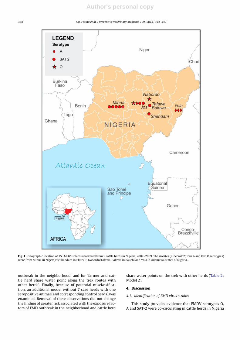
Results from sequencing and phylogenetic analysesrevealed that FMDV serotypes O, A and SAT-2 were co-circulating in cattle herds in Nigeria during 2007–2009. Thegeographic distribution of FMDV serotypes and topotypesidentified in cattle herds in Nigeria during the study periodis presented in Fig. 1.
Two serotype O isolates were recovered from one cat-tle herd in the state of Bauchi in 2007 and one cattle herdin the state of Plateau in 2009. The phylogenetic distancebetween these two isolates = 5.79% (Appendix Table 1). Inaddition, the isolate from 2007 was considered the samestrain as that from 3 isolates from Sudan in 2005 and 7more in 2004. Furthermore, the isolate from 2009 was con-sidered the same strain as that from 3 isolates from Sudanin 2005 (i.e., phylogenetic distance <5%).
Four serotype A isolates were recovered from one cat-tle herd in the state of Plateau (one isolate) and fromone cattle herd in the state of Adamawa (three isolates)in 2009. The phylogenetic distance between the three
isolates from Adamawa = 0.15%, and the distance betweenthe isolates from Adamawa and Plateau = 10.38 to 10.53%(Appendix Table 2). In addition, three SAT-A isolates fromthree different cattle herds in the same neighborhood in thestate of Adamawa were the same strains (i.e., 99.84% iden-tity). Finally, the phylogenetic distance between these fourisolates from Nigeria and other isolates from Cameroon(2000), Mali (2004/2006) and Eritrea (1997) ranged from6.26 to 10.53%.
Nine serotype SAT-2 isolates were recovered from onecattle herd in Bauchi state in 2007 (1 isolate), from onecattle herd in Niger state in 2008 (5 isolates), and from threecattle herds in the same neighborhood in Plateau state in2008 (3 isolates). The phylogenetic distance between thesenine isolates ranged from 0.31 to 1.39% (Appendix Table 3).The distance between the nine isolates from Nigeria andone isolate from Sudan (2007) ranged from 4.78 to 5.25%,and distance from one isolate from Niger Republic (2005)ranged from 7.10 to 7.25%.
3.2. Case–control study
This present study included 68 case herds and 68 con-trol herds in Nigeria. Seven of 68 case herds had one animalthat tested positive to FMDV NSP antibodies. Sixty-one of68 case herds had 2 or more animals that tested posi-tive to FMDV NSP antibodies. Nine of the 68 case herdswere confirmed as FMD-infected by virus isolation andsequencing and phylogenetic analysis of the VP1 gene, and7 additional case herds were confirmed by rt-PCR only.The geographic distribution of case and control farms ispresented in Fig. 2. Most case and control herds wereherds with ≤50 cattle head (see Table 1). In the univari-able analysis, 14 variables had values of P ≤ 0.20 and werefurther analyzed for biological plausibility, magnitude ofassociation, and statistical significance. The explanatoryvariable for ‘FMD outbreak in the neighborhood’ was asso-ciated (P < 0.05) with the variable for ‘cattle market in theneighborhood’. The variable for ‘farmer and cattle herdshare water point along the trek routes with other herds’was associated with the variables for ‘neighboring villageshare water points’ and ‘neighboring village share graz-ing reserves’. The variable ‘neighboring village share waterpoints’ was associated with the variable for ‘neighbor-ing village share grazing reserves’. Finally, the variable for‘neighboring village share grazing reserves’ was associatedwith the variable for ‘farmers and animals share trek route’.
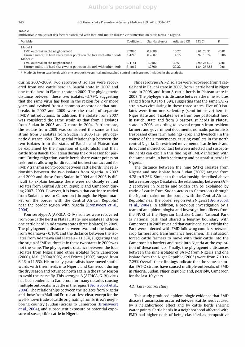
In the multivariable analysis, the variables for ‘FMD out-break in the neighborhood’ and for ‘farmer and cattle herdshare water point along the trek routes with other herds’were retained in the final model (Table 2). Addition to themodel of the interaction term between these two variableswas not significant (P = 0.48) and this term was removedfrom the model. Visual examination of residuals revealedthat delta-beta values for the two variables kept in the finalmodel were not extreme (i.e., not >1), which supportedoverall goodness of fit. Analysis of residuals (set of case andcontrol herds with the largest delta-beta value and lowestfitted value) indicated the existence of influential obser-vations; however, removal of these observations did notchange the finding of greater risk associated with ‘FMD
Author's personal copy
338 F.O. Fasina et al. / Preventive Veterinary Medicine 109 (2013) 334– 342
Fig. 1. Geographic location of 15 FMDV isolates recovered from 9 cattle herds in Nigeria, 2007–2009. The isolates (nine SAT 2; four A and two O serotypes)were from Minna in Niger; Jos/Shendam in Plateau; Nabordo/Tafawa-Balewa in Bauchi and Yola in Adamawa states of Nigeria.
outbreak in the neighborhood’ and for ‘farmer and cat-tle herd share water point along the trek routes withother herds’. Finally, because of potential misclassifica-tion, an additional model without 7 case herds with oneseropositive animal (and corresponding control herds) wasexamined. Removal of these observations did not changethe finding of greater risk associated with the exposure fac-tors of FMD outbreak in the neighborhood and cattle herd
share water points on the trek with other herds (Table 2;Model 2).
4. Discussion
4.1. Identification of FMD virus strains
This study provides evidence that FMDV serotypes O,A and SAT-2 were co-circulating in cattle herds in Nigeria

Author's personal copy
F.O. Fasina et al. / Preventive Veterinary Medicine 109 (2013) 334– 342 339
Fig. 2. Geographic locations of case farms (n = 68) and control farms (n = 68) in Nigeria, 2007–2009.
Table 1Univariable analysis of risk factors associated with foot-and-mouth disease virus infection on cattle farms in Nigeria; showing variables with values P < 0.20.
Variable Category Case farms N = 68 Control farms N = 68 Crude OR 95% CI P
Years of operation 1–5 8 21 1.00 Reference NA6–10 34 27 2.88 1.15, 7.19 0.02>10 25 20 2.82 1.07, 7.43 0.03
Cattle herd residency Sedentary 16 22 1.00 Reference NAPastoralist 29 23 2.04 0.88, 4.75 0.09Cattle market 8 2 5.42 0.97, 30.24 0.05All others 13 20 0.95 0.34, 2.61 0.92
Farmer and cattle herd share water points onthe trek with other herds
No 5 15 1.00 Reference NAYes 62 50 3.50 1.15, 10.63 0.02
Neighboring village shares water points No 26 37 1.00 Reference NAYes 38 28 2.44 1.12, 5.30 0.02
Neighboring village shares grazing reserves No 12 25 1.00 Reference NAYes 50 37 2.57 1.07, 6.16 0.03
Farmer and cattle herd share trek route No 10 21 1.00 Reference NAYes 56 42 2.22 1.01, 4.88 0.04
Farmer and cattle herd share pasture withother herds
No 22 30 1.00 Reference NAYes 35 30 1.87 0.79, 4.42 0.15
Farmer and cattle cross national boundaries No 52 56 1.00 Reference NAYes 15 8 2.00 0.80, 4.95 0.13
Farmer and cattle cross game reserves andparks
No 55 61 1.00 Reference NAYes 11 4 2.50 0.78, 7.97 0.12
Farmer has sighted wild ungulates/ruminants No 49 54 1.00 Reference NAYes 17 10 2.00 0.80, 4.95 0.13
FMD outbreak in the neighborhood No 17 49 1.00 Reference NAYes 42 12 14.50 3.46, 60.76 <0.01
Cattle market in the neighborhood No 13 19 1.00 Reference NAYes 55 45 2.60 0.92, 7.29 0.06
Cattle cross national highways No 31 35 1.00 Reference NAYes 36 24 1.75 0.86, 3.55 0.12
Vehicles have free access to the farm No 15 18 1.00 Reference NAYes 52 42 1.77 0.78, 4.02 0.16
Author's personal copy
340 F.O. Fasina et al. / Preventive Veterinary Medicine 109 (2013) 334– 342
Table 2Multivariable analysis of risk factors associated with foot-and-mouth disease virus infection on cattle farms in Nigeria.
Variable Coefficient Standard error Adjusted OR 95% CI P
Model 1FMD outbreak in the neighborhood 2.7895 0.7680 16.27 3.61, 73.31 <0.01Farmer and cattle herd share water points on the trek with other herds 1.4243 0.7687 4.15 0.92, 18.74 0.06
Model 2a
FMD outbreak in the neighborhood 3.4181 1.0487 30.51 3.90, 283.30 <0.01Farmer and cattle herd share water points on the trek with other herds 3.1012 1.2700 22.22 1.84, 267.83 0.01
a Model 2: Seven case herds with one seropositive animal and matched control herds are not included in the analysis.
during 2007–2009. Two serotype O isolates were recov-ered from one cattle herd in Bauchi state in 2007 andone cattle herd in Plateau state in 2009. The phylogeneticdistance between these two isolates = 5.79%, suggestingthat the same virus has been in the region for 2 or moreyears and evolved from a common ancestor or that out-breaks in 2007 and 2009 were the result of separateFMDV introductions. In addition, the isolate from 2007was considered the same strain as that from 3 isolatesfrom Sudan in 2005 and 7 more in 2004. Furthermore,the isolate from 2009 was considered the same as thatstrain from 3 isolates from Sudan in 2005 (i.e., phyloge-netic distance <5%). The spatial relationship between thetwo isolates from the states of Bauchi and Plateau canbe explained by the migration of pastoralists and theircattle from Bauchi to Plateau during the dry season for pas-ture. During migration, cattle herds share water points ontrek routes allowing for direct and indirect contact and forFMDV transmission to occur between cattle herds. The rela-tionship between the two isolates from Nigeria in 2007and 2009 and those from Sudan in 2004 and 2005 is dif-ficult to explain because there were no closely relatedisolates from Central African Republic and Cameroon dur-ing 2007–2009. However, it is known that cattle are tradedfrom Sudan across to Cameroon (through the Ngoui mar-ket on the border with the Central African Republic)near the border region with Nigeria (Bronsvoort et al.,2004).
Four serotype A (AFRICA, G-IV) isolates were recoveredfrom one cattle herd in Plateau state (one isolate) and fromone cattle herd in Adamawa state (three isolates) in 2009.The phylogenetic distance between two and one isolatesfrom Adamawa = 0.16%, and the distance between the iso-lates from Adamawa and Plateau = 11.38%, suggesting thatthe origin of FMD outbreaks in these two states in 2009 wasnot the same. The phylogenetic distance between the fourisolates from Nigeria and other isolates from Cameroon(2000), Mali (2004/2006) and Eritrea (1997) ranged from6.26 to 11.53%. Historically, pastoralists have moved south-wards with their herds into Nigeria and Cameroon duringthe dry season and returned north again in the rainy seasonto avoid the tsetse fly. This serotype A (AFRICA, G-IV) virushas been endemic in Cameroon for many decades causingmultiple outbreaks in cattle in the region (Bronsvoort et al.,2004). The relationships between the isolates from Nigeriaand those from Mali and Eritrea are less clear, except for thewell-known trade of cattle originating from Eritrea’s neigh-boring country (Sudan) across to Cameroon (Bronsvoortet al., 2004), and subsequent exposure or potential expo-sure of susceptible cattle in Nigeria.
Nine serotype SAT-2 isolates were recovered from 1 cat-tle herd in Bauchi state in 2007, from 1 cattle herd in Nigerstate in 2008, and from 3 cattle herds in Plateau state in2008. The phylogenetic distance between the nine isolatesranged from 0.31 to 1.39%, suggesting that the same SAT-2strain was circulating in these three states. Five of 9 iso-lates were from one sedentary (semi-intensive) herd inNiger state and 4 isolates were from one pastoralist herdin Bauchi state and from 3 pastoralist herds in Plateaustate. In 2008, according to several reports from residentfarmers and government documents, nomadic pastoraliststrespassed other farm holdings (crop and livestock) in thecourse of their movements, causing conflicts in the northcentral Nigeria. Unrestricted movement of cattle herds anddirect and indirect contact between infected and suscepti-ble herds can explain this finding of FMDV infection withthe same strain in both sedentary and pastoralist herds inNigeria.
The distance between the nine SAT-2 isolates fromNigeria and one isolate from Sudan (2007) ranged from4.78 to 5.25%. Similar to the relationship described abovebetween serotype O isolates, the relationship between SAT-2 serotypes in Nigeria and Sudan can be explained bytrade of cattle from Sudan across to Cameroon (throughthe Ngoui market on the border with the Central AfricanRepublic) near the border region with Nigeria (Bronsvoortet al., 2004). In addition, a previous investigation by ateam of an epidemiologist and investigation officers fromthe NVRI at the Nigerian Gashaka-Gumti National Park(a national park that shared a lengthy boundary withCameroon) in 2005 revealed that cattle enclaves within thePark were infected with FMD following conflicts betweencrop farmers and transhumance herdsmen. This situationforced cattle farmers to move with their cattle into theCameroonian borders and back into Nigeria at the expira-tion of these conflicts. Finally, the phylogenetic distancesbetween the nine isolates of SAT-2 from Nigeria and oneisolate from the Niger Republic (2005) were from 7.10 to7.25%. Overall, these findings indicate that the same or sim-ilar SAT-2 strains have caused multiple outbreaks of FMDin Nigeria, Sudan, Niger Republic and, possibly, Cameroonfor the last 10 years.
4.2. Case–control study
This study produced epidemiologic evidence that FMDdisease transmission occurred between cattle herds causedby a neighborhood effect and by cattle herds sharingwater points. Cattle herds in a neighborhood affected withFMD had higher odds of being classified as seropositive
Author's personal copy
F.O. Fasina et al. / Preventive Veterinary Medicine 109 (2013) 334– 342 341
to FMD, compared to herds that were in a neighborhoodnot affected with FMD (adjusted OR = 16.27; 95% CI = 3.61,18.74; P < 0.01). Neighborhoods in rural Nigeria allow fordirect and indirect contact between animals. Cattle herdsare not confined in fenced premises. Usually, cattle (par-ticularly calves) are not penned/tied down at night, andthis practice can be a source of direct and indirect contactbetween cattle. A neighborhood effect for FMDV transmis-sion between herds has been identified in other countries(Ellis-Iversen et al., 2011). Airborne spread of FMD fromswine to nearby cattle and sheep, and an increase in themovements of vehicles and personnel engaged in diseasecontrol efforts perhaps facilitated local spread of FMDduring the epidemic in the UK in 2001 (Mansley et al.,2011). During the outbreak in the UK in 2007, FMDVwas accidently released from a joint vaccine productionand diagnostic laboratory site in Pirbright, UK, most likelyleaked through poorly maintained effluent pipes and trans-ported off-site to two cattle farms nearby via mud or soilon vehicle wheels (Ellis-Iversen et al., 2011). In that out-break, a higher proportion of young stock was observed incase farms compared to other farms, suggesting an associ-ation between age and FMD status. In addition, inadequatebiosecurity was identified as a risk factor for FMD transmis-sion between cattle premises during the outbreak in 2007(Mansley et al., 2011).
In this study, cattle herds that share water points alongthe trek routes with other cattle herds had higher odds ofbeing classified as seropositive to FMD (adjusted OR = 4.15;95% CI = 0.92, 18.74; P < 0.06). In Nigeria, there are estab-lished old grazing reserves and watering points along thetrek routes, and animals originating from different loca-tions share these reserves and watering points. In addition,rivers crossing the routes of these animals are sharedby both FMD-infected and susceptible cattle herds whichcongregate together. A similar type of exposure and dis-ease transmission between FMD-infected and susceptibleanimals has been documented in South Africa. At theKruger National Park, between May and November, thereis scarcity of water in the Park, and buffalo potentiallyinfected with FMDV congregate around available waterpoints providing an opportunity for direct and indirect con-tact with susceptible domestic livestock (Jori et al., 2009).
This study had several limitations. First, FMDV isolateswere recovered from 15 of 50 epithelial/vesicular fluidsamples submitted from cattle located in North Central andNorth East regions of Nigeria. No FMDV isolates were recov-ered from cattle in the South West region. It is possiblethat improper sample collection and processing (or pack-ing) combined with a long distance and time required forsamples to reach the NVRI in Vom could have affected thequality (freshness) of the sample to recover FMDV. Second,7 of 68 case herds (3 from the state of Plateau, 3 from Bauchi,and 1 from Adamawa) had only one seropositive animal,and these herds were not confirmed as infected by virusisolation or rt-PCR. Because the sensitivity of the 3-ABCELISA test is not perfect (Brocchi et al., 2006; Engel et al.,2008), it is possible that one or more case herds with oneseropositive animal were misclassified. Removal of theseseven case herds did not change the finding of greater riskassociated with the exposure factors of FMD outbreak in
the neighborhood and cattle herd share water points on thetrek with other herds. Another limitation was observation(exposure) bias. For example, the accuracy of a producer toclassify his/her cattle herd as vaccinated was not assessedin this study. In Nigeria and other countries in sub-SaharanAfrica, the mobility and dispersion of nomadic pastoral-ists present significant financial and logistic constrains toveterinary services for vaccination of livestock.
Overall, the study results revealed that FMDV serotypesO, A and SAT-2 were co-circulating in cattle herds in Nigeriaduring 2007–2009. Based on results from the phylogeneticanalysis, it is possible that the same SAT-2 strain has causedmultiple outbreaks of FMD in cattle in Nigeria, Sudan, NigerRepublic and, possibly, Cameroon for the last 10 years.Finally, combined results from the phylogenetic analysisand the case–control study suggest that FMD seropositivityin cattle herds was associated with exposure to neighbor-hoods with FMD activity and by cattle herds sharing waterpoints. Results from this study can be used by veterinaryservices in Nigeria and neighboring countries to evaluatecurrent or future FMD control and eradication programs.
Acknowledgements
The authors would like to appreciate David Paton, JeffHammond, Nick Knowles and their team at the IAH, WorldReference Laboratory for Foot and Mouth Disease, PirbrightLaboratory, UK for their robust efforts in confirming thediagnosis and molecular studies and Mr. D. C. Nyam ofthe Viral Research Division for assistance during sampling.The National Animal Disease Information System (NADIS)teams are also thanked for prompt reporting of outbreaksand sample collection and the Management of the NationalVeterinary Research Institute, Vom for providing the envi-ronment for this study. We thank Mrs. Ingrid Booysen ofthe Department of Geography, Geoinformatics and Mete-orology, University of Pretoria for the production of themaps.
Appendix A. Supplementary data
Supplementary data associated with this article can befound, in the online version, at http://dx.doi.org/10.1016/j.prevetmed.2012.10.004.
References
Brocchi, E., Bergmann, I., Dekker, A., Paton, D.J., Sammin, D.J., Greiner, M.,Grazioli, S., De Simone, F., Yadin, H., Haas, B., Bulut, N., Malirat, V.,Neitzert, E., Goris, N., Parida, S., Sorensen, K., De Clercq, K., 2006. Com-parative evaluation of six ELISAs for the detection of antibodies to thenon-structural proteins of foot-and-mouth disease virus. Vaccine 24,6966–6979.
Bronsvoort, B.M., de, C., Radford, A.D., Tanya, V.N., Nfon, C., Kitching, R.P.,Morgan, K.L., 2004. Molecular epidemiology of foot and mouth diseaseviruses in the Adamawa Province of Cameroon. J. Clin. Microbiol. 42(5), 2186–2196.
Cytel Software Corporation, 2000. EGRET for Windows 2000. Cytel Soft-ware Corp., Cambridge, MA, USA.
Durojaiye, A., 1981. Incidence of FMD in Oyo State of Nigeria, 1961-1981.Nig. Vet. J. 10, 7–13.
Ellis-Iversen, J., Smith, R.P., Gibbens, J.C., Sharpe, C.E., Dominguez, M., Cook,A.J.C., 2011. Risk factors for transmission of foot and mouth diseaseduring an outbreak in Southern England in 2007. Vet. Rec. 168 (5),128, http://dx.doi.org/10.1136/vr.c6364.

Author's personal copy
342 F.O. Fasina et al. / Preventive Veterinary Medicine 109 (2013) 334– 342
Engel, B., Buist, W., Orsel, K., Dekker, A., de Clercq, K., Grazioli, S., vanRoermund, H., 2008. A Bayesian evaluation of six disgnostic testsfor foot-and-mouth disease for vaccinated and non-vaccinated cattle.Prev. Vet. Med. 86, 124–138.
Fadiga, M., Jost, C., Ihedioha, J., 2011. Financial costs of diseaseburden, morbidity and mortality from priority livestock dis-eases in Nigeria: disease burden and cost–benefit analysis oftargeted intervention. Available at: http://www.ilri.org/content/financial-costs-disease-burden-morbidity-and-mortality-priority-livestock-diseases-nigeria (accessed 05.03.12).
Food and Agriculture Organization of the United Nations Statis-tics Division (FAO), 2012a. Stock (head/year): Nigeria. Avail-able at: http://faostat.fao.org/site/573/DesktopDefault.aspx?PageID=573#ancor (accessed 10.04.12).
Food and Agriculture Organization of the United Nations (FAO),2012b. WRLFMD Report on the Diagnosis of FMD, Nigeria. Avail-able at: www.wrlfmd.org/fmd genotyping/africa/nig.htm (accessed23.03.12).
Hosmer, D.W., Lemeshow, S., 2000. Logistic regression for matchedcase–control studies. In: Hosmer, D.W., Lemeshow, S. (Eds.), AppliedLogistic Regression. Wiley, New York, pp. 223–259.
Jori, F., Vosloo, W., Du Plessis, B., Bengis, R., Brahmbhatt, D., Gummow,B., Thomson, G.R., 2009. A qualitative risk assessment of factors con-tributing to foot and mouth disease outbreaks in cattle along thewestern boundary of the Kruger National Park. Rev. Sci. Tech. 28 (3),917–931.
Kitching, R.P., Donaldson, A.I., 1987. Collection and transportation of spec-imens for vesicular virus investigation. Rev. Sci. Tech. Off. Int. Epiz. 6,263–272.
Knowles, N.J., Samuel, A.R., 2003. Molecular epidemiology of foot andmouth disease virus. Virus Res. 91, 65–80.
Libeau, J., 1960. Foot-and-mouth disease in Africa, South of the Sahara –the present situation. Bull. Epizoot. Dis. Afr. 8, 152–158.
Ma, L., Zhang, J., Chen, H., Zhou, J., Ding, Y., Liu, Y., 2011. An overview onELISA techniques for FMD. Virol. J. 208, 419.
Mansley, L.M., Donaldson, A.I., Thrushfield, M.V., Honhold, N., 2011.Destructive tension: mathematics versus experience – the progressand control of the 2001 foot and mouth disease epidemic in GreatBritain. Rev. Sci. Tech. 30 (2), 483–498.
Mason, P.W., Grubman, M.J., Baxt, B., 2003. Molecular basis of pathogen-esis FMDV. Virus Res. 91, 9–32.
Nawathe, D.R., Goni, M., 1976. Foot-and-mouth disease in Nigeria. Bull.Anim. Health Prod. Afr. 24, 1–4.
OIE (World Organisation for Animal Health), 2008. Collection andshipment of diagnostic specimens, Chapter 1.1.1, Manual ofDiagnostic Tests and Vaccines for Terrestrial Animals, Web ver-sion, 2008. Available at http://www.oie.int/eng/normes/mmanual/2008/pdf/1.1.01 COLLECTION.pdf (accessed 13.09.08).
Orsel, K., Bouma, A., Dekker, A., Stegeman, J.A., de Jong, M.C.M., 2009. Footand mouth disease virus transmission during the incubation periodof the disease in piglets, lambs, calves and dairy cows. Prev. Vet. Med.88, 158–163.
Owolodun, B.Y., 1971. Foot and mouth disease virus type distribution inNigeria. Bull. Epizoot. Dis. Afr. 8, 152–158.
Rweyemamu, M., Roeder, P., Mackay, D., Sumption, K., Brownlie, J., Lefor-ban, Y., Valarcher, J.F., Knowles, N.J., Saraiva, V., 2008. Epidemiologicalpatterns of foot-and-mouth disease worldwide. Transbound Emerg.Dis. 55 (1), 57–72.
Sahle, M., Venter, E.H., Dwarka, R.M., Vosloo, W., 2004. Molecular epi-demiology of serotype O foot and mouth disease virus isolated fromcattle in Ethiopia between: 1979–2001. Onderstepoort J. Vet. 71,129–138.
Sangare, O., Bastos, A.D., Venter, E.H., Vosloo, W., 2004. A first molecularepidemiological study of SAT-2 type foot-and-mouth disease virusesin West Africa. Epidemiol. Infect. 132 (3), 525–532.
Sorensen, K.J., Madsen, K.G., Madsen, E.S., Salt, J.S., Nqindi, J., Mackay, D.K.J.,1998. Differentiation of infection from vaccination in foot-and-mouthdisease by the detection of antibodies to the non-structural proteins3D, 3AB and 3ABC in ELISA using antigens expressed in baculovirus.Arch. Virol. 143, 1461–1476.
Sumption, K., Pinto, J., Lubroth, J., Morzaria, S., Murray, T., De LaRocque, S., Njeumi, F., 2007. Foot-and-mouth disease: situationworldwide and major epidemiological events in 2005–2006.Emergency Prevention System (EMPRES) 2007, No. 1. Available at:http://www.fao.org/docs/eims/upload/225050/Focus ON 1 07 en.pdf(accessed 09.04.12).
Vosloo, W., Dwarka, R.M., Bastos, A.D.S., Esterhuysen, J.J., Sahle, M.,Sangare, O., 2004. Molecular epidemiological studies of Foot-and-Mouth disease virus in sub-Saharan Africa indicate the presence oflarge numbers of topotypes: implications for local and internationalcontrol. Report of the Session of the Research Group of the StandingTechnical Committee of the European Commission for the Controlof Foot-and-Mouth Disease (EUFMD), Crete, Greece. Availableat: http://www.fao.org/Ag/AGAInfo/commissions/docs/greece04/App22.pdf (accessed 09.04.12).