Folliculin Contributes to VHL Tumor Suppressing Activity in Renal Cancer through Regulation of...
8
Folliculin Contributes to VHL Tumor Suppressing Activity in Renal Cancer through Regulation of Autophagy Prabhat Bastola 1. , Yiwen Stratton 1. , Emily Kellner 1 , Olga Mikhaylova 1 , Ying Yi 1 , Maureen A. Sartor 3 , Mario Medvedovic 3 , Jacek Biesiada 4 , Jarek Meller 3,4,5 , Maria F. Czyzyk-Krzeska 1,2 * 1 Department of Cancer Biology, University of Cincinnati College of Medicine, Cincinnati, Ohio, United States of America, 2 VA Research Service, Department of Veterans Affairs, Cincinnati, Ohio, United States of America, 3 Department of Environmental Health, University of Cincinnati College of Medicine, Cincinnati, Ohio, United States of America, 4 Division of Biomedical Informatics, Cincinnati Children’s Hospital Medical Center, Cincinnati, Ohio, United States of America, 5 Department of Informatics, Nicolas Copernicus University, Torun, Poland Abstract Von Hippel-Lindau tumor suppressor (VHL) is lost in the majority of clear cell renal cell carcinomas (ccRCC). Folliculin (FLCN) is a tumor suppressor whose function is lost in Birt-Hogg-Dube ´ syndrome (BHD), a disorder characterized by renal cancer of multiple histological types including clear cell carcinoma, cutaneous fibrofolliculoma, and pneumothorax. Here we explored whether there is connection between VHL and FLCN in clear cell renal carcinoma cell lines and tumors. We demonstrate that VHL regulates expression of FLCN at the mRNA and protein levels in RCC cell lines, and that FLCN protein expression is decreased in human ccRCC tumors with VHL loss, as compared with matched normal kidney tissue. Knockdown of FLCN results in increased formation of tumors by RCC cells with wild-type VHL in orthotopic xenografts in nude mice, an indication that FLCN plays a role in the tumor-suppressing activity of VHL. Interestingly, FLCN, similarly to VHL, is necessary for the activity of LC3C-mediated autophagic program that we have previously characterized as contributing to the tumor suppressing activity of VHL. The results show the existence of functional crosstalk between two major tumor suppressors in renal cancer, VHL and FLCN, converging on regulation of autophagy. Citation: Bastola P, Stratton Y, Kellner E, Mikhaylova O, Yi Y, et al. (2013) Folliculin Contributes to VHL Tumor Suppressing Activity in Renal Cancer through Regulation of Autophagy. PLoS ONE 8(7): e70030. doi:10.1371/journal.pone.0070030 Editor: Rajvir Dahiya, UCSF/VA Medical Center, United States of America Received April 16, 2013; Accepted June 14, 2013; Published July 29, 2013 Copyright: ß 2013 Bastola et al. This is an open-access article distributed under the terms of the Creative Commons Attribution License, which permits unrestricted use, distribution, and reproduction in any medium, provided the original author and source are credited. Funding: Myrovlytis Trust, Research Grant; NIH/NCI CA122346; BLR&D VAmerit AwardB101BX0011100-02; UC P30-ES006096CEG. The funders had no role in study design, data collection and analysis, decision to publish, or preparation of the manuscript. Competing Interests: The authors have declared that no competing interests exist. * E-mail: [email protected] . These authors contributed equally to this work. Introduction Renal cancer accounts for 2% to 3% of all adult malignancies in the US. In 2011 more than 64,770 new cases and 13,000 deaths were reported, and the incidence is steadily increasing by 2.5% per year. ccRCC represents the majority (85% to 90%) of adult kidney cancers and is the most malignant form. This cancer is characterized by an early loss of VHL tumor suppressor on the short arm of chromosome 3 in the majority of tumors (80%). It is well established that loss of VHL results in increased accumulation and activity of the hypoxia-inducible transcription factor (HIF), which in turns activates expression of genes that promote tumor growth. Among these are angiogenic factors, which lead to increased blood flow through the tumors, thus enhancing the delivery of nutrients and oxygen to cancer cells. Genes that stimulate anaerobic glycolysis are also activated, and this supports adaptation to reduced nutrient availability and shifts cancer cells towards metabolic pathways that promote tumor growth. Recent- ly, we discovered that another tumor-suppressing pathway is regulated by VHL. We found that VHL is a major regulator of the process of macroautophagy (autophagy) [1]. We determined that VHL, by inducing microRNA-204, inhibited LC3B-mediated autophagy which is necessary for ccRCC tumor growth. Moreover, VHL, by inhibiting HIF, induced expression and activity of an LC3B paralog, LC3C. In contrast to LC3B, LC3C acts as a tumor suppressor [1]. In the process of searching for new VHL-induced genes that could participate in the tumor-suppressing activity of VHL, we discovered that reconstitution of wild-type VHL in RCC cells with lost VHL induced specific and significant enrichment for genes expressed from the Smith-Magenis locus (SM) on the short arm of chromosome 17p11.2. Smith-Magenis syndrome is a complex neurobehavioral disorder characterized by intellectual impair- ment, craniofacial and skeletal anomalies, and sleep disturbance [2]. Interestingly, this locus contains another kidney tumor- suppressor gene, folliculin (FLCN), whose activity is lost in the genetic autosomal-dominant disorder, Birt-Hogg-Dube ´ syndrome (BHD) [3]. BHD is characterized by skin fibrofolliculomas, pulmonary cysts, and spontaneous pneumothorax, as well as renal cancer of mixed histological types, including chromophobe, oncocytic, clear cell, and papillary type [3]. The purpose of this work was to determine if FLCN contributes to the tumor suppressing activity of VHL. We show that FLCN contributes to VHL-mediated suppression of tumor growth by affecting LC3B and LC3C autophagic pathways. PLOS ONE | www.plosone.org 1 July 2013 | Volume 8 | Issue 7 | e70030
-
Upload
independent -
Category
Documents
-
view
1 -
download
0
Transcript of Folliculin Contributes to VHL Tumor Suppressing Activity in Renal Cancer through Regulation of...

Folliculin Contributes to VHL Tumor Suppressing Activityin Renal Cancer through Regulation of AutophagyPrabhat Bastola1., Yiwen Stratton1., Emily Kellner1, Olga Mikhaylova1, Ying Yi1, Maureen A. Sartor3,
Mario Medvedovic3, Jacek Biesiada4, Jarek Meller3,4,5, Maria F. Czyzyk-Krzeska1,2*
1 Department of Cancer Biology, University of Cincinnati College of Medicine, Cincinnati, Ohio, United States of America, 2 VA Research Service, Department of Veterans
Affairs, Cincinnati, Ohio, United States of America, 3 Department of Environmental Health, University of Cincinnati College of Medicine, Cincinnati, Ohio, United States of
America, 4 Division of Biomedical Informatics, Cincinnati Children’s Hospital Medical Center, Cincinnati, Ohio, United States of America, 5 Department of Informatics,
Nicolas Copernicus University, Torun, Poland
Abstract
Von Hippel-Lindau tumor suppressor (VHL) is lost in the majority of clear cell renal cell carcinomas (ccRCC). Folliculin (FLCN)is a tumor suppressor whose function is lost in Birt-Hogg-Dube syndrome (BHD), a disorder characterized by renal cancer ofmultiple histological types including clear cell carcinoma, cutaneous fibrofolliculoma, and pneumothorax. Here we exploredwhether there is connection between VHL and FLCN in clear cell renal carcinoma cell lines and tumors. We demonstrate thatVHL regulates expression of FLCN at the mRNA and protein levels in RCC cell lines, and that FLCN protein expression isdecreased in human ccRCC tumors with VHL loss, as compared with matched normal kidney tissue. Knockdown of FLCNresults in increased formation of tumors by RCC cells with wild-type VHL in orthotopic xenografts in nude mice, anindication that FLCN plays a role in the tumor-suppressing activity of VHL. Interestingly, FLCN, similarly to VHL, is necessaryfor the activity of LC3C-mediated autophagic program that we have previously characterized as contributing to the tumorsuppressing activity of VHL. The results show the existence of functional crosstalk between two major tumor suppressors inrenal cancer, VHL and FLCN, converging on regulation of autophagy.
Citation: Bastola P, Stratton Y, Kellner E, Mikhaylova O, Yi Y, et al. (2013) Folliculin Contributes to VHL Tumor Suppressing Activity in Renal Cancer throughRegulation of Autophagy. PLoS ONE 8(7): e70030. doi:10.1371/journal.pone.0070030
Editor: Rajvir Dahiya, UCSF/VA Medical Center, United States of America
Received April 16, 2013; Accepted June 14, 2013; Published July 29, 2013
Copyright: � 2013 Bastola et al. This is an open-access article distributed under the terms of the Creative Commons Attribution License, which permitsunrestricted use, distribution, and reproduction in any medium, provided the original author and source are credited.
Funding: Myrovlytis Trust, Research Grant; NIH/NCI CA122346; BLR&D VAmerit AwardB101BX0011100-02; UC P30-ES006096CEG. The funders had no role in studydesign, data collection and analysis, decision to publish, or preparation of the manuscript.
Competing Interests: The authors have declared that no competing interests exist.
* E-mail: [email protected]
. These authors contributed equally to this work.
Introduction
Renal cancer accounts for 2% to 3% of all adult malignancies in
the US. In 2011 more than 64,770 new cases and 13,000 deaths
were reported, and the incidence is steadily increasing by 2.5% per
year. ccRCC represents the majority (85% to 90%) of adult kidney
cancers and is the most malignant form. This cancer is
characterized by an early loss of VHL tumor suppressor on the
short arm of chromosome 3 in the majority of tumors (80%). It is
well established that loss of VHL results in increased accumulation
and activity of the hypoxia-inducible transcription factor (HIF),
which in turns activates expression of genes that promote tumor
growth. Among these are angiogenic factors, which lead to
increased blood flow through the tumors, thus enhancing the
delivery of nutrients and oxygen to cancer cells. Genes that
stimulate anaerobic glycolysis are also activated, and this supports
adaptation to reduced nutrient availability and shifts cancer cells
towards metabolic pathways that promote tumor growth. Recent-
ly, we discovered that another tumor-suppressing pathway is
regulated by VHL. We found that VHL is a major regulator of the
process of macroautophagy (autophagy) [1]. We determined that
VHL, by inducing microRNA-204, inhibited LC3B-mediated
autophagy which is necessary for ccRCC tumor growth.
Moreover, VHL, by inhibiting HIF, induced expression and
activity of an LC3B paralog, LC3C. In contrast to LC3B, LC3C
acts as a tumor suppressor [1].
In the process of searching for new VHL-induced genes that
could participate in the tumor-suppressing activity of VHL, we
discovered that reconstitution of wild-type VHL in RCC cells with
lost VHL induced specific and significant enrichment for genes
expressed from the Smith-Magenis locus (SM) on the short arm of
chromosome 17p11.2. Smith-Magenis syndrome is a complex
neurobehavioral disorder characterized by intellectual impair-
ment, craniofacial and skeletal anomalies, and sleep disturbance
[2]. Interestingly, this locus contains another kidney tumor-
suppressor gene, folliculin (FLCN), whose activity is lost in the
genetic autosomal-dominant disorder, Birt-Hogg-Dube syndrome
(BHD) [3]. BHD is characterized by skin fibrofolliculomas,
pulmonary cysts, and spontaneous pneumothorax, as well as renal
cancer of mixed histological types, including chromophobe,
oncocytic, clear cell, and papillary type [3]. The purpose of this
work was to determine if FLCN contributes to the tumor
suppressing activity of VHL. We show that FLCN contributes to
VHL-mediated suppression of tumor growth by affecting LC3B
and LC3C autophagic pathways.
PLOS ONE | www.plosone.org 1 July 2013 | Volume 8 | Issue 7 | e70030

Materials and Methods
Cell Culture Methods and TreatmentsOur method for generating pools of human VHL(2) and
VHL(+) 786-O, A498, and Caki-1 cells were described by us
before [1,4,5]. RCC4 cells were gift from Dr. Jamie Cairo
(Jefferson University) and were described by us before [6].
Autophagic fluxes were performed as described before [1]. For
protein or RNA extraction, cells were plated, 24 hr later medium
was changed to that with either 10% or 0.1% serum, and cells
were collected 48 hr later. Cells were then lysed in RIPA buffer
with protease, phosphatase, and proteasomal inhibitors for PAGE
analysis, or in TRI Reagent for RNA extraction. For RNAi
experiments, pLKO.1-based shRNA constructs for FLCN
(TRCN0000005969, TRCN0000005970, and
TRCN0000010979; Open Biosystems) were VSV-G envelope
packaged (Cincinnati Children’s Hospital Medical Center Viral
Vector Core) and infected into cells, which were then treated with
plasmid-appropriate selection reagents. VHL shRNA constructs
were described before [5]. Transductions with lentiviral particles
were performed using 2 mg/ml of polybrene.
Human RCC TumorsFresh-frozen samples of ccRCC tumors (n = 128) and matched
normal kidney samples (n = 114) were obtained and analyzed for
VHL sequence and protein expression, as described before [4].
Specimens were analyzed for expression of protein and mRNA,
also as described before.
Quantitative RT-PCRFor mRNA analysis, the samples were first reverse transcribed
using the TaqMan microRNA Reverse Transcription kit (Applied
Biosystems), and quantified with real-time PCR on an Applied
Biosystems 7900HT Fast Real-Time CRSystem. qRT-PCR was
performed as described before, and using primers described in
Table S4. Description of RNA microarrays is provided in
Supplemental Methods.
Western Blot AnalysisTotal cellular lysates were obtained by lysing cells in RIPA
buffer containing proteinase and phosphatase inhibitors. 5 to
30 mg of extracts were separated on 4% to 12% or 10%
polyacrylamide gels and transferred onto PVDF membranes.
Semi-quantitative immunoblotting for human RCC tumors and
matched normal kidneys was performed as described previously
[4]. The following polyclonal antibodies were obtained from Cell
Signaling Technology (Danvar, MA): anti-FLCN, anti- S6 kinase 1
and T389 phosphorylation; anti- S6 and S240/244 phosphorylation;
anti- 4EBP and T37/46 or S65 phosphorylation. Polyclonal anti-
LC3B (Abcam, Cambridge, MA) and anti-LC3C (Abgent, San
Diego, CA) were used. The following mouse monoclonal
antibodies were used: anti-VHL (Pharmingen; San Jose, CA),
anti-hemagglutinin (Boehringer Ingelheim, Austria); and anti-
GAPDH (Abcam; Cambridge, MA).
Mouse Xenograft ExperimentsFor the intra-kidney injections, 30,000 cells were resuspended in
Matrigel to a final volume of 30 ml, and then slowly injected a few
millimeters under the kidney capsula of 4- to 5-week-old athymic
nude mice. After 10 to 12 weeks mice were sacrificed, tumors with
kidneys were collected and fixed in 4% paraformaldehyde,
paraffin embedded, and H&E stained or processed for S6/S6P
staining in the Histopathology Core at in the Department of
Pathology at CCHMC.
Statistical Analysis of Quantified DataData normalization and analysis of RNA microarrays are
described in Supplemental Methods. The data were deposited to
Gene Expression Omnibus with accession number GSE47106.
For descriptive statistics, data are expressed as mean 6 SEM
unless indicated otherwise. Analysis of differential expression was
performed using t-test or one-way analysis of variance (ANOVA),
followed by Tukey-Kramer multiple comparison tests. For
quantification of the immunoblotting experiments the normalized
values of protein abundance levels were log2 transformed to
facilitate robust, outlier-insensitive analysis. Standard box-and-
whisker plots were used to compare distributions of the normalized
abundance measure for individual proteins. The differences
between mean levels in two types of samples were assessed by
using (two-sided) t-test and further validated by Wilcoxon test.
Differences resulting in P,0.05 were considered to be statistically
significant.
Ethics StatementAll experiments on mice were performed in strict accordance to
the protocol approved by University of Cincinnati Institutional
Animal Care and Use Committee (Protocol Number: 06-07-21-
01). All surgery was performed under isoflurane anesthesia and all
efforts were made to minimize pain. All human samples were
anonymous and exempted from University of Cincinnati Internal
Review Board protocol requirements.
Results
Expression of FLCN is Induced by VHLTo identify the VHL-induced genes that could participate in the
tumor-suppressing activity of VHL, we performed mRNA
microarray experiments in which we compared gene expression
in human RCC 786-O VHL(2) cells and the same cells with
reconstituted, wild-type VHL (786-O VHL(+)). Statistical analysis
identified 1,310 transcripts differentially expressed in the two cell
types (FDR,0.1 and .50% change in expression level) (Table
S1).
Interestingly, among the 588 transcripts induced by the
reconstitution of VHL, we found a significant enrichment in
genes located on the short arm of chromosome 17 (10.71% of all
significantly induced genes), and particularly in cytoband 17p11.2,
which contains the SM locus (1.7% of all significantly induced
genes) (Table 1 and S2). There was also a concentration of genes
induced by VHL on chromosome 14 (7.85% of all significantly
induced genes) with the main cytoband being 14q22.1. VHL-
mediated induction of several of the SM genes was further
confirmed by quantitative RT-PCR (Fig. S1). In contrast, genes
that were repressed by VHL were enriched on chromosome 7 (63
genes, 9.75% of all significantly repressed genes) and chromosome
9 (42 genes, 6.5% of all repressed genes) (Table S3).
The SM locus contains a gene encoding another renal kidney
tumor suppressor, FLCN. Analysis of FLCN mRNA and protein
revealed increased expression of both in 786-O and A498 cells
with reconstituted VHL, as compared with parental VHL(-) cells
(Fig. 1A and B). In addition, FLCN mRNA and protein expression
were repressed in Caki1 renal cancer cells in which we knocked
down endogenous VHL expression by using shRNA (Fig. 1C and
1D). The change in FLCN mRNA expression was in the range of
1.5 to 2 fold, similar to other SM genes (compare Fig. S1).
However, induction at the level of protein was in the 3- to 4-fold
range, an indication that posttranscriptional mechanisms play a
role in this regulation. In these studies we also observed VHL-
mediated induction of p53, another gene located on chromosome
Crosstalk of VHL and FLCN Renal Tumor Suppressors
PLOS ONE | www.plosone.org 2 July 2013 | Volume 8 | Issue 7 | e70030
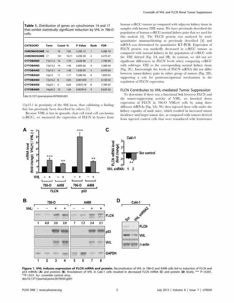
17p13.1 in proximity of the SM locus, thus validating a finding
that has previously been described by others [7].
Because VHL is lost in sporadic clear cell renal cell carcinoma
(ccRCC), we measured the expression of FLCN in lysates from
human ccRCC tumors as compared with adjacent kidney tissue in
samples with known VHL status. We have previously described the
population of human ccRCC-normal kidney pairs that we used for
this analysis [4]. The FLCN protein was analyzed by semi-
quantitative immunoblotting as previously described [4] and
mRNA was determined by quantitative RT-PCR. Expression of
FLCN protein was markedly decreased in ccRCC tumors as
compared with normal kidneys in the population of ccRCC with
the VHL deleted (Fig. 2A and 2B). In contrast, we did not see
significant differences in FLCN levels when comparing ccRCC
with wild-type VHL to the corresponding normal kidney tissue
(Fig. 2C). Interestingly the levels of FLCN mRNA did not differ
between tumor-kidney pairs in either group of tumors (Fig. 2D),
supporting a role for posttranscriptional mechanisms in the
regulation of FLCN expression.
FLCN Contributes to VHL-mediated Tumor SuppressionTo determine if there was a functional link between FLCN and
the tumor-suppressing activity of VHL, we knocked down
expression of FLCN in 786-O VHL(+) cells by using three
different shRNAs (Fig. 3A). We then injected these cells under the
kidney capsules of nude mice, which resulted in increased tumor
incidence and larger tumor size, as compared with tumors derived
from injected control cells that were transduced with lentiviruses
Table 1. Distribution of genes on cytochromes 14 and 17that exhibit statistically significant induction by VHL in 786-Ocells.
CATEGORY Term Count % P Value Rank FDR
CHROMOSOME 14 74 7.85 2.20E-12 1 4.38E-10
CHROMOSOME 17 101 10.71 4.29E-09 2 4.27E-07
CYTOBAND 17p11.2 16 1.70 2.62E-06 3 1.74E-04
CYTOBAND 17p13.3 14 1.48 6.80E-06 4 3.38E-04
CYTOBAND 17p13.1 14 1.48 1.63E-05 5 6.47E-04
CYTOBAND 12p13 11 1.17 5.58E-04 6 1.85E-02
CYTOBAND 17p13.2 8 0.85 0.001459 7 4.15E-02
CYTOBAND 14q22.1 6 0.64 0.003118 8 7.76E-02
CYTOBAND 14q24.3 10 1.06 0.003914 9 8.65E-02
doi:10.1371/journal.pone.0070030.t001
Figure 1. VHL induces expression of FLCN mRNA and protein. Reconstitution of VHL in 786-O and A498 cells led to induction of FLCN andp53 mRNAs (A) and proteins (B). Knockdown of VHL in Caki-1 cells resulted in decreased FLCN mRNA (C) and protein (D) levels; *** P,0.001,**P,0.01. Scr- scramble control virus.doi:10.1371/journal.pone.0070030.g001
Crosstalk of VHL and FLCN Renal Tumor Suppressors
PLOS ONE | www.plosone.org 3 July 2013 | Volume 8 | Issue 7 | e70030

containing scramble sequence (Fig. 3B). The tumors with FLCN
knockdown had a highly malignant, dedifferentiated phenotype
(Fig. 3C).
Because FLCN was shown to inhibit activity of the mTORC1
pathway in UOK257 cells and mouse models of BHD kidney
cancer [8,9], we analyzed the effects of FLCN knockdown on the
activity of this pathway in the cells used for these orthotopic
xenografts. Importantly, knockdown of FLCN resulted in
decreased mTORC1 activity (Fig. 4A), as measured by phosphor-
ylation of S6K, S6 and 4EBP, in cultured cells. We have also
determined expression of S6 and S6-P is sections from xenografts
tumors formed by VHL(+)/scramble transfected cells vs. tumors
formed by these cells with FLCN knocked down. Consistently with
the results obtained from cell culture experiments, sections from
VHL(+)/FLCNKD tumors showed lower staining for S6-P as
compared to the control tissues, while the staining for total S6 was
similar (Fig. 4B). This result suggests that augmented activity of the
mTORC1 pathway does not contribute to increased tumor
growth in these cells.
Recently we have described that VHL suppresses LC3B-
mediated, pro-oncogenic autophagy while it stimulates a novel
tumor suppressing autophagic program, mediated by LC3C [1].
LC3s are well established autophagic regulators necessary for the
formation of the double membrane autophagosomes and for
acquisition of the cargo. Thus we investigated if FLCN may
exercise its tumor suppressing activity downstream from VHL by
regulating LC3C or LC3B autophagy. LC3C is induced by
starvation in VHL(+) cells, thus in these experiments, VHL(+) 786-
O and RCC4 cells were starved in 0.1% serum for 48 hr and then
treated for 1 hr with chloroquine, a lysosomal inhibitor in order to
inhibit autophagic flow and the accumulation of the faster
migrating, lipidated form (LC-II) of LC3B and LC3C was
measured by western blotting. We found that knockdown of
FLCN resulted in a substantial decrease in the accumulation of
LC3C with an increase in LC3B both cell lines (Fig. 5). The
change in LC3B in VHL(+) RCC cells was not associated with any
consistent changes in miR-204 (data not shown). This result
indicates that FLCN regulates autophagy similarly to VHL, and
thus the effects on autophagy may be partially responsible for the
contribution of FLCN to the tumor suppressing activity of VHL.
Discussion
Here we reported that VHL regulates expression of FLCN and
that, in turn, FLCN participates in the tumor suppressing activity
of VHL. The observed effects of FLCN knockdown on tumor
growth are consistent with the negative effect of FLCN overex-
pression on the formation of tumors by 786-O VHL(2) cells, as
observed by others [10]. Importantly, FLCN regulates LC3B and
LC3C autophagic programs in a manner similar to VHL, i.e.
FLCN inhibits LC3B and stimulates LC3C autophagic activity
(Fig. 5D). Because we demonstrated that both LC3B and LC3C
exercise important and opposite effects on growth of ccRCC
tumors [1], we propose that the contribution of FLCN to the
tumor-suppressing activity of VHL results, at least partially, from
its effects on autophagy (Fig. 5D). Regulation of LC3B by FLCN
likely represents a mechanism additional to the previously
described by us VHL-mediated inhibition of LC3B through
miR-204, as we did not measure consistent effects of FLCN on
miR-204. At the same time both tumor suppressors could induce
Figure 2. FLCN protein levels are reduced in human ccRCC with lost VHL. Analysis of FLCN protein (A–C) and mRNA (D) in samples ofhuman ccRCC with known VHL status, as compared with adjacent kidney tissue. A. A representative western blot analysis of FLCN protein in pairs ofkidney (K) and tumor with lost VHL (VHL-DEL) separated on 10% PAGE. The lower band may represent a degradation or posttranslational modificationof FLCN. B and C. Quantification of FLCN protein expression in tumors that had lost VHL (VHL-DEL) and tumors with wild-type VHL (VHL-WT). Theboxes represent lower and upper quartiles separated by the median (thick horizontal line) and the whiskers extend up to the minimum andmaximum values, excluding points that are outside the 1.5 interquartile range from the box (marked as circles). Means 6 SD of each distribution areindicated by closed dots and crosses on the whiskers, respectively. D. Quantitative RT-PCR measurement of FLCN mRNA levels in kidneys and ccRCCVHL-DEL and VHL-WT tumors.doi:10.1371/journal.pone.0070030.g002
Crosstalk of VHL and FLCN Renal Tumor Suppressors
PLOS ONE | www.plosone.org 4 July 2013 | Volume 8 | Issue 7 | e70030
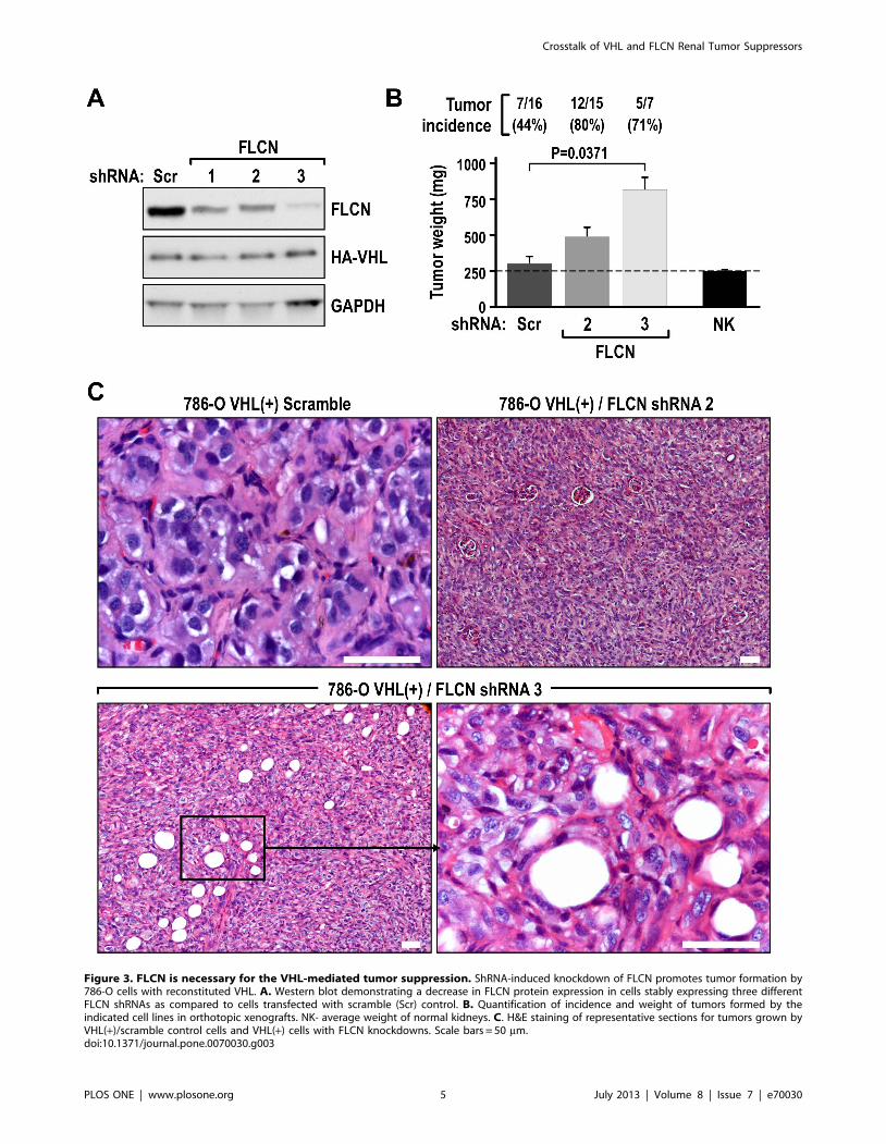
Figure 3. FLCN is necessary for the VHL-mediated tumor suppression. ShRNA-induced knockdown of FLCN promotes tumor formation by786-O cells with reconstituted VHL. A. Western blot demonstrating a decrease in FLCN protein expression in cells stably expressing three differentFLCN shRNAs as compared to cells transfected with scramble (Scr) control. B. Quantification of incidence and weight of tumors formed by theindicated cell lines in orthotopic xenografts. NK- average weight of normal kidneys. C. H&E staining of representative sections for tumors grown byVHL(+)/scramble control cells and VHL(+) cells with FLCN knockdowns. Scale bars = 50 mm.doi:10.1371/journal.pone.0070030.g003
Crosstalk of VHL and FLCN Renal Tumor Suppressors
PLOS ONE | www.plosone.org 5 July 2013 | Volume 8 | Issue 7 | e70030

expression of LC3C through repression of HIF, since LC3C is a
HIF-inhibited gene. VHL is well recognized for its ability to inhibit
HIF-as protein expression and activity. Recently, FLCN has also
been reported to diminish HIF activity, even though it does not
affect the accumulation of HIF-as subunits [11].
The specific role of FLCN in regulation of autophagy is further
supported by the recently identified presence of DENN domain in
the carboxy-terminal region of FLCN [12]. This motif is present
also in folicullin associated protein-1 and 2 (FNIP1 and FNIP2
[13]), and, interestingly in another protein expressed from the SM
locus, SMCR8 [13], which also we found to be induced by VHL
(Fig. S1). These proteins are acting as GDP-GTP exchange factors
(GEFs) for RAB GTPases, potentially necessary for autophagy
associated trafficking and fusion [11,13].
The SM locus has not been linked to ccRCC. Previously, only
one gene from this locus, TNFSF13 (tumor necrosis factor
superfamily member 13), had been studied in this regard. It was
found to exhibit decreased mRNA levels in ccRCC as compared
with normal kidney. In addition, higher levels of this receptor were
shown to be correlated with better overall and disease-free survival
[14]. However it is possible that further analysis will reveal
additional autophagy-related functions of genes expressed from
SM locus.
FLCN contributes to VHL-induced tumor suppression by a
mechanism that does not appear to involve activation of the
mTOR pathway. Indeed, in 786-O cells with reconstituted VHL
and FLCN knocked-down, we actually showed a strong reduction
in several phosphorylation events that are used as indicators of
mTOR activity. Consistently with the inhibition of mTOR we
measured increased accumulation of LC3B, a widely used
autophagic readout, and this increased LC3B-mediated autophagy
may contribute to tumor growth. The observed effects of FLCN
on mTOR activity are consistent with several publications
reporting a positive correlation between levels of FLCN and
mTOR in U251, HEK293, HK-2, ACHN, 786-O VHL(2) cells
[15]. However, the current canonical understanding of the FLCN-
mTOR connection is that FLCN negatively regulates the mTOR
pathway [8,9,16]. In that respect, the human UOK257 cell line
derived from a BHD patient, that does not express VHL, shows a
high level of mTOR activity, and mice with FLCN knockout
demonstrate high levels of S6 phosphorylation [8,9]. The reason
for this discrepancy is currently not understood, but perhaps the
data point towards a compartmentalized effect of FLCN on
mTOR that is affected by the cellular context, such as activity of
VHL. Nevertheless, the connection of VHL to a tumor suppressor
that regulates a metabolic pathway in renal cancer may reveal a
novel aspect of VHL tumor-suppressing activity that is comple-
mentary to its roles in the regulation of angiogenesis and
autophagy.
Importantly, FLCN protein levels are diminished in human
ccRCC with VHL loss, further supporting a role for this tumor
suppressor in the growth of ccRCC. So far, no FLCN mutations or
changes in mRNA levels have been measured in ccRCC. This
convergence in the functions of VHL and FLCN is of special
interest considering the fact that clear cell carcinoma can be part
of the multihistological type of renal cancer that occurs in BHD
syndrome.
The nature of the direct molecular mechanism by which VHL
induces FLCN renal tumor suppressors remains to be determined,
but is likely to involve multiple mechanisms. Because we measured
an effect of VHL on FLCN mRNA steady-state levels in RCC cell
lines, we expect some level of transcriptional induction or
Figure 4. Knockdown of FLCN inhibits the mTOR pathway. A. Western blot analysis of mTOR downstream targets in 786-O VHL(+) cells stablyexpressing either scramble or two different FLCN shRNAs. The GAPDH loading control is shown for each group of immunoblots processed from thesame gel. B. Representative sections of indicated tumors stained for S6 and S6P. Scale bar = 50 mm.doi:10.1371/journal.pone.0070030.g004
Crosstalk of VHL and FLCN Renal Tumor Suppressors
PLOS ONE | www.plosone.org 6 July 2013 | Volume 8 | Issue 7 | e70030

stabilization of FLCN mRNA. In that respect we did not find any
effects of p53 knockdown on FLCN expression. Because we did
not find changes in the FLCN mRNA levels between human
kidney-tumor pairs although we found difference in the steady-
state of FLCN protein, we propose that there is also an important
translational component to this regulation, possibly depending on
the specificity of different kidney cell populations. One explanation
is that VHL may repress expression of specific microRNAs, which
in turn repress FLCN translation.
In summary, with the current study, we have provided first
evidence for a direct connection between the VHL and FLCN
tumor-suppressing pathways converging on regulation of autoph-
agy in renal cancer.
Supporting Information
Figure S1 VHL induces expression of several genesexpressed from the Smith-Magenis locus in 786-0 andA498 cells. The graph shows changes in the expression of
individual mRNAs based on quantitative qRT-PCR measure-
ments. Fold change represents the difference in expression of
mRNAs in VHL(+) versus VHL(2) cells. The sequence of primers
is provided in.
(PDF)
Table S1 List of genes significantly repressed orinduced by VHL in 786-O cells. Fold change represents
VHL (+)/VHL(2) ratio.
(DOCX)
Table S2 Genes significantly induced by VHL andenriched for specific chromosome or cytoband.(XLS)
Table S3 Genes significantly reduced by VHL andenriched for specific chromosome or cytoband.(DOCX)
Table S4 Sequence of primers for the indicated genesused in qRT-PCR.(DOCX)
Acknowledgments
We thank G. Doerman for preparation of Figures and Dr. M. Daston for
editing the manuscript.
Author Contributions
Conceived and designed the experiments: MFCK JM MM. Performed the
experiments: PB YS EK OM YY. Analyzed the data: MAS JB MM.
Contributed reagents/materials/analysis tools: PB YS YY OM EK. Wrote
the paper: MFCK JM MM.
Figure 5. FLCN regulates autophagy. Knockdown of FLCN (shRNA 3) in 786-O (A) or RCC4 (B) VHL(+) cells reduces accumulation of tumorsuppressive LC3C-II but induces expression of oncogenic LC3B-II. Western blots were probed with indicated antibodies. C. Quantification of 3independent experiments as shown in panel A and B. LC3 protein levels measured by optical density were first normalized to GAPDH. Then the ratioof LC3 was calculated between LC3 levels measured in chloroquine treated samples in cells with FLCN knocked down and control cells (compare lane6 to lane 3 in panels A and B). D. Model of the proposed regulation.doi:10.1371/journal.pone.0070030.g005
Crosstalk of VHL and FLCN Renal Tumor Suppressors
PLOS ONE | www.plosone.org 7 July 2013 | Volume 8 | Issue 7 | e70030

References
1. Mikhaylova O, Stratton Y, Hall D, Kellner E, Ehmer B, et al. (2012) VHL-
regulated MiR-204 suppresses tumor growth through inhibition of LC3B-mediated autophagy in renal clear cell carcinoma. Cancer Cell 21: 532–546.
2. Elsea SH, Williams SR (2011) Smith-Magenis syndrome: haploinsufficiency ofRAI1 results in altered gene regulation in neurological and metabolic pathways.
Expert Rev. Mol. Med. 13: e14. Available: http://journals.cambridge.org/
action/displayAbstract?fromPage = online&aid = 8255391 Accessed: 19 April2011.
3. Schmidt LS, Nickerson ML, Warren MB, Glenn GM, et al. (2005) GermlineBHD-mutation spectrum and phenotype analysis of a large cohort of families
with Birt-Hogg-Dube syndrome. Am J Hum Genet 76: 1023–1033.
4. Yi Y, Mikhaylova O, Mamedova A, Bastola P, Biesiada J, et al. (2010) vonHippel-Lindau-dependent patterns of RNA polymerase II hydroxylation in
human renal clear cell carcinomas. Clin. Cancer Res. 16: 5142–5152.5. Mikhaylova O, Ignacak ML, Barankiewicz TJ, Harbaugh SV, Yi Y, et al. (2008)
The von Hippel-Lindau tumor suppressor protein and Egl-9-Type prolinehydroxylases regulate the large subunit of RNA polymerase II in response to
oxidative stress. Mol. Cell. Biol. 28: 2701–2717.
6. Kuznetsova AV, Meller J, Schnell PO, Nash JA, Ignacak ML, Sanchez Y,Conaway JW, Conaway RC, Czyzyk-Krzeska MF (2003) von Hippel-Lindau
protein binds hyperphosphorylated large subunit of RNA polymerase II througha proline hydroxylation motif and targets it for ubiquitination. Proc Natl Acad
Sci USA 100: 2706–2711.
7. Galban S, Martindale JL, Mazan-Mamczarz K, Lopez de Silanes I, Fan J, et al.(2003) Influence of the RNA-binding protein HuR in pVHL-regulated p53
expression in renal carcinoma cells. Mol. Cell. Biol. 23: 7083–7095.8. Baba M, Furihata M, Hong SB, Tessarollo L, Haines DC, et al. (2008) Kidney-
targeted Birt-Hogg-Dube gene inactivation in a mouse model: Erk1/2 and Akt-mTOR activation, cell hyperproliferation, and polycystic kidneys. J. Natl.
Cancer Inst. 100: 140–154.
9. Baba M, Hong SB, Sharma N, Warren MB, Nickerson ML, et al. (2006)
Folliculin encoded by the BHD gene interacts with a binding protein, FNIP1,and AMPK, and is involved in AMPK and mTOR signaling. Proc. Natl. Acad.
Sci. USA 100: 15552–15557.10. Hudon V, Sabourin S, Dydensborg AB, Kottis V, Ghazi A, et al. (2010) Renal
tumor suppressor function of the Birt-Hogg-Dube syndrome gene product
folliculin. J. Med. Genet. 47: 182–189.11. Preston RS, Philp A, Claessens T, Gijezen L, Dydensborg AB, et al. (2011)
Absence of the Birt-Hogg-Dube gene product is associated with increasedhypoxia-inducible factor transcriptional activity and a loss of metabolic
flexibility. Oncogene 30: 1159–1173.
12. Nookala RK, Langemeyer L, Pacitto A, Ochoa-Montano A, Donaldson JC, etal. (2013) Crystal structure of folliculin reveals a hiDENN function in genetically
inherited renal cancer. Open Biol. 2: 120071. Available: http://rsob.royalsocietypublishing.org/content/2/8/120071. Accessed: 2 August 2012.
13. Zhang D, Iyer LM, He F, Aravind L (2012) Discovery of novel DENN proteins:implications for the evolution of eukaryotic intracellular membrane structures
and human disease. Front. Genet. 3: 283. Available: http://www.frontiersin.
org/Bioinformatics_and_Computational_Biology/10.3389/fgene.2012.00283/abstract Accessed: 13 December 2012.
14. Pelekanou V, Notas G, Theodoropoulou K, Kampa M, Takos D, et al. (2011)Detection of the TNFSF members BAFF, APRIL, TWEAK and their receptors
in normal kidney and renal cell carcinomas. Anal. Cell. Pathol. 34: 49–60.
15. Hartman TR, Nicolas E, Klein-Szanto A, Al-Saleem T, Cash TP, et al (2009)The role of the Birt-Hogg-Dube protein in mTOR activation and renal
tumorigenesis. Oncogene 28: 1594–1604.16. Hasumi Y, Baba M, Ajima R, Hasumi H, Valera VA, et al. (2009) Homozygous
loss of BHD causes early embryonic lethality and kidney tumor developmentwith activation of mTORC1 and mTORC2. Proc. Natl. Acad. Sci. USA 106:
18722–18727.
Crosstalk of VHL and FLCN Renal Tumor Suppressors
PLOS ONE | www.plosone.org 8 July 2013 | Volume 8 | Issue 7 | e70030